Article

Sustainable Environmental-Based ZnO Nanoparticles Derived from Pisonia grandis for Future Biological and Environmental Applications

by
Thiyakarajan Sutharappa Kaliyamoorthy
1,
Vijayakumar Subramaniyan
2,*,
Sangeetha Renganathan
3,
Vidhya Elavarasan
2,
Jagatheesvaran Ravi
1,
Praseetha Prabhakaran Kala
4,
Prathipkumar Subramaniyan
5 and
Sekar Vijayakumar
6,*
1
Department of Chemistry, A.V.V.M Sri Pushpam College (Autonomous), Affiliated to Bharathidasan University Tiruchirappalli, Poondi 613503, India
2
PG and Research Department of Botany, A.V.V.M. Sri Pushpam College (Autonomous), Affiliated to Bharathidasan University, Poondi 613503, India
3
Department of Mathematics, A.V.V.M Sri Pushpam College (Autonomous), Affiliated to Bharathidasan University, Tiruchirappalli, Poondi 613503, India
4
Department of Nanotechnology, Noorul Islam Centre for Higher Education, Kumarakoil 629180, India
5
National Institute of Technology, Tiruchirappalli 620015, India
6
Marine College, Shandong University, Weihai 264209, China
*
Authors to whom correspondence should be addressed.
Sustainability 2022, 14(24), 17009; https://doi.org/10.3390/su142417009
Submission received: 7 November 2022 / Revised: 3 December 2022 / Accepted: 13 December 2022 / Published: 19 December 2022

Abstract

:
The bio-synthesis of zinc oxide nanoparticles (ZnO NPs) using aqueous leaf extract of Pisonia grandis is discussed in this work as an effective ecologically beneficial and straightforward method. This strategy intends to increase ZnO nanoparticle usage in the biomedical and environmental sectors, while reducing the particle of hazardous chemicals in nanoparticle synthesis. In the current study, bio-augmented zinc oxide nanomaterials (ZnO-NPs) were fabricated from Pisonia grandis aqueous leaf extracts. Different methods were used to analyze the ZnO-nanoparticles including X-ray diffraction (XRD), Fourier Transforms Infrared (FT-IR), Ultraviolet (UV) spectroscopy, and Field Emission Scanning Electron Microscopy (FE-SEM) with EDX. The synthesized nanoparticles as spheres were verified by FE-SEM analysis; XRD measurements showed that the particle flakes had an average size of 30.32 nm and were very pure. FT-IR analysis was used to validate the functional moieties in charge of capping and stabilizing ZnO nanoparticles. The antimicrobial, cytotoxic, and photodegradation properties of synthesized nanoparticles were assessed using well diffusion, MTT, and UV visible irradiation techniques. The bio-fabricated nanoparticles were proven to be outstanding cytotoxic and antimicrobial nanomaterials. As a result of the employment of biosynthesized ZnO nanoparticles as photocatalytic agents, 89.2% of the methylene blue dye was degraded in 140 min. ZnO nanoparticles produced from P. grandis can serve as promising substrates in biomedicine and applications of environmental relevance due to their eco-friendliness, nontoxic behavior, and cytocompatibility.

Graphical Abstract

1. Introduction

Nowadays, nano-techniques are considered as authenticated state-of-the-art tools with a wide range of contributions to the chemical, pharmaceutical, and food processing sectors. Intriguing applications of nanotechnology may be found in the fields of computers, energy production, optics, therapeutic delivery, and environmental sciences [1]. The emergence of nanotechnology has created several nanoscale devices that have been utilizing a variety of tools, including physico-chemical, and eco-friendly methods. However, the preferred tool is green nanoparticle synthesis, which is simple to manufacture and design [2].
The usage of hazardous substances, lengthy processing times, high costs, and arduous processes are only a few of the downsides of current methods for the creation of nanoparticles. Due to these restrictions, the majority of the pertinent research has focused on compatible and speedy synthesis techniques for the synthesis of nanomaterials. The creation of environmentally friendly processes for producing nano-sized materials has recently received a lot of attention from material scientists. In this regard, the green route for nanoparticle synthesis, specifically when utilizing plant substrates, is a developing inclination that is considered as simple, economical, and less toxic in the green chemical field [3].
In terms of a variety of bio-particulates and metabolic products, such as proteins, vitamins, coenzyme-based intermediaries, flavonoids, phenols, and carbohydrates, plants have a rich genetic diversity. These plant products possess functional groups called hydroxyl, carbonyl, and amines that interact with metal ions to shrink them down to the nanoscale. More specifically, flavonoids have a number of functional groups, but it is thought that the -OH group is primarily in charge of turning metal ions into nanoparticles [4]. These constituents serve a key role in the doping of the nanoparticles, which is crucial for their strength and compatibility, as well as in the bioreduction of the ions to the nanoscale size. Metal ions can be converted into nanoparticles by reducing substances such as phenolic compounds, sterols, and alkaloids, in a single process [5].
The primary factors affecting the nanoparticles ultimately are used in industry and the nature of the metal utilized for nanoparticle biosynthesis is explored. Among them, ZnO is an inorganic substance that happens in nature infrequently. It is typically found in crystals. Manganese impurities in naturally occurring ZnO give it a characteristic red or orange hue [6]. ZnO is a white powder in crystal form that is practically insoluble when it has been refined. ZnO nanoparticles are widely employed for many functions in diagnostics, cosmetics, textiles, and even microelectronics due to their non-toxic and size-dependent features [7]. ZnO nanoparticles have a larger demand to cure epidemic disorders in living beings since ZnO is considered safe (GRAS) and possesses antibacterial characteristics [8].
Pisonia grandis R.Br (Nyctaginaceae) is a common evergreen tree in India known as ‘Leechai kottai keerai’. In the Indian traditional medicine system, tribals used a variety of folkloric medicine so that diabetes, inflammation, wound healing, diuretics, analgesics, filariasis, dysentery, and rheumatic disorders are treated with it [9]. As a result, the present goal of this research is to look into the biosynthesis of ZnO nanoparticles using P. grandis leaf extract. P. grandis leaf extracts included potent bioactive components such as Pinnatol, Allantoin, Sitosterol, -Spinasterol, -Spinasterol glucoside, Octocosanal, Dulcitol, Flavonoids, and Quercetin. These phytocompounds facilitate the conversion of zinc acetate dehydrate to zinc oxide nanoparticles [10].
Here, we describe a quick and environmentally friendly process for making zinc oxide nanoparticles (ZnO NPs) using the plant extracts of Pisonia gradis as reducing reagents and zinc acetate as an initiator for comparing their antibiotic, anticancer and photocatalytic activity. The possibility of using plant-based nanoparticles in the biomedical industry will grow as a result of this research.

2. Materials and Methods

2.1. Chemical

Zinc acetate dehydrate Zn(C2H3O2)2.2H2O, as well as all other chemicals and reagents, sodium hydroxide (NaOH), and 2H2O, were obtained from EMerk in India. The analytical grade of each obtained chemical and reagent allowed for its usage without additional purifying.

2.2. Extraction of Plant Components

We procured P. grandis leaves in and around the college campus. To prepare the extracts, the plant material was powdered using an electric blender. The plant powder (50 g) was heated for 20 min at 60 °C after being immersed in 500 mL of distilled water. The treated powder was left in an incubator overnight at 37 °C for optimum extraction. The solvent was then evaporated from the extract using a vacuum evaporator at 40 °C after it had been filtered using Whatman filter paper. The formed extract was stored at 4 °C in the refrigerator.

2.3. Synthesis of ZnO Nanoparticles

With a few alterations, a previously described process was used to synthesize zinc oxide nanoparticles [11]. In brief, 100 mL of extract was combined with 2.09 gm of zinc acetate dihydrate (Sigma-Aldrich, Chemicals, India) and stirred at 60 °C for 2 h. Once the reaction was finished, the mixture was subjected to centrifugation for 10 min at 10,000 rpm after allowing it to cool at 25 °C. The remaining pellet was rinsed three times in the presence of distilled water, placed in a clean Petri dish, and dried in an oven at 90 °C after the supernatant was removed. The dried plant was then crushed to a fine powder using a mortar and pestle, any impurities were removed by calcining it for 2 h at 80 °C. The blasted powder was utilized for physical analysis and biological applications after being placed in an airtight glass container and labeled as ZnO nanoparticles.

2.4. Characterization of Fabricated ZnO Nanoparticles

The physicochemical characteristics of ZnO nanoparticles derived from P. grandis have been analyzed using a series of analytical procedures. These methods include Ultraviolet (UV) spectroscopy, X-ray diffraction (XRD), Fourier transform infrared spectroscopy (FT-IR), field emission scanning electron microscopy (FE-SEM), transmission electron microscopy (TEM), and energy-dispersive X-ray analysis (EDX). Utilizing UV-visible spectroscopy between the wavelengths of 200 and 700 nm, the reaction that took place between the extract and zinc nitrate reagent was examined. The X-ray diffraction method was employed to understand the components present in the green synthesized zinc oxide nanoparticles. The X-ray diffraction pattern was obtained using a PANalytical X’pert pro-X-ray diffractometer (Netherlands). We employ Scherer’s equation to determine the crystallite’s size. Using Fourier transform infrared (FT-IR) spectroscopy, the active group responsible for the formation of nanoparticles was found in the spectral region between 400 and 4000 cm−1. FE-SEM (JSM-7600F, Japan) and TEM (JEM-2100F, Japan) were used to analyze morphologies and physical dimensions, while Energy-dispersive X-ray (EDX) spectroscopy was used to conduct elemental analysis of nanoparticles.

2.5. Biological Efficiencies

2.5.1. Evaluation of Micro-Morbidity

Utilizing the good diffusion approach, it is revealed that fabricated ZnO nanoparticles made from aqueous plant extract have antimicrobial properties. To obtain the microorganisms such as Staphylococcus aureus (MTCC 737), S. epidermis (MTCC 10656), Pseudomonas aeruginosa (MTCC 429), Serratia marcescens (MTCC 97), Candida albicans (MTCC 227), and Aspergillus niger (MTCC 281) the Eumic Analytical Lab and Research Institute, Tiruchirappalli, India, contacted the Microbial Type Culture Collection (MTCC), Chandigarh, India. These organisms were further pure-cultured in MHA (Muller Hinton agar) and SDA (Sabouraud Dextrose Agar) media at 35 °C and 30 °C, respectively, for bacterial and fungal cultures. The 6 mm-diameter punctures were used to penetrate MHA and SDA plates. Using a micropipette, 20 µL of the substance was injected into each well of every plate. Common antibiotics Gentamycin (50 µg) and Ketoconazole (5 µg) were used as the +ve and -ve controls, respectively. After that, they were kept for subsequent incubation at 30 °C for 24–48 h for bacterial culture and 37 °C for 72–96 h for fungal culture. After incubation, the diameter of the zonation around the well was measured (mm). Triplet plates were used to assure the results.

2.5.2. Evaluation of Cytotoxicity

The human SaOS-2 cancer cell line was provided by the NCCS (National Center for Cell Science) in Pune, India, and sequenced in DMEM media (Dulbecco’s Modified Eagle Medium). In vitro, ZnO nanoparticle therapy was performed on a human SaOS-2 cancer cell line. The cell cultures were assessed using 3-(4,5-dimethylthiazol-2-yl)-2,5-diphenyltetrazolium bromide (MTT, Sigma, Aldrich, Mumbai, India). Human SaOS-2 cancer cell lines (1 × 10⁵ cells/well) were plated and cultivated for 24 h on a 24-well plate. Different ZnO nanoparticle concentrations were created and mixed with the medium for growing cells. We created and homogenized the medium for the cells that were cultured as well as different ZnO nanoparticle concentrations. Then, each well is loaded with 100 L (5 µg/mL) with 5% MTT and kept at 37 °C for 4 h. At 560 nm, the UV-Spectrophotometer was utilized to evaluate the viability of the cells.

2.5.3. Evaluation of Photo-Degradation

To examine the photocatalytic activity of the manufactured ZnO nanoparticles, methylene orange dye in 50 mL of ultrapure water was prepared. After detaching the initial dilution (5 mL), 45 mL of dye mixture was added to the residual solution, which included 25 mg (0.025 g) of ZnO nanoparticles catalyst. After that, samples were obtained every 20 min while the mixture was exposed to UV light for 20 min to maintain the adsorption-desorption equilibrium. A UV-visible spectrophotometer was used to track the dye deterioration while the samples were subjected to centrifugation at 10,000 rpm for 15 min to separate the catalyst. According to Shah et al. [2], the equation was used to calculate the percentage deterioration of the degraded dye.

3. Results and Discussion

3.1. UV Vis Spectrum

Zinc ions in the solution are converted to zinc oxide due to the presence of secondary metabolites in plants. The plant extract functions as stabilizing and reducing agent simultaneously. The examination of the UV-visible spectra between 280 and 800 nm served to corroborate this. A peak in the spectra was visible at 362 nm, which is unique to ZnO nanoparticles (Figure 1). The absorbance peak for ZnO nanoparticles is reported to occur between 310 and 368 nm in wavelength [12]. Eg = 1240/eV was used to compute the band gap energy, which was discovered to be 3.8 eV. This value is close to previously reported band gap energy estimates for ZnO nanoparticles [13].

3.2. XRD

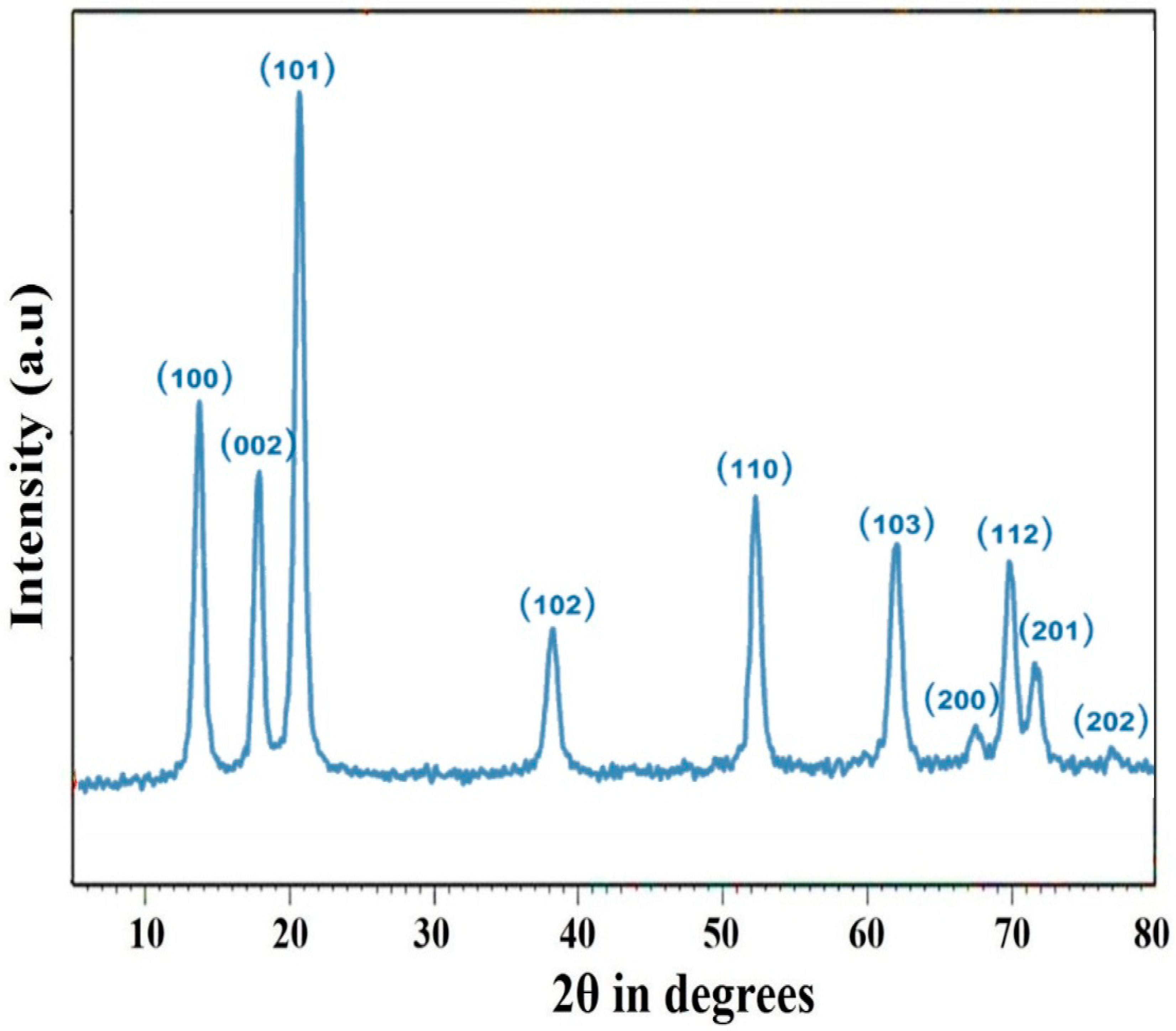
The crystallinity of the fabricated nanoparticles was confirmed by analysis of the diffractogram with a scan rate of 0.02°/min. The X-ray diffraction peaks of green ZnO nanoparticles synthesized by P. grandis at 2θ were found to be at 31.1°, 34.8°, 36.0°, 47.4°, 56.5°, 62.2°, 66.3°, 67.9°, 69.4°, and 77.6°. The crystal was revealed to be hexagonal in form, and the peaks were determined to match the ICDD card number 01-089-0511. As illustrated in Figure 2, crystal planes were detected during the XRD assessment of nanoparticles. These planes relate to the lattice planes (hkl) indexed as (100), (002), (101), (102), (110), (103), (200), (112), (201), and (202), respectively. The current results match the values previously reported [14]. The particle’s average crystalline size was determined to be 30.32 nm.
D = k λ β cos θ
where k is a constant (~0.9), λ is the wavelength of X-ray (~1.5406 Å), and β is the full width at half-maximum of the diffraction peak at 2θ.

3.3. FT-IR Analysis

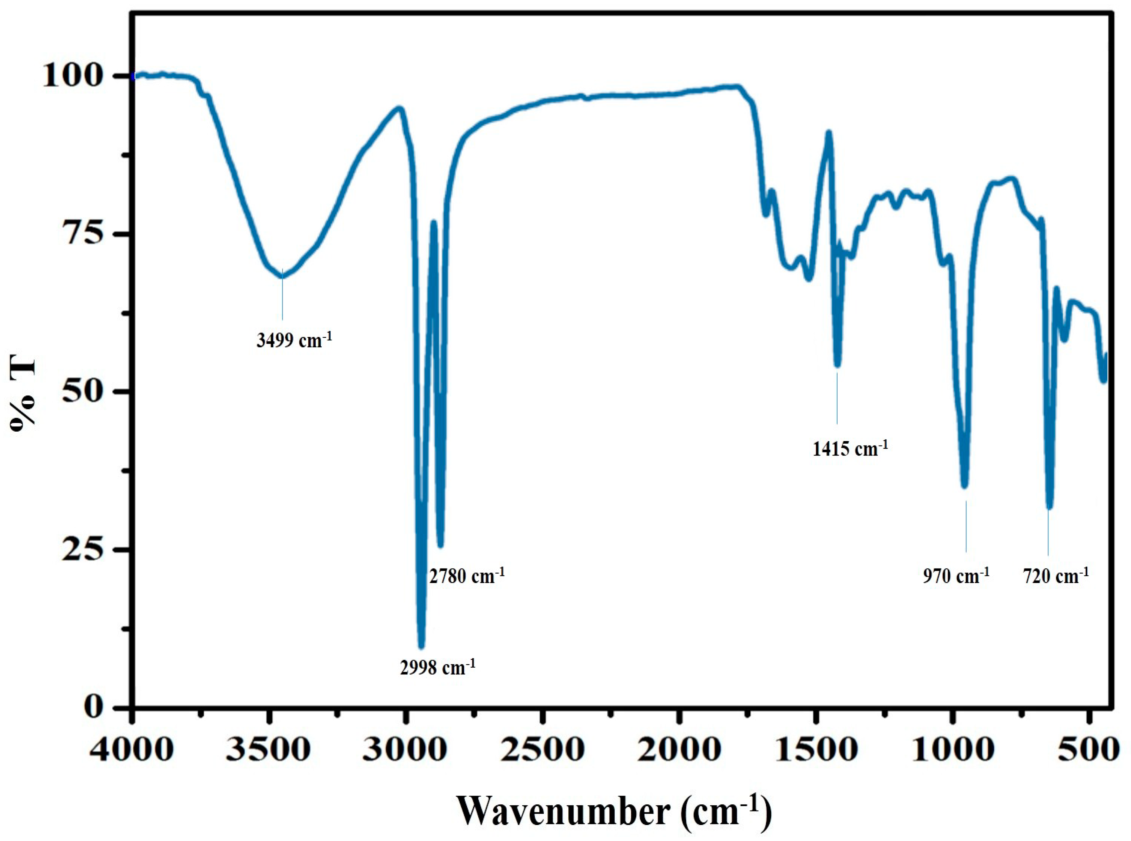
The functional groups contained in the fabricated ZnO nanoparticles are described by the FTIR spectral pattern (Figure 3). It also suggests that interactions between phenolic chemicals, terpenoids, flavonoid alkenes, and amines result in the creation of ZnO nanoparticles. P. grandis was used to manufacture ZnO nanoparticles, and their FT-IR spectra were examined in the 400–4000 cm−1 range. Zinc oxide nanoparticles were produced as a result of the switchover mechanism of zinc ions to zinc oxide, which was visible as bands. Each band corresponds to different stretching and bending motions. The band at 3499 cm−1 is corresponding to the O-H group [15]. The vibrations caused by the C-H stretching are responsible for the bending at 2998 cm−1 [16]. The vibrational modes of the O-H are found in the area at 2780 cm−1 [17]. The absorbance peak at 1415 cm−1 represented the occurrence of the carboxylate anion [18]. The detected band at 970 cm−1 indicated the existence of OH deformation [19], whereas the spectra at 720 cm−1 revealed the presence of C-H rock [20].

3.4. FE-SEM with EDX

By using FE-SEM, the morphology of the green-produced zinc oxide nanoparticles was studied. According to Figure 4a, the particles are strongly agglomerate and have a semi-spherical shape. This demonstrates unambiguously that the particles are dispersed in a homogenous state and that the homogenous nature of nanoparticles performs a significant role in their various functions. The overlaying of particles on top of one another caused the size to rise. By correlating the particle size determined by X-ray diffraction, we are able to affirm the creation of ZnO nanoparticles. Our results are consistent with earlier reports [21]. The EDX picture of zinc oxide nanoparticles can be seen in Figure 4b. Only zinc and oxygen were present, as is obvious from the EDX study. The outcome showed that high-purity zinc nanoparticles made up the reaction product. The surface morphology of zinc oxide nanoparticles was also examined. In earlier research, Keerthana et al. [22] revealed that zinc oxide nanoparticles synthesized during phytosynthesis had a spherical form. The histogram method exposed that the mean particle size of the ZnO nanoparticles was 30 nm (Figure 4c).

3.5. TEM Analysis

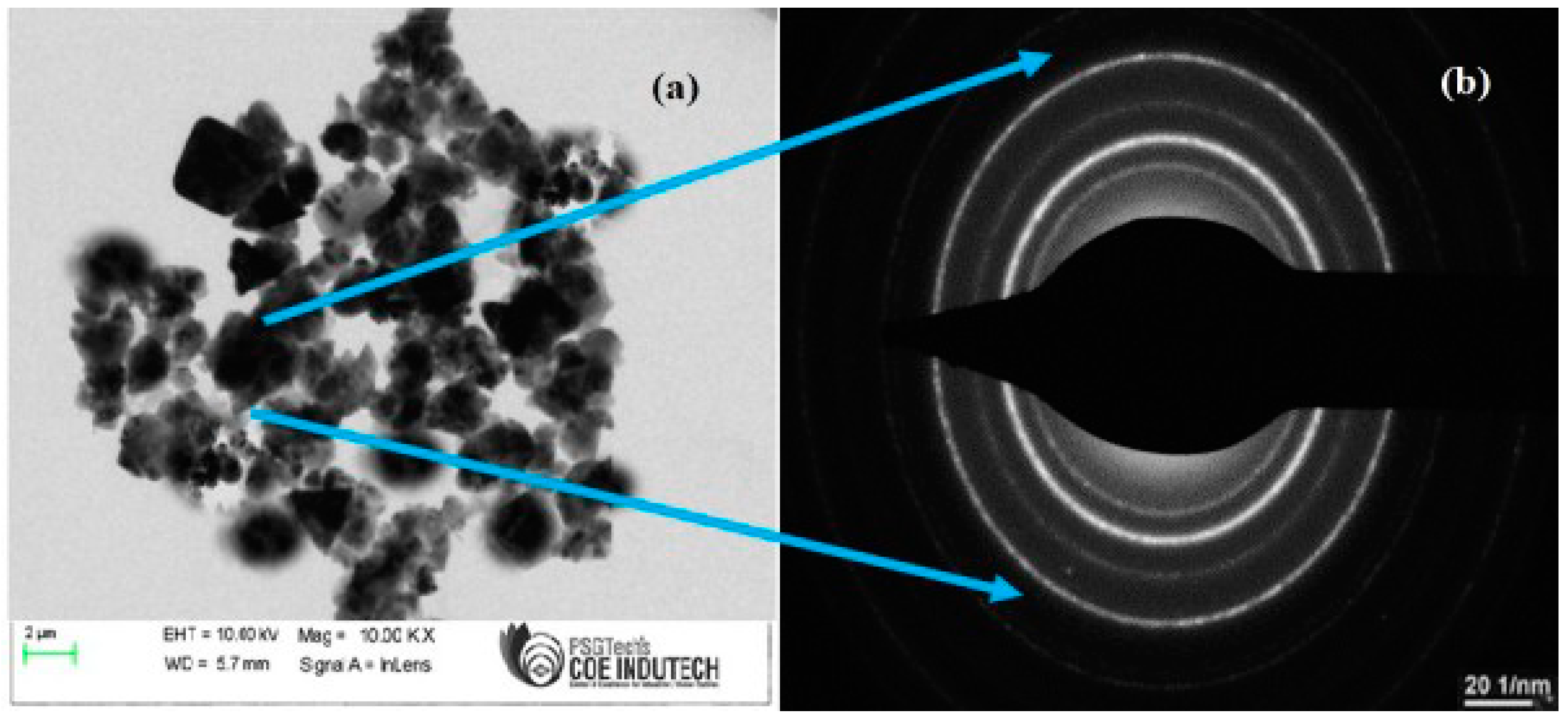
The TEM image and SAED pattern of synthesized ZnO nanoparticles are shown in Figure 5a,b. The average particle size of 27 nm ZnO nanoparticles was determined from Figure 5a, and the results are strongly corroborated by the XRD readings. The described TEM images of the ZnO nanoparticles with spherical and hexagonal shapes can be seen in Figure 5a. The selected area electron diffraction pattern for synthesized ZnO nanoparticles is shown in Figure 5b, which supports the assertion that the produced ZnO nanoparticles are extremely crystalline in nature.

3.6. Antimicrobial Efficacies

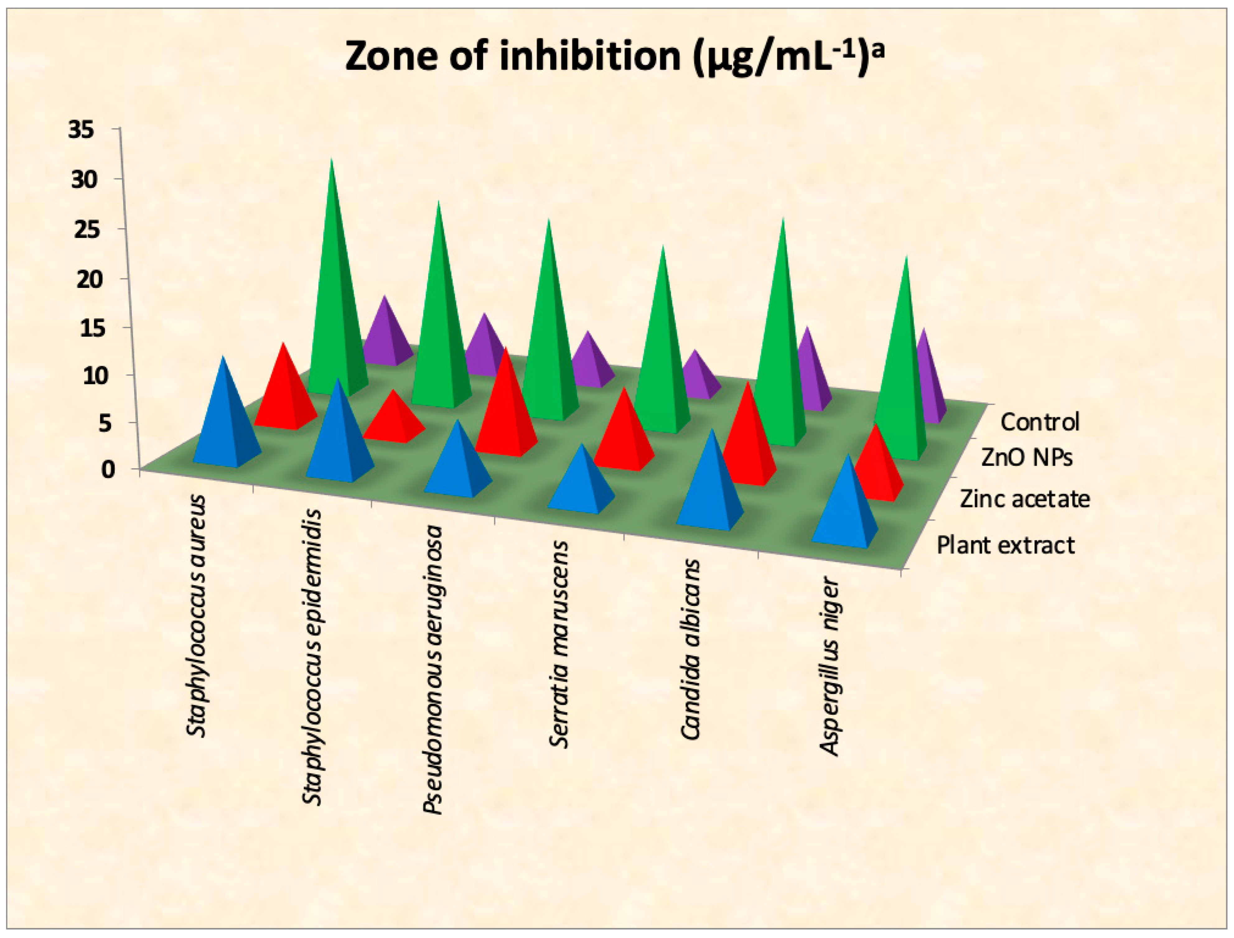
The antimicrobial activities of the synthesized ZnO nanoparticles against S. aureus, S. epidermis, P. aeruginosa, S. maruscens, C. albicans, and A. niger were determined using a well-boring experiment. The findings were shown in Figure 6. Overall, the data showed that synthesized ZnO nanoparticles made from P. grandis leaf extract had a substantial antimicrobial impact on all assessed bacterial strains. S. aureus had the greatest antibacterial zone of inhibition (28 ± 2.06 mm), followed by S. epidermis (23 ± 1.04 mm), P. aeruginosa (22 ± 1.77 mm), and S. maruscens (20 ± 1.55 mm). Whereas C. albicans (24 ± 0.94 mm) and A. niger (21 ± 1.59 mm) showed amazing antifungal activity. Additionally, in comparison to gentamycin (positive control) P. grandis, and zinc acetate, the synthesized ZnO nanoparticles have shown greater antibacterial activity (Figure 7). The antibacterial properties of ZnO nanoparticles vary based on whether the bacteria are Gram-positive or Gram-negative [23]. The biosynthesized ZnO nanoparticles showed greater antibacterial efficacy against Gram +ve strains (S. aureus and S. epidermis) than Gram −ve strains in the current investigation (P. aeruginosa and S. maruscens). According to Vijayakumar et al. [24], they reported that ZnO nanoparticles made from Laurus nobilis leaf extract showed stronger antibiotic action towards S. aureus than P. aeruginosa, which was a similar trend. This may be due to the structure and components of Gram-positive bacteria, namely the peptidoglycan layer, which may enhance the attachment of ZnO nanoparticles to the cell wall whereas Gram-negative bacteria components avoid this connection [23]. Some people hypothesized that the free Zn ions from ZnO nanoparticles have hazardous qualities that block several bacterial cell processes including metabolic and enzymatic activities, which causes bacterial cell death [25,26].

3.7. Cytotoxicity Efficiencies

The cytotoxicity of biologically synthesized ZnO nanoparticles was examined in vitro employing the SaOS-2 cancer cell line. By using the MTT analysis, cell lines were subjected to different dilutions of ZnO NPs (3.9, 7.8, 15.6, 31.2, 62.5, 125, 250, 500, 1000 µg mL) for 48 h before being compared to DMEM as the control (Figure 8). Due to certain enzymes (NAD(P)H-dependent cellular oxidoreductase), reducing the action of MTT reagent and including tetrazolium dyes alters the yellow hue that depends on cellular metabolic processes. Significant percentage decreases in cell viability have been seen after ZnO nanoparticles exposure, and the IC50 was determined to be 39.98 µg/mL. Dead cells in high density and morphological alterations were seen when biosynthesized ZnO nanoparticles were applied to the SaOS-2 cancer cell lines [27]. These morphological modifications highlighted the increased impact of cytotoxicity. The greater cytotoxic impact of the tiny particles might be caused by their high ratio of surface area to volume. Additionally, it is vital to consider that the occurrence of phytoconstituents in the ethanolic leaf extract, apart from tannins, the aqueous extract produced negative results for flavonoids, steroids, furan, alkaloids, anthraquinone, and saponins [28].

3.8. Photodegrataion Efficiencies

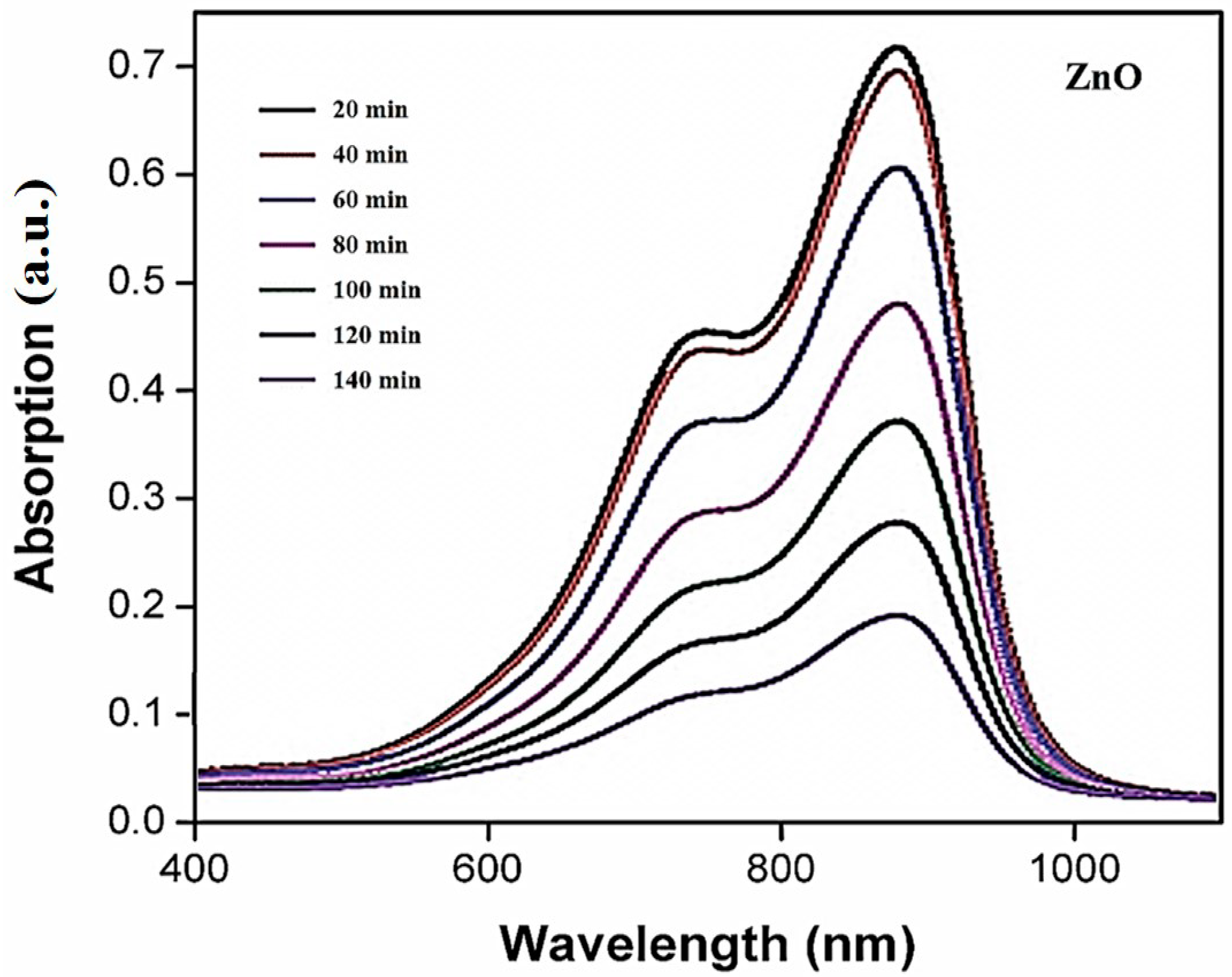
When UV light irradiation was increased for the first 20 min, the deterioration was shown to be 18.4%; however, as time passed, the disintegration of the dye duly climbed and the greatest deformation was seen after 140 min, which was 89.2%, as shown in Figure 9. A similar trend was observed that Shah et al. [2] who reported that MB dye was reduced up to 88% in 140 min.
The efficiency of photodegradation (%) = (C0 − C150/C0 × 100) = (A0 − A150/A0) × 100 where C0 and C150 represent the concentrations of MB solution at t = 0 and t = 140 min, respectively, and A0 and A150 represent the absorption intensity of MB solution [29].
Mohammad et al. [30] reported that the following mechanism may be involved in the photodegradation process. In order to stimulate valence electrons and permit them to move from the valence band to the conduction band, the dye must first be adsorbed onto the surface of the catalyst (in this example, zinc). During this mechanism, a proton hole, designated h+, is elevated in the valence band. Adsorbed water molecules will react with the proton holes and free electrons on the photocatalyst’s surface, causing the moving electrons to translate soluble oxygen into superoxide anion O2•—free radicals and the positive holes to form •OH—radicals as a consequence. The dye molecules are broken down by these light-generated radicals into less complex molecules like CO2 and H2O.

4. Conclusions

This work focuses on the environmentally friendly plant-mediated production of ZnO nanoparticles that are important in biomedicine, using aqueous leaf extracts of P. grandis, a plant that is important in healthcare. XRD analysis confirmed the crystal structure of the generated nanoparticles. Analysis using the Fourier transform infrared (FT-IR) spectrometer confirmed the prevalence of phytochemicals necessary for the translation of metallic ions to nanoparticles (NPs). FE-SEM analysis was used to determine the exact morphology. Synthesized ZnO nanoparticles have successfully demonstrated their ability to combat pathogenic microbial strains. The methylene blue dye could be degraded by the synthesized ZnO nanoparticles with excellent results. The feasibility of cytotoxicity testing using synthesized ZnO nanoparticles was also proven against the SaOS-2 cancer cell lines. According to the findings of our research, the aforementioned biogenic ZnO nanoparticles can be employed in biological and environmental applications.

Author Contributions

T.S.K. has analyzed the data correction. V.S. Writing—review and editing. S.R. has analyzed the formal analysis. V.E. has analyzed antimicrobial activity. J.R. has analyzed the methodology. P.P.K. has analyzed the anticancer activity. P.S. has analyzed the methodology. S.V. Writing—review and editing. All authors have read and agreed to the published version of the manuscript.

Funding

This research was founded by DST-FIST (SR/FST/College-222/2014), DBT-STAR (HRD-11011/18/2022-HRD-DBT).

Institutional Review Board Statement

The study was conducted according to the guidelines of the declaration and approved by the ethics committee of Bharathidasan University, Trichy, and AVVM Sri Pushpam College (Autonomous), Poondi, Thanjavur.

Informed Consent Statement

Not applicable.

Data Availability Statement

On behalf of all the authors, the corresponding author states that our data are available upon reasonable request.

Acknowledgments

We sincerely express our thanks to the management of A.V.V.M. Sri Pushpam College (Autonomous), Poondi, for providing us necessary facilities and support to carry out this work.

Conflicts of Interest

The authors have declared no conflict of interest.

References

  1. Ramsden, J. Nanotechnology: An Introduction; William Andrew is an imprint of Elsevier: New York, NY, USA, 2016. [Google Scholar]
  2. Shah, F.; Hasnain, J.; Sajjad, A.S.; Sumaira, S.; Adnan, K.; Muhammad, T.A.; Muhammad, R.; Faheem, J.; Wajidullah; Noreen, A.; et al. Green Synthesis of Zinc Oxide (ZnO) Nanoparticles Using Aqueous Fruit Extracts of Myristica fragrans: Their Characterizations and Biological and Environmental Applications. ACS Omega 2021, 6, 9709–9722. [Google Scholar] [CrossRef]
  3. Mihir, H.; Siddhivinayak, B.; Kumar, R. Plant-Mediated Green Synthesis of Iron Nanoparticles. J. Nanopart. 2014, 9, 140614. [Google Scholar] [CrossRef] [Green Version]
  4. Makarov, V.V.; Love, A.J.; Sinitsyna, O.V.; Makarova, S.S.; Yaminsky, I.V.; Taliansky, M.E.; Kalinina, N.O. “Green” Nanotechnologies: Synthesis of Metal Nanoparticles Using Plants. Acta Nat. 2014, 6, 35–44. [Google Scholar] [CrossRef] [Green Version]
  5. Minha, N.; Usman, A.; Bushra, K.; Bin, C. Green route to synthesize Zinc Oxide Nanoparticles using leaf extracts of Cassia fistula and Melia azadarach and their antibacterial potential. Sci. Rep. 2020, 10, 9055. [Google Scholar] [CrossRef]
  6. Ahmed, S.; Ahmad, M.; Swami, B.L.; Ikram, S. A review on plants extract mediated synthesis of silver nanoparticles for antimicrobial applications: A green expertise. J. Adv. Res. 2016, 7, 17–28. [Google Scholar] [CrossRef] [PubMed] [Green Version]
  7. Nazmi, S.; Yusuf, Z.; Fuat, B. Green synthesized ZnO nanoparticles using Ganoderma lucidum: Characterization and invitro nano-fertilizer effects. J. Alloys Compd. 2022, 918, 165695. [Google Scholar] [CrossRef]
  8. John Sushma, N.; Mahitha, B.; Mallikarjuna, K.; Deva Prasad, R.B. Bio-inspired ZnO nanoparticles from Ocimum tenuiflorum and their in vitro antioxidant activity. Appl. Phys. 2016, 122, 544. [Google Scholar] [CrossRef]
  9. Vembu, S.; Vijayakumar, S.; Nilavukkarasi, M.; Vidhya, E.; Punitha, V.N. Phytosynthesis of TiO2 nanoparticles in diverse applications: What is the exact mechanism of action? Sens. Int. 2022, 3, 100161. [Google Scholar] [CrossRef]
  10. Aravind, M.; Amalanathan, M.; Sony Michael Mary, M. Synthesis of TiO2 nanoparticles by chemical and green synthesis methods and their multifaceted properties. SN Appl. Sci. 2021, 3, 409. [Google Scholar] [CrossRef]
  11. Vidhya, E.; Vijayakumar, S.; Prathipkumar, S.; Praseetha, P.K. Green way biosynthesis: Characterization, antimicrobial and anticancer activity of ZnO nanoparticles. Gene Rep. 2020, 20, 100688. [Google Scholar] [CrossRef]
  12. Sasani Ghamsari, M.; Alamdari, S.; Han, W.; Park, H. Impact of nanostructured thin ZnO film in ultraviolet protection. Int. J. Nanomed. 2017, 12, 207–216. [Google Scholar] [CrossRef] [PubMed] [Green Version]
  13. Ashwini, J.; Aswathy, T.R.; Achuthsankar, S.N. Green synthesis and characterization of zinc oxide nanoparticles using Cayratia pedata leaf extract. Biochem. Biophys. Rep. 2021, 26, 100995. [Google Scholar] [CrossRef]
  14. Jayachandran, A.; Thankamani, R.A.; Anil, B.R.; Gautham, M.T.; Achuthsankar, S.N. Synthesis and Characterization of Zinc Oxide Nanoparticles Using Acacia caesia Bark Extract and Its Photocatalytic and Antimicrobial Activities. Catalysts 2021, 11, 1507. [Google Scholar] [CrossRef]
  15. Udayachandran Thampy, U.S.; Rama Krishna, C.; Venkata Reddy, C.; Babu, B.; Reddy, Y.P.; Rao, P.S.; Ravikumar, R.V.S.S.N. Spectral Investigations on Cu2+-Doped ZnO Nanopowders. Appl. Magn. Reson. 2011, 41, 69. [Google Scholar] [CrossRef]
  16. Abdul Mathin, S.K.; David Raju, M.; Rama Sekhara Reddy, D.; Jayadev, D.J. Synthesis of Zinc Oxide Nano-particles using Anthocephalus Cadamba Plant Extracts and Explores its Anti-Oxidant, Anti-Inflammatory and Anti-Diabetics Activity. Int. J. Mod. Trends Sci. Technol. 2021, 7, 162–169. [Google Scholar]
  17. Sarangi, S.N.; Nozaki, S.; Sahu, S.N. ZnO Nanorod-Based Non-Enzymatic Optical Glucose Biosensor. J. Biomed. Nanotechnol. 2015, 11, 988–996. [Google Scholar] [CrossRef]
  18. Lei, Z.; Yang, H.; Xue, W.; Ao, Z.; Xianli, G.; Abu El-Gasim, A.Y.; Haile, M.; Cunshan, Z. Ultrasound-Assisted Synthesis of Potentially Food-GradeNano-Zinc Oxide in Ionic Liquids: A Safe, Green, Efficient Approach and Its Acoustics Mechanism. Foods 2022, 11, 1656. [Google Scholar] [CrossRef]
  19. Malte, B.; Frank, G.; Annette, T.; Robert, S. Minerals as Model Compounds for Cu/ZnO Catalyst Precursors: Structural and Thermal Properties and IR Spectra of Mineral and Synthetic (Zincian) Malachite, Rosasite and Aurichalcite and a Catalyst Precursor Mixture. Eur. J. Inorg. Chem. 2009, 2009, 1347–1357. [Google Scholar] [CrossRef]
  20. Al-Fandi, M.; Oweis, R.; Albiss, B.A.; AlZoubi, T.; Al-Akhras, M.-A.; Qutaish, H.; Khwailah, H.; Al-Hattami, S.; Al-Shawwa, E. A prototype Ultraviolet Light Sensor based on ZnO Nanoparticles/Graphene Oxide Nanocomposite Using Low Temperature Hydrothermal Method. IOP Conf. Ser. Mater. Sci. Eng. 2015, 92, 012009. [Google Scholar] [CrossRef] [Green Version]
  21. Pillai, A.M.; Sivasankarapillai, V.S.; Rahdar, A.; Joseph, J.; Sadeghfar, F.; Anuf, A.R.; Rajesh, K.; Kyzas, G.Z. Green synthesis and characterization of zinc oxide nanoparticles with antibacterial and antifungal activity. J. Mol. Struct. 2020, 1211, 128107. [Google Scholar] [CrossRef]
  22. Keerthana, P.; Vijayakumar, S.; Vidhya, E.; Punitha, V.N.; Nilavukkarasi, M.; Praseetha, P.K. Biogenesis of ZnO nanoparticles for revolutionizing agriculture: A step towards anti -infection and growth promotion in plants. Ind. Crops Prod. 2021, 170, 113762. [Google Scholar] [CrossRef]
  23. Ahmed, S.; Abdelbaky, T.A.; Abd El-Mageed, T.A.; Ahmad, O.B.; Samy, S.; Mohamed, A.M.H.A. Green Synthesis and Characterization of ZnO Nanoparticles Using Pelargonium odoratissimum (L.) Aqueous Leaf Extract and Their Antioxidant, Antibacterial and Anti-inflammatory Activities. Antioxidants 2022, 11, 1444. [Google Scholar] [CrossRef]
  24. Sekar, V.; Baskaralingam, V.; Balasubramanian, M.; Malaikkarasu, S. Laurus nobilis leaf extract mediated green synthesis of ZnO nanoparticles: Characterization and biomedical applications. Biomed. Pharm. 2016, 84, 1213–1222. [Google Scholar] [CrossRef]
  25. Happy, A.; Soumya, M.; Venkat Kumar, S.; Rajeshkumar, S. Mechanistic study on antibacterial action of zinc oxide nanoparticles synthesized using green route. Chem. Biol. Interact. 2018, 286, 60–70. [Google Scholar] [CrossRef]
  26. Rexlin, J.; Vijayakumar, S.; Nilavukkarasi, M.; Vidhya, E.; Nahed, S.A.; Maryium, S.; Punitha, V.N.; Praseetha, P.K. Bioengineered ZnO nanoparticles as a nano priming agent in Cyamopsis tetragonoloba (L).Taub. to improve yield and disease resistance. Appl. Nanosci. 2022. 123, 1–9. [CrossRef]
  27. Rizzato, C.; Torres, J.; Kasamatsu, E.; Camorlinga-Ponce, M.; Bravo, M.M.; Canzian, F.; Kato, I. Potential role of biofilm formation in the development of digestive tract cancer with special reference to Helicobacter pylori infection. Front. Microbiol. 2019, 10, 846. [Google Scholar] [CrossRef]
  28. Farina, M.; Preeti, B.; Neelam, P. Phytochemical Evaluation, Antimicrobial Activity, and Determination of Bioactive Components from Leaves of Aegle Marmelos. Biomed. Res. Int. 2014, 2014, 497606. [Google Scholar] [CrossRef] [Green Version]
  29. Mustapha, F.H.; Jail, A.A.; Mohamed, M.; Triwahyono, S.; Hassan, N.S.; Khusnun, N.F.; Hitam, C.N.C.; Rahman, A.F.A.; Firmanshah, L.; Zolkifil, A.S. New insight into self-modified surfaces with defect-rich rutile TiO2 as a visible-light-driven photocatalyst. J. Clean Prod. 2017, 168, 1150–1162. [Google Scholar] [CrossRef]
  30. Mohammad Sajadi, S.; Kamal, K.; Samir, M.H.; Sarbast, A.M.; Azeez, A.B.; Sarbast, M.H. Green Synthesis of the Ag/Bentonite Nanocomposite Using Euphorbia larica Extract: A Reusable Catalyst for Efficient Reduction of Nitro Compounds and Organic Dyes. Chem. Sel. 2018, 3, 12274–12280. [Google Scholar] [CrossRef]
Figure 1. UV-Vis absorption spectrum of ZnO nanoparticles.
Sustainability 14 17009 g001
Figure 2. XRD analysis of ZnO nanoparticles.
Sustainability 14 17009 g002
Figure 3. FT-IR spectrum of ZnO nanoparticles.
Sustainability 14 17009 g003
Figure 4. (a) FE-SEM image of ZnO nanoparticles; (b) EDAX pattern of ZnO nanoparticles; (c) histogram distribution of ZnO nanoparticles.
Sustainability 14 17009 g004
Figure 5. (a) TEM Micrograph of ZnO nanoparticles; (b) SAED pattern of ZnO nanoparticles.
Sustainability 14 17009 g005
Figure 6. Antimicrobial activity of synthesized ZnO nanoparticles on Pisonia grandis.
Sustainability 14 17009 g006
Figure 7. Antimicrobial activity of synthesized ZnO nanoparticles using the well method.
Sustainability 14 17009 g007
Figure 8. Cytotoxic effect of ZnO nanoparticles using MTT assay.
Sustainability 14 17009 g008
Figure 9. Photodegradation efficiencies of ZnO nanoparticles against methylene blue dye.
Sustainability 14 17009 g009
Publisher’s Note: MDPI stays neutral with regard to jurisdictional claims in published maps and institutional affiliations.