Article

Sustainable Production of Ultrathin Ge Freestanding Membranes

1
Institut Interdisciplinaire d’Innovation Technologique (3IT), Université de Sherbrooke, 3000 Boulevard de l’Université, Sherbrooke, QC J1K 0A5, Canada
2
Laboratoire Nanotechnologies Nanosystèmes (LN2), NRS IRL-3463 Institut Interdisciplinaire d’Innovation Technologique (3IT), Université de Sherbrooke, 3000 Boulevard de l’Université, Sherbrooke, QC J1K 0A5, Canada
3
Umicore Electro-Optic Materials, Watertorenstraat 33, 2250 Olen, Belgium
*
Authors to whom correspondence should be addressed.
Sustainability 2024, 16(4), 1444; https://doi.org/10.3390/su16041444
Submission received: 12 January 2024 / Revised: 1 February 2024 / Accepted: 6 February 2024 / Published: 8 February 2024
(This article belongs to the Section Sustainable Materials)

Abstract

:
Germanium (Ge) is a critical material for applications in space solar cells, integrated photonics, infrared imaging, sensing, and photodetectors. However, the corresponding cost and limited availability hinder its potential for widespread applications. However, using Ge freestanding membranes (FSMs) allows for a significant reduction in the material consumption during device fabrication while offering additional advantages such as lightweight and flexible form factor for novel applications. In this work, we present the Ge FSM production process involving sequential porous Ge (PGe) structure formation, Ge membrane epitaxial growth, detachment, substrate cleaning, and subsequent reuse. This process enables the fabrication of multiple high-quality monocrystalline Ge FSMs from the same substrate through efficient substrate reuse at a 100 mm wafer scale by a simple and low-cost chemical cleaning process. A uniform, high-quality PGe layer is produced on the entire recovered substrate. By circumventing the use of conventional high-cost chemical–mechanical polishing or even substantial chemical wet-etching, and by using an optimized PGe structure with reduced thickness, the developed process allows for both cost and an environmental impact reduction in Ge FSMs production, lowering the amount of Ge used per membrane fabrication. Moreover, this process employs large-scale compatible techniques paving the way for the sustainable production of group IV FSMs for next-generation flexible optoelectronics.

1. Introduction

Germanium (Ge) is at the forefront of many applications in optoelectronics and photonics including lasers [1,2], wave guides [3,4], photodetectors [5,6,7], THz transmission [8], thermophotovoltaic [9,10,11], and high-efficiency solar cells [12]. Moreover, thanks to the closely matching thermal and crystallographic properties of Ge and gallium arsenide (GaAs), Ge substrates provide a compelling alternative for epitaxial growth of III-V compounds, while offering wafer diameters up to 300 mm. For these reasons, Ge is considered a critical raw material [13,14]. However, its widespread adoption, outside high added-value markets without alternatives, is hindered by its continuously rising high cost due to the increasing demand for this rare material. Ge, representing a scant 0.00015% of Earth’s crustal composition, is typically not encountered in its free state. This element is predominantly sourced as a secondary product, being derived approximately 75% from zinc ore residues and 25% from the ashes of coal combustion [12,13]. It is estimated that around 30% of the world’s total Ge production comes from recycling. However, it comes predominantly from new scraps generated during the manufacturing process of fiber-optic cables, infrared optics, and substrates, which are reclaimed and fed back to the production process [15,16]. Although the recycling of old scraps has increased during the past decade, it comes mainly from end-of-life fiber-optic cables and infrared optics, as the Ge recovery from electronic devices is a very complicated process that has not been demonstrated at an industrial scale [17,18].
Indeed, while conventional Ge substrates have a thickness of 140 µm, 225 µm, and 450 µm for diameters of 100 mm, 150 mm, and 200 mm [19], respectively, the efficiently required thickness for device operation generally does not exceed 10 µm. Thus, over 90% of the Ge material serves only as mechanical support without any added value to device functionality or performance. This unnecessarily increases the price of optoelectronic devices and increases the amount of hard-to-recover Ge in end-of-life products. A promising solution to this issue consists in using thin, freestanding membranes (FSM) instead of conventional thick substrates [20], to reduce the quantity of used material. Additionally, FSM offers additional advantages such as being lightweight, flexible, and providing an extra degree of freedom for integration compared to conventional heterointegration techniques [21]. For instance, solar cells for space and vehicle applications are a perfect example of a sector that would benefit from the lightweight FSM, as the power-to-mass ratio is an important factor in this domain [22]. Similarly, the thin nature of the Ge FSM brings added benefits for thermophotovoltaic applications, as the presence of the thick substrate underneath the active layer results in an unwanted loss in efficiency due to the parasitic radiative coupling [23,24]. Additionally, the freestanding nature of the membranes allows for their direct integration on the structures incompatible with high-quality monocrystalline growth as well as for stacking of materials with large lattice mismatch, which is impossible with conventional heteroepitaxy [21]. The Ge FSM have already proven their potential for thin, high-efficiency solar cells [25,26,27,28], thermophotovoltaics [29], photodetectors [30,31], and biosensing applications [32,33]. The main domains and applications of Ge FSM are illustrated in Figure 1.
Various techniques have been demonstrated for Ge FSM fabrication, including substrate thinning [34], epitaxial liftoff [35,36], Smart cut technology [37], mechanical spalling [38], 2D-assisted epitaxy [39,40], and Germanium-on-Nothing [41,42]. Among them, the porosification lift-off technique has recently received significant attention and development thanks to its potentially high-throughput and cost-effective process [20,25]. This approach involves the formation of a uniform and tunable porous Ge (PGe) layer [43] using electrochemical etching [44,45,46,47], followed by the deposition of a Ge membrane on top of it. The membrane can then be detached from the parent substrate through the weak nanostructured interface, forming a thin Ge FSM. Moreover, this technique enables the reconditioning of the parent substrate by chemical etching and its reuse for the production of multiple Ge FSM [48], further reducing the fabrication cost by avoiding the need for costly chemical–mechanical polishing [49,50]. Overall porosification liftoff has shown its potential for large-scale production of Ge FSM, with the significantly reduced consumption of Ge material than standard wafering techniques. Even with all the recent advancements this technique holds the potential to be improved and further reduce the quantity of Ge during the production of Ge FSM.
Here, we present an optimized wafers-scale process enabling the Ge FSM fabrication using a porosification lift-off technique, involving the PGe structures with a reduced thickness and porosity, allowing for both reduced Ge consumption and improved Ge FSM surface quality. We demonstrate a successful cleaning of the entire 100 mm substrate after the detachment of the Ge FSM, using slow chemical etching of the PGe residues. The reporosification of the recovered substrate is achieved, resulting in a new high-quality uniform PGe suitable for the further production of Ge FSM from the same substrate. These results highlight the potential of using Ge FSM to reduce rare material consumption during optoelectronic device production, offering a sustainable pathway for the next-generation high-performance devices.

2. Materials and Methods

2.1. Sample Preparation

PGe layers were formed using an optimized bipolar electrochemical etching (BEE) process [43] on top of Ge substrates. The p-type gallium (Ga) doped, 100 mm Ge wafers oriented along the (100) axis, with 6° off-axis miscut towards (111) orientation and resistivity of 8–30 mΩ·cm were used in this study. Before the PGe formation, the Ge substrate was deoxidized in a concentrated hydrofluoric acid (HF, wt%) solution for 5 min, followed by rinsing in anhydrous ethanol (EtOH, 99 wt%) and drying under nitrogen (N2) flow. The BEE was carried out in a custom-built 100 mm porosification cell, consisting of a polytetrafluoroethylene (PTFE) body, copper (Cu) backside electrode, and platinum (Pt) wire working electrode, filled with 300 mL of electrolyte solution composed of HF (49 wt%):EtOH (99 wt%) in 4:1 (V:V) proportions. The SP-50 BioLogic generator was used to apply cyclic square 1 s pluses of etching and passivation with 1.5 and 1.0 mA·cm−2 current density, respectively. Each cycle was separated by 1 s rest time and 420 cycles were applied in total to produce the PGe layer. At the end of the process, PGe substrates were rinsed with EtOH (99 wt%), dried under N2 flow, and subsequently placed into the loading chamber of the growth reactor under a vacuum. Further details on PGe formation can be found in our previous work [43].
The ~1 µm thick Ge membrane was grown in a VG Semicon VG90H CBE reactor at 300 °C, using a solid source of Ge, heated at 1250 °C, with a nominal deposition rate of 500 nm·h−1, as described previously [20].
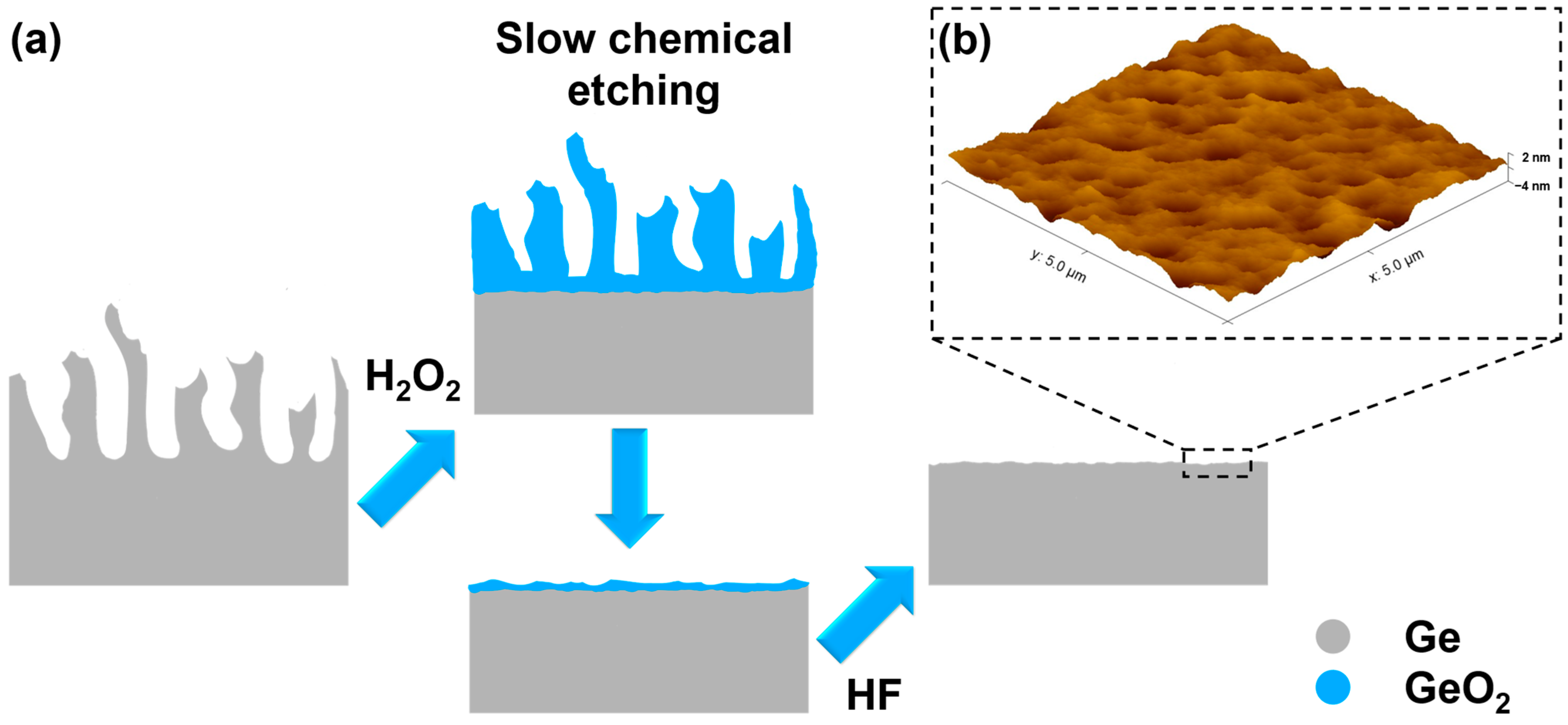
After epitaxial growth, the Ge FSM is detached using an adhesive tape and is transferred onto a flexible Polyvinyl Chloride (PVC) substrate. The recovered Ge substrate has been reconditioned by immersion in a concentrated hydrogen peroxide (H2O2, 30 wt%) solution for 1 min, at room temperature, to transform the remaining PGe crystallites at the surface in germanium dioxide (GeO2) and etch them slowly away. The substrate has been subsequently deoxidized in concentrated HF (49 wt%) to dissolve the remaining Ge oxides on the surface and recover a flat surface. The reconditioned substrate is then reporosified, using the same BEE conditions as on the epi-ready substrate. The complete list of specifications of all the chemicals used in this study can be found in Table S1 of Supplementary Materials.

2.2. Characterization

The PGe thickness, porosity, and their uniformity over the 100 mm wafer were characterized using a J.A. Woollam Co. VASE instrument (Lincoln, NE, USA) in the spectral range between 500 and 900 nm. The measuring points were radially paced every 30° with an in-between point spacing of 5 mm along the radius of the wafer. The PGe thickness was also verified using SEM imaging of the PGe layer cross-section, also revealing the morphology of the nanostructure. The presence/lack of the PGe layer remnants on the substrate after the detachment and cleaning process was observed using optical microscopy (confocal microscope Keyence VK-X1100 (Mississauga, ON, Canada) with 150× lens) and SEM imaging of the substrate’s plan view. All of the SEM observations were performed at a 4.0 mm working distance with Thermo Fisher Scios 2 SEM (Toronto, ON, Canada) using 20 keV acceleration voltage for the electron beam.
The Park system NX20 (Burlington, MA, USA), atomic force microscope (AFM) was used to evaluate the surface topology of the PGe layer, Ge FSM membrane, and of the recovered substrate after cleaning. The AFM scans were performed in tapping mode using a super sharp silicon probe (SSS-NCHR) and a scan resolution of 512 × 512 pixels over a 5 × 5 µm2 area. The scan data were then processed using Gwyddion software (Version 2.64) to obtain the root mean square (RMS) roughness values.
The investigation of the structural properties of Ge FSM was conducted using the Rigaku Smartlab (The Woodlands, TX, USA) high-resolution X-ray diffraction (XRD) system in the in-plane configuration, equipped with Cu Kα X-ray source (wavelength λ(Cu Kα) = 1.5406 Å), Ge (220) × 2 monochromator on the incident beam, and a two-dimensional hybrid pixel array semiconductor X-ray detector (HYPIX-3000). The in-plane pole figure XRD measurements were employed to assess the crystalline quality of the Ge membranes while restricting the depth of beam penetration.

3. Results and Discussion

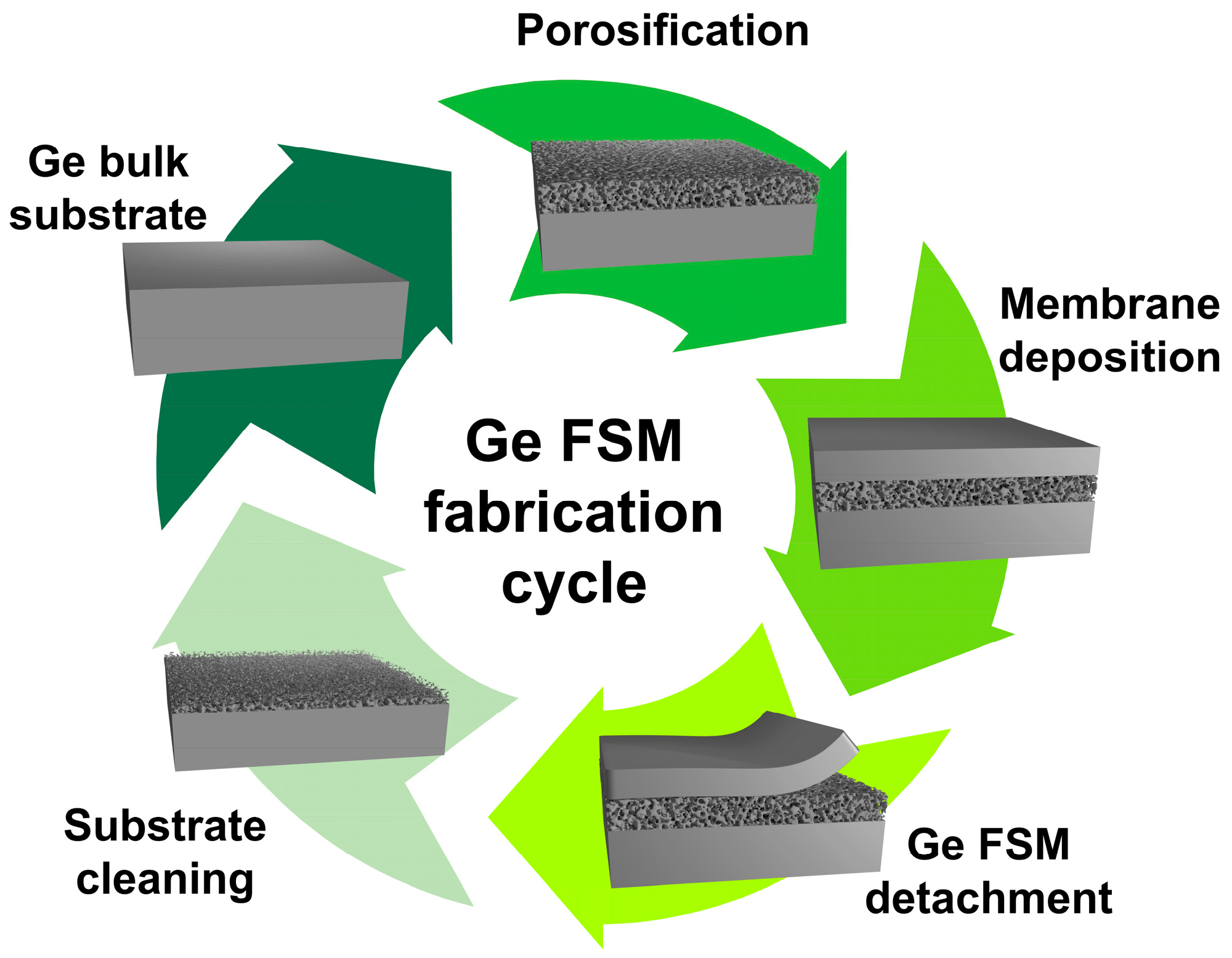
The fabrication process of Ge FSM through the porosification lift-off technique consists of four main steps, as illustrated in Figure 2. First, a high-porosity (60–80%) porous germanium (PGe) layer is formed at the Ge substrate surface by electrochemical etching in a HF:EtOH electrolytic solution. This is followed by a low-temperature deposition of the Ge membrane on top of the PGe layer. The membrane is then mechanically detached from the substrate, which is facilitated by the fragile nanostructured PGe interface between the Ge bulk substrate and the Ge FSM. After detachment, the PGe remnants at the surface of the recovered parent Ge substrate are oxidized using an H2O2 solution followed by their complete dissolution in HF and reuse of the parent Ge substrate for reporosification and repeating the process of Ge FSM fabrication. All steps were previously detailed in the Materials and Methods Section, and the obtained results are discussed further below.
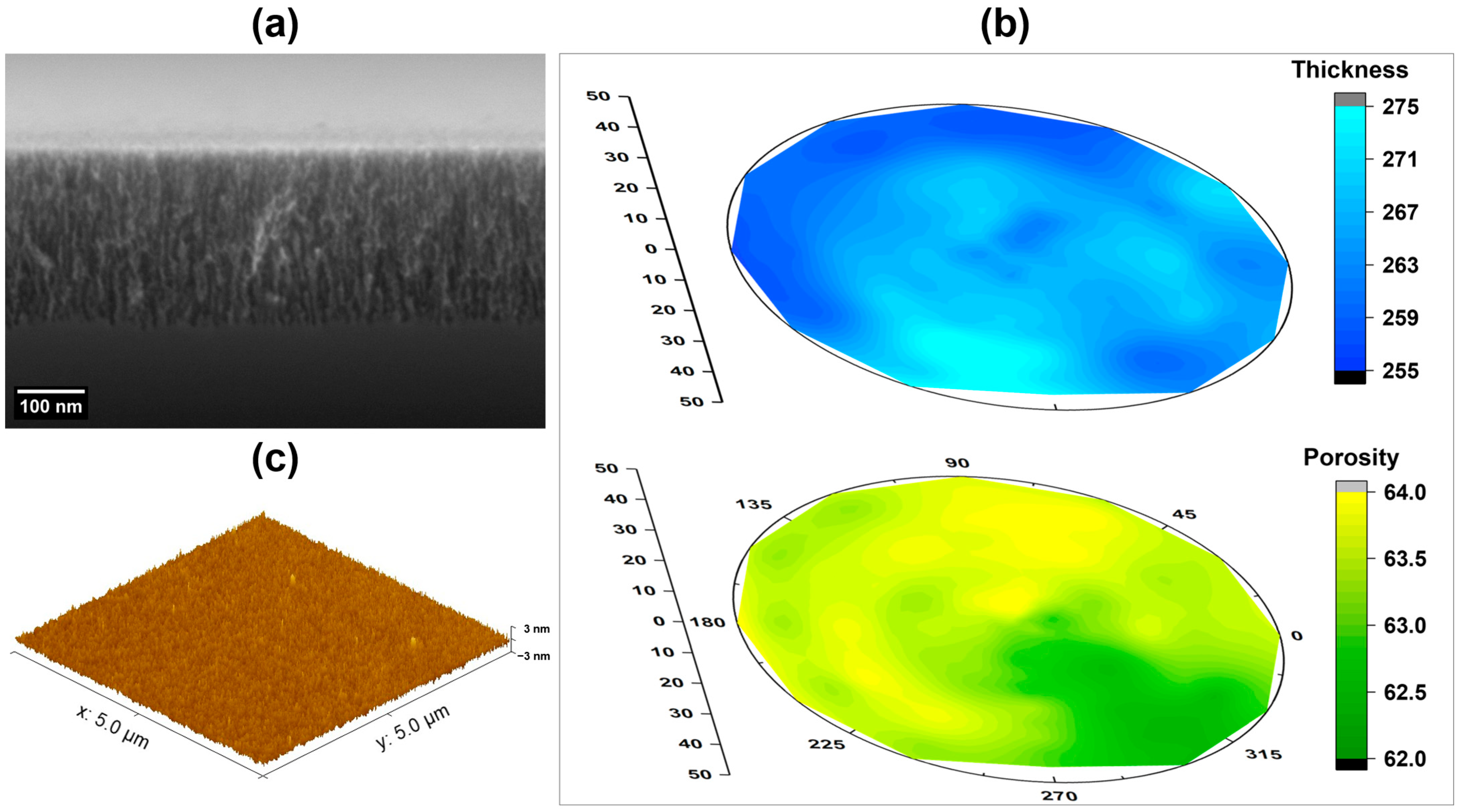
The Ge FSM fabrication cycle begins with the formation of a high-quality PGe layer on top of the Ge substrate. This is a crucial part of the process, as any major defects or inhomogeneities in the PGe structure can be further transferred to the Ge membrane, impacting its quality. Figure 3a shows a SEM cross-sectional micrograph of the PGe layer used in this work. The typical sponge-like porous structure shows a well-defined interface between the PGe layer and bulk material, with ~264 nm thickness and 63% porosity. Furthermore, the ellipsometry mapping of the 100 mm wafer, shown in Figure 3b,c, demonstrates the overall uniformity of the porous nanostructure in both the thickness and porosity with respective variations of ±4 nm and ±1% across the entire surface of the wafer. Moreover, the porous structure manifests a low surface roughness below 2 nm, as illustrated by Figure 3c. The closely packed crystallites of the high-porosity PGe layers (60–80%) make an excellent template for the growth of Ge membrane structures while enabling an easy detachment without additional annealing steps or deposition of the stressor layers to initiate the separation.
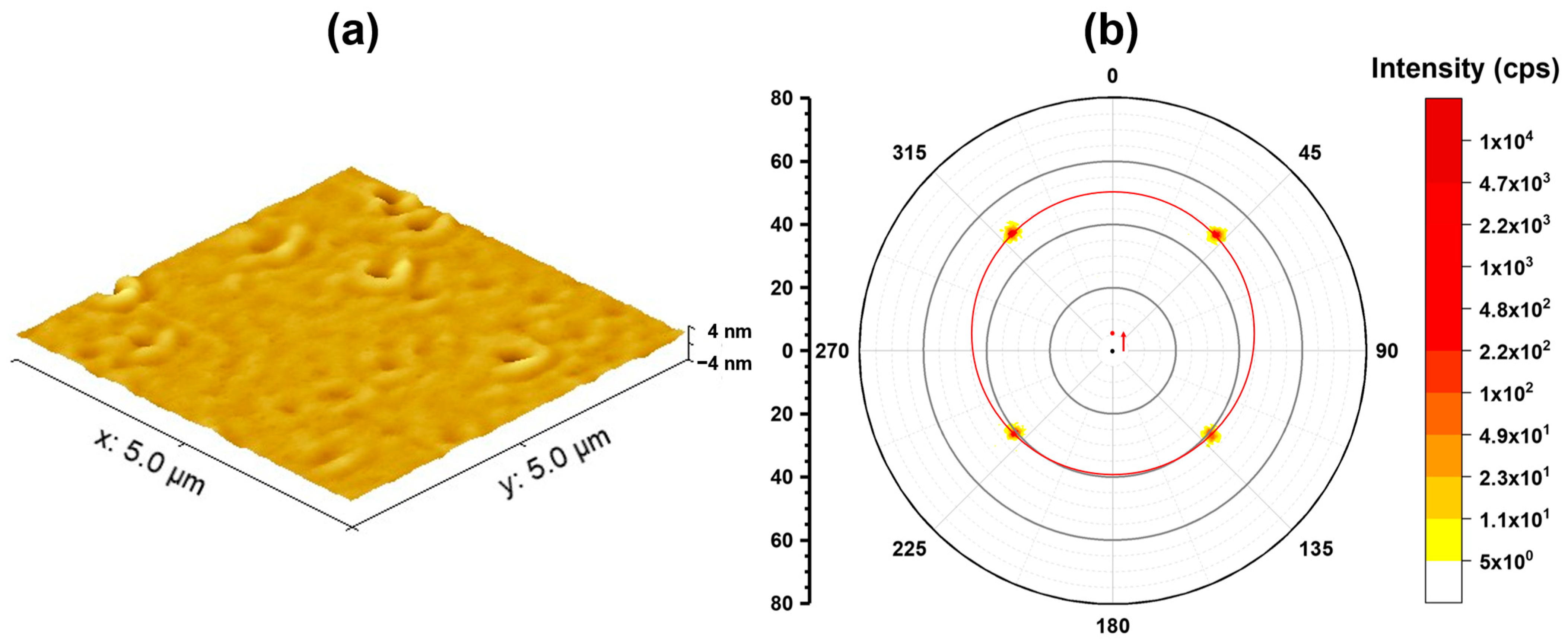
A 1 µm thick Ge membrane is then grown, at 300 °C, on top of the PGe structure, resulting in a smooth surface with RMS roughness around 0.7 nm, as demonstrated by Figure 4a. A few shallow pits and undulations are still identifiable on the surface but can be annihilated by increasing the membrane’s thickness. Depending on the targeted application, the thickness of the membrane can be varied from ~100 nm to few µm. However, for membranes thinner than 1 µm the surface roughness can increase up to 5 nm for the thinnest membranes [20]. This way, the amount of Ge material used for device integration can be directly controlled to use only the quantity of Ge necessary for its function and, hence, limit the waste of material.
The crystalline quality of the membrane is verified using X-ray diffraction in in-plane configuration, to limit the beam penetration. The resulting in-plane pole figure around the Ge (220) axis displays four sharp peaks with 90° rotational symmetry around their central axis, as depicted by Figure 4b. This pattern corresponds to the diamond-cubic crystal structure of Ge, confirming the high crystalline quality of the Ge membrane. Interestingly, the central axis of the sample demonstrates a 6° shift from the measurement axis, as illustrated by the red arrow in Figure 4b. This shift is attributed to the parent substrate’s 6° miscut from (100) orientation towards the (111) axis. Since the porous structure maintains the original substrate orientation, it can transfer even this characteristic to the Ge membrane, resulting in a monocrystalline epilayer with the same 6° off-cut as the parent substrate. The pole figure of the Ge membrane deposited on the on-axis substrate can be found in Figure S1.
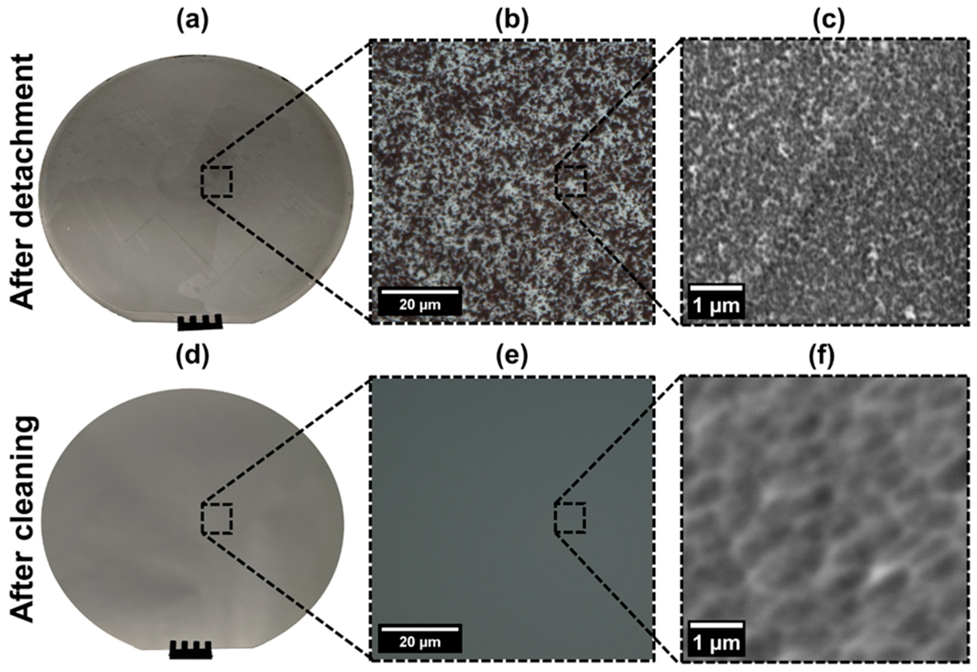
Once the Ge membrane is formed, the high-porosity nanostructure underneath represents a perfect fragile interface allowing for easy detachment and transfer to the host substrate. Using an adhesive polymer tape, the Ge FSM can be separated from the parent substrate and transferred to a flexible PVC holder. After the detachment, irregular remnants of the porous structure are still present on the surface of both the substrate and membrane as illustrated by Figure 5a–c. To eliminate these porous residues, a simple chemical cleaning process is employed. The chemical etching of Ge in aqueous solutions functions on the principles of the formation and dissolution of the GeO2, where H2O2 acts as an oxidizing agent which transforms the Ge surface in GeO2 [51,52]. At the same time, the reduction in the H2O2 at the surface of Ge provides the holes necessary for the dissolution of the oxide [34,53]. When the substrate is immersed in concentrated H2O2, the solution transforms the remaining PGe structure’s high-specific surface area in GeO2 and starts slowly etching it away [54]. This chemical etching of Ge can be described by Equations (1) and (2), where Equation (1) represent the formation of the GeO2 on the Ge surface, and Equation (2) represents its dissolution into H2GeO2, a tetravalent form stable in aqueous solution with pH < 8.5 [51]:
G e + 2 H 2 O 2 G e O 2 + 2 H 2 O
G e O 2 + H 2 O H 2 G e O 3
Compared to techniques involving high-temperature annealing and PGe reconstruction in crystallites with size superior to 100 nm [48,55], the high porosity nanostructure with significantly higher specific surface area and only 5–10 nm thick pore walls, allows for isotropic etching by H2O2 at a very slow rate (few nm/min). This avoids the substantial chemical etching of the substrate (few µm) necessary for the planarization of larger features while maintaining control over the etching process, due to the slow etching rate. The remaining oxides on the substrate’s surface are then dissolved in an HF solution during the deoxidation step prior to the reporosification. This results in a remnant-free surface, as shown in Figure 5d–f.
The entire chemical cleaning process is schematically illustrated in Figure 6a. The AFM scan of the recovered substrate in Figure 6b shows a flat surface with surface roughness below 1 nm. The apparent waving of the cleaned surface represents a slight variation in the initial Ge bulk/PGe interface formed during the BEE process, as the bottoms of the pores are not all perfectly aligned. This effect is then partially mitigated directly by the first anodic step of the BEE process during the formation of the new PGe layer [43,48].
The PGe remnants on the Ge FSM backside have the same nature as they are formed by the separation of the uniform PGe layer. This means that the same approach can be also used for membrane cleaning. However, its necessity should be evaluated depending on the Ge FSM use. In the case of applications where the Ge membrane does not play an active role in the final function and serves mainly as the crystalline substrate for the growth of epitaxial structures, membrane cleaning should not be necessary.
To complete the cycle, the recovered parent substrate then undergoes a second porosification process to obtain a new PGe layer, presented in Figure 7a. For comparison, an optical image of the PGe layer on an epi-ready substrate can be found in Supplementary Materials Figure S2. The BEE conditions are identical to the ones previously used on the epi-ready substrate. It presents the same thickness and porosity with high uniformity across the entire 100 mm wafer as demonstrated by ellipsometry mapping present in Figure 7b,c. The new PGe layer presents a thickness of 265 ± 5 nm and a porosity of 63 ± 1%; both of these values are the same as on the original epi-ready substrate, demonstrating that the small surface undulations on the recovered substrate do not influence the BEE process. This new high-quality PGe structure then allows for a new deposition of the Ge membrane and fabrication of multiple Ge FSM from the same substrate, by repeating the process. This demonstrates that the Ge FSM can be fabricated with a consumption of less than 300 nm of the original Ge substrate, while allowing for easy substrate reuse without the involvement of expensive reconditioning techniques such as chemical–mechanical polishing [49]. In the context of our previous study, the use of an optimized PGe layer enables improvements in both Ge FSM surface quality and material consumption of the substrate per cycle. The lower porosity allows for more closely packed nucleation sites, resulting in a reduction in the membrane’s surface roughness to 0.7 nm from 1.2 nm on 70% porosity PGe substrate [20]. Additionally, the reduced PGe thickness further lowers the substrate consumption by over 70%, while maintaining the ease of detachability of the Ge FMS.
In numbers, around 300 nm of the original substrate is consumed per produced membrane. This value includes the thickness of the PGe layer (~270 nm) and the front/back etching of the substrate during the cleaning process (~30 nm). By considering a conventional 100 mm Ge wafer with a thickness of 175 µm, and its thinnest commercially available counterpart with a thickness of 140 µm, around 35 µm of the substrate can be used for Ge FSM production before losing its structural integrity. This results in over 100 membranes produced from a single 100 mm wafer. The number of reuses can be further increased by the use of thicker substrates, which bring additional benefits such as a reduction in material lost during the sawing process of Ge wafers from a solid ingot grown by the Czochralski method [56,57,58]. Alternatively, the Ge substrate can be bonded on a holder (e.g., Si substrate) for mechanical support, allowing for the use of almost the entire substrate’s thickness and enabling over 500 reuses. This number is expected to further grow with scaling on larger substrates, as larger diameter wafers are considerably thicker.
In comparison, techniques involving high-temperature annealing present large pillars with a diameter superior to 100 nm, and cannot be reconditioned without the etching of substantial material quantity (few µm) [55] or the involvement of costly chemical–mechanical polishing (CMP) [41]. Considering single-sided chemical etching of 5 µm per reuse, this approach presents more limited number of Ge FSM fabricated from a single wafer. However, this approach holds its advantage for applications involving high processing temperatures (>400 °C) as the high specific area PGe structure cannot be maintained under these conditions.
2D-assisted epitaxy, in theory, could eventually offer infinite reuse of the substrate. However, it is still in relatively early research stages, especially in case of group IV materials such as Ge, which were demonstrated for the first time in late 2022 [39], using a new approach on local nucleation and lateral overgrowth on the 2D interface [40,59]. Moreover, other challenges such as large surface growth and the transfer of high-quality interfaces need to be resolved for its viable application [60].
Considering the rarity of the Ge, the complexity of its recovery from end-of-life optoelectronic devices, and the recent geopolitical situation around this material, the Ge FSM represent a sustainable alternative to conventional Ge substrates. It offers to significantly reduce the quantity of Ge material integrated into optoelectronic devices while allowing for cost reduction, thanks to the limited Ge use and substrate recycling.

4. Conclusions

In conclusion, we demonstrated a successful substrate reuse and production of a highly uniform PGe layer on recovered Ge substrate, setting the milestone for the production of multiple Ge FSM from the same substrate. The reduced thickness of the PGe structure allows for a reduction in Ge consumption per cycle, compared to previous studies, while still maintaining the capacity to easily detach and transfer the membrane. Additionally, the optimized porosity helps to improve the surface quality of the Ge FSM membrane. The substrate cleaning is achieved through slow chemical etching of the PGe remaining on the surface, enabling its complete dissolution and obtention of the surface with roughness below 1 nm, without the necessity of substantial etching of the substrate material. The recovered substrate enables the successful reporosification of the entire 100 mm wafer, resulting in a new high-quality PGe layer with physical properties identical to the one formed on the epi-ready surface. This allows for efficient substrate reuse, and the production of multiple Ge membranes, with minimal consumption of the Ge material, due to the optimized PGe structure employed in this work. The Ge FSM production and substrate recovery opens the way for a sustainable alternative to conventional Ge substrates while providing all the advantages of freestanding form for direct heterointegration and flexible electronics. It offers to significantly lower the quantity of Ge material integrated into optoelectronic devices, reducing the quantity of hard-to-recover material in end-of-life products. Furthermore, this approach presents a high potential for the fabrication of FSM from other group IV materials, such as Ge(Si)Sn alloys, presenting a small bandgap ideal for near/mid-IR optoelectronic and photonic applications offering a non-toxic and low-cost alternative to III-V materials.

Supplementary Materials

The following supporting information can be downloaded at: https://www.mdpi.com/article/10.3390/su16041444/s1, Table S1: List of chemicals employed in porosification and chemical cleaning of the substrates, Figure S1: In-plane pole figure of the Ge FSM around Ge (220) axis of the on-axis Ge substrate, Figure S2: Optical image of PGe layer on epi-ready substrate.

Author Contributions

The manuscript was written through the contributions of all authors. T.H.: conceptualization, methodology, investigation, data curation, original draft preparation, review and editing, visualization. B.I.: conceptualization, methodology, supervision, visualization, investigation, review and editing. J.C. and K.D.: review and editing. A.B.: supervision, funding acquisition, review and editing. All authors have read and agreed to the published version of the manuscript.

Funding

The funding for this research was provided by Natural Sciences and Engineering Research Council of Canada (NSERC, CRDPJ 537960-18), Fonds de Recherche du Québec (FRQNT), Innovation en Énergie Électrique (InnovÉÉ, R15-1901), Mitacs (IT25635), Saint-Augustin Canada Electric (Stace), and Umicore.

Institutional Review Board Statement

Not applicable.

Informed Consent Statement

Not applicable.

Data Availability Statement

Data will be made available on request.

Acknowledgments

Special thanks to Philippe-Olivier Provost for the development and fabrication of advanced porosification tools used in this work. We also thank Thierno Mamoudou Diallo and Hubert Pelletier for scientific discussions, Guillaume Bertrand, Julie Ménard, Donald Ducharme, Mathieu Cloutier, and all the technical staff of 3IT for the technical support in the present research work. The authors would also like to express their gratitude to CG Figures for providing free resources and tutorials for the use of Blender in scientific illustrations. LN2 is a joint International Research Laboratory (IRL 3463) funded and co-operated in Canada by Université de Sherbrooke (UdeS) and in France by CNRS as well as ECL, INSA Lyon, and Université Grenoble Alpes (UGA). It is also supported by the Fonds de Recherche du Québec Nature et Technologie (FRQNT).

Conflicts of Interest

Jinyoun Cho and Kristof Dessein are employed by the Umicore Electro-Optic Materials company. The remaining authors declare that the research was conducted in the absence of any commercial or financial relationships that could be construed as a potential conflict of interest.

References

  1. Armand Pilon, F.T.; Lyasota, A.; Niquet, Y.-M.; Reboud, V.; Calvo, V.; Pauc, N.; Widiez, J.; Bonzon, C.; Hartmann, J.M.; Chelnokov, A.; et al. Lasing in strained germanium microbridges. Nat. Commun. 2019, 10, 2724. [Google Scholar] [CrossRef] [PubMed]
  2. Nam, D.; Roy, A.M.; Huang, K.C.Y.; Brongersma, M.L.; Saraswat, K.C. Strained Germanium Membrane using Thin Film Stressor for High Efficiency Laser. In Proceedings of the CLEO: 2011—Laser Applications to Photonic Applications (2011), Paper JTuI85, Baltimore, MD, USA, 1–6 May 2011; Optica Publishing Group: Washington, DC, USA, 2011; p. JTuI85. [Google Scholar]
  3. Lim, J.; Shim, J.; Geum, D.-M.; Kim, S. Experimental Demonstration of Germanium-on-Silicon Slot Waveguides at Mid-Infrared Wavelength. IEEE Photonics J. 2022, 14, 5828709. [Google Scholar] [CrossRef]
  4. Kang, J.; Cheng, Z.; Zhou, W.; Xiao, T.-H.; Gopalakrisna, K.-L.; Takenaka, M.; Tsang, H.K.; Goda, K. Focusing subwavelength grating coupler for mid-infrared suspended membrane germanium waveguides. Opt. Lett. OL 2017, 42, 2094–2097. [Google Scholar] [CrossRef] [PubMed]
  5. Atalla, M.R.M.; Assali, S.; Attiaoui, A.; Lemieux-Leduc, C.; Kumar, A.; Abdi, S.; Moutanabbir, O. All-Group IV Transferable Membrane Mid-Infrared Photodetectors. Adv. Funct. Mater. 2021, 31, 2006329. [Google Scholar] [CrossRef]
  6. Cho, N.; Kim, M.; Ma, Z. Germainum photodetectors coupled with silicon waveguides on a flexible substrate using nanomembrane transfer printing method. In Proceedings of the 10th International Conference on Group IV Photonics, Seoul, Republic of Korea, 28–30 August 2013; pp. 77–78. [Google Scholar]
  7. Wu, S.; Zhou, H.; Chen, Q.; Zhang, L.; Lee, K.H.; Bao, S.; Fan, W.; Tan, C.S. Suspended germanium membranes photodetector with tunable biaxial tensile strain and location-determined wavelength-selective photoresponsivity. Appl. Phys. Lett. 2021, 119, 191106. [Google Scholar] [CrossRef]
  8. Liu, C.; Ma, W.; Chen, M.; Ren, W.; Sun, D. A vertical silicon-graphene-germanium transistor. Nat. Commun. 2019, 10, 4873. [Google Scholar] [CrossRef] [PubMed]
  9. Blanco, E.; Martín, P.; Domínguez, M.; Fernández-Palacios, P.; Lombardero, I.; Sanchez-Perez, C.; García, I.; Algora, C.; Gabás, M. Refractive indices and extinction coefficients of p-type doped Germanium wafers for photovoltaic and thermophotovoltaic devices. Sol. Energy Mater. Sol. Cells 2024, 264, 112612. [Google Scholar] [CrossRef]
  10. Daligou, G.; Soref, R.; Attiaoui, A.; Hossain, J.; Atalla, M.R.M.; Vecchio, P.D.; Moutanabbir, O. Group IV Mid-Infrared Thermophotovoltaic Cells on Silicon. IEEE J. Photovolt. 2023, 13, 728–735. [Google Scholar] [CrossRef]
  11. van der Heide, J.; Posthuma, N.E.; Flamand, G.; Poortmans, J. Development of Low-cost Thermophotovoltaic Cells Using Germanium Substrates. AIP Conf. Proc. 2007, 890, 129–138. [Google Scholar] [CrossRef]
  12. King, R.R.; Law, D.C.; Edmondson, K.M.; Fetzer, C.M.; Kinsey, G.S.; Yoon, H.; Sherif, R.A.; Karam, N.H. 40% efficient metamorphic GaInP/GaInAs/Ge multijunction solar cells. Appl. Phys. Lett. 2007, 90, 183516. [Google Scholar] [CrossRef]
  13. European Commission (EC). Critical Raw Materials Resilience: Charting a Path towards Greater Security and Sustainability. 2020. Available online: https://ec.europa.eu/docsroom/documents/42849 (accessed on 10 January 2024).
  14. Interior Releases 2018′s Final List of 35 Minerals Deemed Critical to U.S. National Security and the Economy|U.S. Geological Survey. Available online: https://www.usgs.gov/news/national-news-release/interior-releases-2018s-final-list-35-minerals-deemed-critical-us (accessed on 10 January 2024).
  15. Thomas, C.L. United States Geological Survey (USGS) 2018 Minerals Yearbook—Germanium. Available online: https://d9-wret.s3.us-west-2.amazonaws.com/assets/palladium/production/atoms/files/myb1-2018-germa.pdf (accessed on 5 January 2024).
  16. United States Geological Survey (USGS) 2023 Mineral Commodity Summaries—Germanium. Available online: https://pubs.usgs.gov/periodicals/mcs2023/mcs2023-germanium.pdf (accessed on 5 January 2024).
  17. Kamran Haghighi, H.; Irannajad, M. Roadmap for recycling of germanium from various resources: Reviews on recent developments and feasibility views. Env. Sci. Pollut. Res. 2022, 29, 48126–48151. [Google Scholar] [CrossRef] [PubMed]
  18. Meshram, P. Abhilash Strategies for Recycling of Primary and Secondary Resources for Germanium Extraction. Min. Metall. Explor. 2022, 39, 689–707. [Google Scholar] [CrossRef]
  19. Germanium Substrates. Available online: https://eom.umicore.com/en/germanium-solutions/products/germanium-substrates/ (accessed on 10 January 2024).
  20. Hanuš, T.; Ilahi, B.; Chapotot, A.; Pelletier, H.; Cho, J.; Dessein, K.; Boucherif, A. Wafer-scale Ge freestanding membranes for lightweight and flexible optoelectronics. Mater. Today Adv. 2023, 18, 100373. [Google Scholar] [CrossRef]
  21. Shin, J.; Kim, H.; Sundaram, S.; Jeong, J.; Park, B.-I.; Chang, C.S.; Choi, J.; Kim, T.; Saravanapavanantham, M.; Lu, K.; et al. Vertical full-colour micro-LEDs via 2D materials-based layer transfer. Nature 2023, 614, 81–87. [Google Scholar] [CrossRef] [PubMed]
  22. Lombardero, I.; Ochoa, M.; Miyashita, N.; Okada, Y.; Algora, C. Theoretical and experimental assessment of thinned germanium substrates for III–V multijunction solar cells. Prog. Photovolt. Res. Appl. 2020, 28, 1097–1106. [Google Scholar] [CrossRef]
  23. Mittapally, R.; Lee, B.; Zhu, L.; Reihani, A.; Lim, J.W.; Fan, D.; Forrest, S.R.; Reddy, P.; Meyhofer, E. Near-field thermophotovoltaics for efficient heat to electricity conversion at high power density. Nat. Commun. 2021, 12, 4364. [Google Scholar] [CrossRef] [PubMed]
  24. Bhatt, G.R.; Zhao, B.; Roberts, S.; Datta, I.; Mohanty, A.; Lin, T.; Hartmann, J.-M.; St-Gelais, R.; Fan, S.; Lipson, M. Integrated near-field thermo-photovoltaics for heat recycling. Nat. Commun. 2020, 11, 2545. [Google Scholar] [CrossRef]
  25. Winter, E.; Schreiber, W.; Schygulla, P.; Souza, P.L.; Janz, S.; Lackner, D.; Ohlmann, J. III-V material growth on electrochemically porosified Ge substrates. J. Cryst. Growth 2023, 602, 126980. [Google Scholar] [CrossRef]
  26. Algora, C.; García, I.; Palacios, P.F.; Gómez-Reboreda, D.; Martín, P.; Sanchez-Perez, C.; Cifuentes, L.; Lombardero, I.; Gabás, M.; Rey-Stolle, I. Advances in Flexible and Lightweight 3J Space Solar Cells for High Power Density Applications. In Proceedings of the 2023 13th European Space Power Conference (ESPC), Elche, Spain, 2–6 October 2023; pp. 1–4. [Google Scholar]
  27. Moon, S.; Kim, K.; Kim, Y.; Kang, H.K.; Park, K.-H.; Lee, J. Ultrathin Flexible Ge Solar Cells for Lattice-Matched Thin-Film InGaP/(In)GaAs/Ge Tandem Solar Cells. Sol. RRL 2023, 7, 2300387. [Google Scholar] [CrossRef]
  28. Cavalli, A.; Alkurd, N.; Johnston, S.; Diercks, D.R.; Roberts, D.M.; Ley, B.E.; Simon, J.; Young, D.L.; Packard, C.E.; Ptak, A.J. Performance of III–V Solar Cells Grown on Reformed Mesoporous Ge Templates. IEEE J. Photovolt. 2022, 12, 337–343. [Google Scholar] [CrossRef]
  29. Martín, P.; Orejuela, V.; Sanchez-Perez, C.; García, I.; Rey-Stolle, I. Device Architectures for Germanium TPV Cells with Efficiencies over 30%. In Proceedings of the 2023 14th Spanish Conference on Electron Devices (CDE), Valencia, Spain, 6–8 June 2023; pp. 1–4. [Google Scholar]
  30. Guo, Q.; Fang, Y.; Zhang, M.; Huang, G.; Chu, P.K.; Mei, Y.; Di, Z.; Wang, X. Wrinkled Single-Crystalline Germanium Nanomembranes for Stretchable Photodetectors. IEEE Trans. Electron Devices 2017, 64, 1985–1990. [Google Scholar] [CrossRef]
  31. Nam, D.; Sukhdeo, D.; Roy, A.; Balram, K.; Cheng, S.-L.; Huang, K.C.-Y.; Yuan, Z.; Brongersma, M.; Nishi, Y.; Miller, D.; et al. Strained germanium thin film membrane on silicon substrate for optoelectronics. Opt. Express OE 2011, 19, 25866–25872. [Google Scholar] [CrossRef] [PubMed]
  32. Zhao, H.; Xue, Z.; Wu, X.; Wei, Z.; Guo, Q.; Xu, M.; Qu, C.; You, C.; Mei, Y.; Zhang, M.; et al. Biodegradable germanium electronics for integrated biosensing of physiological signals. NPJ Flex. Electron. 2022, 6, 1–10. [Google Scholar] [CrossRef]
  33. La Mattina, A.A.; Mariani, S.; Barillaro, G. Bioresorbable Materials on the Rise: From Electronic Components and Physical Sensors to In Vivo Monitoring Systems. Adv. Sci. 2020, 7, 1902872. [Google Scholar] [CrossRef] [PubMed]
  34. Sanchez-Perez, C.; Garcia, I.; Rey-Stolle, I. Fast chemical thinning of germanium wafers for optoelectronic applications. Appl. Surf. Sci. 2022, 579, 152199. [Google Scholar] [CrossRef]
  35. Yoon, J.; Jo, S.; Chun, I.S.; Jung, I.; Kim, H.-S.; Meitl, M.; Menard, E.; Li, X.; Coleman, J.J.; Paik, U.; et al. GaAs photovoltaics and optoelectronics using releasable multilayer epitaxial assemblies. Nature 2010, 465, 329–333. [Google Scholar] [CrossRef] [PubMed]
  36. Maeda, T.; Chang, W.-H.; Irisawa, T.; Ishii, H.; Hattori, H.; Poborchii, V.; Kurashima, Y.; Takagi, H.; Uchida, N. Advanced germanium layer transfer for ultra thin body on insulator structure. Appl. Phys. Lett. 2016, 109, 262104. [Google Scholar] [CrossRef]
  37. Akatsu, T.; Deguet, C.; Sanchez, L.; Allibert, F.; Rouchon, D.; Signamarcheix, T.; Richtarch, C.; Boussagol, A.; Loup, V.; Mazen, F.; et al. Germanium-on-insulator (GeOI) substrates—A novel engineered substrate for future high performance devices. Mater. Sci. Semicond. Process. 2006, 9, 444–448. [Google Scholar] [CrossRef]
  38. Mangum, J.S.; Rice, A.D.; Chen, J.; Chenenko, J.; Wong, E.W.K.; Braun, A.K.; Johnston, S.; Guthrey, H.; Geisz, J.F.; Ptak, A.J.; et al. High-Efficiency Solar Cells Grown on Spalled Germanium for Substrate Reuse without Polishing. Adv. Energy Mater. 2022, 12, 2201332. [Google Scholar] [CrossRef]
  39. Kim, H.; Lee, S.; Shin, J.; Zhu, M.; Akl, M.; Lu, K.; Han, N.M.; Baek, Y.; Chang, C.S.; Suh, J.M.; et al. Graphene nanopattern as a universal epitaxy platform for single-crystal membrane production and defect reduction. Nat. Nanotechnol. 2022, 17, 1054–1059. [Google Scholar] [CrossRef]
  40. Diallo, T.M.; Hanuš, T.; Patriarche, G.; Ruediger, A.; Boucherif, A. Unraveling the Heterointegration of 3D Semiconductors on Graphene by Anchor Point Nucleation. Small 2023, e2306038. [Google Scholar] [CrossRef] [PubMed]
  41. Park, S.; Simon, J.; Schulte, K.L.; Ptak, A.J.; Wi, J.-S.; Young, D.L.; Oh, J. Germanium-on-Nothing for Epitaxial Liftoff of GaAs Solar Cells. Joule 2019, 3, 1782–1793. [Google Scholar] [CrossRef]
  42. Jeong, J.; Kim, T.; Lee, B.J.; Lee, J. PCA-based sub-surface structure and defect analysis for germanium-on-nothing using nanoscale surface topography. Sci. Rep. 2022, 12, 7205. [Google Scholar] [CrossRef] [PubMed]
  43. Hanuš, T.; Arias-Zapata, J.; Ilahi, B.; Provost, P.-O.; Cho, J.; Dessein, K.; Boucherif, A. Large-Scale Formation of Uniform Porous Ge Nanostructures with Tunable Physical Properties. Adv. Mater. Interfaces 2023, 10, 2202495. [Google Scholar] [CrossRef]
  44. Schreiber, W.; Liu, T.; Janz, S. The effect of passivation to etching duration ratio on bipolar electrochemical etching of porous layer stacks in germanium. J. Phys. Chem. Solids 2023, 176, 111265. [Google Scholar] [CrossRef]
  45. Zhu, Y.; Zhang, Y.; Li, B.; Xia, G.; Wen, R.-T. Achieving porous germanium from both p- and n-type epitaxial Ge-on-Si via bipolar potentiostatic etching. Electrochim. Acta 2023, 470, 143307. [Google Scholar] [CrossRef]
  46. Hanuš, T.; Mouchel, L.; Ilahi, B.; Dupuy, A.; Cho, J.; Dessein, K.; Boucherif, A. Potential monitoring during Ge electrochemical etching: Towards tunable double porosity layers. Electrochim. Acta 2024, 474, 143529. [Google Scholar] [CrossRef]
  47. Tutashkonko, S.; Alekseev, S.; Nychyporuk, T. Nanoscale morphology tuning of mesoporous Ge: Electrochemical mechanisms. Electrochim. Acta 2015, 180, 545–554. [Google Scholar] [CrossRef]
  48. Chapotot, A.; Ilahi, B.; Hanuš, T.; Hamon, G.; Cho, J.; Dessein, K.; Darnon, M.; Boucherif, A. Sequential fabrication of multiple Ge nanomembranes from a single wafer: Towards sustainable recycling of Ge substrates. Sustain. Mater. Technol. 2024, 39, e00806. [Google Scholar] [CrossRef]
  49. Ward, J.S.; Remo, T.; Horowitz, K.; Woodhouse, M.; Sopori, B.; VanSant, K.; Basore, P. Techno-economic analysis of three different substrate removal and reuse strategies for III-V solar cells. Prog. Photovolt. Res. Appl. 2016, 24, 1284–1292. [Google Scholar] [CrossRef]
  50. Karagoz, A.; Basim, G.B. Controlling Germanium CMP Selectivity through Slurry Mediation by Surface Active Agents. ECS J. Solid State Sci. Technol. 2015, 4, P5097. [Google Scholar] [CrossRef]
  51. Huygens, I.M.; Gomes, W.P.; Strubbe, K. Etching of Germanium in Hydrogenperoxide Solutions. ECS Trans. 2007, 6, 375–386. [Google Scholar] [CrossRef]
  52. Cerniglia, N.; Wang, P. Dissolution of Germanium in Aqueous Hydrogen Peroxide Solution. J. Electrochem. Soc. 1962, 109, 508. [Google Scholar] [CrossRef]
  53. Turner, D.R. On the Mechanism of Chemically Etching Germanium and Silicon. J. Electrochem. Soc. 1960, 107, 810. [Google Scholar] [CrossRef]
  54. Sioncke, S.; Brunco, D.P.; Meuris, M.; Uwamahoro, O.; Steenbergen, J.V.; Vrancken, E.; Heyns, M.M. Etch Rates of Ge, GaAs and InGaAs in Acids, Bases and Peroxide Based Mixtures. ECS Trans. 2008, 16, 451. [Google Scholar] [CrossRef]
  55. Chapotot, A.; Ilahi, B.; Arias-Zapata, J.; Hanuš, T.; Ayari, A.; Hamon, G.; Cho, J.; Dessein, K.; Darnon, M.; Boucherif, A. Germanium surface wet-etch-reconditioning for porous lift-off and substrate reuse. Mater. Sci. Semicond. Process. 2023, 168, 107851. [Google Scholar] [CrossRef]
  56. Czochralski, J. Ein neues Verfahren zur Messung der Kristallisationsgeschwindigkeit der Metalle. Z. Für Phys. Chem. 1918, 92U, 219–221. [Google Scholar] [CrossRef]
  57. Teal, G.K.; Sparks, M.; Buehler, E. Growth of Germanium Single Crystals Containing p N Junctions. Phys. Rev. 1951, 81, 637. [Google Scholar] [CrossRef]
  58. Depuydt, B.; Theuwis, A.; Romandic, I. Germanium: From the first application of Czochralski crystal growth to large diameter dislocation-free wafers. Mater. Sci. Semicond. Process. 2006, 9, 437–443. [Google Scholar] [CrossRef]
  59. Manzo, S.; Strohbeen, P.J.; Lim, Z.-H.; Saraswat, V.; Arnold, M.S.; Kawasaki, J.K. Pinhole-seeded lateral epitaxy and exfoliation on graphene-terminated surfaces. arXiv 2021, arXiv:2106.00721. [Google Scholar] [CrossRef]
  60. Kim, H.; Kim, J.C.; Jeong, Y.; Yu, J.; Lu, K.; Lee, D.; Kim, N.; Jeong, H.Y.; Kim, J.; Kim, S. Role of transferred graphene on atomic interaction of GaAs for remote epitaxy. J. Appl. Phys. 2021, 130, 174901. [Google Scholar] [CrossRef]
Figure 1. Illustration of Ge FSM applications. The central image depicts freestanding Ge membranes on a flexible holder obtained in this work.
Sustainability 16 01444 g001
Figure 2. Schematic illustration of the Ge freestanding membrane fabrication and substrate reuse.
Sustainability 16 01444 g002
Figure 3. (a) Cross-sectional SEM micrograph of the PGe structure (b) Ellipsometry mapping of the 100 mm wafer showing the uniformity of the PGe layer in thickness (blue) and porosity (green) (c) 5 × 5 µm2 AFM scan of the PGe surface.
Sustainability 16 01444 g003
Figure 4. (a) 5 × 5 µm2 AFM scan of the Ge membrane grown on PGe with surface roughness <1 nm (b) In-plane pole figure of the Ge FSM around Ge (220) axis, the red circle and arrow represent the 6° off-cut orientation of the membrane compared to normal axis.
Sustainability 16 01444 g004
Figure 5. (ac) Typical photo, optical microscope image, and SEM micrograph of the substrate with PGe remnants after the detachment, respectively. (df) Typical photo, optical microscope image, and SEM micrograph of the substrate after cleaning.
Sustainability 16 01444 g005
Figure 6. (a) Schematic illustration of the slow chemical etching cleaning process of the PGe remnants on the substrate’s surface (b) 5 × 5 µm2 AFM scan of the recovered Ge substrate after chemical cleaning with RMS roughness of 0.8 nm.
Sustainability 16 01444 g006
Figure 7. (a) Optical image of the PGe layer on the recovered substrate. (b,c) Ellipsometry mapping of the 100 mm wafer showing the uniformity, in thickness and porosity of the PGe layer on recovered substrate, respectively.
Sustainability 16 01444 g007
Disclaimer/Publisher’s Note: The statements, opinions and data contained in all publications are solely those of the individual author(s) and contributor(s) and not of MDPI and/or the editor(s). MDPI and/or the editor(s) disclaim responsibility for any injury to people or property resulting from any ideas, methods, instructions or products referred to in the content.