Article

Synthesis and Accumulation of Phytocompounds in Field-, Tissue-Culture Grown (Stress) Root Tissues and Simultaneous Defense Response Activity in Glycyrrhiza glabra L.

1
Cellular Differentiation and Molecular Genetics Section, Department of Botany, Jamia Hamdard, New Delhi 110062, India
2
Plant Production Department, College of Food and Agriculture Sciences, King Saud University, Riyadh 11451, Saudi Arabia
3
Research Institute of Nyíregyháza, Institutes for Agricultural Research and Educational Farm (IAREF), University of Debrecen, P.O. Box 12, 4400 Nyíregyháza, Hungary
*
Author to whom correspondence should be addressed.
Sustainability 2024, 16(4), 1613; https://doi.org/10.3390/su16041613
Submission received: 22 December 2023 / Revised: 7 February 2024 / Accepted: 9 February 2024 / Published: 15 February 2024
(This article belongs to the Special Issue Biotechnology and Biochemistry of Crop Plants)

Abstract

:
Harsh climates, i.e., drought, extreme temperatures, and toxic gases, pose issues to agriculture by altering plants’ growth and yield. Biotechnology with biochemical defense approaches is beneficial for generating new plants/varieties with extra resilience to adverse conditions. In response to stress, cultures show an enriched level of secondary metabolite synthesis. Here, an efficient in vitro propagation method using axillary shoot proliferation, along with callus formation, was established in Glycyrrhiza glabra L. The phytochemical composition of in vitro and in vivo grown tissues was analyzed using a gas chromatography–mass spectrometry (GC–MS) technique, and the biochemical attributes were measured and compared in different investigated tissues. Callus formation from root explants was achieved with a frequency of 88.89% on MS medium containing 2.0 mg/L BAP and 0.5 mg/L 2,4-D. Axillary shoot proliferation was obtained from dormant buds when cultured onto MS supplemented with BAP alone, or in combination with, IAA. The maximum shoot proliferation (94.44%) was recorded on MS with 1.0 mg/L BAP with an average shoot length of 10.5 cm. The regenerated shoots were subcultured and transferred to the root induction medium, supplemented with various concentrations of IAA/IBA, wherein 2.0 mg/L IBA resulted in the best rooting frequency (88.89%). The GC–MS-based phytocompounds analysis of the methanolic extracts of root-derived callus and in vivo- and in vitro- grown root tissues was conducted. These samples revealed the presence of more than 35 therapeutically important bioactive compounds, such as methylglabridin, sitosterol, lupeol, squalene, stearic acid, linoleic acid, etc. The biochemical parameters, like total phenolic content, flavonoid content, DPPH scavenging activity, superoxide dismutase, and peroxidase activity were also measured. All the biochemical attributes were found to be higher in in vitro derived roots than the callus and in vivo grown root (donor) samples. These findings demonstrated that callus (root derived) and in vitro roots are a stable and potent source of multiple phytocompounds, encompassing medical significance with wide applications. This study may serve as an alternative opportunity in the sustained and continuous synthesis of important compounds without harming natural vegetation and normal environment.

1. Introduction

Glycyrrhiza glabra L. is widely known as licorice or mulethi, and is a popular member of the Fabaceae family. It is a perennial shrub, endemic to the Mediterranean region, but it has extended to parts of Central and Southwest Asia. The plant is now being introduced in Russia, the United Kingdom, the United States, Italy, France, Germany, Spain, China, and Northern India [1]. The plant is notable to the scientific community for its stoloniferous roots, which contain a wide array of phyto-compounds with diverse therapeutic applications [2]. The main bioactive compound present in the roots is glycyrrhizin (a triterpenoid saponin), which is a natural sweetener and flavoring agent. It has shown several biological activities, such as anti-inflammatory, anti-cancerous, anti-ulcer, anti-spasmodic, anti-allergic, and anti-viral properties [3]. It is also beneficial in the treatment of menstrual cramps, menopausal symptoms, fever, inflamed urinary and respiratory passages, hypoglycemia, and influenza [4].
G. glabra is propagated vegetatively using rhizomes, stolons, or other cuttings. The large-scale production of licorice is often impeded by incomplete seed sets, seed dormancy, low seed vigor, and unfavorable growing conditions [5]. Thus, micropropagation can be a good way to multiply high-quality plantlets under controlled and aseptic conditions [6]. In plant tissue culture, explants, culture mediums, culture conditions, and the application of plant growth regulators (PGRs) all affect the success of establishing an in vitro regeneration system of a plant species [6]. The tissue culture approach has various benefits over traditional methods, including rapid multiplication, germplasm preservation, the possibility of genetic modifications, and even yield improvement [7]. Furthermore, it may also ensure the uniform production of a higher yield of medically important phytocompounds [8].
In vitro conditions, such as the concentrations and combinations of PGRs, photoperiod, humidity, and gas exchange in vessels, and osmotic changes, are the major causes of stress inductions in cultures [9]. In response to these stresses, plant cells trigger a series of physiological reactions, including the increased production and accumulation of reactive oxygen species (ROS), phenolics, flavonoids, and other important phytocompounds [10]. These are detected by tracking various biochemical attributes and the phytochemical composition of cultured tissues. Phytochemical profiling by using the gas chromatography–mass spectrometric (GC–MS) approach has recently gained popularity in various research fields, such as biopharmaceutical and pharmacological sciences. This technique is suitable for the identification and quantification of compounds present in heterogenous mixtures of biological samples containing essential oils, fatty acids, alkaloids, terpenoids, saponins, etc. [11]. The GC–MS-based metabolite profiling has been carried out in various plant species, e.g., yellow elder (Tecoma stans L.) and Indian costus (Saussurea costus) [12,13]. In vitro regeneration studies have been attempted in G. glabra by using seeds, shoot tips, nodal, and leaf explants [14,15,16]. But, until now, no report has been available on the comparative metabolite profiling of in vivo- and in vitro-grown roots and root-derived callus in G. glabra by using the GC–MS method. Therefore, the purpose of this current study was to assess and compare the phytochemical profiles and biochemical variability of different cultured tissues, i.e., the callus- and in vivo- and in vitro-raised roots of G. glabra. The findings of the present investigation could help to provide a continual supply of therapeutically important bioactive compounds to the pharmaceutical sector by means of in vitro culture technology.

2. Material and Methods

2.1. Plant Material and Culture Establishment Conditions

Roots (1–2 cm) and nodal segments of Glycyrrhiza glabra L. were procured from a herbal garden in Jamia Hamdard, New Delhi, and were used as explants for in vitro experiments. Prior to culture establishment, the explants were subject to surface sterilization, following Sathish et al.’s [17] protocol, wherein the explants were initially treated with 30% teepol solution for 8–10 min, followed by washing with running tap water, then surface sterilized with 70% ethanol and 0.1% mercuric chloride solution for 2–3 min, and finally rinsed with autoclaved double-distilled water to remove any particles of sterilizing agents. Later, the explants were aseptically placed on an MS medium [18], supplemented with varying concentrations and combinations of plant growth regulators (PGRs) (depending on the experiment as pictured below), along with 3% sucrose (w/v) and 0.8% agar (w/v). The cultures were grown at a temperature of 24 ± 2 °C under a light intensity of 60 µmolm−2s−1 for 16 h light and 8 h dark photoperiods provided by white fluorescent tubes and with a relative humidity of 60%. The tissues were subcultured at every 3–4 week interval.

2.2. Callus Induction and Proliferation

Callus induction was initiated on disinfected root explants (1–2 cm in size) of G. glabra and was inoculated on the MS medium supplemented with different concentrations (0.5–2.0 mg/L) and combinations of auxins [2,4-dichlorophenoxyacetic acid (2,4-D) and α-naphthalene acetic acid (NAA)] and cytokinin [6-benzylamino purine (BAP)]. In the control treatment, a PGR-free medium was used. Calli were subcultured regularly on the medium with the same PGR every 4 weeks. In each experiment, six explants were used (one explant/culture tube), and every experiment was repeated thrice. After 4 weeks of inoculation, callus induction frequency (%) and callus fresh biomass (in gm) were recorded.
C a l l u s   i n d u c t i o n   f r e q u e n c y   ( % ) = N u m b e r   o f   e x p l a n t s   s h o w i n g   c a l l u s i n g T o t a l   n u m b e r   o f   e x p l a n t s   i n o c u l a t e d × 100

2.3. Axillary Shoot Sprouting and Growth

Surface sterilized nodal segments were cultured onto a shooting medium containing different concentrations of BAP (0.5–4.0 mg/L) alone, or in combination with, indole-3-acetic acid (IAA) at a concentration range of 0.5–1.0 mg/L. The in vitro shoots were then subcultured on the same medium for shoot proliferation every 4 weeks. Each experiment contained six explants, and each of the experiments were repeated three times. The shoot sprouting percentage (%) and the length of the shoots were measured (cm) after 4 weeks of culturing.

2.4. Root of Axillary Shoots

In vitro axillary shoots (4-week-old) were then excised and transferred to both solid and liquid rooting media containing indole acetic acid (IAA) or indole-3-butyric acid (IBA) with a concentration range of 1.0–3.0 mg/L. In each experiment, six explants were used, and every experimental set was repeated thrice. After 4 weeks of culture, the root induction rate (%) and the number of roots per shoot were observed. The healthy in vitro grown roots were then washed with sterile double-distilled water to remove the adhering agar and were subjected to further biochemical and metabolite profiling analyses.

2.5. Preparation of Extracts

The root derived callus and roots of G. glabra grown in vivo and in vitro were collected and shade dried at room temperature for 3 days. About 1.0 g (dry weight) of each shade dried sample was ground into a fine powder using a mortar and pestle and was then individually extracted with a methanol solvent (10 mL) in a rotary shaker for 48 h. After that, the methanolic extracts were filtered using Whatman No. 1 filter paper. Finally, the filtered samples were centrifuged at 10,000 rpm for 5 min and the collected supernatant was kept at a temperature of 4 °C until further use.

2.6. GC–MS Analyses

The GC–MS analysis of methanolic extracts of callus and roots was performed using the equipment GC-MS-QP-2010 (Shimadzu, Tokyo, Japan), following program specifications: helium gas, as a carrier gas, was kept at a constant flow of 1.21 mL/min; the injection temperature of 260 °C; the initial oven temperature was 100 °C with a holding time of 3 min, which was gradually increased to 300 °C for 17 min. The column used in the separation of compounds by GC–MS was the Rxi-5Sil MS GC Capillary Column, 30 m, 0.25 mm ID, 0.25 µm df. The ion source and interface temperatures were operated at 220 °C and 270 °C, respectively, with a solvent cut time of 2.5 min and the GC–MS running time for all samples was 35 min. The bioactive compounds present in each sample were identified by using the mass spectral database of NIST (National Institute of Standards and Technology) library as well as comparing the retention indices, peak area, and peak area % with already identified phytocompounds using GCMS solution software (Version 4.45 SP 1).

2.7. Biochemical Analyses

2.7.1. Estimation of Total Phenolic Content

The total phenolic content (TPC) of the extracts was determined using the Folin–Ciocalteu method [19]. About 0.5 mL of the extract was mixed well with 2.5 mL of 10% (v/v) Folin–Ciocalteu (FC) reagent (Sigma-Aldrich, New York, NY, USA). After the mixture was incubated for 5 min at room temperature, 2.0 mL of 7% sodium carbonate solution was added and subsequently incubated at room temperature for 90 min. Later, absorbance was measured using a UV-Vis spectrophotometer (Biolinkk, BL-295, Delhi, India) at 765 nm against the blank with no extract. The measurement was carried out in triplicates and the total phenolic content was determined by utilizing the calibration curve equation of the standard gallic acid solution. The outcome data were presented as milligrams of gallic acid equivalents per gram of dry weight (mg GAE/g DW).

2.7.2. Estimation of Total Flavonoid Content (TFC)

The total flavonoid content (TFC) was assayed according to the method described by Aryal et al. [20]. Firstly, 0.2 mL of 10% aluminum chloride solution and 0.2 mL of 1 M potassium acetate solution were mixed with 1.0 mL of extract solutions. The final reaction volume was made up to 5.0 mL by adding 3.6 mL distilled water with an incubation period of 30 min at room temperature. The solution was thoroughly mixed, followed by a measurement of their respective absorbance at 415 nm against a blank. The measurement was carried out in triplicates. A standard graph of quercetin of different concentrations was plotted against their respective absorbance. The samples’ TFC were expressed as milligrams of quercetin equivalents per gm of dry weight (mg QE/g DW).

2.7.3. Determination of Free Radical Scavenging Activity by DPPH Assay

Free radical scavenging activity (FRSA) of extract samples of G. glabra was measured using a stable radical 2,2-diphenyl-1-picrylhydrazyl (DPPH) by using Baliyan et al.’s [19] method. An amount of 0.1 mL of extract solutions were briefly added into each test tube containing 3.0 mL of DPPH (0.024% w/v) and 0.1 mL of methanol mixed with 3.0 mL of DPPH was used as a standard. Afterwards, the samples were kept in total darkness for a 90 min incubation period at room temperature. Finally, the absorbance was recorded at 517 nm. The anti-oxidant potential of each sample was checked using the following formula [10]:
Scavenging activity % = (AC − AS/AC) × 100
where AC = absorbance of control and AS = absorbance of sample.

2.7.4. Determination of Peroxidase (POD; EC: 1.11.1.7) Activity

The sample preparation and POD assay were conducted following Haida et al.’s [21] protocol, with slight modifications. Around 1.0 g of fresh samples (callus, in vivo, and in vitro root tissues) were homogenized with 10 mL of 0.1 M extracting phosphate buffer (pH = 6.0). Acquired extracts of samples were then filtered and centrifuged at 12,000 rpm for 30 min. The supernatant was collected and heated at 65 °C for 1 min and stored until further use. For the peroxidase assay, the reaction mixture was composed of 1.0 mL of 10 mM potassium-phosphate buffer (pH = 7.0), 0.5 mL of 1% guaiacol solution, 0.5 mL hydrogen peroxide solution (0.4%), and 0.5 mL of enzyme extract, along with 2.5 mL of distilled water. The control group was prepared with all the reagents, excluding the enzyme extract. Afterwards, the increase in absorbance was checked at 470 nm within a 30 min period, indicating the formation of tetraguaiacol. The enzymatic activity was calculated by the following formula [10]:
A = ΕLC
where, A = absorbance, E = extinction coefficient (6.39 mM−1cm−1), L = path length (1.0 cm) and C = enzyme concentration (mM/g FW), and FW = fresh weight of samples.

2.7.5. Determination of Superoxide Dismutase (SOD; EC: 1.15.1.1) Activity

The enzyme extract preparation and SOD assay were carried out according to the protocol proposed by Mujib et al. [22]. Firstly, the enzyme extract was prepared by homogenization of 1.0 g of fresh tissue samples (callus, in vivo and in vitro root tissues) in 10 mL of 0.5 M of sodium phosphate buffer (pH 7.3) which contained 3.0 mM ethylenediaminetetraacetic acid (EDTA), 1.0% (w/v) polyvinylpyrollidone (PVP), and 1.0% (v/v) Triton X-100. Later, the homogenate was filtered and centrifuged at 11,800 g for 15 min at 4 °C, and finally, the supernatant was collected. For the SOD assay, the final reaction mixture of 3.0 mL contained 50 mM potassium phosphate buffer (pH 7.8), 45 µM methionine, 1.0 M Na2CO3, 2.25 mM Nitro blue tetrazolium (NBT) solution, 3.0 mM EDTA, 10 µM riboflavin, 10 µL of enzyme extract, and distilled water. A control group was taken without any enzyme extract. The mixture was then incubated at 25 °C for 10 min in the presence of 15 W fluorescent lamps. After 10 min of incubation, the absorbance of each sample was recorded at 560 nm using a spectrophotometer. One unit of SOD activity is defined as the amount of enzyme utilized causing 50% inhibition of NBT reduction (units (U)/mg FW).

2.8. Statistical Analysis

All the in vitro culture experiments were performed in a completely randomized design (CRD) in triplicates with six explants per treatment and the biochemical analyses were also replicated thrice. Each data set is represented as mean ± standard error. To statistically analyze the data, one-way analysis of variance (ANOVA) was employed using SPSS software (Ver. 25). The significant differences among the means of samples were determined using Duncan’s multiple range test (DMRT) at p < 0.05 level [23]. GC–MS chromatograms were prepared by GCMS solution software (Shimadzu Corp., Columbia, MD, USA) and biochemical graphs were generated by Microsoft Office Excel ver16.0 (Microsoft Corp., Redmond, WA, USA).

3. Results

3.1. Callus Induction and Proliferation

The roots of G. glabra were used as explants for inducing callus. Within 4 weeks of culturing, white friable callus started to emerge from the surface of explants (Figure 1A). Control treatment (without PGR) did not induce any callus; whereas callus induction was noticed in all MS media containing PGRs. The BAP and 2,4-D combinations were proved to be more efficient in inducing callus (61.11–88.89%) compared to BAP and NAA treatments showing the callus induction range from 11.11–44.44% (Table 1). The highest callus induction frequency (88.89%) was achieved on the MS medium with 2.0 mg/L of BAP + 0.5 mg/L of 2,4-D and the highest fresh biomass (4.4 g/explant) was obtained on the same medium (Figure 1B,C). On the contrary, the lowest callusing rate (11.11%) and fresh biomass (0.8 g/explant) were observed on MS containing 1.0 mg/L BAP and 2.0 mg/L NAA.

3.2. Axillary Shoot Sprouting and Growth

Nodal explants with dormant buds were placed on MS containing either BAP alone, or in combination with IAA. After 6–7 days of culturing, the buds showed the signs of swelling (Figure 2A) and later, the axillary shoots emerged (Figure 2B,C). The highest axillary shoot sprouting frequency (94.44%) was observed in 1.0 mg/L BAP treatment, and this frequency declined significantly when the BAP level was increased to 2.0 mg/L in media (Table 2). The BAP and IAA combinations had a moderate stimulating effect on axillary shoot sprout and growth. The longest axillary shoots (10.5 and 8.7 cm) developed on the MS medium were supplemented with 1.0 mg/L and 0.5 mg/L of BAP, respectively. The axillary shoots were later excised and transferred to the BAP-containing medium for further proliferation (Figure 2D).

3.3. Rooting of Axillary Shoots

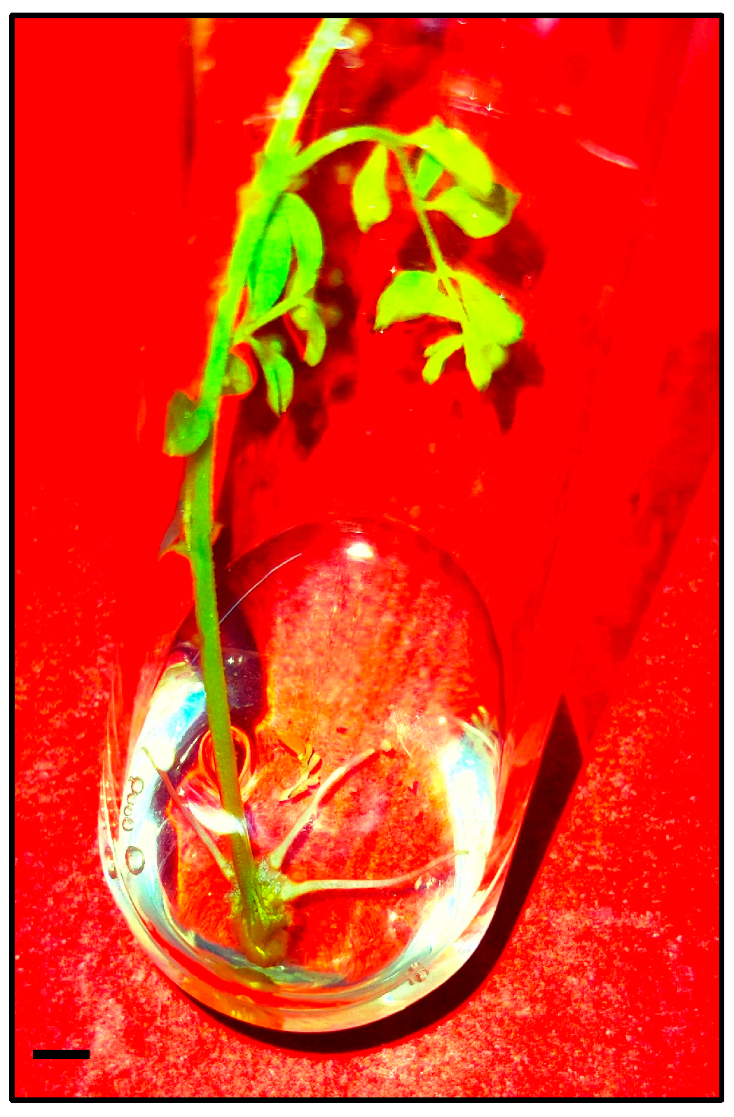
The healthy in vitro axillary shoots were transferred to the MS medium, supplemented with IAA or IBA (1.0–3.0 mg/L) for rooting. Successful root induction was observed within 3–4 weeks of culturing (Figure 3); 2.0 mg/L of IBA proved to be the best in inducing roots which showed 88.89% rooting frequency and average 7.33 root numbers/shoot, followed by 1.0 mg/L of IBA that had 6.67 mean root numbers/shoot with 72.22% rooting frequency (Table 3). On the other hand, with increasing IAA concentrations, there was a significant reduction in rooting and, upon reaching up to 3.0 mg/L IAA, no root development was observed in the axillary shoots.

3.4. GC–MS Analysis

In this current study, metabolite profiling of methanolic extracts of in vivo and in vitro grown roots, as well as root derived callus, was performed using the GC–MS approach, and their respective chromatograms are presented in Figure 4. A comparative chromatographic examination of in vivo and in vitro derived roots revealed the presence of 38 and 36 phytocompounds, respectively (Table 4). The heatmap cluster analysis shown in Figure 5 is based on phytocompounds peak area % measured by GC–MS, and it was found that mome inositol, beta-monoolein, methyl ester of oleic acid, guanosine, dipalimitin, glycidyl oleate, methyl ester of palmitic acid and stearic acid are some of the majorly occurring metabolites in both samples. In total, 24 bioactive compounds have been found to be present in in vivo and in vitro grown samples at varied levels, which include guanosine, glycidyl palmitate, glycidyl oleate, dipalmitin, gamma-sitosterol, stigmasta-3,5-diene, methyl esters of linoleic acid, oleic acid, stearic acid, alpha-spinosterol acetate, etc.
The content of guanosine was found to be almost doubled (8.59%) in in vitro grown roots compared to in vivo grown roots (4.80%). Similarly, the methyl ester content of palmitic acid was found to be higher in the in vitro root (16.26%) sample than in the in vivo grown root sample (10.01%). A similar difference was noted for glycidyl oleate (7.51% in field grown roots and 11.64% in in vitro derived root tissue) as well. On the other hand, some of the metabolites were observed to be lower in in vitro root tissue than the in vivo derived root sample; examples of this are 9-octadecenamide, stigmasta-3,5-diene, gamma-sitosterol and alpha-spinosterol acetate. Certain bioactive compounds are exclusively present in each sample of in vivo grown roots, such as xanthosine (0.37%), squalene (0.76%), 4′-O-methylglabridin (0.32%), 24-epicampesterol (0.30%) and lupeol (0.70%), whereas, in vitro grown roots contained cis-sesquisabinene hydrate (0.28%), 4-methylmannitol (0.55%), 2-monopalmitin (0.16%), alpha-monostearin (0.35%), isopropyl linoleate (0.13%) exclusively.
The phytochemical profile of root derived callus of G. glabra was similarly determined and analyzed. The obtained result of methanolic callus extract depicted a total of 44 phytocompounds; many of which are detected in trace quantities (Table 5). Oleic anhydride (35.65%), methyl galactoside (10.73%), 2,3-dihydroxypropyl laurate (5.96%), Glycidyl palmitate (5.19%), methyl ester of oleic acid (5.18%) were observed as the versatile phytocompounds present in major proportions. Some of the important phytochemicals like palmidrol, 1-monostearin, beta-stigmasterol, 24-epibrassicasterol, squalene, beta-saccharostenone were quantified in minute amounts.

3.5. Total Phenolic Content (TPC), Total Flavonoid Content (TFC) and DPPH Scavenging Activity

The total phenolic content of callus and root extract was estimated by the Folin–Ciocalteu method, using gallic acid as standard. The highest phenolic content was noted in the in vitro grown root, followed by the in vivo grown roots and the least was recorded in the callus extract (Table 6). The TPC value of the in vitro derived root was 9.76 ± 0.21 mg GAE/g DW and the in-field grown root extract was 7.49 ± 0.54 mg GAE/g DW, whereas the lowest TPC was found in the callus extract (3.64 ± 0.45 mg GAE/g DW). The total flavonoid content of different tested samples was determined by the aluminium chloride method using quercetin as the standard and the results were expressed as quercetin equivalent (QE) per gram of dry weight (Table 6). The flavonoid content of extracts ranged from 0.72 to 1.58 mg QE/g DW, showing for about two-fold variation. The flavonoid content of in vivo and in vitro grown root extracts are nearly equal (1.44 and 1.58 mg QE/g DW, respectively), and the lowest TFC was noted to be 0.72 mg QE/g DW in root derived calli. The antioxidant activity of each extract was determined using a DPPH free radical scavenging assay and the activity is presented in Table 6. The antioxidant potential of tested tissues showed a similar trend with TPC and TFC. The level of scavenging activity of the in vitro raised root extract was found to be higher (40.63%) as compared to the in vivo root (31.34%). The lowest scavenging activity percentage (22.90) was observed in callus extract.

3.6. Antioxidant Enzyme Activities

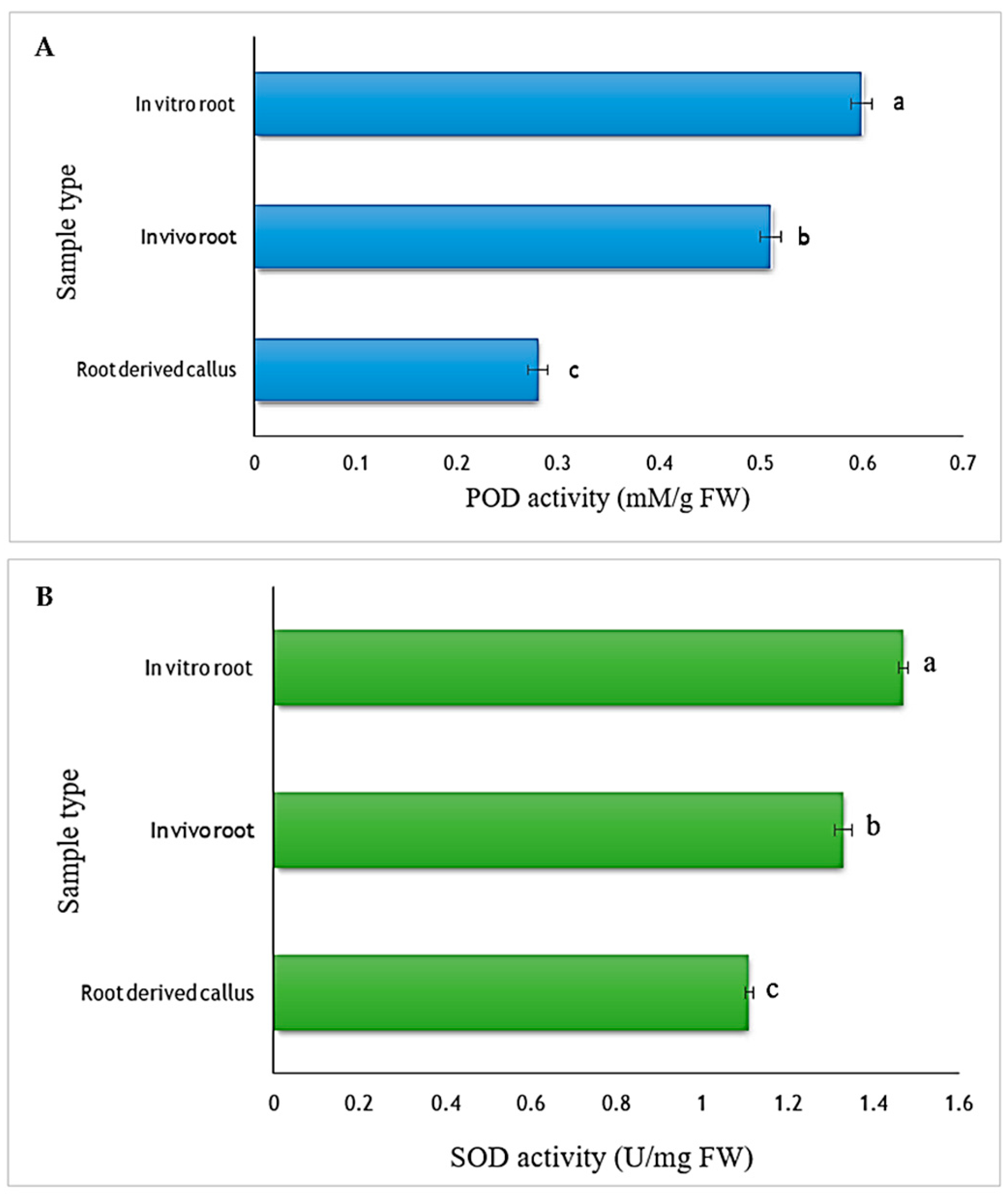
The peroxidase (POD) activity was assessed in callus, in vivo, and in vitro grown roots of G. glabra. Peroxidase activity was found to be significantly higher in root tissue when compared to the root derived callus (Figure 6A). The callus showed the lowest POD activity (0.28 mM/g FW), followed by the in vivo grown root (0.51 mM/g FW). The highest POD activity was displayed by in vitro roots (0.60 mM/g FW), with a two-fold increment in activity as compared to the root derived calli. The superoxide dismutase (SOD) activity was measured in different tissue samples to check the antioxidant potential (Figure 6B). Like POD, a similar trend in SOD activity was detected in different tissues. Considering SOD, callus showed the lowest activity, i.e., 1.11 U/mg FW, followed by the field grown root sample (1.33 U/mg FW). The highest SOD activity was found in roots of tissue cultured shoots (1.47 U/mg FW).

4. Discussion

This study was conducted to study the effects of PGRs, their concentrations, and combinations on the success of some in vitro propagation steps such as callus induction, axillary shoot sprouting, and rooting of in vitro shoots of Glycyrrhiza glabra L. The phytochemical and biochemical profiles of in vitro regenerated tissues were also evaluated. Firstly, the root explants were cultured on MS with varying concentrations of PGRs for callus induction, and the results indicated that BAP, along with 2,4-D, showed good callus growth with the highest fresh biomass as compared to the BAP and NAA combinations. A previous study of callus induction from leaf and stem explants of G. glabra also reported similar observations [5]. The positive effect of 2,4-D with BAP was previously observed in several other plant species, like Digitalis ferruginea [24] and Dioscorea nipponica Makino [25]. Axillary buds are normally dormant under wild conditions, However, shoot formation and proliferation can be induced with the help of cytokinins under in vitro conditions [26]. Here, we demonstrated that the BAP alone, or in combination with IAA, showed better efficiency in breaking axillary bud dormancy leading to high shoot formation. Consistent with our observations, similar effects of BAP on shoot induction from dormant buds were noticed in several plants [27,28]. BAP, along with IAA, also showed moderate effects here, in inducing shoots from dormant axillary buds. In addition to cytokinin, auxins are often useful in inducing shoots as these signaling elements are known to overcome the excessive effects of cytokinins [29]. The promotive effect of auxin with cytokinin in shoot formation has also been documented in previous reports of Glycyrrhiza glabra L. [1] as well as in different plant species such as Curcuma zedoaria [30], Basella rubra L. [31] and Santalum album L. [32]. The axillary shoots were later transferred to a rooting medium containing different concentrations of IAA and IBA. Shoots cultured on MS supplemented with IBA showed a higher rate of rooting than those of shoots treated with IAA. Ayangla et al. [33] reported superior effects of IBA over other auxin treatments in inducing roots in organogenic derived shoots of G. glabra L. IBA showed similar effects on rooting under in vitro conditions in other plants species such as Dracaena sanderiana [34] and Vaccinium corymbosum [35]. The high root induction rate induced by IBA is attributed to its stability and easy translocation to different tissues [36].
GC–MS, a popular analytical technique, is widely utilized in the identification of several phytocompounds, such as volatile compounds, long chain hydrocarbons, sugar alcohols, esters, alkaloids, flavonoids, saponins, etc. [12]. Moreover, this technique also provides impactful insights into the role of various in vitro factors on growth and development by detecting changes in peak area % in metabolite profiles of regenerants and their wild relatives [37]. In this investigation, the obtained chromatographic results showed the presence of more than 35 important bioactive compounds in each tested samples. The comparative metabolite profiling of in vivo and in vitro grown root tissues displayed variation in several detected phytocompounds level. The increased production of phytocompounds in the in vitro derived root (compared to intact in vivo plant tissues) may be attributed to various factors such as media composition, hormonal concentration, genotype, photoperiod, temperature, etc., [13,38] and thus, could be a more potent and reliable source of phytocompounds for pharmacological uses. Previously, Vijayalakshmi and Shourie [39] reported the presence of variety of phytochemicals in leaf, stem, and leaf derived callus in G. glabra. The identification and quantification of bioactive compounds in various plant parts (root, leaf, stem, anther, callus) by GC–MS have recently been described in different plant varieties like Tanacetum sinaicum [40], Catharanthus roseus L. [41] and Amomum nilgiricums [42]. A wide number of therapeutically active phytoconstituents have been detected in G. glabra samples. Squalene, a terpenoid, possesses various biological activities like anti-oxidant, anti-cancerous, detoxifying and moisturizing agents [43]. Glabridin belongs to flavonoid, showing a diverse range of medicinal potentials, such as protection against cardiovascular diseases, diabetes, bacterial infections and possess estrogenic, anti-inflammatory, anti-cancerous effects [44]. Lupeol is a pentacyclic triterpenoid, known to have anti-inflammatory, anti-microbial, anti-angiogenic, anti-diabetic and anti-protozoal agents [45]. Stigmasterol, found in callus samples, was reported to be associated with anti-tumor, anti-osteoarthritis, antibacterial, anti-oxidant, immunomodulatory, anti-parasitic, anti-fungal and neuroprotective properties [46]. Similarly, n-hexadecanoic acid, which was exclusively found in the in vitro derived root tissue, is known to exhibit anti-inflammatory, anti-bacterial and anti-oxidant properties [47,48]. Consistent with our findings, Khan et al. [3] reported the presence of important flavonoids and triterpenoids in the in vitro cultures of G. glabra.
In vitro culture conditions often induce stress in cell lines/regenerated tissues and thereby reduce their rate of survival [9]. Therefore, it is crucial to assess the cellular physiology by monitoring biochemical attributes of tissues at regular interval. The biochemical and antioxidant attributes of in vitro derived tissues were investigated and compared with donor (mother) plant in G. glabra. Various factors like different PGRs used in culturing are involved in up- and down-regulation of phenolics and flavonoid biosynthesis [49]. In our study, the phenolic, as well as flavonoid, synthesis was found to be higher in in vitro root tissues as compared to mother plants. This is in accordance with earlier reported biochemical studies conducted in different plant species [50,51]. The antioxidant activities of in vitro raised tissues were tested using three antioxidant, i.e., DPPH, SOD and POD assays, and the data obtained by these studies revealed that the in vitro derived root tissue showed higher anti-oxidant activities than the field grown root and callus samples. Under stressful conditions, the reactive oxygen species (ROS) production in plant tissues exceeds the normal level, which may be reduced by a number of anti-oxidant enzymes like catalase (CAT), peroxidase (POD), superoxide dismutase (SOD), glutathione reductase (GR), etc. [52]. DPPH is a dark colored, stabilized, organic free radical, which transformed into a light yellow color whenever it accepts an electron or a free radical, indicating the scavenging activity [53]. SOD is a scavenger of superoxide radical and helps to manage H2O2 levels [54], whereas POD oxidizes co-substrates, and promotes decomposition of H2O2 [55]. In this study, the higher degree of antioxidant potential of laboratory grown root tissue is due to the positive correlation of phenolics and flavonoids with antioxidant activity as these molecules donate hydrogen atoms to free radicals, leading to their deactivation [20]. Similar results of antioxidant potential were observed in different plant varieties like Salvia hispanica [56], Zingiber officinale [57], Tylophora indica [58], Thalictrum foliolosum [59], etc. The above evidences elucidates that in vitro derived tissues, such as callus and root, possess immense pharmacological values in terms of phytoconstituents and identifying such compounds could pave the way for novel drug discovery in shorter period of time with minimal cost. Further studies, like molecular docking and bio-prospecting, could be carried out in the future to understand the ligand-protein interaction and biological activities of these significant bioactive compounds.

5. Conclusions

This work focused on the comparative metabolite profiling of in vivo and in vitro derived root tissues of G. glabra for the first time using the GC–MS technique. The analyses displayed a wide variety of phytocompounds ranging from alkaloids, flavonoids, phenolics, terpenoids, sugars, sterols, etc. The root derived callus showed the presence of 44 phytochemicals, which all have diverse therapeutic importance. The biochemical and antioxidant assessment of callus, in vivo, and in vitro grown root tissues were compared, wherein in vitro roots showed a higher presence of phenolics and flavonoids, as well as more antioxidant potential than the other tested tissues. This study reports the presence of various medically beneficial bioactive compounds from in vitro derived plant tissues, which can be utilized by the pharmaceutical industry for their large-scale production.

Author Contributions

Conceptualization, Y.B. and A.M.; methodology, Y.B.; software, S.K.; validation, A.M. and Y.H.D.; formal analysis, J.M. and K.M.-T.; investigation, Y.B.; resources, S.K.; data curation, A.M.; writing—original draft preparation, Y.B.; writing—review and editing, A.M.; visualization, K.M.-T.; supervision, A.M.; project administration, Y.H.D.; funding acquisition, Y.H.D. All authors have read and agreed to the published version of the manuscript.

Funding

The authors are highly thankful to Department of Biotechnology (DBT) and Department of Botany, Jamia Hamdard for receiving financial assistance and other research facilities and Researchers Supporting Project number (RSP-2024R375), King Saud University, Riyadh, Saudi Arabia.

Institutional Review Board Statement

Not applicable.

Informed Consent Statement

There is no involvement of human trial or activities, thus not applicable.

Data Availability Statement

All the datasets generated or analyzed during this current study are included in this published article.

Acknowledgments

The first author is thankful to the Department of Biotechnology (DBT) for financial support given as a Senior Research Fellowship. The authors are also grateful to the laboratory facilities provided by the Department of Botany, Jamia Hamdard, New Delhi. The authors acknowledge the researchers supporting project number (RSP-2024R375), King Saud University, Riyadh, Saudi Arabia.

Conflicts of Interest

The authors declare no conflicts of interest.

References

  1. Yadav, K.; Singh, N. Factors influencing in vitro plant regeneration of liquorice (Glycyrrhiza glabra L.). Iran. J. Biotechnol. 2012, 10, 161–167. [Google Scholar]
  2. Akhtar, R.; Shahzad, A. Alginate Encapsulation in Glycyrrhiza glabra L. with Phyto-Chemical Profiling of Root Extracts of In Vitro Converted Plants Using GC-MS Analysis. Asian Pac. J. Trop. Biomed. 2017, 7, 855–861. [Google Scholar] [CrossRef]
  3. Khan, S.; Pandotra, P.; Manzoor, M.M.; Kushwaha, M.; Sharma, R.; Jain, S.; Ahuja, A.; Amancha, V.; Bhushan, S.; Guru, S.K.; et al. Terpenoid and Flavonoid Spectrum of In Vitro Cultures of Glycyrrhiza glabra Revealed High Chemical Heterogeneity: Platform to Understand Biosynthesis. Plant Cell Tissue Organ Cult. (PCTOC) 2016, 124, 507–516. [Google Scholar] [CrossRef]
  4. Srivastava, M.; Singh, G.; Sharma, S.; Shukla, S.; Misra, P. Elicitation Enhanced the Yield of Glycyrrhizin and Antioxidant Activities in Hairy Root Cultures of Glycyrrhiza glabra L. J. Plant Growth Regul. 2019, 38, 373–384. [Google Scholar] [CrossRef]
  5. Jaiswal, N.; Verma, Y.; Misra, P. High Frequency In Vitro Callogenesis and Plant Regeneration of Glycyrrhiza glabra L. Vegetos 2021, 34, 495–504. [Google Scholar] [CrossRef]
  6. Shaheen, A.; Ali, M.; Ahmad, N.; Dewir, Y.H.; El-Hendawy, S.; Abd-El Gawad, A.M. Micropropagation of licorice (Glycyrrhiza glabra L.) by using intermediate nodal explants. Chil. J. Agric. Res. 2020, 80, 326–333. [Google Scholar] [CrossRef]
  7. Bansal, Y.; Mujib, A.; Siddiqui, Z.H.; Mamgain, J.; Syeed, R.; Ejaz, B. Ploidy Status, Nuclear DNA Content and Start Codon Targeted (SCoT) Genetic Homogeneity Assessment in Digitalis purpurea L., Regenerated In Vitro. Genes 2022, 13, 2335. [Google Scholar] [CrossRef]
  8. Mujib, A.; Ali, M.; Isah, T. Somatic Embryo Mediated Mass Production of Catharanthus roseus in Culture Vessel (Bioreactor)—A Comparative Study. Saudi J. Biol. Sci. 2014, 21, 442–449. [Google Scholar] [CrossRef]
  9. Malik, M.; Wachol, M.; Pawlowska, B. Liquid Culture Systems Affect Morphological and Biochemical Parameters during Rosa canina Plantlets In Vitro Production. Not. Bot. Horti Agrobot. Cluj-Napoca 2018, 46, 58–64. [Google Scholar] [CrossRef]
  10. Khan, H.; Khan, T.; Ahmad, N.; Zaman, G.; Khan, T.; Ahmad, W.; Batool, S.; Hussain, Z.; Drouet, S.; Hano, C.; et al. Chemical Elicitors-Induced Variation in Cellular Biomass, Biosynthesis of Secondary Cell Products, and Antioxidant System in Callus Cultures of Fagonia indica. Molecules 2021, 26, 6340. [Google Scholar] [CrossRef]
  11. Mamgain, J.; Mujib, A.; Syeed, R.; Ejaz, B.; Malik, M.Q.; Bansal, Y. Genome Size and Gas Chromatography-Mass Spectrometry (GC–MS) Analysis of Field-Grown and In Vitro Regenerated Pluchea lanceolata Plants. J. Appl. Genet. 2023, 64, 1–21. [Google Scholar] [CrossRef]
  12. Hussain, S.A.; Ahmad, N.; Anis, M.; Alatar, A.A. Influence of Meta-Topolin on In Vitro Organogenesis in Tecoma stans L., Assessment of Genetic Fidelity and Phytochemical Profiling of Wild and Regenerated Plants. Plant Cell Tissue Organ Cult. 2019, 138, 339–351. [Google Scholar] [CrossRef]
  13. Khan, A.; Shah, A.H.; Ali, N. In-Vitro Propagation and Phytochemical Profiling of a Highly Medicinal and Endemic Plant Species of the Himalayan Region (Saussurea costus). Sci. Rep. 2021, 11, 23575. [Google Scholar] [CrossRef]
  14. Moghbel, N.; Borujeni, M.K.; Bernard, F. Colchicine Effect on the DNA Content and Stomata Size of Glycyrrhiza glabra var. Glandulifera and Carthamus tinctorius L. Cultured In Vitro. J. Genet. Eng. Biotechnol. 2015, 13, 1–6. [Google Scholar] [CrossRef]
  15. Badkhane, Y.; Yadav, A.S.; Bajaj, A. effect of explant sources and different concentrations of plant growth regulators on in vitro micropropagation of Glycyrrhiza glabra L. Indo Am. J. Pharma Res. 2016, 6, 5830–5840. [Google Scholar]
  16. Tahoori, F.; Majd, A.; Nejadsattari, T.; Ofoghi, H.; Iranbakhsh, A. Qualitative and Quantitative Study of Quercetin and Glycyrrhizin in In Vitro Culture of Liquorice (Glycyrrhiza glabra L.) and Elicitation with AgNO3. Not. Bot. Horti Agrobot. Cluj Napoca 2018, 47, 143–151. [Google Scholar] [CrossRef]
  17. Sathish, D.; Vasudevan, V.; Theboral, J.; Elayaraja, D.; Appunu, C.; Siva, R.; Manickavasagam, M. Efficient direct plant regeneration from immature leaf roll explants of sugarcane (Saccharum officinarum L.) using polyamines and assessment of genetic fidelity by SCoT markers. In Vitro Cell. Dev. Biol.-Plant 2018, 54, 399–412. [Google Scholar] [CrossRef]
  18. Murashige, T.; Skoog, F. A Revised Medium for Rapid Growth and Bio Assays with Tobacco Tissue Cultures. Physiol. Plant 1962, 15, 473–497. [Google Scholar] [CrossRef]
  19. Baliyan, S.; Mukherjee, R.; Priyadarshini, A.; Vibhuti, A.; Gupta, A.; Pandey, R.P.; Chang, C.-M. Determination of Antioxidants by DPPH Radical Scavenging Activity and Quantitative Phytochemical Analysis of Ficus religiosa. Molecules 2022, 27, 1326. [Google Scholar] [CrossRef] [PubMed]
  20. Aryal, S.; Baniya, M.K.; Danekhu, K.; Kunwar, P.; Gurung, R.; Koirala, N. Total Phenolic Content, Flavonoid Content and Antioxidant Potential of Wild Vegetables from Western Nepal. Plants 2019, 8, 96. [Google Scholar] [CrossRef] [PubMed]
  21. Haida, Z.; Hakiman, M. A Comprehensive Review on the Determination of Enzymatic Assay and Nonenzymatic Antioxidant Activities. Food Sci. Nutr. 2019, 7, 1555–1563. [Google Scholar] [CrossRef]
  22. Mujib, A.; Fatima, S.; Malik, M.Q. Gamma Ray–Induced Tissue Responses and Improved Secondary Metabolites Accumulation in Catharanthus roseus. Appl. Microbiol. Biotechnol. 2022, 106, 6109–6123. [Google Scholar] [CrossRef]
  23. Duncan, D.B. Multiple Range and Multiple F Tests. Biometrics 1955, 11, 1. [Google Scholar] [CrossRef]
  24. Verma, S.K.; Yucesan, B.; Sahin, G.; Gurel, E. Embryogenesis, Plant Regeneration and Cardiac Glycoside Determination in Digitalis ferruginea subsp. ferruginea L. Plant Cell Tissue Organ Cult. (PCTOC) 2014, 119, 625–634. [Google Scholar] [CrossRef]
  25. Dang, S.; Gao, R.; Zhang, Y.; Feng, Y. In Vitro Regeneration and Its Histological Characteristics of Dioscorea nipponica Makino. Sci. Rep. 2022, 12, 18436. [Google Scholar] [CrossRef]
  26. Nowakowska, M.; Pavlović, Ž.; Nowicki, M.; Boggess, S.L.; Trigiano, R.N. In Vitro Propagation of an Endangered Helianthus verticillatus by Axillary Bud Proliferation. Plants 2020, 9, 712. [Google Scholar] [CrossRef]
  27. Nazirah, A.; Nor-Hasnida, H.; Mohd-Saifuldullah, A.W.; Muhammad-Fuad, Y.; Ahmad-Zuhaidi, Y.; Rozidah, K. Development of an efficient micropropagation protocol for eucalyptus hybrid (E. urophylla × E. grandis) through axillary shoot proliferation. J. Trop. For. Sci. 2021, 33, 391–397. [Google Scholar] [CrossRef]
  28. Tikendra, L.; Dey, A.; Jamir, I.; Sahoo, M.R.; Nongdam, P. Cytokinin Influence on In Vitro Shoot Induction and Genetic Stability Assessment of Dendrocalamus latiflorus Munro: A Commercially Important Bamboo in Manipur, North-East India. Vegetos 2022, 35, 1085–1095. [Google Scholar] [CrossRef]
  29. Novikova, T.I.; Asbaganov, S.V.; Ambros, E.V.; Zaytseva, Y.G. TDZ-Induced Axillary Shoot Proliferation of Rhododendron mucronulatum Turcz and Assessment of Clonal Fidelity Using DNA-Based Markers and Flow Cytometry. In Vitro Cell. Dev. Biol.-Plant 2020, 56, 307–317. [Google Scholar] [CrossRef]
  30. Jena, S.; Ray, A.; Sahoo, A.; Sahoo, S.; Dash, B.; Kar, B.; Nayak, S. Rapid Plant Regeneration in Industrially Important Curcuma zedoaria Revealing Genetic and Biochemical Fidelity of the Regenerants. 3 Biotech 2020, 10, 17. [Google Scholar] [CrossRef]
  31. Kumar, S.S.; Giridhar, P. In Vitro Micropropagation of Basella rubra L. through Proliferation of Axillary Shoots. Plant Cell Tissue Organ Cult. (PCTOC) 2021, 144, 477–483. [Google Scholar] [CrossRef]
  32. Wardani, I.B. Effect of BAP (6-Benzyl Amino Purine) and NAA (Naphtalen Acetic Acid) on the Induction of Axillary Shoots in Sandalwood (Santalum album L.). J. Sci. Technol. Educ. 2022, 1, 23–30. [Google Scholar]
  33. Ayangla, N.W.; Dwivedi, P.; Dey, A.; Pandey, D.K. In vitro propagation, genetic and phytochemical fidelity in Glycyrrhiza glabra L., a potent glycyrrhizin yielding endangered plant. Nucleus 2022, 65, 369–377. [Google Scholar] [CrossRef]
  34. Mujib, A.; Aslam, J.; Bansal, Y. low colchicine doses improved callus induction, biomass growth, and shoot regeneration in in vitro culture of Dracaena sanderiana sander ex mast. Prop. Ornam. Plants 2023, 23, 81–87. [Google Scholar]
  35. Guo, Y.X.; Zhao, Y.Y.; Zhang, M.; Zhang, L.Y. Development of a novel in vitro rooting culture system for the micropropagation of highbush blueberry (Vaccinium corymbosum) seedlings. Plant Cell Tissue Organ Cult. (PCTOC) 2019, 139, 615–620. [Google Scholar] [CrossRef]
  36. Chirumamilla, P.; Gopu, C.; Jogam, P.; Taduri, S. Highly Efficient Rapid Micropropagation and Assessment of Genetic Fidelity of Regenerants by ISSR and SCoT Markers of Solanum khasianum Clarke. Plant Cell Tissue Organ Cult. (PCTOC) 2021, 144, 397–407. [Google Scholar] [CrossRef]
  37. Faisal, M.; Qahtan, A.A.; Alatar, A.A. Thidiazuron Induced In Vitro Plant Regeneration, Phenolic Contents, Antioxidant Potential, GC-MS Profiles and Nuclear Genome Stability of Plectranthus amboinicus (Lour.) Spreng. Horticulturae 2023, 9, 277. [Google Scholar] [CrossRef]
  38. Qahtan, A.A.; Faisal, M.; Alatar, A.A.; Abdel-Salam, E.M. Callus-Mediated High-Frequency Plant Regeneration, Phytochemical Profiling, Antioxidant Activity and Genetic Stability in Ruta chalepensis L. Plants 2022, 11, 1614. [Google Scholar] [CrossRef]
  39. Vijayalakshmi, U.; Shourie, A. Comparative gc-ms analysis of secondary metabolites from leaf, stem and callus of Glycyrrhiza glabra. World J. Pharm. Res. 2019, 8, 1915–1923. [Google Scholar] [CrossRef]
  40. Adel, R.; Abdel-Ghani, A.E.; Abouelenein, D.D.; El-Dahmy, S.I. Variation in the Volatile Constituents of Wild and In Vitro Propagated Tanacetum sinaicum Del. Ex DC through GC-MS Chemical Fingerprint. Ind. J. Nat. Prod. Res. 2021, 12, 238–246. [Google Scholar]
  41. Bansal, Y.; Mujib, A.; Mamgain, J.; Dewir, Y.H.; Rihan, H.Z. Phytochemical Composition and Detection of Novel Bioactives in Anther Callus of Catharanthus roseus L. Plants 2023, 12, 2186. [Google Scholar] [CrossRef]
  42. Konappa, N.; Udayashankar, A.C.; Krishnamurthy, S.; Pradeep, C.K.; Chowdappa, S.; Jogaiah, S. GC–MS analysis of phytoconstituents from Amomum nilgiricum and molecular docking interactions of bioactive serverogenin acetate with target proteins. Sci. Rep. 2020, 10, 16438. [Google Scholar] [CrossRef]
  43. Bhat, M.P.; Rudrappa, M.; Hugar, A.; Gunagambhire, P.V.; Suresh Kumar, R.; Nayaka, S.; Almansour, A.I.; Perumal, K. In-Vitro Investigation on the Biological Activities of Squalene Derived from the Soil Fungus Talaromyces pinophilus. Heliyon 2023, 9, e21461. [Google Scholar] [CrossRef]
  44. Li, C.; Li, T.; Zhu, M.; Lai, J.; Wu, Z. Pharmacological Properties of Glabridin (a Flavonoid Extracted from Licorice): A Comprehensive Review. J. Funct. Foods 2021, 85, 104638. [Google Scholar] [CrossRef]
  45. Sharma, N.; Palia, P.; Chaudhary, A.; Shalini; Verma, K.; Kumar, I. A Review on Pharmacological Activities of Lupeol and Its Triterpene Derivatives. J. Drug Deliv. Ther. 2020, 10, 325–332. [Google Scholar] [CrossRef]
  46. Bakrim, S.; Benkhaira, N.; Bourais, I.; Benali, T.; Lee, L.-H.; El Omari, N.; Sheikh, R.A.; Goh, K.W.; Ming, L.C.; Bouyahya, A. Health Benefits and Pharmacological Properties of Stigmasterol. Antioxidants 2022, 11, 1912. [Google Scholar] [CrossRef]
  47. Aparna, V.; Dileep, K.V.; Mandal, P.K.; Karthe, P.; Sadasivan, C.; Haridas, M. Anti-inflammatory property of n-hexadecanoic acid: Structural evidence and kinetic assessment. Chem. Biol. Drug Des. 2012, 80, 434–439. [Google Scholar] [CrossRef]
  48. Ganesan, T.; Subban, M.; Christopher Leslee, D.B.; Kuppannan, S.B.; Seedevi, P. Structural characterization of n-hexadecanoic acid from the leaves of Ipomoea eriocarpa and its antioxidant and antibacterial activities. Biomass Convers Biorefinery 2022. [Google Scholar] [CrossRef]
  49. Ghosh, A.; Igamberdiev, A.U.; Debnath, S.C. Thidiazuron-Induced Somatic Embryogenesis and Changes of Antioxidant Properties in Tissue Cultures of Half-High Blueberry Plants. Sci. Rep. 2018, 8, 16978. [Google Scholar] [CrossRef]
  50. Jeong, B.R.; Sivanesan, I. Direct Adventitious Shoot Regeneration, In Vitro Flowering, Fruiting, Secondary Metabolite Content and Antioxidant Activity of Scrophularia takesimensis Nakai. Plant Cell Tissue Organ Cult. (PCTOC) 2015, 123, 607–618. [Google Scholar] [CrossRef]
  51. Bhattacharyya, P.; Kumaria, S.; Tandon, P. High Frequency Regeneration Protocol for Dendrobium Nobile: A Model Tissue Culture Approach for Propagation of Medicinally Important Orchid Species. S. Afr. J. Bot. 2016, 104, 232–243. [Google Scholar] [CrossRef]
  52. Meena, M.; Divyanshu, K.; Kumar, S.; Swapnil, P.; Zehra, A.; Shukla, V.; Yadav, M.; Upadhyay, R.S. Regulation of L-Proline Biosynthesis, Signal Transduction, Transport, Accumulation and Its Vital Role in Plants during Variable Environmental Conditions. Heliyon 2019, 5, e02952. [Google Scholar] [CrossRef]
  53. Khorasani Esmaeili, A.; Mat Taha, R.; Mohajer, S.; Banisalam, B. Antioxidant Activity and Total Phenolic and Flavonoid Content of Various Solvent Extracts from In Vivo and In Vitro Grown Trifolium pratense L. (Red Clover). BioMed Res. Int. 2015, 2015, 643285. [Google Scholar] [CrossRef]
  54. Marshall, D.; Siow, Y.; Debnath, S. Somatic Embryogenesis in Vaccinium vitis-Idaea and Genetic Delity Evaluation of Regenerants Using Molecular Markers Together with Their Phytochemical Prole. Sayani Kundu Agric. Agric.-Food Can. Rajesh Barua Agric. Agric.-Food Can. 2023. [Google Scholar] [CrossRef]
  55. Ma, N.; Hu, C.; Wan, L.; Hu, Q.; Xiong, J.; Zhang, C. Strigolactones Improve Plant Growth, Photosynthesis, and Alleviate Oxidative Stress under Salinity in Rapeseed (Brassica napus L.) by Regulating Gene Expression. Front. Plant Sci. 2017, 8, 1671. [Google Scholar] [CrossRef] [PubMed]
  56. Zayova, E.; Nikolova, M.; Dimitrova, L.; Petrova, M. Comparative Study of In Vitro, Ex Vitro and In Vivo Propagated Salvia hispanica (Chia) Plants: Morphometric Analysis and Antioxidant Activity. AgroLife Sci. J. 2016, 5, 166–173. [Google Scholar]
  57. Ali, A.M.A.; El-Nour, M.E.M.; Yagi, S.M. Total Phenolic and Flavonoid Contents and Antioxidant Activity of Ginger (Zingiber officinale Rosc.) Rhizome, Callus and Callus Treated with Some Elicitors. J. Genet. Eng. Biotechnol. 2018, 16, 677–682. [Google Scholar] [CrossRef] [PubMed]
  58. Mamgain, J.; Mujib, A.; Bansal, Y.; Gulzar, B.; Zafar, N.; Syeed, R.; Alsughayyir, A.; Dewir, Y.H. Elicitation Induced α-Amyrin Synthesis in Tylophora indica In Vitro Cultures and Comparative Phytochemical Analyses of In Vivo and Micropropagated Plants. Plants 2024, 13, 122. [Google Scholar] [CrossRef] [PubMed]
  59. Mishra, M.K.; Pandey, S.; Niranjan, A.; Misra, P. Comparative analysis of phenolic compounds from wild and in vitro propagated plant Thalictrum foliolosum and antioxidant activity of various crude extracts. Chem. Pap. 2021, 75, 4873–4885. [Google Scholar] [CrossRef]
Figure 1. Callus induction and proliferation on the root explant of G. glabra on MS medium supplemented with 2.0 mg/L BAP and 0.5 mg/L 2,4-D. Callus initiation after 4 weeks of inoculation (Bar = 0.5 cm) (A). Callus proliferation after regular subculture (Bars (B) = 1.0 cm, (C) = 1.5 cm).
Sustainability 16 01613 g001
Figure 2. In vitro axillary shoot sprouting in G. glabra: Swelling of the axillary bud after 7 days of inoculation (Bar = 0.5 cm) (A). Axillary bud sprouting on 1.0 mg/L BAP fortified MS medium (Bar = 0.5 cm) (B). Elongation of in vitro sprouted axillary shoot on medium with 1.0 mg/L BAP (Bars (C) = 1.0 cm, (D) = 2.0 cm) (C,D).
Sustainability 16 01613 g002
Figure 3. In vitro root induction of the axillary shoots in G. glabra on liquid root induction medium (3.0 mg/L IBA) (Bar = 2.0 cm).
Sustainability 16 01613 g003
Figure 4. GC–MS chromatograms of methanolic extracts of in vivo grown root tissue (A), in vitro derived root tissue (B), and root derived callus of G. glabra (C).
Sustainability 16 01613 g004
Figure 5. Heat-map cluster analysis displaying the relative abundance of phytocompounds identified and quantified by GC–MS technique in methanolic root extracts of G. glabra plants grown in vivo and in vitro.
Sustainability 16 01613 g005
Figure 6. Activities of peroxidase (POD) (A) and superoxide dismutase (SOD) (B) in callus, and in vivo or in vitro root tissues of G. glabra L. Values represent mean ± S.E. of three repeated experiments. Mean values with different letters within the horizontal bar indicate significant mean difference with one another at p ≤ 0.05 level as per DMRT.
Sustainability 16 01613 g006
Table 1. Effect of combinations of cytokinin (BAP) and auxins (2,4-D/NAA) on callus induction frequency and growth of callus using root explants of G. glabra L. after 4 weeks of inoculation.
PGRsConcentration (mg/L)Callusing Frequency (%)Fresh Biomass (g)
Control00 e0 d
BAP + 2,4-D1.0 + 0.572.21 ± 5.56 ab3.9 ± 0.6 ab
1.0 + 2.061.11 ± 5.55 bc3.6 ± 0.4 ab
2.0 + 0.588.89 ± 11.11 a4.4 ± 0.7 a
BAP + NAA1.0 + 0.527.77 ± 5.56 de2.1 ± 0.1 c
1.0 + 2.011.11 ± 5.55 e0.8 ± 0.1 d
2.0 + 1.044.44 ± 5.56 cd2.9 ± 0.2 bc
Values represent mean ± S.E. of six replicates per treatment in three repeated experiments. Mean values with different letters within each column are significant at p ≤ 0.05 level as per DMRT.
Table 2. Effect of BAP alone, or in combination with, IAA on the axillary shoot sprouting from axillary buds of G. glabra L. after 4 weeks of inoculation.
PGRsConcentration (mg/L)Shooting Frequency (%)Shoot Length (cm)Mean Shoot Number
Control00 e0 f0 d
BAP0.577.77 ± 5.56 ab8.7 ± 0.4 b3.33 ± 0.67 ab
1.094.44 ± 5.56 a10.5 ± 0.3 a4.67 ± 0.88 a
2.038.88 ± 14.70 cd4.4 ± 0.6 d2.67 ± 0.88 bc
4.022.21 ± 5.55 de2.1 ± 0.2 e1.33 ± 0.33 cd
BAP + IAA1.0 + 0.2555.55 ± 11.11 bcd6.2 ± 0.4 c3.33 ± 0.33 ab
1.0 + 0.5061.12 ± 14.69 abc6.8 ± 0.8 c3.33 ± 0.33 ab
Values represent mean ± S.E. of six replicates per treatment in three repeated experiments. Mean values with different superscripts within a column indicate significant difference between them at p ≤ 0.05 level as per DMRT.
Table 3. Effect of auxins (IBA/IAA) on in vitro root induction of the axillary shoots of G. glabra L. after 4 weeks of inoculation.
PGRsConcentration (mg/L)Rooting Frequency (%)Mean Root Numbers/Shoot
Control00 c0 d
IBA1.072.22 ± 14.70 ab6.67 ± 1.20 ab
2.088.89 ± 11.11 a7.33 ± 1.33 a
3.061.12 ± 14.70 ab4.67 ± 0.88 abc
IAA1.049.99 ± 9.62 b3.67 ± 0.33 bc
2.038.89 ± 11.11 b2.33 ± 0.67 c
3.00 c0 d
Values represent mean ± S.E. of six replicates per treatment in three repeated experiments. Mean values with different superscripts within a column indicate significant mean difference with one another at p ≤ 0.05 level as per DMRT.
Table 4. GC–MS analysis of phytocompounds present in the methanolic root extracts of G. glabra plants grown in vivo and in vitro.
S.No.Name of the CompoundRetention Time (min)Peak Area %Molecular FormulaMolecular Weight
In Vivo RootIn Vitro RootIn Vivo RootIn Vitro Root
1Pyranone5.0894.9710.681.89C6H8O4144
22-piperidinemethanol5.670-0.47-C6H13NO115.00
3Guanosine9.8359.9154.808.59C10H13N5O5283
4Xanthosine10.440-0.37-C10H12N4O6284
51-Propylpentyl butyrate10.832-0.28-C12H24O2200
6Hexadecanoic acid-10.843-0.71C16H32O2256
7cis-Sesquisabinene hydrate-11.757-0.28C15H26O222
84-Methylmannitol-12.271-0.55C7H16O6196
9Mome inositol13.0312.59427.790.27C7H14O6194
10Ethyl 3-(4-fluorophenyl)-3-oxopropanoate-13.056-0.35C11H11FO3210
114-octanol13.221-1.12-C8H18O130
121-pentadecanol14.144-0.2-C15H32O228
13Butylated Hydroxytoluene-14.454-0.21C15H24O220
14Palmitic acid, methyl ester14.5514.5493.035.09C17H34O2270
15Isopropyl palmitate15.48-0.15-C19H38O2298
16Linoleic acid, methyl ester16.17316.1791.923.19C19H34O2294
17Oleic acid, methyl ester16.23416.23610.0116.26C19H36O2296
18Methyl elaidate16.28516.2860.120.17C19H36O2296
19Linolenic acid, methyl ester16.36616.3720.721.21C19H32O2292
20Stearic acid, methyl ester16.46816.472.464.48C19H38O2298
21Glycol myristate-17.53-0.15C16H32O3272
22Tributyl acetylicitrate17.544-0.3-C20H34O8402
232-Monopalmitin-17.598-0.16C19H38O4330
24Glycidyl palmitate17.99317.9961.652.72C19H36O3312
25Eicosanoic acid, methyl ester18.23318.2350.250.69C21H42O2326
239-octadecenamide18.70318.7020.590.21C18H35NO281
27Oleoyl chloride19.09919.0940.891.58C18H33ClO300
28alpha-Monostearin-19.296-0.35C21H42O4358
29Glycidyl oleate19.48419.4797.5111.64C21H38O3338
30cis-8,11,14-Eicosatrienoic Acid19.622-0.26-C20H34O2306
31Glycidyl stearate19.67119.6781.143.01C21H40O3340
32Dipalmitin19.84719.8521.697.92C35H68O5568
33Pentadecyl hexanoate20.56320.570.290.51C21H42O2326
34.beta.-Monoolein21.22221.22515.1316.53C21H40O4356
35Methyl 12-oxo-9-dodecenoate21.38921.3961.482.21C13H22O3226
36Oleic acid, 3-hydroxypropyl ester21.85721.8611.020.89C21H40O3340
37Squalene21.969-0.76-C30H50410
38Isopropyl linoleate-22.638-0.13C21H38O2322
394′-O-Methylglabridin23.842-0.32-C21H22O4338
40Stigmasta-3,5-diene24.1424.140.250.11C29H48396
41Heneicosanoic acid, 3-ethyl-3-methyl-, methyl ester24.787-0.18-C25H50O2382
4224-Epicampesterol25.413-0.30-C28H48O400
43(22E)-Stigmasta-4,22-dien-3-ol25.66325.6761.201.35C29H48O412
44gamma.-Sitosterol26.32326.3381.040.74C29H50O414
45Lupeol27.438-0.70-C30H50O426
46Ethylcyclodocosane-27.913-0.65C24H48336
47alpha-Spinosterol acetate28.61928.5434.650.79C31H50O2454
48N,N-Dimethylcholestan-6-amine30.995-0.75-C29H53N415
49N-[2-(tetradecyloxy)phenyl]acetamide-31.008-1.2C22H37NO2347
50cis-15-Tetracosenoic acid, propyl ester-31.414-0.68C27H52O2408
Table 5. Phytocompounds identified in the methanolic extract of root derived calli of G. glabra by GC-MS analysis.
S.No.RT (min)Peak Area %Name of the CompoundMolecular FormulaMolecular Weight
110.0142.44GuanosineC10H13N5O5283
212.18910.73Methyl galactosideC7H14O6194
314.5261.59Palmitic acid, methyl esterC17H34O2270
415.0760.88Palmitinic acidC16H32O2256
516.1640.90Linoleic acid, methyl esterC19H34O2294
616.2275.18Oleic acid, methyl esterC19H36O2296
716.3560.45Linolenic acid, methyl esterC19H32O2292
816.4581.26Stearic acid, methyl esterC19H38O2298
916.8392.26cis-9,cis-12-Octadecadienoic acidC18H32O2280
1017.5810.152-MonopalmitinC19H38O4330
1117.9905.19Glycidyl palmitateC19H36O3312
1218.1710.19PalmidrolC18H37NO2299
1318.2310.31Eicosanoic acid, methyl esterC21H42O2326
1418.6800.689-octadecenamideC18H35NO281
1519.0760.14Oleoyl chlorideC18H33ClO300
1619.2890.092-FormylhexadecaneC17H34O254
1719.4331.051,3,14,16-NonadecatetraeneC19H32260
1819.4783.88Glycidyl oleateC21H38O3338
1919.6040.18Ethyl. alpha.-linolenateC20H34O2306
2019.6632.751-MonostearinC21H42O4358
2119.8425.962,3-dihydroxypropyl laurateC15H30O4274
2220.5520.48Pentadecyl hexanoateC21H42O2326
2320.8540.84Linalool oxide, trimethylsilyl etherC13H26O2Si242
2421.1061.20.beta.-StigmasterolC29H48O412
2521.28135.65Oleic anhydrideC36H66O3546
2621.4172.432-StearoylglycerolC21H42O4358
2721.8641.27Oleic acid, 2-hydroxyethyl esterC20H38O3326
2821.9060.25Methyl 2-hydroxy-octadeca-9,12,15-trienoateC19H32O3308
2921.9680.15SqualeneC30H50410
3022.0280.35Octadecyl hexanoateC24H48O2368
3123.0320.19Dodecyl 3-(trifluoromethyl)benzoateC20H29F3O2358
3224.1270.26Stigmasta-3,5-dieneC29H48396
3325.4070.4324-EpicampesterolC28H48O400
3425.6521.11(22E)-Stigmasta-4,22-dien-3-olC29H48O412
3526.3011.51gamma.-SitosterolC29H50O414
3626.7720.19beta.-SaccharostenoneC29H46O410
3727.8890.51EthylcyclodocosaneC24H48336
3828.8230.19DihydroagarofuranC15H26O222
3929.0321.183-Oxo-9.beta.-lanosta-7,22,24-trien-26,23-olideC30H42O3450
4030.9831.02N-[2-(tetradecyloxy)phenyl]acetamideC22H37NO2347
4131.2230.30Methyl hexadecatrienoateC17H28O2264
4231.3840.47cis-15-Tetracosenoic acid, propyl esterC27H52O2408
4333.4161.952-Methylpregn-4-ene-3,20-dioneC22H32O2328
4434.0620.4524-EpibrassicasterolC28H46O398
Table 6. Total phenolic content, total flavonoid content, and DPPH scavenging activity of callus and root tissues of G. glabra.
Sample TypeTPC
(mg GAE/g DW)
TFC
(mg QE/g DW)
DPPH Scavenging
Activity (%)
Root derived callus3.64 ± 0.45 c0.72 ± 0.36 b22.9 ± 0.31 c
In vivo root7.49 ± 0.54 b1.44 ± 0.15 a31.34 ± 1.35 b
In vitro root9.76 ± 0.21 a1.58 ± 0.67 a40.63 ± 2.11 a
Note: TPC: total phenolic content, TFC: total flavonoid content, GAE: gallic acid equivalent, QE: quercetin equivalent, DW: dry weight. Values represent mean ± S.E. of three repeated experiments. Mean values with different superscripts within a column indicate significant mean difference with one another at p ≤ 0.05 level as per DMRT.
Disclaimer/Publisher’s Note: The statements, opinions and data contained in all publications are solely those of the individual author(s) and contributor(s) and not of MDPI and/or the editor(s). MDPI and/or the editor(s) disclaim responsibility for any injury to people or property resulting from any ideas, methods, instructions or products referred to in the content.