Article

Potential Bioactivities of Tamarind Seed Jellose at the Cellular Level for Cosmetic Product Development

by
Pasarat Khongkow
1,2,*,
Suphatsa Khakhong
3,
Chayanee Thammarat
1 and
Thanaporn Amnuaikit
3,4
1
Department of Biomedical Sciences and Biomedical Engineering, Faculty of Medicine, Prince of Songkla University, Songkhla 90110, Thailand
2
Translational Medicine Research Center, Faculty of Medicine, Prince of Songkla University, Songkhla 90110, Thailand
3
Department of Pharmaceutical Technology, Faculty of Pharmaceutical Sciences, Prince of Songkla University, Songkhla 90110, Thailand
4
Drug Delivery System Excellence Center, Faculty of Pharmaceutical Sciences, Prince of Songkla University, Songkhla 90110, Thailand
*
Author to whom correspondence should be addressed.
Sustainability 2024, 16(8), 3114; https://doi.org/10.3390/su16083114
Submission received: 21 September 2023 / Revised: 17 November 2023 / Accepted: 3 April 2024 / Published: 9 April 2024
(This article belongs to the Section Sustainable Products and Services)

Abstract

:
In recent years, the utilization of tamarind seeds as a potential and sustainable ingredient in green cosmetics has gained significant interest. These seeds, previously considered by-products in various food industries, are now being recognized for their interesting value and wide range of bioactive compounds. This study aimed to deeply examine the potential biological activities and underlying molecular mechanisms of tamarind seed jellose (TJ), a natural polysaccharide derived from Tamarindus indica seeds, for various cosmetic applications. Tyrosinase, a key regulator of melanin synthesis and skin color, was the main focus of this study. Through a series of in vitro experiments on skin fibroblasts and B16 melanoma cells, the cytotoxicity, antioxidant activity, and melanogenesis inhibitory potential of tamarind seed jellose were evaluated. Notably, the results revealed that TJ had no obvious cytotoxic effects on skin fibroblast cells at any tested concentrations, ranging from 0 to 10 mg/mL. Interestingly, tamarind seed jellose effectively reduced melanin synthesis by inhibiting tyrosinase expression in a dose-dependent manner. In addition, TJ exhibited a promising antioxidant activity. Collectively, these findings highlight that TJ has the potential to serve as a safe and multi-functional ingredient for green cosmetic applications, offering the potential opportunity to repurpose waste for the beauty industry.

1. Introduction

Melanogenesis, the process responsible for the synthesis of melanin pigments, plays a crucial role in determining skin color and providing photoprotection against ultraviolet (UV) radiation [1,2]. However, abnormal pigmentation, including melasma, freckles, and solar and lentigo (age spots), could indicate skin problems, which continue to be a prevalent concern globally, highlighting the need for effective skin-whitening solutions. Tyrosinase, a key enzyme involved in the production of melanin, is a potential target for treating hyperpigmentation. In recent years, there has been a rising interest in discovering natural and safe skin-whitening ingredients that can inhibit tyrosinase activity [3,4].
Additionally, there has been a notable increase in the demand for sustainable and environmentally friendly skincare and beauty products, commonly known as green cosmetics. Consumers are becoming more aware of the potential harm that conventional cosmetics may pose to their health and the planet. In response to this trend, there is a growing emphasis on the development of green cosmetics that harness the power of natural and environmentally friendly ingredients [4].
Tamarind (Tamarindus indica L.), a well-known medicine plant with a variety of medicinal uses, presents a unique opportunity in this trend. Tamarind seed is a major by-product of the tamarind industry; the seed itself consists of two main parts, including the seed coat (20–30%) and seed kernel (70–75%). Tamarind seed kernels mainly contain large amounts of a natural polysaccharide called xyloglucan (jellose), with molar ratios of o-galactose/o-xylose/D-glucose of 1:2:3 [5,6,7,8]. Although tamarind seed is underutilized, it has recently received intensive attention due to its impressive antioxidant, anti-inflammatory, antibacterial, and UV-protective properties [9,10,11]. A biological activity assessment of tamarind’s seed coat was reported to have antioxidant activity and depigmenting activity [10,12]. Whereas, tamarind seed jellose was reported to be used in various food technology [13] and pharmaceutical applications, such as gelling agents, mucoadhesive buccal films [14], microspheres [15], hydrogels [16], and eyedrops [17]. In addition, it has been shown to reduce the UV-induced hypersensitivity response [18] and exhibit antitumor and immune-stimulating activity, as well as healing properties for dry eye syndrome and cutaneous wounds [19,20,21,22]. Our previous study also revealed that tamarind seed jellose has the potential to be integrated into the formulation of green cosmetics [23].
Although there is an interest in using tamarind seed jellose in cosmetics, to date, the in vitro study underlining the biological effects and benefits of tamarind seed jellose in cosmeceutical applications has not been clearly investigated. In this study, we, therefore, aim to deeply explore the various potential benefits of tamarind seed jellose as an ingredient in green cosmetics, including cytotoxic, antioxidant, and anti-melanogenesis properties. By unraveling the functions and molecular mechanisms of tamarind seed jellose, we expect to promote the sustainable use of tamarind seed jellose as well as the value enhancement of green cosmetics in the beauty industry.

2. Materials and Methods

2.1. Chemicals and Reagents

L-DOPA, mushroom tyrosinase, DPPH (2,2-diphenyl-1-picrylhydrazyl), sulforhodamine B dye, crystal violet, and other chemical reagents were purchased from Sigma-Aldrich (St. Louis, MO, USA). DMEM, penicillin/streptomycin, and trypsin solutions were purchased from Gibco (Baltimore, MD, USA).

2.2. Preparation of Jellose

Tamarind seed jellose was kindly given by Pinphet Corporation Co., Ltd. (Petchaboon, Thailand), and the extraction of jellose powder was described in [24]. The jellose stock solution was prepared in sterile water at a concentration of 10 mg/mL and heated up to 60 °C for 15 min. The solution was then filtered with a 0.22 µm filter for sterilization.

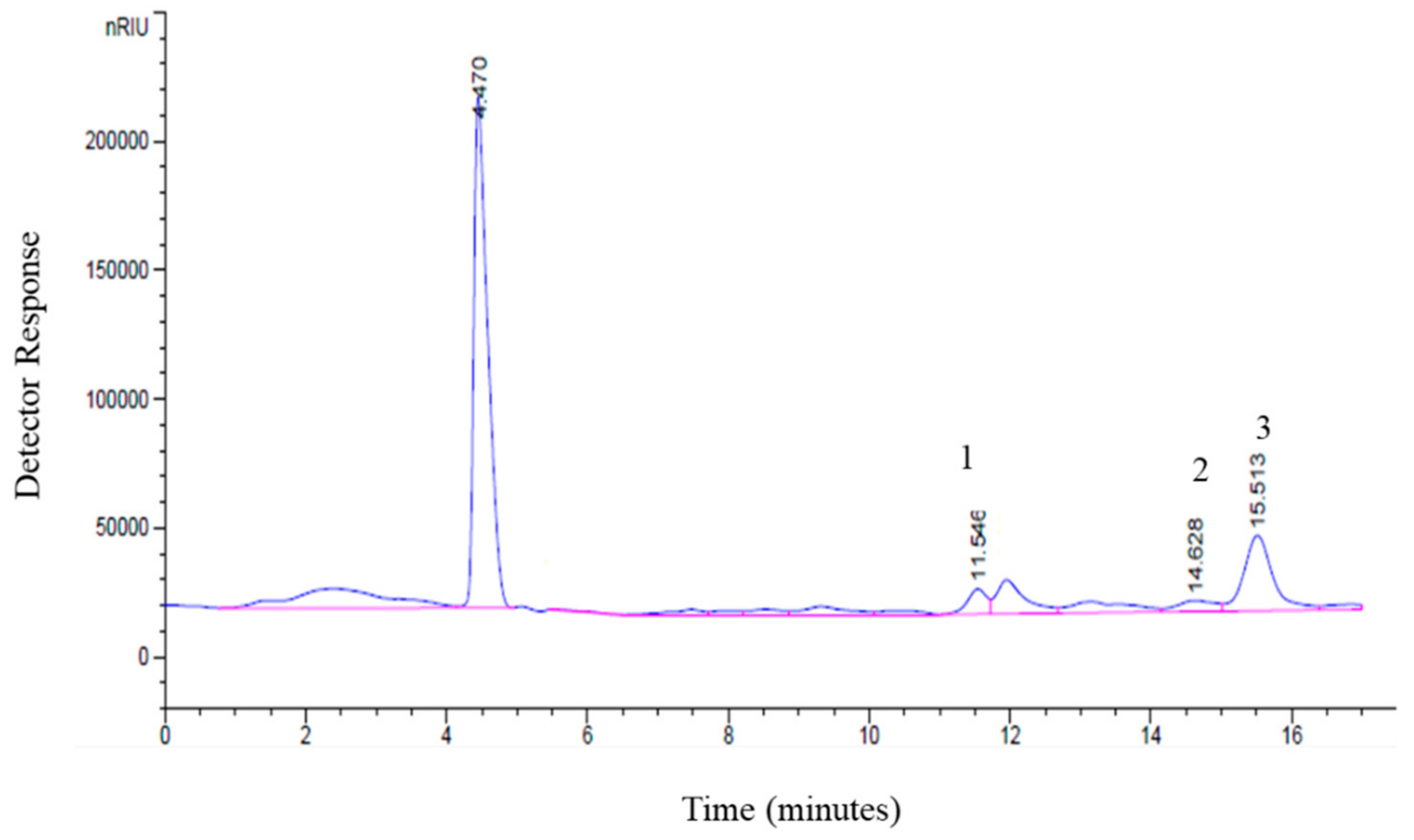
2.3. Quantitative Analysis of Sugars Using High-Performance Liquid Chromatography (HPLC)

To determine the sugar content in tamarind jellose, samples were first hydrolyzed using the acid hydrolysis method [25]. A total of 2 g of the samples was suspended in 2% sulfuric acid. Thermal hydrolysis was performed in a Hirayama sterilizer at 130 °C for 30 mins. Hydrolysis products were then neutralized with 2M NaOH. The hydrolysates were analyzed using an HPLC system with a refractive index detector (RID-10A), column Shodex Asahipak (type NH2P-504E), mobile phase (75 mL acetonitrile, 25 mL water), oven temperature of 40 °C, and column temperature of 40 °C. The analysis was performed at an ambient room temperature of 25 °C, with the flow rate being maintained at 0.6 mL/min. The retention times of the monosaccharide were monitored using a differential refractometer (RID–10A). The retention times obtained were compared to those determined using D-glucose, D-galactose, and D-xylose (Sigma-Aldrich, St. Louis, MO, USA) as standards.

2.4. Mushroom Tyrosinase Assay

The inhibition of tyrosinase was determined by a modification of the dopachrome method using L-DOPA and L-tyrosine as substrates [26]. The samples were dissolved in water at an initial concentration of 20 mg/mL. A total of 50 μL of the samples was diluted in a phosphate buffer (pH 6.8) at concentrations of 200 mM with 50 μL of L-tyrosine added (31 units), then gently shaken before being incubated for 10 min at 30 °C. After incubation, 50 μL of 2.5 mM L-DOPA was added to the sample. The absorbance was measured at 470 nm, and loaded microplates were kept in the dark for more than 20 min at 30 °C. Absorbance was repeatedly measured at the same wavelength, and the calculated % of inhibition was measured by the following equation:
%   I n h i b i t i o n =   [ ( A B ) ( C D ) ]   ( A B ) × 100
A, B, C, and D were the differences in light absorption at a wavelength of 470 nm before and after incubation.

2.5. Determination of Antioxidant Activity

The antioxidant activity of the extracts was measured using a 2,2-diphenyl-1-picrylhydrazyl (DPPH) decolorization assay [27]. A DPPH ethanolic solution was prepared immediately before use. The DPPH solution was modified by being dissolved in 70% ethanol and sonicated for 15 min. Samples were diluted in water. A total of 100 µL of the samples at various concentrations was added to 100 µL of the DPPH solution. After mixing, the absorbance was measured at 517 nm by a microplate reader after exactly 30 min in the dark. Calibration was performed with an ascorbic acid solution. The antioxidation capacity was expressed as % of inhibition following this equation:
%   i n h i b i t i o n = A c o n t r o l A s a m p l e A c o n t r o l × 100
where A c o n t r o l = the absorbance of the control, A s a m p l e = the absorbance of the sample.

2.6. Cell Cultures

The B16 melanoma cell line was a gift kindly provided by Assistant Professor Dr. Sukanya Dejadisai from the Faculty of Pharmaceutical Sciences, Prince of Songkla University (Thailand). B16 melanoma and skin fibroblast cell lines were cultured in Dulbecco’s modified Eagle’s medium (DMEM), supplemented with 10% fetal bovine serum (Gibco), 2 mM glutamine, and 100 units/mL penicillin/streptomycin, and maintained at 37 °C in a humidified incubator with 10% CO2.

2.7. SRB Cell Viability Assay

For the sulforhodamine B (SRB) assay, 5000 cells were seeded in each well of a 96-well plate and incubated at 37 °C with 5% CO2 for 24 h. The cells were then treated with or without various concentrations of the indicated testing reagents. After treatment, cells were fixed with 10% trichloroacetic acid (TCA) for 1 h at 4 °C. The plates were then washed five times with slow-running tap water, and 100 μL of an SRB solution (0.4% SRB in 0.1% acetic acid) was added for staining. After incubation for 1 h at room temperature, the plates were rinsed three times with 1% acetic acid and then air-dried. The stained samples were dissolved in 50 µL of 10 mM Tris base solution to solubilize the protein-bound dye, and the absorbance was measured at 492 nm using a microplate reader (Thermo Fisher Scientific, Waltham, MA, USA). The amount of dye extracted from the stained cells is directly proportional to the living cell mass.

2.8. Clonogenic Assay

A total of 5000 cells were seeded into six-well plates and incubated overnight. The cells were then treated for 48 h with varying concentrations of the tested solutions. The complete media were replaced every 3 days for 14 days. Then, colonies were fixed with 4% paraformaldehyde for fifteen minutes at room temperature and then washed with phosphate-buffered saline. Then, 0.5% crystal violet was used to stain the fixed cells for thirty minutes, and following this, the plates were washed with tap water. The culture plates were dried at room temperature overnight. Finally, the stained colonies were dissolved in 10% acetic acid, and the absorbance was measured at 590 nm using microplate readers (Thermo Fisher Scientific). The cell viability data, derived from the reader, were calculated as the relative percentage of the control group.

2.9. Quantitative Assay of Melanin Content in B16 Melanoma Cells

Cells (5 × 10⁵ cells/dish) were incubated in 60 mm dishes with different concentrations of tested agents. After treatment, the cells were washed twice with PBS, lysed in 200 μL of 1N NaOH, and heated for 1 h at 95 °C to solubilize the melanin. A lytic solution (100 μL) was transferred into a 96-well plate, and the absorbance of the solution was measured at a 405 nm wavelength with a spectrophotometric microplate (Thermo Fisher Scientific). The melanin content was then determined based on the absorbance, divided by the protein concentration in the extract from each cell pellet, and presented as a percentage against that of the untreated control.

2.10. Quantitative Real-Time PCR (qRT-PCR)

The total RNA extraction was performed using an RNeasy Mini Kit (Qiagen, Hilden, Germany), according to the manufacturer’s instructions. Then, 1 µg of the total cellular RNA for each of the conditions was used to generate complementary DNA using Superscript III reverse transcriptase and oligo-dT primers (Invitrogen, Paisley, UK). qPCR was performed using SoFast EvaGreen Supermix (Bio-Rad, Hercules, CA, USA) on a Bio-Rad CFX96 system, according to the manufacturer’s instructions (Bio-Rad). The following gene-specific primers were used for amplification: tyrosinase-sense: 5′-GCCTTCTGTGGAGTTTCCAG-3′ and tyrosinase-antisense: 5′-AGGGGAACTGAGGTCCAGAT-3′; MITF-sense: 5′-GCCCAGGTATGAACAC-GCAC-3′ and MITF-antisense: 5′-GGTTGGCTGGACAGGAGTTG-3′; L19-sense: 5′-GGATCCCAATGAGACCAATG-3′ and L19-antisense: 5′-GGCAG-TACCCTTCCTCTTCC-3′. The results were normalized using L19 mRNA expression as a reference gene. All of the qPCR experiments were repeated for at least three independent experiments, and the relative expression was shown as an average ± S.D. (n = 3). Statistical significance was determined by a Student’s t-test (significant, * p < 0.05, ** p < 0.01, *** p < 0.001).

2.11. Statistics

Data are presented as means ± standard deviation (SD) of at least three independent experiments. Statistical significance was calculated using a Student’s t-test and one-way ANOVA between the control and treated conditions. A p-value of ≤0.05 was considered significantly different between groups.

3. Results

3.1. Quantitative Determination of Sugars and Physicochemical Properties of Tamarind Jellose

Before starting an investigation of the potential bioactivities of tamarind jellose, we first characterized the chemical constituents of tamarind jellose used in this study using an HPLC analysis. As shown in Figure 1, tamarind jellose mainly consisted of the key main monosaccharides galactose, glucose, and xylose at a molar ratio of 1:1.5:1.2. In parallel, the physiochemical properties of tamarind jellose were also characterized to understand its nature (Supplementary Methods S1). As shown in Table 1, tamarind jellose exhibited good and suitable properties for medical and cosmetic applications, as indicated by angle of repose, moisture sorption, and rheology (Table 1).

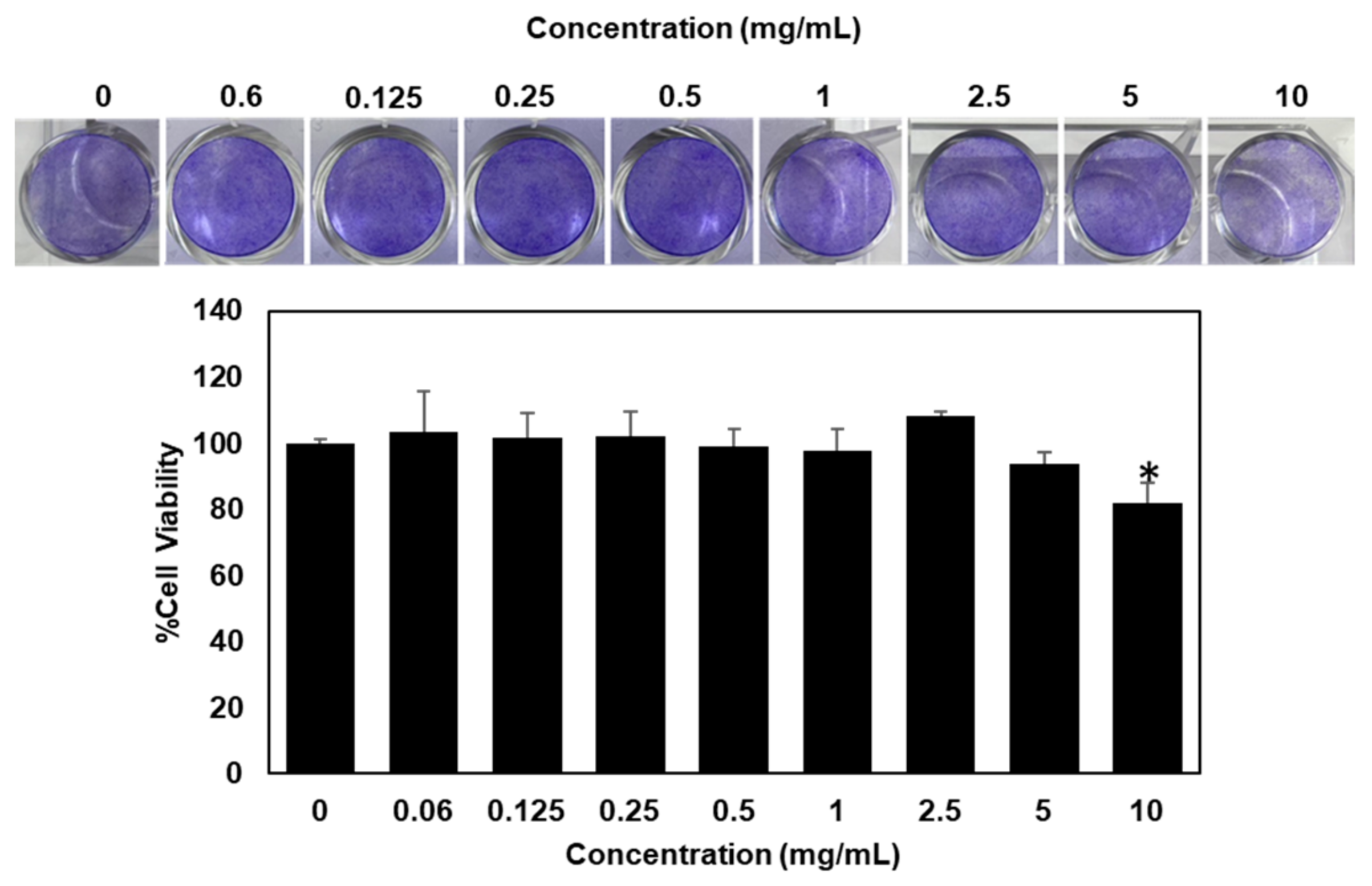
3.2. Cytotoxicity Effect of Tamarind Jellose (TJ) on Skin Fibroblast Cells

In the cosmetics industry, testing for cytotoxicity is crucial to assessing the safety of ingredients. In our experiment, we examined the cytotoxic effects of tamarind jellose (TJ) on skin fibroblast cells using the sulforhodamine B (SRB) assay. The assay relies on the binding of SRB dye with amino acid residues in cell proteins under acidic conditions. The SRB results revealed that tamarind jellose (TJ) at the concentration range of 0–10 mg/mL did not show any cytotoxic effect on skin fibroblast cell growth in vitro, as indicated by the IC50 being over 10 mg/mL at the maximum tested concentrations (Figure 2).

3.3. Long-Term Cytotoxicity Effect of Tamarind Jellose (TJ) on Skin Fibroblast Cells

Chronic exposure to skincare products is closely associated with an increase in skin cell death and inflammation. To further examine the long-term cytotoxicity of tamarind jellose (TJ) on skin fibroblast cells, clonogenic assays were performed. Consistent with the SRB assay, the clonogenic assay results demonstrated that tamarind jellose (TJ) did not show a long-term toxic effect on skin fibroblast cell growth in vitro (Figure 3). These results confirm that TJ is non-toxic and suitable to be utilized as a cosmetic ingredient. The non-toxic concentration range (0–10 mg/mL) was then applied in further cellular activity testing experiments.

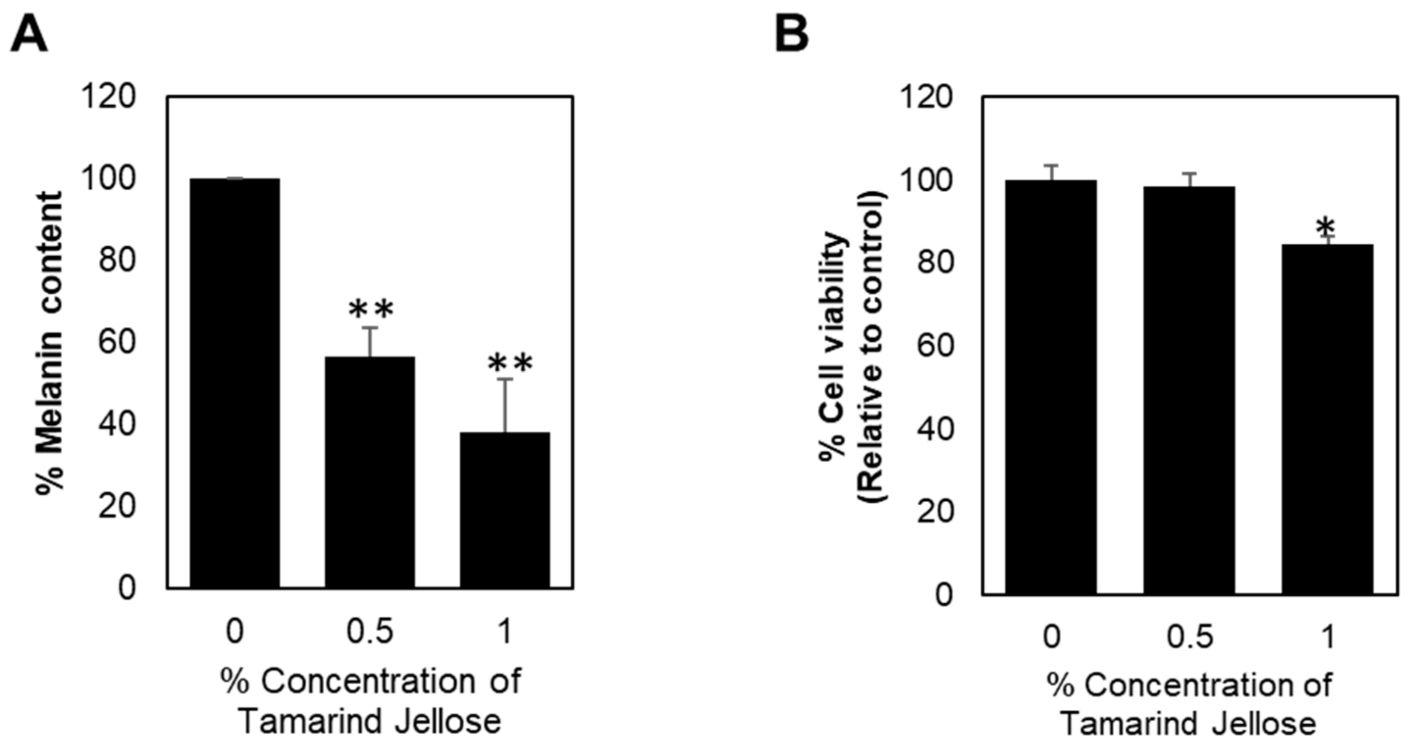
3.4. Inhibitory Effect of Tamarind Jellose on the Production of Melanin in B16 Melanoma Cells

Next, we focused on the potential inhibitory effect of tamarind jellose on the production of melanin in B16 melanoma cells. The cells were treated with tamarind jellose at the indicated concentrations for 48 h. Interestingly, the results in Figure 4 revealed that tamarind jellose significantly decreased the levels of melanin compared to the control group in a dose-dependent manner. After treating the cells with 0.5% and 1% (w/v) concentrations of tamarind jellose, the cellular melanin content was reduced to 56% and 37.9%, respectively, suggesting its potent role as a melanogenesis inhibitor that could be used as a skin-whitening product.

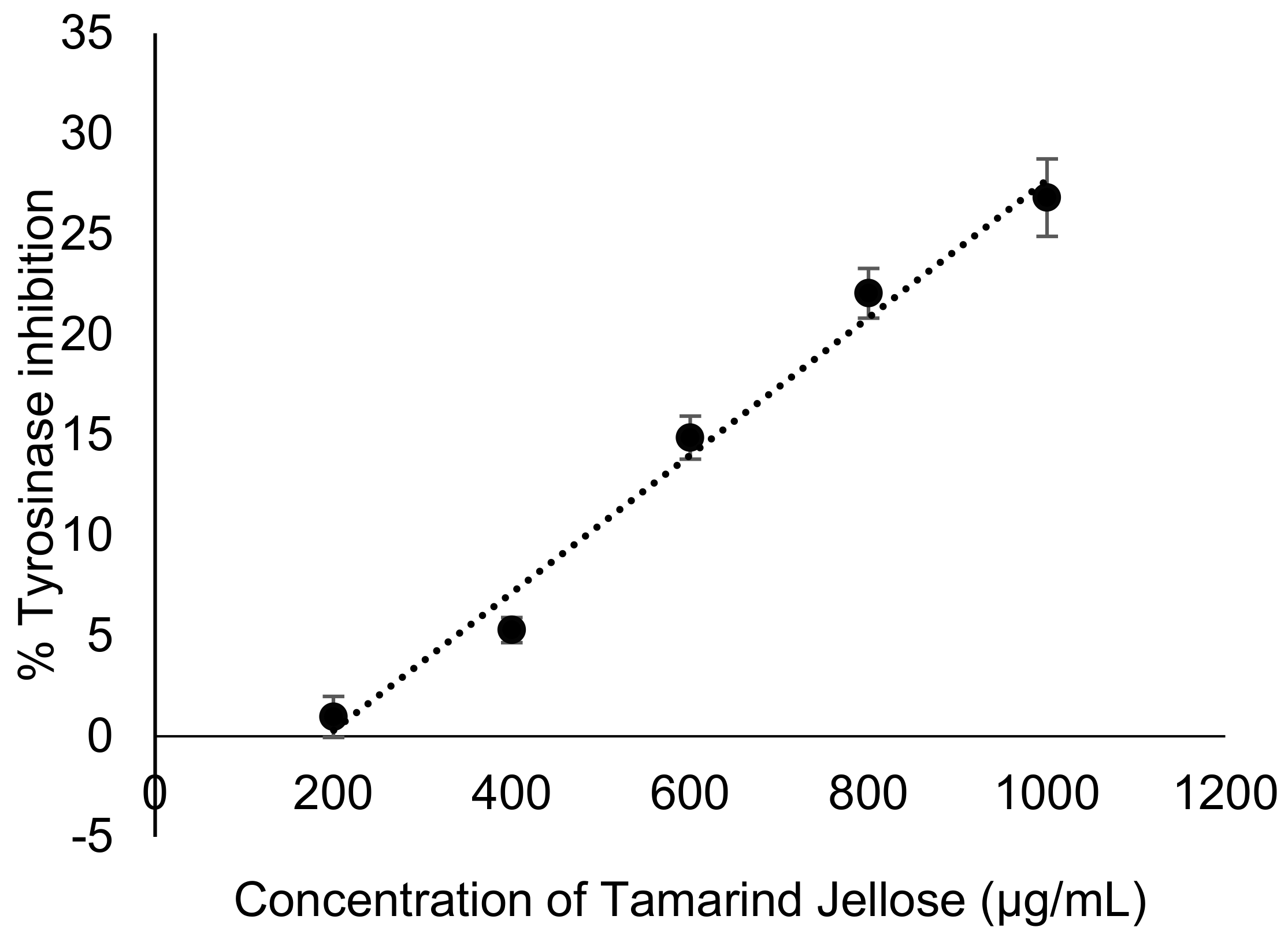
3.5. Tamarind Jellose Inhibits Mushroom Tyrosinase Activity (Cell-Free Methods)

Since tyrosinase is a key rate-limiting enzyme in melanin synthesis, we then investigated whether tamarind jellose reduced melanin content by directly inhibiting tyrosinase activity using a mushroom tyrosinase activity assay. As shown in Figure 5, tamarind jellose at concentrations of 200–1000 µg/mL exhibited a dose-dependent tyrosinase activity inhibition ranging from 0.97 ± 1.02% to 26.82 ± 1.93%. However, it did not reach EC50 due to poor dispersion problems at higher concentrations. In addition, Kojic acid at a concentration of 3–500 µg/mL was used as the positive standard in this experiment, and its percentage of inhibition ranged from 19.14 ± 1.27% to 97.54 ± 0.40%. Therefore, this experiment suggests that tamarind jellose seems to exert a significant inhibitory effect on mushroom tyrosinase activity.

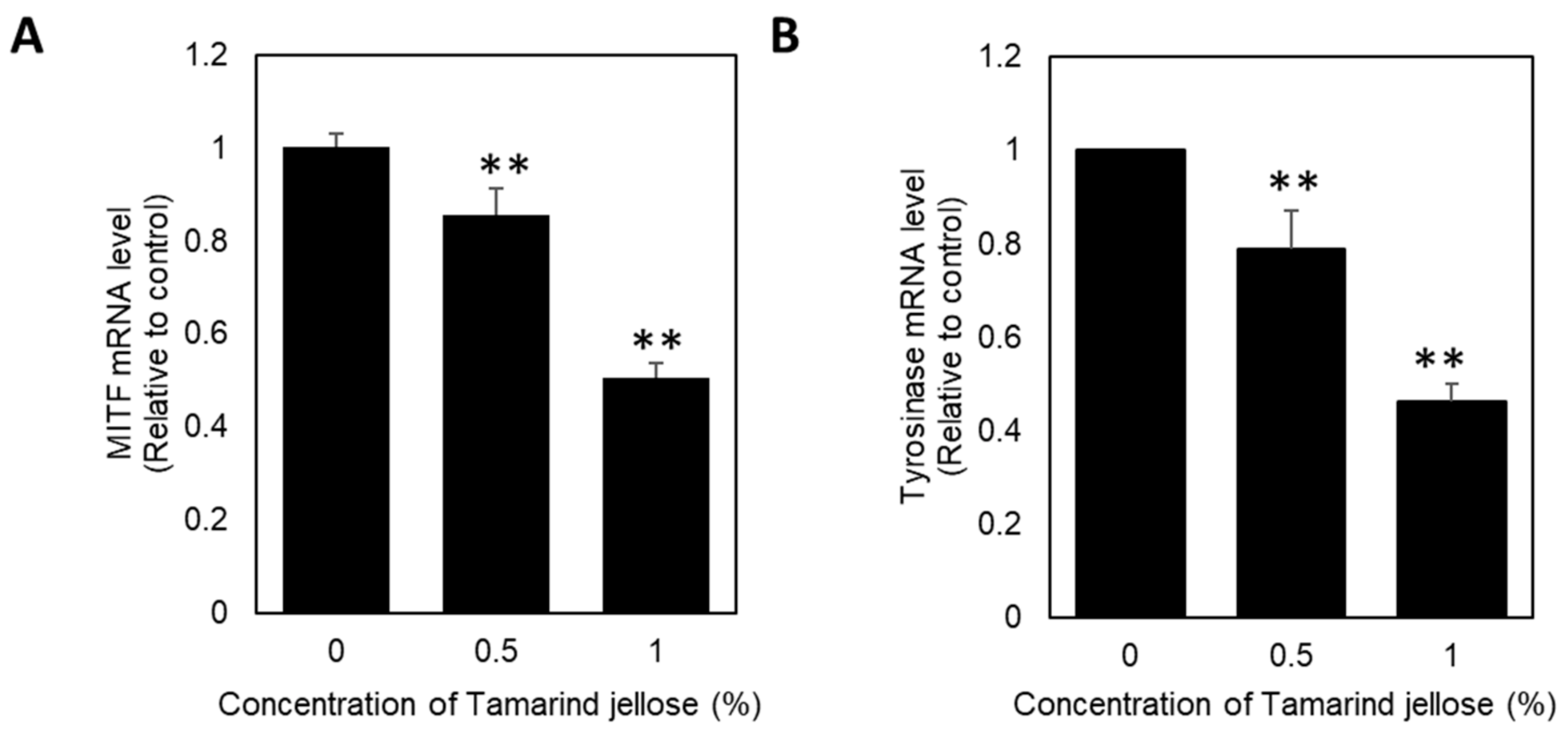
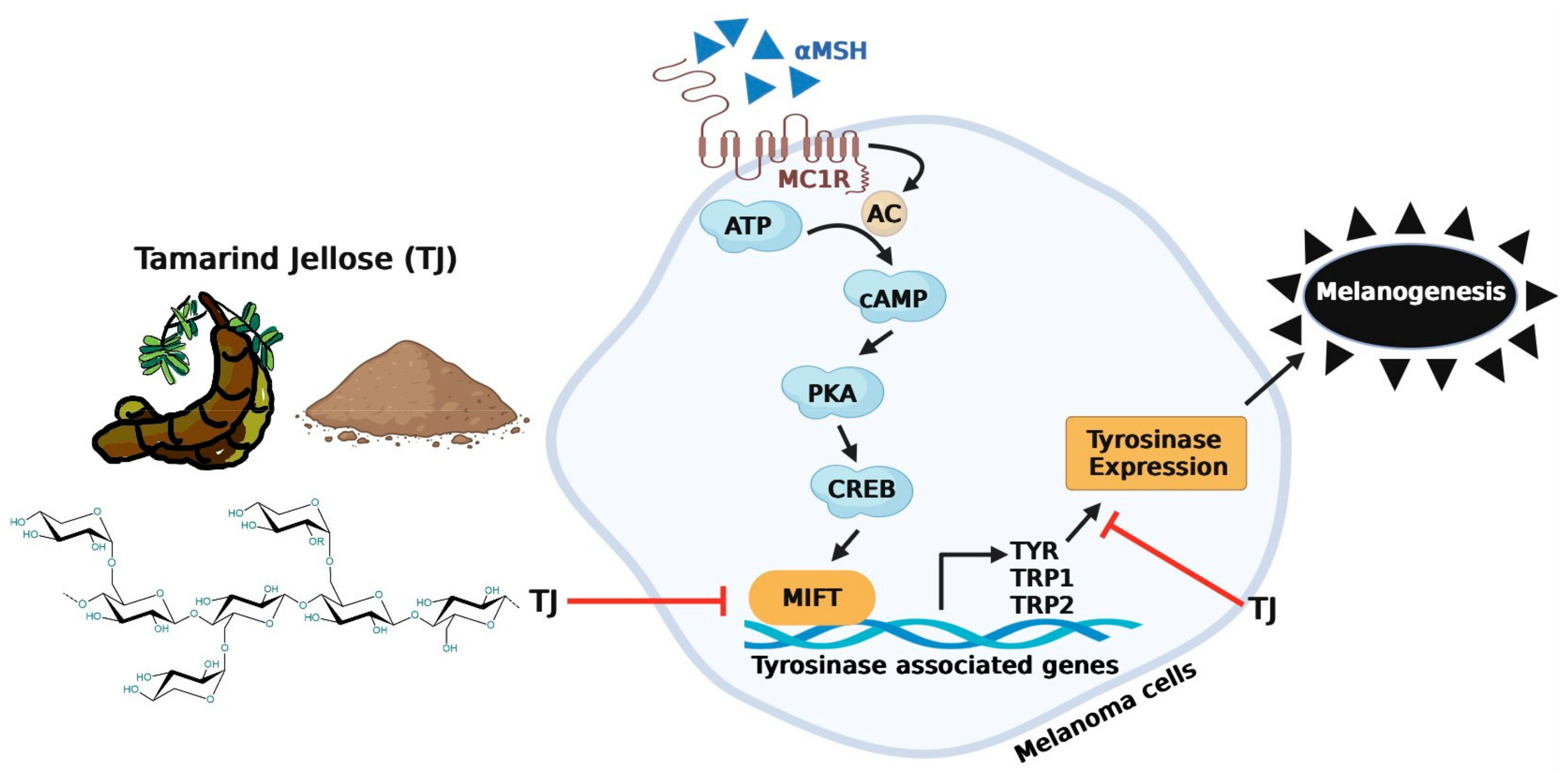
3.6. Effects of Tamarind Jellose on the Transcriptional Expression of Melanogenesis-Related Genes, Including Tyrosinase (TYR) and Microphthalmia-Associated Transcription Factor (MITF) in B16 Melanoma Cells

In order to elucidate, deeply at the cellular level, if tamarind jellose reduced melanin content by inhibiting the expression of key melanogenesis-related genes, including microphthalmia-associated transcription factor (MITF) and its downstream target tyrosinase (TYR), we measured their changes at the mRNA level by a quantitative reverse transcription-polymerase chain reaction (qRT-PCR) analysis after treatment with tamarind jellose at the indicated concentrations for 72 h. The qRT-PCR analysis result showed that the intracellular mRNA expression of MITF and tyrosinase was significantly decreased following treatment with tamarind jellose in a dose-dependent manner (Figure 6). The results suggest that tamarind jellose could be a potential melanogenesis inhibiting agent, and its molecular mechanism is related to the decrease in transcriptional expression of microphthalmia-associated transcription factor (MITF) as well as its downstream tyrosinase (TYR) genes.

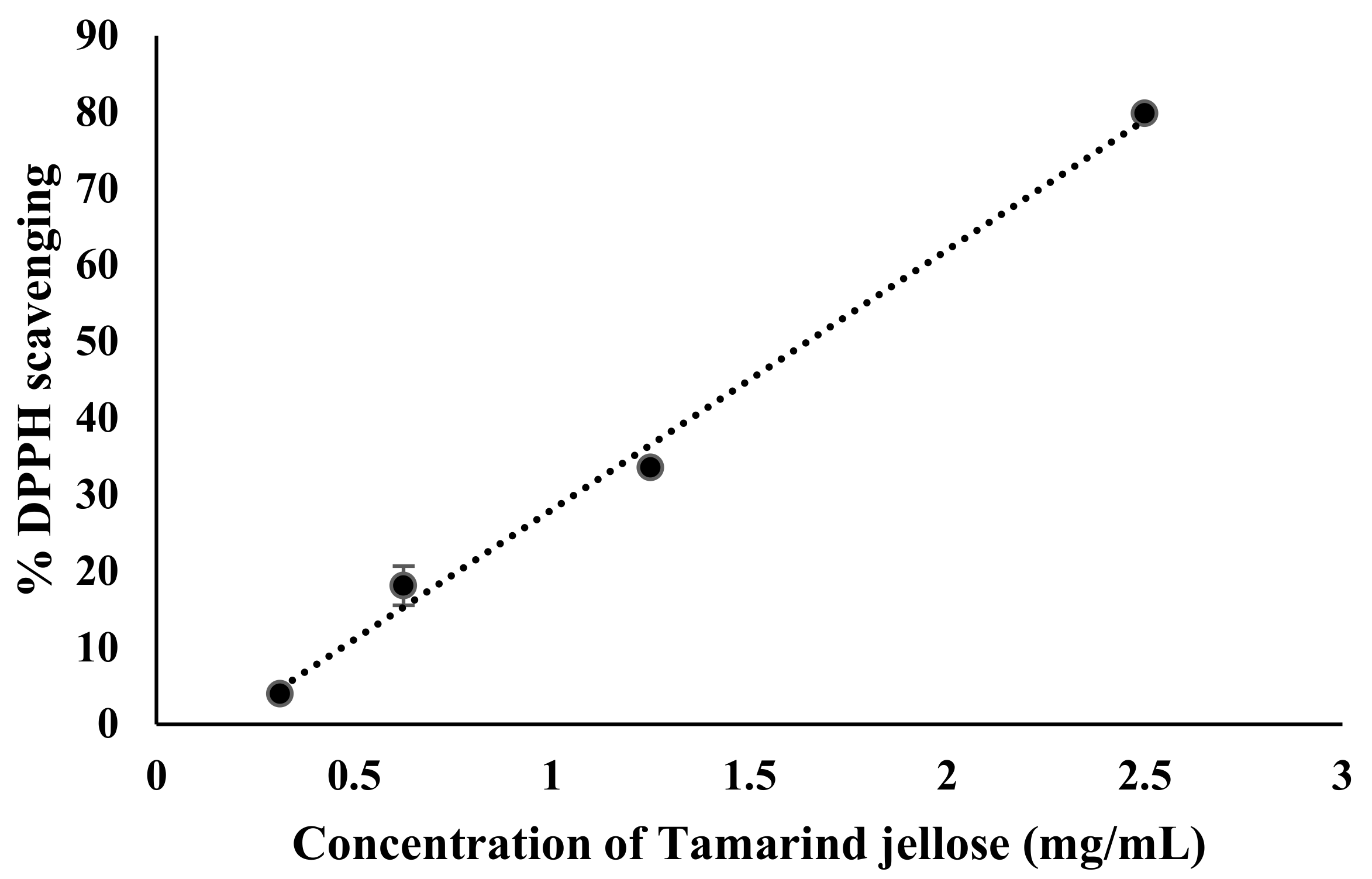
3.7. Antioxidant Activity of Tamarind Jellose

Reactive oxygen species (ROS) have been reported to be one of the key factors in regulating melanin synthesis. In this experiment, we tested the antioxidant property of tamarind jellose by performing a radical scavenging assay using 2,2-diphenyl-1-picrylhydrazyl (DPPH). DPPH is a stable, free radical that is purple in color and can interact with different antioxidant molecules. During the process, it becomes reduced to a colorless compound by monitoring the absorbance at 517 nm using a UV–visible spectrophotometer. As shown in Figure 7, the DPPH free-radical scavenging effects of tamarind jellose were observed in a concentration-dependent manner ranging from 0.3 to 2.5 mg/mL. The IC50 values of tamarind jellose along with L-ascorbic acid (positive standard) for scavenging DPPH were 1.65 and 0.007 mg/mL, respectively. This result clearly indicates that tamarind jellose possesses antioxidant characteristics, which might also contribute to its depigmenting activity.

4. Discussion

In recent years, there has been a growing interest in the development of green or natural cosmetic products that harness the potential of plant-derived ingredients. As consumers become more conscious of the impact of traditional cosmetics on the environment and their own health, there is a need to explore alternative ingredients that are not only safe and sustainable but also possess bioactive properties. Especially, there has been a growing interest in tamarind seed jellose (TJ), a by-product of tamarind processing, as a potential ingredient in green cosmetics. However, comprehensive scientific research on its bioactivity and applicability in cosmetic formulations is still limited. In this study, we performed a series of comprehensive in vitro experiments to reveal the potential bioactivities of tamarind jellose (TJ), including cytotoxicity, antioxidant, and anti-melanogenesis activities, as well as the molecular mechanisms involved, as a functional ingredient in cosmetic product development. We initially characterized the chemical constituents of tamarind jellose used in this study using an HPLC analysis and found that tamarind jellose mainly consisted of galactose, glucose, and xylose at a molar ratio of 1:1.5:1.2 (Figure 1). However, the ratio of sugar compositions in this study was slightly different from previous analytical reports, indicating the ratio of galactose, glucose, and xylose as ~1:3:2 [28,29,30]. These differences might be associated with environmental effects in which the plants were grown during their polysaccharide biosynthesis [7] as well as possible variations in extraction approaches [31,32].
The detection of cytotoxicity is important to evaluate the toxic effects elicited by cosmetic ingredients and identify effective doses. Our short-term and chronic cytotoxic assays (Figure 2 and Figure 3) revealed that TJ at concentrations ranging from 0 to 10 mg/mL is safe and suitable to be utilized as a cosmetic ingredient. Interestingly, we also found that low concentrations of TJ (0.06–5 mg/mL) significantly enhanced cell proliferation. This finding is consistent with a previous study showing that tamarind seed xyloglucans promoted skin regeneration by having a direct influence on cell proliferation and migration of human skin keratinocytes and fibroblasts [32].
The control of melanogenesis is a crucial strategy in the treatment of abnormal hyperpigmentation for both cosmetic and therapeutic purposes. In humans, melanogenesis involves a complex series of oxidative and enzymatic reactions. Tyrosinase (TRY) is one of the most vital enzymes for melanin synthesis. It is the key enzyme in the rate-limiting step in which L-tyrosine is hydroxylated to L-DOPA, which is further oxidized into DOPAquinone. In addition, intracellular microphthalmia-associated transcription factor (MITF) is another key transcription regulator of genes responsible for melanin biosynthesis, including TYR, TRP-1, and TRP-2 [1]. Thus, agents that have an inhibitory effect on TYR activity or the regulation of TYR expression are considered good candidates for skin-whitening product development.
Therefore, the present study also focused on the effect of tamarind jellose on the inhibition of tyrosinase and melanin production using the mushroom tyrosinase assay as well as an in vitro B16F10 mouse melanoma cell culture model. Interestingly, our key findings revealed that TJ showed high inhibitory activities against melanogenesis in a dose-dependent manner (Figure 4). In addition, the depigmenting action of TJ could be attributed to its ability to suppress tyrosinase activity (Figure 5) and MITF-mediated tyrosinase expression (Figure 6), suggesting its potential to be developed as a powerful whitening agent for treating skin hyperpigmentation without undesirable cytotoxicity.
Additionally, reactive oxygen species (ROS) have been reported to be one of the key factors in regulating melanin synthesis, whereas ROS scavengers and ROS inhibitors have been shown to suppress melanogenesis [33]. Our DPPH free-radical scavenging results clearly indicated that tamarind jellose also exhibited a strong antioxidant property (Figure 7).
As shown in Figure 8, this study marks the very first comprehensive exploration of the potential inhibitory effects of tamarind jellose on melanin biosynthesis. The inhibition of melanin production observed could be due to the reduction in tyrosinase activity, MITF-mediated tyrosinase expression, as well as the enhancement of antioxidant activity.

5. Conclusions

In conclusion, these findings provide compelling evidence supporting the efficacy of tamarind jellose serum in brightening skin and reducing spots and wrinkles in middle-aged participants in our previous clinical testing study [23], further strongly confirming that tamarind jellose serves as a safe, effective, multi-functional, and eco-friendly ingredient for beauty products, especially a skin-lightening agent for treating abnormal skin hyperpigmentation for both cosmetic and therapeutic purposes. By incorporating tamarind jellose into skincare and cosmetic products, manufacturers can meet the growing consumer demand for products that prioritize both effectiveness and sustainability. In addition to its functional benefits, tamarind seed jellose presents a promising and valuable resource in cosmetics, offering the beauty industry an opportunity to reduce waste, promote sustainable sourcing practices, and provide consumers with effective, natural, and eco-friendly alternatives for their beauty and skincare products.

Supplementary Materials

The following supporting information can be downloaded at: https://www.mdpi.com/article/10.3390/su16083114/s1, Methods S1: Physiochemical properties of Tamarind Jellose. References [34,35,36] are cited in the Supplementary Materials.

Author Contributions

Conceptualization, T.A. and P.K.; methodology, T.A. and P.K.; investigation and analysis, P.K., S.K., C.T. and T.A.; funding acquisition, T.A.; writing—original draft preparation, P.K., S.K. and T.A.; writing—review and editing, P.K., S.K. and T.A. All authors have read and agreed to the published version of the manuscript.

Funding

This research was supported by the Thailand Research Fund (TRF)—Researchers and Research Funds for Industries (RRi) (Grant No. RDG6050082).

Data Availability Statement

Data are available within the article.

Acknowledgments

Jellose powder in this study was supported by Pinphet Corporation Co., Ltd. (Phetchabun, Thailand).

Conflicts of Interest

All authors report no potential conflicts of interest.

References

  1. D’Mello, S.A.N.; Finlay, G.J.; Baguley, B.C.; Askarian-Amiri, M.E. Signaling Pathways in Melanogenesis. Int. J. Mol. Sci. 2016, 17, 1144. [Google Scholar] [CrossRef] [PubMed]
  2. Videira, I.F.d.S.; Moura, D.F.L.; Magina, S. Mechanisms regulating melanogenesis. An. Bras. Dermatol. 2013, 88, 76–83. [Google Scholar] [CrossRef] [PubMed]
  3. Panzella, L.; Napolitano, A. Natural and Bioinspired Phenolic Compounds as Tyrosinase Inhibitors for the Treatment of Skin Hyperpigmentation: Recent Advances. Cosmetics 2019, 6, 57. [Google Scholar] [CrossRef]
  4. Liu, J.-K. Natural products in cosmetics. Nat. Products Bioprospect. 2022, 12, 40. [Google Scholar] [CrossRef] [PubMed]
  5. Nishinari, K.; Takemasa, M.; Yamatoya, K.; Shirakawa, M. 19—Xyloglucan. In Handbook of Hydrocolloids, 2nd ed.; Phillips, G.O., Williams, P.A., Eds.; Woodhead Publishing Series in Food Science, Technology and Nutrition; Woodhead Publishing: Cambridge, UK, 2009; pp. 535–566. Available online: http://www.sciencedirect.com/science/article/pii/B9781845694142500199 (accessed on 25 December 2022).
  6. Semenzato, A.; Costantini, A.; Baratto, G. Green Polymers in Personal Care Products: Rheological Properties of Tamarind Seed Polysaccharide. Cosmetics 2015, 2, 1–10. [Google Scholar] [CrossRef]
  7. Freitas, R.A.; Martin, S.; Santos, G.L.; Valenga, F.; Buckeridge, M.S.; Reicher, F.; Sierakowski, M.-R. Physico-chemical properties of seed xyloglucans from different sources. Carbohydr. Polym. 2005, 60, 507–514. [Google Scholar] [CrossRef]
  8. Kulkarni, A.D.; Joshi, A.A.; Patil, C.L.; Amale, P.D.; Patel, H.M.; Surana, S.J.; Belgamwar, V.S.; Chaudhari, K.S.; Pardeshi, C.V. Xyloglucan: A functional biomacromolecule for drug delivery applications. Int. J. Biol. Macromol. 2017, 104, 799–812. [Google Scholar] [CrossRef] [PubMed]
  9. Luzia, D.M.M.; Jorge, N. Antioxidant activity, fatty acid profile and tocopherols of Tamarindus indica L. seeds. Food Sci. Technol. 2011, 31, 497–501. [Google Scholar] [CrossRef]
  10. Siddhuraju, P. Antioxidant activity of polyphenolic compounds extracted from defatted raw and dry heated Tamarindus indica seed coat. LWT—Food Sci. Technol. 2007, 40, 982–990. [Google Scholar] [CrossRef]
  11. Nwodo, U.U.; Obiiyeke, G.E.; Chigor, V.N.; Okoh, A.I. Assessment of Tamarindus indica extracts for antibacterial activity. Int. J. Mol. Sci. 2011, 12, 6385–6396. [Google Scholar] [CrossRef] [PubMed]
  12. Phetdee, K.; Rattanamanee, K.; Teaktong, T.; Viyoch, J. Tamarind seed coat extract reduces melanin production via tyrosinase in melanocyte. J. Biol. Sci. 2012, 12, 239–245. [Google Scholar] [CrossRef]
  13. Bonin, E.; Avila, V.D.; Carvalho, V.M.; Cardoso, M.A.P.; Matos, A.M.; Ramos, A.V.G.; Cabral, M.R.P.; Baldoqui, D.C.; Sarragiotto, M.H.; Filho, B.A.d.A.; et al. Study of chemical constituents, antioxidants and antimicrobial activities of Tamarindus indica L. seed. J. Food Sci. 2023, 88, 4639–4652. [Google Scholar] [CrossRef] [PubMed]
  14. Avachat, A.M.; Gujar, K.N.; Wagh, K. V Development and evaluation of tamarind seed xyloglucan-based mucoadhesive buccal films of rizatriptan benzoate. Carbohydr. Polym. 2013, 91, 537–542. [Google Scholar] [CrossRef] [PubMed]
  15. Pal, D.; Nayak, A.K. Novel tamarind seed polysaccharide-alginate mucoadhesive microspheres for oral gliclazide delivery: In vitro–in vivo evaluation. Drug Deliv. 2012, 19, 123–131. [Google Scholar] [CrossRef] [PubMed]
  16. Chen, D.; Guo, P.; Chen, S.; Cao, Y.; Ji, W.; Lei, X.; Liu, L.; Zhao, P.; Wang, R.; Qi, C.; et al. Properties of xyloglucan hydrogel as the biomedical sustained-release carriers. J. Mater. Sci. Mater. Med. 2012, 23, 955–962. [Google Scholar] [CrossRef] [PubMed]
  17. Uccello-Barretta, G.; Nazzi, S.; Zambito, Y.; Di Colo, G.; Balzano, F.; Sansò, M. Synergistic interaction between TS-polysaccharide and hyaluronic acid: Implications in the formulation of eye drops. Int. J. Pharm. 2010, 395, 122–131. [Google Scholar] [CrossRef] [PubMed]
  18. Darvill, A.; Eberhard, S.; Pauly, M.; Albersheim, P.; Strickland, F.M.; Sun, Y. Preservation of the Delayed-Type Hypersensitivity Response to Alloantigen by Xyloglucans or Oligogalacturonide does not Correlate with the Capacity to Reject Ultraviolet-Induced Skin Tumors in Mice. J. Investig. Dermatol. 2001, 116, 62–68. [Google Scholar] [CrossRef] [PubMed]
  19. Aravind, S.R.; Joseph, M.M.; Varghese, S.; Balaram, P.; Sreelekha, T.T. Antitumor and immunopotentiating activity of polysaccharide PST001 isolated from the seed kernel of Tamarindus indica: An in vivo study in mice. Sci. World J. 2012, 2012, 361382. [Google Scholar] [CrossRef] [PubMed]
  20. Burgalassi, S.; Raimondi, L.; Pirisino, R.; Banchelli, G.; Boldrini, E.; Saettone, M.F. Effect of Xyfoglucan (Tamarind Seed Polysaccharide) on Conjunctival Cell Adhesion to Laminin and on Corneal Epithelium Wound Healing. Eur. J. Ophthalmol. 2000, 10, 71–76. [Google Scholar] [CrossRef]
  21. Do Rosário, M.M.T.; Kangussu-Marcolino, M.M.; do Amaral, A.E.; Noleto, G.R.; de Oliveira Petkowicz, C.L. Storage xyloglucans: Potent macrophages activators. Chem. Biol. Interact. 2011, 189, 127–133. [Google Scholar] [CrossRef]
  22. Rolando, M.; Valente, C. Establishing the tolerability and performance of tamarind seed polysaccharide (TSP) in treating dry eye syndrome: Results of a clinical study. BMC Ophthalmol. 2007, 7, 5. [Google Scholar] [CrossRef]
  23. Amnuaikit, T.; Khakhong, S.; Khongkow, P. Formulation Development and Facial Skin Evaluation of Serum Containing Jellose from Tamarind Seeds. J. Pharm. Res. Int. 2019, 31, 1–14. [Google Scholar] [CrossRef]
  24. Sukhawanli, S.; Thamakorn, P. Extraction of tamarind seed jellose under different conditions and their rheological properties. Food Appl. Biosci. J. 1970, 2, 61–68. [Google Scholar] [CrossRef]
  25. González-Hernández, J.C.; Farías Rosales, L.; Zamudio Jaramillo, M.Á.; Álvarez-Navarrete, M.; Vera Villa, J.C.; Martínez Corona, R.; Chávez-Parga, M.; Peña, A. Chemical Hydrolysis of the Polysaccharides of the Tamarind Seed. J. Mex. Chem. Soc. 2012, 56, 395–401. [Google Scholar] [CrossRef]
  26. Chan, G.; Kalaitzidis, D.; Neel, B.G. The tyrosine phosphatase Shp2 (PTPN11) in cancer. Cancer Metastasis Rev. 2008, 27, 179–192. [Google Scholar] [CrossRef] [PubMed]
  27. Brand-Williams, W.; Cuvelier, M.E.; Berset, C. Use of a free radical method to evaluate antioxidant activity. LWT—Food Sci. Technol. 1995, 28, 25–30. [Google Scholar] [CrossRef]
  28. Gidley, M.J.; Lillford, P.J.; Rowlands, D.W.; Lang, P.; Dentini, M.; Crescenzi, V.; Edwards, M.; Fanutti, C.; Grant Reid, J.S. Structure and solution properties of tamarind-seed polysaccharide. Carbohydr. Res. 1991, 214, 299–314. [Google Scholar] [CrossRef] [PubMed]
  29. Patel, T.R.; Morris, G.A.; Ebringerová, A.; Vodeničarová, M.; Velebný, V.; Ortega, A.; Garcia de la Torre, J.; Harding, S.E. Global conformation analysis of irradiated xyloglucans. Carbohydr. Polym. 2008, 74, 845–851. [Google Scholar] [CrossRef]
  30. Goyal, P.; Kumar, V.; Sharma, P. Graft copolymerization of acrylamide onto tamarind kernel powder in the presence of ceric ion. J. Appl. Polym. Sci. 2008, 108, 3696–3701. [Google Scholar] [CrossRef]
  31. Chawananorasest, K.; Saengtongdee, P.; Kaemchantuek, P. Extraction and Characterization of Tamarind (Tamarind indica L.) Seed Polysaccharides (TSP) from Three Difference Sources. Molecules 2016, 21, 775. [Google Scholar] [CrossRef] [PubMed]
  32. Nie, W.; Deters, A.M. Tamarind Seed Xyloglucans Promote Proliferation and Migration of Human Skin Cells through Internalization via Stimulation of Proproliferative Signal Transduction Pathways. Dermatol. Res. Pract. 2013, 2013, 359756. [Google Scholar] [CrossRef] [PubMed]
  33. Funasaka, Y.; Komoto, M.; Ichihashi, M. Depigmenting Effect of α-Tocopheryl Ferulate on Normal Human Melanocytes. Pigment Cell Res. 2000, 13, 170–174. [Google Scholar] [CrossRef] [PubMed]
  34. Manchanda, R.; Arora, S.C.; Manchanda, R. Tamarind seed polysaccharide and its modifications-versatile pharmaceutical excipients—A review. Int. J. Pharm. Technol. Res. 2014, 6, 412–420. [Google Scholar]
  35. Wexler, A.; Hasegawa, S. Relative humidity-temperature relationships of some saturated salt solutions in the temperature range 0 to 50 °C. J. Res. Natl. Bur. Stand. 1954, 53, 19–26. [Google Scholar] [CrossRef]
  36. Kaur, L.; Singh, J.; Singh, H.; McCarthy, O.J. Starch–cassia gum interactions: A microstructure—Rheology study. Food Chem. 2008, 111, 1–10. [Google Scholar] [CrossRef]
Figure 1. Analytical HPLC chromatogram of tamarind jellose. The peaks represent xylose (1), galactose (2), and glucose (3), respectively.
Sustainability 16 03114 g001
Figure 2. Cytotoxic effect of tamarind jellose (TJ) on skin fibroblast cells as determined by an SRB assay. Cells were treated with the indicated testing reagents at different concentrations for 48 h, and then an SRB assay was performed. The % cell viability was calculated relative to non-treated cells. Each bar represents a mean ± SD (n ≥ 3).
Sustainability 16 03114 g002
Figure 3. Long-term clonogenic effect of tamarind jellose (TJ) on skin fibroblast cells as determined by clonogenic assays. Cells were treated with the indicated testing reagents at different concentrations for 14 days, and % cell viability was calculated relative to non-treated cells. Each bar represents a mean ± SD (n ≥ 3); * p < 0.05 denotes significant differences when compared to the control (Student’s t-test).
Sustainability 16 03114 g003
Figure 4. Tamarind jellose reduced melanin production in B16 melanoma cells. (A) Relative melanin contents and (B) cell viability of B16 cells measured at 48 h after treatment. Each bar represents a mean ± SD (n = 3); * p < 0.05 and ** p < 0.01 denoted significant differences when compared to the control (Student’s t-test).
Sustainability 16 03114 g004
Figure 5. In vitro mushroom tyrosinase inhibition activity of tamarind jellose at concentrations from 200 to 1000 µg/mL. All data are expressed as means ± SDs (n = 5).
Sustainability 16 03114 g005
Figure 6. Tamarind jellose inhibits intracellular tyrosinase expression at the mRNA level. B16 cells were treated with tamarin jellose at indicated concentrations for 72 h, and the MITF (A) and tyrosinase (B) mRNA expression levels were determined by qRT-PCR. Each bar represents an average of a relative mRNA level ± SD (n = 3); ** p < 0.01 denotes significant differences when compared to the control (Student’s t-test).
Sustainability 16 03114 g006
Figure 7. Dose response curve for DPPH radical scavenging of tamarind jellose. The plotted data are means ± SDs (n = 3). The dot line is a linear regression that fits the data.
Sustainability 16 03114 g007
Figure 8. Proposed inhibitory mechanism of tamarind jellose on melanin synthesis in B16 melanoma cells. The red line indicates inhibitory effects on tyrosinase activity as well as melanogenesis-related gene expression, including microphthalmia-associated transcription factor (MITF) and tyrosinase (TYR), leading to further hindered melanin synthesis. These data provide insight into the potential anti-melanogenesis effect of tamarind jellose in cosmetic applications.
Sustainability 16 03114 g008
Table 1. Physicochemical properties of tamarind jellose.
ParametersResults
Organoleptic properties
ColorSand brown
OdorRoasted bean
TasteBean
TextureRough powder
pH6.30–7.65
Dispersion (% w/v)2.5
Angle of repose (◦)35.42 ± 0.73
Moisture sorption (% w/w)6.47
RheologyPseudoplastic
Disclaimer/Publisher’s Note: The statements, opinions and data contained in all publications are solely those of the individual author(s) and contributor(s) and not of MDPI and/or the editor(s). MDPI and/or the editor(s) disclaim responsibility for any injury to people or property resulting from any ideas, methods, instructions or products referred to in the content.

