Article

Combining Chlorophyll Fluorescence and Vegetation Reflectance Indices to Estimate Non-Photochemical Quenching (NPQ) of Rice at the Leaf Scale

1
State Key Laboratory of Remote Sensing Science, Faculty of Geographical Science, Beijing Normal University, Beijing 100875, China
2
Beijing Engineering Research Center for Global Land Remote Sensing Products, Faculty of Geographical Science, Beijing Normal University, Beijing 100875, China
3
Key Laboratory of Virtual Geographic Environment, Ministry of Education, Nanjing Normal University, Nanjing 210023, China
4
Jiangsu Center for Collaborative Innovation in Geographical Information Resource Development and Application, Nanjing 210023, China
5
School of Earth Sciences and Engineering, Hohai University, Nanjing 211100, China
6
Jiangxi Academy of Water Science and Engineering, Nanchang 330029, China
*
Author to whom correspondence should be addressed.
Remote Sens. 2023, 15(17), 4222; https://doi.org/10.3390/rs15174222
Submission received: 7 July 2023 / Revised: 23 August 2023 / Accepted: 26 August 2023 / Published: 28 August 2023
(This article belongs to the Section Remote Sensing in Agriculture and Vegetation)

Abstract

:
Non-photochemical quenching (NPQ) is an indicator of crop stress. Until now, only a limited number of studies have focused on how to estimate NPQ using remote sensing technology. The main challenge is the complicated regulatory mechanism of NPQ. NPQ can be divided into energy-dependent (qE) and non-energy-dependent (non-qE) quenching. The contribution of these two components varies with environmental factors, such as light intensity and stress level due to the different response mechanisms. This study aims to explore the feasibility of estimating NPQ using photosynthesis-related vegetation parameters available from remote sensing by considering the two components of NPQ. We concurrently measured passive vegetation reflectance spectra by spectrometer, as well as active fluorescence parameters by pulse-amplitude modulated (PAM) of rice (Oryza sativa) leaves. Subsequently, we explored the ability of the selected vegetation parameters (including the photochemical reflectance index (PRI), inverted red-edge chlorophyll index (IRECI), near-infrared reflectance of vegetation (NIRv), and fluorescence quantum yield (ΦF)) to estimate NPQ. Based on different combinations of these remote sensing parameters, empirical models were established to estimate NPQ using the linear regression method. Experimental analysis shows that the contribution of qE and non-qE components varied under different illumination conditions. Under high illumination, the NPQ was attributed primarily to the qE component, while under low illumination, it was equally attributed to the qE and non-qE components. Among all tested parameters, ΦF was sensitive to the qE component variation, while IRECI and NIRv were sensitive to the non-qE component variation. Under high illumination, integrating ΦF in the regression model captured NPQ variations well (R2 > 0.74). Under low illumination, ΦF, IRECI, and NIRv explained 24%, 62%, and 65% of the variation in NPQ, respectively, while coupling IRECI or NIRv with ΦF considerably improved the accuracy of NPQ estimation (R2 > 0.9). For all the samples under both low and high illumination, the combination of ΦF with at least one of the other parameters (including IRECI, NIRv and PAR) offers a more versatile and reliable approach to estimating NPQ than using any single parameter alone. The findings of this study contribute to the further development of remote sensing methods for NPQ estimation at the canopy scale in the future.

1. Introduction

Photosynthesis is the result of various biophysical and biochemical processes that are closely regulated by environmental conditions such as irradiance intensity [1,2,3]. When light intensity exceeds the ability of vegetation to use the energy in photosynthesis, the excessive light energy can result in photooxidative damage to the photosystem II (PSII) reaction center [4,5]. To prevent this, most photosynthetic plants use responsive photoprotective mechanisms to dissipate excess excitation energy as heat, which is termed the non-photochemical quenching of chlorophyll fluorescence (NPQ) [6,7,8,9]. Based on the mechanism of NPQ relaxation kinetics, NPQ comprises at least four components: (i) energy-dependent quenching (qE) [10], (ii) state transitions (qT) [11], (iii) photoinhibition quenching (qI) [12] and (iv) zeaxanthin-dependent quenching (qZ) [13]. Depending on the relaxation time of these NPQ components, NPQ is simply divided into qE (energy-dependent) and non-qE (non-energy-dependent) components: the former is a fast component, and the latter is a moderate or slow component [13,14,15]. The contribution of these components to NPQ varies with different illumination intensity [16,17], due to each component having its own response mechanism to illumination. Leaf-scale NPQ can be measured by the pulse-amplitude modulated (PAM) fluorescence technique [14,18,19], and has been widely used to monitor vegetation stress, such as high light [20], drought [21], and nutrient deficit [19]. However, few studies have attempted to estimate NPQ through passive remote sensing technology.
The photochemical reflectance index (PRI) is a remote sensing vegetation index, which reflects the activity of the xanthophyll cycle. The xanthophyll cycle is considered a requisite for the generation of qE component [15]. Hence, there exists a positive correlation between PRI and NPQ [22], particularly under conditions of stress [20,23,24,25]. Several studies have attempted to estimate NPQ through PRI [20,26]. However, PRI is affected not only by xanthophyll cycles but also by other factors, such as carotenoid-to-chlorophyll pigment ratios and leaf albedo [27,28,29]. In addition, PRI is not directly linked to the non-qE component of NPQ. Therefore, confounding factors make estimating NPQ by PRI alone challenging.
Several chlorophyll-sensitive remote sensing indices have been found to be closely linked to vegetation photosynthesis [30,31]. The inverted red-edge chlorophyll index (IRECI) makes use of the red-NIR (near-infrared) difference and two narrow bands located in the red-edge region, and was initially designed for the retrieval of chlorophyll content [32], because its narrow bands retain more detailed spectral information related to biochemical properties, such as nitrogen and chlorophyll pigments [31,33]. More recently, IRECI was reported to be suitable for monitoring plant photosynthesis under drought conditions [34]. Another index, the near-infrared reflectance of vegetation (NIRv), is expressed as the product of the normalized difference vegetation index (NDVI) and NIR reflectance, which integrates vegetation structure, light absorption and photosynthetic capacity [30,35]. Previous studies have shown that the NIRv achieves good performances in monitoring vegetation photosynthesis [30,36]. Although IRECI and NIRv both have a good relationship with vegetation’s photochemistry, it is still unknown whether they can be used to estimate NPQ.
In addition to photochemical quenching (PQ) and non-photochemical quenching (NPQ), leaves also dissipate energy through chlorophyll fluorescence (ChIF) [23,37]. ChlF are photons with a spectral region around 650–850 nm that are emitted by chlorophyll pigments after light absorption [23]. ChIF is regarded as the direct proxy of vegetation photosynthesis [38]. Several studies have analyzed the relationship between the quantum yield of fluorescence (ΦF) and photochemistry (ΦP) at leaf scale [23,39]. Their results indicate that ΦF–ΦP typically presents a positive correlation under moderate light conditions, and NPQ would increase while ΦF continues to decrease in this case [23,40]. A similar finding has been reported by Alonso et al. using an ASD spectroradiometer and PAM fluorometer, exhibiting a generally negative ΦF–NPQ relationship [20]. However, the relationship between ΦF and NPQ has not been fully explored. Thus, deeper analysis of how to effectively estimate NPQ by chlorophyll fluorescence quantum yield is necessary.
It is essential to take both the qE and non-qE components into account to accurately estimate NPQ emission. In particular, the non-qE component, despite its mechanism, is still not fully understood, but plays an indispensable role and includes constituents such as qT, qI and qZ [8,13]. These constituents are mostly neglected in NPQ estimation using PRI alone, as they are assumed to represent a rather small proportion and are difficult to measure [20]. However, the non-qE component, which is hidden within the total NPQ, might be larger than the qE component under certain conditions [16]. Hence, it is imperative to further investigate additional remote sensing parameters that are closely related to the non-qE component to enhance the accuracy of NPQ estimation.
In this study, to evaluate the feasibility of estimating NPQ through remote sensing parameters by considering the two components in NPQ, we concurrently measured spectrum and chlorophyll fluorescence parameters at leaf scale in a rice field under different illumination conditions. Then, we calculated vegetation parameters and NPQ (including its components) and explored how to improve the performance in estimating NPQ. The specific objectives of this study were (1) to investigate the different contributions of qE and non-qE components under different illuminations, (2) to explore the ability of different remote sensing parameters (e.g., PRI, NIRv, IRECI, ΦF and PAR) to estimate NPQ, and (3) to explore whether it is necessary to use combinations of remote sensing parameters to improve the accuracy of NPQ estimation.

2. Materials and Methods

2.1. Experimental Site and Design

The experiment was carried out in August 2022 at the Jiangxi Provincial Irrigation Experiment Station, Nanchang city, China (115°58′E, 28°26′N). This region is in a subtropical, humid and monsoonal climate zone, with an average annual air temperature of 17–18 °C and an average annual precipitation of 1634 mm [41]. The soil texture is paddy soil with clay loam.
As shown in Figure 1, there were two 3 × 3 m sampling plots separated from each other by a row 1.5 m wide and alternate observations for two plots every other day. The experimental area consisted of typical irrigated rice (Oryza sativa). Leaf data collection started on 15 August 2022 and ended on 24 August 2022, a period encompassing the range of the booting and heading stages.

2.2. Data Processing

2.2.1. Data Acquisition and Calculation

Chlorophyll fluorescence parameters were recorded using a PAM-2500 portable pulse amplitude meter (Waltz GmbH, Effeltrich, Germany) (Table 1). To calculate different vegetation indices, spectra were collected by a customized QE65Pro spectrometer (Ocean Optics Inc., Dunedin, FL, USA). It had a spectral resolution of ~2.4 nm, a sampling interval of 0.35 nm, a signal-to-noise ratio of 1000, a wavelength range of 480–850 nm and a ~25° field of view. Before the experiments, the spectrometer was wavelength-calibrated with an Ocean Optics HG-1 mercury–argon calibration lamp and radiometrically calibrated with an Ocean Optics LS-1-CAL calibration lamp. Moreover, the photosynthetically active radiation (PAR) recorded by PAM-2500 (named PARPAM-2500) was calibrated with an ML-020P quantum sensor (EKO Instruments Co., Ltd., Tokyo, Japan) (Figure S1).
Measurements were taken on attached leaves of intact plants at 9:00 a.m. and 12:00 a.m. every day. For each measurement, the uppermost, fully expanded leaves were randomly selected. The measurements were performed under weather conditions of almost no wind and clear skies, and these leaves were maintained at their natural attitude. The specific procedures for measuring leaf reflectance and PAM fluorescence parameters are as follows.
In the first step, for each leaf, maximum fluorescence upon a saturating light pulse ( F m ), instantaneous steady-state fluorescence ( F t ) and PARPAM-2500 were assessed using the PAM-2500. Then, a white panel (Spectralon; Labsphere, North Sutton, NH, USA) was placed in parallel with the leaf surface. The spectrometer was used to measure the reflected radiance of the white panel by positioning the fiber-optic probe towards the panel surface. The reflected radiance was regarded as the incident radiance of the leaf. The PAR values were also calculated by integrating the incident irradiance in the 500–700 nm range (named PARQE). The next step was to record the reflected radiance of the leaf by placing the fiber-optic probe toward the leaf.
Subsequently, we measured the maximum fluorescence, termed F E , of the previous leaf once again after dark adaptation for 10 min using dark acclimation clips. In addition, the maximum chlorophyll fluorescence in a dark-adapted state ( F m ) was measured at midnight that day (23:00 local time) using the Leaf-Clip Holder 2030-B of the PAM-2500. Since it is difficult to track leaves measured in the day, we calculated the average F m of 20 randomly selected leaves as the F m estimated value of the day. Moreover, due to differences in geometry and measuring light settings between the dark- and light-acclimated leaf clips (dark acclimation clip DLC-8 vs. light clip 2030-B of the PAM-2500), we multiplied a conversion factor of 2.574 on the F m collected by light-acclimated clip, which was obtained through a separated cross-separated experiment calculating the ratio of Ft acquired by those two leaf clips (see details in [42]). NPQ was estimated according to [43] by the following equation:
NPQ = F m F m F m
To understand the complicated regulatory mechanism of NPQ, we disentangled the energy-dependent quenching (qE) and non-energy-dependent quenching (non-qE) components of NPQ according to their relaxation kinetics [15,44]. We calculated those based on the presentation elaborated in a previous study [20]:
qE = F E F m F m
Non-qE is expressed as NPQ subtracting qE.
Fluorescence quantum yield (ΦF) is a key variable for representing the ability of plant to emit fluorescence photons. The measurement of ΦF can be categorized into two methods: active and passive, depending on the source of excitation light. The passive measurement method calculates ΦF as the ratio of solar-induced chlorophyll fluorescence (SIF) derived from vegetation spectra to the absorbed photosynthetically active radiation (APAR). The active measurement method utilizes a fixed intensity modulated measurement light to excite the steady-state chlorophyll fluorescence (Ft) and record the excited fluorescence. Assuming an unchanged fraction of photosynthetically active radiation (fPAR) of leaves, Ft is determined by fluorescence emission efficiency and is regarded as relative ΦF [45,46]. A previous study found that there is a strong positive correlation between Ft and ΦF [47]. Compared to the active measurement method, the passive measurement method is more susceptible to errors caused by fluctuations in illumination during measurements and limited spectral resolution of the spectrometer. We therefore employed Ft as a proxy for ΦF in this study by fixing the measured light intensity of the PAM fluorometer and controlling the relative angle and distance between its probe and the leaves.
To assess the ability of vegetation indices (VIs) in estimating NPQ, we calculated and analyzed the following three VIs as potential candidates. The photochemical reflectance index (PRI) was used to indicate the activity of the xanthophyll cycle [48]. The chlorophyll-sensitive remote sensing VIs are closely related to vegetation photosynthesis, and thus they exhibit an indirect correlation with NPQ. We have identified two chlorophyll-sensitive indices, namely the inverted red-edge chlorophyll index (IRECI) [32] and the near-infrared reflectance of vegetation (NIRv) [30], which have demonstrated superior performance during our preliminary testing. These indices were computed using the following equations:
PRI = ρ 531 ρ 570 ρ 531 + ρ 570
IRECI = ρ 783 ρ 665 ρ 705 ρ 740
NIR v = NDVI · ρ 800
where ρ is the reflectance, dimensionless, at the near band center measured by the spectrometer, and NDVI is the normalized difference vegetation index ( NDVI = ρ 800 ρ 670 ρ 800 + ρ 670 ) [49].

2.2.2. Data Selection and Modeling

Quality filtering of the raw data was carried out to provide a high-quality leaf dataset. Three main criteria were adopted in the quality filtering to ensure the reliability of the data. First, the data were removed if the near-infrared apparent reflectance was larger than 0.8, as a high near-infrared reflectance was generally obtained due to the temporary mismatch between the irradiance and radiance measurements. Second, the peak in apparent reflectance is also practical for data quality filtering. Due to the in-filling of fluorescence emission, an obvious peak in apparent reflectance around the O2A absorption band could be observed, which indicates that spectral data were reliable. Data without an obvious peak were rejected. Finally, the statistical significance of the correlation between the PARPAM-2500 and PARQE values of all measurements was used as a criterion, which was evaluated with a two-sided t test at a confidence level of 95%. We removed a few outliers caused by fluctuating light or adjacent leaf shadows.
With a strict quality control process as introduced above, 27 sets of leaf data were selected. We categorized these leaves into two groups based on light intensity. Leaves exposed to light intensities below 900 μmol m−2 s−1 were classified as the low-PAR group, while others were classified as the high-PAR group.
To explore the ability of the leaf-level parameters (i.e., VIs, PAR and ΦF) to describe NPQ for all leaf samples or for different PAR conditions, we built a concise model to estimate NPQ using various measurement parameters through the multiple linear regression (MLR) method. Multiple linear regression is a statistical approach that describes the associations of several independent variables with one dependent variable and is used to conduct fitting and modeling analysis of the selected parameters. To improve NPQ estimates, we added and combined PAR, fluorescence parameters and vegetation indices (i.e., PRI, IRECI and NIRv) to test the effect. The NPQ models were obtained as follows:
NPQ = β 0 + β 1 · p 1 + β 2 · p 2 + + β n · p n + ϵ
where β represents the fitting coefficient; p represents various leaf-level parameters; and ϵ is the modeling error. Standard statistics were used to compare the performance of different models in predicting NPQ. A total of 31 combinations of leaf measurement parameters were tested (Table 2). All data processing and statistical analyses were performed using Python 3.9.

2.3. Statistical Analysis

The empirical model was evaluated using the coefficient of determination (R2) and the root mean square error (RMSE), which provides a standard for evaluating the model. The R2 values were used to indicate the proportion of variance in NPQ (including its components) that can be predicted by remote sensing parameter(s).
R 2 = 1 i   ( y i _ m e a s u r e y i _ m o d e l ) 2 i   ( y i _ m e a s u r e y ¯ ) 2
where
y ¯ = 1 n i = 1 n y i _ m e a s u r e
The RMSE can be represented as follows:
RMSE = i = 1 n ( y i _ m e a s u r e y i _ m o d e l ) 2 n
where y i _ m e a s u r e is the actual measured NPQ; y i _ m o d e l is the NPQ predicted by the model; and n is the total amount of data.
The Pearson correlation coefficient ( r ) describes the direction and strength of the linear relationship, with a value between −1 and 1. We utilized r to depict the correlation between NPQ and its two components (qE and non-qE), as well as the correlation between different remote sensing parameters. The specific calculation formula is as follows:
r = i = 1 n ( X i X ¯ ) ( Y i Y ¯ ) i = 1 n ( X i X ¯ ) 2 i = 1 n ( Y i Y ¯ ) 2
where r is the Pearson correlation coefficient; X i and Y i are the ith numbers in X and Y, respectively; and X ¯ and Y ¯ are the means of X and Y, respectively.

3. Results

3.1. Relationship between NPQ, qE and Non-qE

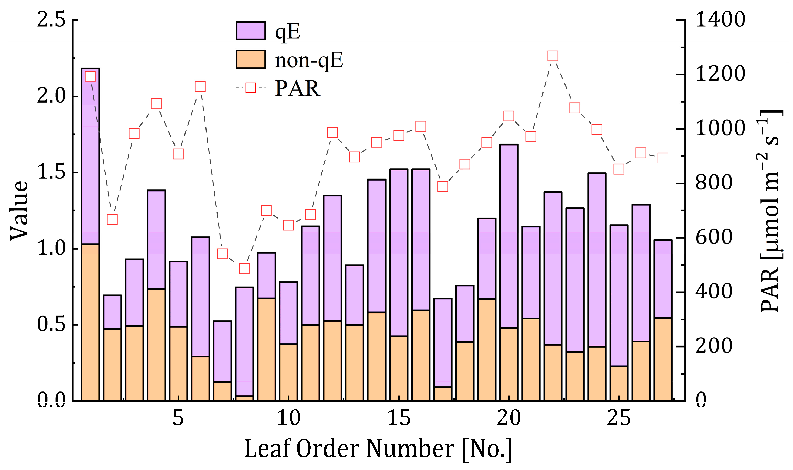
In general, the magnitudes of NPQ increased with PAR, which ranged from ~500 μmol m−2 s−1 to 1300 μmol m−2 s−1. The qE and non-qE components in NPQ both have obvious fluctuations with the change in light intensity (Figure 2). For different leaf groups, the proportions of the qE and non-qE components in NPQ remained relatively constant, with qE ranging from 59% to 62% and non qE ranging from 38% to 41% (Figure 3). The majority of NPQ came from the qE component. However, the contributions of these two components obviously varied under different illumination. The contribution of qE was greater than that of non-qE for all leaves and high-PAR leaves group (Figure 3a,b). For low-PAR leaves (Figure 3c), the contribution of qE to NPQ declined to 0.49, while that of the non-qE component slightly declined to 0.51, indicating that NPQ was contributed by both qE and non-qE components even though there was a greater proportion of the qE component in NPQ. The correlation between the qE and non-qE components became rather high under low-illumination conditions (r = 0.5), possibly suggesting the contribution of qE had shifted from NPQ to non-qE. To sum up, illumination conditions received by leaves is an important factor to determine magnitude and contribution ratio of two main components in NPQ.

3.2. Relationship between NPQ and Leaf Observation Parameters

The correlations between various vegetation parameters and NPQ (including qE and non-qE components) for different illumination conditions were quantitatively analyzed, as shown in Figure 4 and Figure 5. We found that PAR could explain 52% variation in NPQ, highlighting the crucial role of light levels in determining the extent of thermal energy dissipation (Figure 4a). The correlation between PAR and the qE component was higher than the correlation between PAR and the non-qE component (Figure 4b,c). Under high illumination conditions, ΦF showed a strong correlation with NPQ (R2 = 0.75), with PRI displaying moderate correlation (R2 = 0.23), while IRECI and NIRv showed negligible correlations with NPQ (R2 = 0.04 and 0.02, respectively). We found that ΦF accounted for 61% of the variation in the qE component, however its ability to explain the variation in the non-qE components was limited (R2 = 0.13). Although PRI also exhibited a stronger association with the qE component compared to the non-qE component, its performance was not as good as ΦF in explaining qE variability. Under low-illumination conditions, IRECI and NIRv displayed the highest correlation with NPQ (R2 = 0.63 and 0.66, respectively), followed by PRI (R2 = 0.34), while ΦF had the lowest correlation (R2 = 0.24). IRECI and NIRv both showed a rather better relationship with the non-qE component than the qE component, demonstrating that some of the NPQ variability assigned to the non-qE component could be detected by IRECI or NIRv at low light levels. However, PRI was not as effective as IRECI and NIRv in explaining the variability of the non-qE component in NPQ, despite it showing a better relationship with non-qE than qE components. In summary, ΦF exhibits good performance in tracking the qE component variation in NPQ, especially under high light levels. Both IRECI and NIRv demonstrate good performances in tracking the non-qE component variation in NPQ, especially under low light levels. In addition, PRI could partially track the variation in NPQ, but establishing a clear link between PRI and the qE or non-qE components was difficult.
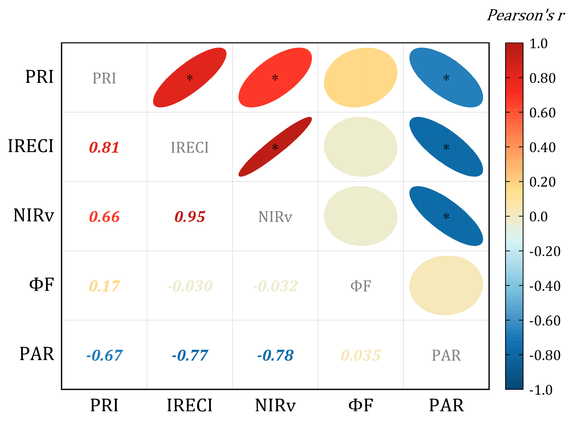
We also quantified the interrelationships between these five leaf observation parameters (Figure 6). NIRv was strongly correlated with IRECI (r = 0.95), indicating these two indices might possess similar biophysical implications despite their utilization of different spectral bands. PAR exhibited strong negative correlations with PRI, IRECI and NIRv. Among these parameters, ΦF demonstrated minimal redundancy with the other parameters (|r| < 0.2).

3.3. Modeling NPQ with Vegetation Reflectance Indices, ΦF and PAR

We built empirical models to estimate NPQ using a linear regression method. The overall accuracy in terms of the coefficient of determination (R2) for 31 NPQ models (Table 2) obtained by a single parameter and combinations of the selected parameters is summarized in Figure 7. The RMSE results are summarized in Figure S2. For leaves exposed to high light levels (Figure 7b), we noticed that NPQ models containing ΦF all had good estimation accuracy (with R2 > 0.74), indicating that ΦF played an important role in estimating NPQ. The addition of other parameters in multi-parameter model did not obviously improve the ability to estimate NPQ. In contrast, NPQ models without ΦF performed poorly with a significantly low level of accuracy (R2 < 0.39). For leaves exposed to relatively low light levels (Figure 7c), ΦF alone, it was difficult to explain the variation in NPQ with R2 = 0.24. We found that the NPQ models that combined ΦF and at least one of IRECI and NIRv all had great performance (R2 > 0.9). For the group containing all leaves (Figure 7a), we found that ΦF alone could not adequately capture NPQ, and its accuracy was not as good as that of one-parameter models built by the other four parameters (including PRI, IRECI, NIRv and PAR, with R2 from 0.44 to 0.52). When integrating ΦF with some parameters (e.g., IRECI, NIRv and PAR), the estimation abilities of those were better than any single-parameter models (R2 > 0.76).
The relative importance of each parameter in model 31 was calculated (Figure 8). Under high-illumination conditions, the NPQ variability was mainly regulated by ΦF, while the remaining parameters exerted minor influence on NPQ. Under low-illumination conditions, the relative contribution of ΦF decreased to 23%; however, NIRv and IRECI became important parameters for governing NPQ. These two parameters together explained over 50% of the variability in NPQ. When considering all leaves, ΦF had the highest relative importance in explaining NPQ variability. It was followed by PAR. PRI had the lowest relative importance in relation to NPQ.

4. Discussion

4.1. Role of Leaf Parameters in Estimating NPQ

Our results indicate that the relative contribution of the qE and non-qE components to NPQ varies with changes in incoming light intensity. Under high-light conditions, the Pearson correlation coefficient between NPQ and qE was 0.80 (Figure 3b), which might be because the formation of qE is closely related to light-driven electron transport [44]. In low-light conditions, the primary driver of NPQ variation shifts away from the qE component. Instead, certain components within the non-qE component gradually gain importance due to their ability to persist for extended periods, even under weak-light conditions [50]. Some studies also found that the contribution of quantum yields of each NPQ components varies with changes in light intensity [16,17]. Therefore, NPQ variation is regulated by both qE and non-qE components under low illumination (Figure 3c). It is worth noting that the estimation of NPQ in this study did not utilize the APAR as a parameter. There are two primary reasons for this: firstly, this study adopted a combined approach of PAR and vegetation index, which effectively accounts for variations in the pigment pool; secondly, the measuring of leaf-level fPAR is challenging to accomplish within a short timeframe.
The relationship between PRI and NPQ is complicated at the leaf scale, where their correlation is influenced not only by PRI observation error but also by non-qE components in NPQ, although several studies have built empirical regression relationships between these two variables [16,17]. Previous studies have classified PRI as “facultative PRI” and “constitutive PRI” according to its timescale response [27,51]. For leaves under high illumination, PRI presented a stronger relationship with the qE component than the non-qE component, which elucidated the finding of Sukhova and Sukhov [52], who showed the connection between ΔPRI (modified formation) and the qE component. The probable explanation is that facultative PRI exhibited a faster response to the variation in the qE component due to the xanthophyll cycle on a short-term scale [27]. For leaves under low illumination, PRI could partly explain non-qE component variation, but could hardly explain qE component variation. The possible reason is that constitutive PRI associated with shifting carotenoid-to-chlorophyll pigment ratios could track the remaining non-qE component well [27,28]. Hence, it is still difficult to establish a clear connection between PRI and its two components. To some extent, estimating NPQ using PRI alone is unreliable.
NIRv utilizes NDVI bands and accounts for NIR band scattering, making it a proxy for APAR due to its ability to reflect the capacity for light harvesting [53]. However, its effectiveness as an indicator of NPQ at leaf scale has not yet been validated. In this study, NIRv exhibited a strong correlation with the non-qE component variation in NPQ under low-illumination conditions (Figure 5g–i). At leaf scale, the reflectance in the near-infrared region is closely linked to their internal cellular structure, and a majority of light photons are scattered with minimal absorption [54]. Therefore, we hypothesized that a potential correlation exists between the NIR band and certain components in non-qE, which may be attributed to the internal leaf scattering effect [55] or the influence of chloroplast movements and distribution [56,57]. This association could result in a strong relationship between NIRv and the non-qE component. Similar to the performance of NIRv, IRECI also exhibited a stronger linear relationship with the non-qE component than the qE component under low-light conditions, while their correlation weakened under high-light conditions. The red-edge bands in IRECI could describe leaf organic ingredients such as pigment pool size and nitrogen content [31,32,58]. It is possible that the size of the pigment pool remained relatively stable throughout the observation period, resulting in no significant change in the red-edge bands in IRECI. Therefore, IRECI is highly correlated with NIRv (r = 0.95), indicating that they convey similar meaning and comparable information despite using different spectral bands (Figure 6). One more thing to be noted was that both IRECI and NIRv values would be compressed to the low-value area with an increase in light intensity, illustrating that low values of these two indices could not distinguish high NPQ. Overall, the utilization of NIRv and IRECI as parameters in NPQ estimation could improve performance in low-light conditions and also simplify the estimation model by reducing reliance on PAR inclusion.
We found that leaf-scale fluorescence quantum yield (ΦF) had a close relationship with NPQ. In this study, ΦF was regarded as the relative ΦF under the assumption that the leaf fPAR remains constant [29,45,59]. There was a strong negative correlation between ΦF and NPQ for the high-PAR group, while ΦF demonstrated only a moderate relationship with NPQ for the low-PAR group and all leaves group (Figure 5j–l). These results also agree with earlier studies, which showed a negative relationship between ΦF and NPQ under high-illumination conditions [20,40]. Under low-illumination conditions or severe stress, the correlation between ΦF and NPQ becomes weak [23,40]. In addition, our results also revealed a stronger correlation between ΦF and the qE component compared to ΦF and the non-qE component for high-PAR leaves. Few studies explicitly identified which component in NPQ was closely related to ΦF. Our findings seem to indirectly demonstrate that ΦF has a strong correlation with variations in the qE component. Under high illumination, NPQ is mainly determined by the qE component. Therefore, ΦF has significant potential for estimating NPQ for leaves with high incoming intensity.

4.2. Performance of Multi-Parameter NPQ Models

We estimated NPQ and its components using simple single-parameter linear regression (Figure 4 and Figure 5), showing that ΦF was better able to explain the variability in the qE component, while IRECI and NIRv were able to explain the variability in the non-qE component. We also noticed that the limitations of single vegetation parameters impeded the precise capture of the dynamics of NPQ. Therefore, our study evaluated multiple-parameter models coupling the complementarity contributions of different kinds of parameters to estimate NPQ, which benefit from the integration of these observation parameters’ advantages. Our results reveal that for high-PAR leaves, the proportion of the qE component in NPQ increased, and ΦF had a strong electronic circulation relationship with qE (Figure 8). Thus, all models that contained ΦF performed well in estimating NPQ (R2 > 0.74) (Figure 7b). Among all selected indices, IRECI and NIRv could better explain the non-qE variation, which has usually been ignored in other studies. For low PAR leaves commonly dominated by qE and non-qE components, IRECI or NIRv could account for over 60% of the variability in NPQ, while ΦF alone explained only 24% of the variation in NPQ. If IRECI and NIRv are combined with ΦF, the accuracy of NPQ estimation has a dramatically improved R2, increasing to 0.9 (Figure 7c). In the analysis of relative importance, we found that IRECI and NIRv were the most influential parameters in reflecting the variation of NPQ under low-illumination conditions (Figure 8). Furthermore, the vegetation canopy comprises sunlit and shaded leaves in actual observations by remote sensing methods. Generally, sunlit leaves receive more direct radiation: shaded leaves receive diffuse radiation and have low incoming intensity. Thus, we mixed all the leaves, making the situation closer to reality. We found that coupling ΦF with one or more of the measurement parameters (e.g., IRECI, NIRv and PAR), the NPQ models displayed better estimation ability (R2 > 0.76) than using each parameter alone (R2 < 0.5) (Figure 7a). The results of the relative importance analysis revealed that the contribution of PAR was second only to ΦF in explaining the variability in NPQ (Figure 8). This finding suggests that the light level to which the leaf is exposed is a key parameter for estimating NPQ. As NPQ represents the process by which absorbed light energy is dissipated as heat [15], it is logical that the light level plays a significant role in determining the extent of this process. Therefore, accurate estimation of NPQ requires considering the illumination conditions in which the leaf is situated. In summary, our findings suggest that the multiparameter NPQ models, which incorporate ΦF along with at least one of the parameters (PAR, IRECI, and NIRv), offer an accurate alternative for estimating NPQ.
In this study, we have chosen to focus primarily on leaf-scale experiments due to the convenience of NPQ measurements at this level and the challenges associated with obtaining accurate NPQ data at the canopy scale. Our finding provides valuable insights into the relationship between NPQ and remote sensing parameters, particularly by considering the two main components of NPQ (qE and non-qE components). We propose that the integration of multiple remote sensing indices can enhance the precision of leaf-level NPQ estimation. Our study provides a comprehensive understanding of the factors that influence NPQ dynamics, enabling more precise and meaningful interpretations of remote sensing signals in relation to NPQ. This lays the foundation for larger-scale estimation of NPQ.
There are still some limitations in this study, and future work needs to address them. Firstly, the amount of leaf samples in our dataset is relatively small. Subsequent studies necessitate the collection of additional samples from various types of vegetation to ascertain the generalizability of the conclusions. With a sufficient number of samples available, advanced machine learning (ML) algorithms can be employed to effectively handle complex relationships between NPQ and develop a more refined NPQ model. Secondly, another limitation of this study is the limited coverage of environmental and stress conditions. Therefore, it is necessary to conduct leaf-level NPQ experiments on different crop types (such as wheat and maize) throughout a wider developmental period that includes stress treatments in the subsequent research. Finally, there are many applications for NPQ estimation at regional and global scales. For instance, one prominent possibility is that utilizing large-scale NPQ remote sensing to monitor vegetation health could be the most direct and effective when plants are exposed to environmental stress (e.g., water stress, heat stress). Accurate quantification of NPQ could also potentially enhance the estimation of gross primary productivity (GPP). If applying our method to remote sensing estimation at the canopy level on even larger scales, it would be important to address the following potential issues. (1) Variation in canopy structure affects the ability to estimate NPQ from satellite or airborne remote sensing data. Canopy structural effects on scaling up NPQ-VIs from leaf to canopy level need to be quantified and mitigated. (2) According to our findings, ΦF plays a key role in estimating NPQ, but accurately retrieving SIF from satellite data is challenging. We argue that the utilization of high-performance sensors, effective SIF retrieval approaches and consideration of actual atmospheric conditions are critical factors to ensure obtaining reliable SIF signals. (3) When upscaling NPQ estimation from leaf to canopy scale, it is inevitable to face certain common challenges in remote sensing, such as directional effects and soil background effects. The systematic and quantitative assessment of these effects on estimating NPQ is needed in future studies.

5. Conclusions

To date, remote sensing approaches to estimating NPQ, a key indicator for detecting vegetation stress status, are still lacking. In this study, based on the spectra and PAM fluorescence parameters of rice leaves, we investigated the feasibility of estimating NPQ using photosynthesis-related parameters. These parameters could be observed by remote sensing technology, including PRI, IRECI, NIRv, fluorescence quantum yield (ΦF) and PAR. Our results identified that ΦF could capture qE variations well. IRECI and NIRv could capture non-qE variation well. NPQ was predominantly regulated by qE components under high light intensity, and it was jointly regulated by qE and non-qE components under low light intensity. Hence, under high-illumination conditions, ΦF had excellent performance in estimating NPQ. Under low-illumination conditions, coupling IRECI or NIRv with ΦF would obviously improve the estimation of NPQ. For all leaves, our results show that coupling ΦF with at least one of IRECI, NIRv and PAR had a more accurate estimation of NPQ than using them alone. In summary, this study presents a promising approach for estimating NPQ by combining multiple parameters, including chlorophyll fluorescence quantum yield (ΦF), IRECI, NIRv and PAR. These findings provide valuable support for the development of remote sensing methods aimed at NPQ estimation at canopy and larger scales.

Supplementary Materials

The following supporting information can be downloaded at https://www.mdpi.com/article/10.3390/rs15174222/s1. Figure S1: Linear correlation between P A R P A M 2500 measured by PAM-2500 and P A R M L 020 P measured by the ML-020P quantum sensor interpolated by the sensor measurement time. The correction formulas are P A R = 2.75 · P A R P A M 2500 + 133.81 and P A R = 3.75 · P A R P A M 2500 + 318.12 when P A R P A M 2500 is lower than 310 and higher than 310, respectively; Figure S2: NPQ model fitting accuracy in terms of RMSE for all leaves (a), high-PAR leaves (b) and low-PAR leaves (c) by means of 31 combinations. The parameter(s) utilized in each combination are represented by color blocks (yellow, PRI; purple, IRECI; blue, NIRv; orange, PAR; green, ΦF).

Author Contributions

Conceptualization, H.J. and Z.L.; methodology, H.J.; formal analysis, Z.L.; investigation, H.J. and Z.L.; resources, H.J., J.W. and R.Z.; writing—original draft preparation, H.J.; writing—review and editing, H.J., Z.L. and P.Y.; visualization, H.J.; supervision, Z.L.; project administration, Z.L.; funding acquisition, Z.L., X.Z., P.Y. and P.Z. All authors have read and agreed to the published version of the manuscript.

Funding

This research was funded by the Natural Science Foundation of China (Grant number. 42071402), the Water Resources Science and Technology Project of Jiangxi Province, China (202023ZDKT10, KT201706) and the Open Fund of the State Key Laboratory of Remote Sensing Science (grant number. OFSLRSS202228).

Data Availability Statement

The data presented in this study are available on request from the corresponding author. The data are not publicly available due to privacy.

Acknowledgments

We thank the anonymous reviewers for their constructive comments. We are also grateful to the Natural Science Foundation of China, the Water Resources Science and Technology Project of Jiangxi Province, China and the Open Fund of the State Key Laboratory of Remote Sensing Science for their support.

Conflicts of Interest

The authors declare no conflict of interest.

References

  1. Farquhar, G.D.; von Caemmerer, S.V.; Berry, J.A. A biochemical model of photosynthetic CO2 assimilation in leaves of C3 species. Planta 1980, 149, 78–90. [Google Scholar] [CrossRef]
  2. Barber, J. Photosynthetic energy conversion: Natural and artificial. Chem. Soc. Rev. 2009, 38, 185–196. [Google Scholar] [CrossRef] [PubMed]
  3. Bambach, N.; Gilbert, M.E. A dynamic model of RuBP-regeneration limited photosynthesis accounting for photoinhibition, heat and water stress. Agric. For. Meteorol. 2020, 285, 107911. [Google Scholar] [CrossRef]
  4. Powles, S.B. Photoinhibition of photosynthesis induced by visible light. Annu. Rev. Plant Physiol. 1984, 35, 15–44. [Google Scholar] [CrossRef]
  5. Yamori, W.; Makino, A.; Shikanai, T. A physiological role of cyclic electron transport around photosystem I in sustaining photosynthesis under fluctuating light in rice. Sci Rep. 2016, 6, 20147. [Google Scholar] [CrossRef]
  6. Horton, P.; Ruban, A.V.; Walters, R.G. Regulation of light harvesting in green plants. Annu. Rev. Plant Physiol. Plant Mol. Biol. 1996, 47, 655–684. [Google Scholar] [CrossRef]
  7. Demmig-Adams, B.; Koh, S.; Cohu, C.M.; Muller, O.; Stewart, J.J.; Adams, W.W. Non-Photochemical Fluorescence Quenching in Contrasting Plant Species and Environments. In Non-Photochemical Quenching and Energy Dissipation in Plants, Algae and Cyanobacteria; Demmig-Adams, B., Garab, G., Adams, W., III, Govindjee, Eds.; Springer: Dordrecht, The Netherlands, 2014; pp. 531–552. [Google Scholar]
  8. Murchie, E.H.; Ruban, A.V. Dynamic non-photochemical quenching in plants: From molecular mechanism to productivity. Plant J. 2020, 101, 885–896. [Google Scholar] [CrossRef]
  9. Nicol, L.; Nawrocki, W.J.; Croce, R. Disentangling the sites of non-photochemical quenching in vascular plants. Nat. Plants 2019, 5, 1177–1183. [Google Scholar] [CrossRef]
  10. Wraight, C.A.; Crofts, A.R. Energy-Dependent Quenching of Chlorophyll a Fluorescence in Isolated Chloroplasts. Eur. J. Biochem. 1970, 17, 319–327. [Google Scholar] [CrossRef]
  11. Allen, J.F.; Bennett, J.; Steinback, K.E.; Arntzen, C.J. Chloroplast protein phosphorylation couples plastoquinone redox state to distribution of excitation energy between photosystems. Nature 1981, 291, 25–29. [Google Scholar] [CrossRef]
  12. Krause, G.H.; Weis, E. Chlorophyll fluorescence and photosynthesis: The basics. Annu. Rev. Plant Biol. 1991, 42, 313–349. [Google Scholar] [CrossRef]
  13. Nilkens, M.; Kress, E.; Lambrev, P.; Miloslavina, Y.; Müller, M.; Holzwarth, A.R.; Jahns, P. Identification of a slowly inducible zeaxanthin-dependent component of non-photochemical quenching of chlorophyll fluorescence generated under steady-state conditions in Arabidopsis. Biochim. Et Biophys. Acta (BBA)-Bioenerg. 2010, 1797, 466–475. [Google Scholar] [CrossRef] [PubMed]
  14. Porcar-Castell, A. A high-resolution portrait of the annual dynamics of photochemical and non-photochemical quenching in needles of Pinus sylvestris. Physiol. Plant. 2011, 143, 139–153. [Google Scholar] [CrossRef] [PubMed]
  15. Ruban, A.V. Nonphotochemical Chlorophyll Fluorescence Quenching: Mechanism and Effectiveness in Protecting Plants from Photodamage. Plant Physiol. 2016, 170, 1903–1916. [Google Scholar] [CrossRef]
  16. Ahn, T.K.; Avenson, T.J.; Peers, G.; Li, Z.; Dall Osto, L.; Bassi, R.; Niyogi, K.K.; Fleming, G.R. Investigating energy partitioning during photosynthesis using an expanded quantum yield convention. Chem. Phys. 2009, 357, 151–158. [Google Scholar] [CrossRef]
  17. Guadagno, C.R.; Virzo De Santo, A.; D’Ambrosio, N. A revised energy partitioning approach to assess the yields of non-photochemical quenching components. Biochim. Et Biophys. Acta (BBA)-Bioenerg. 2010, 1797, 525–530. [Google Scholar] [CrossRef] [PubMed]
  18. Schreiber, U.; Schliwa, U. A solid-state, portable instrument for measurement of chlorophyll luminescence induction in plants. Photosynth. Res. 1987, 11, 173–182. [Google Scholar] [CrossRef] [PubMed]
  19. White, S.; Anandraj, A.; Bux, F. PAM fluorometry as a tool to assess microalgal nutrient stress and monitor cellular neutral lipids. Bioresour. Technol. 2011, 102, 1675–1682. [Google Scholar] [CrossRef]
  20. Alonso, L.; Van Wittenberghe, S.; Amorós-López, J.; Vila-Francés, J.; Gómez-Chova, L.; Moreno, J. Diurnal cycle relationships between passive fluorescence, PRI and NPQ of vegetation in a controlled stress experiment. Remote Sens. 2017, 9, 770. [Google Scholar] [CrossRef]
  21. Flexas, J.; Escalona, J.M.; Evain, S.; Gulías, J.; Moya, I.; Osmond, C.B.; Medrano, H. Steady-state chlorophyll fluorescence (Fs) measurements as a tool to follow variations of net CO2 assimilation and stomatal conductance during water-stress in C3 plants. Physiol. Plant. 2002, 114, 231–240. [Google Scholar] [CrossRef]
  22. Garbulsky, M.F.; Peñuelas, J.; Gamon, J.; Inoue, Y.; Filella, I. The photochemical reflectance index (PRI) and the remote sensing of leaf, canopy and ecosystem radiation use efficiencies: A review and meta-analysis. Remote Sens. Environ. 2011, 115, 281–297. [Google Scholar] [CrossRef]
  23. Porcar-Castell, A.; Tyystjärvi, E.; Atherton, J.; Tol, C.; Flexas, J.; Pfündel, E.; Moreno, J.; Frankenberg, C.; Berry, J. Linking chlorophyll a fluorescence to photosynthesis for remote sensing applications: Mechanisms and challenges. J. Exp. Bot. 2014, 65, 4065–4095. [Google Scholar] [CrossRef] [PubMed]
  24. Chou, S.; Chen, J.M.; Yu, H.; Chen, B.; Zhang, X.; Croft, H.; Khalid, S.; Li, M.; Shi, Q. Canopy-Level Photochemical Reflectance Index from Hyperspectral Remote Sensing and Leaf-Level Non-Photochemical Quenching as Early Indicators of Water Stress in Maize. Remote Sens. 2017, 9, 794. [Google Scholar] [CrossRef]
  25. D’Odorico, P.; Schönbeck, L.; Vitali, V.; Meusburger, K.; Schaub, M.; Ginzler, C.; Zweifel, R.; Velasco, V.M.E.; Gisler, J.; Gessler, A.; et al. Drone-based physiological index reveals long-term acclimation and drought stress responses in trees. Plant Cell Environ. 2021, 44, 3552–3570. [Google Scholar] [CrossRef] [PubMed]
  26. Wang, X.; Chen, J.M.; Ju, W. Photochemical reflectance index (PRI) can be used to improve the relationship between gross primary productivity (GPP) and sun-induced chlorophyll fluorescence (SIF). Remote Sens. Environ. 2020, 246, 111888. [Google Scholar] [CrossRef]
  27. Wong, C.Y.S.; Gamon, J.A. Three causes of variation in the photochemical reflectance index (PRI) in evergreen conifers. New Phytol. 2015, 206, 187–195. [Google Scholar] [CrossRef]
  28. Gitelson, A.A.; Gamon, J.A.; Solovchenko, A. Multiple drivers of seasonal change in PRI: Implications for photosynthesis 1. Leaf level. Remote Sens. Environ. 2017, 191, 110–116. [Google Scholar] [CrossRef]
  29. Yang, P. Exploring the interrelated effects of soil background, canopy structure and sun-observer geometry on canopy photochemical reflectance index. Remote Sens. Environ. 2022, 279, 113133. [Google Scholar] [CrossRef]
  30. Badgley, G.; Field, C.B.; Berry, J.A. Canopy near-infrared reflectance and terrestrial photosynthesis. Sci. Adv. 2017, 3, e1602244. [Google Scholar] [CrossRef]
  31. Chemura, A.; Mutanga, O.; Odindi, J.; Kutywayo, D. Mapping spatial variability of foliar nitrogen in coffee (Coffea arabica L.) plantations with multispectral Sentinel-2 MSI data. Isprs-J. Photogramm. Remote Sens. 2018, 138, 1–11. [Google Scholar] [CrossRef]
  32. Frampton, W.J.; Dash, J.; Watmough, G.; Milton, E.J. Evaluating the capabilities of Sentinel-2 for quantitative estimation of biophysical variables in vegetation. Isprs-J. Photogramm. Remote Sens. 2013, 82, 83–92. [Google Scholar] [CrossRef]
  33. Zeng, Y.; Chen, M.; Hao, D.; Damm, A.; Badgley, G.; Rascher, U.; Johnson, J.E.; Dechant, B.; Siegmann, B.; Ryu, Y.; et al. Combining near-infrared radiance of vegetation and fluorescence spectroscopy to detect effects of abiotic changes and stresses. Remote Sens. Environ. 2022, 270, 112856. [Google Scholar] [CrossRef]
  34. Maleki, M.; Arriga, N.; Roland, M.; Wieneke, S.; Barrios, J.M.; Van Hoolst, R.; Peñuelas, J.; Janssens, I.A.; Balzarolo, M. Soil water depletion induces discrepancies between in situ measured vegetation indices and photosynthesis in a temperate heathland. Agric. For. Meteorol. 2022, 324, 109110. [Google Scholar] [CrossRef]
  35. Zeng, Y.; Badgley, G.; Dechant, B.; Ryu, Y.; Chen, M.; Berry, J.A. A practical approach for estimating the escape ratio of near-infrared solar-induced chlorophyll fluorescence. Remote Sens. Environ. 2019, 232, 111209. [Google Scholar] [CrossRef]
  36. Badgley, G.; Anderegg, L.D.L.; Berry, J.A.; Field, C.B. Terrestrial gross primary production: Using NIRV to scale from site to globe. Glob. Change Biol. 2019, 25, 3731–3740. [Google Scholar] [CrossRef]
  37. Atherton, J.; Nichol, C.J.; Porcar-Castell, A. Using spectral chlorophyll fluorescence and the photochemical reflectance index to predict physiological dynamics. Remote Sens. Environ. 2016, 176, 17–30. [Google Scholar] [CrossRef]
  38. Mohammed, G.H.; Colombo, R.; Middleton, E.M.; Rascher, U.; van der Tol, C.; Nedbal, L.; Goulas, Y.; Pérez-Priego, O.; Damm, A.; Meroni, M.; et al. Remote sensing of solar-induced chlorophyll fluorescence (SIF) in vegetation: 50 years of progress. Remote Sens. Environ. 2019, 231, 111177. [Google Scholar] [CrossRef]
  39. Maguire, A.J.; Eitel, J.U.H.; Griffin, K.L.; Magney, T.S.; Long, R.A.; Vierling, L.A.; Schmiege, S.C.; Jennewein, J.S.; Weygint, W.A.; Boelman, N.T.; et al. On the Functional Relationship Between Fluorescence and Photochemical Yields in Complex Evergreen Needleleaf Canopies. Geophys. Res. Lett. 2020, 47, e2020G–e87858G. [Google Scholar] [CrossRef]
  40. Magney, T.S.; Barnes, M.L.; Yang, X. On the Covariation of Chlorophyll Fluorescence and Photosynthesis Across Scales. Geophys. Res. Lett. 2020, 47, e2020G–e91098G. [Google Scholar] [CrossRef]
  41. Liu, B.; Cui, Y.; Tan, J.; Luo, Y.; Xiang, Z. CO2 fluxes over two paddy fields with different management practices in Southern China. Agric. For. Meteorol. 2021, 310, 108650. [Google Scholar] [CrossRef]
  42. Xu, S.; Atherton, J.; Riikonen, A.; Zhang, C.; Oivukkamäki, J.; MacArthur, A.; Honkavaara, E.; Hakala, T.; Koivumäki, N.; Liu, Z.; et al. Structural and photosynthetic dynamics mediate the response of SIF to water stress in a potato crop. Remote Sens. Environ. 2021, 263, 112555. [Google Scholar] [CrossRef]
  43. Genty, B.; Briantais, J.; Baker, N.R. The relationship between the quantum yield of photosynthetic electron transport and quenching of chlorophyll fluorescence. Biochim. Et Biophys. Acta (BBA)-Gen. Subj. 1989, 990, 87–92. [Google Scholar] [CrossRef]
  44. Mueller-moule, P.; Li, X.; Niyogi, K. Non-Photochemical Quenching. A Response to Excess Light Energy. Plant Physiol. 2001, 125, 1558–1566. [Google Scholar] [CrossRef] [PubMed]
  45. Baker, N.R. Chlorophyll Fluorescence: A Probe of Photosynthesis In Vivo. Annu. Rev. Plant Biol. 2008, 59, 89–113. [Google Scholar] [CrossRef] [PubMed]
  46. Yang, P.; van der Tol, C.; Campbell, P.K.E.; Middleton, E.M. Fluorescence Correction Vegetation Index (FCVI): A physically based reflectance index to separate physiological and non-physiological information in far-red sun-induced chlorophyll fluorescence. Remote Sens. Environ. 2020, 240, 111676. [Google Scholar] [CrossRef]
  47. Cendrero-Mateo, M.P.; Moran, M.S.; Papuga, S.A.; Thorp, K.R.; Alonso, L.; Moreno, J.; Ponce-Campos, G.; Rascher, U.; Wang, G. Plant chlorophyll fluorescence: Active and passive measurements at canopy and leaf scales with different nitrogen treatments. J. Exp. Bot. 2016, 67, 275–286. [Google Scholar] [CrossRef]
  48. Gamon, J.A.; Penuelas, J.; Field, C.B. A narrow-waveband spectral index that tracks diurnal changes in photosynthetic efficiency. Remote Sens. Environ. 1992, 41, 35–44. [Google Scholar] [CrossRef]
  49. Tucker, C.J. Red and photographic infrared linear combinations for monitoring vegetation. Remote Sens. Environ. 1979, 8, 127–150. [Google Scholar] [CrossRef]
  50. Ruban, A.V.; Johnson, M.P.; Duffy, C.D. The photoprotective molecular switch in the photosystem II antenna. Biochim. Et Biophys. Acta (BBA)-Bioenerg. 2012, 1817, 167–181. [Google Scholar] [CrossRef]
  51. Gamon, J.A.; Berry, J.A. Facultative and constitutive pigment effects on the Photochemical Reflectance Index (PRI) in sun and shade conifer needles. Isr. J. Plant Sci. 2012, 60, 85–95. [Google Scholar] [CrossRef]
  52. Sukhova, E.; Sukhov, V. Analysis of Light-Induced Changes in the Photochemical Reflectance Index (PRI) in Leaves of Pea, Wheat, and Pumpkin Using Pulses of Green-Yellow Measuring Light. Remote Sens. 2019, 11, 810. [Google Scholar] [CrossRef]
  53. Wong, C.Y.; D Odorico, P.; Arain, M.A.; Ensminger, I. Tracking the phenology of photosynthesis using carotenoid-sensitive and near-infrared reflectance vegetation indices in a temperate evergreen and mixed deciduous forest. New Phytol. 2020, 226, 1682–1695. [Google Scholar] [CrossRef] [PubMed]
  54. Knipling, E.B. Physical and physiological basis for the reflectance of visible and near-infrared radiation from vegetation. Remote Sens. Environ. 1970, 1, 155–159. [Google Scholar] [CrossRef]
  55. Ayu Purnamasari, R.; Noguchi, R.; Ahamed, T. Land suitability assessments for yield prediction of cassava using geospatial fuzzy expert systems and remote sensing. Comput. Electron. Agric. 2019, 166, 105018. [Google Scholar] [CrossRef]
  56. Nauš, J.; Prokopová, J.; Řebíček, J.; Špundová, M. SPAD chlorophyll meter reading can be pronouncedly affected by chloroplast movement. Photosynth. Res. 2010, 105, 265–271. [Google Scholar] [CrossRef] [PubMed]
  57. Peguero-Pina, J.J.; Pelegrín, E.; Morales, F. Three pools of zeaxanthin in Quercus coccifera leaves during light transitions with different roles in rapidly reversible photoprotective energy dissipation and photoprotection. J. Exp. Bot. 2013, 64, 1649–1661. [Google Scholar] [CrossRef]
  58. Castillo, J.A.A.; Apan, A.A.; Maraseni, T.N.; Salmo, S.G. Estimation and mapping of above-ground biomass of mangrove forests and their replacement land uses in the Philippines using Sentinel imagery. Isprs-J. Photogramm. Remote Sens. 2017, 134, 70–85. [Google Scholar] [CrossRef]
  59. Schreiber, U. Chlorophyll fluorescence: New Instruments for Special Applications. In Photosynthesis: Mechanisms and Effects: Volume I–V: Proceedings of the XIth International Congress on Photosynthesis, Budapest, Hungary, 17–22 August 1998; Garab, G., Ed.; Springer: Dordrecht, The Netherlands, 1998; pp. 4253–4258. [Google Scholar]
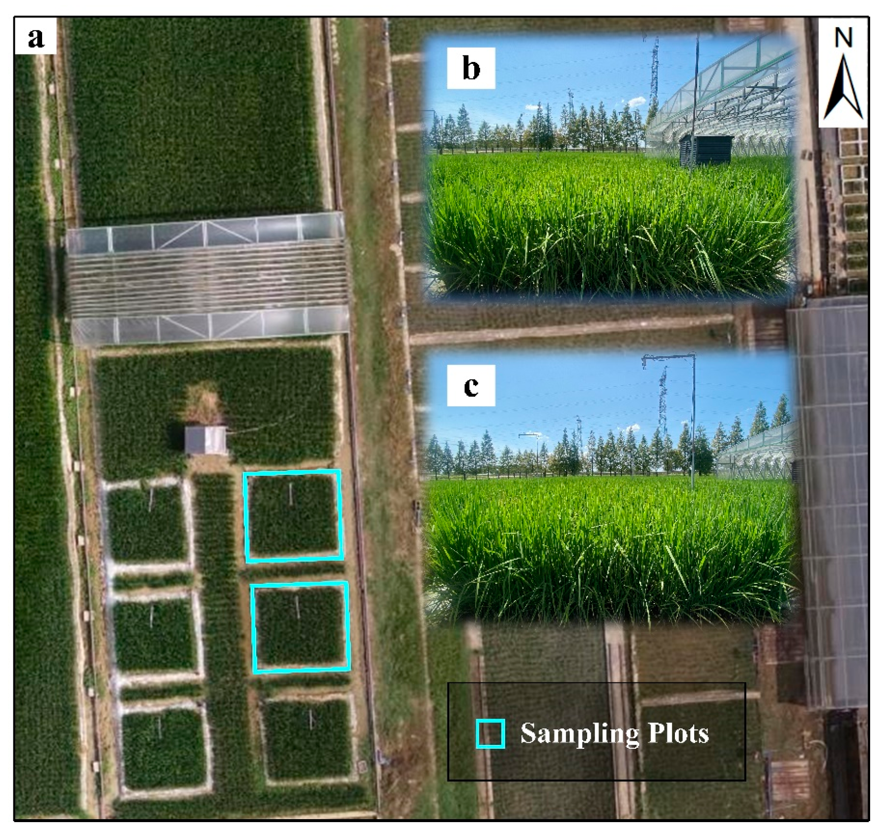
Figure 1. (a) Overview of the plots. The background is an unmanned aerial vehicle (UAV) image (DJI Phantom 4 Da-Jiang Innovations Science and Technology Co., Ltd., Shenzhen., China) acquired on 2 August 2022 at 10:28 CST. (b,c) Photographs of two sampling plots.
Remotesensing 15 04222 g001
Figure 2. The energy-dependent quenching (qE), non-energy-dependent quenching (non-qE) and PAR of different leaves.
Remotesensing 15 04222 g002
Figure 3. r (indicated in black text) between NPQ, qE and non-qE, and proportion of qE and non-qE in NPQ (indicated in blue text) for all leaves (a), high-PAR leaves (>900 μmol m−2 s−1, (b)) and low-PAR leaves (<900 μmol m−2 s−1, (c)).
Remotesensing 15 04222 g003
Figure 4. Relationship between PAR and NPQ (a), qE (b), and non-qE (c). Dotted black lines represent the linear regression lines. “****” denotes significant differences based on a standard ANOVA test with p < 0.0001.
Remotesensing 15 04222 g004
Figure 5. Relationship between different parameters (PRI, IRECI, NIRv and ΦF) and NPQ, qE, and non-qE. Dotted purple, red and black lines represent the linear regression lines for low-PAR leaves, high-PAR leaves and all leaves, respectively, and purple circles and red triangles represent low-PAR leaves and high-PAR leaves, respectively. Results for the relationship between NPQ and PRI (a), IRECI (d), NIRv (g) and ΦF (j). Results for the relationship between qE and PRI (b), IRECI (e), NIRv (h) and ΦF (k). Results for the relationship between non-qE and PRI (c), IRECI (f), NIRv (i) and ΦF (l). “****,” “***,” “**” and “*” denote significant differences based on a standard ANOVA test with p < 0.0001, p < 0.001, p < 0.01, p < 0.05, respectively. “ns” denotes not significant.
Remotesensing 15 04222 g005
Figure 6. The pairwise correlations between the 5 observation parameters at the leaf scale. Correlations were determined using the Pearson correlation coefficient. “*” denotes a significant level with p < 0.05.
Remotesensing 15 04222 g006
Figure 7. NPQ model fitting accuracy in terms of R2 for all leaves (a), high-PAR leaves (b) and low-PAR leaves (c) by means of 31 combinations. The parameter(s) utilized in each combination are represented by color blocks (yellow, PRI; purple, IRECI; blue, NIRv; orange, PAR; green, ΦF).
Remotesensing 15 04222 g007
Figure 8. The relative importance of each parameter (yellow, PRI; purple, IRECI; blue, NIRv; orange, PAR; green, ΦF) in Model 31.
Remotesensing 15 04222 g008
Table 1. Chlorophyll fluorescence parameters measured with the PAM-2500.
AbbreviationDescription
F t Instantaneous steady-state fluorescence measured under ambient light at any point in time.
F m Maximum fluorescence measured upon a saturating light pulse after adequate dark adaptation. Usually, at 23:00 local time in this study.
F m Maximum fluorescence measured upon a saturating light pulse under ambient light at any point in time.
F E Maximum fluorescence measured upon a saturating light pulse after 10 min dark adaptation.
Table 2. Combinations of leaf measurement parameters in the NPQ model.
IDNumber of ParametersParameter(s) Used in the Model
11PRI
2IRECI
3NIRv
4PAR
5ΦF
62IRECI, NIRv
7PRI, IRECI
8PRI, NIRv
9PAR, PRI
10PAR, IRECI
11PAR, NIRv
12ΦF, PRI
13ΦF, IRECI
14ΦF, NIRv
15ΦF, PAR
163PRI, IRECI, NIRv
17PAR, PRI, IRECI
18PAR, PRI, NIRv
19PAR, IRECI, NIRv
20ΦF, IRECI, NIRv
21ΦF, PRI, IRECI
22ΦF, PRI, NIRv
23ΦF, PAR, PRI
24ΦF, PAR, IRECI
25ΦF, PAR, NIRv
264PAR, PRI, IRECI, NIRv
27ΦF, PRI, IRECI, NIRv
28ΦF, PAR, IRECI, NIRv
29ΦF, PAR, PRI, IRECI
30ΦF, PAR, PRI, NIRv
315ΦF, PAR, PRI, IRECI, NIRv
Disclaimer/Publisher’s Note: The statements, opinions and data contained in all publications are solely those of the individual author(s) and contributor(s) and not of MDPI and/or the editor(s). MDPI and/or the editor(s) disclaim responsibility for any injury to people or property resulting from any ideas, methods, instructions or products referred to in the content.