Article

Quercetin Intervention Alleviates Offspring’s Oxidative Stress, Inflammation, and Tight Junction Damage in the Colon Induced by Maternal Fine Particulate Matter (PM2.5) Exposure through the Reduction of Bacteroides

1
Department of Nutrition and Food Hygiene, School of Public Health, Peking University, Beijing 100083, China
2
Beijing Key Laboratory of Toxicological Research and Risk Assessment for Food Safety, Peking University, Beijing 100083, China
*
Author to whom correspondence should be addressed.
Nutrients 2020, 12(10), 3095; https://doi.org/10.3390/nu12103095
Submission received: 2 September 2020 / Revised: 24 September 2020 / Accepted: 30 September 2020 / Published: 11 October 2020
(This article belongs to the Section Nutritional Epidemiology)

Abstract

:
The influences of maternal fine particulate matter (PM2.5) exposure on intestinal oxidative stress, inflammation, tight junctions, and gut microbiota of offspring are not well understood. Moreover, research on the dietary intervention method has not been well studied. In our study, dams received PM2.5 and quercetin intervention during gestation and lactation, and then inflammation biomarkers, oxidative stress indicators, tight junction proteins, and gut microbiota in the colon of offspring were analyzed. Compared with the control group, lower catalase (CAT) and superoxide dismutase (SOD) activities, higher interleukin-17A (IL-17A) and interleukin-22 (IL-22), decreased ZO-1 and occludin expressions, and higher Bacteroides abundance were observed in the offspring mice of the PM2.5 group. However, higher CAT and SOD activities, lower IL-17A and IL-22 levels, increased ZO-1 and occludin expressions, and lower Bacteroides abundance were found in the quercetin groups. In addition, there was a negative correlation between Bacteroides abundance and CAT concentration. Additionally, Bacteroides abundance was positively related to IL-17A and IL-22 levels. These findings suggest that maternal PM2.5 exposure may have some certain effects on intestinal oxidative stress, inflammation, and tight junctions. Quercetin administration may protect the offspring against these adverse effects. Changes of Bacteroides abundance play an important role in the process.

1. Introduction

Epidemiological studies have reported that air pollution contributes to gastrointestinal diseases, such as inflammatory bowel disease [1,2]. These intestinal diseases may be related to the injury of the intestinal mucosal barrier, including oxidative stress, increased inflammation, and damaged tight junctions [3,4,5]. Fine particulate matter (PM2.5) could induce oxidative damage in tissue [6]. Oxidation stress affects the activity of antioxidant enzymes and the permeability and proliferation of epithelial cells; thus, oxidation stress is related to the barrier function of the gut. Vitro and vivo studies show PM2.5 has the ability to induce inflammation responses [7,8]. Occludin and ZO-1 are tight junction proteins, which help the cells of the intestinal epithelium adhere to each other and regulate permeability [9,10,11]. Bernard et al. reported that ZO-1 and occludin could be decreased by PM2.5 intratracheal instillation [12]. Mutlu et al. also found that particulate matter could decrease the epithelial barrier, which might be associated with rearrangement of the epithelial tight junction proteins [13].
Gut microbiota is involved in the maintenance of gut barrier function [14]. A growing body of evidence indicates that gut microbiota dysbiosis is implicated in defects in the gut barrier [15]. The environment has been identified as an important factor to influence gut microbiota. One study demonstrated that gut microbial diversity in the colon can be increased by PM2.5 exposure [16]. It is unknown whether the gut microbiota is related to oxidative stress, inflammation, and tight junctions induced by maternal PM2.5 exposure.
Quercetin, the most common flavonoid in food, is found in a number of plants such as apples, tea, strawberries, and onions [17,18]. According to clinical studies, the pharmacological effects of quercetin on inflammation [19] and oxidative stress have been confirmed. Moreover, quercetin can be transformed into other metabolites with the help of some microbes [20]. These metabolites have a number of biological activities, such as counteracting inflammation and oxidative stress [21,22].
Up to now, few studies have investigated the influences on intestinal oxidative stress, inflammation, tight junctions, and gut microbiota of offspring as their mothers were exposed to PM2.5 during the stages of pregnancy and lactation; moreover, there are no nutritional intervention methods. To explore these effects, dams were given PM2.5 and quercetin, and biomarkers of colon oxidative indicators, inflammation, tight junction proteins, and gut microbiota in the colon of offspring were analyzed.

2. Materials and Methods

2.1. PM2.5 and Quercetin

The dosages of PM2.5 and quercetin were determined in our previous research [23,24,25]. The PM2.5 sample was prepared by several processes: collecting, agitating, filtering, and lyophilizing. A detailed description can be seen in our previous articles [23,24]. Phosphate-buffered saline was used as the solvent to dissolve the PM2.5 powder. The control sample from unexposed filters was disposed equally. A solution of 0.15% carboxymethyl cellulose sodium was used to dissolve the quercetin (Sigma-Aldrich, Shanghai, China).

2.2. Animals and Treatment

Specific pathogen-free 8-week-old ICR mice (Beijing, China, SCXK2016-0010) were used in this study. After being quarantined for 7 days, female mice and male mice were put in the same cage overnight. If the vaginal plug was found the next morning, the day was defined as the gestational day (GD) 0. The day of natural production was defined as postnatal day (PND) 0. Every dam was caged alone. Forty dams were randomly divided into 5 groups, including the normal control (NC) group, the PM2.5 group (PM2.5 group), and 3 quercetin intervention groups (50, 100, and 200 mg/kg), with 8 dams in each subgroup. On PND 3, we kept 8 offspring in each litter, with 4 male mice and 4 female mice. On GD 3, 6, 9, 12, and 15, and PND 2, 5, 8, 11, 14, and 17, the dams were received intratracheal instillation under 3% isoflurane anesthesia. As shown in Table 1, the dams in the PM2.5 group and 3 quercetin intervention groups were exposed to 15 mg/kg PM2.5. The dams in the NC group received the suspension from extracts of a “blank” filter. The dams in the NC and PM2.5 groups were daily given 0.15% carboxymethyl cellulose sodium, while dams in the quercetin groups were daily given different doses of quercetin by gavage. Gavage was conducted every day from GD 0 to PND 21.
On PND 3, 10, 21, and 35, one male and one female offspring born to the same mother were sacrificed. The use of mice was in compliance with the Peking University Guidelines for Animal Research (LA2016284). Figure 1 shows our study design.

2.3. Enzyme-Linked Immunosorbent Assays (ELISA) for Interleukin-17A (IL-17A) and Interleukin-22 (IL-22) in the Colon

Colonic tissue samples were collected on PND 3, 10, 21, and 35. As shown in Figure 1, IL-17A and IL-22 in the colon were measured using ELISA kits according to the kit instructions (Capitalbio, Beijing, China).

2.4. Biochemical Analysis for Catalase (CAT) and Superoxide Dismutase (SOD) in the Colon

Colorimetric assay kits (Nanjing Jiancheng, Jiangsu, China) were used to assay the activities of CAT and SOD in the colon (Figure 1). We conducted the process according to the kit instructions.

2.5. ZO-1 and Occludin in the Colon

ZO-1 and occludin in the colon were assessed by a Western blot. RIPA buffer (Thermal Scientific, USA) was used to homogenize the colon tissue. Then, the sample was centrifuged and prepared to measure the protein concentrations by BCA kits (P1511, Applygen Technologies Inc., Beijing, China). Each sample was loaded to the 8% sodium dodecyl sulfate-polyacrylamide gel electrophoresis and transferred to a polyvinylidene difluoride membrane. After being blocked for 4 h in a Tris-buffered saline/Tween 20, the membrane was incubated with primary antibodies of ZO-1 (1:1000; ab96587, Abcam), occludin (1:1000; ab216327, Abcam), and β-actin (1:5000; ab8227, Abcam) at 4 °C overnight. After that, the membrane was incubated with the secondary antibody. The immunoreactive protein bands were visualized using a Clarity Western ECL substrate kit (Sigma, CA, USA). Scion Image (Frederick, MD, USA) was used to perform the densitometry analysis of protein bands. The values were normalized against the intensity of β-actin (Figure 1).

2.6. Colonic Microbiota Analysis

Each colon content sample of offspring on PND 3, 10, 21, and 35 was used to analyze the gut microbiota (Figure 1). The details of 16S rDNA sequencing were introduced in our article [23]. Briefly, a QIAmp Fast DNA Stool Mini Kit (Qiagen, Germany) was used to extract the microbial genomic DNA in colon contents. Then, the V3-V4 region was amplified by PCR. Primers (5′-CCTACGGGRSGCAGCAG-3′ and 5′-GGACTACVVGGGTATCTAATC-3′) were marked with barcodes. After recovering the amplicons, a HiSeq platform (Illumina Inc., USA) was used to perform sequencing work. Sequencing data were measured using QIIME (version 1.9.1). Sequences with more than 97% identity were clustered into operational taxonomic units by Userach (version 7.0).

2.7. Statistical Analysis

Analysis of variance was used to analyze the effects on biomarkers of gut inflammation (IL-17A and IL-22), oxidative stress indicators (CAT and SOD), and proteins (ZO-1 and occludin). The least significant difference post-hoc test was used if equal variance existed, and Tamhane’s T2 post-hoc test was used if equal variance did not exist. Taxonomic relative abundances were calculated using the Kruskal–Wallis test, or the Mann Whitney U. Association between gut microbes and colonic inflammation or oxidative stress indicators was assessed by Spearman’s rank correlation analysis. p < 0.05 was regarded as statistically significant, and all tests were two-sided.

3. Results

3.1. Effects on Antioxidant Enzymes

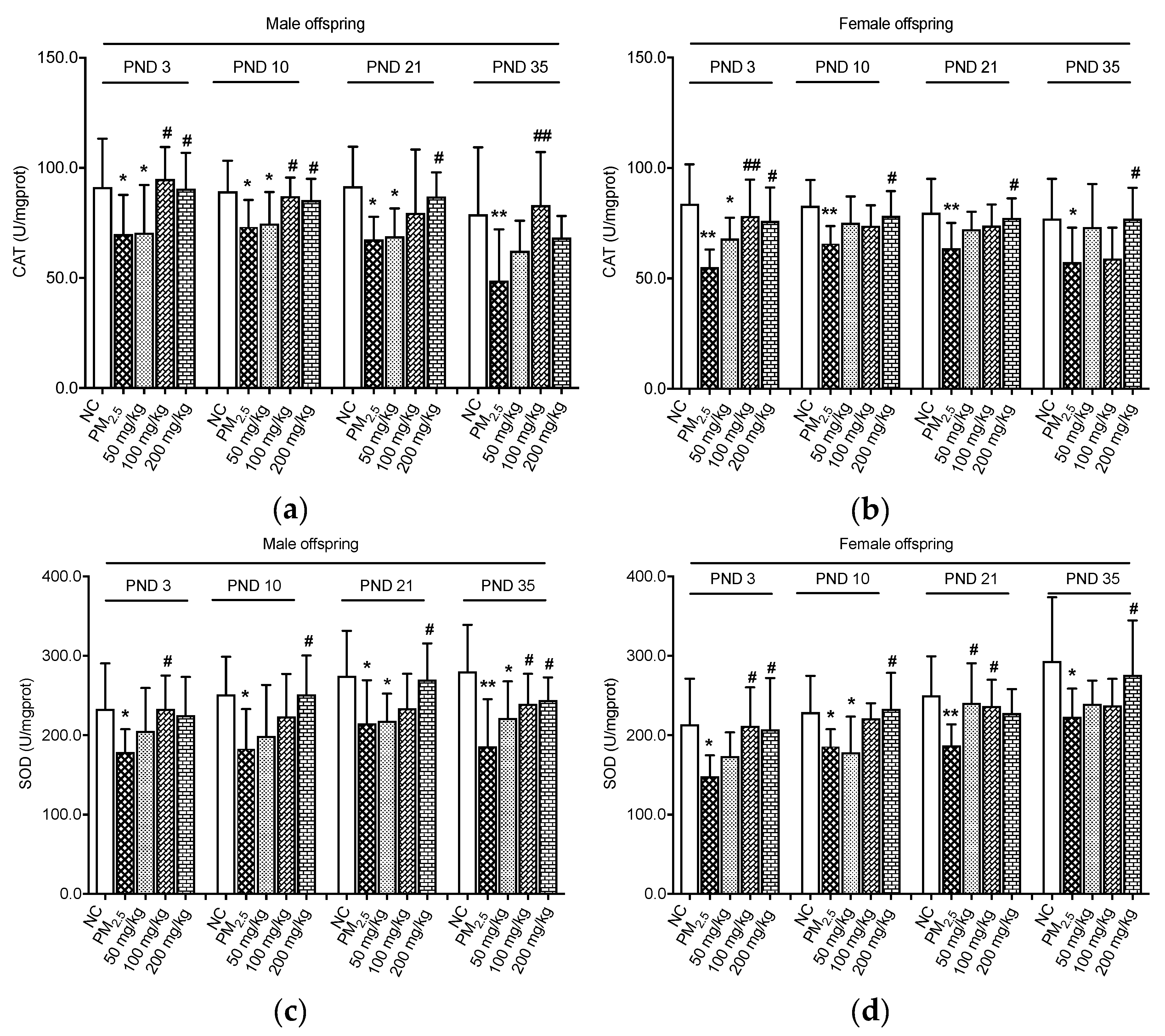
Activities of CAT and SOD of the offspring on PND 3, 10, 21, and 35 were assayed (Figure 2). It was evident that for male offspring, CAT levels in the PM2.5 groups were lower than those in the NC groups (p < 0.05, Figure 2a). On PND 3 and 10, the CAT levels were significantly elevated in the 100 and 200 mg/kg quercetin groups as compared with male samples in the PM2.5 group (p < 0.05). On PND 21, CAT concentrations of male offspring in the 200 mg/kg quercetin group were higher than those of the PM2.5 group (p < 0.05). The same effect was observed in male offspring in the 100 mg/kg quercetin group on PND 35 (p < 0.05). As shown in Figure 2b, the activity of CAT in the female offspring was significantly decreased in the PM2.5 groups compared with the NC groups (p < 0.05). Supplementation with quercetin at the doses of 100 and 200 mg/kg increased CAT activity compared with the PM2.5 group on PND 3 (p < 0.05). Moreover, 200 mg/kg quercetin administration significantly elevated the CAT of female offspring compared with the PM2.5 group on PND 10, 21, and 35 (p < 0.05).
The PM2.5 groups showed lower SOD levels of male offspring as compared with the NC groups (p < 0.05, Figure 2c). The middle dose of quercetin (100 mg/kg) could restore the SOD activity on PND 3 (p < 0.05). Quercetin 200 mg/kg significantly increased SOD concentrations of male offspring on PND 10 and 21 (p < 0.05). Additionally, mice treated with quercetin at doses of 100 and 200 mg/kg showed higher SOD concentrations on PND 35 than that of the PM2.5 group (p < 0.05). Maternal PM2.5 administration led to significant decreases in SOD activities of female offspring as compared with the control groups, as shown in Figure 2d (p < 0.05). Quercetin at two doses (100 and 200 mg/kg) showed protection effects against maternal PM2.5 exposure on PND 3 (p < 0.05). It was also observed that SOD concentrations of female offspring in the 200 mg/kg quercetin groups were higher than those of the PM2.5 groups on PND 10 and 35 (p < 0.05). A protective role of quercetin on SOD levels was also found in the 50 and 100 mg/kg quercetin groups on PND 21 (p < 0.05).

3.2. Effects on Inflammation Indicators

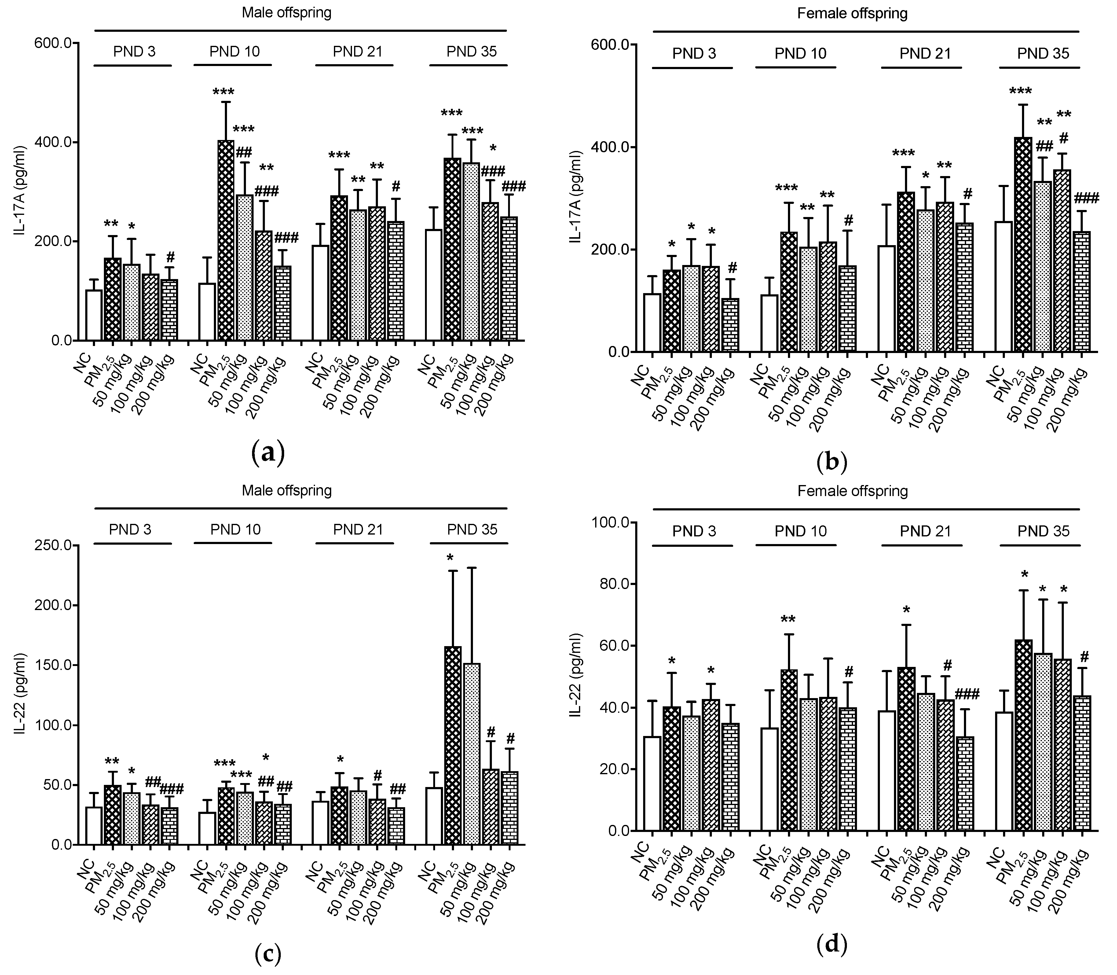
We further evaluated the levels of IL-17A and IL-22 in the colon (Figure 3). The results revealed that on PND 3, 10, 21, and 35, IL-17A concentrations of male offspring in the PM2.5 groups were higher than those in the NC groups (p < 0.05, Figure 3a). We also observed that the level of the anti-inflammatory cytokine IL-17A was down-regulated in the 200 mg/kg quercetin groups on PND 3 and 21, respectively, compared with the PM2.5 groups (p < 0.05). Furthermore, there were significant decreases of IL-17A of male offspring in the three quercetin groups on PND 10 compared with the PM2.5 group (p < 0.05). We found that the male offspring in the quercetin groups (100 and 200 mg/kg) exhibited a significant decrease of IL-17A levels as compared with the males in the PM2.5 group on PND 35 (p < 0.05). IL-17A of female offspring was significantly enhanced by maternal PM2.5 exposure (p < 0.05, Figure 3b). However, compared with the PM2.5 groups, lower IL-17A levels were observed in the 200 mg/kg quercetin groups on PND 3, 10, and 21 (p < 0.05). Similarly, female offspring in three quercetin groups showed striking reductions in the IL-17A expression on PND 35 compared with the control animals (p < 0.05).
As expected, male offspring in the PM2.5 groups showed higher expressions of IL-22 when compared with animals in the NC groups (p < 0.05, Figure 3c). However, the treatment with quercetin (100 and 200 mg/kg) provided marked decreases in IL-22 expression on PND 3, 10, 21, and 35 (p < 0.05). For female offspring, higher IL-22 levels were observed in the PM2.5 groups compared with the NC groups (p < 0.05, Figure 3d). A high dose of quercetin (200 mg/kg) could decrease the IL-22 concentration on PND 10 and 35 (p < 0.05). Likewise, on PND 21, IL-22 concentrations of female offspring in the 100 and 200 mg/kg quercetin groups were lower than those of the PM2.5 group (p < 0.05).

3.3. Effects on Colonic Proteins

Maternal PM2.5 exposure induced significant decreases in ZO-1 expressions in male and female offspring on PND 3, 21, and 35 (p < 0.05, Figure 4c,d). In contrast, the groups supplied with 200 mg/kg quercetin during PM2.5 exposure had significantly higher ZO-1 expression on PND 3, 21, and 35 (p < 0.05).
On PND 10 and 35, occludin expressions of male offspring were decreased by maternal PM2.5 exposure (p < 0.05, Figure 4e). However, quercetin supplementation had no significant effect on the occludin expression in male offspring (p > 0.05, Figure 4e). In addition, maternal PM2.5 exposure caused lower occludin expression in female offspring on PND 3, 10, and 35 (p < 0.05, Figure 4f). On PND 35, occludin expression in female offspring was reversed by quercetin supplementation (p < 0.05).

3.4. Axonomic Composition of Gut Microbiota

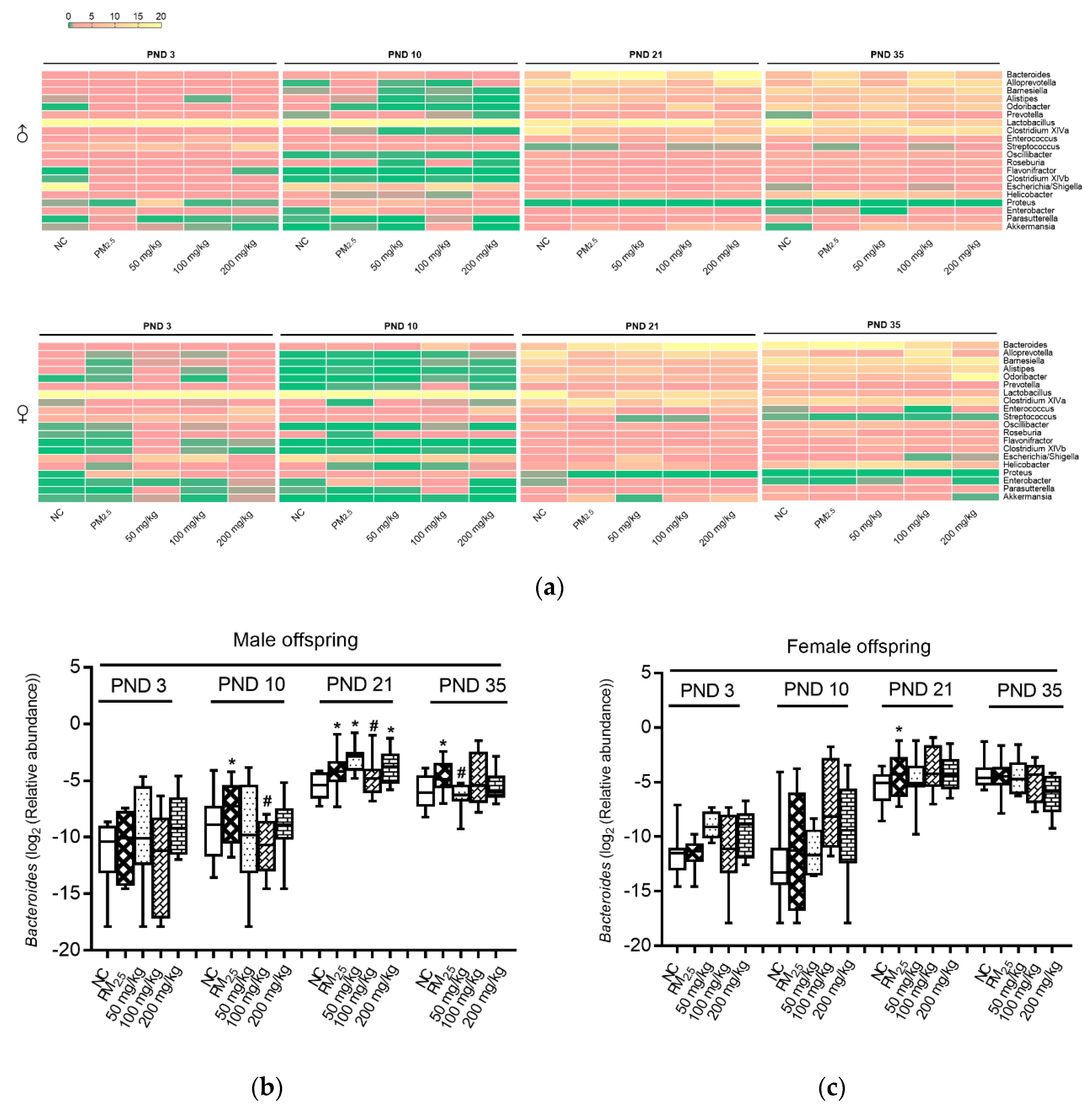
Bacterial genera, including Flavonifractor, Oscillibacter, Bacteroides, Alistipes, Proteus, Enterobacter, and Akkermansia (which were found to be enhanced by PM2.5), were reversed by different doses of quercetin intervention (Figure 5a). In comparison with PM2.5 mice, quercetin intervention enhanced a variety of bacterial genera, including Odoribacter, Streptococcus, Lactobacillus, and Roseburia (Figure 5a; note that these species were initially reduced by PM2.5 compared with the NC group).
Bacteroides of male offspring mice on PND 10, 21, and 35, and female offspring mice on PND 21 in the PM2.5 groups were higher than those in the NC groups (Figure 5b,c). After quercetin intervention, Bacteroides of male offspring mice on PND 10, 21, and 35 in the quercetin groups were lower than those in the PM2.5 groups (Figure 5b).

3.5. Association between Bacteroides and Inflammation and Oxidative Indicators

By Spearman’s correlation analysis (Figure 6), the result demonstrated that Bacteroides was negatively associated with colonic CAT concentration (r = −0.28, p = 0.002), whereas the Bacteroides abundance was positively related to IL-17A levels (r = 0.46, p < 0.001). Moreover, there was a positive correlation between Bacteroides and IL-22 (r = 0.24, p = 0.008). No clear correlation was found between the SOD concentration and the abundance of Bacteroides (p > 0.05).

4. Discussion

It is reported that PM2.5 is related to various health problems. However, there is no study to explore the effects on offspring‘s intestinal barriers when dams were exposed to PM2.5. Moreover, the intervention method has not been well illustrated. Our results indicated that maternal PM2.5 exposure led to crucial influences on intestinal oxidative stress, inflammation, and tight junctions of offspring mice, and quercetin administration may have some certain intervention effects. Gut microbiota may play an important role in this process.
Maternal PM2.5 exposure could affect gut barrier function. First of all, higher oxidative stress was found in the PM2.5 group, showing higher levels of CAT and SOD. Secondly, gut inflammation was observed in the PM2.5 group. IL-17A and IL-22 were both elevated. Thirdly, PM2.5 could have damaged epithelial tight junction proteins, showing low expression of occludin and ZO-1.
Gut barrier changes may be related to changes in gut microbiota. It can be seen that microbiota on PND 3 (colonization), 10 (breastfeeding), 21 (mixed feeding), and 35 (weaning) in the PM2.5 group were different from those in the control group. Polycyclic aromatic hydrocarbons [26], metals [27], and tetrachlorodibenzofuran [28] components were included in the PM2.5 [15], which can interact with intestinal flora. It is noteworthy that the relative Bacteroides abundance of male offspring mice increased in the PM2.5 group, and Bacteroides correlated with colonic CAT, IL-17A, and IL-22 levels. A study demonstrated that a higher level of Bacteroides was found in the colitis rats; moreover, their results also indicated that Bacteroides (r = 0.86, p = 0.001) was positively associated with the histological scores (including cell infiltration and tissue damage) [29]. Thus, higher Bacteroides caused by PM2.5 exposure was related to a damaged colon barrier. The above results indicated that PM2.5 might affect the intestinal barrier by acting on the intestinal flora.
Due to multiple reasons, it is impossible to solve PM2.5 pollution in a short period, so we hoped to reduce injury through diet intervention. Quercetin could improve the gut barrier function. Firstly, oxidative stress could be adjusted by quercetin. Increased levels of CAT and SOD were detected in quercetin groups significantly. Secondly, quercetin showed beneficial effects on inflammation changes. Levels of IL-17A and IL-22 of the quercetin groups were close to the NC groups. Thirdly, the expression of tight junction proteins was also improved. The expression of ZO-1 and occludin in the quercetin groups was higher than that in the PM2.5 group, indicating that quercetin could protect the colon mucosa barrier from damage.
We suspected that quercetin can improve the mucosa barrier by improving gut microbiota evolution of offspring. Gut microbiota abundance of offspring mice in quercetin intervention groups showed intervention effects. Taira et al. demonstrated that in rats, dietary polyphenols ameliorated imbalance in gut microbiota that was caused by a high-fat diet [30]. What is more, polyphenols could significantly inhibit the growth of Bacteroidetes and Firmicutes, and at the same time down-regulate the rate of Bacteroidetes to Firmicutes [31]. Another study demonstrated that the administration of quercetin was found to be effective in attenuating the Firmicutes/Bacteroidetes ratio [32]. In our study, we found that the Bacteroides that had been raised by PM2.5 decreased after the intervention of quercetin. Maternal PM2.5 exposure can cause oxidation and inflammation effects, moreover, the ZO-1 and occludin proteins also decreased. Quercetin may have protective effects on inflammation cytokines, antioxidases, and tight junction protein changes under PM2.5 exposure. These effects might be correlated with the changes in Bacteroides.
Although our study can provide new insights to learn about the influences of PM2.5, several limitations should be discussed. Firstly, our analysis only showed correlations between Bacteroides and indicators, but other associations may also exist with other unreported bacteria. Secondly, the way that gut microbiota influence barrier function has not been illuminated. Thirdly, the study is more about a hypothesis, and more studies will be needed to verify these opinions.

5. Conclusions

PM2.5 tracheal exposure during gestation and lactation could influence intestinal oxidative stress, inflammation, and tight junctions of offspring. Quercetin administration may have some certain intervention effects. Medium- and high-dose quercetin intervention showed better effects. Changes in Bacteroides play an important role in the process.

Author Contributions

Conceptualization, Y.X. and W.L.; methodology, W.L. and Y.Z.; software, Y.Q.; formal analysis, W.L. and Y.Q.; investigation, W.L., Y.Z., and L.Y.; resources, R.L. and Y.C.; writing—original draft preparation, W.L.; writing—review and editing, Y.X.; supervision, Y.X. All authors have read and agreed to the published version of the manuscript.

Funding

This research received no external funding.

Acknowledgments

We are grateful to Quanchao Li for his assistance in animal experiments.

Conflicts of Interest

The authors declare no conflict of interest.

References

  1. Opstelten, J.L.; Beelen, R.J.; Leenders, M.; Hoek, G.; Brunekreef, B.; Van Schaik, F.D.M.; Siersema, P.D.; Eriksen, K.T.; Raaschou-Nielsen, O.; Tjønneland, A.; et al. Exposure to ambient air pollution and the risk of inflammatory bowel disease: A European nested case—Control study. Dig. Dis. Sci. 2016, 61, 2963–2971. [Google Scholar] [CrossRef] [PubMed]
  2. Feng, J.; Cavallero, S.; Hsiai, T.; Li, R. Impact of air pollution on intestinal redox lipidome and microbiome. Free Radic. Biol. Med. 2020, 151, 99–110. [Google Scholar] [CrossRef] [PubMed]
  3. Xavier, R.J.; Podolsky, D.K. Unravelling the pathogenesis of inflammatory bowel disease. Nature 2007, 448, 427–434. [Google Scholar] [CrossRef] [PubMed]
  4. Khor, B.; Gardet, A.; Xavier, R.J. Genetics and pathogenesis of inflammatory bowel disease. Nature 2011, 474, 307–317. [Google Scholar] [CrossRef] [PubMed] [Green Version]
  5. Şengul, N.; Işik, S.; Aslim, B.; Uçar, G.; Demirbağ, A.E. The effect of exopolysaccharide-producing probiotic strains on gut oxidative damage in experimental colitis. Dig. Dis. Sci. 2011, 56, 707–714. [Google Scholar] [CrossRef] [PubMed]
  6. Liu, X.; Li, H.; Meng, Z. Oxidative damage of PM2.5 on hearts, lungs and testicles of rats. Zhongguo Huanjing Kexue 2005, 25, 160–164. Available online: https://europepmc.org/article/cba/515190 (accessed on 10 October 2019).
  7. Al Hanai, A.H.; Antkiewicz, D.S.; Hemming, J.D.C.; Shafer, M.M.; Lai, A.M.; Arhami, M.; Hosseini, V.; Schauer, J.J. Seasonal variations in the oxidative stress and inflammatory potential of PM2.5 in Tehran using an alveolar macrophage model; The role of chemical composition and sources. Environ. Int. 2019, 123, 417–427. [Google Scholar] [CrossRef]
  8. Riva, D.R.; Magalhaes, C.B.; Lopes, A.A.; Lanças, T.; Mauad, T.; Malm, O.; Valenca, S.S.; Saldiva, P.; Faffe, D.; Zin, W.A. Low dose of fine particulate matter (PM2.5) can induce acute oxidative stress, inflammation and pulmonary impairment in healthy mice. Inhal. Toxicol. 2011, 23, 257–267. [Google Scholar] [CrossRef]
  9. Aijaz, S.; Balda, M.S.; Matter, K. Tight junctions: Molecular architecture and function. Int. Rev. Cytol. 2006, 248, 261–298. [Google Scholar] [CrossRef]
  10. Quiros, M.; Nusrat, A. RhoGTPases, actomyosin signaling and regulation of the epithelial apical junctional complex. Semin. Cell Dev. Biol. 2014, 36, 194. [Google Scholar] [CrossRef] [Green Version]
  11. Itallie, C.M.V.; Fanning, A.S.; Bridges, A.; Anderson, J.M. ZO-1 stabilizes the tight junction solute barrier through coupling to the perijunctional cytoskeleton. Mol. Biol. Cell 2009, 20, 3930. [Google Scholar] [CrossRef] [PubMed] [Green Version]
  12. Wei, Y.; Cao, X.N.; Tang, X.L.; Lian-Ju, S.; Lin, T.; He, D.-W.; Wu, S.; Wei, G. Urban fine particulate matter (PM2.5) exposure destroys blood-testis barrier (BTB) integrity through excessive ROS-mediated autophagy. Toxicol. Mech. Methods 2018, 28, 302–319. [Google Scholar] [CrossRef] [PubMed]
  13. Mutlu, E.A.; Engen, P.A.; Soberanes, S.; Urich, D.; Forsyth, C.B.; Nigdelioglu, R.; Chiarella, S.E.; Radigan, K.A.; Gonzalez, A.; Jakate, S.; et al. Particulate matter air pollution causes oxidant-mediated increase in gut permeability in mice. Part. Fibre Toxicol. 2011, 8, 19. [Google Scholar] [CrossRef] [PubMed] [Green Version]
  14. Bischoff, S.C.; Barbara, G.; Buurman, W.; Ockhuizen, T.; Schulzke, J.-D.; Serino, M.; Tilg, H.; Watson, A.J.; Wells, J.M. Intestinal permeability—A new target for disease prevention and therapy. BMC Gastroenterol. 2014, 14, 189. [Google Scholar] [CrossRef] [Green Version]
  15. Salim, S.Y.; Kaplan, G.G.; Madsen, K.L. Air pollution effects on the gut microbiota: A link between exposure and inflammatory disease. Gut Microbes 2014, 5, 215–219. [Google Scholar] [CrossRef]
  16. Mutlu, E.A.; Comba, I.Y.; Cho, T.; Engen, P.; Yazıcı, C.; Soberanes, S.; Hamanaka, R.B.; Nigdelioglu, R.; Meliton, A.Y.; Ghio, A.J.; et al. Inhalational exposure to particulate matter air pollution alters the composition of the gut microbiome. Environ. Pollut. 2018, 240, 817–830. [Google Scholar] [CrossRef]
  17. Santangelo, R.; Silvestrini, A.; Mancuso, C. Ginsenosides, catechins, quercetin and gut microbiota: Current evidence of challenging interactions. Food Chem. Toxicol. 2019, 123, 42–49. [Google Scholar] [CrossRef]
  18. Guo, Y.; Bruno, R.S. Endogenous and exogenous mediators of quercetin bioavailability. J. Nutr. Biochem. 2015, 26, 201–210. [Google Scholar] [CrossRef]
  19. Pfeuffer, M.; Auinger, A.; Bley, U.; Kraus-Stojanowic, I.; Laue, C.; Winkler, P.; Rüfer, C.; Frank, J.; Bösch-Saadatmandi, C.; Rimbach, G.; et al. Effect of quercetin on traits of the metabolic syndrome, endothelial function and inflammation in men with different APOE isoforms. Nutr. Metab. Cardiovasc. Dis. 2013, 23, 403–409. [Google Scholar] [CrossRef]
  20. Zhang, Z.; Peng, X.; Li, S.; Zhang, N.; Wang, Y.; Wei, H. Isolation and identification of quercetin degrading bacteria from human fecal microbes. PLoS ONE 2014, 9, e90531. [Google Scholar] [CrossRef] [Green Version]
  21. Carrasco-Pozo, C.; Gotteland, M.; Castillo, R.L.; Chen, C. 3,4-dihydroxyphenylacetic acid, a microbiota-derived metabolite of quercetin, protects against pancreatic β-cells dysfunction induced by high cholesterol. Exp. Cell Res. 2015, 334, 270–282. [Google Scholar] [CrossRef] [PubMed]
  22. Tang, Y.; Nakashima, S.; Saiki, S.; Myoi, Y.; Abe, N.; Kuwazuru, S.; Zhu, B.; Ashida, H.; Murata, Y.; Nakamura, Y. 3,4-Dihydroxyphenylacetic acid is a predominant biologically-active catabolite of quercetin glycosides. Food Res. Int. 2016, 89, 716–723. [Google Scholar] [CrossRef] [PubMed]
  23. Liu, W.; Zhou, Y.; Li, Y.; Qin, Y.; Yu, L.; Li, R.; Chen, Y.; Xu, Y. Effects of PM2.5 exposure during gestation on maternal gut microbiota and pregnancy outcomes. Chemosphere 2020, 247, 125879. [Google Scholar] [CrossRef] [PubMed]
  24. Liu, W.; Zhang, M.; Feng, J.; Fan, A.; Zhou, Y.; Xu, Y. The influence of quercetin on maternal immunity, oxidative stress, and inflammation in mice with exposure of fine particulate matter during gestation. Int. J. Environ. Res. Public Health 2017, 14, 592. [Google Scholar] [CrossRef]
  25. Liu, W.; Zhou, Y.; Qin, Y.; Li, Y.; Yu, L.; Li, R.; Chen, Y.; Xu, Y. Sex-dependent effects of PM2.5 maternal exposure and quercetin intervention on offspring’s short chain fatty acids. Int. J. Environ. Res. Public Health 2019, 16, 4371. [Google Scholar] [CrossRef] [Green Version]
  26. Tom, V.D.W.; Vanhaecke, L.; Boeckaert, C.; Peru, K.; Headley, J.; Verstraete, W.; Siciliano, S. Human colon microbiota transform polycyclic aromatic hydrocarbons to estrogenic metabolites. Environ. Health. Perspect. 2004, 113, 6–10. [Google Scholar] [CrossRef]
  27. Breton, J.; Massart, S.; Vandamme, P.; De Brandt, E.; Pot, B.; Foligné, B. Ecotoxicology inside the gut: Impact of heavy metals on the mouse microbiome. BMC Pharmacol. Toxicol. 2013, 14. [Google Scholar] [CrossRef] [Green Version]
  28. Zhang, L.; Nichols, R.G.; Correll, J.; Murray, I.A.; Tanaka, N.; Smith, P.B.; Hubbard, T.D.; Sebastian, A.; Albert, I.; Hatzakis, E.; et al. Persistent organic pollutants modify gut microbiota-host metabolic homeostasis in mice through aryl hydrocarbon receptor activation. Environ. Health. Perspect. 2015, 123, 679–688. [Google Scholar] [CrossRef] [Green Version]
  29. Wang, K.; Jin, X.; Li, Q.; Sawaya, A.C.H.F.; Le Leu, R.K.; Conlon, M.A.; Wu, L.; Hu, F. Propolis from different geographic origins decreases intestinal inflammation and Bacteroides spp. populations in a model of DSS-induced colitis. Mol. Nutr. Food Res. 2018, 62, e1800080. [Google Scholar] [CrossRef]
  30. Taira, T.; Yamaguchi, S.; Takahashi, A.; Okazaki, Y.; Yamaguchi, A.; Sakaguchi, H.; Chiji, H. Dietary polyphenols increase fecal mucin and immunoglobulin A and ameliorate the disturbance in gut microbiota caused by a high fat diet. J. Clin. Biochem. Nutr. 2015, 57, 212–216. [Google Scholar] [CrossRef] [Green Version]
  31. Xue, B.; Xie, J.; Huang, J.; Chen, L.; Gao, L.; Ou, S.; Wang, Y.; Peng, X. Plant polyphenols altering a pathway of energy metabolism by inhibiting fecal Bacteriodetes and Firmicutes in vitro. Food Funct. 2016, 7, 1501–1507. [Google Scholar] [CrossRef] [PubMed]
  32. Etxeberria, U.; Arias, N.; Boqué, N.; Macarulla, M.; Portillo, M.P.; Martinez, J.; Milagro, F.I. Reshaping faecal gut microbiota composition by the intake of trans-resveratrol and quercetin in high-fat sucrose diet-fed rats. J. Nutr. Biochem. 2015, 26, 651–660. [Google Scholar] [CrossRef] [PubMed]
Figure 1. Experimental study design. A pair of male and female cubs born to the same mother were sacrificed on PND 3, 10, 21, and 35. Colonic microbiota, inflammatory biomarkers, antioxidases, and related proteins were detected. GD: gestational day; PND: postnatal day; IL: interleukin; CAT: catalase; SOD: superoxide dismutase.
Nutrients 12 03095 g001
Figure 2. Effects on antioxidant enzymes of colon. (a) CAT levels of male offspring; (b) CAT levels of female offspring; (c) SOD levels of male offspring; (d) SOD levels of female offspring. Compared with the NC group, * indicates p < 0.05, ** indicates p < 0.01. Compared with the PM2.5 group, # indicates p < 0.05, ## indicates p < 0.01. NC: normal control; PM2.5: fine particulate matter.
Nutrients 12 03095 g002
Figure 3. Effects on inflammation biomarkers of colon. (a) IL-17A levels of male offspring; (b) IL-17A levels of female offspring; (c) IL-22 levels of male offspring; (d) IL-22 levels of female offspring. Compared with the NC group, * indicates p < 0.05, ** indicates p < 0.01, *** indicates p < 0.001. Compared with the PM2.5 group, # indicates p < 0.05, ## indicates p < 0.01, ### indicates p < 0.001.
Nutrients 12 03095 g003
Figure 4. Effects on proteins of colon. (a) Immunoblots of ZO-1 and occludin expression of male offspring; (b) Immunoblots of ZO-1 and occludin expression of female offspring; (c) ZO-1 expressions of male offspring; (d) ZO-1 expressions of female offspring; (e) Occludin expressions of male offspring; (f) Occludin expressions of female offspring. Compared with the NC group, * indicates p < 0.05, ** indicates p < 0.01. Compared with the PM2.5 group, # indicates p < 0.05. The values were normalized against the intensity of β-actin, n = 3/group/gender/time.
Nutrients 12 03095 g004
Figure 5. Axonomic composition of gut microbiota. (a) Axonomic composition of the top 20 genera; (b) Bacteroides of male offspring mice; (c) Bacteroides of female offspring mice. Compared with the NC group, * indicates p < 0.05. Compared with the PM2.5 group, # indicates p < 0.05.
Nutrients 12 03095 g005
Figure 6. Correlation between Bacteroides and inflammation and oxidative indicators. (a) Correlation between Bacteroides and CAT; (b) Correlation between Bacteroides and SOD; (c) Correlation between Bacteroides and IL-17A; (d) Correlation between Bacteroides and IL-22. Spearman’s correlation analysis was used to analyze the relationship.
Nutrients 12 03095 g006
Table 1. Animal treatment.
GroupNPM2.5 (mg/kg)Quercetin (mg/kg)
NC group8--
PM2.5 group815-
50 mg/kg quercetin group81550
100 mg/kg quercetin group815100
200 mg/kg quercetin group815200
Note: NC: normal control; PM2.5: fine particulate matter.