Article

Up-Regulation of Specific Bioactive Lipids in Celiac Disease

by
Rafael Martín-Masot
1,*,
Jose Daniel Galo-Licona
2,
Natàlia Mota-Martorell
2,
Joaquim Sol
2,3,4,
Mariona Jové
2,
José Maldonado
5,6,7,
Reinald Pamplona
2 and
Teresa Nestares
8
1
Pediatric Gastroenterology and Nutrition Unit, Hospital Regional Universitario de Malaga, 29011 Málaga, Spain
2
Department of Experimental Medicine, Lleida Biomedical Research Institute (IRBLleida), University of Lleida (UdL), 25198 Lleida, Spain
3
Institut Català de la Salut, Atenció Primària, 25198 Lleida, Spain
4
Research Support Unit Lleida, Fundació Institut Universitari per a la Recerca a l’Atenció Primària de Salut Jordi Gol i Gurina (IDIAPJGol), 25198 Lleida, Spain
5
Department of Pediatrics, University of Granada, 18071 Granada, Spain
6
Pediatric Gastroenterology and Nutrition Unit, Hospital Universitario Virgen de las Nieves, 18071 Granada, Spain
7
Maternal and Child Health Network, Carlos III Health Institute, 28029 Madrid, Spain
8
Biomedical Research Centre (CIBM), Department of Physiology and “José MataixVerdú”, Institute of Nutrition and Food Technology (INYTA), University of Granada, 18071 Granada, Spain
*
Author to whom correspondence should be addressed.
Nutrients 2021, 13(7), 2271; https://doi.org/10.3390/nu13072271
Submission received: 13 May 2021 / Revised: 12 June 2021 / Accepted: 25 June 2021 / Published: 30 June 2021
(This article belongs to the Section Nutrition and Public Health)

Abstract

:
Celiac disease (CD) is an autoimmune enteropathy linked to alterations of metabolism. Currently, limited untargeted metabolomic studies evaluating differences in the plasma metabolome of CD subjects have been documented. We engage in a metabolomic study that analyzes plasma metabolome in 17 children with CD treated with a gluten-free diet and 17 healthy control siblings in order to recognize potential changes in metabolic networks. Our data demonstrates the persistence of metabolic defects in CD subjects in spite of the dietary treatment, affecting a minor but significant fraction (around 4%, 209 out of 4893 molecular features) of the analyzed plasma metabolome. The affected molecular species are mainly, but not exclusively, lipid species with a particular affectation of steroids and derivatives (indicating an adrenal gland affectation), glycerophospholipids (to highlight phosphatidic acid), glycerolipids (with a special affectation of diacylglycerols), and fatty acyls (eicosanoids). Our findings are suggestive of an activation of the diacylglycerol-phosphatidic acid signaling pathway in CD that may potentially have detrimental effects via activation of several targets including protein kinases such as mTOR, which could be the basis of the morbidity and mortality connected with untreated CD. However, more studies are necessary to validate this idea regarding CD.

1. Introduction

Celiac disease (CD) is an enteropathy of autoimmune origin with a systemic and chronic trait generated by the dietary gluten and related prolamins in individuals with genetic susceptibility [1,2]. CD appears and develops from the dysfunctional interaction between genetic and environmental factors [3]. The affected subjects, usually children but also adults, display an inflammatory enteropathy with different degrees of severity [4] and clinical manifestations that range from asymptomatic to a wide variety of digestive and/or systemic signs and symptoms. Furthermore, untreated symptomatic CD patients develop relevant morbidity and mortality [5]. Thus, it can be associated to CD-related neurologic diseases; osteoporosis; T-cell lymphoma; and autoimmune endocrine diseases such as Addison’s disease, diabetes (type I), and tiroiditis [5]. Fortunately, when celiac patients are treated with a strict gluten-free diet (GFD), the clinical and histological manifestations reverse [1]. Recent studies indicate that CD affects about 1–3% of the general population in both North America and Europe [6,7,8].
The genetic predisposition of CD is linked to specific class II major histocompatibility complex (MHC) alleles. Thus, the HLA-DQ2 allele is present in more than 90% of CD subjects, whereas the DQ8 allele is present in the remaining 10% of CD patients; importantly, non-HLA genes have been described to contribute to the disease [9]. As around 40–50% of the European general population is positive for the HLA-DQ2 heterodimer [10], it is evident that additional factors are required for the onset and progression of the CD.
Alterations in metabolism seem to also be present in CD. Thus, metabolomic studies, despite being restricted to the use of NMR (nuclear magnetic resonance)-based methods, suggest lesser but significant changes in metabolites belonging to microbiome-derived metabolites, lipid metabolism, and energy metabolism [11,12,13,14,15,16]. Remarkably, differences in methionine, choline, and choline-derived lipids content in CD subjects were described, suggestive of an affectation of one-carbon metabolism. This alteration of methionine metabolism in CD subjects was recently confirmed and extended using a LC–MS/MS approach [17]. In this study, a down-regulation of the trans-sulphuration pathway in CD patients was observed, in particular.
Currently, no untargeted metabolomic analysis using LC-MS/MS or investigations regarding changes in the plasma metabolome of CD subjects have been conducted. For this reason, we designed a metabolomic study with 34 subjects including 17 CD patients and 17 healthy control siblings. The CD subjects were children under a GFD treatment. This election was based on the idea that GFD induces the return of CD signs and symptoms to a healthy state as well as induces histological changes and changes in metabolism. In addition, potential persistent metabolic alterations should be, if present, specific to the CD condition. Blood plasma was used because it is easy to obtain and is the main carrier of metabolites in the body, being well-known as the compositional profile of this biological fluid [18]. Furthermore, its composition is under strict control despite the continuous change induced by different internal and external conditions in physiological and pathological states [19,20]. These facts allow us to ensure the identification of new molecular mechanisms and targets that may lead to healthier states. The plasma metabolome profile was defined using an LC–MS/MS platform to discriminate specific phenotypic patterns linked to genotypes of CD.

2. Materials and Methods

2.1. Subjects

We performed a cross-sectional study in which 17 CD children and 17 healthy control siblings between 4 and 17 years of age were included from January to December of 2018. The study was carried out in the Gastroenterology, Hepatology, and Child Nutrition Service from the “Virgen de las Nieves” University Hospital in Granada, Spain. The diagnosis of CD was made following the European Society for Pediatric Gastroenterology Hepatology and Nutrition (ESPGHAN) criteria of 2012 [21]. Infants with liver or kidney diseases, inflammatory bowel disease, diabetes, and chronic asthma and those consuming dietary supplements containing substances with antioxidant activity were excluded. We also excluded obese patients (according to the criteria of the International Task Force) [22]. All patients signed the written informed consent document and excluded those otherwise from the study. The Ethics Committee of the University of Granada approved the study (reference number 201202400000697), and the Good Clinical Practice guidelines were followed.

2.2. Clinical and Sociodemographics

Participants’ clinical and sociodemographic traits, including adherence to the nutritional intervention (GFD), were evaluated by the same research team.

2.3. Anthropometric Measures

Anthropometric traits (height and weight) were measured in healthy control and CD subjects. Height was determined to the nearest 5 mm using a stadiometer (Seca 22, Hamburg, Germany). Body weight was determined using a mechanical balance scale (Seca200, Hamburg, Germany).

2.4. Blood Samples

Blood samples were obtained via venipuncture in the morning (between 07:00 and 08:00 A.M.) after fasting overnight (8–10 h) and collected in one vacutainer CPT (cell preparation tube; BD, Franklin Lakes, NJ, USA) containing sodium heparin as an anticoagulant. Blood samples were centrifuged, and plasma fractions were collected, immediately frozen in liquid nitrogen, transferred to a −80 °C freezer, and stored for 4 h for the purpose of later metabolomic analysis.

2.5. Untargeted Metabolomics

2.5.1. Chemicals

All reagents were obtained from Sigma-Aldrich and of the highest purity available unless otherwise specified.

2.5.2. Sample Processing

Plasma metabolites’ extraction was made on the basis of a previously described methodology [23]. In brief, 10 µL of plasma was mixed with 30 µL of cold methanol containing 1 μg/mL of Phe-13C as the internal standard and 1 μM BHT as the antioxidant. After incubation at 4 °C for 60 min, the samples were centrifuged at 12,000× g for 3 min. Then, the supernatant was filtrated (CLS8169, Sigma, Madrid, Spain) and transferred to vials with glass inserts (Agilent, Barcelona, Spain) for chemical analysis. Samples were decoded and randomized before the LC-MS analysis. To serve as quality control for metabolite extraction, a pool of plasma samples with internal Phe-13C was used and injected every 5 samples.

2.5.3. Analysis Conditions

Two microliters of metabolic extract were analyzed according to a previously described method [23]. A reversed-phase column (Zorbax SB-Aq 2.1 × 50 mm, 1.8 µm particle size, Agilent Technologies, CA, USA) with a pre-column (Zorbax SB-C8 Rapid Resolution Cartridge, 2.1 × 30 mm, 3.5 µm particle size, Agilent Technologies, CA, USA) set at 60 °C was used for chromatographic separation [23]. Afterwards, electrospray ionization was made in positive and negative ion modes using N2 at a pressure of 50 psi for the nebulizer with a flow of 12 L/min and a temperature of 325 °C.
The analysis was performed using a UHPLC 1290 model coupled to a Q-TOF 6545 instrument (Agilent Technologies, CA, USA) as previously described [23], and data were collected using the MassHunter Data Analysis Software (Agilent Technologies, CA, USA).

2.6. Data Analyses

Mass Hunter Qualitative Analysis Software (Agilent Technologies, Barcelona, Spain) was used to obtain samples’ molecular features, representing different co-migrating ionic species of a given molecular entity using the Molecular Feature Extractor (MFE) algorithm (Agilent Technologies, Barcelona, Spain). The Mass Hunter Mass Profiler Professional Software (Agilent Technologies, Barcelona, Spain) was used to make an untargeted metabolomic analysis over the extracted features. Only features with a minimum of 2 ions were selected. Then, the molecular traits in the samples were aligned using a retention time window of 0.1% ± 0.25 min and 30.0 ppm ± 2.0 m Da. Only features found in at least 70% of the QC samples and with less than 20% of relative standard deviation among QC samples were considered to correct for individual bias. Instrumental drift was corrected using a LOESS approach [24,25]. Multivariate statistics (Principal Component Analysis (PCA), Partial Least Squares-Discriminant Analysis (PLSDA), and Hierarchical and Classification Analyses) were performed using the Metaboanalyst software [26,27].
Statistical analyses were conducted using R version 3.6.1. [28] A paired-t-test was used to evaluate sibling differences in clinical and MS comparisons between groups. The minimum signification level was set at p < 0.05 without multiple-testing adjustment, and Benjamini–Hochberg’s False Discovery Rate (FDR) p-values are reported as complementary information. Heatmaps and forest plots were performed using the heatmap and forest plot packages, respectively [29,30].
Features were defined by the exact mass and retention time using HMDB [31] (accuracy < 30 ppm) and PCDL database (Agilent Technologies, Barcelona, Spain), which uses retention times in a standardized chromatographic system as an orthogonal searchable parameter to complement accurate mass data (accurate mass retention time approach) [32]. Finally, identities were confirmed by MS/MS by checking the MS/MS spectrums using HMDB [31].

3. Results

3.1. Clinical Traits

The baseline clinical traits of the study population are shown in Table 1. A total of 34 children were included in the study, 17 CD and 17 healthy siblings. There were no differences between groups, including factors that could affect metabolomics’ profile such us dietary habits and physical activity.

3.2. Untargeted Metabolomics

With the aim to analyze the effect of the celiac condition in the whole circulating metabolome, an untargeted metabolomic analysis was performed. Baseline correction, peak picking, and peak alignment were determined based on acquired data, resulting in a total of 48,952 molecules from both ionization modes (positive and negative). After quality control assessment, filtering, and correction of the signal, 4893 features remained that were used for statistical analysis. Multivariate statistics suggest that there is not a specific metabolomic signature associated with the celiac condition (data not shown), but when we searched for specific celiac biomarkers, we found 209 molecules statistically different between groups and five of them (lithocholyltaurine, 18-Oxocortisol, 5alpha-Pregnan-3alpha, 20beta-diol disulfate, and an unidentified compound, all up-regulated in CD individuals) retained their significance after applying Benjamini–Hochberg’s FDR (Supplementary Dataset). Among all of the significant molecules, 36 of them were annotated (Figure 1 and Figure 2), wherein 23 (64%) were increased and 13 (36%) decreased in CD patients. Specifically, we described two carboxylic acids and derivates increased in CD, four fatty acyls, five glycerolipids, eleven glycerophospholipids, one organoxigen compound, and two sphingolipids were affected by the CD condition. Furthermore, among 36 molecules with a potential identity, eight of them were lipid species belonging to steroid metabolism and two were involved in bilirubin metabolism. Interestingly, nine of these identified molecules have been involved in cell signalling pathways (diacylglycerides (DG), lysophospholipids, and ceramides) and are mainly increased in CD subjects.

4. Discussion

The variability in clinical manifestations of CD is due to the interindividual differential vulnerability supported by genetic, immunological, and environmental factors. Regarding the diversity of signs and symptoms generated by CD, recent observations suggest that metabolic alterations in subjects with CD must also be considered. Although in most cases nowadays the diagnosis of CD requires confirmation by a positive biopsy, a better understanding of the metabolic processes underlying this pathology could offer the opportunity to uncover potential new physiopathological mechanisms and biomarkers useful for the diagnosis of doubtful cases due to the histological results as well as for follow-up of the disease.
Effectively, available evidence from metabolomic studies certified the presence of minor but significant differences in the metabolic profile between healthy individuals and celiac patients (children and adults) [11,12,13,14,15,16,17]. In these studies, using NMR or MS-based methods, biological samples from different origins including faecalis material, plasma, serum, and urine were analyzed. Overall, the metabolomic changes described are associated with modifications in gut microflora and/or intestinal permeability, malabsorption, and alterations in metabolism [11,12,13,14,15,16,17]. Thus, lower concentrations of amino acids (asparagine, isoleucine, methionine, proline, and valine); diverse metabolites (choline, creatinine, lactate, lipids (phosphatidylcholines), methylamine, methylglutarate, and pyruvate); and higher levels of 3-hydroxybutyric acid, glucose, and triacyl glycerides were the main differences observed in the serum metabolome between CD subjects and healthy controls [11,12,13,14,15,16]. It is also particularly remarkable that the decreased levels of choline, methionine, and phosphatidylcholines [15,16] along with the decreased content of metabolites in the trans-sulphuration pathway [17] clearly suggest defects in the one-carbon metabolism in CD. Importantly, these studies also verified that the introduction of a strict GFD seems to quasi completely reverse the metabolic profile of CD subjects to the healthy condition.
In the present study, our findings demonstrate the persistence of alterations in the whole circulating metabolome of CD patients in spite of dietary intervention. These metabolomic changes are, however, minor as only 209 metabolites seem to be affected, which represent only around 4% of the total plasma metabolome analyzed. Unfortunately, among these 209 metabolites, only 36 were identified, with 23 (64%) increased and 13 (36%) decreased in CD. Interestingly, we identified that molecules mainly belong to lipid families and are involved in cell signaling (DG, lysophospholipids, and ceramides). These lipid species are mostly increased in CD. Furthermore, we found fatty acid derivates involved in inflammatory pathways such as leukotrienes and thromboxanes. Remarkably, eight of the lipid species identified belonged to steroid metabolism and two of them were involved in bilirubin metabolism, suggesting potential alterations at the hepatic level as well as in the endocrine system and, in particular, the adrenal gland. Although it has been described that endocrine manifestations occur in celiac disease [34], this is the first time that potential alterations affecting the functioning of the adrenal gland are reported. More studies, however, are necessary to explore this idea in CD.
Among the observed changes in lipid molecular species, the increased content of DG (three species) and phosphatidic acid (PA) (one species) is particularly relevant and requires special attention. DG has exclusive functions as a constituent of cell membranes, intermediate in lipid metabolism and lipid-mediated signaling, and their levels are strictly regulated [35]. DG signals are particularly relevant in neuronal and immune tissues, playing a central role in the control of neuron communication, phagocytosis, and the control of immune responses [36]. Simply put, DG is generated by hydrolysis of phosphatidylinositol 4,5-bisphosphate, which is also the substrate for phosphatidylinositol 3 kinase activity. DG is phosphorylated in a reaction catalyzed by DG kinases to generate PA, the first step in a series of reactions that replenish phosphatidylinositol 4,5-bisphosphate levels in the phosphatidylinositol cycle. Other DG sources include the pool generated from PC (phosphatidylcholine) and ceramides [36]. Importantly, DG and PA, as lipid second messengers, play important roles in regulating several target proteins including numerous protein kinases such as mTOR [37], among several others. Interestingly, a sustained inflammatory response mediated by the activation of mTOR in CD has been recently described [38]. Therefore, our findings suggest potential defects of these biological processes subsequently to the up-regulation of DG metabolism and for which the consequences should be evaluated in CD patients.
Regarding limitations, the present study’s sample size was relatively small, and consequently, the results must be interpreted with caution. P-values were not adjusted for multiple testing; thus, the results should be further validated with complementary techniques. The fact that five of the celiac children were on a GFD for less than 12 months could influence the results, considering a long-term GFD could act on the metabolic pathways after 12 months, as other studies demonstrated within an adult population [11]. However, this is the first sibling-based metabolomic approach in pediatric CD. Moreover, the fact that study participants were matched with their siblings reduces the differences that can occur in metabolomic processes [39], which could be influenced by several factors, thus decreasing the variability. Another limitation that should be considered is that glycerophospholipid metabolism could not be fully assessed because the metabolite extraction method used is thought to extract more polar components and triacyl glycerides were not extracted and detected. Consequently, the potential role of these molecules as bioenergetics sources has been dismissed.
In conclusion, our results suggest the persistence of minor but significant alterations in the lipid metabolism of CD subjects, despite being under a GFD, that can be an expression of a new biological mechanism underlying the CD pathology, which requires further studies to be verified.

Supplementary Materials

The following are available online at https://www.mdpi.com/article/10.3390/nu13072271/s1, Supplementary Dataset: Plasma metabolome data from CD subjects.

Author Contributions

Conceptualization, R.M.-M., R.P. and T.N.; metabolomic analyses, J.D.G.-L., N.M.-M., J.S., M.J. and R.P.; writting—original draft, R.M.-M., M.J., N.M.-M., R.P. and T.N.; writing—review and editing, R.M.-M., M.J., N.M.-M., R.P., J.M. and T.N.; project administration, T.N.; funding acquisition, J.M. and T.N. All authors have read and agreed to the published version of the manuscript.

Funding

This research was funded by the Regional Government of Andalusia, Excellence Research (Project No P12-AGR-2581); by the Spanish Ministry of Science, Innovation, and Universities (Ministerio de Ciencia, Innovación y Universidades, RTI2018-099200-BI00); by the Generalitat of Catalonia: Agency for Management of University and Research Grants (2017SGR696); and by the Department of Health (SLT002/16/00250) to R.P. This study was co-financed by FEDER funds from the European Union (“A way to build Europe”). IRBLleida is a CERCA Programme/Generalitat of Catalonia. The study was also partially funded by the grant “Investigation grant program by the Association of Celiacs and Sensitive to Gluten of the Community of Madrid”.

Institutional Review Board Statement

The study was conducted according to the guidelines of the Declaration of Helsinki, and approved by the Ethics Committee of the University of Granada (Ref. 201202400000697).

Informed Consent Statement

Written informed consent was obtained from all subjects involved in the study.

Data Availability Statement

The data presented in this study are available on request from the corresponding author.

Acknowledgments

M.J. is a “Serra Húnter” Fellow. N.M.-M. was supported by the Generalitat of the Catalonia Predoctoral Fellowship (AGAUR, ref 647 2018FI_B2_00104). J.D.G-L. was supported by the University of Lleida Predoctoral Fellowship. Rafael Martín-Masot is grateful to the PhD program “MedicinaClínica y SaludPública” from the University of Granada; published results are part of his doctoral thesis.

Conflicts of Interest

The authors declare no conflict of interest.

References

  1. Farrell, R.J.; Kelly, C.P. Celiac Sprue. N. Engl. J. Med. 2002, 346, 180–188. [Google Scholar] [CrossRef]
  2. Green, P.H.; Jabri, B. Coeliac disease. Lancet 2003, 362, 383–391. [Google Scholar] [CrossRef]
  3. Schuppan, D. Current concepts of celiac disease pathogenesis. Gastroenterology 2000, 119, 234–242. [Google Scholar] [CrossRef] [PubMed]
  4. Kagnoff, M.F. Celiac disease: Pathogenesis of a model immunogenetic disease. J. Clin. Investig. 2007, 117, 41–49. [Google Scholar] [CrossRef] [PubMed] [Green Version]
  5. Corrao, G.; Corazza, G.R.; Bagnardi, V.; Brusco, G.; Ciacci, C.; Cottone, M.; Guidetti, C.S.; Usai, P.; Cesari, P.; Pelli, M.A.; et al. Mortality in patients with coeliac disease and their relatives: A cohort study. Lancet 2001, 358, 356–361. [Google Scholar] [CrossRef]
  6. Mäki, M.; Mustalahti, K.; Kokkonen, J.; Kulmala, P.; Haapalahti, M.; Karttunen, T.; Ilonen, J.; Laurila, K.; Dahlbom, I.; Hansson, T.; et al. Prevalence of Celiac Disease among Children in Finland. N. Engl. J. Med. 2003, 348, 2517–2524. [Google Scholar] [CrossRef] [Green Version]
  7. Fasano, A.; Catassi, C. Current approaches to diagnosis and treatment of celiac disease: An evolving spectrum. Gastroenterology 2001, 120, 636–651. [Google Scholar] [CrossRef]
  8. Kivelä, L.; Kurppa, K. Screening for coeliac disease in children. Acta Paediatr. 2018, 107, 1879–1887. [Google Scholar] [CrossRef] [Green Version]
  9. Romanos, J.; van Diemen, C.C.; Nolte, I.M.; Trynka, G.; Zhernakova, A.; Fu, J.; Bardella, M.T.; Barisani, D.; McManus, R.; van Heel, D.; et al. Analysis of HLA and Non-HLA Alleles Can Identify Individuals at High Risk for Celiac Disease. Gastroenterology 2009, 137, 834–840.e3. [Google Scholar] [CrossRef] [Green Version]
  10. Kårhus, L.L.; Thuesen, B.; Skaaby, T.; Rumessen, J.J.; Linneberg, A. The distribution of HLA DQ2 and DQ8 haplotypes and their association with health indicators in a general Danish population. United Eur. Gastroenterol. J. 2018, 6, 866–878. [Google Scholar] [CrossRef] [PubMed]
  11. Bertini, I.; Calabrò, A.; De Carli, V.; Luchinat, C.; Nepi, S.; Porfirio, B.; Renzi, D.; Saccenti, E.; Tenori, L. The Metabonomic Signature of Celiac Disease. J. Proteome Res. 2009, 8, 170–177. [Google Scholar] [CrossRef]
  12. Bernini, P.; Bertini, I.; Calabrò, A.; La Marca, G.; Lami, G.; Luchinat, C.; Renzi, D.; Tenori, L. Are Patients with Potential Celiac Disease Really Potential? The Answer of Metabonomics. J. Proteome Res. 2011, 10, 714–721. [Google Scholar] [CrossRef] [Green Version]
  13. Di Cagno, R.; De Angelis, M.; De Pasquale, I.; Ndagijimana, M.; Vernocchi, P.; Ricciuti, P.; Gagliardi, F.; Laghi, L.; Crecchio, C.; Guerzoni, E.M.; et al. Duodenal and faecal microbiota of celiac children: Molecular, phenotype and metabolome characterization. BMC Microbiol. 2011, 11, 219. [Google Scholar] [CrossRef] [PubMed] [Green Version]
  14. Sellitto, M.; Bai, G.; Serena, G.; Fricke, W.F.; Sturgeon, C.; Gajer, P.; White, J.R.; Koenig, S.S.K.; Sakamoto, J.; Boothe, D.; et al. Proof of Concept of Microbiome-Metabolome Analysis and Delayed Gluten Exposure on Celiac Disease Autoimmunity in Genetically At-Risk Infants. PLoS ONE 2012, 7, e33387. [Google Scholar] [CrossRef] [PubMed] [Green Version]
  15. Kirchberg, F.F.; Werkstetter, K.J.; Uhl, O.; Auricchio, R.; Castillejo, G.; Korponay-Szabo, I.R.; Polanco, I.; Ribes-Koninckx, C.; Vriezinga, S.L.; Koletzko, B.; et al. Investigating the early metabolic fingerprint of celiac disease—A prospective approach. J. Autoimmun. 2016, 72, 95–101. [Google Scholar] [CrossRef] [Green Version]
  16. Sen, P.; Carlsson, C.; Virtanen, S.M.; Simell, S.; Hyoty, H.; Ilonen, J.; Toppari, J.; Veijola, R.; Hyötyläinen, T.; Knip, M.; et al. Persistent Alterations in Plasma Lipid Profiles Before Introduction of Gluten in the Diet Associated With Progression to Celiac Disease. Clin. Transl. Gastroenterol. 2019, 10, e00044. [Google Scholar] [CrossRef]
  17. Martín-Masot, R.; Mota-Martorell, N.; Jové, M.; Maldonado, J.; Pamplona, R.; Nestares, T. Alterations in One-Carbon Metabolism in Celiac Disease. Nutrients 2020, 12, 3723. [Google Scholar] [CrossRef]
  18. Psychogios, N.; Hau, D.D.; Peng, J.; Guo, A.C.; Mandal, R.; Bouatra, S.; Sinelnikov, I.; Krishnamurthy, R.; Eisner, R.; Gautam, B.; et al. The Human Serum Metabolome. PLoS ONE 2011, 6, e16957. [Google Scholar] [CrossRef] [Green Version]
  19. Jové, M.; Maté, I.; Naudí, A.; Mota-Martorell, N.; Portero-Otín, M.; De La Fuente, M.; Pamplona, R. Human Aging Is a Metabolome-related Matter of Gender. J. Gerontol. Ser. A Boil. Sci. Med. Sci. 2016, 71, 578–585. [Google Scholar] [CrossRef] [PubMed] [Green Version]
  20. Burla, B.; Arita, M.; Arita, M.; Bendt, A.K.; Cazenave-Gassiot, A.; Dennis, E.A.; Ekroos, K.; Han, X.; Ikeda, K.; Liebisch, G.; et al. MS-based lipidomics of human blood plasma: A community-initiated position paper to develop accepted guidelines. J. Lipid Res. 2018, 59, 2001–2017. [Google Scholar] [CrossRef] [Green Version]
  21. Husby, S.; Koletzko, S.; Korponay-Szabó, I.; Mearin, M.; Phillips, A.; Shamir, R.; Troncone, R.; Giersiepen, K.; Branski, D.; Catassi, C.; et al. European Society for Pediatric Gastroenterology, Hepatology, and Nutrition Guidelines for the Diagnosis of Coeliac Disease. J. Pediatr. Gastroenterol. Nutr. 2012, 54, 136–160. [Google Scholar] [CrossRef] [PubMed]
  22. Cole, T.J.; Bellizzi, M.C.; Flegal, K.M.; Dietz, W.H. Establishing a standard definition for child overweight and obesity worldwide: International survey. BMJ 2000, 320, 1240. [Google Scholar] [CrossRef] [PubMed] [Green Version]
  23. Cabré, R.; Jové, M.; Naudí, A.; Ayala, V.; Piñol-Ripoll, G.; Gil-Villar, M.P.; Dominguez-Gonzalez, M.; Obis, È.; Berdun, R.; Mota-Martorell, N.; et al. Specific Metabolomics Adaptations Define a Differential Regional Vulnerability in the Adult Human Cerebral Cortex. Front. Mol. Neurosci. 2016, 9, 138. [Google Scholar] [CrossRef] [PubMed]
  24. Dunn, W.B.; Broadhurst, D.; Begley, P.; Zelena, E.; Francis-McIntyre, S.; Anderson, N.; Brown, M.; Knowles, J.D.; Halsall, A.; Haselden, J.N.; et al. Procedures for large-scale metabolic profiling of serum and plasma using gas chromatography and liquid chromatography coupled to mass spectrometry. Nat. Protoc. 2011, 6, 1060–1083. [Google Scholar] [CrossRef]
  25. Broadhurst, D.; Goodacre, R.; Reinke, S.N.; Kuligowski, J.; Wilson, I.D.; Lewis, M.R.; Dunn, W.B. Guidelines and considerations for the use of system suitability and quality control samples in mass spectrometry assays applied in untargeted clinical metabolomic studies. Metabolomics 2018, 14, 72. [Google Scholar] [CrossRef] [Green Version]
  26. Chong, J.; Wishart, D.S.; Xia, J. Using MetaboAnalyst 4.0 for Comprehensive and Integrative Metabolomics Data Analysis. Curr. Protoc. Bioinform. 2019, 68, e86. [Google Scholar] [CrossRef]
  27. Chong, J.; Xia, J. MetaboAnalystR: An R package for flexible and reproducible analysis of metabolomics data. Bioinformatics 2018, 34, 4313–4314. [Google Scholar] [CrossRef] [Green Version]
  28. R Core Team. R: A Language and Environment for Statistical Computing; R Foundation for Statistical Computing: Vienna, Austria, 2018; Available online: http://www.R-project.org/ (accessed on 6 July 2019).
  29. Kolde, R. pheatmap: Pretty Heatmaps. 2019. Available online: https://CRAN.R-project.org/package=pheatmap (accessed on 15 September 2020).
  30. Gordon, M.; Lumley, T. Forestplot: Advanced Forest Plot Using ’Grid’ Graphics. R Package Version 1.10. 2020. Available online: https://CRAN.R-project.org/package=forestplot (accessed on 12 April 2021).
  31. Wishart, D.S.; Feunang, Y.D.; Marcu, A.; Guo, A.C.; Liang, K.; Vázquez-Fresno, R.; Sajed, T.; Johnson, D.; Allison, P.; Karu, N.; et al. HMDB 4.0: The human metabolome database for 2018. Nucleic Acids Res. 2018, 46, D608–D617. [Google Scholar] [CrossRef]
  32. Tenori, L.; Oakman, C.; Morris, P.G.; Gralka, E.; Turner, N.; Cappadona, S.; Fornier, M.; Hudis, C.; Norton, L.; Luchinat, C.; et al. Serum metabolomic profiles evaluated after surgery may identify patients with oestrogen receptor negative early breast cancer at increased risk of disease recurrence. Results from a retrospective study. Mol. Oncol. 2015, 9, 128–139. [Google Scholar] [CrossRef]
  33. Sumner, L.W.; Amberg, A.; Barrett, D.; Beale, M.; Beger, R.; Daykin, C.A.; Fan, T.W.M.; Fiehn, O.; Goodacre, R.; Griffin, J.L.; et al. Proposed minimum reporting standards for chemical analysis. Chemical Analysis Working Group (CAWG). Metabolomics Standards Initiative (MSI). Metabolomics 2007, 3, 211–221. [Google Scholar] [CrossRef] [Green Version]
  34. Freeman, H.J. Endocrine manifestations in celiac disease. World J. Gastroenterol. 2016, 22, 8472–8479. [Google Scholar] [CrossRef] [PubMed]
  35. Carrasco, S.; Merida, I. Diacylglycerol, when simplicity becomes complex. Trends Biochem. Sci. 2007, 32, 27–36. [Google Scholar] [CrossRef] [PubMed]
  36. Almena, M.; Merida, I. Shaping up the membrane: Diacylglycerol coordinates spatial orientation of signaling. Trends Biochem. Sci. 2011, 36, 593–603. [Google Scholar] [CrossRef]
  37. Sakane, F.; Hoshino, F.; Murakami, C. New Era of Diacylglycerol Kinase, Phosphatidic Acid and Phosphatidic Acid-Binding Protein. Int. J. Mol. Sci. 2020, 21, 6794. [Google Scholar] [CrossRef] [PubMed]
  38. Sedda, S.; DiNallo, V.; Marafini, I.; Franzè, E.; Paoluzi, O.A.; Izzo, R.; Giuffrida, P.; Di Sabatino, A.; Corazza, G.R.; Monteleone, G. mTOR sustains inflammatory response in celiac disease. Sci. Rep. 2020, 10, 10798. [Google Scholar] [CrossRef]
  39. Johnson, C.; Gonzalez, F.J. Challenges and opportunities of metabolomics. J. Cell. Physiol. 2011, 227, 2975–2981. [Google Scholar] [CrossRef] [PubMed]
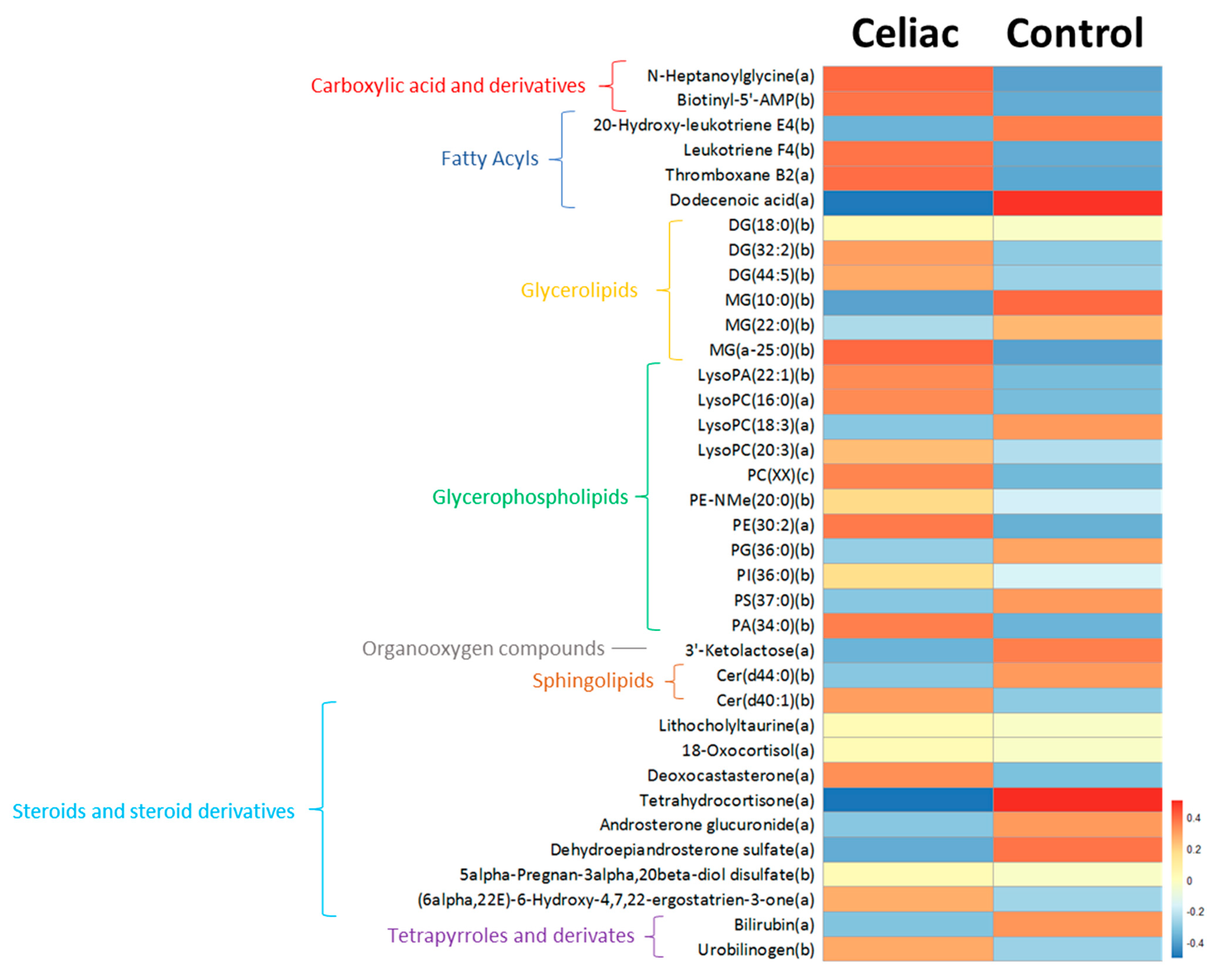
Figure 1. Putatively annotated plasma metabolites significantly different in CD subjects compared to healthy siblings. Levels of statistically significant molecules are represented using a heatmap. Each line of the heatmap represents a lipid species colored by its abundance intensity normalized with an internal standard, log-transformed, and auto-scaled. The scale from blue to red indicates the normalized abundances in arbitrary units. All compounds are putatively annotated based on physicochemical properties and/or spectral similarity with public/commercial spectral libraries [33]. (a) ID based on exact mass, RT, and MS/MS spectrum; (b) ID based on exact mass and RT; and (c) ID based on MS/MS spectrum. PC(XX): the specific phosphatidylcholine species could not be annotated, but its MS/MS spectrum displayed a PC-characteristic pattern.
Nutrients 13 02271 g001
Figure 2. Lipid metabolism seems to be preferentially affected by CD. Levels of statistically significant molecules are represented using a forest plot. Forest plot dots represent the estimated difference in means, and the whiskers represent the confidence intervals. All compounds are putatively annotated based on physicochemical properties and/or spectral similarity with public/commercial spectral libraries [33]. (a) ID based on exact mass, RT, and MS/MS spectrum (high fiability); (b) ID based on exact mass and RT; and (c) ID based on MS/MS spectrum. PC(XX): the specific phosphatidylcholine species could not be annotated, but its MS/MS spectrum displayed a PC-characteristic pattern.
Nutrients 13 02271 g002
Table 1. Demographic and biological characteristics of the study population.
TraitHealthy Siblings (n = 17)Celiac Children (n = 17)p-Value
Sex (female, n (%))10 (58.8)13 (76.4)0.271
Age (years)11.25 (4.23)9.39 (2.77)0.145
Weight (kg)38.54 (16.9)30 (9.63)0.082
Height (cm)140.66 (20.32)131.2 (19.56)0.178
BMI (kg/m2)18.5 (3.98)17 (1.58)0.166
Mediterranean diet (MD) adherence (n (%))
Low1 (5.9)1 (5.9)
Medium8 (47.1)7 (41.2)0.936
High7 (41.2)8 (47.1)
Gluten-free diet (GFD)017
Moderate physical activity (min/week)69.64 (37)81.4 (56.9)0.52
HLA DR-DQ genotype
Negative50
HLA-DQ2+1114
HLA DQ8+00
HLA-DQ2 + DQ8+13
Values shown as mean (standard deviation) unless otherwise indicated.
Publisher’s Note: MDPI stays neutral with regard to jurisdictional claims in published maps and institutional affiliations.