Review

Molecular Mechanisms of the Melatonin Receptor Pathway Linking Circadian Rhythm to Type 2 Diabetes Mellitus

1
Department of Clinical Medicine, School of Medicine, Ningbo University, Ningbo 315211, China
2
Department of Internal Medicine, School of Medicine, Ningbo University, Ningbo 315211, China
3
Department of Preventive Medicine, School of Medicine, Ningbo University, Ningbo 315211, China
4
Department of Biochemistry, School of Medicine, Ningbo University, Ningbo 315211, China
5
Zhejiang Key Laboratory of Pathophysiology, School of Medicine, Ningbo University, Ningbo 315211, China
*
Authors to whom correspondence should be addressed.
Nutrients 2023, 15(6), 1406; https://doi.org/10.3390/nu15061406
Submission received: 16 February 2023 / Revised: 4 March 2023 / Accepted: 10 March 2023 / Published: 15 March 2023
(This article belongs to the Section Nutrition and Diabetes)

Abstract

:
Night-shift work and sleep disorders are associated with type 2 diabetes (T2DM), and circadian rhythm disruption is intrinsically involved. Studies have identified several signaling pathways that separately link two melatonin receptors (MT1 and MT2) to insulin secretion and T2DM occurrence, but a comprehensive explanation of the molecular mechanism to elucidate the association between these receptors to T2DM, reasonably and precisely, has been lacking. This review thoroughly explicates the signaling system, which consists of four important pathways, linking melatonin receptors MT1 or MT2 to insulin secretion. Then, the association of the circadian rhythm with MTNR1B transcription is extensively expounded. Finally, a concrete molecular and evolutionary mechanism underlying the macroscopic association between the circadian rhythm and T2DM is established. This review provides new insights into the pathology, treatment, and prevention of T2DM.

1. Introduction

Circadian rhythm, as the most important inherent biological rhythm, is identified in virtually all eukaryotic organisms. Through the autonomic nervous system and complex hormonal fluctuations, it is deeply involved in the synchronization of physiological functions and daily activities with environmental rhythms [1,2]. If desynchronization occurs, as commonly observed in persons with sleep problems, it will elevate multiple health risks, especially type 2 diabetes, and related cardiovascular complications [3,4,5].
Sleep problems, especially those involving sleep deprivation and circadian rhythm disruption, have been extensively studied. It was estimated that sleep time has markedly reduced, from 8.5 h per day in 2012 to 7.1 h per day in 2021 in China (http://www.zgsmyjh.org, accessed on 11 November 2022), and similar declines have been reported worldwide [6]. As mentioned above, disturbance of the circadian rhythm significantly elevates the risk of type 2 diabetes mellitus (T2DM) [7], which accounts for 90% of diabetes patients [8]. As a rising health burden, the number of adults living with diabetes was estimated to reach 537 million in 2021, corresponding to 10.5% of the world population. Although the epidemiological association between circadian rhythm and T2DM has been reported [9,10], the underlying mechanisms are complex and remain unclear. Melatonin and its receptors serve as intermediaries in synchronizing human endogenous circadian rhythm with environmental rhythms [11]. When people experience circadian rhythm disruption, the central circadian timing system sends incorrect signals that interfere with melatonin release, resulting in the disruption of glucose metabolism, even leading in some cases to T2DM [12]. Numerous studies have explored the intermediary role of the melatonin system, specifically, related signaling pathways, in T2DM. However, few reports have presented a comprehensive summary of the melatonin signaling pathways involved in T2DM.
Thus far, three types of melatonin receptors have been discovered. Among them, MT1 and MT2 are widely distributed in almost all mammals, whereas MT3 is found only in a few animal species. Genome-wide association studies (GWAS) have led to the identification of a strong association between MT2-encoding gene (melatonin receptor 1B, MTNR1B) polymorphisms and an altered risk of T2DM. Importantly, our previous work revealed that MTNR1B is subjected to strong natural selection due to sunshine exposure in human populations worldwide [13]. The combination of two pieces of evidence has suggested the potential molecular and evolutionary mechanisms of MTNR1B action in T2DM, and their regulation through circadian rhythm regulation. Therefore, this review systemically explicates the melatonin signaling pathways related to T2DM, and a reasonable evolutionary mechanism is explained.

2. Melatonin and Its Receptors

Blood melatonin levels are regulated by sunshine exposure; that is, melatonin is released by the pineal gland at night, but its secretion is inhibited in the daytime. Therefore, melatonin release shows a close relationship with sunshine duration [14]. This phenomenon has been verified in experiments with artificial light exposure [15].
As a receptor for light, the retina transforms optical signals into bioelectrical signals, which are then delivered through the optic nerve to the suprachiasmatic nucleus (SCN), the central pacemaker of the circadian timing system. The SCN is critical for regulating the signals transmitted via sympathetic nerves from the paraventricular nucleus (PVN) to the pineal gland, which controls the secretion of melatonin [11]. More specifically, the PVN first communicates with the higher thoracic segments of the intermediolateral spinal column (ISC), from which the sympathetic nerve extends to the superior cervical ganglion, and then, signals are transferred to postsynaptic fibers. After that, the sympathetic postsynaptic nerve reaches the pineal gland, controlling the synthesis of melatonin. From a molecular perspective, melatonin synthesis involves the conversion of tryptophan to melatonin. Specifically, tryptophan is first hydrolyzed by a tryptophan hydroxylase to form 5-hydroxytryptophan, which is further converted into serotonin, and then, serotonin is acetylated by arylalkylamine N-acetyltransferase (AANAT) to yield N-acetylserotonin. Finally, under the catalysis of acetylserotonin O-methyltransferase, N-acetylserotonin is methylated to become N-acetyl-5-methoxytryptamine, namely melatonin [16].
The secretion of melatonin is regulated by different neurotransmitters and nerve conduction systems. As exemplified by the effects of SCN’s regulation on the PVN, astrocytes inside the SCN are first activated to release Glu, which interacts with the NMDA receptor 2 C-terminal (NR2C) in the dark. Next, the activated NR2C promotes neuronal secretion of gamma-aminobutyric acid (GABA). Notably, these neurons are active only in the dark; therefore, GABA cannot be secreted in large amounts during the day. Additionally, GABA has been reported to exert an inhibitory effect on the transmission of sympathetic signals [17], which results in PVN inhibition by the SCN [18]. In other words, PVN activity is inhibited in a dark environment, since a large amount of GABA is released from the SCN at night, but in the daytime, GABA levels are reduced and its inhibitory effect is negligible, allowing PVN to be activated.
Through complex interactions involving nerve impulse transmission, norepinephrine release from the sympathetic postsynaptic fibers, stretching from the superior cervical ganglion, is increased. Subsequently, the released norepinephrine interacts with the β11b adrenergic receptors on the pineal cells, increasing intracellular cAMP concentration, and then, activating protein kinase A (PKA). Next, the activated PKA activates AANAT. Hence, in a dark environment, the transformation of tryptophan into melatonin is activated by norepinephrine release (Figure 1). In contrast, during the day, low SCN neuron activity results in low GABA production, relieving the inhibitory effect on the PVN. Therefore, the postsynaptic nerve in the superior cervical ganglion remains in a resting state, and pineal melatonin synthesis is suppressed.

3. Melatonin Signaling Pathways in T2DM

As typical G protein-coupled receptors (GPCRs), MT1 and MT2 distribute more widely than MT3. Additionally, compared to either MT1 or MT2, MT3 shows lower affinity for melatonin [19].Therefore, human studies tend to focus on the effects of MT1 and MT2 [20,21]. In adult human islet cells, MT2 has been found to be expressed at a level several fold higher than MT1 in both α and β-cells, while MT2 expression has been shown to be similar between these two types of cells [22]. Generally, two circumstances trigger the concrete pathways that mediate the influence of MT1 and MT2 on blood glucose [20]. When MT1 and MT2 are expressed independently, usually in the plasma membranes as monomeric receptors, the Gi protein is recruited and conveys inhibitory signals to adenylate cyclase. However, when MT1 and MT2 form heterodimers, the IP3 pathway is activated, in which the Gq protein is initially recruited and transduces signals to phospholipase C (PLC). The association of these melatonin signaling pathways with T2DM is elucidated in detail below.

3.1. cAMP Pathway

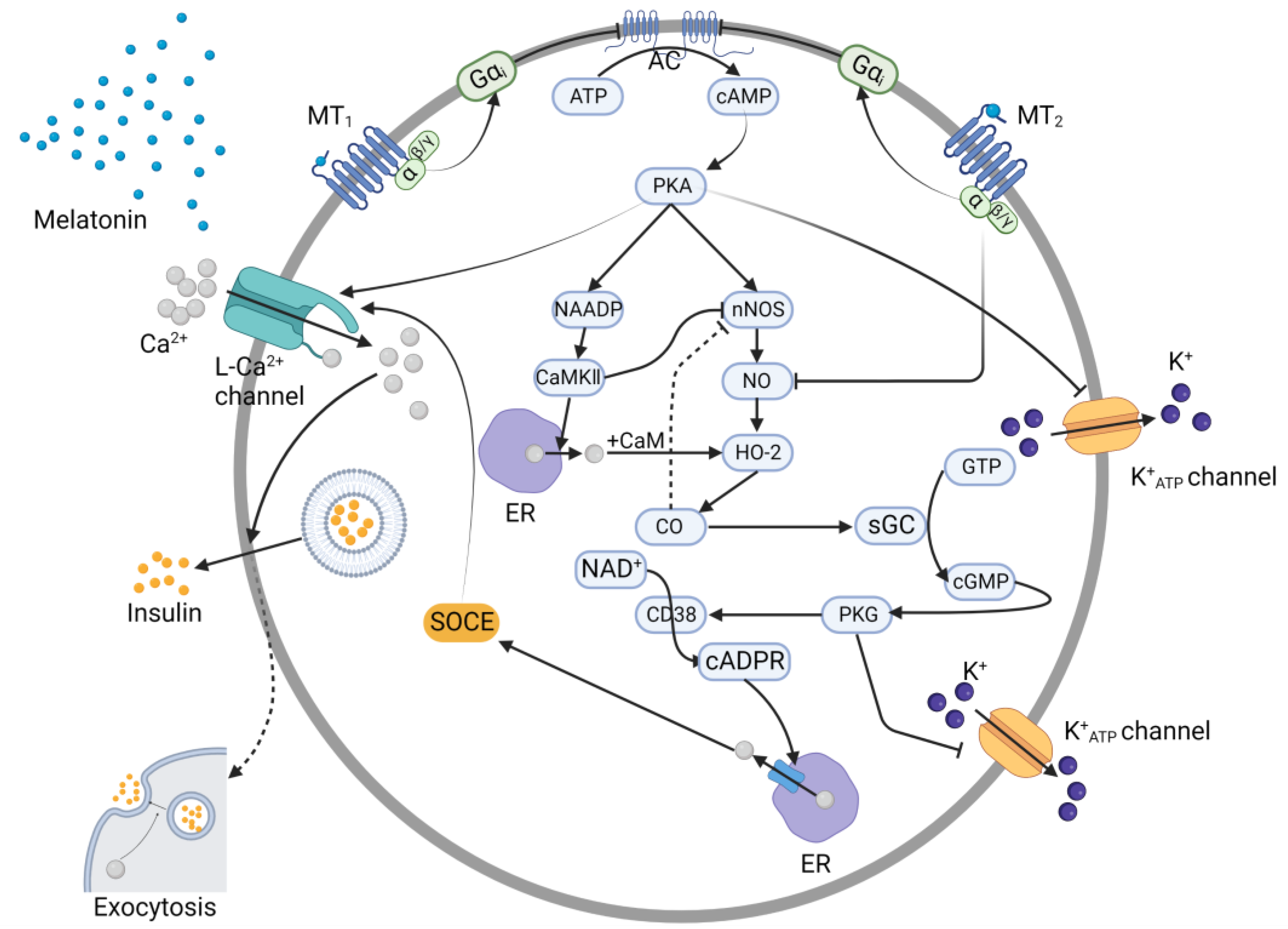
Through a GPCR, the binding of melatonin induces conformational changes in both the monomeric receptors MT1 and MT2, thereby recruiting the trimeric Gi protein to them. After the Gi protein binds to either MT1 or MT2, the α subunit dissociates from the Gi protein, and GDP is converted to GTP. The free Gαi subunit moves to adenylate cyclase, inhibiting its ability to transform ATP into cAMP. As a second messenger, cAMP activates PKA, and then, PKA phosphorylates Ser1928 in the α1 1.2 subunit, and Ser478 and Ser479 in the β2a subunit of the L-type Ca2+ channel [23,24]. With the phosphorylation of Ca2+ channels, the rate of Ca2+ influx increases, which facilitates the fusion of insulin-containing secretory vesicles with the plasma membrane [25]. However, because of the inhibitory effect of the Gαi subunit, the binding of melatonin results in low levels of insulin secretion through the cAMP pathway (Figure 2).

3.2. cGMP Pathway

PKA acts not only directly on L-type Ca2+ channels but also indirectly to regulate Ca2+ channel activity. It induces the production of nicotinic adenine dinucleotide phosphate (NAADP) [26], the initial Ca2+ signal second messenger. NAADP phosphorylates Ca2+/calmodulin-dependent protein kinase II (CaMKII) at Thr286, and then, Ca2+ is mobilized, moving from the endoplasmic reticulum (ER) via the action of phosphorylated CaMKII [27]. Hence, the intracellular Ca2+ concentration increases, and the majority of the Ca2+ in the cytosol is combined with calmodulin (CaM) to form the Ca2+–CaM complex [27], a precursor in the activation of HO-2.
Meanwhile, PKA enhances the activity of nNOS by phosphorylating it at Ser1412 [28], with CaMKII phosphorylating it at Ser847, to exert an inhibitory effect [29]. The comprehensive effect is positive, which can be deduced from the end product of the pathway. Then, NO is synthesized, which in turn activates HO-2 via the action of the Ca2+-CaM complex. HO-2 then produces CO [27], which is considered to be the activator of soluble guanylate cyclase (sGC). When it is in excess, CO can exert an additional inhibitory effect on nNOS. Activated sGC also convert GTPs to cGMP, which in turn activates PKG. PKG targets the CD38 on endosomes. The activation of CD38 leads to the transfer of NAD+ to cADPR, another important Ca2+ signal second messenger. cADPR then mediates Ca2+ release from the ER, a process called store-operated Ca2+ entry (SOCE). Ca2+ continues to induce transmembrane Ca2+ channel opening, resulting in Ca2+ influx and resultant insulin secretion [30]. Noticeably, melatonin-bound MT2 was found to exert an additional negative effect on the synthesis of NO [31], suggesting that the MT2 pathway exerts an additional inhibitory impact on insulin synthesis.
Furthermore, PKG together with PKA exerts an inhibitory effect on K+ATP channels. The K+ATP channel enables K+ efflux, opening when the intracellular ATP concentration decreases, accelerating membrane repolarization. The duration of the action potential decreases; therefore, the activity and the functional duration of the voltage-dependent Ca2+ channel (VDCC) is decreased, and then, the Ca2+ influx is decreased. Since both PKA and PKG inhibit the activity of K+ATP channels, they simultaneously increase Ca2+ influx and insulin secretion [31].
Eventually, as mentioned above, intracellular Ca2+ facilitates the fusion of the insulin-containing secretory vesicles with the plasma membrane, thereby transporting insulin out of the cells (Figure 2). However, due to the inhibitory cascade activated by adenylate cyclase, the whole process originating from melatonin production, exerts a negative impact on insulin secretion. That is, the interaction between melatonin and its receptors decreases the amount of insulin secreted.

3.3. The IP3 Pathway

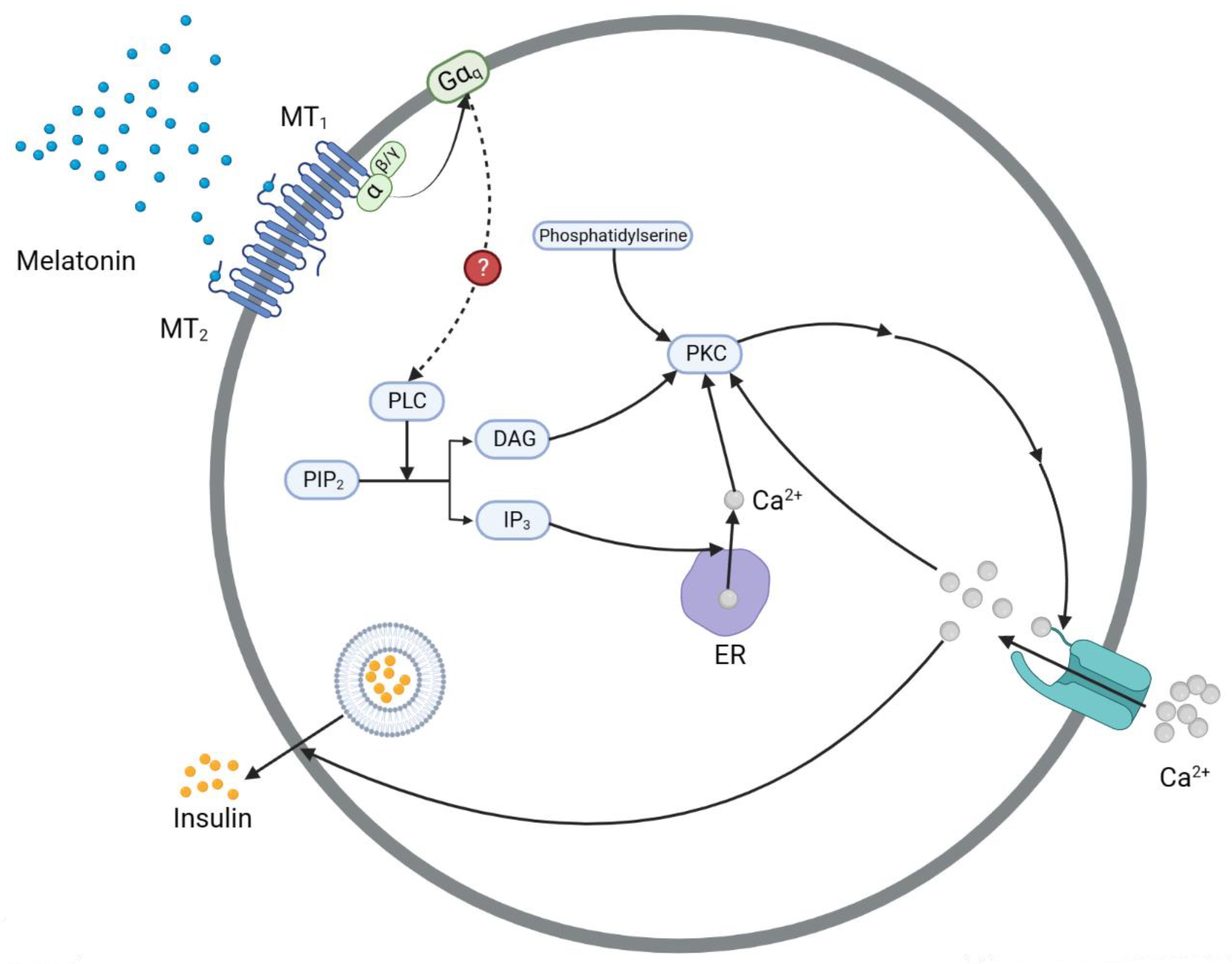
After receptors MT1 and MT2 on β-cells simultaneously bind melatonin, the Gαq subunit is activated, allowing it to leave the trimeric Gq protein complex and bind phospholipase C (PLC) [31]. However, whether the impact of the Gαq subunit on PLC is positive remains unclear. In light of the inhibitory effect of melatonin on gonadotropin-releasing hormone (GnRH)-induced Ca2+ oscillation detected by Zemkova [32,33], it has been speculated that melatonin depresses intracellular Ca2+ store release, which is activated by IP3. Therefore, the impact of the Gαq subunit on PLC is thought to be negative. However, Andreas et al. [34] showed the opposite effect of melatonin on IP3. The difference in outcome may be attributable to the different materials used for these experiments. In Zemkova’s research, the anterior pituitary gland was the subject, while Andreas et al. studied rat insulinoma (INS1) cells.
Nevertheless, regardless of the impact on β-cells, the following pathway is clearly involved [31]: PLC is critical for transforming PIP2 to IP3 and DAG. When IP3 binds to the ER, it causes the efflux of Ca2+ from the ER to the cytosol [35]. Under the combined stimulatory effects of Ca2+, DAG, and phosphatidylserine, PKC is activated, and after successive phosphorylation and activation of various proteins, the transmembrane Ca2+ channel opens, enabling entry of Ca2+ into the cell. Ca2+ influx supplements the endogenous Ca2+ levels, and together, these Ca2+ pools converge to activate PKC in turn. Hence, a PKC–Ca2+ circle forms and endures, through which Ca2+ facilitates insulin secretion (Figure 3). Although the impact of the Gαq subunit on PLC remains unclear, increasing evidence tends to support a positive effect. Therefore, many researchers have attempted to elucidate the comprehensive mechanism underlying both the cAMP and IP3 pathways to explain the overall inhibitory effect of melatonin on insulin secretion considering the premises of the prevailing theory.
Choosing a placebo as a control and 70% fructose as a common solution for the oral glucose tolerance test (OGTT), Patricia et al. [36] found that the insulin level assayed in the morning declines 30 min after the standard OGTT test. However, when it was assayed in the evening, the insulin concentration declined 90 min after the OGTT test. To understand this phenomenon, they analyzed the difference in blood glucose levels and insulin sensitivity indices in both the morning and evening. They concluded that in the evening melatonin impaired glucose tolerance mainly by decreasing insulin sensitivity and insufficient β-cell compensation, but in the morning, it is mainly through melatonin’s direct inhibition of insulin release. At night, the higher levels of insulin resistance led to a greater demand for insulin, which was needed to metabolize large amounts of blood glucose. Therefore, when compared with its effect on the glucose response in the morning, melatonin will elevate plasma concentrations of insulin to a higher peak more slowly at night.
Their results also showed that melatonin exerts a relatively more significant effect on insulin levels in the evening, with insulin levels increasing at first and decreasing later. To explain this phenomenon, several scientists have provided explanations [34,37,38]. In rat cells stimulated with melatonin, the IP3 concentration quickly reached the peak, within the very first minute. Then, it was maintained at an elevated level for at least 15 min and was still at a high level, showing no significant difference compared to the carbachol control group, at 30 min [34]. Although human IP3 pathway-mediated insulin secretion has not been elucidated in detail, it has been speculated that human pancreatic cells show IP3 level changes similar to those in rodent cells; however, the time point at which the IP3 concentration declines may be different [39]. Therefore, insulin levels probably increased through the positive effect of the IP3 pathway. Nevertheless, the subsequent decrease in insulin levels could not be explained solely by the presumably positive effect of the IP3 pathway. Therefore, researchers reanalyzed the cAMP pathway, integrating it with the IP3 pathway to construct a comprehensive signaling system. Many studies [31,37,38] have confirmed that the inhibitory effect of melatonin on the cAMP signaling pathway is realized in approximately 6~8 h. This is much longer than the time required for the activation of the IP3 pathway [37,38,40,41]. Hence, the results from the different researchers’ experiments in which INS1 cells or pancreatic cells were used were compared, and it was concluded that the initial increase in the insulin level was probably caused by the rapidly increasing IP3 level; however, after a relatively long period, when the inhibitory effect of melatonin was predominantly mediated through the cAMP signaling pathway, the K+ATP channel opened, and the L-type Ca2+ channel was inhibited, which led to attenuated Ca2+ influx, and ultimately, inhibited insulin secretion.
Briefly, the melatonin-induced increase in insulin concentration occurs through the melatonin IP3 signaling pathway to compensate for the increasingly fatal insulin resistance at night in humans. This trend ends when the simultaneously activated melatonin cAMP signaling pathway exhibits a higher effect on insulin secretion than the IP3 pathway, inducing a decrease in the insulin level. Additionally, the diminishing insulin level is attributed to the gradually reduced stimulatory effect of the decreasing blood glucose concentration, which further drives insulin levels back to normal.

3.4. Transcription Factor Controlling Pathways

In addition to the three aforementioned signaling pathways, melatonin can also exert an inhibitory impact on insulin secretion at the genetic level. However, few studies have used human cells to explore the regulatory effect of melatonin on insulin gene expression. The majority of the studies performed to date have been carried out with animals, such as rats, serving as proxies for humans, due to their similar expression of insulin-encoding genes.
The insulin-encoding gene in humans is INS, while in rats, it comprises Ins1 and Ins2. In contrast to the majority of vertebrates, rats are exclusively equipped with two independent insulin-encoding genes, Ins1 and Ins2. The evolutionary explanation is based on the complementary functions of two insulin-encoding regions that have enabled adaptation to different environments [42]. In other words, without the simultaneous expression of rat Ins1 and Ins2 genes, insulin could not be synthesized in rats, in contrast to humans, in which a single human INS encodes insulin. Despite a lack of evidence confirming the tight association between INS and Ins1, reports have suggested that the INS and Ins2 structures are very similar, with three exons separated by two introns [42]. To confirm the similar function of INS and Ins2, Karaca et al. [43] were the first to integrate the human INS gene into Ins1/Ins2 double-knockout rats, and they found low insulin production in the transgenic groups. Although the transgenic group expressed only one-half the amount of insulin transcript as the wild-type group, the group concluded that the human INS gene and the rat Ins2 gene resemble each other in structure and transcriptional function. The difference in their expression levels might have been partially attributable to the expression of the Ins1 gene. Hence, to model INS, the study of only the regulatory mechanism underlying Ins2 expression is insufficient. In the following section, we elucidate the concrete signaling pathways involved from melatonin production to insulin gene (Ins1, Ins2) expression.
Using rats as the experimental subjects, Li et al. [44] discovered that the MAPK pathway played an important role in the effect of melatonin on Ins1 and Ins2 expression. The specific signaling process is shown in Figure 4. Through the cAMP-activating process, melatonin activates PKA, which phosphorylates Ras. Then, Raf-1 and MEK1/2 are sequentially activated, and MEK1/2 activates ERK1/2. Finally, ERK1/2 enters the nucleus and promotes the transcription of the Ins1 and Ins2 genes. As studies on the receptor MT1 are lacking, the study group linked only the inhibitory effect of the receptor MT2 to Ins1 and Ins2 gene expression via the Ras/Raf/MEK/ERK pathway, suggesting its regulation was mediated through either transcriptional or translational processes. To describe a more detailed mechanism, we thoroughly reviewed the studies focusing on the transcription of Ins1 and Ins2, which are downstream of the MAPK pathway, together with the vital transcription factors involved. Through meticulously performed experiments, YY1 (Yin Yang 1) and Foxo1 were found to work downstream of ERK1/2, exerting positive effects on Ins1 and Ins2 transcription in pancreatic cells [45,46,47].

3.4.1. YY1

YY1→Ins1, Ins2 Gene

To date, YY1 has been found to regulate insulin expression via three mechanisms. Initially, it can bind to enhancer regions in exon 2 of the Ins1 and Ins2 genes, promoting the activity of pre-proinsulin transcription [48]. In addition, YY1 is an RNA pol II factor, which indicates that by interacting with RNA polymerase II, YY1 helps to stabilize the enhancer–promoter interaction during transcription initiation. Therefore, as the mediator between an enhancer and the polymerase, the binding of YY1 is destined to facilitate the transcription of Ins1 and Ins2.
In addition to its impact on transcription, YY1 also plays a role in translation. As a main component of translation, pre-proinsulin mRNA remains untranslated after Ins1 and Ins2 have been transcribed. However, for eukaryotic rRNAs, when the levels of synthesized 5.8S, 18S, 28S, and their precursor 45S rRNA are changed, the translation or protein expression of insulin is changed. To date, no relevant reports on pancreatic cells have been published. However, Stoeckius et al. [49] discovered that in human vascular smooth muscle cells, increased YY1 levels in the nucleus can stimulate 45S and 5S rRNA synthesis, indicating that YY1 is involved in the pol I/III-catalyzing transcription of 45S and 5S rRNAs, and thus, promotes insulin expression at the translational level.

ERK1/2→YY1

The translocation of YY1 has also been found to be associated with ERK1/2 in the Rb-YY1 cycle [49]. In the cytoplasm, YY1 tends to be coupled with Rb, the retinoblastoma protein, forming the Rb-YY1 complex, and the phosphorylation of this complex by ERK1/2 will facilitate its dissociation. As a result, YY1 is activated and recruited to the nucleoplasm, thereby increasing the transcription levels of the Ins1, Ins2, and rRNA genes. Differently, phosphorylated Rb remains in the cytoplasm, and after dephosphorylation, reenters Rb-YY1 phosphorylation cycle.

3.4.2. Foxo1

In contrast to YY1, Foxo1 has been found to exert a regulatory effect only on the Ins2 gene [45]. However, at least three upstream signaling pathways have been identified thus far, among which the Ras/Raf/MEK/ERK pathway, PI3K pathway, and PKB/Akt pathway seem to have the closest relationships with melatonin and insulin secretions. Therefore, these three pathways are described herein.

ERK1/2→Foxo1→Ins2 Gene

The nuclear import of Foxo1 contributes to the suppression of Ins2 expression [45,50]. More specifically, Foxo1 mainly decreases the transcription level of Ins2 by interacting with a distal promoter [45]. Mezza et al. [47] suggested that ERK1/2 can directly promote Foxo1 export from the nucleus, independent of the insulin signaling pathway. Therefore, ERK1/2 derepresses the inhibitory effect of Foxo1 on Ins2 expression. Generally, since MT2 initially inhibits ERK1/2, the overall MT2 signaling pathway exerts a negative effect on Ins2 expression.

Insulin Receptor→PI3K or PKB/Akt→Foxo1→Ins2 Gene
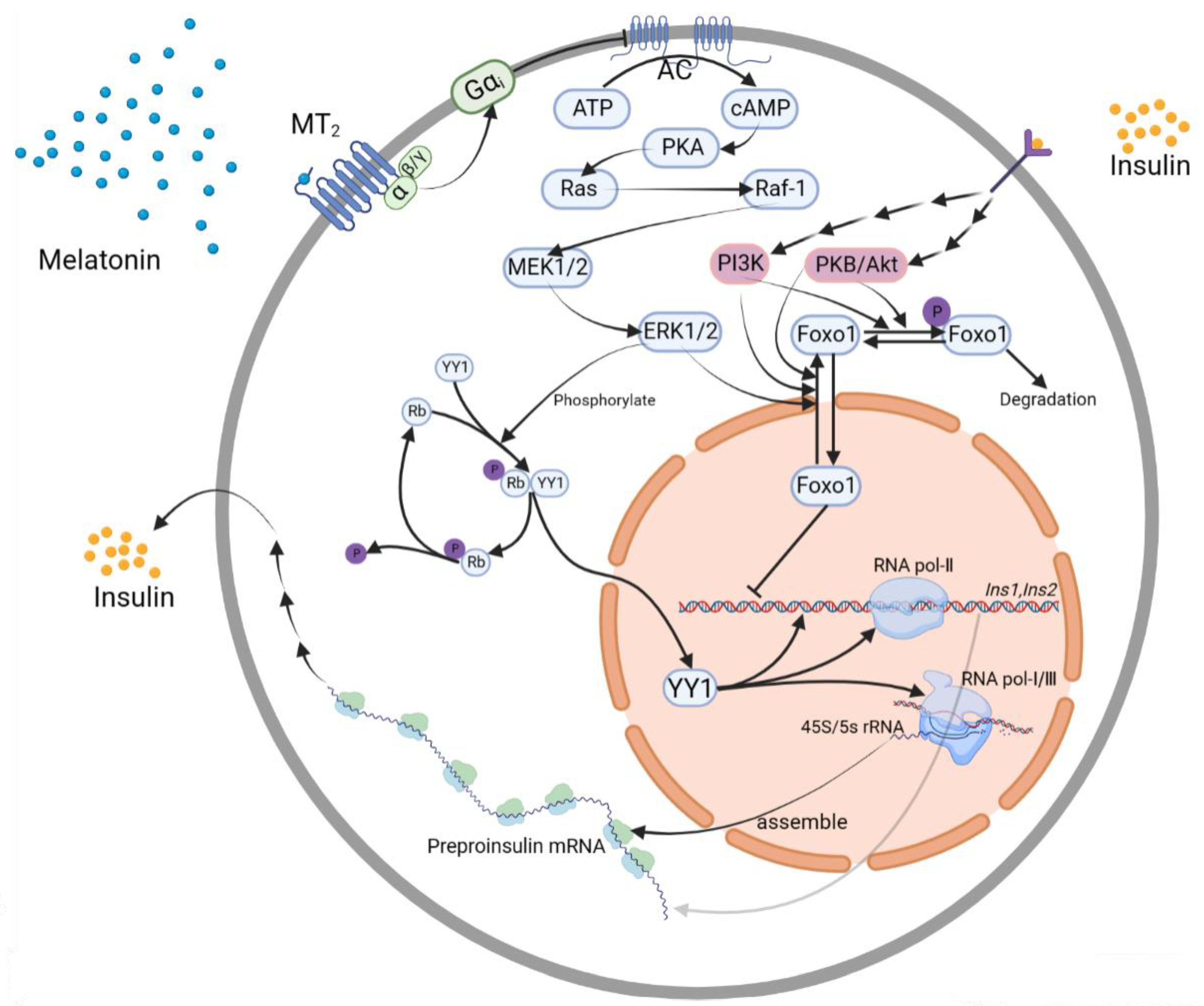
The aforementioned signaling pathways link melatonin to insulin and exert negative effects on insulin secretion. However, insulin may play a positive role in its own secretion [51,52]. The mechanisms, clearly understood, involve the PI3K pathway and PKB/Akt pathway. Once insulin binds to an insulin receptor, the upstream signaling pathway is activated, and when the signal is transduced to PI3K and PKB/Akt, both kinases promote Foxo1 export from the nucleus, and then phosphorylate it, facilitating its degradation. Thus, the inhibitory effect of Foxo1 on Ins2 is derepressed, resulting in enhanced insulin expression. This mechanism is thought to attenuate the negative effect of melatonin on insulin (Figure 4).
Figure 4. The regulation of the melatonin receptor (MT2) to change insulin secretion via insulin gene transcription pathways. The binding of melatonin to MT2 activates the downstream MAPK signaling pathway, which results in ERK1/2 activation. Then, through its interaction with YY1 and Foxo1 separately, ERK1/2 participates in the regulation of pre-proinsulin transcription and translation. Insulin also plays a positive role in its own secretion via the PI3K and PKB/Akt signaling pathways. ERK: extracellular signal-regulated kinase; Foxo1: a member of the large family of forkhead rhabdomyosarcoma transcription factors (FKHRs) [53]; MEK: mitogen-activated protein kinase; PI3K: phosphatidylinositol-3-kinase; PKB/Akt: protein kinase B; Rb: retinoblastoma protein; YY1: Yin Yang 1.
Nutrients 15 01406 g004

4. Regulation of MTNR1B Expression

4.1. Sunshine→MTNR1B Gene Polymorphisms

Since the secretion of melatonin is closely associated with light exposure [11], scientists have questioned whether the expression of melatonin receptors is also regulated by environmental circadian rhythms. Thus, MTNR1A and MTNR1B, encoding MT1 and MT2, respectively, have been extensively studied in vertebrates [54,55]. Mainstream genetic research on glucose metabolism or diabetes [12,56] has focused largely on MTNR1B, with its higher expression levels and stronger genetic susceptibility association. Therefore, we subsequently focused on MTNR1B and MT2 expression.
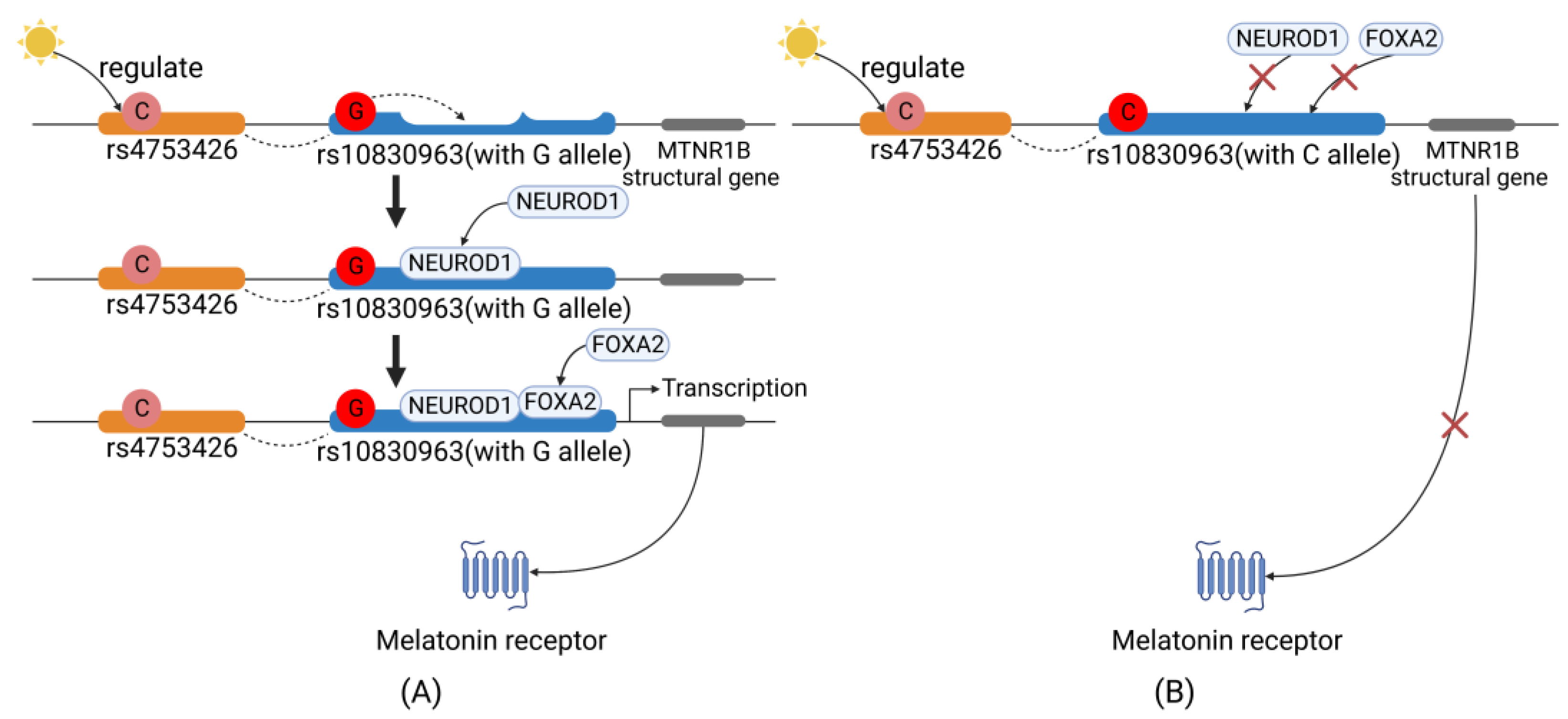
Of all the melatonin pathway-associated disease susceptibility-related single-nucleotide polymorphisms (SNPs), we found that the MTNR1B rs4753426C allele frequency was the only one inversely associated with sunshine duration worldwide, indicating that it has largely undergone natural selection on the basis of sunshine duration [13]. Functionally, rs4753426 is located in the promoter region of MTNR1B [14], suggesting that it plays a regulatory role in gene transcription. Additionally, this SNP has also been suggested to be significantly associated with T2DM [13]. Moreover, another important MTNR1B SNP, rs10830963, has been extensively studied because it is associated with glucose metabolism traits [56,57,58]. Previous studies [59] have demonstrated that the rs4753426C allele and rs10830963G allele are closely linked within the MTNR1B gene (D’ ≥ 0.87, data from the 1000 Genomes Project, Table S1). Therefore, it has been hypothesized that physiological circadian rhythm entrained by environmental sunshine duration probably regulates the expression of melatonin receptor MT2 via its direct or indirect control of rs10830963 and/or rs4753426.

4.2. MTNR1B Gene→MT2 Expression

Sunshine duration regulates the activity of the SCN, the key area controlling the central circadian rhythm, specifically the peripheral circadian rhythm clock activity [11]. The SCN releases endocrine hormones to mediate the transcription of MTNR1B. Studies have revealed that the rs10830963G allele greatly enhances the expression of MT2 [58,60,61,62], with NEUROD1 and FOXA2 [22,63] serving as two important transcription factors in this process. More specifically, as shown in Figure 5, the G allele is carried within a recognition motif in the DNA that matches the consensus sequence of NEUROD1, facilitating NEUROD1 binding to the target site in MTNR1B [64]. In addition, the G allele increases the activity of the FOXA2 binding region [64]. Thus, FOXA2 binds to its cis-acting element and interacts with NEUROD1; through their interaction, the bonds between the DNA and both transcription factors are strengthened. Ultimately, the two transcription factors facilitate the transcription of MTNR1B, contributing to the higher expression of MT2 and lower plasma insulin levels. However, when the G allele is substituted with C, the two transcription factors cannot bind, and MT2 transcription is inhibited, which ultimately leads to lower MT2 levels, and thus, higher insulin levels in plasma [65].

5. Evolutionary Mechanisms

The connection between “low birth weight and high risk of diabetes” was proposed by the thrifty gene hypothesis [66,67], which can also help explain the evolutionary scenario of the MTNR1B gene. According to the out-of-Africa theory, humans originated in Africa approximately 200,000 years ago, and adapted behaviorally and genetically to the local sunshine duration [68]. As they were mainly derived from African ancestors, when ancient people migrated to other continents approximately 50,000~100,000 years ago [69], they may also exchange genes with other ancient non-African human populations [70]. After this migration and gene mixture, the original optimum MTNR1B gene polymorphisms were subjected to a new round of environmental selection. That is, different sunshine durations determined by locations, as well as different climates, induced novel specific advantageous alleles of MTNR1B to accumulate. Then, after a sufficient period of evolution, populations residing in different places demonstrated the alleles in MTNR1B at different frequencies, as shown by the significant inverse correlation between sunshine duration and rs4753426C allele frequency [13], and rs10830963G allele frequency.
Relatively recently, the farming society gradually turned into modern society, creating new selection scenarios. In the farming society, people live to a strict rhythm, working at sunrise and resting at sunset, in order to work and harvest more in a limited time. This pattern of life influenced their genes, which were selected such that the expression of the endocrine hormones and their receptors corresponded to the needs established by the outside environment. On the contrary, modern people often must change their natural life rhythms, especially by shortening their sleep time to spend more time with their families and in businesses; in general, they go to sleep late and get up early. According to a report from Sleep Cycle (https://www.sleepcycle.com, accessed on 2 December 2022), which investigated more than 0.94 million people from 40 countries, the daily sleep time for Chinese people has declined to 6 h 43 min (from 0:59 to 7:42). Moreover, the time at which Chinese people went to sleep in 2021, was 1.5 h later than in 2012. People might stay awake longer than what is considered ideal (7–9 h of sleep required [71]), directly prolonging their light exposure. Hence, these people may display low levels of melatonin [72,73,74], but a normal expression level of melatonin receptors owing to unchanged MTNR1B expression. In these cases, genes and the environment appear to be maladjusted.
This hypothesis is strongly supported by a meta-analysis, through which 17 cohort studies with multiethnic participants were analyzed. The results indicated that short sleep duration significantly increased the risk of T2DM when compared to that of people who slept for a normal duration (risk ratio (RR) = 1.22, p < 0.00001) [75]. Indeed, diminished melatonin secretion was observed in the elderly group in the cohort, who usually slept less on average and for whom the inhibitory effect of melatonin on insulin secretion attenuated. Therefore, these participants displayed elevated insulin levels, representing a greater risk of developing insulin resistance [76], and ultimately, an increase in susceptibility to T2DM. Furthermore, the effect may be pronounced in people eating extra food late at night or eating breakfast early in the morning. Since the insulin levels are not as high as that in the daytime, blood glucose spikes and cannot be absorbed promptly or completely, leading to hyperglycemia and resultant increasing overload of Langerhans’ islets. Accompanied by gradually exacerbating insulin resistance, the untimely rise in blood glucose together with the functionally impaired Langerhans island further contribute to burden the glucose metabolism processes. As a result, these people show a higher possibility of suffering from T2DM.

6. Summary

Characterized by insulin resistance and insufficient insulin secretion, T2DM has become a serious global health burden. Except for the regular risk factors, such as obesity and feeding habits identified by most diabetes research, increasing studies have pointed out that shift work and sleep disorders could also elevate the risk of T2DM, suggesting a physiological mechanism of the circadian disruption involved. However, the underlying molecular and evolutionary mechanism linking circadian disruption and T2DM remains to be elucidated. In this review, we initially summarized four major melatonin-stimulated signaling pathways (the cAMP, cGMP, IP3, and transcription factor-controlled pathways), focusing on their effects on the regulatory mechanisms of insulin secretion. Then, as exemplified by MTNR1B, an evolutionary maladjustment between ancient genes and the present environment was shown to result in circadian rhythm disruption and the exacerbation of impaired glucose metabolism, and even T2DM. Therefore, circadian disruption can be considered a previously unidentified risk factor for T2DM, and the pathological mechanism of this risk was elucidated. This review provides new insights useful to the future prevention, intervention, and treatment of T2DM.
This review also has several limitations. (1) Some studies cited here used non-human species as their experimental materials. Although these animals share a lot in common with humans for glucose metabolism, the differences among species still exist. (2) The evolutionary mechanisms are mainly derived from research on MTNR1B, in future other circadian clock genes, like REV-ERBα and BMAL1 [77,78,79,80], should be further included. (3) This review mainly focused on the association between circadian rhythm disruption and T2DM. However, T2DM also has other noticeable contributing factors like obesity [81] and unhealthy dietary patterns [82]. Hence, a future comprehensive analysis of how circadian disruption works with other factors to finally cause T2DM should be considered. (4) All mechanisms explicated here focus on T2DM, whether they apply to other types of diabetes, such as T1DM, should be further explored. Therefore, more comprehensive studies should be guaranteed to better elucidate the pathological mechanisms of T2DM, from the angle of circadian disruption.

Supplementary Materials

The following supporting information can be downloaded at: https://www.mdpi.com/article/10.3390/nu15061406/s1, Table S1: Linkage disequilibrium statistics (D’) between rs10830963 and rs4753426 in MTNR1B gene.

Author Contributions

This review was conceived by A.-Y.X., J.X. and L.-D.J., A.-Y.X., H.Z., Z.-J.Z., H.-Y.L. and P.-H.W. collected and analyzed the associated publications. A.-Y.X. drafted the manuscript. A.-Y.X., J.X. and L.-D.J. critically revised the work and gave the final approval for the submitted version. All authors have read and agreed to the published version of the manuscript.

Funding

This research was supported by the Zhejiang Public Welfare Technology Application Research Program (LGF20H260009, LGF20H040005), the Zhejiang Medical and Health Science and Technology Program (2022KY1152), and the Ningbo Nonprofit Science and Technology Project (2021S132).

Institutional Review Board Statement

Not applicable.

Informed Consent Statement

Not applicable.

Data Availability Statement

Data authentication is not applicable.

Conflicts of Interest

The authors declare no conflict of interest.

References

  1. Patke, A.; Young, M.W.; Axelrod, S. Molecular mechanisms and physiological importance of circadian rhythms. Nat. Rev. Mol. Cell Biol. 2020, 21, 67–84. [Google Scholar] [CrossRef]
  2. Buijs, R.M.; Escobar, C.; Swaab, D.F. The circadian system and the balance of the autonomic nervous system. Handb. Clin. Neurol. 2013, 117, 173–191. [Google Scholar]
  3. Rizza, S.; Longo, S.; Piciucchi, G.; Romanello, D.; Mavilio, M.; Montagna, M.; Coppeta, L.; Martelli, E.; Magrini, A.; Federici, M. Carotid intimal medial thickness in rotating night shift is related to IL1β/IL6 axis. Nutr. Metab. Cardiovasc. Dis. 2020, 30, 1826–1832. [Google Scholar] [CrossRef] [PubMed]
  4. Rizza, S.; Luzi, A.; Mavilio, M.; Ballanti, M.; Massimi, A.; Porzio, O.; Magrini, A.; Hannemann, J.; Menghini, R.; Lehrke, M.; et al. Alterations in Rev-ERBα/BMAL1 ratio and glycated hemoglobin in rotating shift workers: The EuRhythDia study. Acta Diabetol. 2021, 58, 1111–1117. [Google Scholar] [CrossRef] [PubMed]
  5. Rizza, S.; Luzi, A.; Mavilio, M.; Ballanti, M.; Massimi, A.; Porzio, O.; Magrini, A.; Hannemann, J.; Menghini, R.; Cridland, J.; et al. Impact of light therapy on rotating night shift workers: The EuRhythDia study. Acta Diabetol. 2022, 59, 1589–1596. [Google Scholar] [CrossRef] [PubMed]
  6. Hanson, J.A.; Huecker, M.R. Sleep Deprivation. In StatPearls; StatPearls Publishing: Treasure Island, FL, USA, 2022. [Google Scholar]
  7. Li, Z.-H.; Zhang, P.-D.; Chen, Q.; Gao, X.; Chung, V.C.H.; Shen, D.; Zhang, X.-R.; Zhong, W.-F.; Huang, Q.-M.; Liu, D.; et al. Association of sleep and circadian patterns and genetic risk with incident type 2 diabetes: A large prospective population-based cohort study. Eur. J. Endocrinol. 2021, 185, 765–774. [Google Scholar] [CrossRef]
  8. Sapra, A.; Bhandari, P. Diabetes Mellitus. In StatPearls; StatPearls Publishing: Treasure Island, FL, USA, 2022. [Google Scholar]
  9. Silva-Costa, A.; Rotenberg, L.; Toivanen, S.; Nobre, A.A.; Barreto, S.M.; Schmidt, M.I.; Fonseca, M.D.J.M.D.; Griep, R.H. Lifetime night work exposure and the risk of type 2 diabetes: Results from the longitudinal study of adult health (ELSA-Brasil). Chronobiol. Int. 2020, 37, 1344–1347. [Google Scholar] [CrossRef] [PubMed]
  10. Song, L.; Shen, J.; Wang, J.; Zhang, Y.; Zhou, Z.; Sang, L.; Zhu, L.; Wang, Y.; Zhang, D.; Di Li, D.; et al. Shift patterns, physical exercise, and Type 2 diabetes mellitus (T2DM): A prospective cohort study in China. Transl. Behav. Med. 2023. [Google Scholar] [CrossRef]
  11. Stenvers, D.J.; Scheer, F.A.J.L.; Schrauwen, P.; la Fleur, S.E.; Kalsbeek, A. Circadian clocks and insulin resistance. Nat. Rev. Endocrinol. 2018, 15, 75–89. [Google Scholar] [CrossRef] [Green Version]
  12. Karamitri, A.; Jockers, R. Melatonin in type 2 diabetes mellitus and obesity. Nat. Rev. Endocrinol. 2018, 15, 105–125. [Google Scholar] [CrossRef]
  13. Ji, L.-D.; Xu, J.; Wu, D.-D.; Xie, S.-D.; Tang, N.L.S.; Zhang, Y.-P. Association of disease-predisposition polymorphisms of the melatonin receptors and sunshine duration in the global human populations. J. Pineal Res. 2010, 48, 133–141. [Google Scholar] [CrossRef] [PubMed]
  14. e Silva, A.C.P.; dos Santos, M.J.; Koike, B.; Moreira, M.S.A.; Gitai, D.L.G.; Coelho, J.A.P.D.M.; de Andrade, T.G. Melatonin receptor 1B −1193T>C polymorphism is associated with diurnal preference and sleep habits. Sleep Med. 2018, 53, 106–114. [Google Scholar] [CrossRef]
  15. Touitou, Y.; Reinberg, A.; Touitou, D. Association between light at night, melatonin secretion, sleep deprivation, and the internal clock: Health impacts and mechanisms of circadian disruption. Life Sci. 2017, 173, 94–106. [Google Scholar] [CrossRef] [PubMed]
  16. Cipolla-Neto, J.; Amaral, F.G.D. Melatonin as a Hormone: New Physiological and Clinical Insights. Endocr. Rev. 2018, 39, 990–1028. [Google Scholar] [CrossRef] [PubMed] [Green Version]
  17. Ono, D.; Honma, K.-I.; Yanagawa, Y.; Yamanaka, A.; Honma, S. Role of GABA in the regulation of the central circadian clock of the suprachiasmatic nucleus. J. Physiol. Sci. 2018, 68, 333–343. [Google Scholar] [CrossRef]
  18. Zhang, Q.-J.; Yang, B.-B.; Yang, J.; Wang, Y.-M.; Dai, Y.-T.; Song, N.-H.; Wang, Z.-J.; Xia, J.-D. Inhibitory Role of Gamma-Aminobutyric Receptors in Paraventricular Nucleus on Ejaculatory Responses in Rats. J. Sex. Med. 2020, 17, 614–622. [Google Scholar] [CrossRef]
  19. Tan, D.-X.; Manchester, L.C.; Terron, M.P.; Flores, L.J.; Tamura, H.; Reiter, R.J. Melatonin as a naturally occurring co-substrate of quinone reductase-2, the putative MT3melatonin membrane receptor: Hypothesis and significance. J. Pineal Res. 2007, 43, 317–320. [Google Scholar] [CrossRef]
  20. Liu, J.; Clough, S.J.; Hutchinson, A.J.; Adamah-Biassi, E.B.; Popovska-Gorevski, M.; Dubocovich, M.L. MT1 and MT2 Melatonin Receptors: A Therapeutic Perspective. Annu. Rev. Pharmacol. Toxicol. 2016, 56, 361–383. [Google Scholar] [CrossRef] [Green Version]
  21. Posa, L.; DE Gregorio, D.; Gobbi, G.; Comai, S. Targeting Melatonin MT2 Receptors: A Novel Pharmacological Avenue for Inflammatory and Neuropathic Pain. Curr. Med. Chem. 2018, 25, 3866–3882. [Google Scholar] [CrossRef]
  22. Tuomi, T.; Nagorny, C.L.F.; Singh, P.; Bennet, H.; Yu, Q.; Alenkvist, I.; Isomaa, B.; Östman, B.; Söderström, J.; Pesonen, A.-K.; et al. Increased Melatonin Signaling Is a Risk Factor for Type 2 Diabetes. Cell Metab. 2016, 23, 1067–1077. [Google Scholar] [CrossRef] [Green Version]
  23. Shigeto, M.; Cha, C.Y.; Rorsman, P.; Kaku, K. A role of PLC/PKC-dependent pathway in GLP-1-stimulated insulin secretion. J. Mol. Med. 2017, 95, 361–368. [Google Scholar] [CrossRef]
  24. Seino, S.; Shibasaki, T.; Brown, J.D.; McAnally, D.; Ayala, J.E.; Burmeister, M.A.; Morfa, C.; Smith, L.; Ayala, J.E.; Robichaux, W.G.; et al. PKA-Dependent and PKA-Independent Pathways for cAMP-Regulated Exocytosis. Physiol. Rev. 2005, 85, 1303–1342. [Google Scholar] [CrossRef] [PubMed] [Green Version]
  25. Koneshamoorthy, A.; Seniveratne-Epa, D.; Calder, G.; Sawyer, M.; Kay, T.W.H.; Farrell, S.; Loudovaris, T.; Mariana, L.; McCarthy, D.; Lyu, R.; et al. Case Report: Hypoglycemia Due to a Novel Activating Glucokinase Variant in an Adult—A Molecular Approach. Front. Endocrinol. 2022, 13, 842937. [Google Scholar] [CrossRef] [PubMed]
  26. Kim, B.-J.; Park, K.-H.; Yim, C.-Y.; Takasawa, S.; Okamoto, H.; Im, M.-J.; Kim, U.-H. Generation of Nicotinic Acid Adenine Dinucleotide Phosphate and Cyclic ADP-Ribose by Glucagon-Like Peptide-1 Evokes Ca2+ Signal That Is Essential for Insulin Secretion in Mouse Pancreatic Islets. Diabetes 2008, 57, 868–878. [Google Scholar] [CrossRef] [Green Version]
  27. Rahman, F.U.; Park, D.-R.; Joe, Y.; Jang, K.Y.; Chung, H.T.; Kim, U.-H. Critical Roles of Carbon Monoxide and Nitric Oxide in Ca2+Signaling for Insulin Secretion in Pancreatic Islets. Antioxid. Redox Signal. 2019, 30, 560–576. [Google Scholar] [CrossRef] [PubMed]
  28. Hurt, K.J.; Sezen, S.F.; Lagoda, G.F.; Musicki, B.; Rameau, G.A.; Snyder, S.H.; Burnett, A.L. Cyclic AMP-dependent phosphorylation of neuronal nitric oxide synthase mediates penile erection. Proc. Natl. Acad. Sci. USA 2012, 109, 16624–16629. [Google Scholar] [CrossRef] [PubMed] [Green Version]
  29. Rameau, G.A.; Chiu, L.-Y.; Ziff, E.B. Bidirectional Regulation of Neuronal Nitric-oxide Synthase Phosphorylation at Serine 847 by the N-Methyl-d-aspartate Receptor. J. Biol. Chem. 2004, 279, 14307–14314. [Google Scholar] [CrossRef] [PubMed] [Green Version]
  30. Park, D.-R.; Shawl, A.I.; Ha, T.-G.; Park, K.-H.; Kim, S.-Y.; Kim, U.-H. Arginine Thiazolidine Carboxylate Stimulates Insulin Secretion through Production of Ca2+-Mobilizing Second Messengers NAADP and cADPR in Pancreatic Islets. PLoS ONE 2015, 10, e0134962. [Google Scholar] [CrossRef] [Green Version]
  31. Peschke, E. Melatonin, endocrine pancreas and diabetes. J. Pineal Res. 2007, 44, 26–40. [Google Scholar] [CrossRef]
  32. Zemková, H.; Vaněček, J. Inhibitory Effect of Melatonin on Gonadotropin-Releasing Hormone-Induced Ca2+ Oscillations in Pituitary Cells of Newborn Rats. Neuroendocrinology 1997, 65, 276–283. [Google Scholar] [CrossRef]
  33. Zemková, H.; VaněčEK, J. Differences in Gonadotropin-Releasing Hormone-Induced Calcium Signaling between Melatonin-Sensitive and Melatonin-Insensitive Neonatal Rat Gonadotrophs. Endocrinology 2000, 141, 1017–1026. [Google Scholar] [CrossRef] [PubMed]
  34. Bach, A.G.; Wolgast, S.; Muhlbauer, E.; Peschke, E. Melatonin stimulates inositol-1,4,5-trisphosphate and Ca2+ release from INS1 insulinoma cells. J. Pineal Res. 2005, 39, 316–323. [Google Scholar] [CrossRef]
  35. Wagdi, A.; Malan, D.; Sathyanarayanan, U.; Beauchamp, J.S.; Vogt, M.; Zipf, D.; Beiert, T.; Mansuroglu, B.; Dusend, V.; Meininghaus, M.; et al. Selective optogenetic control of Gq signaling using human Neuropsin. Nat. Commun. 2022, 13, 1765. [Google Scholar] [CrossRef] [PubMed]
  36. Rubio-Sastre, P.; Scheer, F.A.; Gómez-Abellán, P.; Madrid, J.A.; Garaulet, M. Acute Melatonin Administration in Humans Impairs Glucose Tolerance in Both the Morning and Evening. Sleep 2014, 37, 1715–1719. [Google Scholar] [CrossRef] [PubMed] [Green Version]
  37. Peschke, E.; Fauteck, J.-D.; Mußhoff, U.; Schmidt, F.; Beckmann, A.; Peschke, D. Evidence for a melatonin receptor within pancreatic islets of neonate rats: Functional, autoradiographic, and molecular investigations. J. Pineal Res. 2000, 28, 156–164. [Google Scholar] [CrossRef]
  38. Peschke, E.; Mühlbauer, E.; Mußhoff, U.; Csernus, V.J.; Chankiewitz, E.; Peschke, D. Receptor (MT1) mediated influence of melatonin on cAMP concentration and insulin secretion of rat insulinoma cells INS-1. J. Pineal Res. 2002, 33, 63–71. [Google Scholar] [CrossRef]
  39. Rorsman, P.; Braun, M. Regulation of Insulin Secretion in Human Pancreatic Islets. Annu. Rev. Physiol. 2013, 75, 155–179. [Google Scholar] [CrossRef]
  40. Peschke, E.; Peschke, D.; Hammer, T.; Csernus, V. Influence of melatonin and serotonin on glucose-stimulated insulin release from perifused rat pancreatic islets in vitro. J. Pineal Res. 1997, 23, 156–163. [Google Scholar] [CrossRef]
  41. Kemp, D.M.; Ubeda, M.; Habener, J.F. Identification and functional characterization of melatonin Mel 1a receptors in pancreatic β cells: Potential role in incretin-mediated cell function by sensitization of cAMP signaling. Mol. Cell. Endocrinol. 2002, 191, 157–166. [Google Scholar] [CrossRef]
  42. Irwin, D.M. Evolution of the Insulin Gene: Changes in Gene Number, Sequence, and Processing. Front. Endocrinol. 2021, 12, 649255. [Google Scholar] [CrossRef]
  43. Karaca, M.; Durel, B.; Languille, L.; Lamotte, L.; Tourrel-Cuzin, C.; Leroux, L.; Abou Sleymane, G.; Saint-Just, S.; Bucchini, D.; Ktorza, A.; et al. Transgenic expression of human INS gene in Ins1/Ins2 double knockout mice leads to insulin underproduction and diabetes in some male mice. Front. Biosci. 2007, 12, 1586–1593. [Google Scholar] [CrossRef] [PubMed] [Green Version]
  44. Li, Y.; Wu, H.; Liu, N.; Cao, X.; Yang, Z.; Lu, B.; Hu, R.; Wang, X.; Wen, J. Melatonin exerts an inhibitory effect on insulin gene transcription via MTNR1B and the downstream Raf-1/ERK signaling pathway. Int. J. Mol. Med. 2018, 41, 955–961. [Google Scholar] [CrossRef] [PubMed] [Green Version]
  45. Meur, G.; Qian, Q.; da Silva Xavier, G.; Pullen, T.J.; Tsuboi, T.; McKinnon, C.; Fletcher, L.; Tavaré, J.M.; Hughes, S.; Johnson, P.; et al. Nucleo-cytosolic Shuttling of FoxO1 Directly Regulates Mouse Ins2 but Not Ins1 Gene Expression in Pancreatic Beta Cells (MIN6). J. Biol. Chem. 2011, 286, 13647–13656. [Google Scholar] [CrossRef] [Green Version]
  46. Sethi, N.; Wood, H.; Rabbitts, P. The many generations of sequencing technology. Oral Oncol. 2014, 50, e61. [Google Scholar] [CrossRef] [PubMed]
  47. Mezza, T.; Shirakawa, J.; Martinez, R.; Hu, J.; Giaccari, A.; Kulkarni, R.N. Nuclear Export of FoxO1 Is Associated with ERK Signaling in β-Cells Lacking Insulin Receptors. J. Biol. Chem. 2016, 291, 21485–21495. [Google Scholar] [CrossRef] [PubMed] [Green Version]
  48. Liu, D.; Yang, K.Y.; Chan, V.W.; Ye, W.; Chong, C.C.; Wang, C.C.; Wang, H.; Zhou, B.; Cheng, K.K.; Lui, K.O. YY1 Regulates Glucose Homeostasis Through Controlling Insulin Transcription in Pancreatic β-Cells. Diabetes 2022, 71, 961–977. [Google Scholar] [CrossRef] [PubMed]
  49. Stoeckius, M.; Erat, A.; Fujikawa, T.; Hiromura, M.; Koulova, A.; Otterbein, L.; Bianchi, C.; Tobiasch, E.; Dagon, Y.; Sellke, F.W.; et al. Essential Roles of Raf/Extracellular Signal-regulated Kinase/Mitogen-activated Protein Kinase Pathway, YY1, and Ca2+ Influx in Growth Arrest of Human Vascular Smooth Muscle Cells by Bilirubin. J. Biol. Chem. 2012, 287, 15418–15426. [Google Scholar] [CrossRef] [Green Version]
  50. Nagao, M.; Esguerra, J.L.; Asai, A.; Ofori, J.K.; Edlund, A.; Wendt, A.; Sugihara, H.; Wollheim, C.B.; Oikawa, S.; Eliasson, L. Potential Protection Against Type 2 Diabetes in Obesity Through Lower CD36 Expression and Improved Exocytosis in β-Cells. Diabetes 2020, 69, 1193–1205. [Google Scholar] [CrossRef] [PubMed]
  51. Leibiger, I.B.; Leibiger, B.; Berggren, P.-O. Insulin feedback action on pancreatic β-cell function. FEBS Lett. 2002, 532, 1–6. [Google Scholar] [CrossRef] [PubMed] [Green Version]
  52. Halperin, F.; Mezza, T.; Li, P.; Shirakawa, J.; Kulkarni, R.N.; Goldfine, A.B. Insulin regulates arginine-stimulated insulin secretion in humans. Metabolism 2022, 128, 155117. [Google Scholar] [CrossRef] [PubMed]
  53. Remadevi, V.; Muraleedharan, P.; Sreeja, S. FOXO1: A pivotal pioneer factor in oral squamous cell carcinoma. Am. J. Cancer Res. 2021, 11, 4700–4710. [Google Scholar] [PubMed]
  54. Beesley, S.; Lee, J.; Olcese, J. Circadian clock regulation of melatonin MTNR1B receptor expression in human myometrial smooth muscle cells. Mol. Hum. Reprod. 2015, 21, 662–671. [Google Scholar] [CrossRef] [PubMed] [Green Version]
  55. Huang, Y.; Lu, K.; Chao, H.; Chen, A.; Chao, T.; Guo, C.; Hsieh, H.; Shih, H.; Sytwu, H.; Wu, C. The MTNR1A mRNA is stabilized by the cytoplasmic hnRNPL in renal tubular cells. J. Cell. Physiol. 2020, 236, 2023–2035. [Google Scholar] [CrossRef] [PubMed]
  56. Bai, Y.; Tang, L.; Li, L. The roles of ADIPOQ rs266729 and MTNR1B rs10830963 polymorphisms in patients with gestational diabetes mellitus: A meta-analysis. Gene 2019, 730, 144302. [Google Scholar] [CrossRef] [PubMed]
  57. Haljas, K.; Hakaste, L.; Lahti, J.; Isomaa, B.; Groop, L.; Tuomi, T.; Räikkönen, K. The associations of daylight and melatonin receptor 1B gene rs10830963 variant with glycemic traits: The prospective PPP-Botnia study. Ann. Med. 2019, 51, 58–67. [Google Scholar] [CrossRef]
  58. Huang, B.; Wang, Y.-K.; Qin, L.-Y.; Wei, Q.; Liu, N.; Jiang, M.; Yu, H.-P.; Yu, X.-Y. A functional polymorphism rs10830963 in melatonin receptor 1B associated with the risk of gestational diabetes mellitus. Biosci. Rep. 2019, 39, BSR20190744. [Google Scholar] [CrossRef] [Green Version]
  59. Natarajan, R.; Einarsdottir, E.; Riutta, A.; Hagman, S.; Raunio, M.; Mononen, N.; Lehtimäki, T.; Elovaara, I. Melatonin pathway genes are associated with progressive subtypes and disability status in multiple sclerosis among Finnish patients. J. Neuroimmunol. 2012, 250, 106–110. [Google Scholar] [CrossRef]
  60. Li, C.; Zhou, Y.; Qiao, B.; Xu, L.; Li, Y.; Li, C. Association Between a Melatonin Receptor IB Genetic Polymorphism and Its Protein Expression in Gestational Diabetes Mellitus. Reprod. Sci. 2018, 26, 1382–1388. [Google Scholar] [CrossRef]
  61. Garaulet, M.; Gómez-Abellán, P.; Rubio-Sastre, P.; Madrid, J.A.; Saxena, R.; Scheer, F.A. Common type 2 diabetes risk variant in MTNR1B worsens the deleterious effect of melatonin on glucose tolerance in humans. Metabolism 2015, 64, 1650–1657. [Google Scholar] [CrossRef] [Green Version]
  62. Rosta, K.; Al-Aissa, Z.; Hadarits, O.; Harreiter, J.; Nádasdi, A.; Kelemen, F.; Bancher-Todesca, D.; Komlósi, Z.; Németh, L.; Rigó, J.; et al. Association Study with 77 SNPs Confirms the Robust Role for the rs10830963/G of MTNR1B Variant and Identifies Two Novel Associations in Gestational Diabetes Mellitus Development. PLoS ONE 2017, 12, e0169781. [Google Scholar] [CrossRef] [Green Version]
  63. Polychronakos, C.; Alriyami, M. Diabetes in the post-GWAS era. Nat. Genet. 2015, 47, 1373–1374. [Google Scholar] [CrossRef] [PubMed]
  64. Gaulton, K.J.; Ferreira, T.; Lee, Y.; Raimondo, A.; Mägi, R.; Reschen, M.E.; Mahajan, A.; Locke, A.; Rayner, N.W.; Robertson, N.; et al. Genetic fine mapping and genomic annotation defines causal mechanisms at type 2 diabetes susceptibility loci. Nat. Genet. 2015, 47, 1415–1425. [Google Scholar] [CrossRef] [PubMed]
  65. Sparsø, T.; Bonnefond, A.; Andersson, E.; Bouatia-Naji, N.; Holmkvist, J.; Wegner, L.; Grarup, N.; Gjesing, A.P.; Banasik, K.; Cavalcanti-Proença, C.; et al. G-allele of Intronic rs10830963 in MTNR1B Confers Increased Risk of Impaired Fasting Glycemia and Type 2 Diabetes Through an Impaired Glucose-Stimulated Insulin Release: Studies involving 19,605 Europeans. Diabetes 2009, 58, 1450–1456. [Google Scholar] [CrossRef] [PubMed] [Green Version]
  66. Neel, J.V. Diabetes mellitus: A “thrifty” genotype rendered detrimental by “progress”? Am. J. Hum. Genet. 1962, 14, 353–362. [Google Scholar]
  67. Stocker, C.J.; Arch, J.R.S.; Cawthorne, M.A. Fetal origins of insulin resistance and obesity. Proc. Nutr. Soc. 2005, 64, 143–151. [Google Scholar] [CrossRef] [Green Version]
  68. Tattersall, I. Out of Africa: Modern human origins special feature: Human origins: Out of Africa. Proc. Natl. Acad. Sci. USA 2009, 106, 16018–16021. [Google Scholar] [CrossRef] [Green Version]
  69. Pagani, L.; Schiffels, S.; Gurdasani, D.; Danecek, P.; Scally, A.; Chen, Y.; Xue, Y.; Haber, M.; Ekong, R.; Oljira, T.; et al. Tracing the Route of Modern Humans out of Africa by Using 225 Human Genome Sequences from Ethiopians and Egyptians. Am. J. Hum. Genet. 2015, 96, 986–991. [Google Scholar] [CrossRef] [Green Version]
  70. Scerri, E.M.L.; Chikhi, L.; Thomas, M.G. Beyond multiregional and simple out-of-Africa models of human evolution. Nat. Ecol. Evol. 2019, 3, 1370–1372. [Google Scholar] [CrossRef]
  71. Hirshkowitz, M.; Whiton, K.; Albert, S.M.; Alessi, C.; Bruni, O.; DonCarlos, L.; Hazen, N.; Herman, J.; Katz, E.S.; Kheirandish-Gozal, L.; et al. National sleep foundation’s sleep time duration recommendations: Methodology and results summary. Sleep Health 2015, 1, 40–43. [Google Scholar] [CrossRef]
  72. Münch, M.; Knoblauch, V.; Blatter, K.; Schröder, C.; Schnitzler, C.; Kräuchi, K.; Wirz-Justice, A.; Cajochen, C. Age-related attenuation of the evening circadian arousal signal in humans. Neurobiol. Aging 2005, 26, 1307–1319. [Google Scholar] [CrossRef]
  73. Cajochen, C.; Münch, M.; Knoblauch, V.; Blatter, K.; Wirz-Justice, A. Age-related Changes in the Circadian and Homeostatic Regulation of Human Sleep. Chronobiol. Int. 2006, 23, 461–474. [Google Scholar] [CrossRef]
  74. McIntyre, I.M.; Norman, T.R.; Burrows, G.D.; Armstrong, S.M. Human melatonin response to light at different times of the night. Psychoneuroendocrinology 1989, 14, 187–193. [Google Scholar] [CrossRef] [PubMed]
  75. Lu, H.; Yang, Q.; Tian, F.; Lyu, Y.; He, H.; Xin, X.; Zheng, X. A Meta-Analysis of a Cohort Study on the Association between Sleep Duration and Type 2 Diabetes Mellitus. J. Diabetes Res. 2021, 2021, 8861038. [Google Scholar] [CrossRef] [PubMed]
  76. Lauritzen, E.S.; Kampmann, U.; Smedegaard, S.B.; Støy, J. Effects of daily administration of melatonin before bedtime on fasting insulin, glucose and insulin sensitivity in healthy adults and patients with metabolic diseases. A systematic review and meta-analysis. Clin. Endocrinol. 2021, 95, 691–701. [Google Scholar] [CrossRef]
  77. Kou, L.; Chi, X.; Sun, Y.; Han, C.; Wan, F.; Hu, J.; Yin, S.; Wu, J.; Li, Y.; Zhou, Q.; et al. The circadian clock protein Rev-erbα provides neuroprotection and attenuates neuroinflammation against Parkinson’s disease via the microglial NLRP3 inflammasome. J. Neuroinflamm. 2022, 19, 133. [Google Scholar] [CrossRef]
  78. Cui, L.; Xu, F.; Xu, C.; Ding, Y.; Wang, S.; Du, M. Circadian gene Rev-erbα influenced by sleep conduces to pregnancy by promoting endometrial decidualization via IL-6-PR-C/EBPβ axis. J. Biomed. Sci. 2022, 29, 101. [Google Scholar] [CrossRef]
  79. Sehirli, A.; Chukwunyere, U.; Aksoy, U.; Sayiner, S.; Abacioglu, N. The circadian clock gene Bmal1: Role in COVID-19 and periodontitis. Chronobiol. Int. 2021, 38, 779–784. [Google Scholar] [CrossRef] [PubMed]
  80. McKee, C.A.; Lee, J.; Cai, Y.; Saito, T.; Saido, T.; Musiek, E.S. Astrocytes deficient in circadian clock gene Bmal1 show enhanced activation responses to amyloid-beta pathology without changing plaque burden. Sci. Rep. 2022, 12, 1796. [Google Scholar] [CrossRef] [PubMed]
  81. Bell, J.A.; Kivimaki, M.; Hamer, M. Metabolically healthy obesity and risk of incident type 2 diabetes: A meta-analysis of prospective cohort studies. Obes. Rev. 2014, 15, 504–515. [Google Scholar] [CrossRef] [Green Version]
  82. Bellou, V.; Belbasis, L.; Tzoulaki, I.; Evangelou, E. Risk factors for type 2 diabetes mellitus: An exposure-wide umbrella review of meta-analyses. PLoS ONE 2018, 13, e0194127. [Google Scholar] [CrossRef] [Green Version]
Figure 1. The regulation of melatonin synthesis in the dark. In the dark, melatonin synthesis in the pineal gland is regulated through successively transmitted nerve impulses. ① Astrocytes in the SCN are activated and release Glu, which interacts with NR2C, inducing the release of GABA. ② The sympathetic postsynaptic nerves release norepinephrine to activate AANAT, which promotes the synthesis of melatonin. AANAT: arylalkylamine-N-acetyltransferase; GABA: γ-aminobutyric acid; Glu: glutamate; NE: norepinephrine; NR2C: NMDA receptor 2 C-terminal; Trp: tryptophan. Both the red arrows in graph ① and black arrows in graph ② show the directions of signaling pathways, while the red upward arrows in graph ② represent concentration increases of each molecules.
Nutrients 15 01406 g001
Figure 2. The regulation of melatonin receptors (MT1/MT2) on the secretion of insulin mediated via cAMP and cGMP pathways. Through MT1 and MT2 activation, the catalytic activity of AC is downregulated, leading to reduced activation of PKA. Then, PKA is inactivated in one of three pathways, the cAMP pathway, the cGMP pathway, and the K+ATP channel, to regulate insulin secretion in combination. AC: adenylate cyclase; ATP: adenosine triphosphate; cADPR: cyclic ADP-ribose; cAMP: cyclic AMP; CaMKII: Ca2+/calmodulin-dependent protein kinase II; cGMP: cyclic GMP; CO: carbon monoxide; ER: endoplasmic reticulum; Gαi: the α subunit of the G protein; GTP: guanosine triphosphate; HO-2: heme oxygenase 2; nNOS: neuronal nitric oxide synthase; NAD+: nicotinamide adenine dinucleotide; NAADP: nicotinic acid adenine dinucleotide phosphate; NO: nitric oxide; PKA: protein kinase A; PKG: protein kinase G; sGC: soluble guanylyl cyclase; and SOCE: store-operated Ca2+ entry.
Nutrients 15 01406 g002
Figure 3. The regulatory effect of melatonin receptors (MT1, MT2) on the secretion of insulin via the IP3 pathway. Through the PKC-dependent signaling pathway, the activation of MT1 and MT2 heterodimers ultimately causes Ca2+ to traverse the plasma membrane, changing the level of insulin excreted. DAG: diacylglycerol; IP3: inositol-1,4,5-trisphosphate; PKC: protein kinase C; PLC: phospholipase C. PLC catalyzes PIP2 transformation to DAG and IP3.
Nutrients 15 01406 g003
Figure 5. Molecular regulation of sunshine duration on the transcription of the MTNR1B gene. Specifically, rs10830963 allelic polymorphism show different MT2 expression patterns in response to sunshine duration. (A) rs10830963G allele-containing MTNR1B and (B) rs10830963C allele-containing MTNR1B. The dash line indicates linkage disequilibrium between rs4753426C and rs10830963G.
Nutrients 15 01406 g005
Disclaimer/Publisher’s Note: The statements, opinions and data contained in all publications are solely those of the individual author(s) and contributor(s) and not of MDPI and/or the editor(s). MDPI and/or the editor(s) disclaim responsibility for any injury to people or property resulting from any ideas, methods, instructions or products referred to in the content.