Article

Anti-Steatotic Effects of Chlorella vulgaris, Nannochloropsis gaditana and Gracilaria vermiculophylla Algae Extracts in AML-12 Hepatocytes

by
Maitane González-Arceo
1,
Jenifer Trepiana
1,2,3,*,
Leixuri Aguirre
1,2,3,*,
Jone Ibarruri
4,
Marta Martínez-Sanz
5,
Marta Cebrián
4,
Isidra Recio
5,
María P. Portillo
1,2,3 and
Saioa Gómez-Zorita
1,2,3
1
Nutrition and Obesity Group, Department of Nutrition and Food Science, University of the Basque Country (UPV/EHU) and Lucio Lascaray Research Institute, 01006 Vitoria-Gasteiz, Spain
2
CIBEROBN Physiopathology of Obesity and Nutrition, Institute of Health Carlos III, 01006 Vitoria-Gasteiz, Spain
3
Bioaraba Health Research Institute, 01006 Vitoria-Gasteiz, Spain
4
AZTI, Food Research, Basque Research and Technology Alliance (BRTA), Parque Tecnológico de Bizkaia, Astondo Bidea, 609, 48160 Derio, Spain
5
Instituto de Investigación en Ciencias de la Alimentación, CIAL (CSIC-UAM), Nicolás Cabrera, 9, 28049 Madrid, Spain
*
Authors to whom correspondence should be addressed.
Nutrients 2023, 15(8), 1960; https://doi.org/10.3390/nu15081960
Submission received: 21 March 2023 / Revised: 13 April 2023 / Accepted: 18 April 2023 / Published: 19 April 2023
(This article belongs to the Special Issue Food and Botanical Derived Phytoextracts for Health Benefits)

Abstract

:
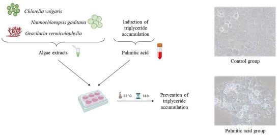
Non-alcoholic fatty liver disease (NAFLD) is considered the most common chronic liver alteration whose prevalence is increasing in Western countries. Microalgae and macroalgae have attracted great interest due to the high content in bioactive compounds with beneficial effects on health. The aim of the present study is to assess the potential interest of extracts rich in proteins obtained from the microalgae Chlorella vulgaris and Nannochloropsis gaditana and the macroalga Gracilaria vermiculophylla in the prevention of lipid accumulation in AML-12 hepatocytes. Toxicity was not observed at any of the tested doses. Both microalgae and the macroalga were effective in preventing triglyceride accumulation, with Nannochloropsis gaditana being the most effective one. Although the three algae extracts were able to increase different catabolic pathways involved in triglyceride metabolism, the mechanisms underlying the anti-steatotic effect were different in each algae extract. In conclusion, the present study demonstrates that Chlorella vulgaris, Nannochloropsis gaditana and Gracilaria vermiculophylla extracts are able to partially prevent the accumulation of triglycerides induced by palmitic acid in cultured hepatocytes, a model used to mimic the steatosis induced in liver by dietary patterns rich in saturated fat.

Graphical Abstract

1. Introduction

Non-alcoholic fatty liver disease (NAFLD), characterised by the excess of lipid accumulation within hepatocytes (>5%) [1], is the most common chronic liver disease in Western countries, affecting approximately 25% of the adult population [2]. Its prevalence continues to grow due to the increase in sedentary behaviours and westernisation of the diet, factors that contribute to the above-mentioned metabolic alteration. The development of NAFLD is closely related to obesity and insulin resistance [3]. Recently, a panel of experts has proposed renaming NAFLD as metabolic dysfunction-associated fatty liver disease (MAFLD) since it more accurately reflects the heterogeneous pathogenesis and the underlying metabolic abnormalities [4].
Nowadays, there is no pharmacological treatment approved for liver steatosis. Diet and physical activity are the main common strategies used to prevent or treat this hepatic alteration [5]. Consequently, there is a growing interest in the development of new therapies [2] based, for instance, on bioactive molecules present in foodstuffs and plants. In this context, many authors are focusing their attention on marine algae, which contribute to the sustainable blue growth in the European Union, a strategy that ranges from responsible food systems to decarbonisation, as well as biodiversity and coastal resilience to circularity [6]. Algae are rich in several bioactive compounds such as pigments, polyunsaturated fatty acids, phenolic compounds, peptides, lipids, vitamins, polysaccharides or sterols, among others. It has been reported that they exert antioxidant, anti-inflammatory, anti-obesity, anti-diabetic, hypolipidemic, anti-hypertensive and anti-cancer activities [7,8,9]. As a result, diets supplemented with these organisms, both micro- and macroalgae, may have positive effects on chronic diseases [10,11,12,13].
Regarding the effects of algae on hepatic steatosis, although several species of both microalgae and macroalgae have demonstrated to have a positive effect on this alteration, the reported studies are still scarce [10]. The aim of the present study is to assess the potential interest of extracts obtained from the microalgae Chlorella vulgaris and Nannochloropsis gaditana and from the macroalga Gracilaria vermiculophylla in the prevention of liver steatosis. To our knowledge, the activity of these algae on hepatic triglyceride accumulation has yet to be studied. Furthermore, our aim is to identify the mechanisms responsible for this effect.

2. Materials and Methods

2.1. Algae Extract Preparation

Gracilaria vermiculophylla was manually collected in September 2019 in the Bidasoa estuary (Hondarribia, Gipuzkoa, Spain). After collection and washing with marine water in the field, a second washing with fresh water was carried out in the lab to remove the impurities. After, samples were vacuum packed and frozen at −20 °C. Both microalgae (Chlorella vulgaris and Nannochloropsis gaditana) were provided by NEOALGAE (Gijón, Asturias, Spain) in frozen fresh-paste format (20–25% total solids) and stored at −20 °C until use. Before extraction, frozen biomass was thawed at 40 °C and diluted to the desired solid content with distilled water.
Microalgae extracts were produced by adapting the methodology of Safi et al. [14,15]. A suspension of microalgae at 10% dry weight and adjusted to pH 12 with NaOH 10 M was prepared. The suspension was vacuum percolated through 100 μm filters to avoid issues with the Ultra High-Pressure Homogenizer (UHPH) equipment MicroDeBee (Bee International, South Easton, MA, USA) and stored in cold and dark atmosphere to avoid deterioration. The applied UHPH conditions were 250 MPa, 250 µm orifice and three cycles. The samples were submerged in an ice-water bath for proper preservation. After the UHPH process, samples were centrifuged at 10,000× g for 10 min at 4 °C (avoiding direct light). Soluble proteins were collected in the supernatant. These proteins were precipitated by adjusting the pH to 3.0 with 6 M HCl. Samples were again centrifuged at 10,000× g for 10 min at 4 °C, and the pellet was resuspended in 0.1 M phosphate buffer with a pH of 7.5.
Macroalga extract was prepared as follows: frozen samples were thawed in a water bath at 40 °C and crushed to a particle size between 1 and 5 mm. A suspension of the macroalga in water at 5% of dry weight solids was prepared, grinded with a homogeniser (Ultra-turrax-IKA-T25, Staufen, Germany) for two minutes at 18,000 rpm and adjusted to a pH of 12 with NaOH 10 M. The suspension was then treated by ultrasounds (VibraCell 75042, Bioblock Scientific, Illkirch, France) for 1 h and 30 min, in cycles of 59 s ON and 15 s OFF. This treatment was also submerged in an ice-water bath to avoid an increase in temperature and protected from light to prevent alterations in bioactive compounds. After the extraction process, samples were handled in the same way as the microalgae suspensions after the UHPH procedure.

2.2. Algae Extract Composition

The proximate composition of the extracts was analysed according to the Association of Official Analytical Chemists (AOAC) Official Methods [16]. The moisture of the samples was determined through a drying process at 100 °C to a constant weight (method 934.01). Crude protein content was determined by Kjeldahl methodology, using a conversion factor for nitrogen-to-protein of N × 6.25 and 4.59 for microalgae and macroalga, respectively (method 955.04). Crude fat content was determined by Soxhlet methodology (method 920.39). Carbohydrate content was calculated by the difference in 100 g of algae extract. The total phenolic content was measured following the procedure described by Singleton and Rossi [17].

2.3. Characterization of the Protein Fraction

Sodium dodecyl sulphate-polyacrylamide gel electrophoresis (SDS-PAGE) was used on the algae extracts. Based on the protein content estimated by Kjeldahl, the extracts were dissolved in sample buffer, which contained Tris-HCl (0.05 M, pH 6.8, SDS (1.6% w:v), glycerol (8% v:v), β-mercaptoethanol (2% v:v) and bromophenol blue indicator (0.002% w:v) at protein concentrations of 0.8, 1.0 and 1.5 mg/mL. The samples were heated at 95 °C for 5 min and were subsequently loaded on the 12% Bis-Tris polyacrylamide gel (Criterion XT, Bio-Rad, Richmond, CA, USA). Electrophoretic separations were carried out at 150 V using XT-MES as a running buffer (Bio-Rad) in the Criterion cell (Bio-Rad). The gels were stained with Coomassie Blue (Instant Blue, Expedeon, Swavesey, UK), and images were taken with a Molecular Imager VersaDoc™ MP 5000 system (Bio-Rad, Hercules, CA, USA).
The protein/peptide size distribution was evaluated with high-performance size-exclusion chromatography (HPLC-SEC) using an ultra-high performance liquid chromatography instrument (Waters) equipped with a bioZenTM 1.8 µm 150 × 4.6 mm column and a bioZenTM SEC-2, 4.6 mm pre-column (Phenomenex). The mobile phase was a 55:45:0.1 water:acetonitrile:TFA (v/v/v) solution with a flow of 100 µL/min. The algae extracts were dissolved in water at a concentration of 3 mg/mL, diluted in the mobile phase to obtain a protein concentration of 1.5 mg/mL and centrifuged for 5 min at 12,800× g at room temperature to separate the soluble fraction. A total of 3 μL of each sample was injected into the system and the absorbance was registered at wavelengths of 214 and 280 nm. Peptides of known molecular weight (insulin with 5800 Da and peptides VPFPGPI and VYII with 888 Da and 506 Da, respectively) were used as molecular weight markers using the same chromatographic conditions.

2.4. Cell Culture and Treatment

A mouse hepatocyte AML12 cell line (alpha mouse liver 12; ATCC® CRL-2254™), obtained from the American Type Culture Collection (ATCC, Manassas, VA, USA), was maintained in 75 cm2 flasks in DMEM/HAM’s F12 Glutamax and supplemented with 10% heat inactivated foetal bovine serum, 5 μg/mL insulin, 5 μg/mL transferrin, 5 ng/mL selenium, 40 ng/mL dexamethasone and 1% penicillin/streptomycin (10,000 U/mL) at standard cell culture conditions (37 °C, 5% CO2). When the cell monolayer reached 75% of confluence, cells were detached with a solution of trypsin-EDTA and harvested to perform subsequent experiments.
AML12 cells were incubated with palmitic acid (PA) to create an in vitro model of steatotic hepatocytes [18]. Cells were briefly exposed or not to 0.3 mM of PA to induce triglyceride accumulation and co-incubated with algae extracts for 18 h, adjusting the final concentration of each algae extract in culture media to 10, 25, 50 or 150 μg/mL. The control groups received the same amount of the vehicle. Each experiment was performed at least three times.
After 18 h, the incubation medium was collected, and cells were harvested according to the subsequent analysis. Cells used for triglyceride determination and protein immunodetection were collected in 10 mM Tris-HCl pH 7.4, 150 mM NaCl and 1 mM ethylenediaminetetraacetic acid (EDTA) buffer through scrapping. The cell suspension was then sonicated with 5 s bursts in a Branson Sonifier SFX550 (San Luis, MO, USA) fitted with a microtip. Cells used for RNA extraction were collected in TRIzol® reagent.

2.5. Cell Viability Assay

Cell viability was assessed using the crystal violet assay, based on cell staining with crystal violet [19]. Following treatment, AML12 cells were briefly washed with phosphate buffered saline (PBS), fixed in 3.7% formaldehyde and stained with 0.25% crystal violet in the dark for 20 min. Lastly, the resulting crystals were solubilised with 33% acetic acid, and the absorbance was registered at 590 nm in an iMark microplate reader (Bio-Rad, Hercules, CA, USA). Cell viability was expressed as the percentage of the control cells.

2.6. Determination of Triglyceride Content

Triglyceride content in cell suspension was measured using a commercial kit (Spinreact, Girona, Spain). Protein measurements were performed using the Bradford method [20]. Triglyceride content values were obtained as mg triglycerides/mg protein and expressed as the percentage of the control cells.

2.7. Detection of Alanine Aminotransferase (ALT/GPT) Levels in Cell Culture Medium

To determine ALT/GPT levels in cell culture medium, a commercial kit (BioSystems, Barcelona, Spain) was used.

2.8. Analysis of Gene Expression by Real-Time PCR

RNA was extracted from cells using RNeasy Mini Spin Columns (Qiagen, Carlsbad, CA, USA) according to the manufacturer’s instructions. After DNase treatment (Ambion, Foster City, CA, USA), the integrity of the RNA was verified and quantified using an RNA 6000 Nano Assay (Thermo Scientific, Wilmington, DE, USA). A total of 1.5 μg of total RNA in a total reaction volume of 30 μL from each sample was reverse-transcribed into complementary DNA (cDNA) using an iScript cDNA Synthesis Kit (Bio-Rad, Hercules, CA, USA). Reactions were incubated initially at 25 °C for 5 min, subsequently at 42 °C for 30 min and finally at 85 °C for 5 min.
Adipose triglyceride lipase (Atgl), carnitine palmitoyltransferase I a (Cpt1a), citrate synthase (Cs), fatty acid synthase (Fasn), long chain acyl-CoA dehydrogenase (Acadl), mitochondrial transcription factor A (Tfam) and uncoupling protein 2 (Ucp2) mRNA levels were quantified. Beta actin (Actb) served as housekeeping for posterior normalisation. An aliquot of 4.75 μL of diluted cDNA sample was amplified by Real-Time PCR in a 12.5 μL reaction volume. The cDNA samples were amplified in an iCycler-MyiQ Real-Time PCR Detection System (Bio-Rad, Hercules, CA, USA) in the presence of SYBR Green Master Mix (Applied Biosystems, Foster City, CA, USA) and the sense and antisense primers (300 nM each except for Fas, where a concentration of 600 nM was used). The primer sequences are described in Table 1. The PCR parameters were as follows: initial 2 min at 50 °C, denaturation at 95 °C for 10 min, followed by 40 cycles of denaturation at 95 °C for 15 s, annealing at 60 °C for 30 s and extension at 60 °C for 30 s. For Acadl and Fasn, the annealing temperature was 61 and 62.2 °C, respectively.
Acetyl-CoA carboxylase (Acc) and diacylglycerol acyltransferase (Dgat2) were amplified using TaqMan probes. Actb mRNA levels were similarly measured and served as the reference gene. In total, 4.5 μL of each diluted cDNA sample was added to the PCR reagent mixture (final volume of 10 μL), which consisted of TaqMan Fast Advanced Master Mix (Applied Biosystems, Vilna, Lithuania) and TaqMan Gene Expression Assay Mix (Applied Biosystems, Foster City, Ca, USA) containing specific primers and probes (Table 1). The PCR parameters were as follows: initial 2 min at 50 °C, denaturation at 95 °C for 2 min, followed by 40 cycles of denaturation at 95 °C for 3 s and combined annealing and extension at 60 °C for 30 s.
In all cases, the results were expressed as fold changes of the threshold cycle (Ct) value relative to controls using the 2−ΔΔCt method [21].

2.9. Analysis of Protein Expression by Western Blot

Solute carrier family 27 member 2 (FATP2) and microsomal triglyceride transfer protein (MTTP) were assessed by western blot. The protein concentration was determined according to the Bradford protocol [20]. Protein samples (40 μg) were denaturalised at 95 °C for 3 min in Laemmli buffer [22] and loaded into 4–15% Mini-PROTEAN TGX Precast Gels (BioRad, Hercules, CA, USA). The proteins were then transferred onto PVDF membranes (Millipore, MA, USA) by electroblotting and later blocked with 5% casein and 0.5% bovine serum albumin (BSA) PBS-tween buffer for 2 h at room temperature. Subsequently, membranes were incubated with anti-FATP2 (1:500) (Santa Cruz Biotech, Santa Cruz, CA, USA), anti-MTTP (1:500) (Abcam, Cambridge, UK) and anti-α-tubulin (1:2000) (Cell Signaling, Beverly, MA, USA) for at least 1 h at room temperature and kept at 4 °C overnight. After washing, membranes were incubated with secondary antibodies (mouse anti-goat IgG; 1:5000 and mouse anti-rabbit IgG; 1:5000) (Santa Cruz Biotech, Santa Cruz, CA, USA); the immunoreactive proteins were detected by the Forte Western HRP substrate (Millipore; Burlington, MA, USA), and the blots were imaged by scanning with the ChemiDoc™MP Imaging System (Bio-Rad, Hercules, CA, USA). α-tubulin was used as housekeeping.

2.10. Statistical Analysis

The results are presented as mean ± standard error of the mean (SEM). Statistical analysis was performed using SPSS 26.0 (SPSS, Chicago, IL, USA). The normal distribution of the data was tested using the Shapiro–Wilk test. Data from the groups treated with algae extracts were compared with control cells or with PA cells using the Student’s t test or Mann–Whitney’s U test as appropriate. Statistical significance was established at the p < 0.05 level.

3. Results

3.1. Composition of Algae Extracts

The proximal composition of algae extracts is shown in Table 2. Dry weights were 90.27 ± 0.80 for Chlorella vulgaris, 90.02 ± 0.13 for Nannochloropsis gaditana and 91.70 ± 2.38 for Gracilaria vermiculophylla. The lowest fat percentage was found in Gracilaria vermiculophylla extract, and the highest one was found in the Chlorella vulgaris extract. By contrast, the percentage of protein was quite similar in the three extracts. Finally, regarding minerals, the Nannochloropsis gaditana extract showed a two-fold percentage compared to the other two extracts.
The Chlorella vulgaris extract contained 12.27 ± 0.39 mg of total polyphenols/dry weight, calculated as gallic acid equivalents, Nannochloropsis gaditana 9.08 ± 1.56 mg and Gracilaria vermiculophylla 2.72 ± 0.08 mg. Thus, the extract of Chlorella vulgaris had the highest concentration and Gracilaria vermiculophylla had the lowest.
The molecular weight (MW) distribution of proteins and peptides (above 10 kDa) in the algae extracts was studied through SDS-PAGE under denaturising conditions. As shown in Figure 1A, while some bands were clearly visible in the two microalgae extracts, it was not possible to identify protein bands in the case of the Gracilaria macroalga, even at the highest protein concentration tested. This was most likely caused by the lower solubility of this extract and its higher polysaccharide concentration, thus, hindering the transport and staining of the protein through the well. Regarding the Chlorella microalga, the most intense band was detected within the range of 20–25 kDa. Moreover, a coloured band lower than 2 kDa was also detected. In the case of the Nannochloropsis microalga, the main bands corresponded to 37 kDa, 50 kDa and 100 kDa.
The molecular weight distribution of the extracts was further studied by means of HPLC-SEC, and representative chromatograms obtained at wavelengths of 214 nm and 280 nm (specific for amino acid residues with aromatic rings) are shown in Figure 1B,C, respectively. With regards to the Chlorella vulgaris extract, the chromatogram at 214 nm showed a very heterogeneous size distribution, with peaks appearing in a wide range of MW, while at 280 nm, only small peptides (<500 Da) were detected. The extract from Nannochloropsis gaditana revealed more intense peaks, specifically within the range of 1–0.5 kDa and <0.5 kDa. Contrarily, the extract from the macroalga Gracilaria showed a very low intensity, once again suggesting the low solubility of the proteins from this extract. Thus, only two small peaks corresponding to low MW compounds (<1 kDa) were visible in the chromatograms.

3.2. Cell Viability

Hepatocytes incubated with 10, 25, 50 or 150 μg/mL of each algae extract, together with PA, showed no loss of viability compared with cells incubated with PA alone (Figure 2A–C). However, cell viability was decreased by 36% in the PA group with regards to the control group.

3.3. Effects on Triglyceride Accumulation

When cells were treated with Chlorella vulgaris, only those receiving 50 or 150 μg/mL of the microalga extract showed a decrease in triglyceride content (−22% and −24%, respectively) (Figure 3A). Nannochloropsis gaditana extract prevented triglyceride accumulation when cells were exposed to 10 or 25 μg/mL (−27% and −34%, respectively), but not at higher doses (Figure 3B). After treatment with Gracilaria vermiculophylla, the dose of 10 μg/mL proved ineffective, whereas the other three doses significantly avoided triglyceride accumulation induced by PA (−18%, −22% and −24%, respectively) (Figure 3C). In all cases, with the exception of Nannochloropsis gaditana at 25 μg/mL, the prevention of triglyceride accumulation was partial because values did not reach those found in the control hepatocytes.

3.4. Detection of ALT/GPT Level in Cell Culture Medium

The ALT/GPT concentration was measured in cell culture medium after the treatment of hepatocytes, with the dose of each algae extract showing the greatest reduction in triglyceride content: that is, 150 μg/mL for Chlorella vulgaris and Gracilaria vermiculophylla and 25 μg/mL for Nannochloropsis gaditana.
In PA cells, ALT/GPT level increased significantly compared to the control cells. Regarding those treated with the algae extracts, ALT/GPT levels were significantly reduced in Chlorella vulgaris and Gracilaria vermiculophylla cells when compared to PA cells. The concentration in the incubation medium of cells treated with Nannochloropsis gaditana was reduced (−37%), although statistical significance was not reached (Figure 4).

3.5. Effects on Genes and Proteins Involved in Triglyceride Metabolism

Gene and protein expressions were analysed in hepatocytes treated with the dose of each algae extract showing the greatest reduction in triglyceride content: that is, 150 μg/mL for Chlorella vulgaris and Gracilaria vermiculophylla and 25 μg/mL for Nannochloropsis gaditana.
Concerning the metabolic pathways that contribute to triglyceride accumulation in liver, the expression of genes and proteins involving fatty acid synthesis, fatty acid uptake and triglyceride assembly were analysed. Regarding de novo lipogenesis, gene expressions of Acc (Figure 5A) and Fasn (Figure 5B) were not significantly modified by any of the treatments. Gene expression of Fatp2, which is a transmembrane protein in charge of exogenous long chain fatty acid uptake, was not detected, and mRNA levels of Dgat2, involved in triglyceride assembly, remained unchanged (Figure 5D). In view of the fact that Fatp2 gene expression was not detected, FATP2 protein expression was measured, and it was observed that it remained unchanged after the treatment with algae extracts. Nevertheless, it is worth mentioning that a non-statistically significant trend towards reduced levels (−50%) was appreciated in hepatocytes treated with Gracilaria vermiculophylla (p = 0.084) (Figure 5C) when compared to the PA group.
With regard to triglyceride secretion, only hepatocytes treated with Chlorella vulgaris showed a significant increase in MTTP protein expression; the rest of the algae treatments induced no changes (Figure 6A). Gene expression of Atgl, which is responsible for triglyceride hydrolysis, was not modified by any of the algae treatments when compared with PA incubation alone, although its expression was increased by 133% in the GV group (Figure 6B). Regarding fatty acid oxidation, gene expression of Cpt1a and Acadl were analysed; the first one is responsible for the transportation of long-chain fatty acids into mitochondrial matrix for beta-oxidation, and hile the second gene catalyses the first step of mitochondrial fatty acid beta-oxidation. Cpt1a showed a significant increase in cells treated with the three algae extracts (Figure 6C). Lastly, only Gracilaria vermiculophylla extract increased mRNA levels of Acadl (Figure 6D).
mRNA levels of Cs and Tfam, related to mitochondrial density and mitochondriogenesis, respectively, were not modified by algae treatments in comparison with cells incubated only with PA. However, it is worth mentioning that an increase of 145% was observed in the GV group. Concerning Ucp2, a possible indicator of NAFLD development, the treatment with algae extracts did not modify its expression in comparison with the PA group (Figure 7).

4. Discussion

The increasing prevalence of NAFLD has encouraged the interest in new strategies for its prevention and treatment. In this scenario, microalgae and macroalgae extracts have received great attention from the scientific community in recent years. Data from the literature show that, in addition to the effects attributed to the pigments present in algae, some extracts containing high percentages of protein-like compounds and carbohydrates have been reported to be able to modulate lipid metabolism [23,24,25,26,27,28,29]. In the present study, the efficacy on liver steatosis of extracts rich in proteins and peptides, obtained from the microalgae Chlorella vulgaris and Nannochloropsis gaditana and the macroalga Gracilaria vermiculophylla, was assessed.
Regarding algae extract composition, in that obtained from Chlorella vulgaris, the most intense protein band was detected within the range of 20–25 kDa. In this line, a previous study showed that the main band for the proteins extracted from Chlorella vulgaris at alkaline pH was located at 30 kDa. However, an intense band corresponding to proteins of more than 670 kDa and 50–75 kDa was also observed [30]. In another study carried out in a different species of Chlorella, protein extracts produced by alkaline solubilisation corresponded to ~8 kDa [31]. In the extract obtained from Nannochloropsis gaditana, the main protein bands corresponded to 37 kDa, 50 kDa and 100 kDa. In this case, only the band at 50 kDa had been previously detected for this microalga species [32]. In the study reported by Vizcaíno et al. [32], molecular weight bands lower than 14 kDa were also visualised in the native microalga, but not in the present study. Moreover, in a piece of research addressed with another species of Nannochloropsis, proteins extracted by alkaline solubilisation showed major peaks corresponding to 10 kDa and 1.6 kDa [31]. Regarding the macroalga Gracilaria vermiculophylla, it seems that the proteins present in the extract had very limited solubility, probably due to the formation of protein–polysaccharide complexes, thus hindering their characterisation by SDS-PAGE and HPLC-SEC. Interestingly, in the case of the Chlorella vulgaris, low molecular weight peptides may be interacting with pigments. The differences observed between the results obtained in the present study and those reported in the literature can be due to the extraction protocol applied. A limitation of the present study is that the proteins and peptides present in the extracts have not been identified. Thus, a more precise characterisation is an interesting aspect to be addressed in future studies.
In the present study, the concentrations of the extracts utilised for hepatocyte incubation ranged from 10 μg/mL to 150 μg/mL, a range of doses commonly used with algae extracts. In this experiment, palmitic acid was used to stimulate triglyceride accumulation into the cells; and to mimic the influx of excess free fatty acids into hepatocytes, which takes place under overfeeding conditions and obesity, and thus, to induce steatosis. Under the present experimental conditions, the data demonstrate that the three algae extracts partially prevented the accumulation of triglycerides induced by palmitic acid in cells; the percentages of prevention ranged from 18% to 34%. The microalga Nannochloropsis gaditana was the most effective because it induced the highest reduction in triglyceride accumulation (−34%); it was also the most powerful since it was effective at a dose lower than the other algae (10 μg/mL). This fact could be due to their high content in soluble proteins and peptides. In the case of this microalga, surprisingly, whereas 10 and 25 μg/mL were effective, higher doses, 50 and 150 μg/mL, were not. This phenomenon has also been observed in studies devoted to analysing the metabolic effects of phytochemicals such as phenolic compounds, where on occasion, a lower dose appears to be more effective than a higher one [18,33,34]. Nevertheless, the reasons that justify this phenomenon have not been described to date. In contrast, the Gracilaria vermiculophylla extract showed a dose response pattern on triglyceride accumulation.
These beneficial effects are probably caused by the proteins and peptides present in the extracts. Moreover, in the case of Nannochloropsis gaditana, it has been observed that its protein and peptides are biologically active [26]. In fact, it has been recently discovered that it contains UCA01, a protein of around 25 and 37 KDa with anti-tumoral, antioxidant and mitochondrial protective effects on HepG2 hepatocytes (patent number: 201930775) [35,36].
Although algae have been recognised and exploited as alternative food supplements, it has been reported that some species can produce compounds with toxic effects [6,37]. In order to address this issue, hepatocyte viability was assessed, and no significant effects were observed for any algae at any of the doses tested. Moreover, a decrease in ALT levels was found when cells were treated with Chlorella vulgaris and Gracilaria vermiculophylla, indicating a protective effect on hepatocytes. In the case of the Nannochloropsis gaditana extract, no differences on its secretion were identified between this group and the PA group.
After confirming the positive effects of the algae extracts, the potential mechanisms underlying the anti-steatotic effects were assessed. For this purpose, the sets of cells treated with the dose of each algae extract showing the greatest reduction in triglyceride content were used (Chlorella vulgaris at 150 μg/mL, Nannochloropsis gaditana at 25 μg/mL and Gracilaria vermiculophylla at 150 μg/mL.) The genes and proteins analysed are related to the main metabolic pathways involved in hepatic triglyceride accumulation: (a) pathways favouring triglyceride accumulation, de novo lipogenesis and fatty acid uptake for the synthesis and triglyceride assembly, and (b) pathways avoiding triglyceride accumulation, triglyceride mobilization, fatty acid oxidation and triglyceride secretion.
In the case of Chlorella vulgaris, the results suggest that one mechanism involved in the prevention of triglyceride accumulation intensified triglyceride secretion from hepatocytes, mediated by a boost in MTTP protein expression. Cpt1a was also increased, suggesting enhanced fatty acid oxidation, and thus, a lower availability of free fatty acids for triglyceride synthesis. According to data concerning Cpt1a gene expression, the triglyceride-lowering effect of Nannochloropsis gaditana was probably caused by an increase in fatty acid oxidation. In the case of Gracilaria vermiculophylla, the results show a trend towards reduced fatty acid uptake (FATP2) and an augmented lipolysis (Atgl) that can be linked to the increase in fatty acid oxidation (Cpt1a, Acadl and Tfam).
Due to the fact that a great number of compounds with potential anti-steatotic effect are present in the three algae extracts, it is not possible to state which of them is truly responsible for the positive effects induced by the algae in hepatocytes. However, the identification of the bioactive compounds or metabolites from algae extracts that induce the beneficial effects will be crucial in the upcoming years for the development of new therapies targeting fatty liver and other metabolic diseases; thus, it requires further research.
In conclusion, the present study demonstrates for the first time that Chlorella vulgaris, Nannochloropsis gaditana and Gracilaria vermiculophylla extracts abundant in proteins are able to partially prevent the accumulation of triglycerides induced by palmitic acid in cultured hepatocytes, a model used to mimic the steatosis induced in liver by dietary patterns rich in saturated fat. The three algae extracts analysed prevent hepatic steatosis by acting on different metabolic pathways involved in hepatic lipid metabolism. Nevertheless, further research is needed to validate these results in in vivo studies and to test the effects of these extracts on other processes involved in the development of liver steatosis and its progression to steatohepatitis, such as oxidative stress and inflammation.

Author Contributions

Conceptualization, S.G.-Z., L.A., I.R. and M.P.P.; experimental work, M.G.-A., J.T., M.M.-S., J.I. and M.C.; writing-original draft preparation, S.G.-Z., J.T., M.G.-A. and L.A.; writing-review and editing, J.T., L.A., S.G.-Z. and M.P.P.; funding acquisition, M.P.P. and M.C. All authors have read and agreed to the published version of the manuscript.

Funding

This study was supported by Instituto de Salud Carlos III (CIBERobn) under Grant CB12/03/30007, the Government of the Basque Country under Grants KK-2019-00031 and IT1482-22, the Grant TED2021-129711B-I00, funded by MCIN/AEI/10.13039/501100011033 and, as appropriate, by “ERDF A way of making Europe”, by the “European Union” or by the “European Union NextGenerationEU/PRTR”.

Institutional Review Board Statement

Not applicable.

Informed Consent Statement

Not applicable.

Data Availability Statement

Not applicable.

Conflicts of Interest

The authors declare no conflict of interest.

References

  1. Bessone, F.; Razori, M.V.; Roma, M.G. Molecular pathways of nonalcoholic fatty liver disease development and progression. Cell. Mol. Life Sci. 2019, 76, 99–128. [Google Scholar] [CrossRef]
  2. Drescher, H.K.; Weiskirchen, S.; Weiskirchen, R. Current Status in Testing for Nonalcoholic Fatty Liver Disease (NAFLD) and Nonalcoholic Steatohepatitis (NASH). Cells 2019, 8, 845. [Google Scholar] [CrossRef] [PubMed]
  3. Tanaka, N.; Kimura, T.; Fujimori, N.; Nagaya, T.; Komatsu, M.; Tanaka, E. Current status, problems, and perspectives of non-alcoholic fatty liver disease research. World J. Gastroenterol. 2019, 25, 163–177. [Google Scholar] [CrossRef] [PubMed]
  4. Eslam, M.; Sanyal, A.J.; George, J.; Panel, I.C. MAFLD: A Consensus-Driven Proposed Nomenclature for Metabolic Associated Fatty Liver Disease. Gastroenterology 2020, 158, 1999–2014.e1. [Google Scholar] [CrossRef]
  5. Makri, E.; Goulas, A.; Polyzos, S.A. Epidemiology, Pathogenesis, Diagnosis and Emerging Treatment of Nonalcoholic Fatty Liver Disease. Arch. Med. Res. 2021, 52, 25–37. [Google Scholar] [CrossRef]
  6. Berdalet, E.; Fleming, L.E.; Gowen, R.; Davidson, K.; Hess, P.; Backer, L.C.; Moore, S.K.; Hoagland, P.; Enevoldsen, H. Marine harmful algal blooms, human health and wellbeing: Challenges and opportunities in the 21st century. J. Mar. Biol. Assoc. UK 2015, 2015, 61–91. [Google Scholar] [CrossRef] [PubMed]
  7. Biris-Dorhoi, E.S.; Michiu, D.; Pop, C.R.; Rotar, A.M.; Tofana, M.; Pop, O.L.; Socaci, S.A.; Farcas, A.C. Macroalgae—A Sustainable Source of Chemical Compounds with Biological Activities. Nutrients 2020, 12, 3085. [Google Scholar] [CrossRef]
  8. Khan, M.I.; Shin, J.H.; Kim, J.D. The promising future of microalgae: Current status, challenges, and optimization of a sustainable and renewable industry for biofuels, feed, and other products. Microb. Cell Fact. 2018, 17, 36. [Google Scholar] [CrossRef]
  9. Saadaoui, I.; Rasheed, R.; Abdulrahman, N.; Bounnit, T.; Cherif, M.; Al Jabri, H.; Mraiche, F. Algae-Derived Bioactive Compounds with Anti-Lung Cancer Potential. Mar. Drugs 2020, 18, 197. [Google Scholar] [CrossRef] [PubMed]
  10. González-Arceo, M.; Gómez-Zorita, S.; Aguirre, L.; Portillo, M.P. Effect of Microalgae and Macroalgae Extracts on Non-Alcoholic Fatty Liver Disease. Nutrients 2021, 13, 2017. [Google Scholar] [CrossRef]
  11. Gómez-Zorita, S.; González-Arceo, M.; Trepiana, J.; Eseberri, I.; Fernández-Quintela, A.; Milton-Laskibar, I.; Aguirre, L.; González, M.; Portillo, M.P. Anti-Obesity Effects of Macroalgae. Nutrients 2020, 12, 2378. [Google Scholar] [CrossRef]
  12. Gómez-Zorita, S.; Trepiana, J.; González-Arceo, M.; Aguirre, L.; Milton-Laskibar, I.; González, M.; Eseberri, I.; Fernández-Quintela, A.; Portillo, M.P. Anti-Obesity Effects of Microalgae. Int. J. Mol. Sci. 2019, 21, 41. [Google Scholar] [CrossRef] [PubMed]
  13. Attjioui, M.; Ryan, S.; Ristic, A.K.; Higgins, T.; Goñi, O.; Gibney, E.R.; Tierney, J.; O’Connell, S. Comparison of edible brown algae extracts for the inhibition of intestinal carbohydrate digestive enzymes involved in glucose release from the diet. J. Nutr. Sci. 2021, 10, e5. [Google Scholar] [CrossRef] [PubMed]
  14. Safi, C.; Frances, C.; Ursu, A.V.; Laroche, C.; Pouzet, C.; Vaca-Garcia, C.; Pontalier, P.-Y. Understanding the effect of cell disruption methods on the diffusion of Chlorella vulgaris proteins and pigments in the aqueous phase. Algal Res. 2015, 8, 61–68. [Google Scholar] [CrossRef]
  15. Safi, C.; Charton, M.; Pignolet, O.; Silvestre, F.; Vaca-Garcia, C.; Pontalier, P.-Y. Influence of microalgae cell wall characteristics on protein extractability and determination of nitrogen-to-protein conversion factors. J. Appl. Phycol. 2013, 25, 523–529. [Google Scholar] [CrossRef]
  16. Cunniff, P. Association of Official Analytical Chemists. Official Methods of Analysis of AOAC International, 16th ed.; Association of Official Analytical Chemists: Rockville, MD, USA, 1995. [Google Scholar]
  17. Singleton, V.L.; Rossi, J.A. Colorimetry of Total Phenolics with Phosphomolybdic-Phosphotungstic Acid Reagents. Am. J. Enol. Vitic. 1965, 16, 144. [Google Scholar] [CrossRef]
  18. Trepiana, J.; Krisa, S.; Renouf, E.; Portillo, M.P. Resveratrol Metabolites Are Able to Reduce Steatosis in Cultured Hepatocytes. Pharmaceuticals 2020, 13, 285. [Google Scholar] [CrossRef]
  19. Gillies, R.J.; Didier, N.; Denton, M. Determination of cell number in monolayer cultures. Anal. Biochem. 1986, 159, 109–113. [Google Scholar] [CrossRef] [PubMed]
  20. Bradford, M.M. A rapid and sensitive method for the quantitation of microgram quantities of protein utilizing the principle of protein-dye binding. Anal. Biochem. 1976, 72, 248–254. [Google Scholar] [CrossRef] [PubMed]
  21. Livak, K.J.; Schmittgen, T.D. Analysis of relative gene expression data using real-time quantitative PCR and the 2(-Delta Delta C(T)) Method. Methods 2001, 25, 402–408. [Google Scholar] [CrossRef]
  22. Laemmli, U.K. Cleavage of structural proteins during the assembly of the head of bacteriophage T4. Nature 1970, 227, 680–685. [Google Scholar] [CrossRef] [PubMed]
  23. Baldrick, F.R.; McFadden, K.; Ibars, M.; Sung, C.; Moffatt, T.; Megarry, K.; Thomas, K.; Mitchell, P.; Wallace, J.M.W.; Pourshahidi, L.K.; et al. Impact of a (poly)phenol-rich extract from the brown algae Ascophyllum nodosum on DNA damage and antioxidant activity in an overweight or obese population: A randomized controlled trial. Am. J. Clin. Nutr. 2018, 108, 688–700. [Google Scholar] [CrossRef] [PubMed]
  24. Zhao, B.; Cui, Y.; Fan, X.; Qi, P.; Liu, C.; Zhou, X.; Zhang, X. Anti-obesity effects of Spirulina platensis protein hydrolysate by modulating brain-liver axis in high-fat diet fed mice. PLoS ONE 2019, 14, e0218543. [Google Scholar] [CrossRef]
  25. Fan, X.; Cui, Y.; Zhang, R.; Zhang, X. Purification and identification of anti-obesity peptides derived from Spirulina platensis. J. Funct. Foods 2018, 47, 350–360. [Google Scholar] [CrossRef]
  26. Paterson, S.; Gómez-Cortés, P.; de la Fuente, M.A.; Hernández-Ledesma, B. Bioactivity and Digestibility of Microalgae Tetraselmis sp. and Nannochloropsis sp. as Basis of Their Potential as Novel Functional Foods. Nutrients 2023, 15, 477. [Google Scholar] [CrossRef] [PubMed]
  27. Yang, T.-H.; Chiu, C.-Y.; Lu, T.-J.; Liu, S.-H.; Chiang, M.-T. The Anti-Obesity Effect of Polysaccharide-Rich Red Algae (Gelidium amansii) Hot-Water Extracts in High-Fat Diet-Induced Obese Hamsters. Mar. Drugs 2019, 17, 532. [Google Scholar] [CrossRef]
  28. Kolsi, R.B.A.; Jardak, N.; Hajkacem, F.; Chaaben, R.; Jribi, I.; Feki, A.E.; Rebai, T.; Jamoussi, K.; Fki, L.; Belghith, H.; et al. Anti-obesity effect and protection of liver-kidney functions by Codium fragile sulphated polysaccharide on high fat diet induced obese rats. Int. J. Biol. Macromol. 2017, 102, 119–129. [Google Scholar] [CrossRef]
  29. Rashed, Z.E.; Grasselli, E.; Khalifeh, H.; Canesi, L.; Demori, I. Brown-Algae Polysaccharides as Active Constituents against Nonalcoholic Fatty Liver Disease. Planta Med. 2022, 88, 9–19. [Google Scholar] [CrossRef] [PubMed]
  30. Ursu, A.V.; Marcati, A.; Sayd, T.; Sante-Lhoutellier, V.; Djelveh, G.; Michaud, P. Extraction, fractionation and functional properties of proteins from the microalgae Chlorella vulgaris. Bioresour. Technol. 2014, 157, 134–139. [Google Scholar] [CrossRef]
  31. Chen, Y.; Chen, J.; Chang, C.; Chen, J.; Cao, F.; Zhao, J.; Zheng, Y.; Zhu, J. Physicochemical and functional properties of proteins extracted from three microalgal species. Food Hydrocoll. 2019, 96, 510–517. [Google Scholar] [CrossRef]
  32. Vizcaíno, A.J.; Sáez, M.I.; Martínez, T.F.; Acién, F.G.; Alarcón, F.J. Differential hydrolysis of proteins of four microalgae by the digestive enzymes of gilthead sea bream and Senegalese sole. Algal Res. 2019, 37, 145–153. [Google Scholar] [CrossRef]
  33. Gómez-Zorita, S.; Fernández-Quintela, A.; Aguirre, L.; Macarulla, M.T.; Rimando, A.M.; Portillo, M.P. Pterostilbene improves glycaemic control in rats fed an obesogenic diet: Involvement of skeletal muscle and liver. Food Funct. 2015, 6, 1968–1976. [Google Scholar] [CrossRef] [PubMed]
  34. Cho, S.J.; Jung, U.J.; Choi, M.S. Differential effects of low-dose resveratrol on adiposity and hepatic steatosis in diet-induced obese mice. Br. J. Nutr. 2012, 108, 2166–2175. [Google Scholar] [CrossRef] [PubMed]
  35. Carrasco-Reinado, R.; Bermudez-Sauco, M.; Escobar-Niño, A.; Cantoral, J.M.; Fernández-Acero, F.J. Development of the “Applied Proteomics” Concept for Biotechnology Applications in Microalgae: Example of the Proteome Data in. Mar. Drugs 2021, 20, 38. [Google Scholar] [CrossRef] [PubMed]
  36. Carrasco-Reinado, R.; Escobar-Niño, A.; Fajardo, C.; Morano, I.M.; Amil-Ruiz, F.; Martinez-Rodríguez, G.; Fuentes-Almagro, C.; Capilla, V.; Tomás-Cobos, L.; Soriano-Romaní, L.; et al. Development of New Antiproliferative Compound against Human Tumor Cells from the Marine Microalgae Nannochloropsis gaditana by Applied Proteomics. Int. J. Mol. Sci. 2021, 22, 96. [Google Scholar] [CrossRef]
  37. Meinita, M.D.N.; Harwanto, D.; Tirtawijaya, G.; Negara, B.F.S.P.; Sohn, J.H.; Kim, J.S.; Choi, J.S. Fucosterol of Marine Macroalgae: Bioactivity, Safety and Toxicity on Organism. Mar. Drugs 2021, 19, 545. [Google Scholar] [CrossRef]
Figure 1. (A) SDS-PAGE protein profiles of the algae extracts at different protein concentrations. The arrows point towards the most intense bands. (B,C) HPLC-SEC chromatograms of the algae extracts detected at wavelengths of 214 nm (B) and 280 nm (C). The chromatograms for the three samples are shown at the same intensity scale.
Nutrients 15 01960 g001
Figure 2. Cell viability in AML12 hepatocytes exposed to 0.3 mM of palmitic acid (PA) with or without Chlorella vulgaris (CV) (A), Nannochloropsis gaditana (NG) (B) and Gracilaria vermiculophylla (GV) (C) at 10, 25, 50 or 150 μg/mL for 18 h. Data are means ± SEM (standard error of the mean).
Nutrients 15 01960 g002
Figure 3. Triglyceride (TG) content in AML12 hepatocytes exposed or not to 0.3 mM of palmitic acid (PA) with or without Chlorella vulgaris (CV) (A), Nannochloropsis gaditana (NG) (B), and Gracilaria vermiculophylla (GV) (C) at 10, 25, 50 or 150 μg/mL for 18 h. Data are means ± SEM (standard error of the mean). * p < 0.05, ** p < 0.01 and *** p < 0.001 vs. control cells, # p < 0.05 and ## p < 0.01 vs. PA cells.
Nutrients 15 01960 g003
Figure 4. Alanine aminotransferase (ALT/GPT) level in cell culture medium of AML12 hepatocytes exposed or not to 0.3 mM of palmitic acid (PA) with or without Chlorella vulgaris (CV) at 150 μg/mL, Nannochloropsis gaditana (NG) at 25 μg/mL and Gracilaria vermiculophylla (GV) at 150 μg/mL for 18 h. Data are means ± SEM (standard error of the mean). * p < 0.05 vs. control cells, # p < 0.05 vs. PA cells.
Nutrients 15 01960 g004
Figure 5. Gene expression of Acc (A) and Fasn (B), protein expression of FATP2 (C) and gene expression of Dgat2 (D) in AML12 hepatocytes exposed or not to 0.3 mM of palmitic acid (PA) with or without Chlorella vulgaris (CV) at 150 μg/mL, Nannochloropsis gaditana (NG) at 25 μg/mL and Gracilaria vermiculophylla (GV) at 150 μg/mL for 18 h. Data are means ± SEM (standard error of the mean). The western blot bands shown are representative of 6 samples/group. Acc: acetyl-CoA carboxylase; Dgat2: diacylglycerol acyltransferase; Fasn: fatty acid synthase; FATP2: fatty acid transport protein 2.
Nutrients 15 01960 g005
Figure 6. Protein expression of MTTP (A) and gene expression of Atgl (B), Cpt1a (C) and Acadl (D) in AML12 hepatocytes exposed or not to 0.3 mM of palmitic acid (PA) with or without Chlorella vulgaris (CV) at 150 μg/mL, Nannochloropsis gaditana (NG) at 25 μg/mL and Gracilaria vermiculophylla (GV) at 150 μg/mL for 18 h. Data are means ± SEM (standard error of the mean). * p < 0.05, ** p < 0.01 vs. control cells. # p < 0.05 and ## p < 0.01 vs. PA cells. Acadl: long chain acyl-CoA dehydrogenase; Atgl: adipose triglyceride lipase; Cpt1a: carnitine palmitoyltransferase 1a; MTTP: microsomal triglyceride transfer protein.
Nutrients 15 01960 g006
Figure 7. Gene expression of Tfam (A) and Cs (B) and Ucp2 (C) in AML12 hepatocytes exposed or not to 0.3 mM of palmitic acid (PA) with or without Chlorella vulgaris (CV) at 150 μg/mL, Nannochloropsis gaditana (NG) at 25 μg/mL and Gracilaria vermiculophylla (GV) at 150 μg/mL for 18 h. Data are means ± SEM (standard error of the mean).* p < 0.05, ** p < 0.01 and *** p < 0.001 vs. control group. Cs: citrate synthase; Tfam: mitochondrial transcription factor A, Ucp2: uncoupling protein 2.
Nutrients 15 01960 g007
Table 1. Primer sequences, gene accession and assay ID for quantitative Real-Time PCR amplification.
SYBR Green RT-PCR
GeneGene AccessionSense Primer 5′-3′Antisense Primer 5′-3′
AcadlNM_007381.4TGG GGA CTT GCT CTC AAC AGGC CTG TGC AAT TGG AGT
ActbNM_007393.5ACG AGG CCC AGA GCA AGA GGGT GTG GTG CCA GAT CTT CTC
AtglNM_025802.3GAG CTT CGC GTC ACC AACCAC ATC TCT CGG AGG ACC A
Cpt1aNM_013495.2CGG TTC AAG AAT GGC ATC ATCTCA CAC CCA CCA CCA CGA T
CsNM_026444.4GCC TCT GCA TGG ACT AGC AAATTG CCG ACT TCC TTC TGT AGC T
FasnNM_007988.3AGC CCC TCA AGT GCA CAG TTGC CAA TGT GTT TTC CCT G
TfamNM_009360.4AAG CTT ATC CAT GAC AGC TAA AGGGGC TGG CTC ACC ACA GTT
Ucp2NM_011671.5TAC TCT CCT GAA AGC CAA CCT CCAA TGA CGG TGG TGC AGA AG
Taqman RT-PCR
GeneGene AccessionAssay ID
AccNM_133360.2Mm01304285_m1
ActbNM_007393.5Mm02619580_g1
Dgat2NM_026384.3Mm00499536_m1
Acadl: long chain acyl-CoA dehydrogenase; Acc: acetyl-CoA carboxylase; Actb: beta actin Atgl: adipose triglyceride lipase; Cpt1a: carnitine palmitoyltransferase I A, Cs: citrate synthase; Dgat2: diacylglycerol acyltransferase; Fasn: fatty acid synthase; Tfam: mitochondrial transcription factor A; Ucp2: uncoupling protein 2.
Table 2. Proximal composition of the algae extracts used in the present study.
Protein (%)Fat (%)Ash (%)Carbohydrates (%)
Chlorella vulgaris48.0 ± 0.211.8 ± 2.020.5 ± 0.621.1 ± 0.6
Nannochloropsis gaditana42.0 ± 9.25.6 ± 0.142.6 ± 4.29.9 ± 4.4
Gracilaria vermiculophylla41.4 ± 0.73.5 ± 0.122.1 ± 3.233.4 ± 3.6
% of dry weight (DW). Carbohydrates were calculated by difference.
Disclaimer/Publisher’s Note: The statements, opinions and data contained in all publications are solely those of the individual author(s) and contributor(s) and not of MDPI and/or the editor(s). MDPI and/or the editor(s) disclaim responsibility for any injury to people or property resulting from any ideas, methods, instructions or products referred to in the content.