Article

The Impact of Fermented Milk Products Containing Bifidobacterium longum BB536 on the Gut Environment: A Randomized Double-Blind Placebo-Controlled Trial

1
Innovative Research Institute, Morinaga Milk Industry Co., Ltd., 1-83, 5-Chome, Higashihara, Zama 252-8583, Kanagawa, Japan
2
Food Research & Development Institute, Morinaga Milk Industry Co., Ltd., 1-83, 5-Chome, Higashihara, Zama 252-8583, Kanagawa, Japan
3
Matsumoto City Hospital, 4417-180 Hata, Matsumoto 390-1401, Nagano, Japan
*
Author to whom correspondence should be addressed.
Nutrients 2024, 16(21), 3580; https://doi.org/10.3390/nu16213580
Submission received: 4 October 2024 / Revised: 17 October 2024 / Accepted: 17 October 2024 / Published: 22 October 2024
(This article belongs to the Section Prebiotics and Probiotics)

Highlights

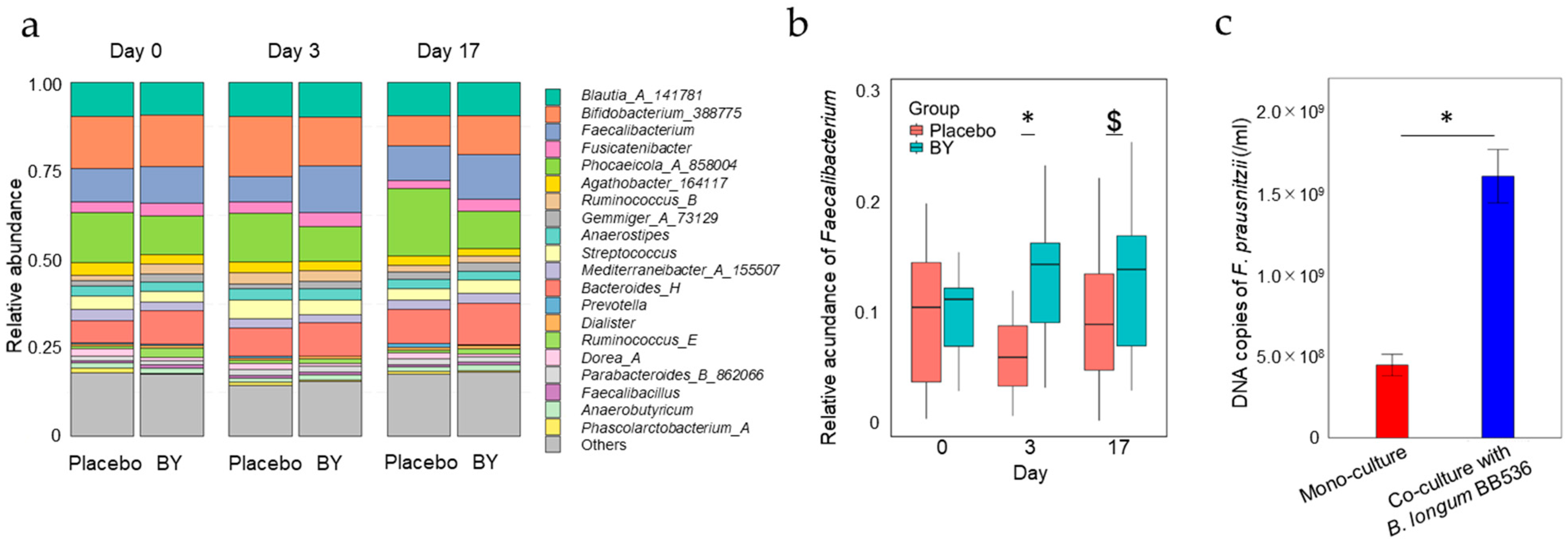
  • Consumption of fermented milk containing BB536 increases the relative abundance of Faecalibacterium.
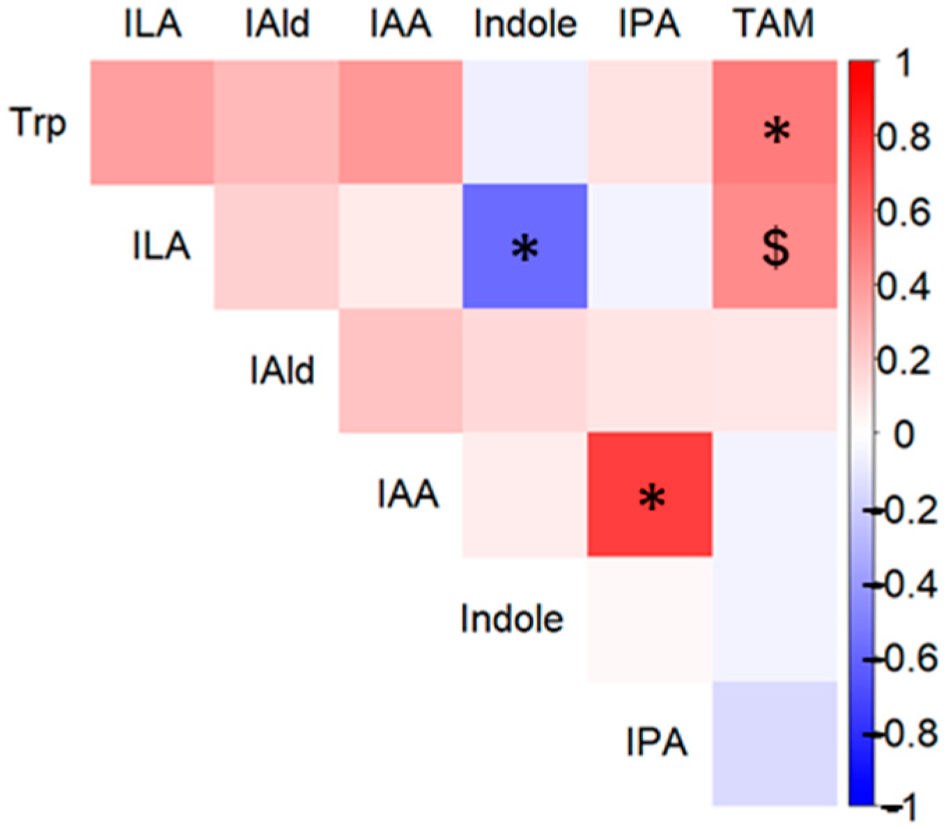
  • Consumption of fermented milk containing BB536 affects the concentrations of Tryptophan metabolites.
  • The number of viable BB536 bacteria in the gut is positively correlated with the abundance of Faecalibacterium and Tryptophan metabolites.
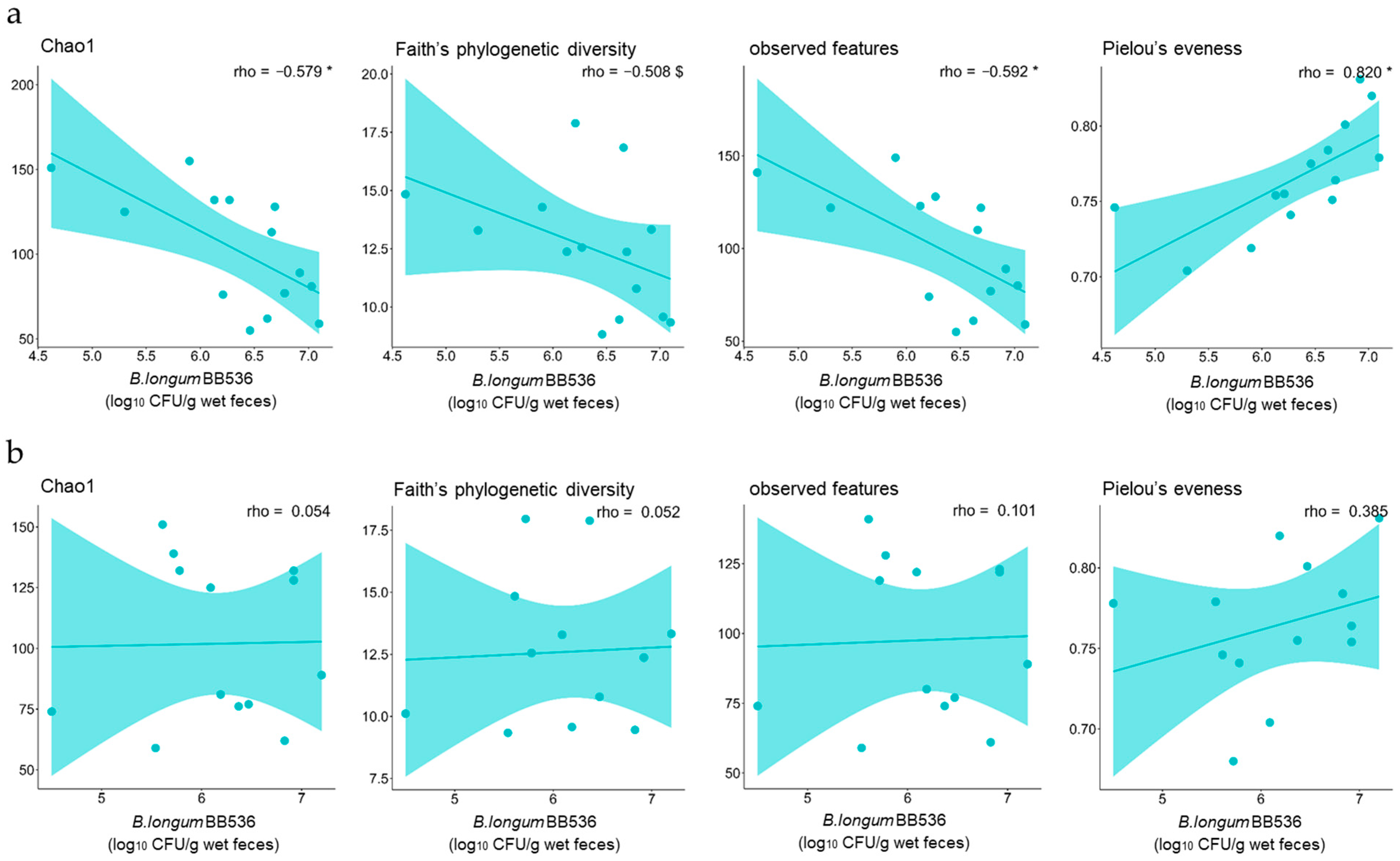
  • The alpha diversity of fecal microbiota affects the survival rate of BB536.

Abstract

:
Probiotics, particularly those native to the gut microbiota, have a profound influence on the gut environment. In this study, we conducted a randomized placebo-controlled, double-blind, parallel-group comparison trial to investigate the effects of Bifidobacterium longum BB536 (B. longum BB536) on the fecal microbiota and metabolite compositions in healthy individuals. We compared the effects of fermented milk produced solely with Streptococcus thermophiles and Lactobacillus bulgaricus (placebo group) and fermented milk supplemented with B. longum BB536 (BY group). Our findings revealed a significantly greater relative abundance of Faecalibacterium in the BY group than in the placebo group by the 3rd day, a trend that persisted until the end of the trial on the 17th day. Additionally, the BY group presented significantly increased concentrations of tryptophan (Trp), Indole-3-lactic acid, and Indole-3-aldehyde on the 17th day. A significant positive correlation was observed between the relative abundance of Faecalibacterium and the number of viable B. longum BB536 bacteria in the feces. The concentrations of Trp and Indole-3-acetic acid were also significantly correlated with the number of viable B. longum BB536 bacteria in the feces. Our results suggest that B. longum BB536 intake can modulate the gut microbiota and metabolite profiles, which are general indicators for monitoring the gut environment, potentially conferring health benefits to the host.

1. Introduction

The human gut is a complex ecosystem that is home to trillions of microorganisms known as the gut microbiota [1]. This ecosystem is crucial for maintaining host health and is intricately involved in various physiological functions, such as intestinal barrier maintenance [2], metabolism [3], immunity [4], and signal transmission in the nervous system [5]. Disruptions in this ecosystem could increase the risk of various diseases, highlighting the importance of maintaining a healthy gut environment [6].
Probiotics, living microorganisms that confer health benefits to the host when consumed in adequate amounts [7], can influence the immune system and intestinal epithelial cells directly and indirectly through the modulation of the gut environment. One of the common sources of these beneficial probiotics is fermented milk, which also possesses beneficial properties such as antioxidant, anti-hypertensive, and anti-allergic effects. These benefits may be attributed to the presence of Streptococcus thermophiles and Lactobacillus bulgaricus, which are Gram-positive facultative anaerobic and are classified as animal-derived lactic acid bacteria. However, considering the indirect effect by the microbes, species native to the gut microbiota may have a more profound impact on the gut environment due to their enhanced interaction with other gut bacteria. From this perspective, the influence may fundamentally differ between fermented milk containing only species not originally inhabiting the human gut, such as S. thermophiles and L. bulgaricus, and that which includes species such as Bifidobacterium, a part of the gut microbiota.
Bifidobacterium longum BB536 (B. longum BB536) is a human residential Bifidobacterium (HRB) strain that has been safely utilized worldwide for over 50 years. B. longum BB536 has been reported to confer various health benefits, including the regulation of the gut environment [8]. Previous studies have reported that the intake of products containing B. longum BB536 can regulate the gut environment and bowel movements [9], potentially contribute to immune system regulation [10,11], and reduce visceral and neutral fat levels when it is coingested with Bifidobacterium breve MCC1274 [12]. Notably, B. longum BB536 is associated with the production of tryptophan metabolites [13], which play an important role in host health. Tryptophan and its metabolite products not only satisfy the nutritional requirements of the host and microorganisms [14] but also contribute to the suppression of intestinal inflammation as ligands for various receptors [15,16,17]. These findings suggest that B. longum BB536 could play a pivotal role in maintaining the gut environment through the production of these metabolites. However, few clinical trials have comprehensively reported the effects of consuming fermented milk containing BB536 on both the gut microbiota and these metabolites.
In this study, we conducted a randomized placebo-controlled, double-blind, parallel-group comparison trial (RCT) in healthy individuals, in which traditional fermented milk produced only with S. thermophiles and L. bulgaricus (placebo group) and fermented milk to which B. longum BB536 was added (BY group) were used as test foods. We previously reported the number of viable B. longum BB536 bacteria in the feces using this experimental protocol [18]. In the present study, we evaluated the differences in the fecal microbiota and metabolites, which are general indicators for monitoring the environment of the large intestine [19,20], between the BY group and the placebo group in the same experiment, considering short-term (3rd day of intake) and medium-term (17th day of intake) effects. The objective of this study was to elucidate the mechanism by which B. longum BB536 confers various beneficial effects to the host.

2. Materials and Methods

2.1. Clinical Trial Design

This study was a double-blind, randomized, placebo-controlled trial conducted at the Matsumoto Health Laboratory, Japan. The trial aimed to investigate the effect of B. longum BB536 on the intestinal environment of healthy adults. The primary outcome of this study was the improvement of the intestinal environment. Changes in gut metabolite abundance and gut microbiota composition served as the outcome indicators. Healthy adults aged 18–64 years were enrolled, with exclusion criteria as previously described [18]. Briefly, participants were randomly assigned to consume either commercial fermented milk containing 20 million CFU/g B. longum BB536 or an identical placebo daily for a 17-day intervention period following a 1-week preobservation period. All dietary intake during the intervention was recorded with the food-recording app Calomeal® (Life Log Technology, Inc., Tokyo, Japan). Compliance was monitored by checking the remaining quantity of fermented milk and app entries. The analyses of the per-protocol set that were performed in this study were the same as those of the participants described in a previous study [18], and the per-protocol set was composed of 17 participants in the placebo group and 17 participants in the BY group. Fecal samples were collected on specific days using Raku-Ryu cups, transferred to a specific collection tube, enclosed in an AneroPouch Kenki (Mitsubishi Gas Chemical Co., Tokyo, Japan), and transported to the laboratory within 24 h. This trial was registered at the University Hospital Medical Research Network under UMIN000052110.

2.2. Bacterial Strains and Culture Conditions

The B. longum BB536 frozen culture was obtained from Morinaga Milk Industry (Tokyo, Japan), and F. prausnitzii JCM 39207 was purchased from the Japan Collection of Microorganisms (Tsukuba, Japan). All the strains used in this study were precultured in YCFA medium [21] under anaerobic conditions at 37 °C for 24 h. To evaluate the effect of B. longum BB536 on F. prausnitzii JCM 39207, the strains (1 × 10⁶ CFU/mL for both) were cultured under anaerobic conditions at 37 °C for 24 h in Gifu anaerobic medium broth (Shimadzu Diagnostics Co., Ltd., Kyoto, Japan).

2.3. 16S rRNA Gene Analysis

Bacterial DNA was extracted from the sample and amplified as previously described [21]. The V3-V4 region of the bacterial 16S rRNA gene was paired-end sequenced using the Illumina NextSeq 1000 platform with a NextSeq 1000/2000 P1 reagent kit (600 cycles) (Illumina, Inc., San Diego, CA, USA). The sequences were analyzed via QIIME2 (version 2022.8) [22]. The demultiplexed reads were processed via the following steps: filtering, denoising, merging, chimera removal, and generating the amplicon sequence variants (ASVs) via DADA2 [23]. The ASVs were taxonomically assigned on the basis of the Greengeens2 database (version 2022.10).

2.4. Faecalibacterium-Specific Quantitative PCR Analysis

Quantitative PCR analysis was performed by using a CFX96 Thermocycler (Bio-Rad Laboratories, Hercules, CA, USA) with a real-time PCR detection kit (Clostridium cluster iv: RI-0001) (TechnoSuruga Laboratory Co., Ltd., Nagasaki, Japan) and TB Green Premix Ex Taq II (TaKaRa Bio Inc., Shiga, Japan) according to the manufacturer’s instructions.

2.5. Metabolomic Analysis of Fecal Metabolites

2.5.1. Chemicals and Reagents

All chemicals used for target analysis, including internal standards, were purchased from Tokyo Chemical Industry Co., Ltd. (Tokyo, Japan), Sigma-Aldrich (St. Louis, MO, USA), and FUJIFILM Wako Pure Chemical Corporation (Osaka, Japan) (Tables S1 and S2). LCMS-grade water, methanol, and formic acid were purchased from FUJIFILM Wako Pure Chemical Corporation. The mobile phase and pH buffering solution were specifically purchased from SHIMADZU GLC Ltd. (Kyoto, Japan).

2.5.2. Extraction of Metabolites from Fecal Samples

Approximately 100 mg (±5 mg) of each fecal sample was suspended in 1000 μL of IS solution (1 μM MO in 50% water/methanol) or water. These suspensions were homogenized with 0.1 mm zirconia/silica beads by vigorous shaking (25 Hz, 10 min) via a TissueLyser instrument and centrifuged at 20,380× g for 5 min. The supernatants were then transferred into NANOSEP 3K OMEGA (Cytiva, Marlborough, MA, USA) or ULTRAFREE MC PLHCC 5K (Human Metabolome Technologies, Inc., Yamagata, Japan) media. The supernatants transferred to the ultrafiltration filters were then centrifuged at 9100× g for 3–5 h. The filtered samples were stored at −80 °C until further analysis.

2.5.3. Liquid Chromatography–Tandem Mass Spectrometry (LC-MS/MS) Analysis

LC-MS/MS analysis was performed using a Nexera X2 system (Shimadzu, Kyoto, Japan) equipped with two LC-40D XR pumps, a DGU-405 degasser, a SIL-40C XR autosampler, a CTO-40C column oven, and a CBM-40 control module coupled with an LC-8045 triple quadrupole mass spectrometer (Shimadzu). An XBridge® C8 column (4.6 × 150 mm, 5 μm; Waters Corporation, Milford, MA, USA) was used for the separation of metabolites. The mobile phase was composed of A: 0.1% (v/v) formic acid in water and B: 0.1% (v/v) formic acid in methanol. The flow rate, column temperature, and injection volume were set as 0.2 mL/min, 40 °C, and 2 µL, respectively. The gradient program for mobile phase B was as follows: 0 min, 30%; 5 min, 30%; 42 min, 95%; 47 min, 95%; 50.5 min, 30%; and 60 min, 30%. The mass spectrometer was equipped with an electrospray ionization (ESI) source under the following conditions: nebulizing gas flow, 3 L/min; heating gas flow, 10 L/min; interface temperature, 300 °C; desolvation line temperature, 250 °C; heating block temperature, 400 °C; drying gas flow, 10 L/min; and collision-induced dissociation gas pressure, 230 kPa.

2.5.4. HPLC Analysis

The HPLC analysis was performed using a Nexera X2 system (Shimadzu) equipped with two LC-40D pumps, a DGU-405 degasser, a SIL-40C autosampler, a CTO-40C column oven, a CBM-40 control module, and a CDD-10A VP conductivity detector. A tandem ion exclusion column (Shim-pack SCR-102H, 300 mm × 8.0 mm, 7 µm; SHIMADZU GLC Ltd.) was used for the separation of metabolites. The mobile phase and the pH buffering solution were delivered at a flow rate of 0.8 mL/min. The column temperature and injection volume were set as 50 °C and 10 µL, respectively.

2.6. Statistical Analysis

Intergroup differences in the fecal microbiota on days 3 and 17 were analyzed with Quade’s nonparametric analysis of covariance (Quade’s nonparametric ANCOVA) after centered log-ratio (CLR) transformation. Similarly, intergroup differences in fecal metabolites on days 3 and 17 were analyzed with Quade’s nonparametric ANCOVA. Other outcomes were compared between groups with Student’s t test and the Mann–Whitney U test, as appropriate. Correlation analysis was performed via Spearman’s correlation coefficient. Statistical analyses were performed via IBM SPSS Statistics (ver. 29.0.2.0 (20); Armonk, NY, USA), except Spearman’s correlation analysis, which was conducted via R (ver. 4.3.1). When data were missing, we performed listwise deletion to handle these missing values. A p value of <0.05 was considered statistically significant.

2.7. Data Availability

The raw sequence data have been deposited in the DNA Data Bank of Japan (DDBJ) Sequence Read Archive (DRR596307-DRR596404) under BioProject no. PRJDB18808. This project includes links and access to fecal sample data (BioSample SAMD00819537-SAMD00819634).

3. Results

3.1. Differences in Fecal Microbiota Composition Between the BY and Placebo Groups

We evaluated the impact of milk fermented with B. longum BB536 (BY) on the fecal microbiota via 16S rRNA gene sequencing analysis of fecal samples collected before intake and on days 3 and 17. The alpha diversity of the fecal microbiota was compared between groups using Chao1, observed features, Pielou’s evenness, and Faith’s phylogenetic diversity metrics as scoring metrics. However, no significant disparities were observed between the placebo and BY groups (Figure S1). We then compared the relative abundance at the genus level. Compared with the placebo group, the BY group presented a significantly greater relative abundance of Faecalibacterium on day 3, and this trend was still observed on day 17 (Figure 1a,b). Additionally, significant differences in the abundances Phascolarctobacterium_A on days 3 and 17 and Holdemanella on day 17 were also observed between the two groups (Tables S3 and S4). Intriguingly, on day 3, the relative abundance of Faecalibacterium was positively correlated with the number of viable B. longum BB536 bacteria detected in the fecal samples on the same day (Figure S2). To validate the beneficial relationship, B. longum BB536 and a representative Faecalibacterium, Faecalibacterium prausnitzii, were cocultured. The results indicated that compared with monoculture with F. prausnitzii alone, coculture with B. longum BB536 significantly promoted the growth of F. prausnitzii (Figure 1c).

3.2. Differences in Fecal Metabolite Abundance Between the BY and Placebo Groups

We evaluated the impact of BY intake on the abundance of fecal metabolites, short-chain fatty acids (SCFAs), and tryptophan (Trp) metabolites. Fecal samples were collected before intake and on days 3 and 17 of the experiment and analyzed via HPLC and LC-MS/MS. Sixteen types of metabolites were detected in both the BY and placebo groups, five of which exhibited different concentrations (Table S5). Trp, Indole-3-lactic acid (ILA), and Indole-3-aldehyde (IAld) were present at significantly higher concentrations in the BY group than in the control group on day 17, and Indole-3-acetic acid (IAA) also tended to be present at higher concentrations (Figure 2a). The concentration of formic acid in feces tended to be lower in the BY group than in the placebo group on day 17. Spearman’s correlation analysis was conducted between the five metabolites with significant differences or trends between groups on day 17 and the number of viable B. longum BB536 bacteria detected in feces. This analysis revealed that the concentrations of Trp and IAA in feces were significantly positively correlated on day 17 (Figure 2b). Although significant correlations among different Trp metabolites were not detected, the concentration of several metabolites demonstrated a positive correlation with that of Trp (Figure 3). Interestingly, a significant negative correlation was observed between indole and ILA. Conversely, no significant differences were observed in dietary nutrient intake during the trial period between the two groups on the basis of dietary records (Table S6). Furthermore, the levels of most Trp metabolites did not significantly differ between the BY and placebo milk products. However, Trp, which showed the most notable difference, demonstrated a significantly lower concentration in the BY-enriched milk product, a finding that is intriguing given its inverse relationship with fecal Trp concentrations (Figure S3).

3.3. Correlations Between the Number of B. longum BB536 Bacteria That Survived Digestion and the Diversity of the Fecal Microbiota

Given the significant correlations observed between the abundances of certain gut bacteria or the concentrations of certain metabolites and the number of surviving B. longum BB536 bacteria in feces, it is hypothesized that the ability of these bacteria to survive in the gut is crucial for their beneficial effect on the gut environment. Therefore, to elucidate the factors affecting the survival of bacteria in the gut environment, a correlation analysis was conducted between preintake information from the study participants and the number of viable B. longum BB536 bacteria detected in feces. The results showed that the number of viable B. longum BB536 bacteria detected in feces on day 3 was significantly negatively correlated with Chao1, observed features, and Faith’s phylogenetic diversity and significantly positively correlated with Pielou’s evenness (Figure 4a). Conversely, no significant correlations were detected between the number of viable B. longum BB536 detected in feces on day 17 and any of the scores (Figure 4b).

4. Discussion

The association between probiotic intake and host health has garnered considerable attention in recent years [24]. Probiotics can influence the host directly by acting on immune cells [25] and modifying the gut microbiota composition [26] and indirectly by regulating metabolite abundance [27]. The strain B. longum BB536, used in our study, has been reported to impact the host immune function and gut microbiota composition [28]. However, the specific benefits to the host from fermented milk containing this strain, compared with general fermented milk using only lactic acid bacteria, remain to be fully elucidated. In this study, we conducted an RCT in which study participants consumed fermented milk containing only lactic acid bacteria or fermented milk fortified with B. longum BB536 and the effects on both the fecal microbiota and metabolite profiles were assessed. These elements serve as general indicators for monitoring the gut environment. Our findings underscore the impact of consuming BY on the gut environment, as evidenced by significant changes in these indicators.
Compared with the placebo group, the BY group presented an increased relative abundance of Faecalibacterium on day 3 (Figure 1a,b), and in vitro assays revealed an increase in F. prausnitzii in the presence of B. longum BB536 (Figure 1c). As Faecalibacterium has been reported to proliferate by utilizing acetic acid [29], it is plausible that the acetic acid produced by B. longum BB536 facilitated the growth of Faecalibacterium. The butyric acid produced by Faecalibacterium not only functions as a vital energy source for colon cancer cells [30] but has also been reported to exhibit anti-inflammatory effects [31], bolster intestinal wall integrity [32], and confer protection against colon cancer [33]. However, no significant difference in the fecal concentration of butyric acid in the feces was detected between the two groups (Table S5). This could be attributed to insufficient diet-derived nutrients for Faecalibacterium to produce butyric acid. Additionally, given that blood SCFAs are reportedly more sensitive to probiotic intake than are fecal SCFAs, the amount of SCFAs absorbed by the host may vary, leading to different blood concentrations [34]. The relative abundance of F. prausnitzii reportedly decreases in patients with inflammatory bowel disease (IBD) and colon cancer [35], and we postulate that maintaining a high proportion of Faecalibacterium itself may play a pivotal role in preserving host health. However, further investigations are warranted to examine the effects of probiotics on the production of SCFAs.
Among the substances produced by gut microbes, Trp metabolites are of paramount importance. The gut microbiota is believed to contribute to host health by converting Trp into various metabolites [36]. These metabolites function as ligands for aryl hydrocarbon receptors (AhRs) and hydroxycarboxylic acid receptor 3 (HCA3), which play crucial roles in maintaining host homeostasis [37]. In this study, we observed that, compared with the placebo group, the BY group presented elevated fecal concentrations of Trp metabolites such as ILA, IAA, and IAld on day 17 (Figure 2a). ILA, produced by Bifidobacterium, has been reported to play an important role in maintaining infant health by decreasing Th2, Th17, and IFN-β production, thereby contributing to the suppression of intestinal inflammation [38,39]. The fecal concentration of IAA is known to decrease in patients with IBD and alcoholic hepatitis [40,41]. IAld has been reported to potentially play a role in suppressing Candida albicans infection induced by IL-22 [42] and enhancing intestinal barrier function [43].
Notably, the Trp concentration was significantly greater in the BY group than in the placebo group on day 3, and this difference became more pronounced on day 17 (Figure 2a). Trp may be absorbed from the colon via the large neutral amino acid transporter small subunit 2 (LAT2) to meet the nutritional requirements [44] of the host. Trp is reportedly converted into various biological signaling molecules through two major metabolic pathways in the host, namely, the kynurenine pathway and the serotonin pathway [45]. Kynurenine, produced by the kynurenine pathway, has been reported to regulate the activity of immune cells and to exhibit anti-inflammatory effects [46]. Conversely, serotonin and 5-hydroxy tryptophan, which are produced via the serotonin pathway, function as neurotransmitters and play crucial roles in regulating sleep [47]. They have also been associated with mental disorders such as depression and anxiety [48]; therefore, our findings may support a previous study that reported that B. longum BB536 may act as a psychobiotic [49].
In this study, we observed that the high concentrations of Trp metabolites may have been produced by gut microbes that naturally inhabit the gut metabolizing the increased Trp from B. longum BB536 supplementation. Many previous studies have demonstrated that gut microbes can convert Trp into various Trp metabolites. For example, IAA is reportedly produced by a variety of bacteria, including Bifidobacterium, Bacteroides, Clostridium, Parabacteroides, and Eubacterium, while IAld is also reported to be produced by Lactobacillus, among others [50]. Indeed, the fecal culture results from a study using a human digestive tract model have indicated that Trp supplementation increases the concentration of Trp metabolites [51]. Conversely, the mechanism by which probiotic intake increases the concentration of Trp in feces remains elusive. A previous report demonstrated that the intake of B. breve CCFM1025 significantly increased the concentration of Trp in the feces, but the underlying mechanism remains unclear [52]. We hypothesized that the differences in the diet or the consumed test food may have an effect, but no difference was observed in the diet of either group in this study (Table S6). In contrast, Trp was present at significantly lower concentrations in the fermented milk supplemented with B. longum BB536 (Figure S3). Previous reports have indicated that gut bacteria maintain a symbiotic relationship in the gut by complementing each other’s nutrient needs [53]. In particular, the results of an in silico analysis revealed that Trp contributes to the diversity and homeostasis of the microbiota as a nutrient for gut bacteria [14]. On the basis of these previous studies, it is plausible that Trp accumulates only under certain conditions where B. longum BB536 coexists with other gut bacteria. Nonetheless, these are merely hypotheses, and further research is warranted in the future.
Previous in vitro studies have suggested that HRB converts indole to ILA [13]. However, in this study, no significant difference in indole content was detected between the two groups (Table S5). Nevertheless, the significant negative correlation observed between the concentrations of indole and ILA on day 17 (Figure 3) implies that the conversion from indole to ILA occurred. The absence of a significant difference may be attributed to the large disparity in the concentration of Trp or the presence of different indole producers between the groups.
In this study, a positive correlation was observed between some of the gut bacteria and metabolites that differed in abundance between the two groups and the number of viable B. longum BB536 bacteria detected in feces (Figure 2b and Figure S2). Therefore, it is highly plausible that identifying factors that affect the number of live bacteria reaching the gut can enhance the beneficial effects of B. longum BB536 intake on the gut environment. We observed that the number of viable B. longum BB536 bacteria that reached the gut after 3 days was related to the alpha diversity of the fecal microbiota before intake (Figure 4a). The negative correlation with Chao1, observed features, and Faith’s phylogenetic diversity indicates that greater species diversity decreases the percentage of viable bacteria that reach the gut, whereas a positive correlation with Pielou’s evenness suggests that an even distribution of species increases this percentage. These findings suggest that the greater the diversity of the gut microbiota is, the more challenging it is for newly arrived B. longum BB536 to survive in the gut. In fact, similar phenomena have been confirmed in reports on environmental bacteria [54]. Conversely, the number of live B. longum BB536 bacteria reaching the gut on day 17 did not have a notable relationship with this diversity (Figure 4b). Therefore, it is predicted that the richness and evenness of species in the gut microbiota are important factors in determining the number of viable B. longum BB536 bacteria reaching the gut and that their influence diminishes as the intake period is extended.
This study, while providing valuable insights, has several limitations. First, the trial included only 34 subjects who met the criteria across both groups. To obtain more robust data, expanding the sample size in future clinical trials would be beneficial. Second, the mechanism underlying the observed increase in Trp concentration in the BY group in this study remains unclear. To elucidate this phenomenon, further verification experiments are needed to elucidate the intricate crosstalk between bacteria. By addressing these challenges, we can shed more light on the complex effects of probiotics, including B. longum BB536, and contribute to the development of more effective probiotic products.

5. Conclusions

In conclusion, our study demonstrated that the intake of milk products containing B. longum BB536 bacteria led to an increase in the relative abundance of Faecalibacterium, a key indicator of gut health, by the 3rd day of intake. Furthermore, a significant alteration in the concentration of Trp metabolites, which function as signaling molecules and serve as another general indicator for monitoring gut health, was observed in the fecal samples on day 17. These changes, which were more pronounced with a greater number of viable B. longum BB536 bacteria present in the fecal samples, suggest an improvement in gut health following BY intake. We posit that these findings lend support to the notion that the regular intake of B. longum BB536 bacteria can contribute to overall health by regulating the gut environment.

Supplementary Materials

The following supporting information can be downloaded at: https://www.mdpi.com/article/10.3390/nu16213580/s1, Figure S1: Alpha diversity indices of the fecal microbiota; Figure S2: Correlation of CLR-transformed abundance of Faecalibacterium and viable B. longum BB536 count on day 3; Figure S3: Concentrations of Trp, IAA, ILA, and IAld in fermented milk. * p < 0.05, with Student’s t test; Table S1: List of Trp metabolites and internal standards for LC-MS/MS; Table S2: List of SCFAs for HPLC; Table S3: Comparison of the relative abundances of the top 50 bacterial genera on day 3; Table S4: Comparison of the relative abundances of the top 50 bacterial genera on day 17; Table S5: Comparison of the concentrations of metabolites in feces; Table S6: Comparison of estimated total nutrient intake during the trial period.

Author Contributions

Conceptualization, R.E., T.O. and M.T.; methodology, R.E., A.S., M.K., S.A., N.M., A.H., K.K., M.N. (Manabu Nakano) and T.O.; validation, R.E. and T.O.; formal analysis, R.E., R.M., S.A. and T.O.; investigation, R.E., R.M., A.S., K.Y., E.M. and H.K.; resources, Y.K. and H.M.; data curation, R.E., R.M., A.S. and T.O.; writing—original draft preparation, R.E. and T.O.; writing—review and editing, R.E., K.K., N.I., M.N. (Manabu Nakano), T.O. and M.T.; visualization, R.E. and T.O.; supervision, M.N. (Masahiko Nakamura) and M.T.; project administration, N.I., M.N. (Manabu Nakano), T.O. and M.T. All authors have read and agreed to the published version of the manuscript.

Funding

This research received no external funding.

Institutional Review Board Statement

This study was conducted in compliance with the Declaration of Helsinki (Fortaleza, revised in 2013) and the Ethical Guidelines for Life Sciences and Medical Research Involving Human Subjects (Ministry of Education, Culture, Sports, Science and Technology; Ministry of Health, Labour and Welfare; and Ministry of Economy, Trade and Industry Notification No. 1, 2021). This study was approved by the Research Ethics Committee of Matsumoto City Hospital and the reference number is 509 (date 18 August 2023).

Informed Consent Statement

Informed consent was obtained from all participants involved in the study.

Data Availability Statement

The data collected in this study can be found in the published article.

Acknowledgments

We would like to thank the participants and staff, including Matsumoto City and Matsumoto Health Lab staff, for their cooperation in this study.

Conflicts of Interest

R.E., R.M., A.S., K.Y., E.M., H.K., M.K., S.A., N.M., A.H., K.K., Y.K., H.M., N.I., M.N., T.O. and M.T. are employed at Morinaga Milk Industry Co., Ltd. The authors declare that the research was conducted in the absence of any commercial or financial relationships that could be construed as a potential conflict of interest.

References

  1. Daniel, N.; Lécuyer, E.; Chassaing, B. Host/microbiota interactions in health and diseases-Time for mucosal microbiology! Mucosal Immunol. 2021, 14, 1006–1016. [Google Scholar] [CrossRef] [PubMed]
  2. Di Tommaso, N.; Gasbarrini, A.; Ponziani, F.R. Intestinal barrier in human health and disease. Int. J. Environ. Res. Public Health 2021, 18, 12836. [Google Scholar] [CrossRef] [PubMed]
  3. Bohan, R.; Tianyu, X.; Tiantian, Z.; Ruonan, F.; Hongtao, H.; Qiong, W.; Chao, S. Gut microbiota: A potential manipulator for host adipose tissue and energy metabolism. J. Nutr. Biochem. 2019, 64, 206–217. [Google Scholar] [CrossRef]
  4. Yoo, J.Y.; Groer, M.; Dutra, S.V.O.; Sarkar, A.; McSkimming, D.I. Gut microbiota and immune system interactions. Microorganisms 2020, 8, 1587. [Google Scholar] [CrossRef] [PubMed]
  5. Margolis, K.G.; Cryan, J.F.; Mayer, E.A. The microbiota-gut-brain axis: From motility to mood. Gastroenterology 2022, 160, 1486–1501. [Google Scholar] [CrossRef]
  6. Levy, M.; Kolodziejczyk, A.A.; Thaiss, C.A.; Elinav, E. Dysbiosis and the immune system. Nat. Rev. Immunol. 2017, 17, 219–232. [Google Scholar] [CrossRef]
  7. Markowiak, P.; Śliżewska, K. Effects of probiotics, prebiotics, and synbiotics on human health. Nutrients 2017, 9, 1021. [Google Scholar] [CrossRef]
  8. Wong, C.B.; Odamaki, T.; Xiao, J.-Z. Insights into the reason of Human-Residential Bifidobacteria (HRB) being the natural inhabitants of the human gut and their potential health-promoting benefits. FEMS Microbiol. Rev. 2020, 44, 369–385. [Google Scholar] [CrossRef]
  9. Nakamura, Y.; Suzuki, S.; Murakami, S.; Nishimoto, Y.; Higashi, K.; Watarai, N.; Umetsu, J.; Ishii, C.; Ito, Y.; Mori, Y.; et al. Integrated gut microbiome and metabolome analyses identified fecal biomarkers for bowel movement regulation by Bifidobacterium longum BB536 supplementation: A RCT. Comput. Struct. Biotechnol. J. 2022, 20, 5847–5858. [Google Scholar] [CrossRef]
  10. Odamaki, T.; Xiao, J.-Z.; Iwabuchi, N.; Sakamoto, M.; Takahashi, N.; Kondo, S.; Miyaji, K.; Iwatsuki, K.; Togashi, H.; Enomoto, T.; et al. Influence of Bifidobacterium longum BB536 intake on faecal microbiota in individuals with Japanese cedar pollinosis during the pollen season. J. Med. Microbiol. 2007, 56, 1301–1308. [Google Scholar] [CrossRef]
  11. Li, Y.; Arai, S.; Kato, K.; Iwabuchi, S.; Iwabuchi, N.; Muto, N.; Motobayashi, H.; Ebihara, S.; Tanaka, M.; Hashimoto, S. The potential immunomodulatory effect of Bifidobacterium longum subsp. longum BB536 on healthy adults through plasmacytoid dendritic cell activation in the peripheral blood. Nutrients 2023, 16, 42. [Google Scholar] [CrossRef] [PubMed]
  12. Sato, S.; Arai, S.; Kato, K.; Yoshida, K.; Iwabuchi, N.; Sagami, T.; Tanaka, M. Effects of Bifidobacterium longum BB536 and Bifidobacterium breve MCC1274 on body composition in normal and overweight adults in randomized placebo-controlled study. Nutrients 2024, 16, 815. [Google Scholar] [CrossRef] [PubMed]
  13. Yong, C.C.; Sakurai, T.; Kaneko, H.; Horigome, A.; Mitsuyama, E.; Nakajima, A.; Katoh, T.; Sakanaka, M.; Abe, T.; Xiao, J.-Z.; et al. Human gut-associated Bifidobacterium species salvage exogenous indole, a uremic toxin precursor, to synthesize indole-3-lactic acid via tryptophan. Gut Microbes 2024, 16, 2347728. [Google Scholar] [CrossRef]
  14. Starke, S.; Harris, D.M.M.; Zimmermann, J.; Schuchardt, S.; Oumari, M.; Frank, D.; Bang, C.; Rosenstiel, P.; Schreiber, S.; Frey, N.; et al. Amino acid auxotrophies in human gut bacteria are linked to higher microbiome diversity and long-term stability. ISME J. 2023, 17, 2370–2380. [Google Scholar] [CrossRef]
  15. Sun, M.; Ma, N.; He, T.; Johnston, L.J.; Ma, X. Tryptophan (Trp) modulates gut homeostasis via aryl hydrocarbon receptor (AhR). Crit. Rev. Food Sci. Nutr. 2019, 60, 1760–1768. [Google Scholar] [CrossRef] [PubMed]
  16. Illés, P.; Krasulová, K.; Vyhlídalová, B.; Poulíková, K.; Marcalíková, A.; Pečinková, P.; Sirotová, N.; Vrzal, R.; Mani, S.; Dvořák, Z. Indole microbial intestinal metabolites expand the repertoire of ligands and agonists of the human pregnane X receptor. Toxicol. Lett. 2020, 334, 87–93. [Google Scholar] [CrossRef]
  17. Sakurai, T.; Horigome, A.; Odamaki, T.; Shimizu, T.; Xiao, J.-Z. Production of hydroxycarboxylic acid receptor 3 (HCA3) ligands by Bifidobacterium. Microorganisms 2021, 9, 2397. [Google Scholar] [CrossRef]
  18. Sen, A.; Kimura, M.; Ejima, R.; Arai, S.; Mitsuyama, E.; Kaneko, H.; Mishima, R.; Muto, N.; Hiraku, A.; Kato, K.; et al. Assessing probiotic viability in the gastrointestinal tract in randomized placebo controlled trial: A combined approach of molecular biology and novel cultivation techniques. Benef. Microbes, 2024; submitted. [Google Scholar]
  19. Martín, R.; Rios-Covian, D.; Huillet, E.; Auger, S.; Khazaal, S.; Bermúdez-Humarán, L.G.; Sokol, H.; Chatel, J.-M.; Langella, P. Faecalibacterium: A bacterial genus with promising human health applications. FEMS Microbiol. Rev. 2023, 47, 1–18. [Google Scholar] [CrossRef]
  20. McCann, J.R.; Rawls, J.F. Essential amino acid metabolites as chemical mediators of host-microbe interaction in the gut. Annu. Rev. Microbiol. 2023, 77, 479–497. [Google Scholar] [CrossRef]
  21. Murakami, R.; Hashikura, N.; Yoshida, K.; Xiao, J.-Z.; Odamaki, T. Growth-promoting effect of alginate on Faecalibacterium prausnitzii through cross-feeding with Bacteroides. Food Res. Int. 2021, 144, 110326. [Google Scholar] [CrossRef]
  22. Bolyen, E.; Rideout, J.R.; Dillon, M.R.; Bokulich, N.A.; Abnet, C.C.; Al-Ghalith, G.A.; Alexander, H.; Alm, E.J.; Arumugam, M.; Asnicar, F.; et al. Reproducible, interactive, scalable and extensible microbiome data science using QIIME 2. Nat. Biotechnol. 2019, 37, 852–857. [Google Scholar] [CrossRef] [PubMed]
  23. Callahan, B.J.; McMurdie, P.J.; Rosen, M.J.; Han, A.W.; Johnson, A.J.A.; Holmes, S.P. DADA2: High-resolution sample inference from Illumina amplicon data. Nat. Methods 2016, 13, 581–583. [Google Scholar] [CrossRef] [PubMed]
  24. Sánchez, B.; Delgado, S.; Blanco-Míguez, A.; Lourenço, A.; Gueimonde, M.; Margolles, A. Probiotics, gut microbiota, and their influence on host health and disease. Mol. Nutr. Food Res. 2016, 61, 1600240. [Google Scholar] [CrossRef] [PubMed]
  25. Galdeano, C.M.; Cazorla, S.I.; Dumit, J.M.L.; Vélez, E.; Perdigón, G. Beneficial effects of probiotic consumption on the immune system. Ann. Nutr. Metab. 2019, 74, 115–124. [Google Scholar] [CrossRef]
  26. Wieërs, G.; Belkhir, L.; Enaud, R.; Leclercq, S.; de Foy, J.-M.P.; Dequenne, I.; de Timary, P.; Cani, P.D. How probiotics affect the microbiota. Front. Cell. Infect. Microbiol. 2020, 9, 454. [Google Scholar] [CrossRef]
  27. Markowiak-Kopeć, P.; Śliżewska, K. The effect of probiotics on the production of short-chain fatty acids by human intestinal microbiome. Nutrients 2020, 12, 1107. [Google Scholar] [CrossRef]
  28. Lau, A.S.Y.; Yanagisawa, N.; Hor, Y.Y.; Lew, L.C.; Ong, J.S.; Chuah, L.O.; Lee, Y.Y.; Choi, S.B.; Rashid, F.; Wahid, N.; et al. Bifidobacterium longum BB536 alleviated upper respiratory illnesses and modulated gut microbiota profiles in Malaysian pre-school children. Benef. Microbes 2018, 9, 61–70. [Google Scholar] [CrossRef]
  29. Ramirez-Farias, C.; Slezak, K.; Fuller, Z.; Duncan, A.; Holtrop, G.; Louis, P. Effect of inulin on the human gut microbiota: Stimulation of Bifidobacterium adolescentis and Faecalibacterium prausnitzii. Br. J. Nutr. 2008, 101, 541–550. [Google Scholar] [CrossRef]
  30. Louis, P.; Young, P.; Holtrop, G.; Flint, H.J. Diversity of human colonic butyrate-producing bacteria revealed by analysis of the butyryl-CoA:acetate CoA-transferase gene. Environ. Microbiol. 2010, 12, 304–314. [Google Scholar] [CrossRef]
  31. Zhou, L.; Zhang, M.; Wang, Y.; Dorfman, R.G.; Liu, H.; Yu, T.; Chen, X.; Tang, D.; Xu, L.; Yin, Y.; et al. Faecalibacterium prausnitzii produces butyrate to maintain Th17/Treg balance and to ameliorate colorectal colitis by inhibiting histone deacetylase 1. Inflamm. Bowel Dis. 2018, 24, 1926–1940. [Google Scholar] [CrossRef]
  32. Zhuang, M.; Shang, W.; Ma, Q.; Strappe, P.; Zhou, Z. Abundance of probiotics and butyrate-production microbiome manages constipation via short-chain fatty acids production and hormones secretion. Mol. Nutr. Food Res. 2019, 63, 1801187. [Google Scholar] [CrossRef]
  33. Zagato, E.; Pozzi, C.; Bertocchi, A.; Schioppa, T.; Saccheri, F.; Guglietta, S.; Fosso, B.; Melocchi, L.; Nizzoli, G.; Troisi, J.; et al. Endogenous murine microbiota member Faecalibaculum rodentium and its human homologue protect from intestinal tumour growth. Nat. Microbiol. 2020, 5, 511–524. [Google Scholar] [CrossRef] [PubMed]
  34. Zhang, Q.; Li, G.; Zhao, W.; Wang, X.; He, J.; Zhou, L.; Zhang, X.; An, P.; Liu, Y.; Zhang, C.; et al. Efficacy of Bifidobacterium animalis subsp. lactis BL-99 in the treatment of functional dyspepsia: A randomized placebo-controlled clinical trial. Nat. Commun. 2024, 15, 227. [Google Scholar] [CrossRef]
  35. Touchefeu, Y.; Duchalais, E.; des Varannes, S.B.; Alameddine, J.; Mirallie, E.; Matysiak-Budnik, T.; Le Bastard, Q.; Javaudin, F.; Rimbert, M.; Jotereau, F.; et al. Concomitant decrease of double-positive lymphocyte population CD4CD8αα and Faecalibacterium prausnitzii in patients with colorectal cancer. Eur. J. Gastroenterol. Hepatol. 2020, 32, 149–156. [Google Scholar] [CrossRef]
  36. Gupta, S.K.; Vyavahare, S.; Blanes, I.L.D.; Berger, F.; Isales, C.; Fulzele, S. Microbiota-derived tryptophan metabolism: Impacts on health, aging, and disease. Exp. Gerontol. 2023, 183, 112319. [Google Scholar] [CrossRef]
  37. Laursen, M.F.; Sakanaka, M.; von Burg, N.; Mörbe, U.; Andersen, D.; Moll, J.M.; Pekmez, C.T.; Rivollier, A.; Michaelsen, K.F.; Mølgaard, C.; et al. Bifidobacterium species associated with breastfeeding produce aromatic lactic acids in the infant gut. Nat. Microbiol. 2021, 6, 1367–1382. [Google Scholar] [CrossRef] [PubMed]
  38. Henrick, B.M.; Rodriguez, L.; Lakshmikanth, T.; Pou, C.; Henckel, E.; Arzoomand, A.; Olin, A.; Wang, J.; Mikes, J.; Tan, Z.; et al. Bifidobacteria-mediated immune system imprinting early in life. Cell 2021, 184, 3884–3898.e11. [Google Scholar] [CrossRef] [PubMed]
  39. Sakurai, T.; Odamaki, T.; Xiao, J.-Z. Production of indole-3-lactic acid by Bifidobacterium strains isolated fromhuman infants. Microorganisms 2019, 7, 340. [Google Scholar] [CrossRef] [PubMed]
  40. Lamas, B.; Richard, M.L.; Leducq, V.; Pham, H.-P.; Michel, M.-L.; Da Costa, G.; Bridonneau, C.; Jegou, S.; Hoffmann, T.W.; Natividad, J.M.; et al. CARD9 impacts colitis by altering gut microbiota metabolism of tryptophan into aryl hydrocarbon receptor ligands. Nat. Med. 2016, 22, 598–605. [Google Scholar] [CrossRef]
  41. Hendrikx, T.; Duan, Y.; Wang, Y.; Oh, J.-H.; Alexander, L.M.; Huang, W.; Stärkel, P.; Ho, S.B.; Gao, B.; Fiehn, O.; et al. Bacteria engineered to produce IL-22 in intestine induce expression of REG3G to reduce ethanol-induced liver disease in mice. Gut 2019, 68, 1504–1515. [Google Scholar] [CrossRef]
  42. Zelante, T.; Iannitti, R.G.; Cunha, C.; De Luca, A.; Giovannini, G.; Pieraccini, G.; Zecchi, R.; D’Angelo, C.; Massi-Benedetti, C.; Fallarino, F.; et al. Tryptophan catabolites from microbiota engage aryl hydrocarbon receptor and balance mucosal reactivity via interleukin-22. Immunity 2013, 39, 372–385. [Google Scholar] [CrossRef] [PubMed]
  43. Scott, S.A.; Fu, J.; Chang, P.V. Microbial tryptophan metabolites regulate gut barrier function via the aryl hydrocarbon receptor. Proc. Natl. Acad. Sci. USA 2020, 117, 19376–19387. [Google Scholar] [CrossRef] [PubMed]
  44. Bröer, S. Intestinal amino acid transport and metabolic health. Annu. Rev. Nutr. 2023, 43, 73–99. [Google Scholar] [CrossRef]
  45. Xue, C.; Li, G.; Zheng, Q.; Gu, X.; Shi, Q.; Su, Y.; Chu, Q.; Yuan, X.; Bao, Z.; Lu, J.; et al. Tryptophan metabolism in health and disease. Cell Metab. 2023, 35, 1304–1326. [Google Scholar] [CrossRef]
  46. Siska, P.J.; Jiao, J.; Matos, C.; Singer, K.; Berger, R.S.; Dettmer, K.; Oefner, P.J.; Cully, M.D.; Wang, Z.; QuinnIii, W.J.; et al. Kynurenine induces T cell fat catabolism and has limited suppressive effects in vivo. eBioMedicine 2021, 74, 103734. [Google Scholar] [CrossRef]
  47. Wieckiewicz, M.; Martynowicz, H.; Lavigne, G.; Lobbezoo, F.; Kato, T.; Winocur, E.; Wezgowiec, J.; Danel, D.; Wojakowska, A.; Mazur, G.; et al. An exploratory study on the association between serotonin and sleep breathing disorders. Sci. Rep. 2023, 13, 11800. [Google Scholar] [CrossRef]
  48. Walden, K.E.; Moon, J.M.; Hagele, A.M.; Allen, L.E.; Gaige, C.J.; Krieger, J.M.; Jager, R.; Mumford, P.W.; Pane, M.; Kerksick, C.M. A randomized controlled trial to examine the impact of a multi-strain probiotic on self-reported indicators of depression, anxiety, mood, and associated biomarkers. Front. Nutr. 2023, 10, 1219313. [Google Scholar] [CrossRef] [PubMed]
  49. Pinto-Sanchez, M.I.; Hall, G.B.; Ghajar, K.; Nardelli, A.; Bolino, C.; Lau, J.T.; Martin, F.-P.; Cominetti, O.; Welsh, C.; Rieder, A.; et al. Probiotic Bifidobacterium longum NCC3001 reduces depression scores and alters brain activity: A pilot study in patients with irritable bowel syndrome. Gastroenterology 2017, 153, 448–459.e8. [Google Scholar] [CrossRef]
  50. Su, X.; Gao, Y.; Yang, R. Gut microbiota-derived tryptophan metabolites maintain gut and systemic homeostasis. Cells 2022, 11, 2296. [Google Scholar] [CrossRef]
  51. Koper, J.E.; Troise, A.D.; Loonen, L.M.; Vitaglione, P.; Capuano, E.; Fogliano, V.; Wells, J.M. Tryptophan supplementation increases the production of microbial-derived AhR agonists in an in vitro simulator of intestinal microbial ecosystem. J. Agric. Food Chem. 2022, 70, 3958–3968. [Google Scholar] [CrossRef]
  52. Tian, P.; Chen, Y.; Zhu, H.; Wang, L.; Qian, X.; Zou, R.; Zhao, J.; Zhang, H.; Qian, L.; Wang, Q.; et al. Bifidobacterium breve CCFM1025 attenuates major depression disorder via regulating gut microbiome and tryptophan metabolism: A randomized clinical trial. Brain Behav. Immun. 2022, 100, 233–241. [Google Scholar] [CrossRef] [PubMed]
  53. Sharma, V.; Rodionov, D.A.; Leyn, S.A.; Tran, D.; Iablokov, S.N.; Ding, H.; Peterson, D.A.; Osterman, A.L.; Peterson, S.N. B-vitamin sharing promotes stability of gut microbial communities. Front. Microbiol. 2019, 10, 1485. [Google Scholar] [CrossRef] [PubMed]
  54. van Elsas, J.D.; Chiurazzi, M.; Mallon, C.A.; Elhottova, D.; Kristufek, V.; Salles, J.F. Microbial diversity determines the invasion of soil by a bacterial pathogen. Proc. Natl. Acad. Sci. USA 2012, 109, 1159–1164. [Google Scholar] [CrossRef] [PubMed]
Figure 1. Impact of B. longum BB536 bacteria on the fecal microbiota composition: (a) stacked bar graph showing the 20 most abundant genera in the fecal microbiota; (b) the relative abundance of Faecalibacterium in fecal samples; (c) DNA copy number of F. prausnitzii JCM 39207 when cocultured with B. longum BB536 in GAM medium at 37 °C for 24 h. Statistical significance is indicated as follows: $ p < 0.1; * p < 0.05 with (b) Quade’s nonparametric ANCOVA and (c) Student’s t test.
Nutrients 16 03580 g001
Figure 2. Correlation between fecal metabolite concentrations and viable B. longum BB536 counts: (a) quantitative measurements of fecal Trp, IAA, ILA, IAld, and formic acid concentrations; (b) correlation of the concentrations of the same five metabolites on day 17 and the viable B. longum BB536 count. Statistical significance is indicated as follows: $ p < 0.1; * p < 0.05; with (a) Quade’s nonparametric ANCOVA and (b) Spearman’s correlation.
Nutrients 16 03580 g002
Figure 3. Heatmap of Spearman’s correlations among Trp metabolites on day 17. The map displays pairwise comparisons of Trp, ILA, IAA, IAld, Indole, Indole-3-propionic acid (IPA), and tryptamine (TAM) on day 17. Statistical significance is indicated as follows: $ p < 0.1; * p < 0.05.
Nutrients 16 03580 g003
Figure 4. Correlation between alpha diversity of the fecal microbiota and viable B. longum BB536 count. Correlation of preintake fecal microbiota alpha diversity with viable B. longum BB536 count on (a) day 3 and (b) day 17. Alpha diversity was scored by Chao1, Faith’s phylogenetic diversity, observed features, and Pielou’s evenness tests. Statistical significance is indicated as follows: $ p < 0.1; * p < 0.05, with Spearman’s correlation.
Nutrients 16 03580 g004
Disclaimer/Publisher’s Note: The statements, opinions and data contained in all publications are solely those of the individual author(s) and contributor(s) and not of MDPI and/or the editor(s). MDPI and/or the editor(s) disclaim responsibility for any injury to people or property resulting from any ideas, methods, instructions or products referred to in the content.

