Article

Bifidobacterium longum K5 Prevents Enterohaemorrhagic Escherichia coli O157:H7 Infection in Mice through the Modulation of the Gut Microbiota

1
Key Laboratory of Dairy Science, Ministry of Education, Northeast Agricultural University, Harbin 150030, China
2
Food College, Northeast Agricultural University, Harbin 150030, China
*
Author to whom correspondence should be addressed.
Nutrients 2024, 16(8), 1164; https://doi.org/10.3390/nu16081164
Submission received: 21 March 2024 / Revised: 11 April 2024 / Accepted: 11 April 2024 / Published: 13 April 2024
(This article belongs to the Section Prebiotics and Probiotics)

Abstract

:
Enterohemorrhagic Escherichia coli (EHEC) serotype O157:H7 is a commonly encountered foodborne pathogen that can cause hemorrhagic enteritis and lead to hemolytic uremic syndrome (HUS) in severe cases. Bifidobacterium is a beneficial bacterium that naturally exists in the human gut and plays a vital role in maintaining a healthy balance in the gut microbiota. This study investigated the protective effects of B. longum K5 in a mouse model of EHEC O157:H7 infection. The results indicated that pretreatment with B. longum K5 mitigated the clinical symptoms of EHEC O157:H7 infection and attenuated the increase in myeloperoxidase (MPO) activity in the colon of the mice. In comparison to the model group, elevated serum D-lactic acid concentrations and diamine oxidase (DAO) levels were prevented in the K5-EHEC group of mice. The reduced mRNA expression of tight junction proteins (ZO-1, Occludin, and Claudin-1) and mucin MUC2, as well as the elevated expression of virulence factors Stx1A and Stx2A, was alleviated in the colon of both the K5-PBS and K5-EHEC groups. Additionally, the increase in the inflammatory cytokine levels of TNF-α and IL-1β was inhibited and the production of IL-4 and IL-10 was promoted in the K5-EHEC group compared with the model group. B. longum K5 significantly prevented the reduction in the abundance and diversity of mouse gut microorganisms induced by EHEC O157:H7 infection, including blocking the decrease in the relative abundance of Roseburia, Lactobacillus, and Oscillibacter. Meanwhile, the intervention with B. longum K5 promoted the production of acetic acid and butyric acid in the gut. This study provides insights into the use of B. longum K5 for developing probiotic formulations to prevent intestinal diseases caused by pathogenic bacterial infections.

1. Introduction

Escherichia coli is a prevalent bacterial species found in the human intestinal tract, which plays a vital role in food digestion. However, certain pathogenic strains can lead to severe illnesses [1]. EHEC is responsible for foodborne epidemics [2,3]. EHEC O157:H7, the most prevalent strain of EHEC, has caused numerous fatalities worldwide. Antibiotics are commonly used to treat such diseases in clinical practice. But Shiga toxin (Stx) might be induced when EHEC is treated with antibiotics, resulting in fatal disease. Moreover, the use of antibiotics may disrupt the dynamic equilibrium of the gut microbiota and contribute to increased drug resistance among pathogenic microorganisms, posing a risk of exacerbating the severity of the condition. Consequently, there is an urgent need for effective preventive measures against EHEC O157:H7 infections [4].
Bifidobacteria, Gram-positive prokaryotes commonly inhabiting the human gastrointestinal tract, demonstrate valuable functions [5]. Supplementation with Bifidobacteria can regulate the balance of the gut microbiota and prevent intestinal diseases and pathogen infections [6]. In addition, Bifidobacteria can inhibit the adhesion of pathogenic bacteria to intestinal epithelial cells, suppress the expression of virulence factors in different pathogens, and impede the proliferation of pathogenic bacteria through the production of metabolites such as SCFAs and bacteriocins [7,8,9]. B. longum is one species of Bifidobacterium which is prevalent in breast-fed infants and the adult intestinal tract [10]. Recent studies have emphasized the inhibitory impact of B. longum on EHEC O157:H7. Specifically, B. longum NCC2705 has been shown to offer protection against potentially fatal EHEC O157:H7 infection. It achieves this by effectively suppressing the production of Stx within the cecum and preventing its translocation from the intestinal cavity into the bloodstream [11].
The adhesion capacity is a critical parameter for assessing the potential of probiotics [12]. Probiotics with a high adhesion capacity may exert antagonistic effects on pathogens by outcompeting them, producing antimicrobial compounds, and limiting the adhesion of pathogenic microorganisms [13]. Our previous investigation indicated that B. longum K5 had enhanced adhesion capability on HT-29 cells and had the potential to alleviate symptoms associated with inflammatory bowel disease (IBD) [14,15]. In this study, BALB/c mice were supplemented with B. longum K5 for one week prior to establishing the EHEC O157:H7-induced mouse model. The expression of tight junction proteins, inflammatory cytokines, and virulence factors in the colon of the mice was examined to confirm the protective effect of B. longum K5 against EHEC O157:H7 infection. Additionally, to investigate the association between the gut microbiota and short-chain fatty acids (SCFAs), both the gut microbiota and SCFA concentrations were detected.
The main objective of this study was to uncover the protective mechanisms and the impacts of B. longum K5 against EHEC O157:H7 infection in mice. The findings of this study will establish a theoretical foundation for the development of probiotic formulations containing B. longum K5 as a preventive measure against EHEC O157:H7 infection.

2. Materials and Methods

2.1. Bacterial Strains

B. longum K5 was obtained from the Key Laboratory of Dairy Science (KLDS) at Northeast Agricultural University (NEAU) in Harbin, China. The EHEC O157:H7 strain was sourced from the China Center of Industrial Culture Collection (CICC).

2.2. Animals and Experimental Design

Thirty-six six-week-old BALB/c male mice were obtained from Beijing Vital River Laboratory Animal Technology Co., Ltd. (Beijing, China). They were housed under controlled conditions with a temperature of 25 ± 2 °C, a humidity of 50 ± 5%, and a 12 h light/dark cycle. The animal experiment received ethical approval from the Animal Ethics Committee of Northeast Agricultural University, with the ethical approval code NEAUEC20220363. All the mice were acclimatized for 7 days before the experiment and randomly divided into 4 groups. The experimental groups of mice are described in Table 1. From day 1 to day 7, 0.2 mL of PBS was given to the control and model groups, and 0.2 mL of B. longum K5 was given to the K5-EHEC and K5-PBS groups. From day 8 to day 14, 0.2 mL of PBS was given to the control and K5-PBS groups, and 0.2 mL of EHEC O157:H7 was given to the model and K5-EHEC groups. It was worth mentioning that the concentration of EHEC O157:H7 was adjusted to 1.0 × 10⁸ CFU/mL, while B. longum K5 was adjusted to a concentration of 1.0 × 10⁹ CFU/mL for gavage.

2.3. Collection of Samples

The weights of the mice were recorded before euthanasia. Immediately following euthanasia, blood samples were obtained by retro-orbital bleeding. The colons of the mice were extracted and measured for their length, and intestinal contents from all mice were collected. All the collected samples were stored at −80 °C for the subsequent experiments.

2.4. Disease Activity Index (DAI) Score

The mice were weighed every two days, and the DAI score was calculated based on their weight changes, fecal consistency, and bloody stools, following the criteria outlined in Table S1.

2.5. Determination of MPO Activity

Colonic MPO activity in the mice was measured following the instructions provided in the ELISA kit (Chenglin, Beijing), as detailed in reference [16].

2.6. H&E and AB-PAS Staining

Hematoxylin and eosin (H&E) staining was conducted for histological evaluation after euthanizing the mice, as described in reference [17], and the scores were computed according to the criteria provided in Table S2.
Alcian Blue/Periodic Acid Schiff (AB-PAS) staining was conducted according to the kit instructions to assess the extent of damage to the colonic mucous layer, the samples were examined under a microscope at 100× magnification, and images were captured. We quantified goblet cells in the visual field using Image-Pro Plus 6.0 software.

2.7. Quantitative RT-PCR

We evaluated the expression levels of tight junction proteins (ZO-1, Occludin, and Claudin-1), mucins (MUC2) in colon tissue, and virulence factors (Stx1A and Stx2A) in cecal contents using RT-qPCR, following the method described in reference [18]. The total RNA was extracted from the cells according to the method provided by the kit, and cDNA was synthesized using the reverse transcription kit. Then, RT-qPCR detection was performed using HotStart™ 2X SYBR Green qPCR Master Mix (APExBIO, Houston, TX, USA). The primer details can be found in Table S3. The relative expressions of the target genes were analyzed using the 2−ΔΔCT calculation method, and Gapdh was used as the internal reference gene.

2.8. Detection of Gut Microbiota

DNA from each group of mouse colon contents was extracted using Fast DNA SPIN extraction kits (MP Biomedicals, Santa Ana, CA, USA). The region V3–V4 of the 16S rRNA gene was amplified with 338F/806R primers. Sequencing was performed using the Illumina MiSeq platform with MiSeq Reagent Kit v3 at Shanghai Personal Biotechnology Co., Ltd. (Shanghai, China) [19]. We employed PICRUSt to predict the KEGG functional profiling of microbial communities, and the analysis of functional profiling differences between various groups was performed using STAMP (v 2.1.3) software, as outlined in previous references [20,21].

2.9. Detection of SCFAs

GC-MS was used to analyze the SCFAs in the intestinal contents, following the methods outlined by Yan et al. [22]. A quantity of 100 mg of colon contents was diluted with 1 mL deionized water and mixed thoroughly in an iced water bath. Then, 200 μL of 50% sulfuric acid, 100 μL of 10 mg/L cyclohexanone solution, and 1 mL of ether were added and the mixture was homogenized for 1 min. Samples were centrifuged at 12,000 rpm for 10 min at 4℃, and the supernatants were obtained and analyzed.

2.10. Statistical Analysis

The data was analyzed by SPSS 24.0 software, with statistical significance set at p < 0.05. Graphs were created by GraphPad Prism 8.0 software.

3. Results

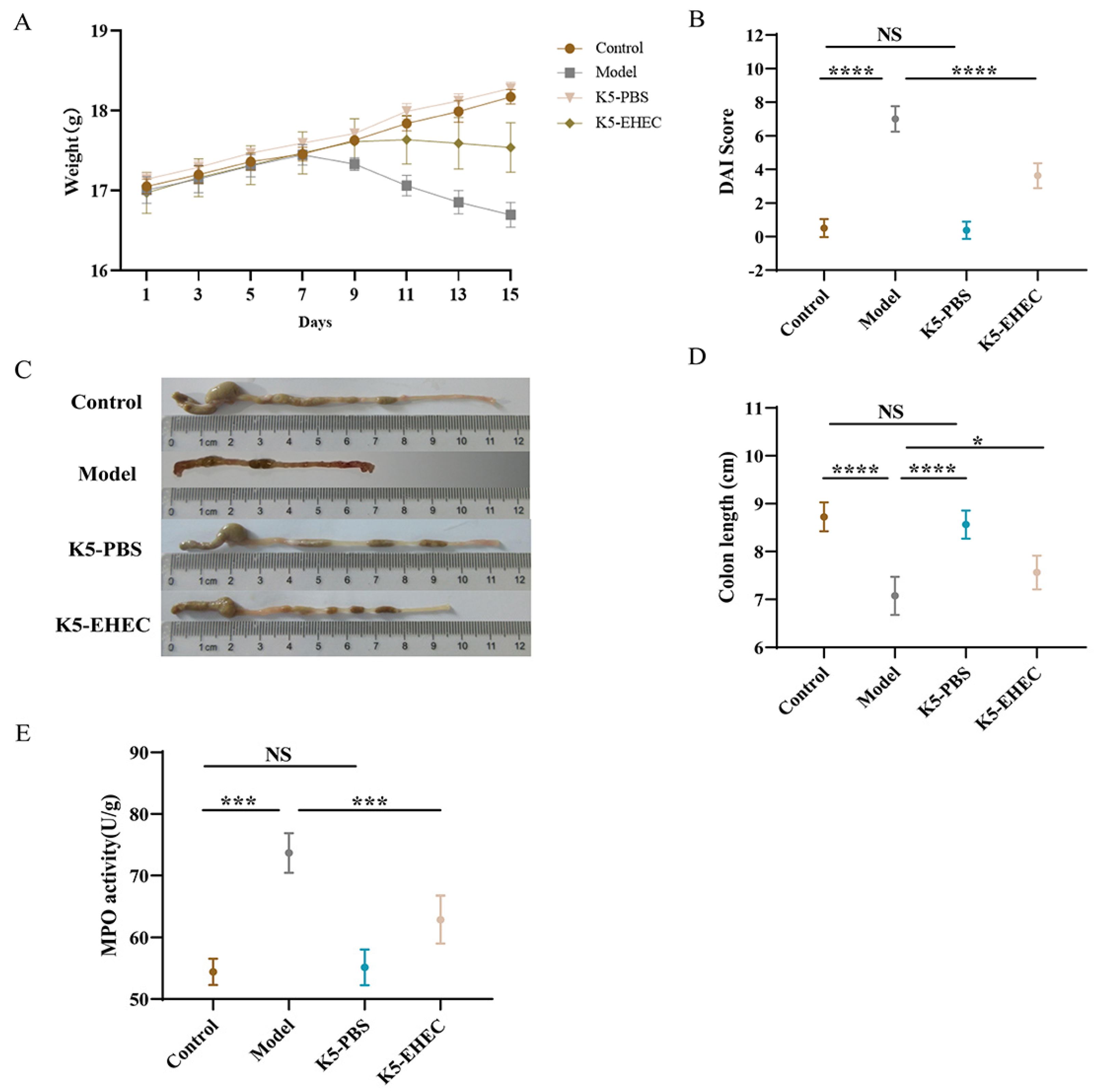
3.1. Effects of B. longum K5 in Alleviating Clinical Symptoms

Initially, all the experimental groups of mice had identical body weights. However, after being administered EHEC O157:H7, the mice in the model and K5-EHEC groups experienced weight loss. The K5-EHEC group of mice had lesser weight loss when compared to the model group, indicating that B. longum K5 may be effective in protecting against the weight loss caused by EHEC O157:H7 (Figure 1A). In contrast to those in the control group, DAI scores in the model group showed a significant increase following EHEC O157:H7 infection (p < 0.0001) (Figure 1B). The K5-EHEC group markedly inhibited the elevation of DAI scores as compared to the model group (p < 0.0001). Additionally, EHEC O157:H7 infection caused a reduction in colon length in the mice, which was significantly lower in the model group in comparison to the control and K5-PBS groups (p < 0.0001). B. longum K5 prevented the shortening of the colon, with a significant increase observed in colon length in the K5-EHEC group compared to the model group (p < 0.05) (Figure 1C,D).
The level of neutrophil infiltration in response to inflammatory stimuli can be indicated by colonic MPO activity. The model group showed a significant elevation in colonic MPO activity when compared to the control group (p < 0.001). The activity of MPO in the K5-EHEC group was observed to be significantly lower than that in the model group (p < 0.001) (Figure 1E). Pretreatment with B. longum K5 halted the increase in MPO activity in the colon tissue of the mice.

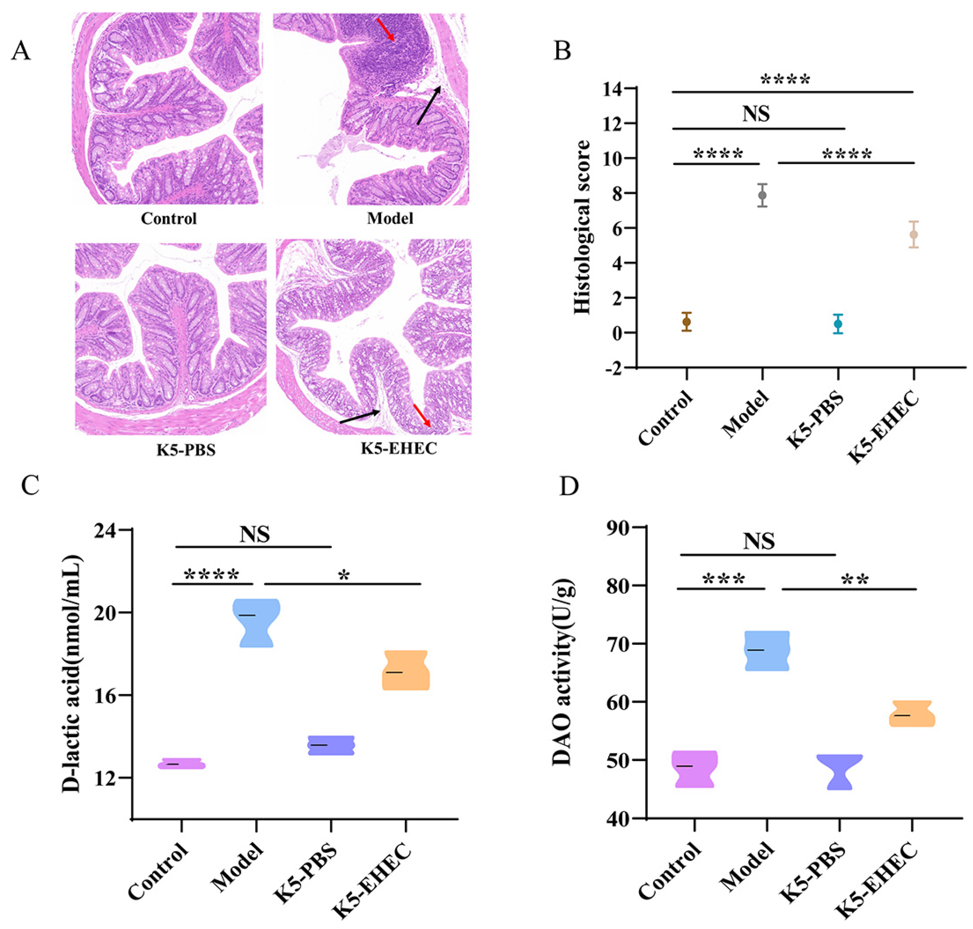
3.2. Effects of B. longum K5 on Intestinal Pathological Changes

EHEC O157:H7 infection led to substantial colonic tissue damage. In the control group, mice displayed normal histological features in the colon. Conversely, the model group exhibited a significant deterioration in colonic epithelial integrity, marked by a reduction in goblet cell counts, expanded submucosal space, and widespread necrotic damage within the intestinal mucosa. In contrast to the model group, in the K5-EHEC group, the tissue damage caused by EHEC O157:H7 was alleviated and the major structures of colonic epithelial cells were preserved (Figure 2A).
The histopathological scores revealed a significant increase in colonic tissue scores in both the model and K5-EHEC groups following EHEC O157:H7 stimulation (p < 0.0001). The histopathological scores of the K5-EHEC group were lower than those of the model group after the B. longum K5 intervention (p < 0.0001) (Figure 2B). These findings indicate the efficacy of B. longum K5 in mitigating pathological damage to the gut in EHEC O157:H7- infected mice, thus safeguarding the integrity of the gut.

3.3. Effect of B. longum K5 on Intestinal Permeability

Serum D-lactate and DAO activity levels were used as markers to evaluate intestinal permeability. Comparative analysis revealed a marked increase in serum D-lactic acid levels and DAO activity in the model group compared to the control group (p < 0.001). Both the increases in serum D-lactate levels and DAO activity were effectively prevented in the K5-EHEC group compared to the model group (p < 0.05) (Figure 2C,D). These findings underscore the potential effectiveness of B. longum K5 in alleviating the increased intestinal permeability induced by EHEC O157:H7.

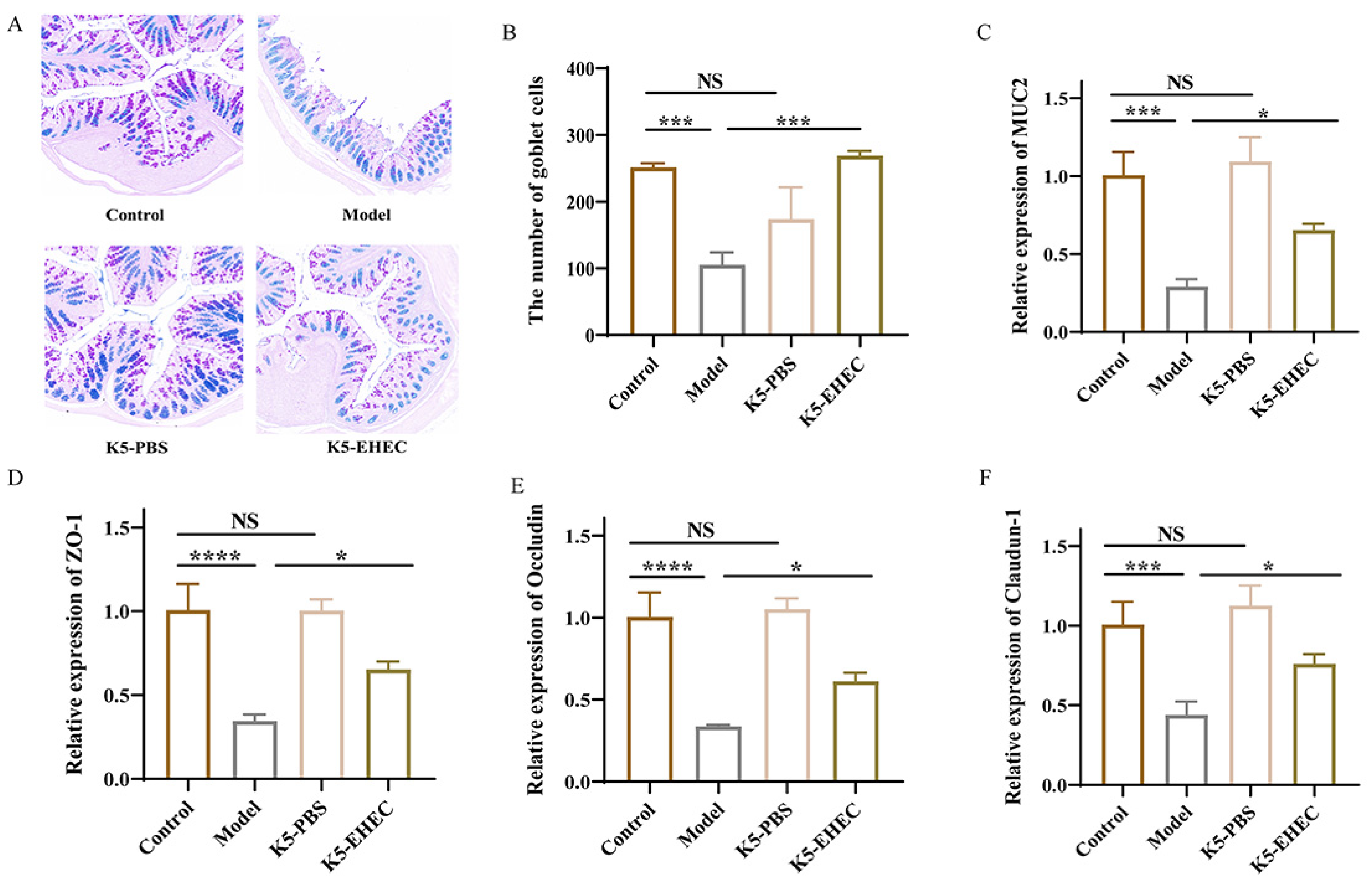
3.4. Effect of B. longum K5 on Gut Barrier Function

AB-PAS staining was utilized to visualize goblet cells and mucin secretion in the gut. Colonic mucin secretion was diminished in the model mice compared to both the control and K5-PBS groups. The K5-EHEC group exhibited elevated levels of colonic mucin compared to the model group (Figure 3A). Additionally, the number of goblet cells in the model group was considerably less than that in the control group (p < 0.001). The K5-EHEC group exhibited a prevention of the reduction in the number of colonic goblet cells compared to the model group (p < 0.001) (Figure 3B).
Compared with that in the control group, the mRNA expression level of MUC2 in the colon tissue of the K5-PBS group was slightly increased, but the difference was not significant (p > 0.05). EHEC O157:H7 infection led to a significant decrease in the mRNA expression levels of MUC2 in the colons of mice in the model and K5-EHEC groups compared with the control group (p < 0.001). However, the K5-EHEC group prevented the reduction in MUC2 mRNA expression compared to the model group (p < 0.05) (Figure 3C). These findings indicate a reduction in MUC2 expression levels due to EHEC O157:H7 infection, whereas pretreatment with B. longum K5 protected intestinal epithelial cells and mitigated the decrease in MUC2 expression.
The expression levels of Occludin and Claudin-1 mRNA in the colonic tissues of mice in the K5-PBS group were slightly elevated when compared to those in the control group, but the difference was not significant (p > 0.05). The mRNA expression of tight junction proteins was significantly reduced in the colonic tissues of mice in the model group compared to the control group (p < 0.001). Notably, the K5-EHEC group effectively halted the decrease in mRNA expression levels of tight junction proteins compared with the model group (p < 0.05) (Figure 3D–F). These observations emphasize the effectiveness of pretreatment with B. longum K5 in mitigating the decrease in mRNA expression of tight junction proteins (ZO-1, Occludin, and Claudin-1) induced by EHEC O157:H7 infection in colonic tissues. This played a crucial role in reinforcing the function of the gut barrier.

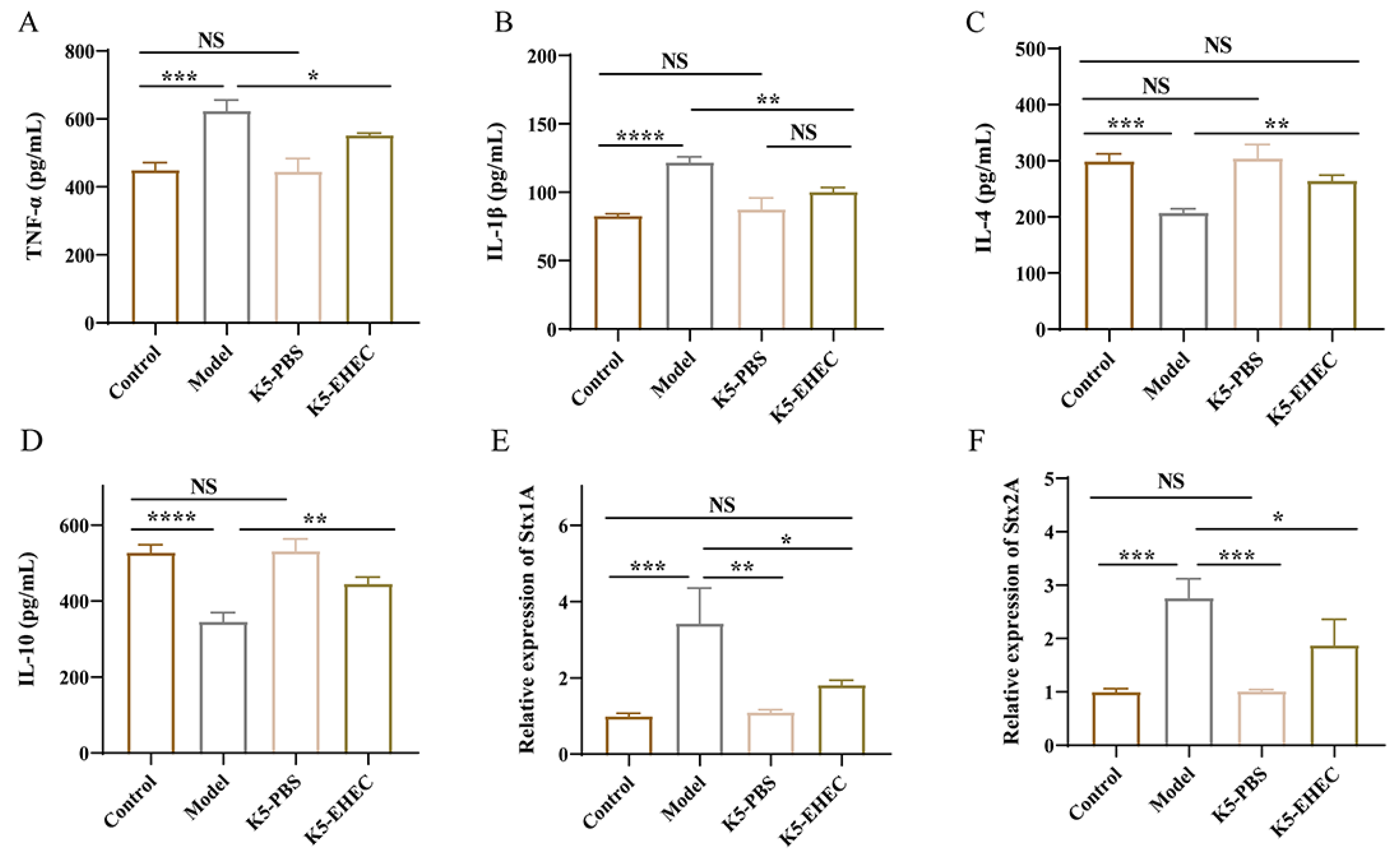
3.5. Effect of B. longum K5 on Gut Inflammation

The levels of TNF-α, IL-1β, IL-4, and IL-10 were measured in mouse colon tissue to reflect the extent of gut inflammation. Compared with those in the control group, the concentrations of TNF-α and IL-1β exhibited a marked increase in the colons of mice in the model group (p < 0.001). The concentrations of IL-4 and IL-10 in the colons of the model group were notably reduced compared to those of the control group (p < 0.001). As depicted in Figure 4A–D, the K5-EHEC group notably inhibited the elevation of TNF-α and IL-1β levels and prevented the reduction in IL-4 and IL-10 levels compared to the model group (p < 0.05). Remarkably, IL-1β levels in the K5-EHEC group did not show a significant difference compared to those in the K5-PBS group (p > 0.05). IL-4 levels in the K5-EHEC group were comparable to those in the control group (p > 0.05). These findings emphasize the ability of B. longum K5 to mitigate the inflammatory response induced by EHEC O157:H7 infection through the inhibition of pro-inflammatory cytokine production and the promotion of anti-inflammatory cytokine production.

3.6. Effect of B. longum K5 on the Expression of Virulence Factors

The analysis of cecal contents from the mice in the model group revealed a substantial increase in the mRNA expression levels of Stx1A and Stx2A compared to the control and K5-PBS groups (p < 0.01). This trend, however, was reversed in the K5-EHEC group, where a significant suppression of Stx1A and Stx2A expression was observed (p < 0.05), as detailed in Figure 4E,F. Remarkably, the mRNA expression level of Stx1A in the K5-EHEC group did not exhibit a significant deviation from that of the control group (p > 0.05). This implies that pretreatment with B. longum K5 alleviated the elevated expression levels of Stx1A and Stx2A induced by EHEC O157:H7 infection, underscoring the role of B. longum K5 in mitigating the effects of this infection.

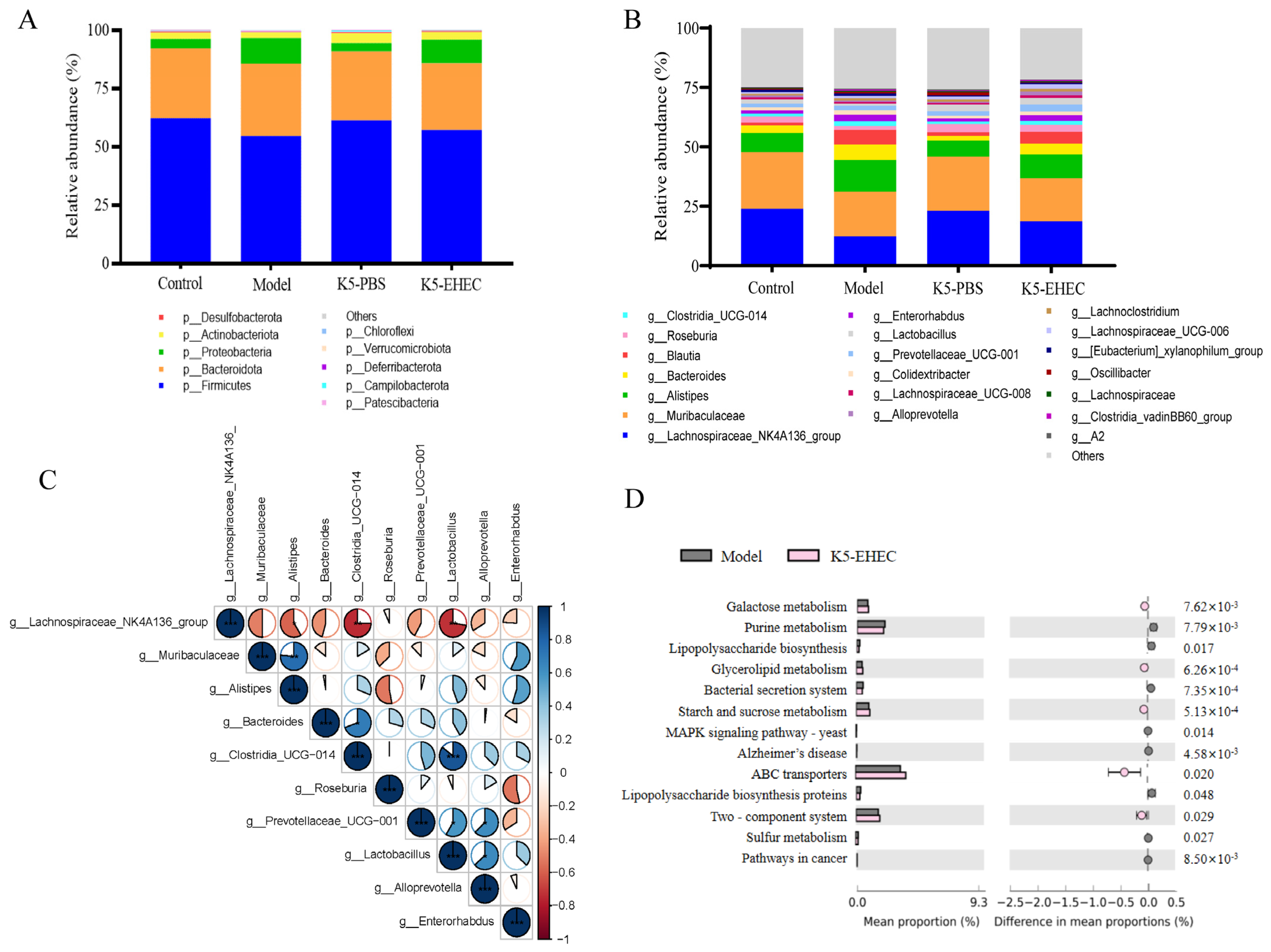
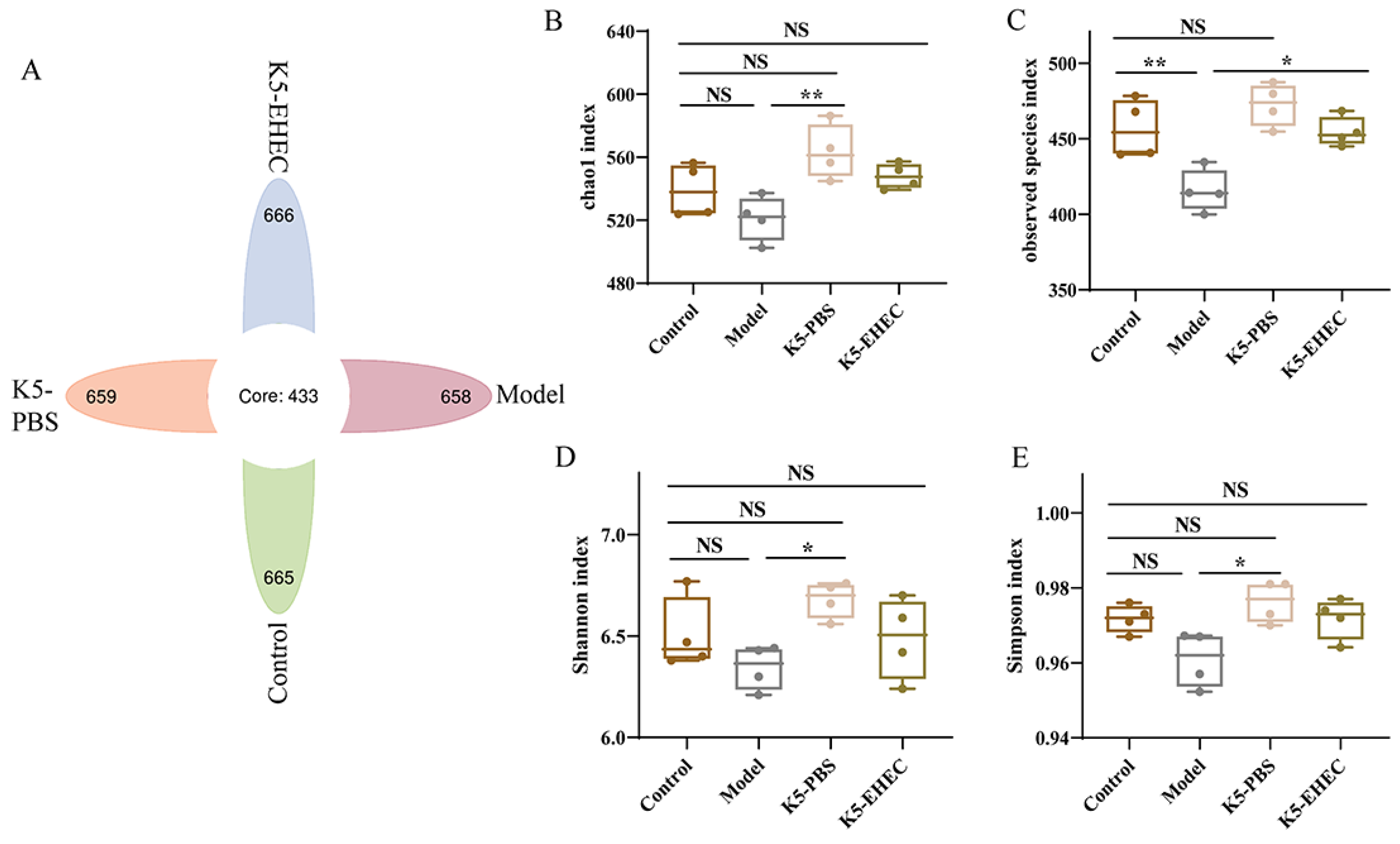
3.7. Effect of B. longum K5 on the Diversity of the Gut Microbiota

The impact of B. longum K5 on the gut microbiota was investigated through 16S rRNA sequencing. The flower plot results revealed that the unique operational taxonomic unit (OTU) counts for the control, model, K5-PBS, and K5-EHEC groups were 665, 658, 666, and 659, respectively (Figure 5A). The Chao1, observed species, Shannon, and Simpson indices were used to evaluate the diversity of the gut microbiota. There was no significant difference in species richness and diversity between the K5-PBS group and the control group (p > 0.05). A marked decrease in the observed species index was observed in the model group compared to the control group (p < 0.01). However, the K5-EHEC group mitigated this significant decrease in the observed species index compared to the model group (p < 0.05), and the Chao1, Shannon, and Simpson indices of the K5-EHEC group were not significantly different from those of the control group (p > 0.05) (Figure 5B–E). These findings indicate that EHEC O157:H7 infection diminished the gut microbial diversity in the mice, while pretreatment with B. longum K5 appeared to prevent the reduction in microbial diversity in the gut environment caused by EHEC O157:H7 infection.

3.8. Effect of B. longum K5 on the Structure of the Gut Microbiota

In this study, we examined the effect of EHEC O157:H7 infection on the intestinal microbial composition, focusing on both the phylum and genus levels. In the control and K5-PBS groups, the gut microbiota predominantly comprised Firmicutes, Bacteroidota, and Proteobacteria. Notably, the K5-PBS group exhibited a marginal rise in Actinobacteriota. The model group had a reduction in the abundance of Firmicutes and Bacteroidota, accompanied by a surge in Proteobacteria. Conversely, pretreatment with K5 in the K5-EHEC group lessened the increase in Proteobacteria and the decrease in Firmicutes (Figure 6A). At the genus level, the model group demonstrated an elevation in the relative abundance of Alistipes, Bacteroides, Blautia, and Desulfovibrio following infection with EHEC O157:H7. Meanwhile, the relative abundance of the Lachnospiraceae NK4A136 group, Muribaculaceae, Roseburia, and Lactobacillus decreased in the model group. These findings demonstrate that pretreatment with B. longum K5 effectively prevented the decrease in abundance of the Lachnospiraceae NK4A136 group, Roseburia, and Lactobacillus while inhibiting the proliferation of Alistipes associated with EHEC O157:H7 infection. There was a slight increase in the abundance of Lactobacillus in the K5-PBS group compared to the control group (Figure 6B).
Correlation analyses of the primary genera within the gut microorganisms of K5-EHEC demonstrated a notably negative correlation between the levels of Clostridia UCG-014 and Lactobacillus and the levels of Lachnospiraceae NK4A136 (p < 0.01). Lactobacillus and Clostridia UCG-014 exhibited a significantly positive correlation with each other (p < 0.001); a highly positive correlation was also observed between Alistipes and Muribaculaceae (p < 0.01) (Figure 6C). This indicates that EHEC O157:H7 disrupted the typical composition of the gut microbiota in mice, whereas B. longum K5 seems to have reduced the induction of this effect by promoting the growth of beneficial microbiota. The functional prediction of the KEGG pathway indicated that B. longum K5 prevented the decrease in the abundance of microbial genes related to galactose, glycerolipid, starch, and sucrose metabolism induced by EHEC O157:H7 infection. Treatment with B. longum K5 concurrently halted the downregulation of genes associated with ABC transporter proteins and the two-component system. On the contrary, the increase in genes associated with the bacterial secretion system, sulfur metabolism, lipopolysaccharide biosynthesis proteins, and pathways related to Alzheimer’s disease, the MAPK signaling pathway, and cancer was significantly inhibited (Figure 6D).

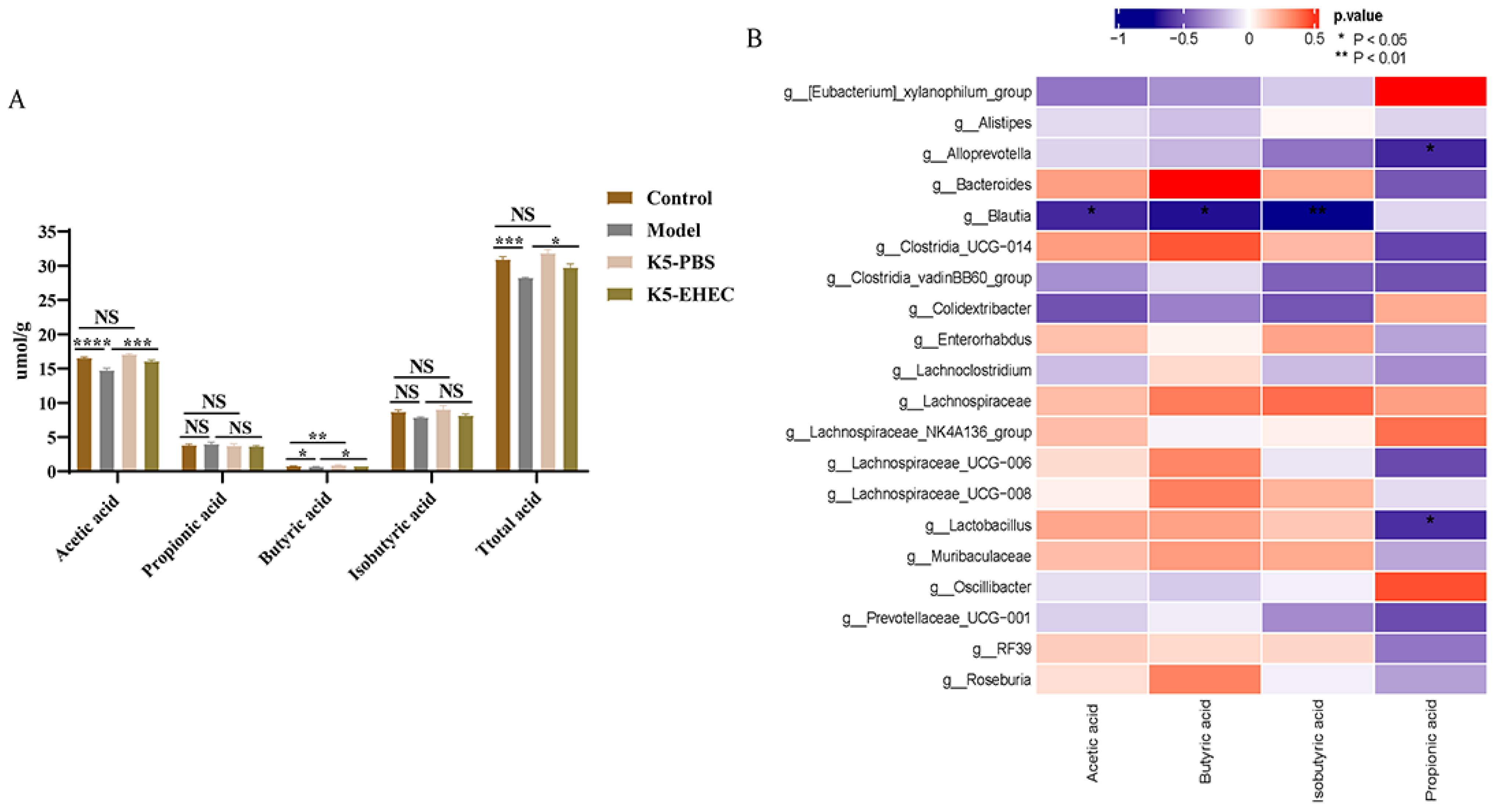
3.9. Production of SCFAs

The results of SCFAs in the intestinal contents of the mice revealed a marginal increase in the levels of acetic, isobutyric, and total acids in the K5-PBS group compared to the control group. However, the difference was not statistically significant (p > 0.05). The concentrations of butyric acid were significantly higher in the K5-PBS group compared to the control group (p < 0.01). And the model group exhibited the lowest levels of acetic acid, butyric acid, and total acid. Remarkably, pretreatment with B. longum K5 in the K5-EHEC group significantly promoted butyric acid and total acid production as compared to the model group (p < 0.05) (Figure 7A).
A correlation analysis of SCFAs with the main genera of gut microorganisms showed that Alloprevotella and Lactobacillus were significantly and negatively correlated with the propionic acid content, and Blautia was significantly and negatively correlated with acetic, butyric, and isobutyric acids (p < 0.05) (Figure 7B). B. longum K5 demonstrated the potential to promote the production of SCFAs, particularly butyric acid, in the intestinal contents of mice.

4. Discussion

EHEC O157:H7 is considered a highly dangerous foodborne pathogen capable of colonizing the epithelial surface of the intestinal mucosa [23,24], forming A/E lesions, and impairing the function of the intestinal epithelial barrier [25]. Stx produced by EHEC O157:H7 can be transmitted into the bloodstream, causing toxemia and extensive histopathological damage and increasing the risk of HUC [26]. Probiotics have garnered significant attention for their potential role in preventing pathogenic infections. A study conducted by Wang et al. revealed that L. casei LC2W effectively inhibited the intestinal colonization of EHEC O157:H7 by directly or indirectly antagonizing the bacterial association with epithelial cells [27]. The fermented broth of L. acidophilus has been demonstrated to prevent the intestinal barrier dysfunction induced by EHEC O157:H7 [28]. B. longum has been reported to have health-promoting benefits of inhibiting harmful intestinal bacteria [29], lowering cholesterol levels [30], modulating the gut microbiota, and enhancing immune response [16,31]. Bifidobacterium is known to secrete various antimicrobial agents, including organic acids and bacteriocins [32,33]. Moreover, the mechanisms through which Bifidobacteria exert their antibacterial effects include suppressing the expression of virulence factors, competing for nutrients, and obstructing the adhesion and invasion of pathogenic bacteria to host cells by contending for binding sites [34]. Therefore, our research utilized a previously established EHEC O157:H7-infected mouse model [35] to explore the protective effects of B. longum K5 and to elucidate the mechanism of action.
The experimental group of mice exhibited symptoms such as diarrhea, shortened colons, weight loss, and intestinal wall thinning after one week of gavage with EHEC O157:H7, confirming the effective establishment of the EHEC O157:H7 infection model in comparison to the control group. Mice in the K5-EHEC group pre-supplemented with B. longum K5 showed mitigation of these symptoms, including the weight loss and shortened colon. The increase in the DAI score further indicated the efficacy of B. longum K5 in alleviating infection symptoms in the mice. These observations were in agreement with the study by Gagnon et al., where B. thermophilum RBL 71 enhanced food intake and substantially reduced weight loss in infected mice [36]. Our research showed that introducing B. longum K5 effectively attenuated the EHEC O157:H7-induced rise in MPO activity, suggesting a reduction in neutrophil infiltration. This aligns with the findings reported by Wang et al. [27], emphasizing the potential therapeutic role of B. longum K5 in controlling inflammation.
The structural integrity of the intestinal epithelium is crucial in resisting pathogenic bacterial invasion [37]. In our study, EHEC O157:H7 disrupted this integrity in mice, evidenced by damage to the intestinal epithelial microvilli and mucosal lining. Conversely, mice treated with B. longum K5 showed attenuated intestinal damage, less severe inflammatory infiltration, better-preserved intestinal epithelial structures, and significantly improved colon tissue pathology, corroborating the findings of Wang et al. [32]. The functionality of the gut barrier is connected to numerous clinical conditions, including bacterial infections [38]. Pathogenic organisms can undermine this barrier by altering the expression of TJ proteins and producing toxins, thereby causing cell damage, apoptosis, and heightened permeability in intestinal epithelial cells [39]. EHEC O157:H7 infection significantly reduced the mRNA expression of Claudin-1, Occludin, and ZO-1. And there was a notable increase in serum DAO and D-lactic acid levels. Contrastingly, B. longum K5 treatment effectively counteracted these changes. This aligns with Bao et al.’s observations, wherein Bacillus amyloliquefaciens TL106 was found to mitigate TJ protein expression reduction in the colon and decrease DAO and D-lactic acid levels in serum [40]. Mucins, serving as initial barriers on cell surfaces, play a pivotal role in hindering the invasion of pathogenic microorganisms [41]. MUC2, predominantly expressed in goblet cells, is a critical component of the mucus layer, essential for maintaining intestinal balance [41,42]. The pathogen EHEC O157:H7 can degrade mucin via its StcE secretion, while probiotics are known to promote MUC2 production in goblet cells, thereby reinforcing the gut barrier [43,44]. Our experiment revealed that pre-supplementation of mice with B. longum K5 prevented the EHEC O157:H7-induced decrease in MUC2 mRNA expression and resulted in less damage to goblet cells. This is consistent with the findings of Wang et al., who reported that Lactobacillus casei LC2W can bolster the colonic mucosal layer by enhancing MUC2 expression [45].
The balance between pro-inflammatory and anti-inflammatory cytokines determines the inflammation outcome during bacterial infections [46,47]. Consistent with prior findings [27,48], our study observed heightened levels of pro-inflammatory cytokines TNF-α and IL-1β in the colons of mice infected with EHEC O157:H7. Notably, the pretreatment with B. longum K5 prevented these elevations in the concentrations of TNF-α and IL-1β in the colon tissue. Key anti-inflammatory cytokines like IL-4 and IL-10 are essential in modulating inflammatory responses. EHEC O157:H7 infection led to a significant diminishment of anti-inflammatory factors in colon tissue, a trend that was notably prevented in mice treated with B. longum K5. This aligns with the observations of Bao et al. [40], who reported that Bacillus amylolyticus TL106 curbed the production of pro-inflammatory factors like TNF-α, IL-1β, IL-6, and IL-8 and mitigated the decline in IL-10 levels in the intestinal tissues of mice infected with EHEC O157:H7. Stx1A and Stx2A are key virulence factors in EHEC O157:H7, playing a pivotal role in the development of hemorrhagic diarrhea and HUC by disrupting protein synthesis in eukaryotic cells. Research by Yoshimura et al. highlighted that certain strains of Bifidobacteria can markedly lower Shiga toxin levels in both the cecal contents and serum of mice [11]. In line with these findings, our study also observed that the elevated levels of Stx1A and Stx2A resulting from EHEC O157:H7 infection were inhibited in both the serum and cecal contents of mice pretreated with B. longum K5. Furthermore, a study by Saito et al. involved pre-inoculating mice with Bacteroides fragilis and vulgatus before EHEC O157:H7 infection. Their results showed a notable reduction in Stx1 and Stx2 levels and a significant decrease in mortality rates compared to those in mice infected only with EHEC O157:H7 [49].
The gut microbiota, nestled within the mucous layer, acts as a natural defense, curbing the proliferation of pathogenic bacteria. This is achieved through various mechanisms, such as competing for colonization sites and nutrients and secreting antimicrobial agents [50]. A balanced gut microbiota is critical in modulating the host’s immune response, thereby reducing the inflammatory response induced by bacterial infections [51]. In this study, we found that B. longum K5 was effective in preventing the loss of microbial diversity in the gut microbiota of mice caused by EHEC O157:H7 infection. Mice treated with B. longum K5 exhibited a gut microbiota structure similar to that in the control group. At the genus level, a decrease in the relative abundance of the Lachnospiraceae NK4A136 group, Muribaculaceae, and Roseburia was observed following EHEC O157:H7 infection, while B. longum K5 administration prevented the decrease in these genera. B. longum K5 also raised the relative abundance of Roseburia, Lactobacillus, and Oscillibacter. These observations align with the findings of Hu et al., wherein L. johnsonii, L. plantarum, and L. rhamnosus were shown to elevate the relative abundance of Lachnospira, Ruminococcus, Roseburia, and Blautia in the intestine of EHEC-infected mice [52]. These findings suggest that early B. longum K5 intervention can bolster gut microbiota diversity, counteracting the microbial dysbiosis induced by EHEC O157:H7 infection. PICRUSt predictions indicated that the model group, infected with EHEC O157:H7, showed an upregulation of genes linked to lipopolysaccharide biosynthesis. In contrast, the increase in these genes was prevented in the K5-EHEC group. This pattern is consistent with our observation that B. longum K5 substantially mitigated the levels of inflammatory cytokines. The model group infected with EHEC O157:H7 demonstrated an elevated gene abundance in sulfur metabolism. This aligns with our earlier observation of an increased relative abundance of Desulfovibrio in the gut microbiota of these mice.
SCFAs, including acetic acid, propionic acid, and butyric acid, are the primary products of complex nutrient fermentation by the gut microbiota [53]. SCFAs play a multifaceted role: they serve as energy sources for intestinal epithelial cells and influence the expression of virulence genes in pathogenic bacteria, thereby impacting their cellular metabolism and pathogenicity [54,55]. In mice infected with EHEC O157:H7, we observed a significant alteration in SCFA production. There was a substantial decrease in the total acid content, with both the acetic acid and butyric acid levels dropping below those in the control group. However, treatment with B. longum K5 mitigated these changes. This aligns with prior research showing elevated SCFA concentrations in the fecal samples of mice treated with Bifidobacterium [56]. Moreover, our findings are consistent with those of Fukuda et al., who demonstrated that Bifidobacterium, through acetate production, can hinder the transport of Shiga toxins from the intestinal tract to the bloodstream, thereby offering protection against EHEC O157:H7-induced mortality in mice [8]. Indeed, in the event of an EHEC epidemic, pre-treatment with probiotics, such as Bifidobacterium, could be considered for individuals at heightened risk of infection or complications. This approach may involve providing prophylactic Bifidobacterium supplementation to vulnerable populations, such as individuals with weakened immune systems, young children, or the elderly. Prophylactic Bifidobacterium supplementation could contribute to the management and control of the epidemic by bolstering gut health and potentially reducing the susceptibility to EHEC infection or mitigating its severity.

5. Conclusions

In summary, our research demonstrated the potential protective effects of B. longum K5 against EHEC O157:H7 infection in mice. B. longum K5 prevented EHEC O157:H7-induced weakening of the gut barrier, preserved the abundance of beneficial microbes, and promoted the production of SCFAs in the colons of the mice. Moreover, it inhibited the increase in the concentration of pro-inflammatory cytokines and pathogenic virulence factors. These insights underscore the potential of B. longum K5 as a novel therapeutic agent in preventing infections induced by EHEC O157:H7.

Supplementary Materials

The following supporting information can be downloaded at: https://www.mdpi.com/article/10.3390/nu16081164/s1. Table S1: Disease Activity Index (DAI) Scoring System. Table S2: Histopathological Scoring System. Table S3: Primer Sequences for RT-qPCR Assays in Colonic Tissue.

Author Contributions

Conceptualization, D.L., C.L., Y.Y., S.L., Y.C., F.L., G.H. and B.L.; Methodology, D.L., X.L., S.L., Y.C., G.H. and B.L.; Validation, D.L. and X.L.; Formal analysis, T.C.; Data curation, C.L.; Writing—original draft, D.L.; Writing—review & editing, Y.Y., F.L., G.H. and B.L.; Visualization, T.C. All authors have read and agreed to the published version of the manuscript.

Funding

This research was funded by the National Natural Science Foundation of China (32072190).

Institutional Review Board Statement

The animal study protocol was approved by the Animal Ethics Committee of Northeast Agricultural University (ethical approval code NEAUEC20220363, approval date 8 September 2022).

Informed Consent Statement

Not applicable.

Data Availability Statement

The data presented in this study are available from the author upon reasonable request.

Acknowledgments

We thank Zengbo Wang and Yuqi Wang for their constructive revisions and contributions to enhancing the linguistic quality of the article.

Conflicts of Interest

The authors declare no conflicts of interest in this study.

References

  1. Croxen, M.A.; Law, R.J.; Scholz, R.; Keeney, K.M.; Wlodarska, M.; Finlay, B.B. Recent advances in understanding enteric pathogenic Escherichia coli. Clin. Microbiol. Rev. 2013, 26, 822–880. [Google Scholar] [CrossRef] [PubMed]
  2. Srutkova, D.; Schwarzer, M.; Hudcovic, T.; Zakostelska, Z.; Drab, V.; Spanova, A.; Rittich, B.; Kozakova, H.; Schabussova, I. Bifidobacterium longum CCM 7952 promotes epithelial barrier function and prevents acute DSS-induced colitis in strictly strain-specific manner. PLoS ONE 2015, 10, e0134050. [Google Scholar] [CrossRef]
  3. Nguyen, Y.; Sperandio, V. Enterohemorrhagic E. coli (EHEC) pathogenesis. Front. Cell. Infect. Microbiol. 2012, 2, 90. [Google Scholar] [CrossRef] [PubMed]
  4. Lim, J.Y.; Yoon, J.W.; Hovde, C.J. A brief overview of Escherichia coli O157: H7 and its plasmid O157. J. Microbiol. Biotechnol. 2010, 20, 5–14. [Google Scholar] [CrossRef] [PubMed]
  5. Sharma, M.; Wasan, A.; Sharma, R.K. Recent developments in probiotics: An emphasis on Bifidobacterium. Food Biosci. 2021, 41, 100993. [Google Scholar] [CrossRef]
  6. Jung, H.L.; Soon, H.S. Antimicrobial and Immunomodulatory Effects of Bifidobacterium Strains: A Review. J. Microbiol. Biotechnol. 2020, 30, 1793. [Google Scholar]
  7. Delcaru, C.; Alexandru, I.; Podgoreanu, P.; Cristea, V.C.; Bleotu, C.; Chifiriuc, M.C.; Bezirtzoglou, E.; Lazar, V. Antagonistic activities of some Bifidobacterium sp. strains isolated from resident infant gastrointestinal microbiota on Gram-negative enteric pathogens. Anaerobe 2016, 39, 39–44. [Google Scholar] [CrossRef]
  8. Fukuda, S.; Toh, H.; Hase, K.; Oshima, K.; Nakanishi, Y.; Yoshimura, K.; Tobe, T.; Clarke, J.M.; Topping, D.L.; Suzuki, T. Bifidobacteria can protect from enteropathogenic infection through production of acetate. Nature 2011, 469, 543–547. [Google Scholar] [CrossRef] [PubMed]
  9. Westermann, C.; Gleinser, M.; Corr, S.C.; Riedel, C.U. A critical evaluation of bifidobacterial adhesion to the host tissue. Front. Microbiol. 2016, 7, 1220. [Google Scholar] [CrossRef]
  10. Zhang, C.; Yu, Z.; Zhao, J.; Zhang, H.; Zhai, Q.; Chen, W. Colonization and probiotic function of Bifidobacterium longum. J. Funct. Foods 2019, 53, 157–165. [Google Scholar] [CrossRef]
  11. Yoshimura, K.; Matsui, T.; Itoh, K. Prevention of Escherichia coli O157: H7 infection in gnotobiotic mice associated with Bifidobacterium strains. Antonie Van Leeuwenhoek 2010, 97, 107–117. [Google Scholar] [CrossRef]
  12. Juge, N. Microbial adhesins to gastrointestinal mucus. Trends Microbiol. 2012, 20, 30–39. [Google Scholar] [CrossRef]
  13. Monteagudo-Mera, A.; Rastall, R.A.; Gibson, G.R.; Charalampopoulos, D.; Chatzifragkou, A. Adhesion mechanisms mediated by probiotics and prebiotics and their potential impact on human health. Appl. Microbiol. Biotechnol. 2019, 103, 6463–6472. [Google Scholar] [CrossRef]
  14. Zhao, L.; Wang, S.; Dong, J.; Shi, J. Identification, Characterization, and Antioxidant Potential of Bifidobacterium longum subsp longum Strains Isolated From Feces of Healthy Infants. Front. Microbiol. 2021, 12, 3154. [Google Scholar]
  15. Zhao, L.; Xie, Q.; Evivie, S.E.; Yue, Y.; Yang, H.; Lv, X.; Liu, F.; Li, B.; Huo, G. Bifidobacterium longum subsp longum K5 alleviates inflammatory response and prevents intestinal barrier injury induced by LPS in vitro based on comparative genomics. J. Funct. Foods 2022, 92, 105030. [Google Scholar]
  16. Dong, J.; Ping, L.; Xie, Q.; Liu, D.; Zhao, L.; Evivie, S.E.; Wang, Z.; Li, B.; Huo, G. Lactobacillus plantarum KLDS1.0386 with antioxidant capacity ameliorates the lipopolysaccharide-induced acute liver injury in mice by NF-κB and Nrf2 pathway. Food Biosci. 2022, 47, 101589. [Google Scholar] [CrossRef]
  17. Kwon, J.; Lee, C.; Heo, S.; Kim, B.; Hyun, C.-K. DSS-induced colitis is associated with adipose tissue dysfunction and disrupted hepatic lipid metabolism leading to hepatosteatosis and dyslipidemia in mice. Sci. Rep. 2021, 11, 5283. [Google Scholar] [CrossRef] [PubMed]
  18. Jiaqi, G.; Fei, L.; Sijia, Z.; Etareri, E.S.; Jialu, S.; Na, L.; Li, Z.; Yingxue, Y.; Qinggang, X.; Guicheng, H.; et al. Effect of Bifidobacterium longum subsp. longum on the proliferative and tight-junction activities of Human Fetal Colon Epithelial Cells. J. Funct. Foods 2021, 86, 104715. [Google Scholar]
  19. Na, L.; Shengnan, L.; Qingxue, C.; Lina, Z.; Bailiang, L.; Guicheng, H. Distinct gut microbiota and metabolite profiles induced by delivery mode in healthy Chinese infants. J. Proteom. 2021, 232, 104071. [Google Scholar]
  20. Langille, M.G.I.; Zaneveld, J.; Caporaso, J.G.; McDonald, D.; Knights, D.; Reyes, J.A.; Clemente, J.C.; Burkepile, D.E.; Vega Thurber, R.L.; Knight, R. Predictive functional profiling of microbial communities using 16S rRNA marker gene sequences. Nat. Biotechnol. 2013, 31, 814–821. [Google Scholar] [CrossRef]
  21. Parks, D.H.; Tyson, G.W.; Hugenholtz, P.; Beiko, R.G. STAMP: Statistical analysis of taxonomic and functional profiles. Bioinformatics 2014, 30, 3123–3124. [Google Scholar] [CrossRef] [PubMed]
  22. Fenfen, Y.; Na, L.; Jialu, S.; Huizhen, L.; Yingxue, Y.; Wenshu, J.; Nana, W.; Yue, S.; Guicheng, H.; Bailiang, L. Lactobacillus acidophilus alleviates type 2 diabetes by regulating hepatic glucose, lipid metabolism and gut microbiota in mice. Food Funct. 2019, 10, 5804–5815. [Google Scholar]
  23. Cookson, A.L.; Woodward, M.J. The role of intimin in the adherence of enterohaemorrhagic Escherichia coli (EHEC) O157: H7 to HEp-2 tissue culture cells and to bovine gut explant tissues. Int. J. Med. Microbiol. 2003, 292, 547–553. [Google Scholar] [CrossRef] [PubMed]
  24. Moxley, R.A. Escherichia coli 0157:H7: An update on intestinal colonization and virulence mechanisms. Anim. Health Res. Rev. 2004, 5, 15–33. [Google Scholar] [CrossRef] [PubMed]
  25. Lebeis, S.L.; Sherman, M.A.; Kalman, D. Protective and destructive innate immune responses to enteropathogenic Escherichia coli and related A/E pathogens. Future Microbiol. 2008, 3, 315–328. [Google Scholar] [CrossRef] [PubMed]
  26. Obrig, T.G. Shiga toxin mode of action in E. coli O157:H7 disease. Front. Biosci. 1997, 2, 635–642. [Google Scholar] [CrossRef] [PubMed]
  27. Wang, G.; Zhang, Y.; Song, X.; Xia, Y.; Lai, P.F.H.; Ai, L. Lactobacillus casei LC2W can inhibit the colonization of Escherichia coli O157: H7 in vivo and reduce the severity of colitis. Food Funct. 2019, 10, 5843–5852. [Google Scholar] [CrossRef]
  28. Bonvicini, F.; Pagnotta, E.; Punzo, A.; Calabria, D.; Simoni, P.; Mirasoli, M.; Passerini, N.; Bertoni, S.; Ugolini, L.; Lazzeri, L.; et al. Effect of Lactobacillus acidophilus fermented broths enriched with Eruca sativa seed extracts on intestinal barrier and inflammation in a co-culture system of an enterohemorrhagic Escherichia coli and human intestinal cells. Nutrients 2020, 12, 3064. [Google Scholar] [CrossRef]
  29. Yun, B.; Song, M.; Park, D.J.; Oh, S. Beneficial effect of Bifidobacterium longum ATCC 15707 on survival rate of Clostridium difficile infection in mice. Korean J. Food Sci. Anim. Resour. 2017, 37, 368. [Google Scholar] [CrossRef]
  30. Jiang, J.; Wu, C.; Zhang, C.; Zhang, Q.; Yu, L.; Zhao, J.; Zhang, H.; Narbad, A.; Chen, W.; Zhai, Q. Strain-specific effects of Bifidobacterium longum on hypercholesterolemic rats and potential mechanisms. Int. J. Mol. Sci. 2021, 22, 1305. [Google Scholar] [CrossRef]
  31. Finamore, A.; Roselli, M.; Donini, L.; Brasili, E.; Rami, R.; Carnevali, P.; Mistura, L.; Pinto, A.; Giusti, A.; Mengheri, E. Supplementation with Bifidobacterium longum Bar33 and Lactobacillus helveticus Bar13 mixture improves immunity in elderly humans (over 75 years) and aged mice. Nutrition 2019, 63, 184–192. [Google Scholar] [CrossRef] [PubMed]
  32. Martinez, F.A.C.; Balciunas, E.M.; Converti, A.; Cotter, P.D.; de Souza Oliveira, R.P. Bacteriocin production by Bifidobacterium spp. A review. Biotechnol. Adv. 2013, 31, 482–488. [Google Scholar] [CrossRef] [PubMed]
  33. Makras, L.; De Vuyst, L. The in vitro inhibition of Gram-negative pathogenic bacteria by Bifidobacteria is caused by the production of organic acids. Int. Dairy J. 2006, 16, 1049–1057. [Google Scholar] [CrossRef]
  34. Nair, M.S.; Amalaradjou, M.A.; Venkitanarayanan, K. Antivirulence properties of probiotics in combating microbial pathogenesis. Adv. Appl. Microbiol. 2017, 98, 1–29. [Google Scholar]
  35. Bian, X.; Wang, T.T.; Xu, M.; Evivie, S.E.; Luo, G.W.; Liang, H.Z.; Yu, S.F.; Huo, G.C. Effect of Lactobacillus Strains on Intestinal Microflora and Mucosa Immunity in Escherichia coli O157:H7-Induced Diarrhea in Mice. Curr. Microbiol. 2016, 73, 65–70. [Google Scholar] [CrossRef] [PubMed]
  36. Mélanie, G.; Ehab, E.K.; Nassra, D.; Denis, R. Effect of Bifidobacterium thermacidophilum probiotic feeding on enterohemorrhagic Escherichia coli O157:H7 infection in BALB/c mice. Int. J. Food Microbiol. 2006, 111, 26–33. [Google Scholar]
  37. Peterson, L.W.; Artis, D. Intestinal epithelial cells: Regulators of barrier function and immune homeostasis. Nat. Rev. Immunol. 2014, 14, 141–153. [Google Scholar] [CrossRef] [PubMed]
  38. König, J.; Wells, J.; Cani, P.D.; García-Ródenas, C.L.; MacDonald, T.; Mercenier, A.; Whyte, J.; Troost, F.; Brummer, R.-J. Human intestinal barrier function in health and disease. Clin. Transl. Gastroenterol. 2016, 7, e196. [Google Scholar] [CrossRef]
  39. Groschwitz, K.R.; Hogan, S.P. Intestinal barrier function: Molecular regulation and disease pathogenesis. J. Allergy Clin. Immunol. 2009, 124, 21–22. [Google Scholar] [CrossRef]
  40. Bao, C.L.; Liu, S.Z.; Shang, Z.D.; Liu, Y.J.; Wang, J.; Zhang, W.X.; Dong, B.; Cao, Y.H. Bacillus amyloliquefaciens TL106 protects mice against enterohaemorrhagic Escherichia coli O157: H7-induced intestinal disease through improving immune response, intestinal barrier function and gut microbiota. J. Appl. Microbiol. 2021, 131, 470–484. [Google Scholar] [CrossRef]
  41. Kim, J.J.; Khan, W.I. Goblet cells and mucins: Role in innate defense in enteric infections. Pathogens 2013, 2, 55–70. [Google Scholar] [CrossRef] [PubMed]
  42. Gundamaraju, R.; Chong, W.C. Consequence of distinctive expression of MUC2 in colorectal cancers: How much is actually bad? Biochim. Biophys. Acta BBA Rev. Cancer 2021, 1876, 188579. [Google Scholar] [CrossRef] [PubMed]
  43. Martens, E.C.; Neumann, M.; Desai, M.S. Interactions of commensal and pathogenic microorganisms with the intestinal mucosal barrier. Nat. Rev. Microbiol. 2018, 16, 457–470. [Google Scholar] [CrossRef] [PubMed]
  44. Liu, Y.; Yu, X.; Zhao, J.; Zhang, H.; Zhai, Q.; Chen, W. The role of MUC2 mucin in intestinal homeostasis and the impact of dietary components on MUC2 expression. Int. J. Biol. Macromol. 2020, 164, 884–891. [Google Scholar] [CrossRef]
  45. Wang, G.; Tang, H.; Zhang, Y.; Xiao, X.; Xia, Y.; Ai, L. The intervention effects of Lactobacillus casei LC2W on Escherichia coli O157: H7-induced mouse colitis. Food Sci. Hum. Wellness 2020, 9, 289–294. [Google Scholar] [CrossRef]
  46. Gobert, A.P.; Coste, A.; Guzman, C.A.; Vareille, M.; Hindré, T.; de Sablet, T.; Girardeau, J.-P.; Martin, C. Modulation of chemokine gene expression by Shiga-toxin producing Escherichia coli belonging to various origins and serotypes. Microbes Infect. 2008, 10, 159–165. [Google Scholar] [CrossRef]
  47. Shi, Y.F. The role of inflammatory cytokines in bacterial infection. Int. J. Intern. Med. 2009, 36, 112–115. [Google Scholar]
  48. Zhao, X.; Wang, L.; Zhu, C.; Xia, X.; Zhang, S.; Wang, Y.; Zhang, H.; Xu, Y.; Chen, S.; Jiang, J. The antimicrobial peptide mastoparan X protects against enterohemorrhagic Escherichia coli O157: H7 infection, inhibits inflammation, and enhances the intestinal epithelial barrier. Front. Microbiol. 2021, 12, 644887. [Google Scholar] [CrossRef] [PubMed]
  49. Saito, K.; Suzuki, R.; Koyanagi, Y.; Isogai, H.; Yoneyama, H.; Isogai, E. Inhibition of enterohemorrhagic Escherichia coli O157: H7 infection in a gnotobiotic mouse model with pre-colonization by Bacteroides strains. Biomed. Rep. 2019, 10, 175–182. [Google Scholar] [CrossRef]
  50. Lawley, T.D.; Walker, A.W. Intestinal colonization resistance. Immunology 2013, 138, 1–11. [Google Scholar] [CrossRef]
  51. Kelly, D.; Conway, S.; Aminov, R. Commensal gut bacteria: Mechanisms of immune modulation. Trends Immunol. 2005, 26, 326–333. [Google Scholar] [CrossRef] [PubMed]
  52. Hu, Y.; Zhao, M.; Lu, Z.; Lv, F.; Zhao, H.; Bie, X.L. L. johnsonii, L. plantarum, and L. rhamnosus alleviated Enterohaemorrhagic Escherichia coli-induced diarrhoea in mice by regulating gut microbiota. Microb. Pathog. 2021, 154, 104856. [Google Scholar] [CrossRef] [PubMed]
  53. Den Besten, G.; Van Eunen, K.; Groen, A.K.; Venema, K.; Reijngoud, D.-J.; Bakker, B.M. The role of short-chain fatty acids in the interplay between diet, gut microbiota, and host energy metabolism. J. Lipid Res. 2013, 54, 2325–2340. [Google Scholar] [CrossRef] [PubMed]
  54. Hamer, H.M.; Jonkers, D.; Venema, K.; Vanhoutvin, S.; Troost, F.J.; Brummer, R.J. The role of butyrate on colonic function. Aliment. Pharmacol. Ther. 2008, 27, 104–119. [Google Scholar] [CrossRef] [PubMed]
  55. Peng, M.; Biswas, D. Short chain and polyunsaturated fatty acids in host gut health and foodborne bacterial pathogen inhibition. Crit. Rev. Food Sci. Nutr. 2017, 57, 3987–4002. [Google Scholar] [CrossRef]
  56. Wang, L.; Hu, L.; Xu, Q.; Jiang, T.; Fang, S.; Wang, G.; Zhao, J.; Zhang, H.; Chen, W. Bifidobacteria exert species-specific effects on constipation in BALB/c mice. Food Funct. 2017, 8, 3587–3600. [Google Scholar] [CrossRef]
Figure 1. Effect of B. longum K5 on EHEC-induced clinical symptoms. Note: (A) changes in mouse body weight; (B) Disease Activity Index (DAI) scores; (C) representative images of mouse colons; (D) colon length; (E) MPO activity. Comparisons with different significance levels: (*) p < 0.05, (***) p < 0.001, and (****) p ≤ 0.0001. NS = not significant.
Nutrients 16 01164 g001
Figure 2. Effect of B. longum K5 on intestinal permeability. Note: (A) hematoxylin and eosin (H&E) staining (magnification ×100); Red arrows: inflammatory infiltration, Black arrows: increased tissue interstitial space (B) histological score; (C) D-lactic acid level; (D) DAO activity. Comparisons with different significance levels: (*) p < 0.05, (**) p < 0.01, (***) p < 0.001, and (****) p ≤ 0.0001. NS = not significant.
Nutrients 16 01164 g002
Figure 3. Effect of B. longum K5 on intestinal barrier function. Note: (A) AB-PAS staining of colon tissue sections (AB-PAS staining, magnification ×100); (B) number of goblet cells; (C) MUC2; (D) ZO-1; (E) Occludin; (F) Claudin-1. Comparisons with different significance levels: (*) p < 0.05, (***) p < 0.001, and (****) p ≤ 0.0001. NS = not significant.
Nutrients 16 01164 g003
Figure 4. Effect of B. longum K5 on intestinal inflammation. Note: (A) TNF-α; (B) IL-1β; (C) IL-4; (D) IL-10; (E) Stx1A; (F) Stx2A. Comparisons with different significance levels: (*) p < 0.05, (**) p < 0.01, (***) p < 0.001, and (****) p ≤ 0.0001. NS = not significant.
Nutrients 16 01164 g004
Figure 5. Effect of B. longum K5 on intestinal flora diversity. Note: (A) OTU flower plot; (B) Chao1 index; (C) observed species index; (D) Shannon index; (E) Simpson index. Comparisons with different significance levels: (*) p < 0.05, (**) p < 0.01. NS = not significant.
Nutrients 16 01164 g005
Figure 6. Effect of B. longum K5 on intestinal microflora structure and microbiota functional prediction using PICRUSt. Note: (A) phylum level; (B) genus level; (C) correlation analysis of gut microbial alteration; (D) prediction of the gut microbiota function. Comparisons with different significance levels: (*) p < 0.05, (**) p < 0.01, (***) p < 0.001.
Nutrients 16 01164 g006
Figure 7. Levels of short-chain fatty acids in intestinal contents. Note: (A) short-chain fatty acids; (B) heatmap diagram of the correlations between SCFAs and genera with high abundance in the gut microbiota. Comparisons with different significance levels: (*) p < 0.05, (**) p < 0.01, (***) p < 0.001, and (****) p ≤ 0.0001. NS = not significant.
Nutrients 16 01164 g007
Table 1. Experimental design for the animal model.
Group−6–0 d1–7 d7–14 d
ControlDiet ad libitum0.2 mL PBS0.2 mL PBS
ModelDiet ad libitum0.2 mL PBS0.2 mL EHEC O157:H7
K5-PBSDiet ad libitum0.2 mL B. longum K50.2 mL PBS
K5-EHECDiet ad libitum0.2 mL B. longum K50.2 mL EHEC O157:H7
Disclaimer/Publisher’s Note: The statements, opinions and data contained in all publications are solely those of the individual author(s) and contributor(s) and not of MDPI and/or the editor(s). MDPI and/or the editor(s) disclaim responsibility for any injury to people or property resulting from any ideas, methods, instructions or products referred to in the content.

