Article

The Antioxidant and Chemopreventive Activity of a Nutraceutical Derived from Brassicaceae Seed Extracts for Colorectal Cancer

by
Ana Guzmán-Carrasco
1,2,
Cristina Mesas
1,3,
Kevin Doello
1,3,4,5,
Jesús M. Porres
2,
Alejandro García-Beltrán
2,
Rosario Martínez
2,
Francisco Bermúdez
6,
Mercedes Peña
1,3,
Consolación Melguizo
1,3,5,* and
Jose Prados
1,3,5
1
Instituto de Investigación Biosanitaria de Granada, ibs.GRANADA, 18012 Granada, Spain
2
Department of Physiology, Institute of Nutrition and Food Technology (INyTA), Biomedical Research Center (CIBM), Sport and Health University Research Institute (IMUDS), Universidad de Granada, 18016 Granada, Spain
3
Institute of Biopathology and Regenerative Medicine (IBIMER), Center of Biomedical Research (CIBM), University of Granada, 18100 Granada, Spain
4
Medical Oncology Service, Virgen de las Nieves Hospital, 18016 Granada, Spain
5
Department of Anatomy and Embryology, Faculty of Medicine, University of Granada, 18071 Granada, Spain
6
Seed for Innovation S.L., Scientific Headquarters of the Almería Technology Park, Universidad de Almería, 04128 Almería, Spain
*
Author to whom correspondence should be addressed.
Nutrients 2025, 17(8), 1358; https://doi.org/10.3390/nu17081358
Submission received: 19 March 2025 / Revised: 12 April 2025 / Accepted: 15 April 2025 / Published: 16 April 2025
(This article belongs to the Special Issue Effects of Plant Extracts on Human Health—2nd Edition)

Abstract

:
Background. Worldwide, colorectal cancer is the third most commonly diagnosed cancer. It is the second leading cause of cancer-related mortality. Recent studies establish a relationship between natural compounds from plants with the prevention and treatment of cancer. Specifically, glucosinolates with antitumoral capacity and polyphenols with the ability to scavenge free radicals that can cause cell damage have been identified in the Brassicaceae family. Objectives. Based on the previously mentioned factors, this study aimed to develop a nutraceutical made with extracts from different Brassicaceae seeds and study its antioxidant and antiproliferative action in vitro and in vivo using the AOM/DSS model in CC57BL6J mice. Results. Extract from the seeds of Eruca sativa and Sinapis alba showed the highest antioxidant capacity among the different species studied and were selected for nutraceutical formulation, which was potentially absorbable (73%) after an in vitro digestion process. In total, thirty compounds were identified in the nutraceutical that could be responsible for its antioxidant and tumoral prevention capacity. The intake of nutraceutical was a successful intervention to prevent the development of polyps by 31.6% and their size by 53.9%. When the nutritional intervention was used in combination with a physical exercise protocol, these parameters dropped to 52.3% and 62.6%, respectively. Conclusions. These findings suggest that the consumption of a diet rich in bioactive compounds from Brassica species, in combination with physical activity, is a valuable prevention strategy for colorectal cancer. However, more research is required to evaluate the efficacy and safety of these interventions in clinical settings.

Graphical Abstract

1. Introduction

Colorectal cancer (CRC), defined as the abnormal and excessive proliferation of cells in the colon and rectum [1], is the third most common cancer worldwide. According to the World Health Organization (WHO), 1.93 million new cases of CRC were reported in 2020, affecting 1.07 million men and 865,630 women. It is also the second cause of cancer-related deaths, with 916,000 fatalities reported in 2022 (515,639 men and 419,536 women) [2]. The study published by Santucci et al. [3] evaluates cancer mortality data in the European Union from 1970–1974 to 2015–2019, showing a stronger decline from the year 2000 onwards. According to the prediction of these investigators, this trend is expected to continue. The predicted CRC mortality rate for 2024 (compared with data from 2015 to 2019) showed a decrease of 6.9% and 11.8% for men and women, respectively, considering all age groups. However, an increase in CRC-associated mortality is estimated in the 25–49 age group. Specifically, in Spain, an increase of 5.5% is expected in men, while in Italy, both sexes are affected. The sharpest increases for the predicted CRC mortality rate have been detected in the UK, with 26.1% in men and 38.6% in women. By 2040, the global mortality burden of CRC is expected to rise to 1.6 million deaths.
Risk factors for the development of CRC include (i) age, as it is diagnosed more frequently in individuals over 50 years old; (ii) predisposing diseases, especially the presence of intestinal polyps or inflammatory bowel disease; (iii) a history of CRC, since individuals with a previous CRC diagnosis are at higher risk of recurrence and metachronous intestinal tumors; (iv) genetic and familial factors, with approximately 25% of cases presenting a family history of CRC and 10% involving a hereditary component; (v) lifestyle factors, particularly physical inactivity; and (vi) dietary factors, which play an essential role and are under continuous research. In fact, this pathology has been associated with excessive alcohol intake, overweight and obesity, and certain types of food (e.g., processed meat) [2].
A consistent inverse association has been observed between the intake of legumes, vegetables, and fruits and the risk of cancer development. Thus, a higher consumption of vegetables is linked to a reduced cancer risk [2,4]. Recommended vegetables include carrots, green leafy vegetables, and vegetables of the genus Allium and the Brassicaceae family, among others. The recommendation of Brassicaceae consumption for CRC prevention is based on the content of molecules naturally present in this family known as glucosinolates, which are converted into bioactive products known as isothiocyanates (ITCs) through the action of the enzyme myrosinase and the intestinal microbiota. In addition, their high polyphenol content contributes to chemoprevention by scavenging free radicals that can cause cellular damage and enhancing the activity of detoxifying enzymes that inhibit the early stages of CRC development [5,6]. There are many factors that negatively affect the population’s direct intake of Brassicacea such as its characteristic bitter taste and smell [7]. This effect is mainly due to the presence of glucosinolates with a thiourea fraction which can negatively influence the preference in individuals more sensitive to this taste, an effect that is genetically determined [8,9]. In addition, these compounds can be altered and lose their functionality during cooking techniques involving high temperatures [10]. Specifically, between 20 and 40% of these compounds are lost during boiling [11].
However, despite their promising chemopreventive effects, the direct ingestion of Brassicaceae vegetables for further absorption and conversion into ITCs may not be sufficient to achieve therapeutic activity [12]. In this context, the use of nutraceuticals becomes an effective strategy in the prevention of CRC. Nutraceuticals are defined as food-derived products containing bioactive components that contribute to disease prevention and treatment. In oncology, these compounds have demonstrated significant efficacy in the prevention of tumor development by modulating the proliferation, differentiation, apoptosis, inflammation, angiogenesis, and metastasis of cancer cells. Moreover, their antioxidant properties may protect cells from free radicals that could cause DNA alterations [13,14]. Glucosinolates and phenolic compounds present in Brassica exert numerous health benefits, including antioxidant, anticancerogenic, antiaggregant, and detoxifying enzyme-inducing activities [15,16].
Based on the above-mentioned information, we hypothesize that a diet rich in bioactive compounds from Brassica species, combined with a structured physical exercise protocol, represents an effective therapeutic approach for CRC prevention. Additionally, dietary supplementation with a nutraceutical derived from Brassica seed extracts with high antioxidant capacity may help reduce oxidative stress and modulate gut microbiota. Therefore, this study aimed to (i) design a nutraceutical with high antioxidant and detoxifying enzyme-inducing capacity using different Brassica seed extracts, (ii) develop an animal experimental model of in situ CRC in mice, and (iii) assess the positive effects of a dietary intervention with the designed nutraceuticals and a mixed exercise program on oxidative stress, inflammation, and intestinal dysbiosis, aiming to expand our understanding of the molecular mechanisms underlying our experimental findings.

2. Materials and Methods

All procedures were carried out collaboratively between the Biomedical Research Center (CIBM) and the Sport and Health University Research Institute (IMUDS) of the University of Granada, Spain.

2.1. Plant Material

All the seeds that were analyzed in this study were supplied by the company Beyond Seeds S.L. (Almería, Spain). A total of six different plants from the Brassicaceae family were studied, including three different species, namely Brassica oleracea, Eruca sativa (rocket), and Sinapis alba (mustard), and three varieties of Brassica oleracea, Brassica oleracea var. sabellica (Kale), Brassica oleracea var. italica (broccoli), and Brassica oleracea var. botrytis (cauliflower).

2.2. Functional Extracts and Nutraceutical Formulation

Two distinct methods were employed to extract bioactive compounds from the plant material: (i) ethanolic extraction for the enhanced extraction of phenolic compounds and glucosinolates and (ii) the hydrolysis of proteins to produce bioactive peptides.
Ethanolic extraction: Seeds or freeze-dried plants were mixed with a hydroalcoholic solution (ethanol:water:HCl, 50:50:0.2 v/v/v) and extracted sequentially for 30 min under continuous stirring at 4 °C and at a pH of 2. This was followed by centrifugation (3500 rpm, 10 min), and the resulting pellet was reused for a new extraction procedure. All supernatants were pooled to form the final ethanolic extract, which was stored at –20 °C. To prevent oxidation, nitrogen (N2) was used following the method described by Kapravelou et al. [17].
Protein extraction and hydrolysis: Samples were mixed with distilled water (1:4) and extracted for 30 min under the following conditions: pH 8.8, T = 33 °C, and continuous stirred, as described by Kapravelou et al. [18]. After centrifugation (3500 rpm, 5 min), the pellet was subjected to a new extraction process, and combined supernatants were heated to 47 °C for 15 min, followed by the addition of 0.1 M CaCl2 and MgSO4 (1:100 v/v). Protein hydrolysis was conducted through the sequential addition of proteases from Bacillus licheniformis (0.3 AU/g protein) and Aspergillus oryzae (100 AU/g protein), each for 30 min at a pH of 8.8 and 47 °C.
Nutraceutical formulation: The nutraceutical was formulated by combining extracts to maximize their synergistic effects on oxidative stress, inflammation, detoxifying enzymes, and intestinal microbiome. The final selection was based on total polyphenol content, which was indicative of the antioxidant capacity. Because of differences in the freeze-drying behavior between ethanolic extracts and protein hydrolysates, the final formulation consisted of 10% ethanolic extract from Eruca sativa seeds, 10% from Sinapis alba seeds, 40% protein hydrolysate from Eruca sativa, and 40% from Sinapis alba.
The volumes of each extract were determined based on the yields after lyophilization (Cryodos-50, TELSTAR, Madrid, Spain). Prior to freezing, ethanol was removed from ethanolic extracts via vacuum evaporation (ThermoSci, Waltham, MA, USA).

2.3. In Vitro Digestion

The designed nutraceutical was subjected to an in vitro digestion process following the methodology described by Porres et al. [19], with slight modifications [20]. The process consisted of three phases: (i) gastric digestion for 2 h, (ii) pH equilibration for 30 min, and (iii) digestion and intestinal absorption through a 2 h equilibrium dialysis process (12,000–14,000 Da, Medicell Membranes Ltd., London, UK). To carry out the in vitro digestion process, 5 g of the sample was mixed with 100 mL of 0.01 N HCl, and the solution was adjusted to a pH of 2 using 1 N HCl. For gastric digestion, 20 mL of the digestion aliquot was mixed with 1 mL of the pepsin solution (0.16 g/mL in 0.1 N HCl). Following the initial preparation steps, dialysis membranes were introduced into the digestion vessels prior to pH adjustment using 0.1 N sodium bicarbonate (NaHCO3). Subsequently, 5 mL of 0.1 N NaHCO3 with bile salts (25 mg/mL) and pancreatin (4 mg/mL) was added to simulate intestinal digestion conditions. Upon completion of the digestion process, the contents inside the dialysis membranes, representing the dialyzed and potentially absorbable fraction, and those remaining outside, representing non-absorbed components potentially reaching the colon, were separately collected and stored at –20 °C for further analyses. Negative control assays were conducted using an equivalent volume of 0.01 N HCl. All procedures were carried out at 37 °C under constant agitation to mimic physiological conditions.

2.4. Antioxidant Activity

The quantification of total polyphenolic content was carried out via a modified Folin–Ciocalteu method according to Martínez et al. [21]. A gallic acid standard curve (0–600 µg/L) was constructed, and absorbance was measured at 760 nm using a Multiskan FC Microplate Photometer (Thermo Fisher Scientific, Waltham, MA, USA). The results were expressed as micrograms of gallic acid equivalents per milligram of extract (µg GAE/mg). A radical cation decolorization assay, ABTS [2,2′-azino-bis(3-ethylbenzothiazoline-6-sulfonic acid)], was assessed using the protocol established by Miller et al. [22]. Briefly, 6 µL of the extract or gallic acid standard solution (0–600 µg/L) was combined with 294 µL of the ABTS solution and incubated for 1–5 min. Absorbance was recorded at 620 nm, and the antioxidant capacity was expressed as µg GAE/mg of extract.
The Fe2+ chelating activity of the extracts was determined spectrophotometrically according to the method by Dinis et al. [23], while the reducing power (Fe3+ to Fe2+) was determined based on the procedures described by Oyaizu [24] and Duh et al. [25].

2.5. Chromatographic Studies via UPLC-QTOF Analysis

High-resolution mass spectrometric analysis of bioactive compounds in the nutraceutical formulation was performed using ultra-performance liquid chromatography coupled with quadrupole time-of-flight mass spectrometry (UPLC-QTOF; ACQUITY H CLASS and SYNAP G2, Waters, Milford, MA, USA). Prior to analysis, samples were filtered through 0.22 µm nylon syringe filters (Millipore, Burlington, MA, USA), and 10 µL aliquots were injected into the chromatographic system.
Phenolic compounds were separated using an ACQUITY HSS T3 analytical column (100 mm × 2.1 mm, 1.8 µm particle size), in accordance with the method detailed by Martínez et al. [26]. Mass spectrometry was performed under negative electrospray ionization (ESI) mode by using high-purity nitrogen as both the desolvation gas (600 L/h) and the cone gas (30 L/h). Spectra were acquired across a mass-to-charge (m/z) range of 50–1200. Compound identification was tentatively performed based on retention times and characteristic mass fragmentation patterns using the MassLynx software v4.1 (Waters Corp., Milford, MA, USA).

2.6. In Vivo Experimental Design

2.6.1. Animals

For the in vivo assay, 55 male C57BL/6J mice (body weight 15.3 ± 1.1 g at 4 weeks of age) were acquired from Charles River Laboratories (Barcelona, Spain). The mice were randomly assigned to four experimental groups and housed in groups of five per cage in a properly aerated and temperature-regulated space (21 ± 2 °C) (Animal Experimental Unit, CIC, University of Granada). The experiment lasted for 17 weeks, with the first week dedicated to allowing the animals to acclimate to their diet, housing, and training conditions. All animal procedures were conducted in accordance with the ethical guidelines for the care and use of laboratory animals and were approved by the Animal Experimentation Ethics Committee of the University of Granada, Spain (5/10/2021/148). The number of animals used (n = 15 for experimental group and 10 for CT−) was calculated following the principles of the 3Rs to ensure ethical research conduct [27]. It is worth noting that no mouse was excluded during the experimentation.

2.6.2. Experimental Design

The experimental period lasted 17 weeks. Mice were randomly allocated to the following groups: CT−, the healthy control group that received a normocaloric standard diet (TD110675; 3.6 kcal/g, Teklad); CT+, which received a normocaloric standard diet and was subjected to CRC induction.; NT, which received a standard diet supplemented with 0.6% of the formulated nutraceutical following CRC induction; and NT + Ex, which received the same nutraceutical-supplemented diet as the NT group and underwent a structured training protocol during the whole experimental period. Animals were maintained under a reversed light/dark cycle (12:12 h), with ad libitum access to filtered water (type 2) and their respective diets. Daily food consumption was recorded, and body weight was measured weekly at a consistent time point. To ensure blinding, one of the authors was responsible for randomly assigning the experimental groups and providing the diet. At the conclusion of the experimental period, mice were anesthetized via intraperitoneal (IP) injection of ketamine (75 mg/kg) and xylazine (10 mg/kg). Organs were harvested, weighed, and examined for macroscopic abnormalities and immediately immersed in formaldehyde for histological processing. For colon tissue, the Swiss roll technique was used to facilitate the visualization of the intestinal epithelial morphology [28]. Additionally, a section of the colon was preserved in RNAlater (Ambion) and stored at –80 °C for subsequent RNA extraction.

2.6.3. Tumor Induction in the Colon

Tumor induction was performed following the protocol described by Mesas et al. [29], with minor modifications. The development of a tumor was induced by an intraperitoneal injection of azoxymethane (Ref. A5486, Sigma-Aldrich, Madrid, Spain) at a dose of 12 mg/kg and three doses of 2% DSS (Ref. 42867-100MG, Sigma-Aldrich, Madrid, Spain) in drinking water.

2.6.4. Exercise Protocol

To assess maximal aerobic capacity, an incremental treadmill running test was conducted to determine the maximal oxygen consumption (VO2 max) of the animals, following the protocol by Yang et al. [30], with minor modifications. Testing was conducted using a single-lane treadmill (Panlab LE8708) connected to an LE405 gas analyzer (Panlab, Harvard Apparatus, Barcelona, Spain). The initial treadmill speed of 20 cm/s was progressively increased by 3 cm/s every minute until a maximum speed of 100 cm/s was reached or until the animal reached exhaustion. For the exercise intervention, animals in the NT + Ex group were trained using a motorized rodent treadmill (Panlab LE8710RTS). During the first week, mice underwent an adaptation period consisting of 10 min of treadmill running per day at 12 cm/s on a 0° incline. This was followed by a structured training program developed by our research group (see Table 1). The protocol consisted of moderate-to-high intensity aerobic exercise performed 5 days per week during the animals’ dark cycle, with each session lasting 25 min. In brief, the protocol included a 5 min warm-up at 20% VO2 max, followed by a first moderate-intensity cycle that consisted of 1.5 min of a progressive speed increase from 30 to 45% VO2 max and 1 min at 45% VO2 max. This was followed by four moderate-intensity sets in which the speed of 45% VO2 max was maintained for 2 min, with additional intervals at 30% VO2 max for 1 min and 1.5 min rest.

2.6.5. Morphological and Histological Analysis

The colons of the mice were excised, opened, and photographed using the ImageJ software v1.54d for polyp counting and measurement. The fixation of both organs and Swiss roll sections was performed in paraformaldehyde (24 h) and embedded in paraffin blocks. Subsequently, 5 µm sections were cut on a rotary microtome (Leica, Wetzlar, Germany). Finally, the sections were deparaffinized, hydrated, and stained with hematoxylin and eosin (H&E).

2.6.6. Gene Expression Assays

After homogenizing the colon aliquots, total RNA was extracted using 1 mL of Tri-Reagent (Sigma-Aldrich), solubilized in RNase-free water, and treated with DNase (Applied Biosystems, Waltham, MA, USA) to eliminate any DNA. Reverse transcription of RNA (100–250 ng) was then performed in a Lifepro thermal cycler (Bioer Serves Life, Hangzhou, China) according to standard protocols. Quantitative RT-PCR was performed with the Quantum Studio 12 K Flex Real-Time PCR System using primers (Applied Biosystems) for genes involved in oxidative metabolism, detoxification pathways, inflammatory processes, or in tumor development (Table 2). For the PCR reaction, the master mix consisted of the first-strand cDNA template, primers, 2X TaqMan Fast Universal PCR Master Mix, and No AmpErase UNG (Applied Biosystems). ß-actin was used as an internal control. Data analysis was performed using the 2 ΔΔCt method with the control group as the reference.

2.6.7. Metagenomic Analysis

Cecal contents were collected for the isolation of genomic DNA (gDNA). Extraction was performed using the QIAamp® (Venlo, The Netherlands) PowerFecal® (Woodcliff Lake, NJ, USA) DNA kit according to the manufacturer’s protocol for process automation using the QIAcube robot. The concentration of gDNA was determined using fluorometry (qubit). The V4 region (233 bp) of 16S ribosomal RNA (rRNA) genes was sequenced by the MiSeq system (Illumina, San Diego, CA, USA) for the analysis of gDNA samples. The Institute of Parasitology and Biomedicine López-Neyra (IPBLN) of the Spanish National Research Council (CSIC, Granada, Spain) performed the library preparation, pooling, and miniSeq sequencing.

2.7. Statistical Analysis

Descriptive statistical methods were employed, like absolute frequency statistics which are expressed as the mean and standard deviation and relative frequency ones which are expressed as percentages. Significant differences were analyzed using one-way analysis of variance (ANOVA), and Tukey’s test was used to distinguish differences between means. Results are reported as the mean plus standard deviation (SD) of three replicates for in vitro experiments and of ten (the CT− group) and fifteen (CT+, NT, and NT + Ex groups) replicates for the in vivo study. Eight animals were included in the gene expression analysis. For in vitro digestion results, student’s t-tests were applied to analyze significant differences. Statistical Package for Social Sciences (IBM SPSS for Windows®, version 22.0, Armonk, NY, USA) was used for all the analyses, and the significance level was set at p < 0.05.

3. Results

3.1. Antioxidant Capacity of Brassicaceae Extracts

The extraction yield and antioxidant capacity of the different functional extracts obtained from Brassicaceae seeds, as well as the nutraceutical formulated, are shown in Table 3. Regarding the ethanolic extracts, Sinapis alba seeds showed a higher extraction yield (120.8 ± 1.3 mg of extract per gram of flour), as well as a significantly higher concentration of total polyphenols (60.5 ± 1.0 µg gallic acid equivalent per mg of extract). The ethanolic extract of Eruca sativa seeds demonstrated the highest antioxidant capacity in the rest of the tests. Specifically, in the ABTS test and iron-reducing capacity assay, it achieved values of 15.0 ± 0.0 and 43.5 ± 1.4 µg gallic acid equivalent per mg of extract, respectively, while its iron-chelating activity was 3.99 ± 0.06 chelating activity units per mg of extract (CAU/mg).
A similar trend of results was obtained from the protein hydrosylates. The protein hydrolysate from Eruca sativa seeds exhibited significantly higher values than the other species tested in their ability to scavenge ABTS free radicals (17.0 ± 0.4 µg gallic acid/mg hydrolysate) and their iron-chelating activity (1.34 ± 0.02 CAU/mg HP), whereas the protein hydrolysate from Sinapis alba seeds yielded the highest extraction efficiency (31.9 ± 1.1%). In this case, the highest concentration of total polyphenols was observed in the hydrolysates of both seeds, with 33.6 ± 0.4, and 31.7 ± 0.4 µg gallic acid/mg hydrolysate for Eruca sativa and Sinapis alba, respectively. Based on these findings, a nutraceutical was developed using these two plant species, which maintained the antioxidant capacity in all chemical assays (Table 3).

3.2. In Vitro Digestion

The results of the in vitro digestion tests are summarized in Table 4. After the digestion process, 73.1% of the formulated nutraceutical was found to be potentially absorbable. Additionally, in vitro digestion did not affect the antioxidant capacity of either the bioavailable (dialyzed) fraction or the non-absorbable components (retained) that reach the colon. In both cases, the antioxidant capacity remained significantly higher than that of the blank.

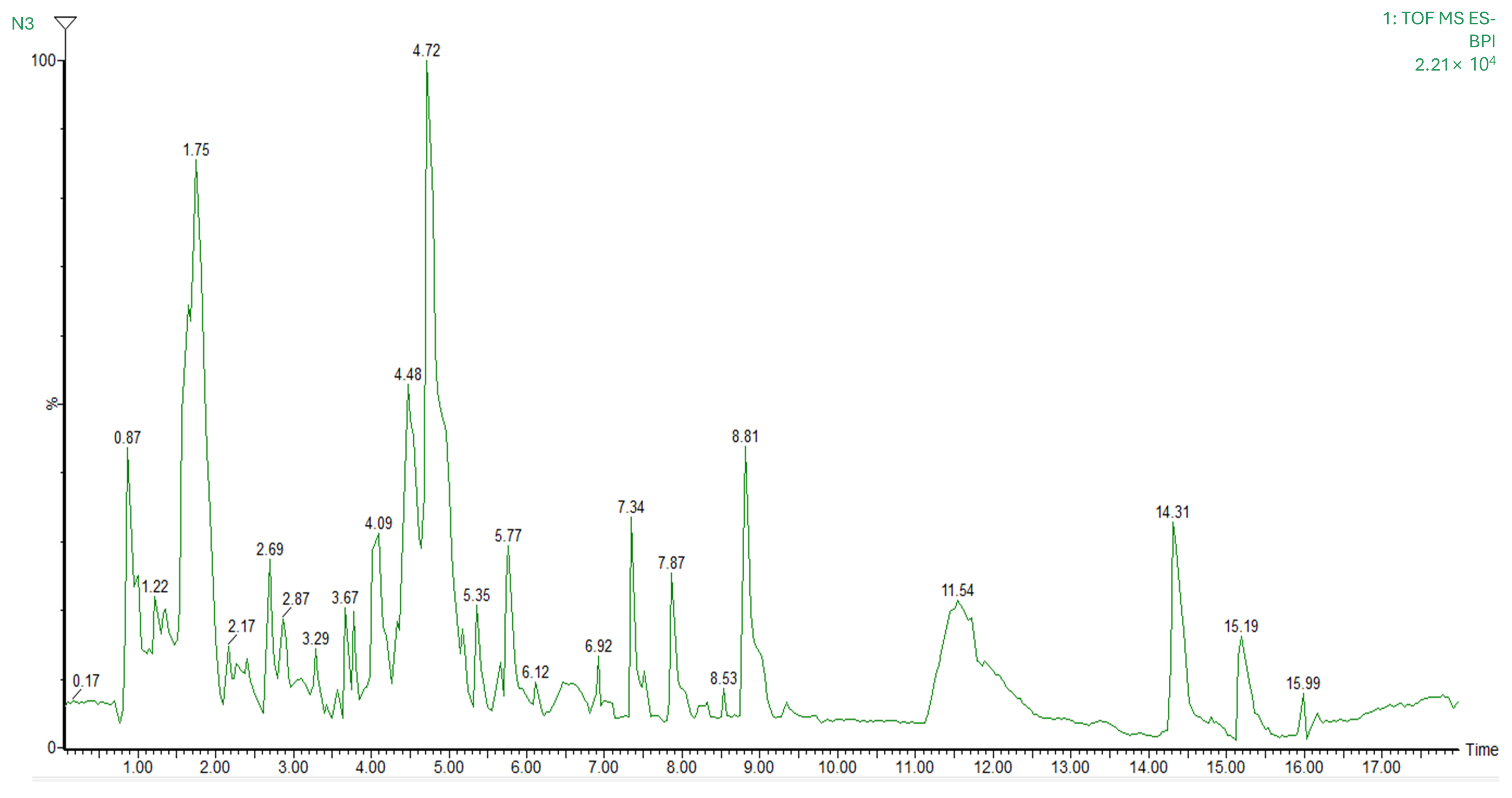
3.3. Mass Spectrometry Identification of Bioactive Compounds

The chromatographic profile and the main compounds tentatively identified in the nutraceutical formulation are presented in Figure 1 and Table 5, respectively. A total of 30 biocompounds were identified, with flavonoids being the most representative class including 10 compounds (cucumerin B, dichamanetin, epigallocatechin, eucalyptin, gallocatechin, moracenin D, nobiletin, pectolinarigenin, prenylnaringenin, and quercetin), followed by terpenoids with 6 compounds (chamissonolide, hydrangenoside A, kirenol, ligustrosidic acid, lucidumoside C, and pyrohyperforin). In addition, two compounds were identified within the phenolic acid class (curculigoside A and populin), as well as two phenilpropanoids (atharticin and nothoapiole) and two glucosids (marinoid D and prunioside A). Other identified compounds belonged to the following groups: alkaloid (nesodine), chromones (cimifugin), quinone (rubialatin A), isoflavans, furans (mumefural), isoflavones (glycitin), ether (crotepoxide), long-chain fatty alcohol (persealide), and lignans (diphyllin).

3.4. In Vivo Experimental Model

3.4.1. Body Weight and Food/Water Intake

Figure 2 illustrates the effects of colon cancer induction and interventions on body weight and food/water intake in mice throughout the experimental period. The initial animal weight was 15 ± 1 g, and all animals reached 22 ± 2 g by the end of the study period (Figure 2A). A temporary weight loss was observed at weeks 5, 8, and 11 in the CT+, NT, and NT + Ex groups, corresponding to the administration of DSS in drinking water for tumor induction. Regarding food intake (Figure 2B), no significant differences were observed between groups, except at weeks 5, 8, and 10, when a decrease in intake was detected in the tumor-bearing groups (CT+, NT, and NT + Ex) due to DSS-induced discomfort. However, there were no significant differences in the intake of drinking water when the DSS was administered (Figure 2C).

3.4.2. Tissue and Organ Weights

The effects of tumor development and the different interventions performed on organ weight and colon length are presented in Table 6. Tumor development led to a significant increase in the weight of the colon, spleen, and liver, although the increment in liver weight was not statistically significant. In addition, tumor development led to a shortening of the colon and the thickening of its wall. Only the nutritional intervention (NT group) significantly reduced colon weight (0.25 ± 0.02 g) and prevented colon shortening, with values comparable to the healthy control group (6.28 ± 0.15 cm vs. 6.88 ± 0.18 cm, respectively).

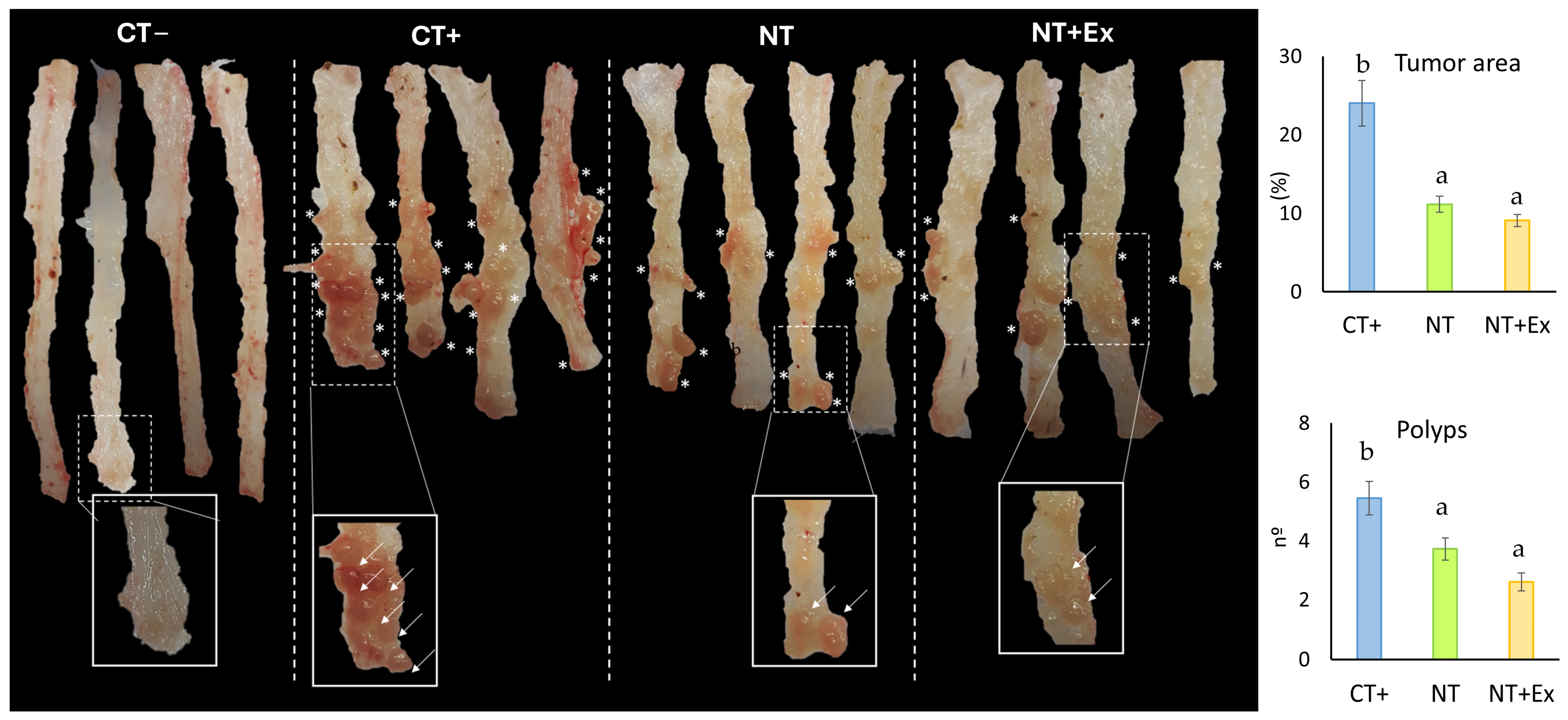
3.4.3. Modulation of Polyp Size and Number

The effect of the nutritional intervention, alone or combined with the physical exercise protocol, on the number of polyps and the area occupied by these polyps is shown in Figure 3. Tumor induction resulted in polyp development in the positive control group (CT+) of 24.3 ± 2.9% of the total colon area. In the NT group, polyps were significantly smaller, occupying 11.2 ± 1.0% of the total colon area. Although not statistically significant, the area occupied by the tumor was further reduced to 9.1 ± 0.8% when the nutraceutical was combined with the exercise program (NT + Ex). Regarding the number of polyps, the CT+ group showed a mean of 5.45 ± 0.52 polyps, whereas the NT group significantly reduced this number to 3.73 ± 0.38 polyps. In the NT + Ex group, polyps were further reduced to 2.60 ± 0.31.

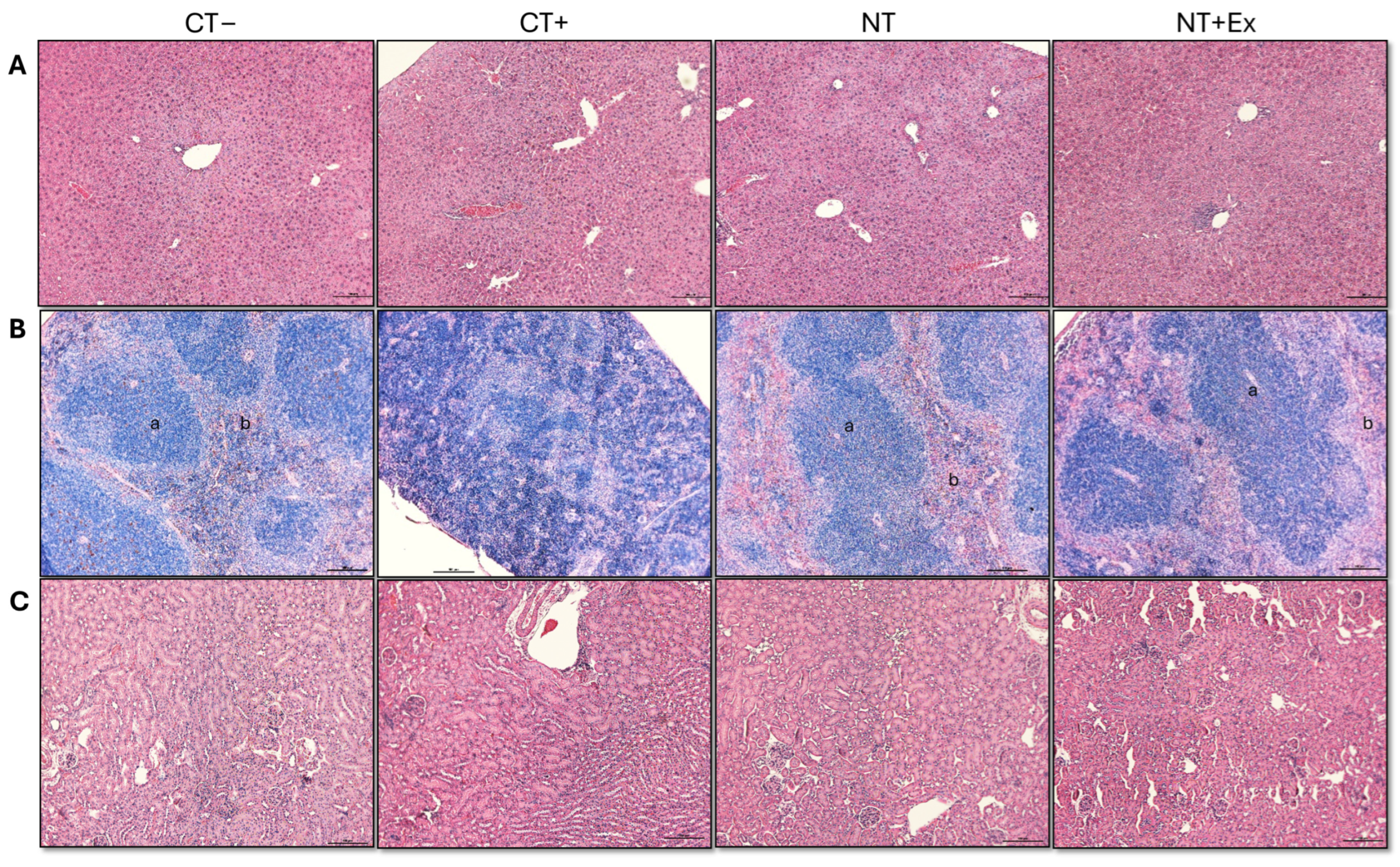
3.4.4. Histological Analysis

The histological analyses using H&E staining of samples from the Swiss Roll colon, liver, spleen, and kidney are shown in Figure 4 and Figure 5. The positive control (CT+) exhibited disrupted colonic crypt architecture, the absence of goblet cells, and dysplastic polyps marked by colonic epithelial cells with abnormal nuclei. In contrast, the Swiss roll colon samples from treated mice (NT and NT + Ex) showed a significantly lower number of polyps, and those that developed polyps exhibited a lower degree of dysplasia compared to those in the untreated group (CT+). In the CT+ group, a disruption in the organization between the white pulp and the red pulp of the spleen was also observed. The groups that ingested the nutraceutical throughout the experimental period were able to reverse this effect, showing an organization very similar to that of the healthy control group.

3.4.5. Gene Expression

Table 7 presents the gene expression levels in the colon of experimental animals. Tumor induction and development led to a significant increase in the expression of the slc20, pik3cd, and IL-1b genes, whereas the expression of gpx2, sod1, gsta1, and IL-6 was reduced. The different interventions successfully restored the expression levels of gpx2, sod1, pik3cd, and slc20 to normal values. Moreover, they significantly increased the expression of noq1 and decreased the expression of cadm1, cdc42, and IL-6, even below the levels observed in the control group.

3.4.6. Metagenomic Analysis

Figure 6 illustrates the effect of tumor development, nutritional interventions, and physical exercise on the relative abundance of bacteria in the cecum content of the experimental groups. Tumor development resulted in a significant increase in the phylum Verrucomicrobia that was not reversed with the different interventions applied. No additional significant differences related to tumor development were observed, but the presence of bacteria belonging to the phylum Firmicutes and Actinobacteria was reduced in the groups that had developed a tumor and had been fed the nutraceutical, while the presence of Bacteroidetes was increased in the group that also underwent the physical exercise protocol. Consequently, the ratio of Firmicutes/Bacteroidetes was significantly decreased by the nutritional intervention. The development of CRC significantly increased the presence of bacteria belonging to the Verricomicrobiaceae family and other unclassified families and decreased that of Lachnospiraceae and Porphyromonadaceae. These last two were reverted to the standard values of healthy mice when the nutraceutical was administered in the diet and the physical exercise protocol was performed.
At the genus level, Blautia was significantly reduced due to tumor development. However, its concentration increased with the nutritional intervention treatment, reaching levels comparable to those of healthy mice (CT−) when combined with the physical exercise protocol (NT + Ex). The Lactobacillus genus significantly increased its concentration with tumor development and was influenced by the nutritional intervention, showing similar values to the control group, that is, those animals that had been fed the nutraceutical (NT). The presence of the genus Akkermansia showed a significant increase in all groups that had developed the tumor.

4. Discussion

Natural products are a promising tool for treating or preventing CRC. Nearly 50% of the currently available treatments for CRC have been developed directly or indirectly from natural compounds [31,32]. Among these, the Brassicaceae family has been identified as an excellent source of bioactive compounds with antitumor properties [33]. Of all the Brassicaceae extracts analyzed in this study, the highest extraction yield and the highest polyphenol content were obtained in the ethanolic extract of Sinapis alba seeds. For ABTS, lipid peroxidation inhibition, and iron-chelating or iron-reducing capacity tests, the best results were achieved with the Eruca sativa seed extract. These results are consistent with the findings by Khalil et al. [34], who reported that Eruca sativa seeds showed a significantly enhanced ability to scavenge 2,2-diphenyl-1-picrylhydrazyl (DPPH) free radicals in comparison to other Brassicaceae species.
To induce in situ CRC in C57BL-6J mice, an intraperitoneal administration of AOM and three administrations of DSS in the drinking water were employed. This CRC induction model has been successfully validated in previous studies from our research group [29] and is widely used in the study of CRC, as it accurately replicates the full process of colon carcinogenesis, from the initial crypt proliferation phase to the final development of carcinoma [35]. Specifically, as we demonstrate in histological studies, the induction by AOM and DSS induces dysplastic polyps that are the previous step to invasive carcinoma, as described by Robertis et al. [36] AOM/DSS-induced tumors have mutations of Kas and β-catenin but not p53 mutations.
The nutritional intervention, either on its own or combined with the training protocol, significantly reduced the number of polyps developed in the colon with respect to the untreated control group (CT+). This effect can be attributed to the bioactive compounds identified in the nutraceutical, as in vitro digestibility tests confirmed that dialysates and retained fractions from nutraceutical-enriched diets exhibited higher antioxidant activity than those from the control diet. These findings suggest that some of the antioxidant compounds in the nutraceutical were potentially absorbable, which facilitates the exertion of beneficial systemic effects [37,38]. Specifically, several bioactive molecules identified in the nutraceutical have been previously studied for their anticancer properties in CRC. The flavonoid pectolinarigenin has demonstrated anticancer effects both in vitro and in vivo by inhibiting the cell growth, migration, and invasion of colon cancer cells (HCT116 and CT26). Furthermore, it has been shown to reduce the size and number of tumor nodules in a subcutaneous cancer model of Balb/c mice induced with CT26 colon cancer cells [39], T24 bladder cancer cells [40], or ACS gastric cancer cells [41]. Other authors have reported that the flavonoid epigallocatechin is able to inhibit tumor cell growth via the PI3K pathway and can be used as a chemotherapeutic [42], a chemopreventive agent [43], or as combinatorial therapy with anti-CTLA4 [44] against CRC. Dichamentin has shown a cytotoxic effect against HCT-116 CRC cells while demonstrating no cytotoxic effects on the non-cancerous lung cell line MRC-5 [45]. Quercetin exerts chemopreventive effects by inducing a large number of transcription factors that regulate metabolic pathways such as cell cycle, cell adhesion, gene transcription, or the immune response [46]. Nobiletin is another flavonoid with preventive effects on CRC progression, which is mediated by modulating the Akt signaling pathway, suppressing angiogenesis, and inhibiting CRC progression [47].
The preventive effect of Brassicaceae consumption against tumor development has been demonstrated in several studies. For instance, Baenas et al. [48] reported that the administration of 100 mg/kg of an aqueous extract from broccoli sprouts 30 days before tumor induction significantly reduced brain tumor size and progression in an experimental rat model. Similarly, Brassicaceae consumption has been shown to reduce preneoplastic colon lesions as well as the expression of cancer stem cell markers in rats [49]. This effect has been attributed to the set of phenolic compounds, isothiocyanates, vitamins, and nutrients present in these species, which act as an effective and favorable matrix in the stimulation of detoxifying enzymes and activators of the cellular antioxidant defense process [48]. Several population-based studies have examined the relationship between Brassica consumption and the decrease in the risk of developing CRC [50]. In the study by Mori et al. [51], no significant association was found between Brassica consumption and the risk of developing CRC in the Japanese population. However, these authors identified a positive association between plant-derived polyphenol consumption and CRC prevention, which is in agreement with our findings.
In the present study, no metabolic alterations or organ lesions derived from nutraceutical ingestion were found. In fact, the null toxicity of ethanolic extracts of Eruca sativa and Sinapis alba seeds has been reported by other authors. For instance, Xian et al. [52] demonstrated that the ingestion of up to 2500 mg/kg (maximum tested dose) of an ethanolic extract of Sinapis alba seeds proved to be innocuous in an experimental animal model of Balb/c mice, showing no signs of toxicity, deterioration, or death in the animals. Similarly, the nasogastric administration of up to 200 mg/kg of an ethanolic extract of Eruca sativa seeds also showed no signs of toxicity in an experimental Wistar rat model [53].
CRC is associated with oxidative stress and chronic inflammation [54,55]. In this experiment, tumor development led to a reduced expression of antioxidant enzymes (gpx and sod1) and the detoxification enzyme gst, along with an increased expression of transcription factors related to inflammatory activity such as IL-1ß. All these parameters were reversed in the groups that ingested the nutraceutical. Furthermore, the former intervention increased the transcript expression of quinone reductase enzyme (nqo1), which is involved in cellular detoxification, and decreased the expression of IL-6. These effects are assignable to the high phenolic content of the nutraceutical, as phenolic compounds are potent antioxidants capable of neutralizing free radicals; preventing the oxidation of lipids, proteins, and nucleic acids; and reducing colonic mucosa inflammation [56]. Other authors have reported that the administration of 200 mg/kg of an ethanolic extract from Sinapis alba seeds reduced the mRNA expression of TNFα, IL-1ß, and IL-6 in the colon of Balb/c mice [52]. Similarly, the ingestion of 200 mg/kg of an ethanolic extract from Eruca sativa seeds increased the activity of the antioxidant enzymes sod and gpx in the testes of Wistar rats exposed to acrylamide-induced oxidative stress [53]. Therefore, these compounds may reduce the risk of exposure and susceptibility to carcinogenesis [5]. In addition to their previously reported antiproliferative in vitro and in vivo effects, flavonoids play a pivotal role in tumor prevention by modulating oxidative stress and chronic inflammation. As discussed earlier, reactive oxygen species (ROS) can activate transcription factors involved in tumor generation and progression by increasing genomic instability, cell proliferation, and the risk of DNA damage or mutation. Furthermore, the tumor microenvironment is highly oxidative, which inhibits the activity of endogenous antioxidant enzymes such as glutathione peroxidase. Therefore, the antioxidant capacity conferred by these molecules is essential for counteracting these tumor-promoting factors [14,57].
As described by Mesas et al. [29], the effect in the prevention of new dysplastic polyps in colonic mucosa could be related to the anti-inflammatory effect of the extract that could reduce ROS production and the mutagenesis related to polyp formation.
Tumor development increased pik3cd and slc20a1 expression with respect to the healthy control group. The overexpression of pik3cd correlates with increased β-catenin levels, which translates to increased signaling of the Wnt/β-catenin pathway, which is involved in CRC initiation and progression [58]. Although Slc20a1 is not a specific CRC marker, its overexpression has been related with a poor prognosis of prostate cancer [59] and breast cancer [60]. Moreover, Slc20a1 is involved in Wnt/β-catenin signaling pathway activation [61]. The expression of these markers was reversed following the different interventions implemented, with the effect being more pronounced in the group combining both interventions (NT + Ex). Cdc42 is crucial in tumor generation, invasion, and metastasis because of its role in essential physiological processes, including cytoskeleton and microtubule regulation, transcription, cell cycle progression, and apoptosis [62]. Therefore, its overexpression is strongly associated with CRC development [63]. However, in our study, cdc42 expression did not show significant differences with tumor development, although its expression was reduced in animals that receive the nutritional intervention, either alone or combined with physical exercise. Similar results were reported in a study by Guo et al. [13], where tumor development in AOM/DSS-induced CRC in C57BL/6 mice led to an increase in pik3cd and cdc42 expression, whereas it was reduced in the group of mice that ingested 2% curcumin for 12 weeks.
The significant impact of nutraceutical administration on the microbiome under our experimental conditions can be clarified by the large percentage of ingested flavonoids that are not absorbed in the small intestine and reach the colon directly, where they interact with the microbiota, stimulating the production of short-chain fatty acids and the proliferation of beneficial bacteria [64]. The composition of the microbiome in animals that have developed CRC revealed an increased concentration of the phylum Verrucomicrobia, whereas the relative abundance of the phylum Firmicutes and Actinobacteria was significantly lower compared to the healthy control group (CT−). These findings are consistent with those reported by Hidalgo-García et al. and Song et al. [65,66]. However, while these authors also observed an increase in the phylum Bacteroidetes, such a shift was not detected in our study. These abnormal changes in microbiome composition may involve the destruction of the intestinal barrier by increasing the inflammatory response and damage to the gut mucosa [65,67]. The observed decrease in Firmicutes abundance could be due to the pro-inflammatory state associated with tumor induction, as this CRC model is characterized by inflammation-driven tumorigenesis [66,68].
The Firmicutes/Bacteroidetes (F/B) ratio is typically reduced in CRC development and tends to increase following dietary or therapeutic interventions [65,69,70]. This effect may be linked to an increase in Firmicutes, which are associated with butyrate production, a short-chain fatty acid that has been shown to induce apoptosis in colon cancer cells, reduce inflammation, and preserve intestinal mucosal integrity [71]. Under our experimental conditions, the F/B ratio was reduced following CRC induction and further decreased after the nutraceutical and exercise interventions. However, it is important to note that while the F/B ratio is often used as a biomarker for gut dysbiosis, its interpretation remains controversial, as its significance may vary across different pathological conditions [66,72].
As previously mentioned, none of the interventions carried out were able to reverse these alterations. Conversely, an increase in the abundance of Bateroidetes, a phylum associated with CRC susceptibility and promotion [71], was observed. However, since Bacteroidetes was not represented at the genus level, it is unlikely that there was an increased presence of harmful species such as Bacteroides fragilis, a key contributor to CRC development due to its ability to induce inflammation and alter gut ecology, thereby facilitating the colonization of pathogenic bacteria such as E. coli and Fusobacterium nucleatum [73]. In fact, under our experimental conditions, the presence of Blautia (phylum Bacillota, family Lachnospiraceae) was increased compared to CT+. Blautia has been reported to inhibit CRC progression by enhancing CD8+ immune cell activity [74]. Similarly, we detected an increase in Akkermansia (phylum Verrucomicrobia, family Verrucomicrobiaceae), specifically Akkermansia muciniphila, which has been shown to protect against CRC development in murine models by inhibiting the AhR/β-catenin signaling pathway [75], in addition to promoting the release of extracellular vesicles that have a multitude of regulatory effects on health, including improved mucosal integrity, reduced intestinal permeability, and an increase in beneficial bacteria [76]. Consequently, this genus is associated with a healthy microbiome that can protect against CRC development [65]. However, to establish a definitive link between microbiome modulation and CRC prevention, further studies are needed to identify the abundance and composition of microbial populations in precancerous tissues [73].
The gut microbiome can exert a direct influence on the organism’s oxidative and inflammatory status. Specifically, microorganisms can activate the host’s antioxidant defense mechanisms or exert their effects by activating their own antioxidant enzymes, including superoxide dismutase (SOD) and catalase. Additionally, they produce metabolites such as short-chain fatty acids (SCFAs), which can regulate the organism’s oxidative status. Specifically, SCFAs (acetate, propionate, and butyrate) induce the activity of the transcription factor nuclear factor erythroid 2-related factor 2 (Nrf2) in epithelial cells, which regulates the expression of antioxidant and detoxifying enzymes, thereby enhancing antioxidant activity [77]. Any disruption of the microbiome may lead to the proliferation of pro-oxidant bacteria such as Listeria and Clostridium, which can impair epithelial cell metabolic function by producing hydrogen sulfide (H2S) through the degradation of sulfur-containing amino acids. This metabolite inhibits the mitochondrial electron transport chain in epithelial cells, thereby promoting oxidative stress.
It is well-established that an imbalance in oxidative status triggers the expression of pro-inflammatory cytokines. These changes increase membrane permeability, facilitating the translocation of bacteria into the lamina propria and further amplifying ROS production and the inflammatory response. Sustained disruption of this balance over time contributes to the development of colitis and colorectal cancer [77].
The limitations of this study are that the AOM/DSS model for polyp development will be affected by the type of mouse strain used, since the mouse strains used present different sensitivity to the treatment. Another limitation is the impossibility to control the daily intake of DSS consumed by each mouse. Therefore, we propose to experimenters who intend to reproduce the study in the future to evaluate the dose of AOM/DSS to be used in their study in a previous trial to minimize environmental variations in their trial and to place an automatic water system, if possible. Another factor to consider is that animals during treatment with AOM/DSS can lose a significant amount of weight, so we recommend weighing the animals daily (during DSS intake and one week after), and in case of marked weight loss and weakness, administer 1mL of saline solution via IP.

5. Conclusions

The outcomes of this study reveal the beneficial effects of Brassicaceae extracts on health and support their potential use as dietary supplements for the prevention of pathological conditions in which oxidative stress plays a key role. Bioactive compounds derived from this family could be a key element in reducing the risk and incidence of CRC by scavenging unstable molecules such as reactive oxygen species (ROS), which can initiate the process of carcinogenesis. The intake of a Brassica seed-based nutraceutical proved to be an effective intervention, reducing polyp development by 31.6% and polyp size by 53.9%, this effect was increased to 52.3% and 62.6%, respectively, when the nutritional intervention was combined with a physical exercise protocol. Despite the promising results of this study, further trials are needed to define whether similar results can be achieved in other animal models and to transfer their potential applicability to CRC prevention in humans.

Author Contributions

Conceptualization, F.B., J.M.P., C.M. (Consolación Melguizo) and J.P.; methodology, A.G.-C., C.M. (Cristina Mesas), K.D., J.M.P. and R.M.; software, A.G.-C., A.G.-B. and M.P.; validation, K.D.; formal analysis, A.G.-C., C.M. (Cristina Mesas), A.G.-B. and M.P.; investigation, A.G.-C., C.M. (Cristina Mesas), K.D., J.M.P. and R.M.; resources, F.B.; writing—original draft preparation, A.G.-C. and C.M. (Cristina Mesas); writing—review and editing, C.M. (Consolación Melguizo) and J.P.; visualization, J.P.; supervision, J.M.P., R.M. and J.P.; funding acquisition, F.B. and C.M. (Consolación Melguizo). All authors have read and agreed to the published version of the manuscript.

Funding

This research was funded by the University of Granada, Junta de Andalucía, Spanish Ministry of Science, Innovation, and Universities as well as the European Union through project RTC2019-006870-1 and the FEDER program. In addition, this work was supported by funds from research groups CTS107 and AGR145 (Andalusian Government).

Institutional Review Board Statement

All animal procedures were conducted in accordance with the ethical guidelines for the care and use of laboratory animals and were approved by the Animal Experimentation Ethics Committee of the University of Granada, Spain (5 October 2021; number 148).

Informed Consent Statement

Not applicable.

Data Availability Statement

Data are available upon request due to time limitations.

Acknowledgments

We want to thank Susana Ibáñez from the analytical unit (CIC, UGR) for her dedicated and skillful assistance.

Conflicts of Interest

The authors declare no conflicts of interest. F.B. was employed by Seed for Innovation S.L. The remaining authors declare that the research was conducted in the absence of any commercial or financial relationships that could be construed as a potential conflict of interest.

References

  1. Gómez-España, M.A.; Montes, A.F.; Garcia-Carbonero, R.; Mercadé, T.M.; Maurel, J.; Martín, A.M.; Pazo-Cid, R.; Vera, R.; Carrato, A.; Feliu, J. SEOM Clinical Guidelines for Pancreatic and Biliary Tract Cancer (2020). Clin. Transl. Oncol. 2021, 23, 988–1000. [Google Scholar] [CrossRef]
  2. WHO. Colorectal Cancer. Available online: https://www.who.int/news-room/fact-sheets/detail/colorectal-cancer (accessed on 5 February 2025).
  3. Santucci, C.; Mignozzi, S.; Malvezzi, M.; Boffetta, P.; Collatuzzo, G.; Levi, F.; Vecchia, C.L.; Negri, E. European Cancer Mortality Predictions for the Year 2024 with Focus on Colorectal Cancer. Ann. Oncol. 2024, 35, 308–316. [Google Scholar] [CrossRef]
  4. Hardt, L.; Mahamat-Saleh, Y.; Aune, D.; Schlesinger, S. Plant-Based Diets and Cancer Prognosis: A Review of Recent Research. Curr. Nutr. Rep. 2022, 11, 695–716. [Google Scholar] [CrossRef]
  5. Macharia, J.M.; Mwangi, R.W.; Rozmann, N.; Zsolt, K.; Varjas, T.; Uchechukwu, P.O.; Wagara, I.N.; Raposa, B.L. Medicinal Plants with Anti-Colorectal Cancer Bioactive Compounds: Potential Game-Changers in Colorectal Cancer Management. Biomed. Pharmacother. 2022, 153, 113383. [Google Scholar] [CrossRef]
  6. Sanchez-Guzman, X.; Alvarez-Domínguez, L.; Ramírez-Torres, M.F.; Montes-Alvarado, J.B.; Garcia-Ibañez, P.; Moreno, D.A.; Domínguez, F.; Maycotte, P. Cruciferous Plant Extracts, Their Isothyocianate or Indol Derivatives, and Their Effect on Cellular Viability of Breast Cancer Cell Lines. J. Med. Food 2024, 27, 1183–1192. [Google Scholar] [CrossRef]
  7. Favela-González, K.M.; Hernández-Almanza, A.Y.; De la Fuente-Salcido, N.M. The Value of Bioactive Compounds of Cruciferous Vegetables (Brassica) as Antimicrobials and Antioxidants: A Review. J. Food Biochem. 2020, 44, e13414. [Google Scholar] [CrossRef]
  8. Yang, W.Y.; Lim, K.Y.; Yen, P.L.; Ong, S.H.; Naumovski, N.; Jani, R. The Association between Consumption of Bitter-Taste Vegetables in Asian Culture and Metabolic Syndrome Risk Factors in Children: A Narrative Review. Explor. Res. Hypothesis Med. 2024, 9, 47–54. [Google Scholar] [CrossRef]
  9. Mohd Nor, N.D.; Houston-Price, C.; Harvey, K.; Methven, L. The Effects of Taste Sensitivity and Repeated Taste Exposure on Children’s Intake and Liking of Turnip (Brassica Rapa Subsp. Rapa); a Bitter Brassica Vegetable. Appetite 2021, 157, 104991. [Google Scholar] [CrossRef]
  10. Nowak, K.; Rohn, S.; Halagarda, M. Impact of Cooking Techniques on the Dietary Fiber Profile in Selected Cruciferous Vegetables. Molecules 2025, 30, 590. [Google Scholar] [CrossRef]
  11. Baenas, N.; Marhuenda, J.; García-Viguera, C.; Zafrilla, P.; Moreno, D.A. Influence of Cooking Methods on Glucosinolates and Isothiocyanates Content in Novel Cruciferous Foods. Foods 2019, 8, 257. [Google Scholar] [CrossRef]
  12. Pereira, S.S.; Guimarães, M.; Monteiro, M.P. Towards Precision Medicine in Bariatric Surgery Prescription. Rev. Endocr. Metab. Disord. 2023, 24, 961–977. [Google Scholar] [CrossRef] [PubMed]
  13. Guo, Y.; Wu, R.; Gaspar, J.M.; Sargsyan, D.; Su, Z.-Y.; Zhang, C.; Gao, L.; Cheng, D.; Li, W.; Wang, C.; et al. DNA Methylome and Transcriptome Alterations and Cancer Prevention by Curcumin in Colitis-Accelerated Colon Cancer in Mice. Carcinogenesis 2018, 39, 669–680. [Google Scholar] [CrossRef]
  14. Marino, P.; Pepe, G.; Basilicata, M.G.; Vestuto, V.; Marzocco, S.; Autore, G.; Procino, A.; Gomez-Monterrey, I.M.; Manfra, M.; Campiglia, P. Potential Role of Natural Antioxidant Products in Oncological Diseases. Antioxidants 2023, 12, 704. [Google Scholar] [CrossRef] [PubMed]
  15. Lučić, D.; Pavlović, I.; Brkljačić, L.; Bogdanović, S.; Farkaš, V.; Cedilak, A.; Nanić, L.; Rubelj, I.; Salopek-Sondi, B. Antioxidant and Antiproliferative Activities of Kale (Brassica oleracea L. Var. Acephala DC.) and Wild Cabbage (Brassica incana Ten.) Polyphenolic Extracts. Molecules 2023, 28, 1840. [Google Scholar] [CrossRef]
  16. Zhang, Y.; Lv, X.; Wang, D.; Zheng, C.; Chen, H.; Yuan, Y.; Wei, F. Metabolomics Combined with Biochemical Analyses Revealed Phenolic Profiles and Antioxidant Properties of Rapeseeds. Food Chem. 2025, 466, 142250. [Google Scholar] [CrossRef]
  17. Kapravelou, G.; Martínez, R.; Andrade, A.M.; Chaves, C.L.; López-Jurado, M.; Aranda, P.; Arrebola, F.; Cañizares, F.J.; Galisteo, M.; Porres, J.M. Improvement of the Antioxidant and Hypolipidaemic Effects of Cowpea Flours (Vigna Unguiculata) by Fermentation: Results of in Vitro and in Vivo Experiments. J. Sci. Food Agric. 2015, 95, 1207–1216. [Google Scholar] [CrossRef] [PubMed]
  18. Kapravelou, G.; Martínez, R.; Andrade, A.M.; Sánchez, C.; Chaves, C.L.; López-Jurado, M.; Aranda, P.; Cantarero, S.; Arrebola, F.; Fernández-Segura, E.; et al. Health Promoting Effects of Lupin (Lupinus albus var. multolupa) Protein Hydrolyzate and Insoluble Fiber in a Diet-Induced Animal Experimental Model of Hypercholesterolemia. Food Res. Int. 2013, 54, 1471–1481. [Google Scholar] [CrossRef]
  19. Porres, J.M.; Aranda, P.; López-Jurado, M.; Urbano, G. Nutritional Evaluation of Protein, Phosphorus, Calcium and Magnesium Bioavailability from Lupin (Lupinus albus var. multolupa)-Based Diets in Growing Rats: Effect of α-Galactoside Oligosaccharide Extraction and Phytase Supplementation. Br. J. Nutr. 2006, 95, 1102–1111. [Google Scholar] [CrossRef]
  20. Martínez, R.; Mesas, C.; Guzmán, A.; Galisteo, M.; López-Jurado, M.; Prados, J.; Melguizo, C.; Bermúdez, F.; Porres, J.M. Bioavailability and Biotransformation of Linolenic Acid from Basil Seed Oil as a Novel Source of Omega-3 Fatty Acids Tested on a Rat Experimental Model. Food Funct. 2022, 13, 7614–7628. [Google Scholar] [CrossRef]
  21. Martínez, R.; Kapravelou, G.; Donaire, A.; Lopez-Chaves, C.; Arrebola, F.; Galisteo, M.; Cantarero, S.; Aranda, P.; Porres, J.M.; López-Jurado, M. Effects of a Combined Intervention with a Lentil Protein Hydrolysate and a Mixed Training Protocol on the Lipid Metabolism and Hepatic Markers of NAFLD in Zucker Rats. Food Funct. 2018, 9, 830–850. [Google Scholar] [CrossRef]
  22. Miller, N.J.; Rice-Evans, C.; Davies, M.J.; Gopinathan, V.; Milner, A. A Novel Method for Measuring Antioxidant Capacity and Its Application to Monitoring the Antioxidant Status in Premature Neonates. Clin. Sci. 1993, 84, 407–412. [Google Scholar] [CrossRef]
  23. Dinis, T.C.; Maderia, V.M.; Almeida, L.M. Action of Phenolic Derivatives (Acetaminophen, Salicylate, and 5-Aminosalicylate) as Inhibitors of Membrane Lipid Peroxidation and as Peroxyl Radical Scavengers. Arch. Biochem. Biophys. 1994, 315, 161–169. [Google Scholar] [CrossRef] [PubMed]
  24. Oyaizu, M. Studies on Products of Browning Reaction. Antioxidative Activities of Products of Browning Reaction Prepared from Glucosamine. Jpn. J. Nutr. Diet. 1986, 44, 307–315. [Google Scholar] [CrossRef]
  25. Duh, P.-D.; Tu, Y.-Y.; Yen, G.-C. Antioxidant Activity of Water Extract of Harng Jyur (Chrysanthemum Morifolium Ramat). LWT-Food Sci. Technol. 1999, 32, 269–277. [Google Scholar] [CrossRef]
  26. Martínez, R.; Kapravelou, G.; Porres, J.M.; Melesio, A.M.; Heras, L.; Cantarero, S.; Gribble, F.M.; Parker, H.; Aranda, P.; López-Jurado, M. Medicago sativa L., a Functional Food to Relieve Hypertension and Metabolic Disorders in a Spontaneously Hypertensive Rat Model. J. Funct. Foods 2016, 26, 470–484. [Google Scholar] [CrossRef]
  27. Hubrecht, R.C.; Carter, E. The 3Rs and Humane Experimental Technique: Implementing Change. Animals 2019, 9, 754. [Google Scholar] [CrossRef]
  28. Bialkowska, A.B.; Ghaleb, A.M.; Nandan, M.O.; Yang, V.W. Improved Swiss-Rolling Technique for Intestinal Tissue Preparation for Immunohistochemical and Immunofluorescent Analyses. J. Vis. Exp. 2016, 113, e54161. [Google Scholar] [CrossRef]
  29. Mesas, C.; Martínez, R.; Doello, K.; Ortiz, R.; López-Jurado, M.; Bermúdez, F.; Quiñonero, F.; Prados, J.; Porres, J.M.; Melguizo, C. In Vivo Antitumor Activity of Euphorbia Lathyris Ethanol Extract in Colon Cancer Models. Biomed. Pharmacother. 2022, 149, 112883. [Google Scholar] [CrossRef]
  30. Yang, W.; Liu, Y.; Yang, G.; Meng, B.; Yi, Z.; Yang, G.; Chen, M.; Hou, P.; Wang, H.; Xu, X. Moderate-Intensity Physical Exercise Affects the Exercise Performance and Gut Microbiota of Mice. Front. Cell Infect. Microbiol. 2021, 11, 712381. [Google Scholar] [CrossRef]
  31. Hossain, M.S.; Karuniawati, H.; Jairoun, A.A.; Urbi, Z.; Ooi, D.J.; John, A.; Lim, Y.C.; Kibria, K.M.K.; Mohiuddin, A.K.M.; Ming, L.C.; et al. Colorectal Cancer: A Review of Carcinogenesis, Global Epidemiology, Current Challenges, Risk Factors, Preventive and Treatment Strategies. Cancers 2022, 14, 1732. [Google Scholar] [CrossRef]
  32. Huang, X.-M.; Yang, Z.-J.; Xie, Q.; Zhang, Z.-K.; Zhang, H.; Ma, J.-Y. Natural Products for Treating Colorectal Cancer: A Mechanistic Review. Biomed. Pharmacother. 2019, 117, 109142. [Google Scholar] [CrossRef]
  33. Peña, M.; Guzmán, A.; Martínez, R.; Mesas, C.; Prados, J.; Porres, J.M.; Melguizo, C. Preventive Effects of Brassicaceae Family for Colon Cancer Prevention: A Focus on in Vitro Studies. Biomed. Pharmacother. 2022, 151, 113145. [Google Scholar] [CrossRef] [PubMed]
  34. Khalil, N.; Gad, H.A.; Al Musayeib, N.M.; Bishr, M.; Ashour, M.L. Correlation of Glucosinolates and Volatile Constituents of Six Brassicaceae Seeds with Their Antioxidant Activities Based on Partial Least Squares Regression. Plants 2022, 11, 1116. [Google Scholar] [CrossRef] [PubMed]
  35. Dzhalilova, D.; Zolotova, N.; Fokichev, N.; Makarova, O. Murine Models of Colorectal Cancer: The Azoxymethane (AOM)/Dextran Sulfate Sodium (DSS) Model of Colitis-Associated Cancer. PeerJ 2023, 11, e16159. [Google Scholar] [CrossRef]
  36. Robertis, M.D.; Massi, E.; Poeta, M.L.; Carotti, S.; Morini, S.; Cecchetelli, L.; Signori, E.; Fazio, V.M. The AOM/DSS Murine Model for the Study of Colon Carcinogenesis: From Pathways to Diagnosis and Therapy Studies. J. Carcinog. 2011, 10, 9. [Google Scholar] [CrossRef] [PubMed]
  37. Di Lorenzo, C.; Colombo, F.; Biella, S.; Stockley, C.; Restani, P. Polyphenols and Human Health: The Role of Bioavailability. Nutrients 2021, 13, 273. [Google Scholar] [CrossRef]
  38. Qin, W.; Ketnawa, S. Exploring the Bioaccessibility of Roasted Japanese Green Tea: Impact of Simulated Gastrointestinal Digestion. Foods 2025, 14, 311. [Google Scholar] [CrossRef] [PubMed]
  39. Gan, C.; Li, Y.; Yu, Y.; Yu, X.; Liu, H.; Zhang, Q.; Yin, W.; Yu, L.; Ye, T. Natural Product Pectolinarigenin Exhibits Potent Anti-Metastatic Activity in Colorectal Carcinoma Cells in Vitro and in Vivo. Bioorg. Med. Chem. 2019, 27, 115089. [Google Scholar] [CrossRef]
  40. Deng, Z.; Shen, D.; Yu, M.; Zhou, F.; Shan, D.; Fang, Y.; Jin, W.; Qian, K.; Li, S.; Wang, G.; et al. Pectolinarigenin Inhibits Bladder Urothelial Carcinoma Cell Proliferation by Regulating DNA Damage/Autophagy Pathways. Cell Death Discov. 2023, 9, 214. [Google Scholar] [CrossRef]
  41. Lee, H.J.; Kwon, Y.S.; Lee, J.H.; Moon, Y.G.; Choi, J.; Hyun, M.; Tak, T.K.; Kim, J.-H.; Heo, J.D. Pectolinarigenin Regulates the Tumor-Associated Proteins in AGS-Xenograft BALB/c Nude Mice. Mol. Biol. Rep. 2024, 51, 305. [Google Scholar] [CrossRef]
  42. Ding, F.; Yang, S. Epigallocatechin-3-Gallate Inhibits Proliferation and Triggers Apoptosis in Colon Cancer via the Hedgehog/Phosphoinositide 3-Kinase Pathways. Can. J. Physiol. Pharmacol. 2021, 99, 910–920. [Google Scholar] [CrossRef]
  43. Wubetu, G.Y.; Shimada, M.; Morine, Y.; Ikemoto, T.; Ishikawa, D.; Iwahashi, S.; Yamada, S.; Saito, Y.; Arakawa, Y.; Imura, S. Epigallocatechin Gallate Hinders Human Hepatoma and Colon Cancer Sphere Formation. J. Gastroenterol. Hepatol. 2016, 31, 256–264. [Google Scholar] [CrossRef]
  44. Lei, J.; Chen, J.; Chen, J.; Fang, J.; Zhou, Z.; Xu, A. Epigallocatechin-3-Gallate Induces Immunogenic Cell Death and Enhances Cancer Immunotherapy in Colorectal Cancer. Biochem. Biophys. Res. Commun. 2024, 736, 150907. [Google Scholar] [CrossRef]
  45. Costa, E.V.; Soares, L.d.N.; Chaar, J.d.S.; Silva, V.R.; Santos, L.d.S.; Koolen, H.H.F.; da Silva, F.M.A.; Tavares, J.F.; Zengin, G.; Soares, M.B.P.; et al. Benzylated Dihydroflavones and Isoquinoline-Derived Alkaloids from the Bark of Diclinanona Calycina (Annonaceae) and Their Cytotoxicities. Molecules 2021, 26, 3714. [Google Scholar] [CrossRef]
  46. Huminiecki, L. Evidence for Multilevel Chemopreventive Activities of Natural Phenols from Functional Genomic Studies of Curcumin, Resveratrol, Genistein, Quercetin, and Luteolin. Int. J. Mol. Sci. 2022, 23, 14957. [Google Scholar] [CrossRef] [PubMed]
  47. Kisacam, M.A. Nobiletin Is Capable of Regulating Certain Anti-Cancer Pathways in a Colon Cancer Cell Line. Naunyn-Schmiedeberg’s Arch. Pharmacol. 2023, 396, 547–555. [Google Scholar] [CrossRef] [PubMed]
  48. Baenas, N.; Vega-García, A.; Manjarrez-Marmolejo, J.; Moreno, D.A.; Feria-Romero, I.A. The Preventive Effects of Broccoli Bioactives against Cancer: Evidence from a Validated Rat Glioma Model. Biomed. Pharmacother. 2023, 168, 115720. [Google Scholar] [CrossRef] [PubMed]
  49. Kim, S.; Trudo, S.P.; Gallaher, D.D. Apiaceous and Cruciferous Vegetables Fed During the Post-Initiation Stage Reduce Colon Cancer Risk Markers in Rats. J. Nutr. 2019, 149, 249–257. [Google Scholar] [CrossRef]
  50. Tse, G.; Eslick, G.D. Cruciferous Vegetables and Risk of Colorectal Neoplasms: A Systematic Review and Meta-Analysis. Nutr. Cancer 2014, 66, 128–139. [Google Scholar] [CrossRef]
  51. Mori, N.; Murphy, N.; Sawada, N.; Achaintre, D.; Yamaji, T.; Scalbert, A.; Iwasaki, M.; Inoue, M.; Gunter, M.J.; Tsugane, S. Prediagnostic Plasma Polyphenol Concentrations and Colon Cancer Risk: The JPHC Nested Case–Control Study. Clin. Nutr. 2022, 41, 1950–1960. [Google Scholar] [CrossRef]
  52. Xian, Y.-F.; Hu, Z.; Ip, S.-P.; Chen, J.-N.; Su, Z.-R.; Lai, X.-P.; Lin, Z.-X. Comparison of the Anti-Inflammatory Effects of Sinapis Alba and Brassica Juncea in Mouse Models of Inflammation. Phytomedicine 2018, 50, 196–204. [Google Scholar] [CrossRef] [PubMed]
  53. Abd-Elsalam, R.M.; El Badawy, S.A.; Ogaly, H.A.; Ibrahim, F.M.; Farag, O.M.; Ahmed, K.A. Eruca Sativa Seed Extract Modulates Oxidative Stress and Apoptosis and Up-Regulates the Expression of Bcl-2 and Bax Genes in Acrylamide-Induced Testicular Dysfunction in Rats. Environ. Sci. Pollut. Res. Int. 2021, 28, 53249–53266. [Google Scholar] [CrossRef]
  54. Fan, Q.; Wang, J.; Tian, M.; Sawut, A.; Xiao, D.; Yi, Z.; Chen, L. Circulating Inflammatory Cytokines and Colorectal Cancer: New Insights from Mendelian Randomization. Medicine 2025, 104, e41331. [Google Scholar] [CrossRef]
  55. Nelson, V.K.; Nuli, M.V.; Mastanaiah, J.; Saleem, T.S.M.; Birudala, G.; Jamous, Y.F.; Alshargi, O.; Kotha, K.K.; Sudhan, H.H.; Mani, R.R.; et al. Reactive Oxygen Species Mediated Apoptotic Death of Colon Cancer Cells: Therapeutic Potential of Plant Derived Alkaloids. Front. Endocrinol. 2023, 14, 1201198. [Google Scholar] [CrossRef]
  56. Zhang, H.; Tang, J.; Cao, H.; Wang, C.; Shen, C.; Liu, J. Effect and Mechanism of Magnolia Officinalis in Colorectal Cancer: Multi-Component-Multi-Target Approach. J. Ethnopharmacol. 2025, 338, 119007. [Google Scholar] [CrossRef] [PubMed]
  57. Cid-Gallegos, M.S.; Sánchez-Chino, X.M.; Álvarez-González, I.; Madrigal-Bujaidar, E.; Vásquez-Garzón, V.R.; Baltiérrez-Hoyos, R.; Villa-Treviño, S.; Dávila-Ortíz, G.; Jiménez-Martínez, C. Modification of In Vitro and In Vivo Antioxidant Activity by Consumption of Cooked Chickpea in a Colon Cancer Model. Nutrients 2020, 12, 2572. [Google Scholar] [CrossRef]
  58. Chen, J.-S.; Huang, J.-Q.; Luo, B.; Dong, S.-H.; Wang, R.-C.; Jiang, Z.-K.; Xie, Y.-K.; Yi, W.; Wen, G.-M.; Zhong, J.-F. PIK3CD Induces Cell Growth and Invasion by Activating AKT/GSK-3β/β-Catenin Signaling in Colorectal Cancer. Cancer Sci. 2019, 110, 997–1011. [Google Scholar] [CrossRef]
  59. Okamoto, T.; Onaga, C.; Matsuoka, I.; Ozaki, A.; Matsuda, C.; Kasai, T.; Xiong, Y.; Harada, Y.; Sato, T.; Nakano, Y.; et al. High SLC20A1 Expression Indicates Poor Prognosis in Prostate Cancer. Cancer Diagn. Progn. 2023, 3, 439–448. [Google Scholar] [CrossRef] [PubMed]
  60. Onaga, C.; Tamori, S.; Matsuoka, I.; Ozaki, A.; Motomura, H.; Nagashima, Y.; Sato, T.; Sato, K.; Tahata, K.; Xiong, Y.; et al. High SLC20A1 Expression Is Associated With Poor Prognosis for Radiotherapy of Estrogen Receptor-Positive Breast Cancer. Cancer Diagn. Progn. 2022, 2, 429–442. [Google Scholar] [CrossRef]
  61. Li, J.; Dong, W.; Li, Z.; Wang, H.; Gao, H.; Zhang, Y. Impact of SLC20A1 on the Wnt/Β-catenin Signaling Pathway in Somatotroph Adenomas. Mol. Med. Rep. 2019, 20, 3276–3284. [Google Scholar] [CrossRef]
  62. Murphy, N.P.; Binti Ahmad Mokhtar, A.M.; Mott, H.R.; Owen, D. Molecular Subversion of Cdc42 Signalling in Cancer. Biochem. Soc. Trans. 2021, 49, 1425–1442. [Google Scholar] [CrossRef]
  63. Cai, J.-W.; Huang, X.-M.; Li, X.-L.; Qin, S.; Rong, Y.-M.; Chen, X.; Weng, J.-R.; Zou, Y.-F.; Lin, X.-T. An 11-Gene Signature for the Prediction of Systemic Recurrences in Colon Adenocarcinoma. Gastroenterol. Rep. 2021, 9, 451–460. [Google Scholar] [CrossRef]
  64. Wang, L.; Li, M.; Gu, Y.; Shi, J.; Yan, J.; Wang, X.; Li, B.; Wang, B.; Zhong, W.; Cao, H. Dietary Flavonoids-Microbiota Crosstalk in Intestinal Inflammation and Carcinogenesis. J. Nutr. Biochem. 2024, 125, 109494. [Google Scholar] [CrossRef] [PubMed]
  65. Hidalgo-García, L.; Ruiz-Malagon, A.J.; Huertas, F.; Rodríguez-Sojo, M.J.; Molina-Tijeras, J.A.; Diez-Echave, P.; Becerra, P.; Mirón, B.; Morón, R.; Rodríguez-Nogales, A.; et al. Administration of Intestinal Mesenchymal Stromal Cells Reduces Colitis-Associated Cancer in C57BL/6J Mice Modulating the Immune Response and Gut Dysbiosis. Pharmacol. Res. 2023, 195, 106891. [Google Scholar] [CrossRef]
  66. Song, C.-H.; Kim, N.; Nam, R.H.; Choi, S.I.; Jang, J.Y.; Lee, H.-N. Changes in Gut Microbiome upon Orchiectomy and Testosterone Administration in AOM/DSS-Induced Colon Cancer Mouse Model. Cancer Res. Treat. 2023, 55, 196–218. [Google Scholar] [CrossRef] [PubMed]
  67. Zhang, M.-M.; Yin, D.-K.; Rui, X.-L.; Shao, F.-P.; Li, J.-C.; Xu, L.; Yang, Y. Protective Effect of Pai-Nong-San against AOM/DSS-Induced CAC in Mice through Inhibiting the Wnt Signaling Pathway. Chin. J. Nat. Med. 2021, 19, 912–920. [Google Scholar] [CrossRef] [PubMed]
  68. Parang, B.; Barret, C.W.; Williams, C.S. AOM/DSS Model of Colitis-Associated Cancer. Methods Mol. Biol. 2016, 1422, 297–307. [Google Scholar] [CrossRef]
  69. Deng, Y.; Huang, X.; Chen, X.; Wang, M.; Tian, L.; Zhou, H.; Yang, W.; He, F.; Yin, W. Chemopreventive Effects of Polysaccharides and Flavonoids from Okra Flowers in Azomethane/Dextran Sulfate Sodium-Induced Murine Colitis-Associated Cancer. Nutrients 2023, 15, 4820. [Google Scholar] [CrossRef]
  70. Zhu, J.; Liu, W.; Bian, Z.; Ma, Y.; Kang, Z.; Jin, J.; Li, X.; Ge, S.; Hao, Y.; Zhang, H.; et al. Lactobacillus Plantarum Zhang-LL Inhibits Colitis-Related Tumorigenesis by Regulating Arachidonic Acid Metabolism and CD22-Mediated B-Cell Receptor Regulation. Nutrients 2023, 15, 4512. [Google Scholar] [CrossRef]
  71. Pal, P.; Shastry, R.P. Exploring the Complex Role of Gut Microbiome in the Development of Precision Medicine Strategies for Targeting Microbial Imbalance-Induced Colon Cancer. Folia Microbiol. 2023, 68, 691–701. [Google Scholar] [CrossRef]
  72. Yin, Y.; Yang, T.; Tian, Z.; Shi, C.; Yan, C.; Li, H.; Du, Y.; Li, G. Progress in the Investigation of the Firmicutes/Bacteroidetes Ratio as a Potential Pathogenic Factor in Ulcerative Colitis. J. Med. Microbiol. 2025, 74, 001966. [Google Scholar] [CrossRef] [PubMed]
  73. Gong, D.; Adomako-Bonsu, A.G.; Wang, M.; Li, J. Three Specific Gut Bacteria in the Occurrence and Development of Colorectal Cancer: A Concerted Effort. PeerJ 2023, 11, e15777. [Google Scholar] [CrossRef] [PubMed]
  74. Zhang, X.; Yu, D.; Wu, D.; Gao, X.; Shao, F.; Zhao, M.; Wang, J.; Ma, J.; Wang, W.; Qin, X.; et al. Tissue-Resident Lachnospiraceae Family Bacteria Protect against Colorectal Carcinogenesis by Promoting Tumor Immune Surveillance. Cell Host Microbe 2023, 31, 418–432.e8. [Google Scholar] [CrossRef]
  75. Zhang, L.; Ji, Q.; Chen, Q.; Wei, Z.; Liu, S.; Zhang, L.; Zhang, Y.; Li, Z.; Liu, H.; Sui, H. Akkermansia Muciniphila Inhibits Tryptophan Metabolism via the AhR/β-Catenin Signaling Pathway to Counter the Progression of Colorectal Cancer. Int. J. Biol. Sci. 2023, 19, 4393–4410. [Google Scholar] [CrossRef] [PubMed]
  76. Zheng, T.; Hao, H.; Liu, Q.; Li, J.; Yao, Y.; Liu, Y.; Zhang, T.; Zhang, Z.; Yi, H. Effect of Extracelluar Vesicles Derived from Akkermansia Muciniphila on Intestinal Barrier in Colitis Mice. Nutrients 2023, 15, 4722. [Google Scholar] [CrossRef]
  77. Kunst, C.; Schmid, S.; Michalski, M.; Tümen, D.; Buttenschön, J.; Müller, M.; Gülow, K. The Influence of Gut Microbiota on Oxidative Stress and the Immune System. Biomedicines 2023, 11, 1388. [Google Scholar] [CrossRef]
Figure 1. Chromatographic profile of bioactive compounds identified in the nutraceutical using high-performance liquid chromatography.
Nutrients 17 01358 g001
Figure 2. (A) Body weight, (B) food intake, and (C) water intake of mice fed different experimental diets. CT−, healthy animals fed the control diet. CT+, tumor-induced animals fed the control diet. NT, tumor-induced animals fed the control diet supplemented with the nutraceutical. NT + Ex, tumor-induced animals fed the control diet supplemented with the nutraceutical combined with the exercise protocol. Results are expressed as the means plus the SD (vertical bars) of 15 replicates.
Nutrients 17 01358 g002
Figure 3. Effect of the nutritional intervention (NT), alone or in combination with physical exercise (NT + Ex), on the number of polyps and the area occupied by polyps. Asterisks (*) indicate each of the polyps identified in the colons of the animals. Arrows point to the polyps located in the enlarged segment of the colon. Results are expressed as the means plus the SD (parenthesis) of 15 replicates. Means values with different letters (a and b) represent significant differences (ANOVA, p < 0.05).
Nutrients 17 01358 g003
Figure 4. Representative H&E-stained histological images following tumor induction via AOM/DSS. The Swiss roll technique was used to analyze the colon (scale bar = 1000 μm [top row] and 100 μm [bottom row]). Arrows indicate polyps with dysplasia. CT−, healthy animals fed the control diet. CT+, tumor-induced animals fed the control diet. NT, tumor-induced animals fed the control diet supplemented with the nutraceutical. NT + Ex, tumor-induced animals fed the control diet supplemented with the nutraceutical combined with the exercise protocol.
Nutrients 17 01358 g004
Figure 5. Representative images of the histological analysis of organs from different experimental groups. The H&E method was used to analyze the liver (A), spleen (B), and kidneys (C). In spleen samples, the white pulp and the red pulp are indicated by (a) and (b), respectively. Bars, 100 µm.
Nutrients 17 01358 g005
Figure 6. Metagenomic analysis of caecum content in the experimental groups, showing bacterial abundance at the level of phylum (A) and genus (B). CT−, healthy animals fed the control diet. CT+, tumor-induced animals fed the control diet. NT, tumor-induced animals fed the control diet supplemented with the nutraceutical. NT + Ex, tumor-induced animals fed the control diet supplemented with the nutraceutical combined with the exercise protocol. Results are the means of 8 replicates.
Nutrients 17 01358 g006
Table 1. Training protocol.
PhaseTime% VO2 Max
Warm-up5′20
Exercise program
1′30″20 → 45
1′45
2′45
1′30
1′30″Rest
Table 2. Reference of primers for genes analyzed in the study.
FunctionGenesPrimers
Oxidative metabolismcatMm00437992
gpx2Mm01286848
sod1Mm01344233_g1
Detoxification pathwaysnqo1Mm00500822_g1
gsta1Mm03019257_g1
Inflammatory processIL-1bMm00434228_m1
IL-6Mm00446190_m1
Tumor developmentcadm1Mm00457551_m1
cdc42Mm01194005_g1
pik3cdMm00435674_m1
slc20Mm00489378_m1
Table 3. Antioxidant capacity of the different Brassicaceae seed extracts and the nutraceutical.
Yield
(mg/g)
TPC
(µg GA)
ABTS
(µg GA)
ICC
(CAU)
IRC
(µg GA)
EtOH
BO var. sabellica115.4 ± 2.9 b30.9 ± 0.5 b11.6 ± 0.4 bc1.56 ± 0.11 b25.9 ± 1.4 b
BO var. italica115.3 ± 9.7 b31.4 ± 0.9 b12.8 ± 0.1 c1.29 ± 0.09 b27.7 ± 0.3 b
BO var. botrytis132.1 ± 4.6 b27.9 ± 0.4 a10.2 ± 0.7 b0.87 ± 0.01 a20.1 ± 0.6 a
Eruca sativa86.9 ± 1.9 a43.5 ± 0.4 c15.0 ± 0.0 d3.99 ± 0.06 c43.5 ± 1.4 c
Sinapis alba120.8 ± 1.3 b60.5 ± 1.0 d7.4 ± 0.1 a0.98 ± 0.05 a18.8 ± 1.2 a
HP
BO var. sabellica24.4 ± 0.2 b28.5 ± 0.3 b12.2 ± 0.7 a1.00 ± 0.03 a13.1 ± 0.3 b
BO var. italica27.3 ± 0.3 c26.2 ± 0.3 ab10.2 ± 0.3 a1.10 ± 0.02 ab12.9 ± 0.2 b
BO var. botrytis29.3 ± 0.6 cd25.1 ± 1.1 a11.5 ± 0.7 a1.19 ± 0.07 b13.0 ± 0.2 b
Eruca sativa20.3 ± 0.4 a33.6 ± 0.4 c17.0 ± 0.4 b1.34 ± 0.02 c12.8 ± 0.1 b
Sinapis alba31.9 ± 1.1 d31.7 ± 0.4 c11.1 ± 0.4 a0.98 ± 0.02 a8.0 ± 0.1 a
NT 32.5 (0.3)7.8 (0.1)0.93 (0.04)9.1 (0.1)
BO, Brassica oleracea. CAU, chelating activity unit. EtOH, ethanolic extract. GA, gallic acid. HP, protein hydrolysate. ABTS, 2,2′-azino-bis3-ethylbenzothiazoline-6-sulfonic acid. ICC, iron-chelating capacity. IRC, iron-reducing capacity. NT, nutraceutical. TPC, total polyphenol content. Results are the means plus the SD (in parenthesis) of four replicates expressed per milligram of extract. Means with different letters (a, b, c and d) indicate significant differences (ANOVA p < 0.05).
Table 4. Dializability and antioxidant activity of NT after an in vitro digestion.
DialyzedRetained
BlankNTBlank NT
Dializability (%) 73.1 ± 2.3
TPC (µg GA)7.21 ± 0.2354.5 ± 1.1 *8.24 ± 0.456.8 ± 1.2 *
ABTS (µg GA)8.96 ± 0.2316.0 ± 0.4 *7.05 ± 0.2715.7 ± 0.4 *
ICC (CAU)0.59 ± 0.031.44 ± 0.01 *0.41 ± 0.022.48 ± 0.03 *
IRC (µg GA)0.80 ± 0.1111.1 ± 0.55 *1.53 ± 0.168.9 ± 0.3 *
CAU, chelating activity unit. GA, gallic acid. ICC, iron-chelating capacity. IRC, iron-reducing capacity. NT, nutraceutical. TPC, total polyphenol content. Results are the means plus the SD (in parenthesis) of four replicates expressed per milligram of sample. (*) Statistical significance compared to the blank according to Student’s t-test (p < 0.01).
Table 5. Identification of bioactive compounds in the nutraceutical.
RTMSCOMPOUNDMF [H]% FITF1F2F3
0.95313.0712PectolinarigeninC17H13O696.65225.0631180.0354120.0542
1.22299.0403MumefuralC12H11O998.65240.0316225.069220.0431
2.17379.0818DiphyllinC21H15O734.36293.0519280.1175105.0269
2.41471.0927Epigallocatechin (1)C23H19O1199.45331.0149253.0714209.0065
2.67471.108Rubialatin AC27H19O869.13347.0891293.041267.1032
2.69361.0923CrotepoxideC18H17O899.94220.0375159.0295119.0415
2.70505.171Prunioside AC25H29O1193.83347.0891293.041267.1032
3.57305.066(+)-GallocatechinC15H13O799.1267.0891239.0623221.0522
3.78465.1397Curculigoside AC22H25O1199.5347.0685331.0488253.0787
4.01167.1553Cimifugin (2) C22H27O1199.33227.0043220.0349209.0298
4.16583.2027Lucidumoside CC27H35O1489.84301.1084262.0572220.0384
4.19551.1553Cucumerin BC29H27O112.85315.1268294.0228262.0588
4.33553.1557Ligustrosidic acidC25H29O1499.07294.0379267.0679253.0775
4.35553.1557Marinoid DC25H29O1499.07317.038267.0788250.0633
4.65445.1135GlycitinC22H21O101.49317.0562267.0958250.0111
4.68771.1773Quercetin (3)C36H35O198.75267.0869253.084227.0035
4.72467.1495DichamanetinC29H23O610.39253.0823239.0681195.0144
5.73401.1236NobiletinC21H21O891.69347.0704253.0765239.0632
6.12709.2285Moracenin DC40H37O1238.63668.2699267.0976239.0707
6.92551.2856LokundjosideC29H43O1084.27287.1523267.1325165.0612
6.95389.1236Populin/PopulosideC20H21O884.36301.1513220.0353195.0285
7.34323.1495ChamissonolideC17H23O699.75309.135262.0421221.0459
7.87251.0919NothoapioleC13H15O5n/a220.036207.0571158.9771
8.32753.2242CatharticinC34H41O1915.22331.0123253.071239.0514
8.53325.1076EucalyptinC19H17O594.85299.1306267.0681220.025
8.81619.2391Hydrangenoside AC31H39O1354.88556.2574262.052220.054
9.86339.12328-PrenylnaringeninC20H19O598.19339.1318239.0277105.0285
11.36533.3631PyrohyperforinC35H49O416.01400.2627301.1781159.0122
12.91433.2015NesodineC27H29O525.23317.0427267.1048239.073
14.31337.2379KirenolC20H33O4n/a323.22136.0556119.0428
15.19339.2535PersealideC20H35O4n/a325.2375207.0567159.0379
F1, F2, and F3: fragments. MF, molecular formula. MS, mass. RT, retention time. n/a, not applicable. (1) Epigallocatechin-O-(3-O-methyl) gallate. (2) Cimifugin ß-D-glucopyranoside. (3) Quercetin 3-O-a-6‴-caffeoylglucosyl-ß-1.2-rhamnoside.
Table 6. Effect of tumor development and nutritional or physical exercise interventions on organ weights (g) and colon length (cm).
CT−CT+NTNT + Ex
Liver0.798 ± 0.026 a0.863 ± 0.026 ab0.868 ± 0.014 ab0.886 ± 0.017 b
Kidneys0.108 ± 0.002 a0.109 ± 0.002 a0.113 ± 0.002 a0.109 ± 0.002 a
Heart0.113 ± 0.002 a0.105 ± 0.002 a0.109 ± 0.002 a0.107 ± 0.003 a
Spleen0.071 ± 0.001 a0.165 ± 0.017 b0.132 ± 0.009 b0.150 ± 0.016 b
Plantaris0.013 ± 0.000 a0.012 ± 0.001 a0.013 ± 0.000 a0.013 ± 0.000 a
Cecum0.096 ± 0.005 a0.103 ± 0.004 a0.099 ± 0.002 a0.096 ± 0.004 a
Gastrocnemius0.095 ± 0.003 a0.095 ± 0.002 a0.103 ± 0.002 a0.105 ± 0.003 a
Colon0.119 ± 0.015 a0.338 ± 0.031 c0.248 ± 0.022 b0.262 ± 0.009 bc
Colon length6.88 ± 0.18 b5.99 ± 0.28 a6.28 ± 0.15 ab6.08 ± 0.13 a
CT−, healthy animals fed the control diet. CT+, tumor-induced animals fed the control diet. NT, tumor-induced animals fed the control diet supplemented with the nutraceutical. NT + Ex, tumor-induced animals fed the control diet supplemented with the nutraceutical combined with the exercise protocol. Results are expressed as the means plus the SD (parenthesis) of 15 replicates. Means values with different letters (a, b, and c) represent significant differences (ANOVA, p < 0.05).
Table 7. Gene expression levels in the colon of mice at the end of the experimental period.
CT−CT+NTNT + Ex
cat1.00 ± 0.18 b0.77 ± 0.08 ab0.93 ± 0.04 ab0.75 ± 0.04 a
gpx21.00 ± 0.12 b0.64 ± 0.08 a1.21 ± 0.09 b1.05 ± 0.04 b
sod11.00 ± 0.16 c0.38 ± 0.05 a0.86 ± 0.06 bc0.72 ± 0.03 b
nqo11.00 ± 0.18 a0.79 ± 0.14 a2.36 ± 0.11 b2.23 ± 0.11 b
gsta11.00 ± 0.14 c0.65 ± 0.08 b0.72 ± 0.08 b0.27 ± 0.02 a
cadm11.00 ± 0.16 b1.00 ± 0.14 b0.75 ± 0.03 a0.67 ± 0.03 a
cdc421.00 ± 0.15 b1.23 ± 0.23 b0.64 ± 0.04 a0.55 ± 0.01 a
IL-1ß1.00 ± 0.11 a1.73 ± 0.17 b1.56 ± 0.18 b1.29 ± 0.09 ab
IL-61.00 ± 0.21 c0.54 ± 0.09 b0.39 ± 0.03 ab0.18 ± 0.01 a
pik3cd1.00 ± 0.09 b2.64 ± 0.28 c0.70 ± 0.07 ab0.52 ± 0.04 a
slc201.00 ± 0.09 a4.49 ± 0.40 b0.97 ± 0.08 a0.70 ± 0.03 a
CT−, healthy animals fed the control diet. CT+, tumor-induced animals fed the control diet. NT, tumor-induced animals fed the control diet supplemented with the nutraceutical. NT + Ex, tumor-induced animals fed the control diet supplemented with the nutraceutical combined with the exercise protocol. Results are expressed as means plus the SD (parenthesis) of 15 replicates. Mean values with different letters (a, b, and c) represent significant differences (ANOVA, p < 0.05).
Disclaimer/Publisher’s Note: The statements, opinions and data contained in all publications are solely those of the individual author(s) and contributor(s) and not of MDPI and/or the editor(s). MDPI and/or the editor(s) disclaim responsibility for any injury to people or property resulting from any ideas, methods, instructions or products referred to in the content.

