Article

Overexpressing the cpr1953 Orphan Histidine Kinase Gene in the Absence of cpr1954 Orphan Histidine Kinase Gene Expression, or Vice Versa, Is Sufficient to Obtain Significant Sporulation and Strong Production of Clostridium perfringens Enterotoxin or Spo0A by Clostridium perfringens Type F Strain SM101

by
Iman Mehdizadeh Gohari
1,
Jessica L. Gonzales
2,
Francisco A. Uzal
2 and
Bruce A. McClane
1,*
1
Department of Microbiology and Molecular Genetics, University of Pittsburgh School of Medicine, Pittsburgh, PA 15219, USA
2
California Animal Health and Food Safety Laboratory System, School of Veterinary Medicine, University of California Davis, San Bernardino, CA 92408, USA
*
Author to whom correspondence should be addressed.
Toxins 2024, 16(4), 195; https://doi.org/10.3390/toxins16040195
Submission received: 13 March 2024 / Revised: 3 April 2024 / Accepted: 10 April 2024 / Published: 18 April 2024
(This article belongs to the Special Issue Toxins: 15th Anniversary)

Abstract

:
The CPR1953 and CPR1954 orphan histidine kinases profoundly affect sporulation initiation and Clostridium perfringens enterotoxin (CPE) production by C. perfringens type F strain SM101, whether cultured in vitro (modified Duncan–Strong sporulation medium (MDS)) or ex vivo (mouse small intestinal contents (MIC)). To help distinguish whether CPR1953 and CPR1954 act independently or in a stepwise manner to initiate sporulation and CPE production, cpr1953 and cpr1954 null mutants of SM101 were transformed with plasmids carrying the cpr1954 or cpr1953 genes, respectively, causing overexpression of cpr1954 in the absence of cpr1953 expression and vice versa. RT-PCR confirmed that, compared to SM101, the cpr1953 mutant transformed with a plasmid encoding cpr1954 expressed cpr1954 at higher levels while the cpr1954 mutant transformed with a plasmid encoding cpr1953 expressed higher levels of cpr1953. Both overexpressing strains showed near wild-type levels of sporulation, CPE toxin production, and Spo0A production in MDS or MIC. These findings suggest that CPR1953 and CPR1954 do not function together in a step-wise manner, e.g., as a novel phosphorelay. Instead, it appears that, at natural expression levels, the independent kinase activities of both CPR1953 and CPR1954 are necessary for obtaining sufficient Spo0A production and phosphorylation to initiate sporulation and CPE production.
Key Contribution: Sporulation plays a critical role in disease transmission and enterotoxin production by Clostridium perfringens type F strains. Results of this study suggest that the CPR1953 and CPR1954 orphan histidine kinases work together independently and additively to initiate sporulation and enterotoxin production by these strains.

1. Introduction

Clostridium perfringens is a Gram-positive, rapidly-growing, spore-forming, rod-shaped, anaerobic, fermentative bacterium of the phylum Firmicutes [1,2,3]. This bacterium is widely-dispersed in nature, including such environments as sewage, decaying vegetation, and soil [1,4,5,6]. Although C. perfringens is an important human and animal pathogen [7], it can also be found in normal human and animal intestinal microbiota [1,4].
The virulence of C. perfringens involves its ability to produce potent exotoxins. This bacterium secretes a multitude (more than 20) of different toxins, which allows it to cause a broad array of diseases. Those diseases occur in both humans and other animals and range from myonecrosis (gas gangrene) to enterocolitis and fatal enterotoxemia (involving damage to non-intestinal organs caused by toxins absorbed into the circulation from the intestines) [7,8].
C. perfringens strains are currently categorized into seven types (A–G) based upon their carriage of genes encoding six toxins [2]. By definition, C. perfringens type F isolates must harbor the alpha toxin and enterotoxin (cpe) genes but cannot carry the genes encoding the other four typing toxins [2]. Type F isolates cause human food-poisoning and human non-foodborne gastrointestinal illnesses such as antibiotic-associated diarrhea and sporadic diarrhea [1,9,10,11,12,13,14]. Epidemiological studies, as well as molecular Koch’s postulate analyses, have supported the importance of enterotoxin (CPE) production for the virulence of type F C. perfringens isolates [1,15].
Besides producing toxins, another feature of C. perfringens is its ability to form spores that are metabolically inert and highly resistant to harsh environmental conditions [16]. In addition, production of CPE is strictly dependent upon sporulation [16,17]. Therefore, sporulation plays a critical role in the survival, transmission, and pathogenicity of type F isolates.
In C. perfringens, like other Clostridia and Bacillus spp., a hierarchical cascade of regulation is required for sporulation and, subsequently, cpe expression driven by sporulation-associated sigma factors [17,18,19,20,21]. Sporulation initiation is mediated by the master transcriptional regulator Spo0A, which is activated by phosphorylation [22]. In Bacillus spp., Spo0A is activated through a phosphorylation process involving a multicomponent phosphorelay [23,24]. However, that phosphorelay is absent from C. perfringens [25,26,27], leading to proposals that C. perfringens initiates sporulation by phosphorylating Spo0A using sporulation-specific orphan histidine kinases (OHKs), i.e., histidine kinases encoded by genes not located with genes encoding a cognate response regulator. To evaluate that hypothesis, nucleotide and protein BLAST analyses were performed [26,28] to identify putative OHKs in SM101, which is a transformable derivative of C. perfringens type F food poisoning strain NCTC 8798 [17]. Those searches indicated that the SM101 genome encodes seven apparent OHKs, which are designated CPR0195, CPR1055, CPR1316, CPR1493, CPR1728, CPR1953, and CPR1954. Interestingly, while these putative OHKs are unique to C. perfringens, they are encoded by almost all genome-sequenced C. perfringens isolates available in the GenBank database [26,28].
In 2019, it was reported [26] that disrupting the cpr0195 gene caused a partial, but significant, decrease in sporulation and CPE production by SM101 when grown in modified Duncan–Strong laboratory sporulation medium (MDS). However, MDS cultures of an isogenic cpr1055 null mutant still sporulated and produced CPE at similar levels as wild-type SM101, demonstrating that not all putative orphan histidine kinases are required for sporulation and CPE production in MDS cultures. This study also showed that the purified kinase domain of CPR0195 can directly phosphorylate Spo0A in vitro [26].
More recently [28], the remaining five putative orphan kinase genes (cpr1316, cpr1493, cpr1728, cpr1953, and cpr1954) were disrupted in SM101 and those null mutants, along with the cpr0195 and cpr1055 null mutants prepared by Freedman et al. [26], were used to investigate the contribution, if any, of each apparent OHK to sporulation and CPE production under two different culture conditions, i.e., MDS or mouse intestinal contents (MIC) as a pathophysiologically relevant ex vivo model. The results identified several phenotypic patterns. The CPR1055 and CPR1728 OHKs are not required for sporulation or CPE production whether SM101 is grown in MDS or MIC. In contrast, CPR0195, CPR1316, and CPR1493 are important for sporulation and CPE production when SM101 is grown in MDS but they are unnecessary for either sporulation or CPE production when SM101 is cultured in MIC. Finally, CPR1953 and CPR1954 are virtually essential for sporulation and CPE production by SM101, whether cultured in MDS or MIC [28].
Our previous work [28] revealed that the cpr0195, cpr1316 and cpr1493 null mutants make less Spo0A protein compared to wild-type SM101. Furthermore, the cpr1953 and cpr1954 null mutants of SM101 produced much less Spo0A than even those other OHK null mutants. While CPR0195 was shown to directly phosphorylate Spo0A in vitro [26], this ability has not yet been examined for the CPR1316, CPR1493, CPR1953, or CPR1954 OHKs. However, studies using Phos-Tag gels demonstrated that the cpr1954 null mutant cannot activate the reduced amount of Spo0A that it does produce [28]; whether CPR1954 OHK directly or indirectly mediates Spo0A phosphorylation remains undetermined. Collectively, the decreased levels of Spo0A production by the cpr1953 and cpr1954 null mutants and lack of Spo0A phosphorylation (at least for the cpr1954 null mutant) offer explanations for the defective sporulation and CPE production of these OHK mutants.
Importantly, it remains unclear how CPR1953 and CPR1954 function to initiate sporulation and CPE production, i.e., do they have redundant, but additive, functions or must they function together stepwise, for example as a novel phosphorelay? To help distinguish between those possibilities, the current study evaluated the effects on SM101 sporulation and CPE production when the cpr1953 gene is overexpressed in the absence of cpr1954 expression or the cpr1954 gene is overexpressed in the absence of cpr1953 expression.

2. Results

2.1. Construction and Characterization of cpr1953 or cpr1954 Overexpressing Strains

For this study, we prepared an SM101 derivative overexpressing cpr1954 in a background free of cpr1953 expression and a second SM101 derivative overexpressing cpr1953 in a background free of cpr1954 expression. This was accomplished by transforming the previously constructed [28] CPR1953KO and CPR1954KO strains (where “KO” represents isogenic null mutant) with plasmids carrying, respectively, either the cpr1954 or cpr1953 genes. Specifically, CPR1953KO was transformed by electroporation with the previously prepared [28] plasmid pJIR750-cpr1954COM (carrying the cpr1954 gene), which restored near wild-type levels of sporulation and CPE production when transformed into CPR1953KO. Similarly, CPR1954KO was transformed with pJIR750-cpr1953COM (carrying the cpr1953 gene), which restored near wild-type levels of sporulation and CPE production when transformed into CPR1954KO. The resultant strains were named CPR1953KO-1954COM and CPR1954KO-1953COM.
Our previous study [28] had also prepared two plasmids named pJIR750-cpr1954COM-H410A and pJIR750-cpr1953COM-H430A, where the codon encoding a critical His residue in the predicted phosphotransfer region of, respectively, CPR1954 or CPR1953 was replaced by a codon encoding an alanine substitution. Those substitutions rendered the variant proteins unable to restore sporulation or CPE production when pJIR750-cpr1954COM-H410A or pJIR750-cpr1953COM-H430A, respectively, were transformed into the CPR1954KO or CPR1953KO mutants [28]. Therefore, as a control to evaluate whether the kinase activity of CPR1953 or CPR1954 is important for the phenotypes of CPR1953KO-1954COM and CPR1954KO-1953COM, the CPR1953KO and CPR1954KO mutants were also transformed, respectively, with pJIR750-cpr1954COM-H410A or pJIR750-cpr1953COM-H430A to create CPR1953KO-1954COM-H410A and CPR1954KO-1953COM-H430A.
To verify that the newly constructed strains still carried disruptions in their relevant OHK gene, PCR assays were conducted using primers specific for internal sequences upstream and downstream of the intron insertion site in the cpr1953 or cpr1954 open reading frame (ORF). Using DNA from wild-type SM101 (Figure 1A and Figure 2A), the cpr1953 primer pair amplified a 365 bp amplicon, while the cpr1954 primer pair amplified a 596 bp amplicon. The same internal primers amplified a larger amplicon using DNA from strains carrying the cpr1953 or cpr1954 null mutations due to the targeted insertion of a 900 bp intron into, respectively, their cpr1953 or cpr1954 genes (Figure 1A and Figure 2A). Specifically, PCR using primers to internal cpr1953 ORF sequences amplified an ~1265 bp product using DNA from CPR1953KO, CPR1953KO-1954COM, or CPR1953KO-1954COM-H410A (Figure 1A). Similarly, PCR using primers to internal cpr1954 ORF sequences amplified a 1496 bp product using DNA from CPR1954KO, CPR1954KO-1953COM, or CPR1954KO-1953COM-H430A (Figure 2A). For CPR1953KO-1953COM and CPR1954KO-1954COM, PCR using the same primer sets to internal ORF sequences in cpr1953 or cpr1954, respectively, amplified a product matching the size of the PCR product using DNA from wild-type SM101, confirming transformation (Figure 1A and Figure 2A).
To confirm that the newly constructed transformants carry, as specified, the pJIR750-cpr1953COM, pJIR750-cpr1954COM, pJIR750-cpr1954COM-H410A, or pJIR750-cpr1953COM-H430A plasmids, PCR assays were performed using specific primer sets that included a pJIR750 forward primer and a reverse primer to either cpr1953 or cpr1954 ORF sequences. As shown in Figure 1B and Figure 2B, PCR products were amplified for these newly prepared transformant strains, but not SM101 or the cpr1953 or cpr1954 null mutants, verifying creation of the correct constructs.
Reverse Transcription-PCR (RT-PCR) was performed to substantiate the loss of expression of each mutated kinase gene. Initially, the quality and purity of each extracted RNA sample was confirmed by performing RT-PCR for expression of the 16S and polC genes, using a PCR reaction performed in the presence or absence of reverse transcriptase (Figure 1C and Figure 2C). Using the same RNA preparations, RT-PCR then demonstrated cpr1953 expression by SM101 and CPR1953KO-1953COM but not by CPR1953KO, CPR1953KO-1954COM, or CPR1953KO-1954COM-H410A (Figure 1C). Similarly, cpr1954 expression was detected in SM101 and CPR1954KO-1954COM but not by CPR1954KO, CPR1954KO-1953COM, or CPR1954KO-1953COM-H430A (Figure 2C).
Finally, the growth phenotype of each C. perfringens strain in MDS sporulation medium was evaluated at different time points. Those analyses indicated that the growth rate of all mutants, as well as their derivative transformants carrying pJIR750-based plasmids, was similar to that of wild-type SM101 (Figure 1D and Figure 2D).

2.2. Evidence for Overexpression of the cpr1953 or cpr1954 Genes in, Respectively, CPR1954KO-1953COM or CPR1953KO-1954COM

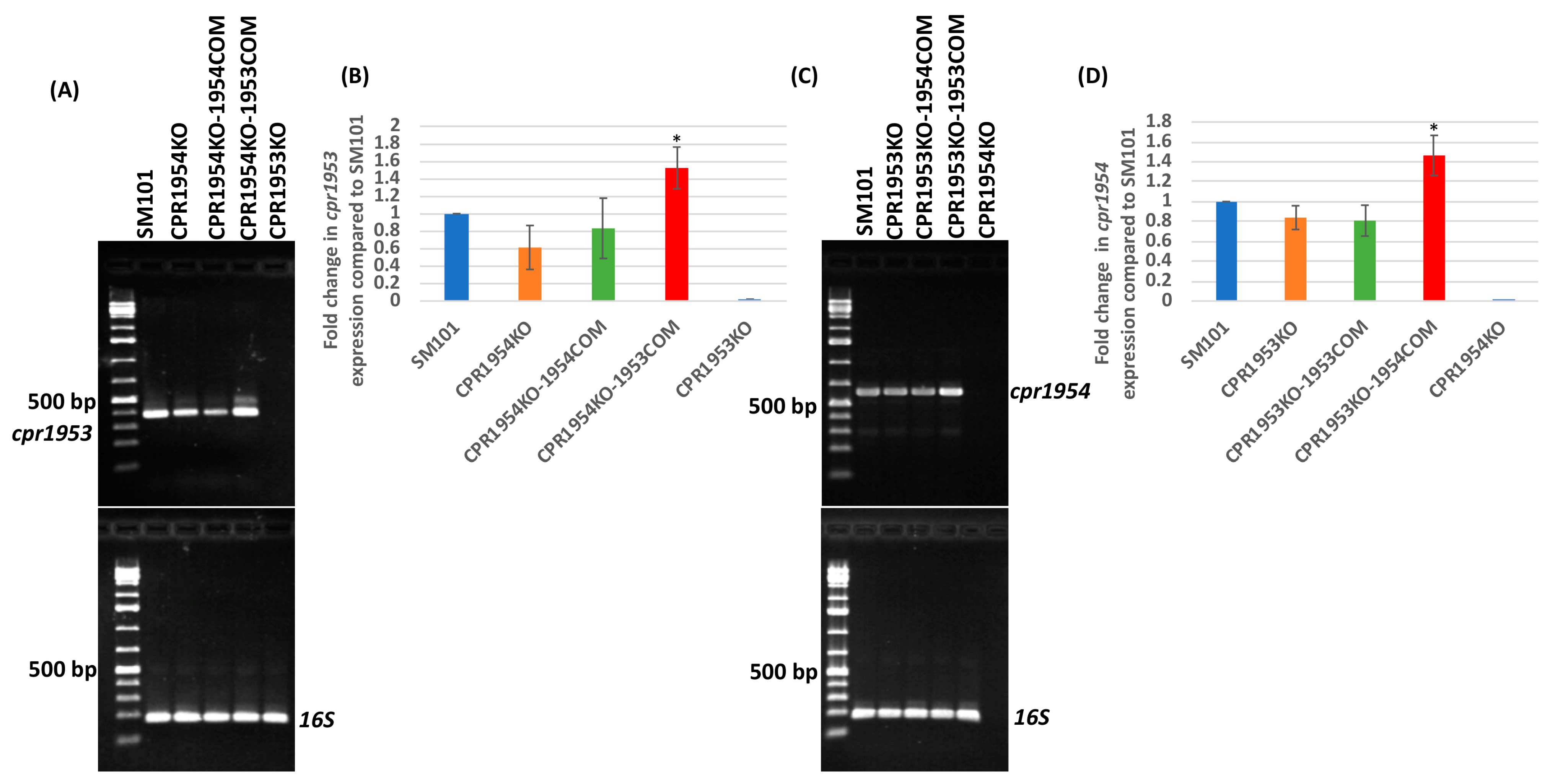
Expression levels of the cpr1953 or cpr1954 genes were then compared between CPR1954KO-1953COM or CPR1953KO-1954COM vs. wild-type SM101 to assess whether these were overexpressing strains. Since qRT-PCR primers could not be designed for cpr1953 or cpr1954, RT-PCR was performed using purified RNA from wild-type SM101 and its derivative OHK null mutants or transformants carrying pJIR750-based plasmids.
Results of those RT-PCR analyses demonstrated that CPR1953KO-1954COM and CPR1954KO-1953COM expressed their cpr1954 or cpr1953 genes, respectively, at significantly higher levels compared to SM101 (Figure 3A–D). No significant difference was noted in cpr1953 expression levels between SM101 vs. CPR1954KO or CPR1954KO-1954COM (Figure 3A,B). Similarly, no significant differences were apparent in cpr1954 expression levels between SM101 vs. CPR1953KO or CPR1953-1953COM (Figure 3C,D). In this experiment, all strains were shown by RT-PCR to express similar levels of the 16S RNA gene, used as a loading control (Figure 3A,C).

2.3. Overexpression of cpr1953 or cpr1954 Genes, Respectively, by the cpr1954 or cpr1953 Null Mutants Is Sufficient to Restore Sporulation and CPE Production to near Wild-Type Levels in MDS Cultures

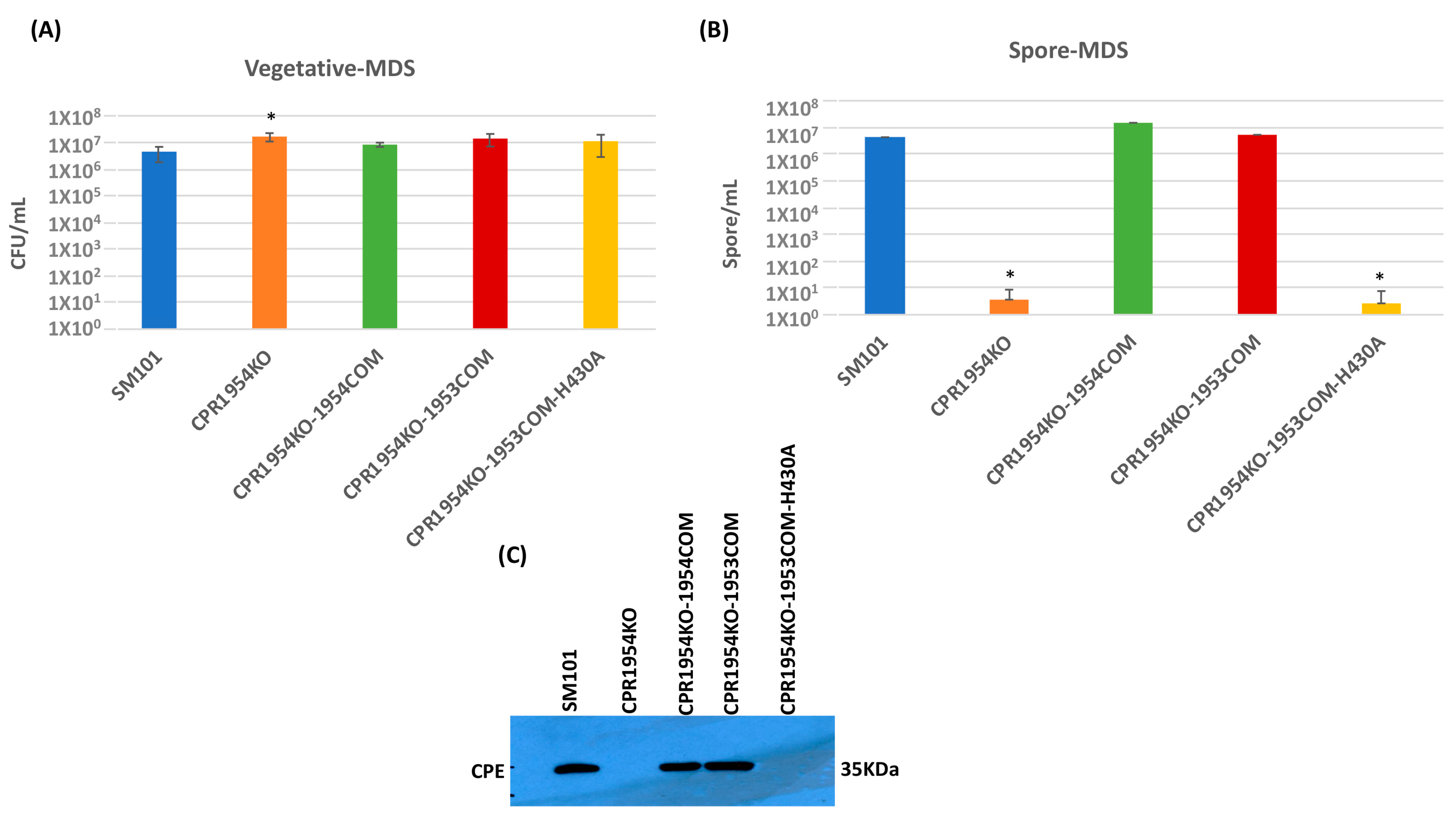
An experiment then evaluated whether overexpression of cpr1953 in the absence of cpr1954 expression, or overexpression of cpr1954 in the absence of cpr1953 expression, is sufficient to affect vegetative cell viability, spore formation, or CPE production when SM101 is cultured in MDS. Consistent with previous findings [28], overnight MDS cultures of CPR1953KO and CPR1954KO produced slightly, but significantly, more viable vegetative cells compared to wild-type SM101 (Figure 4A and Figure 5A). Overnight MDS cultures of CPR1953KO-1954COM, CPR1953KO-1954COM-H410, CPR1954KO-1953COM, and CPR1954KO-1953COM-H430 did not contain significantly more viable vegetative cells compared to SM101 MDS cultures (Figure 4A and Figure 5A).
In those overnight MDS cultures, the CPR1953KO mutant produced virtually no spores (Figure 4B), as expected from our previous study [28]. As we also reported previously, complementation of the CPR1953KO mutant to restore cpr1953 expression significantly enhanced sporulation back to the same levels as detected for SM101 MDS cultures. Importantly, these experiments also revealed for the first time that overexpressing cpr1954 in the absence of cpr1953 expression was sufficient to obtain significant sporulation, i.e., MDS cultures of CPR1953KO-1954COM produced 106-fold more spores than CPR1953KO, although the sporulation efficiency of this overexpressing strain was slightly lower than that of wild-type SM101 cultured in MDS (Figure 4B). Demonstrating the involvement of CPR1954 kinase activity for CPR1953KO-1954COM-induced sporulation, MDS cultures of CPR1953KO-1954COM-H410 still produced very low levels of sporulation, similar to CPR1953KO (Figure 4B).
In MDS, CPE production by those strains closely corresponded to their sporulation phenotype. Unlike the strong CPE production exhibited by SM101 MDS cultures, MDS cultures of CPR1953KO made no detectable CPE, while MDS cultures of the CPR1953KO-1953COM complementing strain made CPE at levels similar to wild-type SM101 (Figure 4C). The CPR1953KO-1954COM strain overexpressing cpr1954 in the absence of cpr1953 also made significantly more CPE than CPR1953KO when cultured in MDS, although CPR1953KO-1954COM produced slightly less CPE compared to wild-type SM101 when both are cultured in MDS (Figure 4C). Under the same MDS growth condition, the CPR1953KO-1954COM-H410 strain produced no detectable CPE (Figure 4C).
Similarly, when cultured in MDS, CPR1954KO sporulated extremely poorly (<10 spores/mL) and produced no detectable CPE (Figure 5B,C), consistent with our previous report [28]. Also consistent with those previous findings, complementation of the cpr1954 null mutant to restore cpr1954 expression significantly increased sporulation and CPE production back to approximately wild-type levels. Importantly, this study showed for the first time that, when cultured in MDS, the CPR1954KO-1953COM strain overexpressing cpe1953 in the absence of cpr1954 makes significantly more spores and CPE than CPR1954KO, producing spores and CPE at near wild-type SM101 levels (Figure 5B,C). Confirming the importance of CPR1953 kinase activity for CPR1954KO-1953COM-induced CPE production, transformation of CPR1953KO with pJIR750-cpr1953COM-H430A did not increase sporulation or CPE production (Figure 5B,C) compared to MDS cultures of CPR1954KO.

2.4. Overexpression of cpr1953 in the Absence of cpr1954 Expression or Vice Versa, Is Also Sufficient to Obtain near Wild-Type Levels of Sporulation and CPE Production in MIC

Our previous study [28] reported that the contributions of individual OHKs to SM101 sporulation and CPE production can vary between MDS and MIC cultures. Therefore, the vegetative cell viability, sporulation, and CPE production characteristics of the overexpressing strains were also evaluated in overnight MIC cultures.
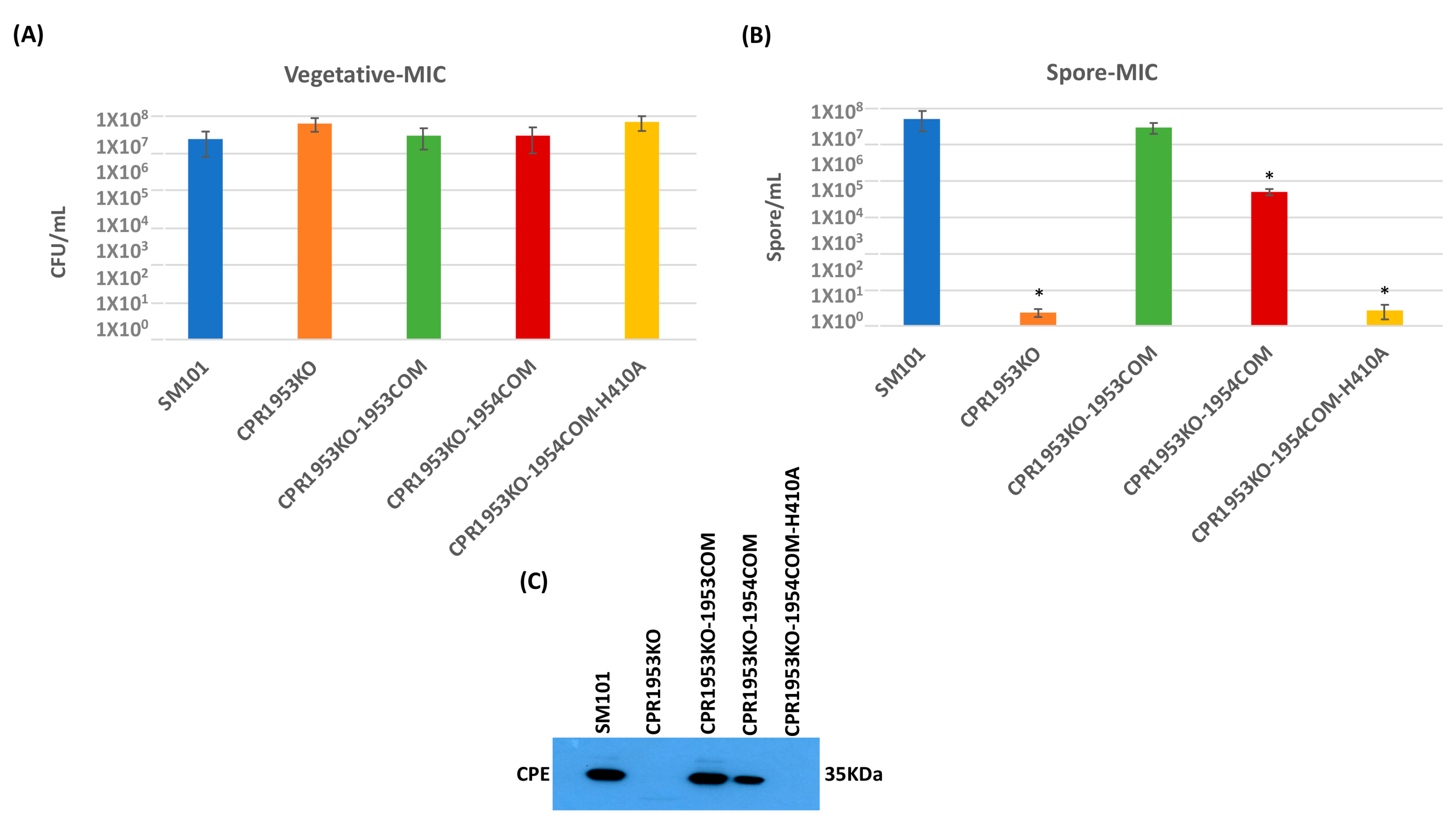
The results of those analyses indicated that overnight MIC cultures of all isogenic mutants, complementing strains and overexpressing strains contained the same level of viable vegetative cells as similar cultures of the SM101 parental strain (Figure 6A and Figure 7A). As expected from previous results [28], the CPR1953KO and CPR1954KO strains sporulated extremely poorly and did not produce CPE in MIC (Figure 6B,C and Figure 7B,C). However, MIC cultures of the CPR1953-1953COM and CPR1954-1954COM strains showed sporulation and CPE production at near wild-type levels. Importantly, this study showed for the first time that the overexpressing CPR1953KO-1954COM and CPR1954KO-1953COM strains also exhibit near wild-type levels of sporulation and CPE production in overnight MIC culture (Figure 6B,C and Figure 7B,C). In contrast, this study also determined that the CPR1953KO-1954COM-H410 and CPR1954KO-1953COM-H430 control strains did not sporulate or produce CPE (Figure 6B,C and Figure 7B,C).

2.5. Analysis of Spo0A Production Levels by Strains Overexpressing cpr1953 in the Absence of cpr1954 Expression or Vice Versa

To evaluate whether, relative to CPR1953KO or CPR1954KO, the increased sporulation and CPE production observed for CPR1953KO-1954COM and CPR1954KO-1953COM strains might involve increased Spo0A production, wild-type SM101 and its derivatives were grown in MDS broth medium for 3 h at 37 °C. Subsequently, equal amounts of total protein extracted from equal number of cells were subjected to Western blot assay for comparison of strain Spo0A production levels.
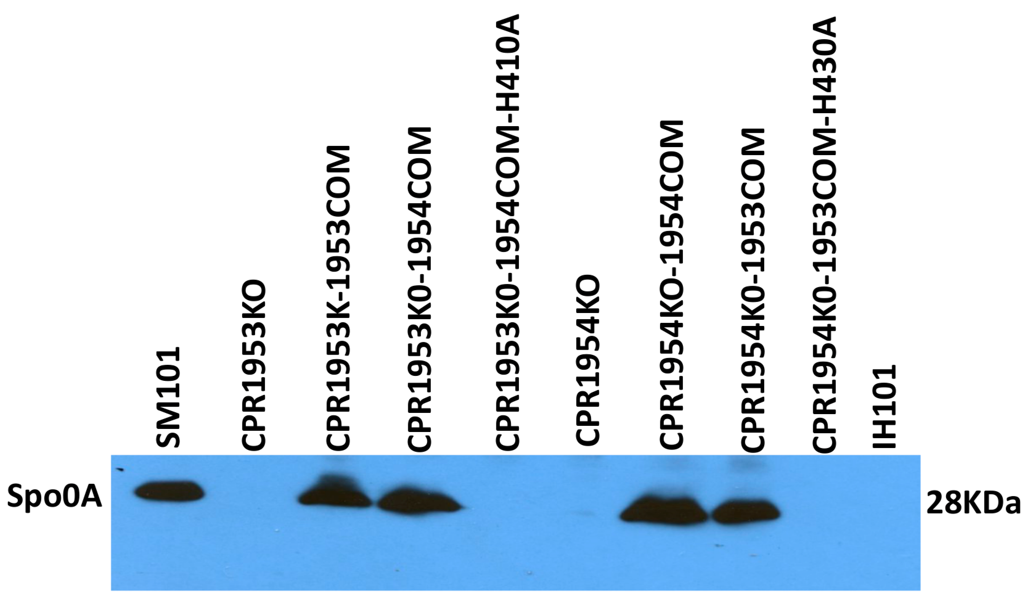
Results of this experiment determined that, as reported previously, the SM101 parental strain produces readily detectable levels of Spo0A, while the CPR1953KO and CPR1954KO mutants produce greatly reduced amounts of Spo0A. Furthermore, as also expected from previous results [28], the CPR1953KO-1953COM and CPR1954KO-1954COM complementing strains exhibited near wild-type levels of Spo0A production. Importantly, results shown in Figure 8 also demonstrate for the first time that the overexpressing CPR1953KO-1954COM and CPR1954KO-1953COM strains produce Spo0A at wild-type levels (Figure 8). This effect required the kinase activity of CPR1954 or CPR1953, respectively, since the CPR1953KO-1954COM-H410 and CPR1954KO-1953COM-H430 strains did not produce Spo0A protein. In this study, spo0A null mutant (1H101) of SM101 was used as a negative control.

3. Discussion

As briefly mentioned in the Introduction, most Bacilli and Clostridia can sporulate in the presence of environmental or metabolic stress signals such as nutrient limitations or population density [18,29,30,31]. In those endospore-forming bacteria, Spo0A phosphorylation initiates sporulation. However, different endospore-formers accomplish Spo0A phosphorylation by varying mechanisms. Bacillus subtilis, the paradigm model for sporulation by Bacilli, uses a phosphorelay involving five histidine kinases named KinA-E [23,32,33], with KinA and KinB usually considered as the major kinases for B. subtilis sporulation [32,34,35]. KinA and KinB transfer a phosphoryl group to the cognate regulator Spo0F, which is followed by sequential transfer of that phosphoryl group to the phosphotransferase Spo0B and then on to Spo0A [23,32,33,36].
When Clostridial genome sequences first became available, it was believed that all Clostridia lack the classical sporulation phosphorelay of Bacilli. However, the phosphorelay of Bacilli was later identified in some Clostridia [37], although it still has not been found in industrially important, solvent-producing Clostridia or pathogenic Clostridia, including C. perfringens. Instead, it was proposed that those Clostridia lacking the classical phosphorelay instead use OHKs to phosphorylate Spo0A and begin their sporulation. As discussed below, support for the OHK/sporulation hypothesis has been obtained for several nonpathogenic solvent-producing Clostridia, where mutants unable to produce particular OHKs showed decreased sporulation [26,28,38,39,40].
Among the pathogenic Clostridia, perhaps the strongest support for the OHK/sporulation hypothesis has been provided by studies of C. perfringens. As mentioned in the Introduction, at least five OHKs have been identified that contribute to C. perfringens sporulation in MDS, although only two of those OHKs (CPR1953 and CPR1954) also contribute to sporulation in MIC [26,28]. In either MDS or MIC cultures, those two OHKs also had by far the greatest impact on SM101 sporulation of all C. perfringens OHKs. Specifically, in either MDS or MIC, CPR1953KO and CPR1954KO producing < 10 spores/mL, which represents a >105-fold reduction in sporulation compared to wild-type SM101 [28].
There are some insights into how CPR1953 and CPR1954 affect sporulation and sporulation-dependent CPE production by SM101. CPR1954KO and CPR1953KO were shown to produce substantially reduced levels of Spo0A in 3 h MDS cultures and decreased levels of spo0A expression in 3 h MIC culture. Those observations may reflect feedback enhancement of spo0A expression by phosphorylated Spo0A [28,41]. A C. perfringens OHK named CPR0195 has been shown to directly phosphorylate Spo0A in vitro [26], but this ability has not yet been assessed for CPR1953 or CPR1954. However, Phos-Tag gels did not detect any phosphorylation of the reduced amount of Spo0A produced in 3 h MDS cultures of CPR1954KO, which would be consistent with CPR1954 being required for Spo0A phosphorylation [28]; whether this is a direct or indirect effect of CPR1954 on Spo0A phosphorylation is unclear. Nonetheless, reduced Spo0A production and phosphorylation in the absence of CPR1953 and CPR1954 likely help to explain the profound effects of these OHKs on sporulation.
Several possible mechanisms could explain why CPR1953 and CPR1954 are both needed, at natural production levels, for SM101 sporulation. For example, these two proteins might represent a novel phosphorelay, where they function together in a step-wise manner. Alternatively, at natural production levels, CPR1953 and CPR1954 might function independently but both be necessary to obtain sufficient levels of Spo0A production and phosphorylation to initiate sporulation.
To start distinguishing between those two possibilities, the current study examined the effects on sporulation and CPE production when cpr1954 is overexpressed in a background free of cpr1953 and vice versa. To accomplish this, we introduced a plasmid carrying the cpr1954 gene into CPR1953KO, which still carries the wild-type cpr1954 gene. RT-PCR analyses showed that the resultant CPR1953KO-1954COM strain expressed significantly higher levels of the cpr1954 gene than CPR1953KO or CPR1953KO-1953COM, consistent with a gene dosage effect increasing cpr1954 expression in CPR1953KO-1954COM (Figure 3C,D). Similarly, the CPR1954KO-1953COM strain overexpressed cpr1953 relative to either CPR1954KO or CPR1954KO-1954COM, also consistent with a gene dosage effect increasing cpr1953 expression in CPR1954KO-1953COM (Figure 3A,B). Characterization of those strains showed that overexpression of either OHK gene, in the absence of expression of the other OHK gene, is sufficient to obtain near wild-type levels of sporulation and CPE production, which is sporulation-dependent. These findings are consistent with a model where CPR1953 and CPR1954 work independently but additively. This model suggests that, at natural production levels an unidentified trigger increases the total kinase activity of both of these OHKs, which leads to sufficient levels of Spo0A production and phosphorylation for SM101 to start sporulation and CPE production.
The involvement of two major kinases in initiating C. perfringens sporulation is somewhat reminiscent of the dual contributions of KinA and KinB to sporulation initiation in B. subtilis. However, the strong sporulation impairment exhibited by both the CPR1953KO and CPR1954KO mutants of SM101 contrasts with the partial redundancy of KinA and KinB in B. subtilis sporulation, i.e., single kinA or kinB null mutants show only slight sporulation impairments but a double kinA/kinB null mutant exhibits a substantial sporulation deficiency [35]. The situation in some nonpathogenic, solvent-producing Clostridia seems to more closely resemble the apparently independent roles of CPR1953 and CR1954 in regulating C. perfringens sporulation. For example, several OHKs increase sporulation in the solvent-producing species C. acetobutylicum and those OHKs have been shown to directly phosphorylate Spo0A in vitro [37,39,42]. As this study suggests for CPR1953 and CPR1954 of C. perfringens, it appears those C. acetobutylicum OHKs function independently, but collectively, to initiate sporulation. Interestingly, those C. acetobutylicum OHKs do not appear to be close homologs of CPR1953 and CPR1954, supporting diversity in sporulation initiation amongst the Clostridia. Ongoing further exploration of the mechanistic contributions of CPR1953 and CPR1954 to C. perfringens sporulation and CPE production are needed to further elucidate their critical role in these processes.

4. Materials and Methods

4.1. C. perfringens Isolates, Plasmids, Media, and Culture Condition

All C. perfringens isolates and plasmids used in this study are listed in Table 1. C. perfringens type F strain SM101 was used for this study as the wild-type parental strain. The wild-type strain and all complemented isolates were stored at −20 °C in cooked meat medium (CMM; Oxoid, Hampshire, UK). Stock cultures of all derivatives were made in 35% glycerol and stored at −80 °C. The C. perfringens strains were grown anaerobically using a BD GasPak EZ container system at 37 °C in the following broth media: Fluid thioglycollate medium (FTG; Difco Laboratories, Detroit, MI, USA), and Modified Duncan–Strong medium (MDS; 0.4% yeast extract (Becton, Dickinson, Franklin Lakes, NJ, USA), 1.5% proteose peptone [Becton, Dickinson], 1% disodium phosphate heptahydrate (Fisher Scientific, Rockingham County, NH, USA), 0.4% raffinose (Sigma-Aldrich, St. Louis, MO, USA), 0.1% sodium thioglycollate (Sigma-Aldrich), and 1mM caffeine (Sigma-Aldrich)). Brain Heart Infusion (BHI) agar (Research Products International, Mt. Prospect, IL, USA) was used for growth of C. perfringens strains on solid media. When required, the culture media were supplemented with chloramphenicol (15 μg/mL) (Sigma-Aldrich) for selection of complementing strains as well as counting of vegetative or spore cells of complementing strains.

4.2. Construction of Overexpressing Strains

To construct the overexpressing strains designated as CPR1953KO-1954COM and CPR1954KO-1953COM, the pJIR750-cpr1954COM and pJIR750-cpr1953COM plasmids were transferred by electroporation into CPR1953KO or CPR1954KO, respectively, and chloramphenicol resistant (CMr) clones were selected. The resultant transformants were confirmed by PCR using the primers listed in Table 2.
In addition, CPR1953KO-1954COM-H410A and CPR1954KO-1953COM-H430A were constructed by electroporation of pJIR750-cpr1954COM-H410A and pJIR750-cpr1953COM-H430A plasmids into CPR1953KO or CPR1954KO, respectively. CMr transformants were selected and then confirmed by PCR using the primers listed in Table 2. The pJIR750-cpr1954COM-H410A and pJIR750-cpr1953COM-H430A plasmids were constructed in our previous study [28]. Briefly, these plasmids encode an alanine substitution for the key functional His residue involved in phosphotransfer for the CPR1953 and CPR1954 orphan histidine kinases.

4.3. Preparation of Pathophysiologically-Relevant Ex Vivo MIC

The ex vivo (mouse intestinal content (MIC)) model used in this study for growth of C. perfringens strains was developed previously [28]. Briefly, the small intestinal contents of five healthy male or female BALB/c mice were pooled and then mixed (1:1) with PBS (phosphate buffer saline, Corning, Corning, NY, USA). The diluted sample was then centrifuged at 15,000× g for 10 min to eliminate the solid materials. Subsequently, the supernatant was diluted (1:5) with PBS and sterilized by passage through a 0.2 μm syringe filter.

4.4. Comparison of Growth Kinetics between C. perfringens Strains Cultured in MDS Sporulation Medium

For comparison of strain growth characteristics in MDS broth, a 200 μL aliquot of an overnight FTG culture of SM101 or its derivative strains was inoculated into 10 mL of freshly prepared MDS medium. The cultures were incubated anaerobically at 37 °C for 0, 2, 4, 6, 8, or 24 h. Then, a 1 mL aliquot culture of each isolate was collected and OD600 was measured using a Bio-Rad SmartSpec spectrophotometer (Hercules, CA, USA). This experiment was conducted with three biological replicates.

4.5. Measurement of Viable Vegetative Cells or Heat-Resistant Spores

Quantitative counts of viable vegetative cells or heat-resistant spores of SM101 and its derivative strains were evaluated as previously described [28]. Briefly, for enumeration of viable vegetative cells, a 200 μL or 40 μL aliquot of an overnight FTG of each isolate was added into 10 mL of freshly prepared MDS medium or 1 mL of MIC containing 5% Oxyrase (Oxyrase, Inc., Mansfield, OH, USA), respectively. After overnight anaerobic incubation at 37 °C, those cultures were serially diluted with sterile PBS and plated onto BHI agar plates. The colonies on each BHI agar plates were enumerated after incubation overnight at 37 °C.
To enumerate spore formation, samples were prepared in MDS or MIC as described above. However, before plating the cell suspensions, each culture was heated at 70 °C for 20 min to kill the remaining vegetative cells and induce spore germination. Those heat-shocked samples were then serially diluted with sterile PBS and plated on BHI agar plates. After overnight anaerobic incubation at 37 °C, the colonies were counted. This experiment was conducted with three biological replicates.

4.6. Western Blot Analyses of CPE Production

For CPE Western blot analyses, C. perfringens cultures were prepared as described earlier in the “measurement of vegetative cells” section. After overnight anaerobic incubation at 37 °C, each MDS or MIC culture was adjusted to equal OD600. After this step, those adjusted samples were centrifuged at 15,000× g for 5 min. Equal volumes of each supernatant were mixed with 5× concentrated loading buffer containing bromophenol blue dye and SDS (sodium dodecyl sulfate) and then electrophoresed onto a 12% SDS-PAGE gel, followed by transfer onto a polyvinylidene difluoride (PVDF) membrane (Bio-Rad). Subsequently, the blot was blocked at room temperature for 1 h with TBST buffer (Tris buffered saline, 0.1% Tween 20) containing 5% w/v nonfat dry milk. The blocked blot was then incubated with rabbit polyclonal CPE antibody [44], 1:5000 dilution in TBST buffer containing 5% nonfat dry milk at 4 °C overnight. After 3 washes with TBST buffer for 10 min each, the membrane was incubated with a horseradish peroxidase-conjugated secondary anti-rabbit antibody (Sigma-Aldrich) at dilution of 1:10,000 for 1 h at room temperature. Excess secondary antibody was eliminated by washing 3 times with TBST buffer. Finally, the membrane was developed using SuperSignal West Pico substrate (ThermoFisher, Waltham, MA, USA).

4.7. Western Blot Analysis of Spo0A Production

Spo0A Western blot analysis was performed as previously described [28]. Briefly, wild-type SM101 and its derivative strains were grown in 10 mL of freshly prepared MDS medium for 3 h at 37 °C. Each C. perfringens culture was then adjusted to equal OD600 using a Bio-Rad SmartSpec spectrophotometer. Equal volume of those adjusted cultures was harvested by centrifugation, washed with PBS buffer, lysed with B-PER reagent (ThermoFisher Scientific) containing protease inhibitor cocktail II (Research Products International), and then boiled for 5 min at 95 °C. The whole-cell extracts were then subjected to 12% SDS-PAGE followed by Western blot with rabbit polyclonal antibody made against Clostridioides difficile Spo0A at dilution of 1:2000. The primary antibody was detected by horseradish peroxidase-conjugated secondary anti-rabbit antibody at dilution of 1:10,000, and Western blot was visualized using SuperSignal West Pico substrate. In this experiment, the spo0A knock-out mutant 1H101 [22] was served as a negative control.

4.8. DNA Extraction and PCR Analysis

Genomic DNA was extracted from wild-type SM101 and its derivative strains using the MasterPure Gram-positive bacterial DNA purification kit (Epicentre, Paris, France). All previously constructed [28] plasmids used in this study were isolated from C. perfringens type A strain 13, the cloning host strain, using a QIAprep Spin Miniprep Kit (Qiagen, Hilden, Germany), according to the manufacturer’s instructions.
The PCR reactions for confirming the isogenic mutants and complemented strains were conducted using 2× DreamTaq Green PCR Master Mix (ThermoFisher Scientific) and appropriate primers listed in Table 2. The following PCR amplification condition was used for this experiment: (1) 95 °C for 5 min; (2) 35 cycles of 95 °C for 30 s, 55 °C for 30 s, 72 °C for 90 s, and (3) a final extension for 5 min at 72 °C.
In addition, LongAmp Taq polymerase (New England Biolabs, Ipswich, MA, USA) was used for confirmation of CPR1953KO-1954COM, CPR1954KO-1953COM, CPR1953KO-1954COM-H410A, and CPR1954KO-1953COM-H430A constructs. The following PCR parameters were used: (1) 95 °C for 5 min; (2) 35 cycles of 95 °C for 30 s, 55 °C for 30 s, 65 °C for 50 s per Kb, and (3) a final extension for 10 min at 65 °C. Finally, an aliquot of each PCR reaction (20 μL) was electrophoresed on a 2% agarose gel and then visualized after staining with ethidium bromide.

4.9. RNA Isolation and RT-PCR Analysis

A 200 μL aliquot of an overnight FTG culture of SM101 or its derivative strains was transferred into 10 mL of MDS broth and anaerobically incubated at 37 °C for 3 h. Total RNA was isolated from pelleted cells using the saturated phenol (Fisher Scientific) protocol, as previously described [45]. Purified RNA was quantified by NanoDrop spectrophotometer (ThermoFisher Scientific) and then stored in a −80 °C freezer. Furthermore, the purity and quality of each extracted RNA sample was evaluated by conducting PCR assay for polC and 16S genes in the absence or presence of reverse transcriptase enzyme. RT-PCR assay was performed using one-step RT-PCR containing 200 ng of purified RNA, avian myeloblastosis virus (AMV) reverse transcriptase (4 U; Promega, Madison, WI, USA), RNA and DNA free dH2O, 2× DreamTaq Green PCR Master Mix (ThermoFisher Scientific), and appropriate primer sets listed in Table 2. Finally, the RT-PCR assay was carried out using the following parameters: (1) 95 °C for 4 min; (2) 42 °C for 45 min (for cDNA synthesis); (3) 95 °C for 2 min; (4) 30 cycles of 95 °C for 30 s, 55 °C for 30 s, 72 °C for 30 s, and (5) a final extension for 5 min at 72 °C.

4.10. Statistical Analysis

Differences among the results were determined by using either ordinary one-way analysis of variance (ANOVA; GraphPad Prism 8) or Student’s unpaired t test from at least three independent experiments. If the p value was less than 0.05, the result was considered statistically significant.

Author Contributions

Conceptualization, I.M.G. and B.A.M.; Methodology, I.M.G., B.A.M. and F.A.U.; Formal analysis, I.M.G. and B.A.M.; Investigation, I.M.G., B.A.M. and J.L.G., Data curation, I.M.G. and B.A.M., Writing—original draft preparation, I.M.G. and B.A.M., Writing—review and editing, I.M.G., B.A.M., J.L.G. and F.A.U., Supervision, B.A.M.; Funding acquisition, B.A.M. and F.A.U. All authors have read and agreed to the published version of the manuscript.

Funding

This work was generously supported by grant R01AI019844-41 (to B.A.M.) from the National Institute of Allergy and Infectious Diseases. The content is solely the responsibility of the authors and does not necessarily represent the official views of the National Institutes of Health.

Institutional Review Board Statement

The experiment involving mice intestinal contents was reviewed and approved by the University of California Davis Institutional Animal Care and Use Committee (IACUC protocol 23182, approved 2 February 2023 and IACUC protocol 23482, approved 18 July 2023).

Informed Consent Statement

Not applicable.

Data Availability Statement

All the data that support the findings of this study are available from the corresponding author upon reasonable request.

Acknowledgments

The authors thank Aimee Shen for the antiserum against Clostridioides difficile Spo0A. We also thank Mahfuzur Sarker for providing the isogenic IH101 spo0A null mutant.

Conflicts of Interest

The authors declare no conflicts of interest.

References

  1. McClane, B.A.; Robertson, S.L.; Li, J. Clostridium perfringens. In Food Microbiology: Fundamentals and Frontiers, 4th ed.; Doyle, M.P., Buchanan, R.L., Eds.; ASM Press: Washington, DC, USA, 2013; pp. 465–489. [Google Scholar]
  2. Rood, J.I.; Adams, V.; Lacey, J.; Lyras, D.; McClane, B.A.; Melville, S.B.; Moore, R.J.; Popoff, M.R.; Sarker, M.R.; Songer, J.G.; et al. Expansion of the Clostridium perfringens toxinotyping scheme. Anaerobe 2018, 53, 5–10. [Google Scholar] [CrossRef] [PubMed]
  3. Grenda, T.; Jarosz, A.; Sapała, M.; Grenda, A.; Patyra, E.; Kwiatek, K. Clostridium perfringens-opportunistic foodborne pathogen, its diversity and epidemiological significance. Pathogens 2023, 12, 768. [Google Scholar] [CrossRef] [PubMed]
  4. Carman, R.J.; Sayeed, S.; Li, J.; Genheimer, C.W.; Hiltonsmith, M.F.; Wilkins, T.D.; McClane, B.A. Clostridium perfringens toxin genotypes in the feces of healthy North Americans. Anaerobe 2008, 14, 102–108. [Google Scholar] [CrossRef]
  5. Voidarou, C.; Bezirtzoglou, E.; Alexopoulos, A.; Plessas, S.; Stefanis, C.; Papadopoulos, I.; Vavias, S.; Stavropoulou, E.; Fotou, K.; Tzora, A.; et al. Occurrence of Clostridium perfringens from different cultivated soils. Anaerobe 2011, 17, 320–324. [Google Scholar] [CrossRef] [PubMed]
  6. Stelma, G.N. Use of bacterial spores in monitoring water quality and treatment. J. Water Health 2018, 16, 491–500. [Google Scholar] [CrossRef] [PubMed]
  7. Mehdizadeh Gohari, I.; Navarro, M.; Li, J.; Shrestha, A.; Uzal, F.A.; McClane, B.A. Pathogenicity and virulence of Clostridium perfringens. Virulence 2021, 12, 723–753. [Google Scholar] [CrossRef] [PubMed]
  8. Kiu, R.; Hall, L.J. An update on the human and animal enteric pathogen Clostridium perfringens. Emerg. Microbes. Infect. 2018, 6, 141. [Google Scholar] [CrossRef] [PubMed]
  9. McClane, B.A.; Lyerly, D.M.; Wilkins, T.D. Enterotoxic clostridia: Clostridium perfringens type A and Clostridium difficile. In Gram-positive Pathogens, 3rd ed.; Fischetti, V.A., Novick, R.P., Ferretti, J.J., Portnoy, D.A., Rood, J.I., Eds.; ASM Press: Washington, DC, USA, 2006; pp. 703–714. [Google Scholar]
  10. Carman, R.J. Clostridium perfringens in spontaneous and antibiotic-associated diarrhoea of man and other animals. Rev. Med. Microbiol. 1997, 8, 546. [Google Scholar] [CrossRef]
  11. Larcombe, S.; Hutton, M.L.; Lyras, D. Involvement of bacteria other than Clostridium difficile in antibiotic-associated diarrhoea. Trends. Microbiol. 2016, 24, 463–476. [Google Scholar] [CrossRef] [PubMed]
  12. Mpamugo, O.; Donovan, T.; Brett, M.M. Enterotoxigenic Clostridium perfringens as a cause of sporadic cases of diarrhoea. J. Med. Microbiol. 1995, 43, 442–445. [Google Scholar] [CrossRef] [PubMed]
  13. Borriello, S.P.; Larson, H.E.; Welch, A.R.; Barclay, F.; Stringer, M.F.; Bartholomew, B.A. Enterotoxigenic Clostridium perfringens: A possible cause of antibiotic-associated diarrhoea. Lancet 1984, 11, 305–307. [Google Scholar] [CrossRef] [PubMed]
  14. García, S.; Vidal, J.E.; Heredia, N.; Juneja, V.K. Clostridium perfringens. In Food Microbiology: Fundamentals and Frontiers, 5th ed.; Doyle, M.P., Diez-Gonzalez, F., Hill, C., Eds.; ASM Press: Washington, DC, USA, 2019; pp. 513–540. [Google Scholar]
  15. Sarker, M.R.; Carman, R.J.; McClane, B.A. Inactivation of the gene (cpe) encoding Clostridium perfringens enterotoxin eliminates the ability of two cpe-positive C. perfringens type A human gastrointestinal disease isolates to affect rabbit ileal loops. Mol. Microbiol. 1999, 33, 946–958. [Google Scholar] [CrossRef] [PubMed]
  16. Li, J.; Paredes-Sabja, D.; Sarker, M.R.; McClane, B.A. Clostridium perfringens sporulation and sporulation-associated toxin production. Microbiol. Spectr. 2016, 4, TBS-0022-2015. [Google Scholar] [CrossRef] [PubMed]
  17. Zhao, Y.; Melville, S.B. Identification and characterization of sporulation-dependent promoters upstream of the enterotoxin gene (cpe) of Clostridium perfringens. J. Bacteriol. 1998, 180, 136–142. [Google Scholar] [CrossRef] [PubMed]
  18. Shen, A.; Edwards, A.N.; Sarker, M.R.; Paredes-Sabja, D. Sporulation and germination in Clostridial pathogens. Microbiol. Spect. 2019, 6, GGP3-0017. [Google Scholar]
  19. Harry, K.H.; Zhou, R.; Kroos, L.; Melville, S.B. Sporulation and enterotoxin (CPE) synthesis are controlled by the sporulation-specific factors SigE and SigK in Clostridium perfringens. J. Bacteriol. 2009, 191, 2728–2742. [Google Scholar] [CrossRef] [PubMed]
  20. Li, J.; McClane, B.A. Evaluating the involvement of alternative sigma factors SigF and SigG in Clostridium perfringens sporulation and enterotoxin synthesis. Infect. Immun. 2010, 78, 4286–4293. [Google Scholar] [CrossRef] [PubMed]
  21. Fimlaid, K.A.; Shen, A. Diverse mechanisms regulate sporulation sigma factor activity in the Firmicutes. Curr. Opin. Microbiol. 2015, 24, 88–95. [Google Scholar] [CrossRef] [PubMed]
  22. Huang, I.H.; Waters, M.; Grau, R.R.; Sarker, M.R. Disruption of the gene (spo0A) encoding sporulation transcription factor blocks endospore formation and enterotoxin production in enterotoxigenic Clostridium perfringens type A. FEMS Microbiol. Lett. 2004, 233, 233–240. [Google Scholar] [CrossRef] [PubMed]
  23. Burbulys, D.; Trach, K.A.; Hoch, J.A. Initiation of sporulation in B. subtilis is controlled by a multicomponent phosphorelay. Cell 1991, 64, 545–552. [Google Scholar] [CrossRef] [PubMed]
  24. Hoch, J.A. Regulation of the phosphorelay and the initiation of sporulation in Bacillus subtilis. Annu. Rev. Microbiol. 1993, 47, 441–465. [Google Scholar] [CrossRef] [PubMed]
  25. Shimizu, T.; Ohtani, K.; Hirakawa, H.; Ohshima, K.; Yamashita, A.; Shiba, T.; Naotake, O.; Masahira, H.; Satoru, K.; Hideo, H. Complete genome sequence of Clostridium perfringens, an anaerobic flesh-eater. Proc. Natl. Acad. Sci. USA 2002, 99, 996–1001. [Google Scholar] [CrossRef] [PubMed]
  26. Freedman, J.; Li, J.; Mi, E.; McClane, B.A. Identification of an important orphan histidine kinase for the initiation of sporulation and enterotoxin production by Clostridium perfringens type F strain SM101. mBio 2019, 10, e02674-18. [Google Scholar] [CrossRef] [PubMed]
  27. Myers, G.S.; Rasko, D.A.; Cheung, J.K.; Ravel, J.; Seshadri, R.; DeBoy, R.T.; Ren, Q.; Varga, J.; Awad, M.M.; Brinkac, L.M.; et al. Skewed genomic variability in strains of the toxigenic bacterial pathogen, Clostridium perfringens. Genome Res. 2006, 16, 1031–1040. [Google Scholar] [CrossRef] [PubMed]
  28. Mehdizadeh Gohari, I.; Li, J.; Navarro, M.A.; Mendonça, F.S.; Uzal, F.A.; McClane, B.A. Identification of orphan histidine kinases that impact sporulation and enterotoxin production by Clostridium perfringens type F strain SM101 in a pathophysiologically-relevant ex vivo mouse intestinal contents model. PLoS Pathog. 2023, 19, e1011429. [Google Scholar] [CrossRef] [PubMed]
  29. Al-Hinai, M.A.; Shawn, W.J.; Papoutsakis, E.T. The Clostridium sporulation programs: Diversity and preservation of endospore differentiation. Microbiol. Mol. Biol. Rev. 2015, 79, 19–37. [Google Scholar] [CrossRef] [PubMed]
  30. Tan, I.S.; Ramamurthi, K.S. Spore formation in Bacillus subtilis. Environ. Microbiol. Rep. 2014, 6, 212–225. [Google Scholar] [CrossRef]
  31. Riley, E.P.; Lopez-Garrido, J.; Sugie, J.; Liu, R.B.; Pogliano, K. Metabolic differentiation and intercellular nurturing underpin bacterial endospore formation. Sci. Adv. 2021, 7, eabd6385. [Google Scholar] [CrossRef] [PubMed]
  32. Perego, M.; Cole, S.P.; Burbulys, D.; Trach, K.; Hoch, J.A. Characterization of the gene for a protein kinase which phosphorylates the sporulation-regulatory proteins Spo0A and Spo0F of Bacillus subtilis. J. Bacteriol. 1989, 171, 6187–6196. [Google Scholar] [CrossRef] [PubMed]
  33. Jiang, M.; Shao, W.; Perego, M.; Hoch, J.A. Multiple histidine kinases regulate entry into stationary phase and sporulation in Bacillus subtilis. Mol. Microbiol. 2000, 38, 535–542. [Google Scholar] [CrossRef] [PubMed]
  34. Trach, K.A.; Hoch, J.A. Multisensory activation of the phosphorelay initiating sporulation in Bacillus subtilis: Identification and sequence of the protein kinase of the alternate pathway. Mol. Microbiol. 1993, 8, 69–79. [Google Scholar] [CrossRef] [PubMed]
  35. LeDeaux, J.R.; Yu, N.; Grossman, A.D. Different roles for KinA, KinB, and KinC in the initiation of sporulation in Bacillus subtilis. J. Bacteriol. 1995, 177, 861–863. [Google Scholar] [CrossRef] [PubMed]
  36. Trach, K.A.; Burbulys, D.; Strauch, M.; Wu, J.J.; Dhillon, N.; Jonas, R.; Hanstein, C.; Kallio, P.; Perego, M.; Bird, T. Control of the initiation of sporulation in Bacillus subtilis by a phosphorelay. Res. Microbiol. 1991, 142, 815–823. [Google Scholar] [CrossRef] [PubMed]
  37. Davidson, P.; Rory, E.; Redler, B.; Hiller, N.L.; Laub, M.T.; Durand, D. Flexibility and constraint: Evolutionary remodeling of the sporulation initiation pathway in Firmicutes. PLoS Genet. 2018, 14, e1007470. [Google Scholar] [CrossRef] [PubMed]
  38. Underwood, S.; Guan, S.; Vijayasubhash, V.; Baines, S.D.; Graham, L.; Lewis, R.J.; Wilcox, M.H.; Stephenson, K. Characterization of the sporulation initiation pathway of Clostridium difficile and its role in toxin production. J. Bacteriol. 2009, 191, 7296–7305. [Google Scholar] [CrossRef] [PubMed]
  39. Steiner, E.D.; Angel, E.D.; Young, D.I.; Heap, J.T.; Minton, N.P.; Hoch, J.A.; Young, M. Multiple orphan histidine kinases interact directly with Spo0A to control the initiation of endospore formation in Clostridium acetobutylicum. Mol. Microbiol. 2011, 80, 641–654. [Google Scholar] [CrossRef]
  40. Mearls, E.B.; Lee, R.L. The identification of four histidine kinases that influence sporulation in Clostridium thermocellum. Anaerobe 2014, 28, 109–119. [Google Scholar] [CrossRef] [PubMed]
  41. Fujita, M.; Sadaie, Y. Feedback loops involving Spo0A and AbrB in in vitro transcription of the genes involved in the initiation of sporulation in Bacillus subtilis. J. Biochem. 1998, 124, 98–104. [Google Scholar] [CrossRef]
  42. Du, G.; Zhu, C.; Wu, Y.; Kang, W.; Xu, M.; Yang, S.T.; Xue, C. Effects of orphan histidine kinases on clostridial sporulation progression and metabolism. Biotechnol. Bioeng. 2022, 119, 226–235. [Google Scholar] [CrossRef] [PubMed]
  43. Bannam, T.L.; Rood, J.I. Clostridium perfringens-Escherichia coli shuttle vectors that carry single antibiotic resistance determinants. Plasmid 1993, 29, 223–235. [Google Scholar] [CrossRef] [PubMed]
  44. McClane, B.A.; Strouse, R.J. Rapid detection of Clostridium perfringens type A enterotoxin by enzyme-linked immunosorbent assay. J. Clin. Microbiol. 1984, 19, 112–115. [Google Scholar] [CrossRef] [PubMed]
  45. Li, J.; Ma, M.; Sarker, M.R.; McClane, B.A. CodY is a global regulator of virulence-associated properties for Clostridium perfringens type D strain CN3718. mBio 2013, 4, e00770-13. [Google Scholar] [CrossRef] [PubMed]
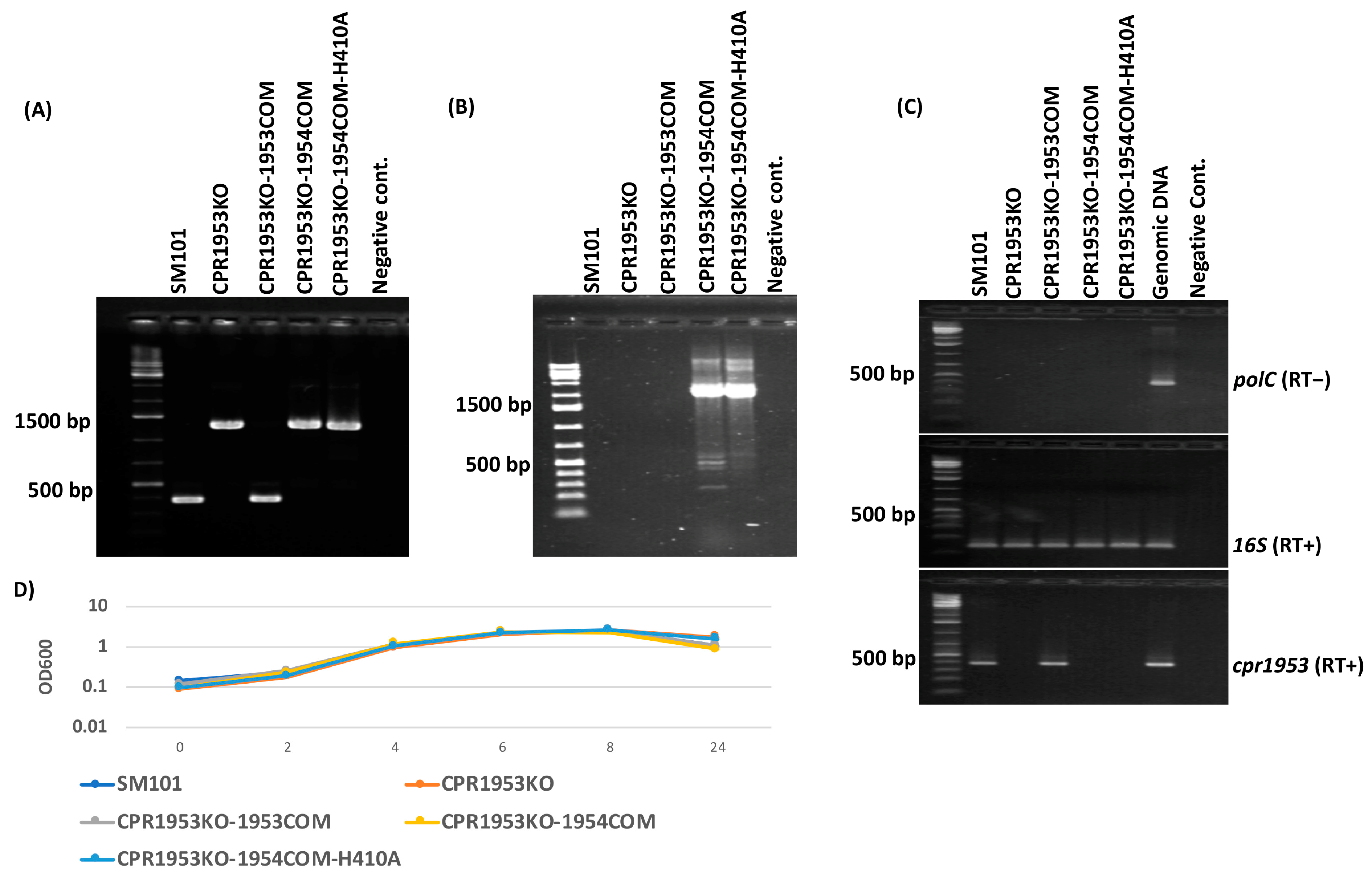
Figure 1. Preparation and characterization of CPR1953KO-1954COM and CPR1953KO-1954COM-H410A. (A) PCR assay verifying that CPR1953KO-1954COM and CPR1953KO-1954COM-H410A carry intron disruptions in their cpr1953 gene. PCR was conducted using internal cpr1953 gene primer sets which amplified a larger amplicon from CPR1053KO (1265 bp) versus SM101 wild-type strain (365 bp) due to insertion of a 900 bp group II intron into the specific DNA target site of the cpr1953 gene. The complementing strain CPR1953KO-1953COM amplified a PCR product similar in size to that of wild-type SM101. As expected, the overexpressing transformants amplified amplicons similar in size to that of isogenic CPR1953KO using the same primer sets, confirming those strains still carried a disrupted cpr1953 gene. (B) PCR assay confirmed the presence of pJIR750-based plasmids in transformants using specific primers that were able to amplify PCR products from contiguous pJIR750 and cpr1954 sequences. As shown, no PCR products were observed for SM101, CPR1953KO, and CPR1953KO-1953COM. (C) RT-PCR analyses for polC gene (top), 16S gene (middle), or cpr1953 gene (bottom) transcription by wild-type SM101, CPR1953KO, CPR1953KO-CPR1953COM, CPR1953KO-1954COM, and CPR1953KO-1954COM-H410A strains. Purified genomic DNA was used as a positive control. (D) Growth curve analysis for wild-type SM101 and its derivative strains cultured in MDS medium at 37 °C for different time points. Error bars depict SDs. All data were collected from three independent repetition. p value < 0.05 was considered as significant differences among the results.
Toxins 16 00195 g001
Figure 2. Preparation and characterization of CPR1954KO-1953COM and CPR1954KO-1953COM-H430A strains. (A) PCR assay verifying CPR1954KO-1953COM and CPR1954KO-1953COM-H430A carry intron disruptions in their cpr1954 genes. PCR was conducted using internal cpr1954 gene primer sets which amplified a larger amplicon from CPR1954KO (1496 bp) versus wild-type SM101 (596 pb) due to insertion of a 900 bp group II intron into the specific DNA target site of the cpr1954 gene. The complementing strain CPR1954KO-1954COM amplified a PCR product similar to that of the wild-type SM101 strain. As expected, the CPR1954KO-1953COM and CPR1954KO-1953COM-H430A transformants amplified products similar in size to that of isogenic CPR1954KO using the same primer sets, confirming those strains still carry intron disruptions in their cpr1954 gene. (B) PCR assay confirmed the presence of pJIR750-based plasmids in these transformants using specific primers, which were able to amplify PCR products from contiguous pJIR750 and cpr1953 sequences. As shown, no PCR products were observed for SM101, CPR1954KO, and CPR1954KO-1954COM. (C) RT-PCR analyses for transcription of the polC gene (top), 16S gene (middle), or cpr1954 gene (bottom) by wild-type SM101, CPR1954KO, CPR1954KO-CPR1954COM, CPR1954KO-1953COM, and CPR1954KO-1953COM-H430A. Purified genomic DNA was used as a positive control. (D) Growth curve analysis for wild-type SM101 and its derivative strains cultured in MDS medium at 37 °C for different time points. Error bars depict SDs. All data were collected from three independent repetitions. p value < 0.05 was considered as significant differences among the results.
Toxins 16 00195 g002
Figure 3. Comparing the expression levels of the cpr1953 and cpr1954 genes in various strains. (A) RT-PCR comparing the expression levels of the cpr1953 gene between SM101, CPR1954KO, CPR1954KO-1954COM, CPR1954KO-1953COM, and CPR1953KO. (B) Comparison of cpr1953 expression levels in SM101 versus its derivative isogenic null mutants or complementing strains as determined by densitometry using Image J software (version 1.53t). (C) RT-PCR comparing the expression levels of cpr1954 between SM101, CPR1953KO, CPR1953KO-1953COM, CPR1953KO-1954COM, and CPR1954KO. (D) Comparison of cpr1954 expression levels in SM101 versus its derivative strains as determined by densitometry using Image J software. In this assay, RT-PCR for 16S RNA was used as loading control. Figure is shown as a representation of three independent experiments. * p < 0.05 compared to SM101.
Toxins 16 00195 g003
Figure 4. Evaluating the effect of overexpressing the cpr1953 gene in the absence of cpr1954 expression on vegetative cell viability, spore formation, and CPE production when SM101 and its derivative strains were cultured in MDS. (A) Viable vegetative cell numbers (CFU/mL) when SM101, CPR1953KO, CPR1953KO-1953COM, CPR1953KO-1954COM, or CPR1953KO-1954COM-H1410A were anaerobically cultured overnight at 37 °C in MDS. (B) Heat-resistant spore counts (CFU/mL) in aliquots of those same MDS cultures after heating at 70 °C for 20 min to kill the remaining vegetative cells and induce spore germination. (C) Western blot analysis of CPE levels in supernatants of those same overnight MDS cultures for SM101, CPR1953KO, CPR1953KO-1953COM, CPR1953KO-1954COM, and CPR1953KO-1954COM-H1410A. The size of the protein band is shown at right. Error bars depict SDs. All data were collected from three independent repetitions. Asterisk represents p < 0.05 versus SM10. Western blot is representative of three independent experiments.
Toxins 16 00195 g004
Figure 5. Evaluating the effect of overexpressing the cpr1954 gene in the absence of cpr1953 expression on vegetative cell viability, spore formation, and CPE production when SM101 and its derivative strains were cultured in MDS. (A) Viable vegetative cell numbers (CFU/mL) when SM101, CPR1954KO, CPR1954KO-1954COM, CPR1954KO-1953COM, and CPR1954KO-1953COM-H430A were anaerobically cultured overnight at 37 °C in MDS medium. (B) Heat-resistant spore counts (CFU/mL) in aliquots of those same MDS cultures after heating at 70 °C for 20 min to kill the remaining vegetative cells and spore germination induction. (C) Western blot analysis of CPE levels in supernatants of those overnight MDS cultures for SM101, CPR1954KO, CPR1954KO-1954COM, CPR1954KO-1953COM, and CPR1954KO-1953COM-H430A. The size of the protein band is shown at right. Error bars depict SDs. All data were collected from three independent repetition. Asterisk represents p < 0.05 versus SM101. Western blot is representative of three independent experiments.
Toxins 16 00195 g005
Figure 6. Evaluating the effect of overexpressing the cpr1953 gene in the absence of cpr1954 expression of vegetative cell viability, spore formation, and CPE production when SM101 and its derivatives were cultured in MIC. (A) Viable vegetative cells (CFU/mL) numbers for SM101, CPR1953KO, CPR1953KO-1953COM, CPR1953KO-1954COM, and CPR1953KO-1954COM-H1410A anaerobically cultured overnight at 37 °C in MIC containing 5% Oxyrase. (B) Heat-resistant spore counts (CFU/mL) in aliquots of those same MIC cultures after heating at 70 °C for 20 min to kill the remaining vegetative cells and spore germination induction. (C) Western blot analysis of CPE levels in supernatants of those overnight MIC cultures for SM101, CPR1953KO, CPR1953KO-1953COM, CPR1953KO-1954COM, and CPR1953KO-1954COM-H1410A. The size of the protein band is shown at right. Error bars depict SDs. All data were collected from three independent repetition. Asterisk represents p < 0.05 versus SM10. Western blot is representative of three independent experiments.
Toxins 16 00195 g006
Figure 7. Evaluating the effects of overexpressing the cpr1954 gene in the absence of cpr1953 expression on vegetative cell viability, spore formation, and CPE production when SM101 and its derivative isogenic mutant or complementing strains were cultured in MIC. (A) Viable vegetative cell numbers (CFU/mL) for SM101, CPR1954KO, CPR1954KO-1954COM, CPR1954KO-1953COM, and CPR1954KO-1953COM-H430A anaerobically cultured overnight at 37 °C in MIC containing 5% Oxyrase. (B) Heat-resistant spore counts (CFU/mL) in aliquots of those same MDS cultures after heating at 70 °C for 20 min to kill the remaining vegetative cells and spore germination induction. (C) Western blot analysis of CPE levels in supernatants of those overnight MDS cultures from SM101, CPR1954KO, CPR1954KO-1954COM, CPR1954KO-1953COM, and CPR1954KO-1953COM-H430A. The size of the protein band is shown at right. Error bars depict SDs. All data were collected from three independent repetition. Asterisk represents p < 0.05 versus SM101. Western blot is representative of three independent experiments.
Toxins 16 00195 g007
Figure 8. Evaluation the effects of overexpressing cpr1953 in the absence of cpr1954 expression or vice versa on Spo0A production in MDS culture. Western blot result for Spo0A production in pelleted cells from 3 h MDS broth medium cultures of wild-type SM101 or its derivatives. The assay was conducted three times, and the result shown is representative of three independent repetitions. The size of protein is shown at right.
Toxins 16 00195 g008
Table 1. Strains and plasmids used in this study.
Strains or PlasmidsDescriptionReference
Strains
SM101Transformable derivative of a type F food poisoning strain[17]
Strain 13Soil isolate[25]
CPR1953KOSM101 cpr1953 null mutant[28]
CPR1954KOSM101 cpr1954 null mutant[28]
CPR1953KO-1953COMSM101 cpr1953 null mutant complement with cpr1953[28]
CPR1954KO-1954COMSM101 cpr1954 null mutant complemented with cpr1954[28]
CPR1953KO-1954COMSM101 cpr1953 null mutant complement with cpr1954This study
CPR1954KO-1953COMSM101 cpr1954 null mutant complemented with cpr1953This study
CPR1953KO-1954COM-H410ASM101 cpr1953 null mutant complemented with cpr1954-H410AThis study
CPR1954KO-1953COM-H430ASM101 cpr1954 null mutant complemented with cpr1953-H430AThis study
Plasmids
pJIR750C. perfringensE. coli shuttle plasmid[43]
pJIR750-cpr1953COMUsed for construction of CPR1954KO-1953COM[28]
pJIR750-cpr1954COMUsed for construction of CPR1953KO-1954COM[28]
pJIR750-cpr1953COM-H430AUsed for construction of CPR1954KO-1953COM-H430A[28]
pJIR750-cpr1954COM-H410AUsed for construction of CPR1953KO-1954COM-H410A[28]
pUC57E. coli cloning vectorGenScript
pUC57-cpr1953COM-H430AUsed for construction of CPR1953COM-H430A plasmid[28]
pUC57-cpr1954COM-H410AUsed for construction of a CPR1954COM-H410A plasmid[28]
Table 2. Primers used in this study.
Primer NamePurposeSequence (5′ -> 3′)PCR Product Size (bp)
1953KO-FScreen for intron insertion in cpr1953; cpr1953 RT-PCR; Screen for CPR1953KO-1953COM; CPR1953KO-1954COM and CPR1953KO-1954COM-H410AGTGGAATACAAAGTGGAATATGAWild-type: (365); Mutant: (1265)
1953KO-RCCTAATGCTATAACAAATACAATAA
1954KO-FScreen for intron insertion in cpr1954; cpr1954 RT-PCR; Screen for CPR1954KO-1954COM; CPR1954KO-1953COM and CPR195KO-1953COM-H430AGATGTAATTGTGTTTGGGGTGATWild-type: (596); Mutant: (1496)
1954KO-RCCTGAAGAATATGAAGCTTCTCC
polC-FRT-PCR analysis of polCACTTCCCTGCAAGCCTCTTCTCCT300
polC-RTGGTTCAGCTTGTGAAGCAGGGC
16S-FRT-PCR analysis of 16SCCTTACCTACACTTGACATCCC118
16S-RGGACTTAACCCAACATCTCACG
PJIR750-FScreen for CPR1954KO-1953COM and CPR1954KO-1953COM-H430A strainsGTTGGCCGATTCATTAATGCA1354
CPR1953-RGAATAAGAAGCCAAAATATCCCTA
PJIR750-FScreen for CPR1953KO-1954COM and CPR1953KO-1954COM-H410A strainsGTTGGCCGATTCATTAATGCA2300
CPR1954-RTCTTTAAGTCATGAGATATGCTTGA
Disclaimer/Publisher’s Note: The statements, opinions and data contained in all publications are solely those of the individual author(s) and contributor(s) and not of MDPI and/or the editor(s). MDPI and/or the editor(s) disclaim responsibility for any injury to people or property resulting from any ideas, methods, instructions or products referred to in the content.