Article

Molecular Screening for Cyanobacteria and Their Cyanotoxin Potential in Diverse Habitats

1
Department of Genetic Toxicology and Cancer Biology, National Institute of Biology, 1000 Ljubljana, Slovenia
2
Biotechnical Faculty, University of Ljubljana, 1000 Ljubljana, Slovenia
3
Department of Biotechnology and Systems Biology, National Institute of Biology, 1000 Ljubljana, Slovenia
4
Karst Research Institute, Research Centre of the Slovenian Academy of Sciences and Arts, 6230 Postojna, Slovenia
5
Materials Research Laboratory, University of Nova Gorica, 5000 Nova Gorica, Slovenia
6
UNESCO Chair on Karst Education, University of Nova Gorica, 5271 Vipava, Slovenia
*
Authors to whom correspondence should be addressed.
Toxins 2024, 16(8), 333; https://doi.org/10.3390/toxins16080333
Submission received: 29 May 2024 / Revised: 16 July 2024 / Accepted: 25 July 2024 / Published: 27 July 2024

Abstract

:
Cyanobacteria are adaptable and dominant organisms that exist in many harsh and extreme environments due to their great ecological tolerance. They produce various secondary metabolites, including cyanotoxins. While cyanobacteria are well studied in surface waters and some aerial habitats, numerous other habitats and niches remain underexplored. We collected 61 samples of: (i) biofilms from springs, (ii) aerial microbial mats from buildings and subaerial mats from caves, and (iii) water from borehole wells, caves, alkaline, saline, sulphidic, thermal, and iron springs, rivers, seas, and melted cave ice from five countries (Croatia, Georgia, Italy, Serbia, and Slovenia). We used (q)PCR to detect cyanobacteria (phycocyanin intergenic spacer—PC-IGS and cyanobacteria-specific 16S rRNA gene) and cyanotoxin genes (microcystins—mcyE, saxitoxins—sxtA, cylindrospermopsins—cyrJ), as well as amplicon sequencing and morphological observations for taxonomic identification. Cyanobacteria were detected in samples from caves, a saline spring, and an alkaline spring. While mcyE or sxtA genes were not observed in any sample, cyrJ results showed the presence of a potential cylindrospermopsin producer in a biofilm from a sulphidic spring in Slovenia. This study contributes to our understanding of cyanobacteria occurrence in diverse habitats, including rare and extreme ones, and provides relevant methodological considerations for future research in such environments.
Key Contribution: Cyanobacteria were found in caves and in some extreme environments, such as saline and alkaline springs, where they have not been reported before, using a molecular approach. Furthermore, the presence of a potential cylindrospermopsin producer was detected in biofilm from a sulphidic spring.

Graphical Abstract

1. Introduction

Cyanobacteria, the earliest photosynthetic life forms and often referred to as “ecosystem engineers” [1], stand out as primary colonizers. They are dominant in diverse environments, including harsh and extreme conditions due to their long-term evolution, great ecological tolerance, morphological plasticity, and adaptability. Harsh and extreme environments, characterized by extreme temperature, pressure, salinity, ionizing radiation, pH, and high metal/salt levels, are challenging for organisms [2,3,4]. Cyanobacteria synthetize diverse biomolecules that help them to survive under such conditions [5]. They have evolved robust mechanisms to protect the photosynthetic machinery from desiccation [6], specialized pigments to capture a broad spectrum of light, and robust cell walls to withstand environmental fluctuations. Their ability to fix nitrogen provides essential nutrients in nitrogen-poor areas [7], while some species produce protective mucilage that protects them from desiccation [8]. They can also tolerate extreme pH levels and high salinity. Adaptability and remarkable resistance to desiccation and radiation also ensure survival in harsh environments [9]. Recent advances in metagenomics and experimental research have brought to light significant metabolic adaptations in response to changing conditions such as desiccation/rehydration cycles. These adaptations include production of extracellular polysaccharides, enhanced DNA repair, formation of protein complexes to maintain integrity, adaptation to low water activity through compatible solute synthesis and regulation of ion channels, and protection against oxidative stress [10,11,12].
As a result, cyanobacteria have been found in polar regions, hot deserts, alkaline lakes, acidic environments [13], hypersaline environments, hot springs [14], caves, ice, snow [15], and ambient springs, i.e., springs with a temperature approaching the mean annual air temperature in the catchment area [16]. It has been shown that cyanobacteria can tolerate an extreme stratospheric environment similar to Mars [17] and that certain species such as Chroococcidiopsis are a promising model for synthetic biology in space [9]. Whether a species can be a successful colonizer in the long term depends not only on its physiological fitness, but also on the presence of and interaction with other species and suitable environmental parameters [18]. For some species, their frequency and abundance are largely influenced by human activities [19].
Cyanobacteria produce various secondary metabolites, including different groups of cyanotoxins that cause toxic effects in vertebrates [20] and bioaccumulate in biomass and other material through absorption processes [21]. Due to the toxicity of these compounds and their occurrence in aquatic environments, the World Health Organization has established guidelines for drinking and recreational water for the most commonly occurring cyanotoxins worldwide [22]. The presence of cyanotoxins has been confirmed in many water habitats [23], but a complete list is still missing. The most common cyanotoxins in water environments are hepatotoxic microcystins and nodularins, hepato- and cytotoxic cylidrospermopsins, and neurotoxic anatoxins and saxitoxins [24,25]. Microcystins are toxic to humans, molluscs, crustaceans, fish, amphibians, mammals, and birds, and have adverse effects in the form of hepatotoxicity, reproductive toxicity, cardiotoxicity, neurotoxicity, immunotoxicity, and endocrine disruption [26]. Cylindrospermopsins can damage the liver, kidneys, and other organs, and may also have immunotoxic effects. Saxitoxins and anatoxins target the nervous system and lead to muscle paralysis and respiratory failure [20]. In addition, some of these toxins can be genotoxic [27]. While toxin-producing cyanobacteria are well studied in surface waters, much less is known about their occurrence in extreme and rare habitats, although they could also affect the associated biota and ecosystem services there. For example, cyanotoxins have been detected in the poorly studied lampenflora community (microbial mat that forms around lamps in show caves) in Mammoth Caves, USA [28] and in public hot springs used for bathing and recreation [29]. Cyanobacteria in hot springs have been associated with flamingo poisoning [30]. Recent advances in molecular methods allow us to study such environments using microbial community DNA. This can provide information on community composition (e.g., [31]), functional traits (e.g., [19]), or the ability to produce certain secondary metabolites, such as cyanotoxins (e.g., [32]).
Cyanobacteria, a phylum of bacteria, have recently received more scientific attention to better understand their ecology and role in various habitats such as freshwater, marine ecosystems, soils, rocks, and extreme environments such as hot springs and polar regions. They can be a constitutive or even dominant component of biofilms, microbial mats, and aquatic habitats. A microbial mat is a multi-layered complex structure of microorganisms, in which different layers harbour different microbial populations that carry out different metabolic processes. A biofilm is a structured community of microorganisms embedded in a self-produced extracellular polymeric substance that protects the community from environmental stresses. Aquatic ecosystems can range from common habitats, such as rivers, lakes, or the sea, to rare environments, such as cave water and cave ice, or springs with extreme environmental conditions (e.g., alkaline, saline, or thermal springs). Cyanotoxins have been detected in all these sample types—microbial mats, biofilms, and water—in various rare habitats, but studies are sparse and geographically limited [14].
In this study, we analysed randomly selected and accessible sites in different geographical locations in Europe for the presence of cyanobacteria and cyanotoxin genes. These include various water and microbial mat samples from common habitats, habitats with rare distribution, and habitats known for extreme environmental parameters. We analysed samples of biofilms from springs, aerial microbial mats from buildings and subaerial mats from caves, and water from borehole wells, caves, alkaline, saline, sulphidic, thermal, and iron springs, rivers, the sea, and melted cave ice. The reason for selecting the sites was an initial screening to determine the potential distribution of cyanobacteria and the cyanotoxin potential in different habitats that have not yet been investigated using molecular approach. Samples were screened for the presence of major cyanotoxin genes using end-point PCR, quantitative PCR (qPCR), and amplicon sequencing, which supported and expanded the existing knowledge of cyanobacteria occurrence. The results confirmed the presence of cyanobacteria in caves, a saline spring, and an alkaline spring. Molecular screening for cyanotoxins did not reveal the presence of microcystin and saxitoxin synthesis genes in any of the samples, but suggested a potential occurrence of cylindrospermopsin producers in biofilm from a sulphidic spring.

2. Results and Discussion

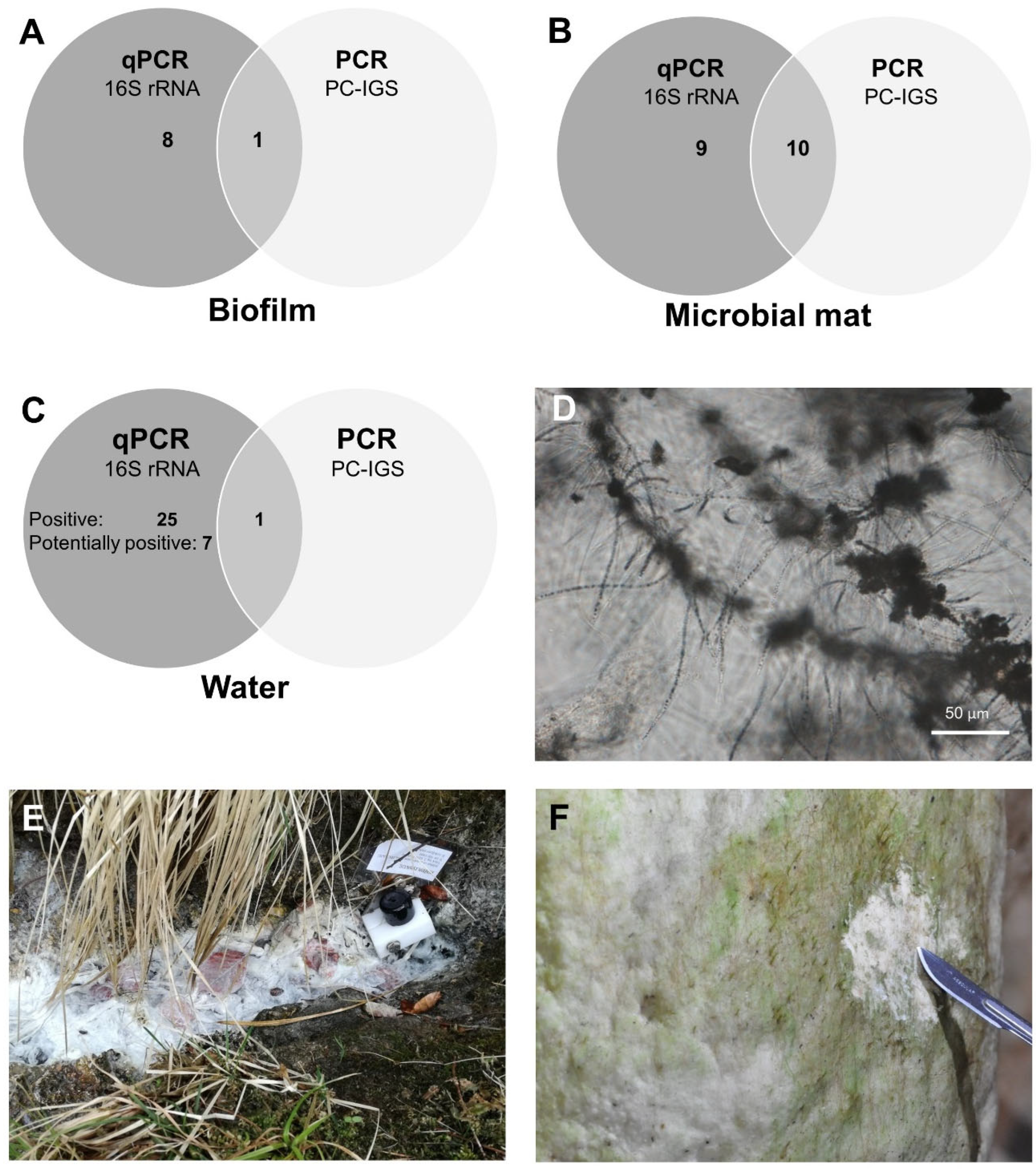
2.1. Molecular Detection of Cyanobacteria: Findings and Constraints

Molecular analysis indicated the presence of autotrophs in all habitat types studied (Figure 1A–C, Table 1). Molecular signals for cyanobacteria-specific 16S rRNA gene were detected in waters with a pH of 2.5 to 12.4, a temperature range of 10 to 83 °C, in sulphidic water (~8.5 mg/L H2S in Žveplenica sulphidic spring [33]), cave ice [34], the sea, terrestrial saline spring (2 mg/L NaCl, unpublished data), cave seepage water, and discharge from borehole wells (Table 1). The screened microbial mats were collected in environments exposed to light. Their macroscopic greenish appearance indicated the presence of phototrophs (Figure 1F), which was confirmed by the molecular tests. In the biofilm samples, the presence of phototrophs was not so obvious, as none of the samples showed a colour typical of photosynthetic pigments (Figure 1E). Nevertheless, the initial qPCR screening with a cyanobacteria-specific 16S rRNA assay indicated the presence of autotrophs in 56 of the 61 samples (92%). The remaining five samples (8%), all originating from water, showed low signal (only one or two out of three replicates tested were positive) and were considered potentially positive (Figure 1C, Table 1). Fifty samples produced results within the quantification range, and the calculated cell concentrations of autotrophs ranged from 15 to 6.6 × 10⁵ cells per μL DNA, while the remaining 11 samples (two biofilm and nine water samples) were positive but below quantification limit (Supplementary Table S1).
To confirm the presence of cyanobacteria in the samples, phycocyanin intergenic spacer (PC-IGS) was amplified by PCR (Figure 2). Amplification products were observed in 34 of 60 samples (57%), but the results were inconclusive in many cases due to the presence of products of different lengths on the gel (Figure 2B), so amplification was repeated to confirm the results. The presence of multiple bands on the gel might be a consequence of non-specific amplification of other similar sequences of the bacterial genomes. In addition, a significant variation in fragment length is expected in this target region [35], and mixed populations of cyanobacteria are expected in environmental samples. However, twelve samples (20%) produced a distinct product of 500–800 bp in both repetitions and were therefore considered positive for PC-IGS with high confidence (Table 1), and sequencing of those amplicons was attempted. It should be noted that the remaining 22 samples showed a weak amplification product in the first repetition of the experiment, which was not reproducible in the second repetition. This could be explained by either a very low concentration of the target gene (stochastic effect—target region is randomly amplified or not) [36] or degradation of DNA due to multiple freeze/thaw cycles, and these samples were not considered positive.
The observed discrepancies between the results of the PC-IGS and 16S rRNA assays (samples positive for 16S rRNA gene only, Table 1) could be a consequence of cross-reactivity of the 16S rRNA qPCR assay with chloroplasts of plants or algae [37], resulting in the detection of eukaryotic algae in the samples. In addition, the high sensitivity of the qPCR and the design of qPCR assays to amplify short DNA fragments improve the probability of detecting target sequences compared to PCR. The PC-IGS PCR assay targets much longer fragments (over 500 bp) than the 16S rDNA qPCR assay (about 70 bp), meaning that qPCR could also amplify partially degraded DNA, and this could negatively affect PCR amplification [38].
The PC-IGS region has previously been used as a phylogenetic marker for cyanobacteria (e.g., [35,39,40]), as it is specific only to this taxa group, and carries sufficient genetic variation to distinguish between different taxa [41]. In previous studies, the PC-IGS-based phylogeny was mostly consistent with the 16S rRNA-based phylogeny [42,43,44], but showed better coverage and specificity compared to the 16S rRNA gene region [39], as well as better resolution between closely related strains [41,44,45]. In this study, the presence of the PC-IGS genetic region and thus the presence of cyanobacteria was confirmed by sequencing in seven of the twelve samples; five samples of microbial mat from caves (M-10, M-14, M-15, M-17, M-19 from Postojnska jama and Škocjanske jame) in Slovenia, one biofilm sample (B-01) from an alkaline spring in Serbia, and one water sample (W-01) from a saline spring in Croatia (Table 1). In four of these seven samples (B-01, M-10, M-14, M-15), taxa could not be reliably determined due to low read quality, indicating a mixed cyanobacterial community. The latter observation was already made during PCR amplification of this gene, where several PCR products were amplified (Figure 2B). Since Sanger sequencing approach is designed for single template reactions and not for mixed communities, amplicon cloning and sequencing of a number of clones would be required to determine the taxa from those mixed communities. Furthermore, there are significant gaps in the taxonomic completeness of reference databases [46], which is why some taxa may remain unidentified. While Salmaso et al. [46] highlighted the importance of this gap for the widely studied 16S rRNA gene, it is likely even more pronounced for PC-IGS with more than 25 times fewer records within the cyanobacterial phylum (NCBI Nucleotide, accessed 17 July 2023). Therefore, further studies on the phylogenetic composition of these samples, employing cloning of PCR products and sequencing of different phylogenetic markers, would be needed to clearly determine the composition of the cyanobacterial community.
In three of seven samples, a single cyanobacterial taxon was determined with PC-IGS sequencing. A taxon phylogenetically close to Cyanothece sp. was identified in an aerophytic microbial mat from the Škocjanske jame cave entrance (M-17; query cover 91–94%, identity 75–77%), and Calothrix sp. was identified in the community of a stromatolitic stalagmite from the same cave (M-19, query cover 100%, identity 91%). The presence of Cyanothece was previously confirmed by microscopic examination of field material from several sites in this cave [47], as well as Calothrix and other cyanobacterial taxa, e.g., Aphanocapsa, Aphanothece, Chroococcus, Gloeocapsa, Homoeohtrix, Leptolyngbya, Lyngbya, Nostoc, Oscillatoria, Phormidium, Planktolyngbya, Pseudoanabaena, Schizohtrihy, Scytonema, Syhnecohcystis, and Trichodesmium [48]. The presence of most of these taxa was also confirmed microscopically in other caves [49,50,51]. In a terrestrial saline spring, Synechocystis sp. was identified (W-01; query cover 100%, identity 91–93%) (Table 1). Some Synechocystis strains are known to be halotolerant, growing in saline or sea waters, or saline (brackish) swamps [52], and the marine strain Synechocystis sp. PCC 7338 was found to have several genes related to adaptation to high salinity and high osmotic pressure [53]. Other studies on saline springs have found Calothrix pulvinata, Phormidium tergestinum, Tapinothrix violacea, and Rivularia sp. aff. bullata to be the characteristic taxa in such environments [16], but we did not find these taxa in the studied terrestrial saline spring in Croatia.
Nevertheless, this study confirmed the presence of cyanobacteria in some extreme environments, e.g., in biofilms from alkaline and saline springs, for which there were no previous reports. However, additional and systematic sampling in different locations and sample types is needed to explore further the presence of cyanobacteria in such rare and diverse habitats.

2.2. Cylindrospermopsins: Potential Presence in Sulphidic Springs

The potential for cyanotoxin production (microcystin, saxitoxin, and cylindrospermopsin synthesis genes) was studied with different qPCR assays applied to all samples positive with either 16S rRNA gene, PC-IGS, or both. Microcystin and saxitoxin producers were not clearly detected in any sample (a weak qPCR signal for Planktothrix-specific mcyE gene was observed in the sample W-18 (Table 1), but this was not investigated further). However, the presence of cylindrospermopsin producers was indicated in four (7%) of the 61 samples (B-07, B-08, M-14, M-15; Table 1). The calculated cell concentrations of potential cylindrospermopsin producers were between 67 and 801 cells per μL DNA (Supplementary Table S2), but the qPCR results were inconclusive. To confirm the qPCR results, the cyrJ region was amplified by end-point PCR targeting a longer gene fragment, and a product of the expected length was detected and sequenced in two biofilm samples from Žveplenica (B-08) and Riharjev studenec (B-07) sulphidic springs. In Riharjev studenec, the 563 bp long fragment (100% overlap between F and R reads) showed 95% similarity to the sulphur-oxidizing bacteria Thiothrix fructosivorans ATCC 49748. The overlapping region corresponded to gene QTX09933.1 (NCBI Nucleotide) in T. fructosivorans, which has been annotated as a response regulator by automated computational analysis.
In Žveplenica, the taxa could not be reliably determined based on cyrJ sequence, but the results indicate the presence of organisms with genetic potential for cylindrospermopsin production. More specifically, the forward cyrJ read was full of overlapping bases, suggesting a mixed community, but the reverse cyrJ read was of good quality and matched cyanobacterial cyrJ genes in the NCBI GenBank database (Oscillatoria strains AWQC-PHO021 and PCC 6506, 95% identity). This may indicate that the forward primer coincidentally aligned with a DNA fragment from another organism, possibly Thiothrix. A previous molecular study on the biodiversity of Žveplenica sulphidic spring has shown that Thiothrix dominates the biofilm (Mulec and Summers Engel, 2019). To investigate this further, a microscopic analysis of the Žveplenica biofilm was performed. The microscopic examination revealed the presence of filaments belonging to the sulphur-oxidizing bacteria Thiothrix (Figure 1D); the only cyanobacterial taxon identified in the sample was Tolypothrix, but the identification was not straightforward, as the filaments were not clearly visible due to the abundant Thiothrix filaments. It is worth mentioning that some publications describe heterocysts in Tolypothrix [54], which were not observed morphologically in the Žveplenica sample.
Nevertheless, sulphidic springs are often colonized by thick aggregates of sulphur-oxidizing Thiothrix [55]. Microscopic observations of Thiothrix in Žveplenica and the sequencing results suggest that the positive signal of the cyrJ target region (forward read) could also originate from Thiothrix. To test whether this was due to non-specific primer alignment or homology, we blasted the QTX09933.1 gene of Thiothrix fructosivorans strain ATCC 49748 against the cyanobacteria/melaionabacteria group, and a cyrJ reference gene from Cylindrospermopsis raciborskii AWT205 (ABX60159.1) against Thiothrix/Francisella group in the NCBI Protein database (blastp), but no significant hits were found. While parts (13 bp) of primer sequences cynsulF and cylnamR (see 4.2. Molecular Analyses) matched 100% with various Thiothrix strains, alignment of the reference cyrJ gene from Cylindrospermopsis raciborskii AWT205 (EU140798.1) with the Thiothrix group from the NCBI Nucleotide database revealed only short matching fragments, so non-specific primer alignment and amplification of different genome fragments (due to low annealing temperature of the primers) is the more likely explanation. This explanation may apply not only to Žveplenica (B-08) but also to Riharjev studenec (B-07). The Žveplenica sulphidic spring has already been discovered as a biodiversity hotspot and a unique habitat with a rich and diverse microbial biofilm and copepods as the most abundant invertebrate species [33]. In this spring, the presence of cyanobacteria, e.g., Oscillatoria, has already been detected by molecular analyses [56], but their characteristics and role have not been explored. Cyanobacteria in microbial mats of sulphidic springs can simultaneously perform oxygenic and anoxygenic photosynthesis [57]. While the evolutionary history of metabolically versatile cyanobacteria remains unknown, data suggest the possibility of coevolution of sulphate reduction and anoxygenic cyanobacterial photosynthesis in microbial mat systems where the local sulphur cycle is driven by a dense biofilm population [58]. The existence of cyanotoxin-producing strains in such a complex environment is therefore highly possible, but due to the lack of biological material required for toxin analyses in the majority of samples, the presence of toxins was not assessed.
Despite the potential presence of cylindrospermopsin in the microbial mat from the cave entrance in Škocjanske jame (M-14, M-15), its presence could not be confirmed by downstream sequencing analysis (Table 1). The presence of cyanotoxins in caves should be further investigated, as cave microbial mat, i.e., lampenflora, has already demonstrated the presence of microcystins [28].
The cyrJ gene encodes a sulfotransferase, a tailoring reaction enzyme involved in the biosynthesis of cylindrospermopsin, which catalyses the sulphation of the C-12 atom [59]. This gene was proposed as a good candidate for a toxin assay because it is more unique than nonribosomal peptide synthetase (NRPS) and polyketide synthase (PKS) genes (such as some other genes in the cylindrospermopsin biosynthesis gene cluster, i.e., cyrB, cyrC, cyrD, cyrE, and cyrF) and is expected to have less cross-reactivity with other gene clusters containing these genes, commonly found in cyanobacteria (Mihali et al., 2008). The assay we used has already been successfully used for screening and sequencing of the cyrJ gene in isolated strains [60] and some environmental samples [61], but our results suggest that there might be some cross-reactivity with sulphur-oxidizing bacteria. However, this is not necessarily a consequence of low primer specificity but could also be due to a low annealing temperature used for PCR amplification.
This study could be expanded by a chemical analysis of cyanotoxins to confirm their presence. Metagenomic analysis by next-generation sequencing is another option for in-depth characterization of the community structure of cyanobacteria and evaluation of the cyanotoxin production potential, for example microcystin [62], saxitoxin [63], and cylindrospermopsin [64]. Our findings demonstrate the importance of selecting (q)PCR assays that are optimised for the analysis of environmental samples. Although the results of the study did not clearly confirm the presence of cyanotoxins in the studied habitats, this is an important consideration for public health in natural waters that may be used for drinking, recreational, or industrial purposes, such as public hot springs or tourist caves.

3. Conclusions

We performed molecular screening for the presence of cyanobacteria and cyanotoxin genes in samples from diverse habitats, covering a wide range of environmental conditions. Cyanobacteria were detected in various sample types: biofilms, microbial mats, and water. Molecular assays did not confirm the presence of genes for microcystin and saxitoxin synthesis in any of the samples. However, the analysis indicated the potential presence of cylindrospermopsin synthesis genes in a biofilm from a sulphidic spring. Further analysis is required to confirm the presence of cylindrospermopsins, as the cross-reactivity of the cyrJ assay with genomic DNA from the sulphur-oxidizing bacteria Thiothrix found in the sample makes the results inconclusive. Chemical analysis of cyanotoxins or metagenomic analysis could be performed to confirm the presence of cylindrospermopsins or to identify the organism carrying the target gene cyrJ in our samples. Several challenges were identified that need to be addressed in future research on cyanobacteria and their toxicity, in particular the lack of reference genes in databases and the specificity of molecular assays. Particular attention should be paid to the selection of molecular techniques and assays used to study mixed environmental communities, as cross-reactivity of molecular assays can bias results or on the other hand reveal new genomic traits in a complex sample. Ideally, a combination of different methods (e.g., molecular, chemical and morphological) targeting different properties of cyanobacteria and their metabolic characteristics should be used to gain a thorough and in-depth insight into environmental samples. Nevertheless, this study contributes to the limited data on the presence of cyanobacteria and cyanotoxin genes in underexplored habitats.

4. Material and Methods

4.1. Study Sites, Sampling and DNA Extraction

The samples were collected during several campaigns from randomly selected and accessible sites in different geographical locations from Croatia, Georgia, Italy, Serbia, and Slovenia in the period from 2011 to 2020 in the frame of occasional or regular monitoring (Table 1). Three different types of samples were collected: biofilm with nine samples (labelled B-01 to B-09), microbial mat with 19 samples (M-01 to M-19), and water with 33 samples (W-01 to W-33). We collected water and microbial mat samples from common, rare and extreme habitats that have not yet been analysed for cyanobacteria and cyanotoxins using a molecular approach and represent a temporal snapshot of the cyanobacterial community. Biofilm was considered to be a film of microorganisms embedded in extracellular polymers adhered on submerged surfaces or in aquatic environments. A microbial mat was considered to be a complex, multilayered structure of microorganisms collected from aerial and subaerial habitats.
Several samples (21%) originated from common habitats, i.e., aerial (M-01, M-02, M-03, M-04 and M-05) and aquatic habitats (W-03, W-05, W-08, W-09, W-11, W-17, W-18, W-22). Furthermore, 41% of the samples were collected from rare habitats, i.e., subaerial microbial mat from caves (M-06, M-07, M-08, M-09, M-10, M-11, M-12, M-13, M-14, M-15, M-16, M-17, M-18, M-19) and from waters in caves (W-07, W-23, W-24, W-25, W-26, W-27, W-28, W-29, W-30, W-31, W-33). The remaining 38% of the samples were from extreme habitats, i.e., alkaline environment (pH > 12; B-01 and W-15), saline spring (2 mg NaCl/L; W-01), low pH iron spring (pH ≤ 5.5; W-13 and W-14), sulphidic environment (B-02, B-03, B-04, B-05, B-06, B-07, B-08, B-09 and W-32), thermal spring (T ≥ 37 °C; W-02, W-04, W-06, W-10, W-12 and W-16), and samples from cave ice (W19, W20 and W21).
Initially, 11 additional water samples were included, but they were negative for both the 16S rRNA gene and phycocyanin intergenic spacer (PC-IGS) (q)PCR assays, and since the quality of the DNA could not be confirmed, they were excluded from further analyses. Biofilms were collected from alkaline and sulphidic springs and from a cave. Sulphidic springs emitted a characteristic odour of hydrogen sulphide. Multi-layered microbial mats in aerophytic habitats were sampled from buildings (façade, roof) and cave surfaces. In caves, this included lampenflora, i.e., the microbial mat around lamps from show caves [65], the microbial mat that developed in the cave entrance illuminated by sunlight, and a stromatolitic stalagmite. Stromatolitic stalagmites are calcareous deposits in cave entrances that are oriented towards the incoming sunlight due to the biolithogenic activity of cyanobacteria [66]. The water samples originated from different types of springs (acidic, alkaline, iron, saline, sulphidic, and thermal), borehole wells, cave seeping water, rivers, the sea, and melted ice from a cave. pH and temperature of the sampled waters were measured with a portable meter WTW Multiline 3420 (Xylem Analytics, Weilheim, Germany).
Samples were collected aseptically and transferred to a laboratory where the total community DNA was extracted. In the field, surfaces with biofilms and microbial mats were scraped with a sterile scalpel or knife and collected in sterile tubes. Water samples were collected in plastic bottles and filtered through 0.22 μm pore size filters (Merck Millipore, Burlington, MA, USA). Generally, 500 to 1000 mL of water volume was sufficient to obtain DNA, but some extractions required larger volumes, up to 10 L. Depending on the sample type and following the manufacturer’s instructions, three different kits were used for DNA isolation: PowerBiofilm DNA Isolation kit (MO BIO Laboratories, Carlsbad, CA, USA) for biofilm samples, PowerWater DNA Isolation kit (MO BIO Laboratories) for water samples, and NucleoSpin Soil kit (Macherey-Nagel, Düren, Germany) for microbial mats. DNA purity and concentration were determined spectrophotometrically using standard absorbance at 260 nm and 280 nm. After isolation DNA was stored at −20 °C until molecular analyses.

4.2. Molecular Analyses

The screening for cyanobacteria and cyanotoxins comprised several steps with different PCR and qPCR assays (Table 2). First, qPCR was performed with a cyanobacteria-specific 16S rRNA assay. Second, samples were screened with an end-point PCR assay targeting PC-IGS to confirm the results of the first step. Samples were further tested with five qPCR assays targeting genes involved in cyanotoxin synthesis: cyrJ (cylindrospermopsins), sxtA (saxitoxins), and mcyE (microcystins; Dolichospermum-, Microcystis-, and Planktothrix-specific). Finally, the PC-IGS and cyrJ regions were re-amplified by PCR in positive samples and sequenced.

4.2.1. PCR Screening for PC-IGS

PCR amplification for PC-IGS (Table 2) was performed on the Mastercycler Nexus Gradient thermal cycler (Eppendorf, Hamburg, Germany). Reaction mixtures with a final volume of 10 µL were prepared in 0.2 mL 8-strip PCR tubes (Starlab, Hamburg, Germany), and contained 1× DreamTaq PCR buffer (Thermo Fisher Scientific, Waltham, MA, USA), 1 mM MgCl2, 0.3 µM each dNTP, 0.3 µM each forward and reverse primer, 0.25 U DreamTaq polymerase (Thermo Fisher Scientific), and 1 µL undiluted DNA template. PCR amplification included an initial denaturation of 3 min at 94 °C, followed by 35 cycles of 30 s at 94 °C, 30 s at 50 °C, and 1 min at 72 °C, with a final extension of 5 min at 72 °C. Positive controls (DNA from the cyanobacterial culture Planktothrix agardhii NIVA-CYA 126/8) and negative controls (nuclease-free water, Sigma-Aldrich, St. Louis, MO, USA) were included in each experiment. PCR products were visualised on a 0.8% agarose gel containing 0.5 × Tris-Borate-EDTA buffer, stained with Midori Green Advance DNA Stain (NIPPON Genetics EUROPE, Düren, Germany) at 120 V for 60 min. Then, 5 µL of the PCR product was loaded onto the gel together with pstI-digested λ DNA to determine the product size. In addition to the visual analysis of the gels, GelAnalyzer 19.1 [72] was used to determine band intensity and size.

4.2.2. qPCR Reactions for Cyanobacteria and Toxin-Related Genes

The qPCR reactions were performed on the Applied Biosystems 7900HT qPCR cycler (Thermo Fisher Scientific) using SYBR Green I chemistry (Thermo Fisher Scientific). The reaction mixtures and amplification conditions were as described in Zupančič et al. (2021). Briefly, reactions were performed in triplicates with a final volume of 10 μL (5 μL of SYBR Green PCR Master Mix (Thermo Fisher Scientific), 0.3 μM (Microcystis-specific mcyE assay) or 0.9 μM (all other assays) of each primer, and 2 μL of 10-fold diluted DNA template. Primer sequences and references are listed in Table 2. Reactions were performed in clear 384-well PCR plates (Thermo Fisher Scientific) with MicroAmp™ optical adhesive film (Thermo Fisher Scientific). PCR amplification conditions included 2 min at 50 °C, an initial denaturation of 10 min at 95 °C, followed by 45 cycles of 15 s at 95 °C and 1 min at 60 °C, and a dissociation stage (15 s at 95 °C, 15 s at 60 °C, gradually increased to 95 °C). Positive controls (specific synthetic DNA fragments; [37]) and negative controls (nuclease-free water, Sigma-Aldrich) were included in each experiment.
Results were analysed with SDS software (version 2.4.1, Thermo Fisher Scientific) and data analyses were performed as described in Zupančič et al. (2021). Samples were considered positive if: (i) all three technical replicates were positive and (ii) the melting temperature (Tm) of the amplicon matched the Tm (±0.9 °C for 16S rRNA, ±0.5 °C for all other assays) of cyanobacterial cultures described in [37]. Samples with one or two positive replicates were considered potentially positive. Quantification of target cells (autotrophs based on 16S rRNA gene, and potential cylindrospermopsin producers based on cyrJ gene) was performed as described in [37]. For all positive and potentially positive samples, average Cq values were calculated from Cq values of all positive replicates. Relative quantification was performed using calibration curves approach. The calibration curves were generated from eight subsequent dilutions of DNA from cyanobacterial cultures as described in [37].

4.2.3. Sequencing of PC-IGS and cyrJ

Amplification of PC-IGS and cyrJ genes (Table 2) for sequencing was performed on GeneAmp PCR system 9700 (Thermo Fisher Scientific). Reaction mixtures with a final volume of 30 µL contained 1x DreamTaq PCR buffer (Thermo Fisher Scientific), 1 mM MgCl2, 0.3 µM of each dNTP, 0.3 µM of each forward and reverse primer, 0.75 U Taq polymerase, and 3 µL undiluted DNA template. PCR amplification conditions included an initial denaturation of 3 min at 94 °C, followed by 35 cycles of 30 s at 94 °C, 30 s at 50 °C, and 1 min at 72 °C, with a final extension of 5 min at 72 °C. The PCR products were run on a 1.2% agarose gel with modified Tris-acetate-EDTA buffer (Montage DNA Gel Extraction Kit, Merck Millipore) stained with ethidium bromide at 100 V for 100 min. 25 µL of the product was loaded onto the gel together with GeneRuler 100 bp Plus DNA Ladder (Thermo Fisher Scientific) to determine the product size. Visible bands were extracted from the gel under UV light and purified using the Montage DNA Gel Extraction Kit (Merck Millipore). The products were sequenced using Sanger technology (Eurofins Genomics, Ebersberg, Germany). DNA sequences were analysed in BioEdit (v. 7.2.5), where forward and reverse reads were aligned (optimal global alignment), poor quality regions were removed, and trimmed sequences were aligned with BLAST [73] against the NCBI Nucleotide database [74]. The sequences were deposited in the NCBI GenBank database under the accession numbers PP061425–PP061428. DNA extracted from Aphanizomenon ovalisporum ILC-164 was used as a positive control for both PCR amplifications and sequenced together with the samples, and high quality reads were obtained from both target regions. For PC-IGS, the 573 bp long read showed 100% identity with PC-IGS genes from other A. ovalisporum sequences deposited in NCBI Nucleotide (strains UAM287, UAM289, APH033B, KA1.1, UAM291, and UAM290, as well as Anabaena bergii ANA366B). For cyrJ, the 488 bp long read showed 100% identity with cyrJ genes from strains ILC-164 and LK_11a.

4.3. Microscopy of Sulphidic Biofilm

Although the focus of this work was on molecular methods used for screening, microscopy was employed as an additional tool to observe morphological characteristics of one sulphidic biofilm sample. The biofilm from Žveplenica sulphidic spring was sampled after molecular analysis that indicated the presence of potential cylindrospermopsin producers in the sample. Microscopic observation of the fresh material was performed under phase contrast with 400×, 600× and 1000× magnification (Nikon Eclipse TE300, Tokyo, Japan). The autotrophic community was determined according to the taxonomic key in [75,76,77].

Supplementary Materials

The following supporting information can be downloaded at: https://www.mdpi.com/article/10.3390/toxins16080333/s1, Table S1: qPCR results and the corresponding calculated target cell concentrations obtained with the cyanobacteria-specific 16S rRNA assay targeting autotrophs; Table S2: qPCR results and the corresponding calculated target cell concentrations obtained with the cyrJ assay targeting potential cylindrospermopsin producers.

Author Contributions

Conceptualization, M.J, T.E., P.K., A.O.-M. and J.M.; formal analysis, M.J., T.E. and J.M.; investigation, M.J., T.E., S.S., A.O.-M. and J.M.; data curation, J.M.; writing—original draft preparation, M.J., T.E., P.K., A.O.-M. and J.M.; writing—review and editing, M.J., T.E., P.K. and J.M.; visualization, M.J., T.E. and J.M.; supervision, J.M.; funding acquisition, T.E., P.K. and J.M. All authors have read and agreed to the published version of the manuscript.

Funding

This work was supported by the Slovenian Research and Innovation Agency [research core funding no. P6-0119, P1-0245, P4-0407, P2-0412, project funding J2-4428, and Young researcher grant number 10040146 (MZ)].

Institutional Review Board Statement

Not applicable.

Informed Consent Statement

Not applicable.

Data Availability Statement

Data are available from the corresponding authors upon request. The nucleotide sequences produced in this study were deposited in the NCBI GenBank database under the accession numbers PP061425–PP061428.

Acknowledgments

The authors thank George Melikadze, Nino Kapanadze, Dejan Milenić, Ana Vranješ, Metka Petrič, Andrej Mihevc, Mojca Zega, Spomenka Vlahović and Samo Šturm for assistance with field work, to Rainer Kurmayer for enabling a part of analyses in their laboratory, and to three anonymous reviewers for helpful suggestions for improving this manuscript.

Conflicts of Interest

The authors have no conflicts of interest to disclose.

References

  1. Roncero-Ramos, B.; Muñoz-Martín, M.Á.; Chamizo, S.; Fernández-Valbuena, L.; Mendoza, D.; Perona, E.; Cantón, Y.; Mateo, P. Polyphasic evaluation of key cyanobacteria in biocrusts from the most arid region in Europe. PeerJ 2019, 7, e6169. [Google Scholar] [CrossRef] [PubMed]
  2. Oarga, A. Life in extreme environments. Rev. Biol. Ciências Terra 2009, 9, 1–10. [Google Scholar]
  3. Shu, W.S.; Huang, L.N. Microbial diversity in extreme environments. Nat. Rev. Microbiol. 2022, 20, 219–235. [Google Scholar] [CrossRef]
  4. Carré, L.; Zaccai, G.; Delfosse, X.; Girard, E.; Franzetti, B. Relevance of earth-bound extremophiles in the search for extraterrestrial life. Astrobiology 2022, 22, 322–367. [Google Scholar]
  5. Anwar, U.B.; Zwar, I.P.; de Souza, A.O. Biomolecules produced by extremophiles microorganisms and recent discoveries. In New and Future Developments in Microbial Biotechnology and Bioengineering; Gupta, V.K., Ed.; Elsevier: Amsterdam, The Netherlands, 2020; pp. 247–270. [Google Scholar]
  6. Xu, H.F.; Dai, G.Z.; Qiu, B.S. Weak red light plays an important role in awakening the photosynthetic machinery following desiccation in the subaerial cyanobacterium Nostoc flagelliforme. Environ. Microbiol. 2019, 21, 2261–2272. [Google Scholar] [CrossRef]
  7. Bustos-Díaz, E.D.; Barona-Gómez, F.; Cibrián-Jaramillo, A. Cyanobacteria in nitrogen-fixing symbioses. In Cyanobacteria; Mishra, A.K., Tiwari, D.N., Rai, A.N., Eds.; Academic Press: Cambridge, MA, USA, 2019; pp. 29–42. [Google Scholar]
  8. Nayaka, S.; Toppo, K.; Verma, S. Adaptation in algae to environmental stress and ecological conditions. In Plant Adaptation Strategies in Changing Environment; Shukla, V., Kumar, S., Kumar, N., Eds.; Springer: Singapore, 2017; pp. 103–115. [Google Scholar]
  9. Billi, D. Challenging the Survival Thresholds of a Desert Cyanobacterium under Laboratory Simulated and Space Conditions. In Extremophiles as Astrobiological Models; Seckbach, J., Stan-Lotter, H., Eds.; Wiley: Hoboken, NJ, USA, 2020; pp. 183–195. [Google Scholar]
  10. Xu, H.F.; Raanan, H.; Dai, G.Z.; Oren, N.; Berkowicz, S.; Murik, O.; Kaplan, A.; Qiu, B.S. Reading and surviving the harsh conditions in desert biological soil crust: The cyanobacterial viewpoint. FEMS Microbiol. Rev. 2021, 45, fuab036. [Google Scholar] [CrossRef] [PubMed]
  11. Nelson, C.; Giraldo-Silva, A.; Garcia-Pichel, F. A symbiotic nutrient exchange within the cyanosphere microbiome of the biocrust cyanobacterium, Microcoleus vaginatus. ISME J. 2020, 15, 282–292. [Google Scholar] [CrossRef]
  12. Dabravolski, S.A.; Isayenkov, S.V. Metabolites facilitating adaptation of desert cyanobacteria to extremely arid environments. Plants 2022, 11, 3225. [Google Scholar] [CrossRef]
  13. Steinberg, C.E.W.; Schäfer, H.; Beisker, W. Do acid-tolerant cyanobacteria exist? Acta Hydrochim. Et Hydrobiol. 1998, 26, 13–19. [Google Scholar] [CrossRef]
  14. Cirés, S.; Casero, M.C.; Quesada, A. Toxicity at the Edge of Life: A Review on Cyanobacterial Toxins from Extreme Environments. Mar. Drugs 2017, 15, 233. [Google Scholar] [CrossRef]
  15. Gärtner, G.; Stoyneva-Gärtner, M.; Uzunov, B. Algal Toxic Compounds and Their Aeroterrestrial, Airborne and other Extremophilic Producers with Attention to Soil and Plant Contamination: A Review. Toxins 2021, 13, 322. [Google Scholar] [CrossRef]
  16. Cantonati, M.; Komárek, J.; Montejano, G. Cyanobacteria in ambient springs. Biodivers. Conserv. 2015, 24, 865–888. [Google Scholar] [CrossRef]
  17. Li, Q.; Hu, C.; Yang, H. Responses of cyanobacterial crusts and microbial communities to extreme environments of the stratosphere. Microorganisms 2022, 10, 1252. [Google Scholar] [CrossRef] [PubMed]
  18. Piano, E.; Bona, F.; Falasco, E.; La Morgia, V.; Badino, G.; Isaia, M. Environmental drivers of phototrophic biofilms in an Alpine show cave (SW-Italian Alps). Sci. Total Environ. 2015, 536, 1007–1018. [Google Scholar] [CrossRef] [PubMed]
  19. Ai, J.; Guo, J.; Li, Y.; Zhong, X.; Lv, Y.; Li, J.; Yang, A. The diversity of microbes and prediction of their functions in karst caves under the influence of human tourism activities—A case study of Zhijin Cave in Southwest China. Environ. Sci. Pollut. Res. 2022, 29, 25858–25868. [Google Scholar] [CrossRef] [PubMed]
  20. Buratti, F.M.; Manganelli, M.; Vichi, S.; Stefanelli, M.; Scardala, S.; Testai, E.; Funari, E. Cyanotoxins: Producing organisms, occurrence, toxicity, mechanism of action and human health toxicological risk evaluation. Arch. Toxicol. 2017, 91, 1049–1130. [Google Scholar] [CrossRef]
  21. Scarlett, K.R.; Kim, S.; Lovin, L.M.; Chatterjee, S.; Scott, J.T.; Brooks, B.W. Global scanning of cylindrospermopsin: Critical review and analysis of aquatic occurrence, bioaccumulation, toxicity and health hazards. Sci. Total Environ. 2020, 738, 139807. [Google Scholar] [CrossRef]
  22. Chorus, I.; Welker, M. (Eds.) Toxic Cyanobacteria in Water—A Guide to Their Public Health Consequences, Monitoring and Management, 2nd ed.; World Health Organization: Geneva, Switzerland, 2021. [Google Scholar]
  23. Mehinto, A.C.; Smith, J.; Wenger, E.; Stanton, B.; Linville, R.; Brooks, B.W.; Sutula, M.A.; Howard, M.D. Synthesis of ecotoxicological studies on cyanotoxins in freshwater habitats—Evaluating the basis for developing thresholds protective of aquatic life in the United States. Sci. Total Environ. 2021, 795, 148864. [Google Scholar] [CrossRef]
  24. Svirčev, Z.; Lalić, D.; Bojadžija Savić, G.; Tokodi, N.; Drobac Backović, D.; Chen, L.; Meriluoto, J.; Codd, G.A. Global geographical and historical overview of cyanotoxin distribution and cyanobacterial poisonings. Arch. Toxicol. 2019, 93, 2429–2481. [Google Scholar] [CrossRef]
  25. Du, X.; Liu, H.; Yuan, L.; Wang, Y.; Ma, Y.; Wang, R.; Chen, X.; Losiewicz, M.D.; Guo, H.; Zhang, H. The diversity of cyanobacterial toxins on structural characterization, distribution and identification: A systematic review. Toxins 2019, 11, 530. [Google Scholar] [CrossRef]
  26. Chen, L.; Giesy, J.P.; Adamovsky, O.; Svirčev, Z.; Meriluoto, J.; Codd, G.A.; Mijovic, B.; Shi, T.; Tuo, X.; Li, S.-C.; et al. Challenges of using blooms of Microcystis spp. in animal feeds: A comprehensive review of nutritional, toxicological and microbial health evaluation. Sci. Total Environ. 2021, 764, 142319. [Google Scholar] [CrossRef] [PubMed]
  27. Yilmaz, S.; Ülger, T.G.; Göktaş, B.; Öztürk, Ş.; Karataş, D.Ö.; Beyzi, E. Cyanotoxin genotoxicity: A review. Toxin Rev. 2021, 41, 699–712. [Google Scholar] [CrossRef]
  28. Nelson, S.; Young, D.; Toomey, R.; Byl, T. Terrestrial cyanobacteria of Mammoth Cave National Park produce microcystin toxin. In Proceedings of the 28th Tennessee Water Resources Symposium, Montgomery Bell State Park, Burns, TN, USA, 10–12 April 2019. [Google Scholar]
  29. Mohamed, Z.A. Toxic cyanobacteria and cyanotoxins in public hot springs in Saudi Arabia. Toxicon 2008, 51, 17–27. [Google Scholar] [CrossRef] [PubMed]
  30. Krienitz, L.; Ballot, A.; Kotut, K.; Wiegand, C.; Pütz, S.; Metcalf, J.S.; Codd, G.A.; Stephan, P. Contribution of hot spring cyanobacteria to the mysterious deaths of Lesser Flamingos at Lake Bogoria, Kenya. FEMS Microbiol. Ecol. 2003, 43, 141–148. [Google Scholar] [CrossRef] [PubMed]
  31. Strunecký, O.; Kopejtka, K.; Goecke, F.; Tomasch, J.; Lukavský, J.; Neori, A.; Kahl, S.; Pieper, D.H.; Pilarski, P.; Kaftan, D.; et al. High diversity of thermophilic cyanobacteria in Rupite hot spring identified by microscopy, cultivation, single-cell PCR and amplicon sequencing. Extremophiles 2019, 23, 35–48. [Google Scholar] [CrossRef] [PubMed]
  32. Kleinteich, J.; Wood, S.A.; Puddick, J.; Schleheck, D.; Küpper, F.C.; Dietrich, D. Potent toxins in Arctic environments—Presence of saxitoxins and an unusual microcystin variant in Arctic freshwater ecosystems. Chem. Biol. Interact. 2013, 206, 423–431. [Google Scholar] [CrossRef] [PubMed]
  33. Mulec, J.; Oarga-Mulec, A.; Schiller, E.; Perşoiu, A.; Holko, L.; Šebela, S. Assessment of the physical environment of epigean invertebrates in a unique habitat: The case of a karst sulfidic spring, Slovenia. Ecohydrology 2014, 8, 1326–1334. [Google Scholar] [CrossRef]
  34. Mulec, J.; Oarga-Mulec, A.; Holko, L.; Pašić, L.; Kopitar, A.N.; Eleršek, T.; Mihevc, A. Microbiota entrapped in recently-formed ice: Paradana Ice Cave, Slovenia. Sci. Rep. 2021, 11, 1993. [Google Scholar] [CrossRef] [PubMed]
  35. Neilan, B.A.; Jacobs, D.; Goodman, A.E. Genetic diversity and phylogeny of toxic cyanobacteria determined by DNA polymorphisms within the phycocyanin locus. Appl. Environ. Microbiol. 1995, 61, 3875–3883. [Google Scholar] [CrossRef]
  36. Taylor, S.C.; Nadeau, K.; Abbasi, M.; Lachance, C.; Nguyen, M.; Fenrich, J. The Ultimate qPCR Experiment: Producing Publication Quality, Reproducible Data the First Time. Trends Biotechnol. 2019, 37, 761–774. [Google Scholar] [CrossRef]
  37. Zupančič, M.; Kogovšek, P.; Šter, T.; Remec Rekar, Š.; Cerasino, L.; Baebler, Š.; Krivograd Klemenčič, A.; Eleršek, T. Potentially Toxic Planktic and Benthic Cyanobacteria in Slovenian Freshwater Bodies: Detection by Quantitative PCR. Toxins 2021, 13, 133. [Google Scholar] [CrossRef] [PubMed]
  38. Hughes-Stamm, S.R.; Ashton, K.J.; van Daal, A. Assessment of DNA degradation and the genotyping success of highly degraded samples. Int. J. Leg. Med. 2011, 125, 341–348. [Google Scholar] [CrossRef] [PubMed]
  39. Jiang, Y.; Xiao, P.; Liu, Y.; Wang, J.; Li, R. Targeted deep sequencing reveals high diversity and variable dominance of bloom-forming cyanobacteria in eutrophic lakes. Harmful Algae 2017, 64, 42–50. [Google Scholar] [CrossRef]
  40. Kim, S.G.; Rhee, S.K.; Ahn, C.Y.; Ko, S.R.; Choi, G.G.; Bae, J.W.; Park, Y.H.; Oh, H.M. Determination of cyanobacterial diversity during algal blooms in Daechung Reservoir, Korea, on the basis of cpcBA intergenic spacer region analysis. Appl. Environ. Microbiol. 2006, 72, 3252–3258. [Google Scholar] [CrossRef] [PubMed]
  41. Teneva, I.; Dzhambazov, B.; Mladenov, R.; Schirmer, K. Molecular and phylogenetic characterization of Phormidium species (cyanoprokaryota) using the cpcB-IGS-cpcA locus. J. Phycol. 2005, 41, 188–194. [Google Scholar] [CrossRef]
  42. Piccin-Santos, V.; Brandão, M.M.; Bittencourt-Oliveira, M.D.C. Phylogenetic study of Geitlerinema and Microcystis (Cyanobacteria) using PC-IGS and 16S-23S ITS as markers: Investigation of horizontal gene transfer. J. Phycol. 2014, 50, 736–743. [Google Scholar] [CrossRef] [PubMed]
  43. Dadheech, P.K.; Ballot, A.; Casper, P.; Kotut, K.; Novelo, E.; Lemma, B.; Pröschold, T.; Krienitz, L. Phylogenetic relationship and divergence among planktonic strains of Arthrospira (Oscillatoriales, Cyanobacteria) of African, Asian and American origin deduced by 16S–23S ITS and phycocyanin operon sequences. Phycologia 2010, 49, 361–372. [Google Scholar] [CrossRef]
  44. Robertson, B.R.; Tezuka, N.; Watanabe, M.M. Phylogenetic analyses of Synechococcus strains (cyanobacteria) using sequences of 16S rDNA and part of the phycocyanin operon reveal multiple evolutionary lines and reflect phycobilin content. Int. J. Syst. Evol. Microbiol. 2001, 51, 861–871. [Google Scholar] [CrossRef] [PubMed]
  45. Wang, Z.; Liu, Y.; Xu, Y.; Xiao, P.; Li, R. The divergence of cpcBA-IGS sequences between Dolichospermum and Aphanizomenon (Cyanobacteria) and the molecular detection of Dolichospermum flos-aquae in Taihu Lake, China. Phycologia 2013, 52, 447–454. [Google Scholar] [CrossRef]
  46. Salmaso, N.; Vasselon, V.; Rimet, F.; Vautier, M.; Elersek, T.; Boscaini, A.; Donati, C.; Moretto, M.; Pindo, M.; Riccioni, G.; et al. DNA sequence and taxonomic gap analyses to quantify the coverage of aquatic cyanobacteria and eukaryotic microalgae in reference databases: Results of a survey in the Alpine region. Sci. Total Environ. 2022, 834, 155175. [Google Scholar] [CrossRef]
  47. Mulec, J.; Kosi, G.; Vrhovšek, D. Characterization of cave aerophytic algal communities and effects of irradiance levels on production of pigments. J. Caves Karst Stud. 2008, 70, 3–12. [Google Scholar]
  48. Mulec, J.; Kosi, G.; Vrhovšek, D. Algae promote growth of stalagmites and stalactites in karst caves (Škocjanske jame, Slovenia). Carbonates Evaporites 2007, 22, 6–9. [Google Scholar] [CrossRef]
  49. Saiz-Jimenez, C. Microbiological and environmental issues in show caves. World J. Microbiol. Biotechnol. 2012, 28, 2453–2464. [Google Scholar] [CrossRef]
  50. Urzì, C.; De Leo, F.; Bruno, L.; Albertano, P. Microbial Diversity in Paleolithic Caves: A Study Case on the Phototrophic Biofilms of the Cave of Bats (Zuheros, Spain). Microb. Ecol. 2010, 60, 116–129. [Google Scholar] [CrossRef] [PubMed]
  51. Popović, S.; Krizmanić, J.; Vidaković, D.; Jakovljević, O.; Trbojević, I.; Predojević, D.; Vidović, M.; Subakov Simić, G. Seasonal Dynamics of Cyanobacteria and Algae in Biofilm from the Entrance of Two Caves. Geomicrobiol. J. 2020, 37, 315–326. [Google Scholar] [CrossRef]
  52. Guiry, M.D.; Guiry, G.M. AlgaeBase. World-Wide Electronic Publication; National University of Ireland: Galway, Ireland, 2023; Available online: https://www.algaebase.org (accessed on 25 July 2023).
  53. Jeong, Y.; Hong, S.J.; Cho, S.H.; Yoon, S.; Lee, H.; Choi, H.K.; Kim, D.M.; Lee, C.G.; Cho, S.; Cho, B.K. Multi-Omic Analyses Reveal Habitat Adaptation of Marine Cyanobacterium Synechocystis sp. PCC 7338. Front. Microbiol. 2021, 12, 667450. [Google Scholar] [CrossRef] [PubMed]
  54. Cantonati, M. Cyanoprokaryotes and algae other than diatoms in springs and streams of the Dolomiti Bellunesi National Park (Northern Italy). Algol. Stud. 2008, 126, 113–136. [Google Scholar] [CrossRef]
  55. Strauss, H.; Chmiel, H.; Christ, A.; Fugmann, A.; Hanselmann, K.; Kappler, A.; Königer, P.; Lutter, A.; Siedenberg, K.; Teichert, B. Multiple sulphur and oxygen isotopes reveal microbial sulphur cycling in spring waters in the Lower Engadin, Switzerland. Isot. Environ. Health Stud. 2015, 52, 75–93. [Google Scholar] [CrossRef]
  56. Mulec, J.; Summers Engel, A. Karst spring microbial diversity differs across an oxygen-sulphide ecocline and reveals potential for novel taxa discovery. Acta Carsologica 2019, 48, 129–143. [Google Scholar] [CrossRef]
  57. Klatt, J.M.; de Beer, D.; Häusler, S.; Polerecky, L. Cyanobacteria in Sulfidic Spring Microbial Mats Can Perform Oxygenic and Anoxygenic Photosynthesis Simultaneously during an Entire Diurnal Period. Front. Microbiol. 2016, 7, 1973. [Google Scholar] [CrossRef]
  58. Hamilton, T.; Klatt, J.; de Beer, D.; Macalady, J.L. Cyanobacterial photosynthesis under sulfidic conditions: Insights from the isolate Leptolyngbya sp. strain hensonii. ISME J. 2018, 12, 568–584. [Google Scholar] [CrossRef] [PubMed]
  59. Mihali, T.K.; Kellmann, R.; Muenchhoff, J.; Barrow, K.D.; Neilan, B.A. Characterization of the gene cluster responsible for cylindrospermopsin biosynthesis. Appl. Environ. Microbiol. 2008, 74, 716–722. [Google Scholar] [CrossRef] [PubMed]
  60. Kokociński, M.; Mankiewicz-Boczek, J.; Jurczak, T.; Spoof, L.; Meriluoto, J.; Rejmonczyk, E.; Hautala, H.; Vehniäinen, M.; Pawełczyk, J.; Soininen, J. Aphanizomenon gracile (Nostocales), a cylindrospermopsin-producing cyanobacterium in Polish lakes. Environ. Sci. Pollut. Res. 2013, 20, 5243–5264. [Google Scholar] [CrossRef] [PubMed]
  61. Mankiewicz-Boczek, J.; Kokociński, M.; Gagała, I.; Pawełczyk, J.; Jurczak, T.; Dziadek, J. Preliminary molecular identification of cylindrospermopsin-producing cyanobacteria in two Polish lakes (Central Europe). FEMS Microbiol. Lett. 2012, 326, 173–179. [Google Scholar] [CrossRef] [PubMed]
  62. Linz, D.M.; Sienkiewicz, N.; Struewing, I.; Stelzer, E.A.; Graham, J.L.; Lu, J. Metagenomic mapping of cyanobacteria and potential cyanotoxin producing taxa in large rivers of the United States. Sci. Rep. 2023, 13, 2806. [Google Scholar] [CrossRef]
  63. Kim, K.H.; Yoon, Y.; Hong, W.Y.; Kim, J.; Cho, Y.C.; Hwang, S.J. Application of metagenome analysis to characterize the molecular diversity and saxitoxin-producing potentials of a cyanobacterial community: A case study in the North Han River, Korea. Appl. Biol. Chem. 2018, 61, 153–161. [Google Scholar] [CrossRef]
  64. Feng, J.; Zhou, L.; Zhao, X.; Chen, J.; Li, Z.; Liu, Y.; Ou, L.; Xie, Z.; Wang, M.; Yin, X.; et al. Evaluation of environmental factors and microbial community structure in an important drinking-water reservoir across seasons. Front. Microbiol. 2023, 14, 1091818. [Google Scholar] [CrossRef]
  65. Mulec, J. Lampenflora, Definition of. In Encyclopedia of Caves; White, W.B., Culver, D.C., Pipan, T., Eds.; Academic/Elsevier Press: Cambridge, MA, USA, 2019; pp. 635–641. [Google Scholar]
  66. Mulec, J. Phototrophs in caves. In Cave Ecology; Moldovan, O.T., Kováč, L., Halse, S., Eds.; Springer: Cham, Switzerland, 2018; pp. 91–106. [Google Scholar]
  67. Al-Tebrineh, J.; Mihali, T.K.; Pomati, F.; Neilan, B.A. Detection of saxitoxin-producing cyanobacteria and Anabaena circinalis in environmental water blooms by quantitative PCR. Appl. Environ. Microbiol. 2010, 76, 7836–7842. [Google Scholar] [CrossRef]
  68. Campo, E.; Lezcano, M.-Á.; Agha, R.; Cirés, S.; Quesada, A.; El-Shehawy, R. First TaqMan Assay to identify and quantify the cylindrospermopsin-producing cyanobacterium Aphanizomenon ovalisporum in water. Adv. Microbiol. 2013, 03, 430–437. [Google Scholar] [CrossRef]
  69. Rantala, A.; Fewer, D.P.; Hisbergues, M.; Rouhiainen, L.; Vaitomaa, J.; Börner, T.; Sivonen, K. Phylogenetic evidence for the early evolution of microcystin synthesis. Proc. Natl. Acad. Sci. USA 2004, 101, 568–573. [Google Scholar] [CrossRef]
  70. Vaitomaa, J.; Rantala, A.; Halinen, K.; Rouhiainen, L.; Tallberg, P.; Mokelke, L.; Sivonen, K. Quantitative Real-Time PCR for Determination of Microcystin Synthetase E Copy Numbers for Microcystis and Anabaena in Lakes. Appl. Environ. Microbiol. 2003, 69, 7289–7297. [Google Scholar] [CrossRef] [PubMed]
  71. Rantala, A.; Rajaniemi-Wacklin, P.; Lyra, C.; Lepistö, L.; Rintala, J.; Mankiewicz-Boczek, J.; Sivonen, K. Detection of microcystin-producing cyanobacteria in Finnish lakes with genus-specific microcystin synthetase gene E (mcyE) PCR and associations with environmental factors. Appl. Environ. Microbiol. 2006, 72, 6101–6110. [Google Scholar] [CrossRef] [PubMed]
  72. Lazar, I., Jr.; Lazar, I., Sr. GelAnalyzer 19.1. Available online: www.gelanalyzer.com (accessed on 1 April 2021).
  73. Altschul, S.F.; Gish, W.; Miller, W.; Myers, E.W.; Lipman, D.J. Basic local alignment search tool. J. Mol. Biol. 1990, 215, 403–410. [Google Scholar] [CrossRef] [PubMed]
  74. Nucleotide; National Library of Medicine (US), National Center for Biotechnology Information: Bethesda, MD, USA, 2004. Available online: https://www.ncbi.nlm.nih.gov/nucleotide/ (accessed on 25 July 2023).
  75. John, D.M.; Whitton, B.A.; Brook, A.J. The Freshwater Algal Flora of the British Isles: An Identification Guide to Freshwater and Terrestrial Algae; Cambridge University Press: Cambridge, UK, 2011. [Google Scholar]
  76. Komárek, J. Cyanoprokaryota. 3. Heterocytous genera. In Süswasserflora von Mitteleuropa/Freshwater Flora of Central Europe; Büdel, B., Gärtner, G., Krienitz, L., Schagerl, M., Eds.; Springer Spektrum: Berlin, Germany, 2013; pp. 1–1130. [Google Scholar]
  77. Komárek, J.; Anagnostidis, K. Modern approach to the classification system of Cyanophytes 4—Nostocales. Arch. Hydrobiol. Suppl. 1989, 82, 247–345. [Google Scholar]
Figure 1. Comparison of molecular tests qPCR (16S rRNA in autotrophs) and PCR (PC-IGS—phycocyanin intergenic spacer) for detection of cyanobacteria for different sample types: biofilm (A), microbial mat (B), water (C). Number of positive samples determined with each method and with both methods is given. (D) a microphotograph of a sample from Žveplenica sulphidic spring, Slovenia, with abundant filaments of sulphur-oxidizing bacteria Thiothrix. (E) white biofilm of Žveplenica sulphidic spring and temperature datalogger at spring’s orifice. (F) lampenflora in Postojnska jama.
Toxins 16 00333 g001
Figure 2. Examples of specific (A) and non-specific (B) PCR amplification products of the phycocyanin intergenic region visualized on 0.8% agarose gel containing 0.5 × Tris-Borate-EDTA buffer, stained with Midori Green Advance DNA Stain (NIPPON Genetics EUROPE, Düren, Germany) at 120 V for 60 min. Samples: 1—W-06, 2—W-11, 3—W-03, 4—W-05, 5—W-02, 6—W-04, 7—W-18, 8—W-19, 9—W-14, 10—W-12, 11—M-16, 12—M-07, 13—M-08, 14—M-09, 15—M-19, 16—M-04, 17—B-08 (see Table 1 for details). Size marker (M): pstI-digested λ DNA; a—1700 bp, b—805 bp, c—514 bp. Expected product size 500–740 bp.
Toxins 16 00333 g002
Table 1. List of samples collected in different locations (country, location and coordinates are given) on different sampling dates. Biofilm, microbial mat and water samples were collected in diverse environments, some of them extreme (high/low temperature and/or pH). All samples were tested with PC-IGS PCR assay, and 16S rRNA gene, cyrJ, mcyE, and sxtA qPCR assays, and results are reported as + (positive) and − (negative). Quantitative results are available in Supplementary Tables S1 and S2. PC-IGS and cyrJ amplicons of selected samples were sequenced, and the most similar genus is indicated.
SampleCountryLocationCoordinatesSampling Date [dd/mm/yyyy]Sample TypeDescriptionT [°C]pHqPCR
(16S rRNA)
PCR
PC-IGS
Seq.
PC-IGS (Genus)
qPCR cyrJSeq. cyrJqPCR stxAqPCR mcyE (Dol.)qPCR mcyE (Mic.)qPCR mcyE (Pla.)
B-01SerbiaBela voda, Zlatibor43°42′13″ N, 19°34′43″ E29/09/2011biofilmalkaline spring15.612.40++(+)NS
B-02SloveniaHajnsko45°12′22″ N, 15°35′26″ E13/04/2016biofilmsulphidic spring13.67.13+NSNS
B-03SloveniaMatijeva jama45°41′21″ N, 14°15′53″ E31/08/2016biofilmbiofilm in a caveNANA+NSNS
B-04SloveniaSmrdljivec45°39′48″ N, 13°59′57″ E24/08/2017biofilmsulphidic spring19.77.28+NSNS
B-05SloveniaSmrdljivec45°39′48″ N, 13°59′57″ E24/08/2017biofilmsulphidic spring19.77.28+NSNS
B-06SloveniaSmrdljivec45°39′48″ N, 13°59′57″ E24/08/2017biofilmsulphidic spring19.77.28+NSNS
B-07SloveniaRiharjev studenec46°18′50″ N, 14°42′07″ E22/03/2016biofilmsulphidic spring10.27.76+NS(+)+
Thio-thrix
B-08 *SloveniaŽveplenica46°03′59″ N, 13°49′38″ E10/04/2016biofilmsulphidic spring10.57.54+NS+(+)
B-09SloveniaŽvepovnik46°21′00″ N, 14°50′43″ E22/03/2016biofilmsulphidic spring10.47.32+NSNS
M-01SloveniaLjubljana46°02′59″ N, 14°29′58″ E08/06/2015microbial matfacade, city centreNANA+NSNS
M-02SloveniaLjubljana46°04′57″ N, 14°30′47″ E08/06/2015microbial matfacade, city outskirtNANA+NSNS
M-03SloveniaLjubljana46°04′57″ N, 14°30′47″ E08/06/2015microbial matfacade, city outskirtNANA+NSNS
M-04SloveniaLjubljana46°04′14″ N, 14°30′51″ E05/06/2015microbial matroof, city outskirtNANA+NSNS
M-05SloveniaLjubljana46°04′14″ N, 14°30′51″ E05/06/2015microbial matroof, city outskirtNANA+NSNS
M-06SloveniaMatijeva jama45°41′21″ N, 14°15′53″ E08/07/2019microbial matmicrobial mat at a cave entranceNANA++NS
M-07SloveniaPostojnska jama45°46′59″ N, 14°12′14″ E24/01/2020microbial matlampenflora in a caveNANA+NSNS
M-08SloveniaPostojnska jama45°46′59″ N, 14°12′14″ E24/01/2020microbial matlampenflora in a caveNANA+NSNS
M-09SloveniaPostojnska jama45°46′59″ N, 14°12′14″ E24/01/2020microbial matlampenflora in a caveNANA+NSNS
M-10SloveniaPostojnska jama45°46′59″ N, 14°12′14″ E24/01/2020microbial matlampenflora in a caveNANA++(+)NS
M-11SloveniaPostojnska jama45°46′59″ N, 14°12′14″ E24/01/2020microbial matlampenflora in a caveNANA++NS
M-12 *SloveniaŠkocjanske jame45°39′57″ N, 13°59′24″ E08/04/2016microbial matmicrobial mat at a cave entranceNANA+NSNS
M-13SloveniaŠkocjanske jame45°39′57″ N, 13°59′24″ E23/01/2020microbial matmicrobial mat at a cave entranceNANA++NS
M-14SloveniaŠkocjanske jame45°39′57″ N, 13°59′24″ E23/01/2020microbial matmicrobial mat at a cave entranceNANA++(+)(+)
M-15SloveniaŠkocjanske jame45°39′57″ N, 13°59′24″ E23/01/2020microbial matmicrobial mat at a cave entranceNANA++(+)(+)
M-16SloveniaŠkocjanske jame45°39′57″ N, 13°59′24″ E23/01/2020microbial matmicrobial mat at a cave entranceNANA++NS
M-17SloveniaŠkocjanske jame45°39′57″ N, 13°59′24″ E23/01/2020microbial matmicrobial mat at a cave entranceNANA+++ Cyano-theceNS
M-18 *SloveniaŠkocjanske jame45°39′57″ N, 13°59′24″ E08/04/2016microbial matmicrobial mat at a cave entranceNANA++NS
M-19 *SloveniaŠkocjanske jame45°39′57″ N, 13°59′24″ E08/04/2016microbial matstromatolitic stalagmite at a cave entranceNANA+++ CalothrixNS
W-01CroatiaSlanci, Slanje46°13′48″ N, 16°32′56″ E24/03/2019watersaline spring #17.67.97+++ Synecho-cystisNS
W-02GeorgiaAnalisopeli41°49′15″ N, 41°47′54″ E13/07/2012waterdischarge from a borehole well36.97.50+NSNS
W-03GeorgiaSighnaghi41°36′23″ N, 45°56′02″ E30/06/2012waterspring12.37.50+NSNS
W-04GeorgiaTbilisi, Lisi41°43′13″ N, 44°45′30″ E02/07/2012waterdischarge from a borehole well63.47.50+NSNS
W-05GeorgiaTbilisi, Ortachala41°40′25″ N, 44°50′30″ E04/07/2012waterdischarge from a borehole well21.58.00+NSNS
W-06GeorgiaVardzia Cave Monastery41°23′30″ N, 43°18′33″ E08/07/2012waterdischarge from a borehole well51.09.30(+)NSNS
W-07GeorgiaVardzia Cave Monastery41°22′52″ N, 43°17′03″ E08/07/2012waterseeping water in a cave10.57.00(+)NSNS
W-08ItalySoča/Isonzo River45°56′17″ N, 13°36′05″ E20/06/2019waterriver16.48.30+NSNS
W-09ItalyVipava/Vipacco River45°53′17″ N, 13°35′27″ E20/06/2019waterriver20.98.30+NSNS
W-10ItalyTržič/Monfalcone45°47′32″ N, 13°33′57″ E20/06/2019waterthermal spring37.47.10+NSNS
W-11ItalyAdriatic sea45°46′51″ N, 13°32′23″ E20/06/2019watersea25.88.20+NSNS
W-12ItalyTržič/Monfalcone45°47′31″ N, 13°33′56″ E20/06/2019waterdischarge from a borehole well39.27.00(+)NSNS
W-13SerbiaCrveno vrelo, Đavolja Varoš42°59′26″ N, 21°23′48″ E28/09/2012wateriron spring16.95.50+NSNS
W-14SerbiaĐavolje vrelo, Đavolja Varoš42°59′19″ N, 21°23′35″ E28/09/2012wateriron spring25.52.50+NSNS
W-15SerbiaMokra Gora, Zlatibor43°47′28″ N, 19°32′13″ E28/09/2010wateralkaline spring15.412.03(+)NSNS
W-16SerbiaVranjska Banja42°33′00″ N, 22°00′23″ E29/09/2012waterthermal spring83.08.50(+)NSNS
W-17SloveniaKlariči45°48′49″ N, 13°35′54″ E20/06/2019waterdischarge from a borehole well14.37.50+NSNS
W-18SloveniaReka River45°39′21″ N, 14°03′21″ E20/06/2019waterriver20.28.50+NSNS(+)
W-19SloveniaParadana45°59′19″ N, 13°50′41″ E09/05/2016
04/06/2016 **
waterice from caveNA8.21 **+NANSNS
W-20SloveniaParadana45°59′19″ N, 13°50′41″ E09/05/2016
04/06/2016 **
waterice from caveNA8.62 **+NSNS
W-21SloveniaParadana45°59′19″ N, 13°50′41″ E09/05/2016
04/06/2016 **
waterice from caveNA8.46 **+NSNS
W-22SloveniaPivka River, Postojna45°46′55″ N, 14°12′14″ E02/07/2019waterriver before ponor in a cave22.67.59+NSNS
W-23SloveniaPlaninska jama45°49′15″ N, 14°14′48″ E17/07/2017
15/11/2023 **
waterseeping water in a cave11.2 **8.12 **+NSNS
W-24SloveniaPlaninska jama45°49′15″ N, 14°14′48″ E11/10/2017
15/11/2023 **
waterseeping water in a cave11.2 **8.12 **+NSNS
W-25SloveniaPlaninska jama45°49′15″ N, 14°14′48″ E11/10/2017
15/11/2023 **
waterseeping water in a cave10.9 **7.95 **+NSNS
W-26SloveniaPlaninska jama45°49′15″ N, 14°14′48″ E11/10/2017
15/11/2023 **
waterseeping water in a cave10.9 **7.88 **+NSNS
W-27SloveniaPlaninska jama45°49′15″ N, 14°14′48″ E14/11/2017
15/11/2023 **
waterseeping water in a cave11.2 **8.12 **+NSNS
W-28SloveniaPlaninska jama45°49′15″ N, 14°14′48″ E14/11/2017
15/11/2023 **
waterseeping water in a cave10.9 **7.95 **+NSNS
W-29SloveniaPlaninska jama45°49′15″ N, 14°14′48″ E14/11/2017
15/11/2023 **
waterseeping water in a cave10.9 **7.88 **+NSNS
W-30SloveniaPostojnska jama45°46′59″ N, 14°12′14″ E11/10/2017waterseeping water in a cave10.68.04+NSNS
W-31SloveniaPostojnska jama45°46′59″ N, 14°12′14″ E14/11/2017waterseeping water in a cave10.68.04+NSNS
W-32SloveniaSmrdljivec45°39′48″ N, 13°59′57″ E18/07/2017watersulphidic spring21.57.28+NSNS
W-33SloveniaŠkocjanske jame45°39′57″ N, 13°59′24″ E13/12/2017waterseeping water in a cave18.78.16+NSNS
PC-IGS—phycocyanin intergenic spacer; 16S—16S RNA gene (cyanobacteria-specific); qPCR results: + 3/3 technical replicates positive; (+) 1 or 2 replicates positive; Seq (sequencing): + target gene confirmed and taxa determined; (+) target gene confirmed, but taxa could not be determined; Dol.—Dolichospermum; Mic.—Microcystis; Pla.—Planktothrix; NA—not analysed; NS—not sequenced; * two technical replicates (sample aliquots) were analysed and the results were merged; ** date of measurements differed from sampling; # 2 mg/L (unpublished).
Table 2. qPCR and PCR assays used in this study.
Type of AnalysisTarget OrganismsTarget GenePrimer LabelNucleotide Sequence
(5′ → 3′)
Amplicon Length [bp]Reference
qPCRCyanobacteria16S rRNAcyano-real16S-FAGC CAC ACT GGG ACT GAG ACA73[67]
cyano-real16S-RTCG CCC ATT GCG GAA A
PCR, sequencingCyanobacteriaPC-IGSPCβFGGC TGC TTG TTT ACG CGA CA500–740[35]
PCαRCCA GTA CCA CCA GCA ACT AA
qPCRCylindrospermopsin-producerscyrJcyrJ207-FCCC CTA CAA CCT GAC AAA GCT T77[68]
cyrJ207-RCCC GCC TGT CAT AGA TGC A
PCR, sequencingCylindrospermopsin-producerscyrJcynsulFACT TCT CTC CTT TCC CTA TC586[59]
cylnamRGAG TGA AAA TGC GTA GAA CTT G
qPCRSaxitoxin-producerssxtAsxtA-FGAT GAC GGA GTA TTT GAA GC125[67]
sxtA-RCTG CAT CTT CTG GAC GGT AA
qPCRMicrocystin-
producers (genus Dolichospermum)
mcyEmcyE-F2GAA ATT TGT GTA GAA GGT GC247[69]
AnamcyE-12RCAA TCT CGG TAT AGC GGC[70]
qPCRMicrocystin-
producers (genus Microcystis)
mcyEmcyE-F2GAA ATT TGT GTA GAA GGT GC247[69]
MicmcyE-R8CAA TGG GAG CAT AAC GAG[70]
qPCRMicrocystin-
producers (genus Planktothrix)
mcyEmcyE-F2GAA ATT TGT GTA GAA GGT GC249[69]
PlamcyE-R3CTC AAT CTG AGG ATA ACG AT[71]
Disclaimer/Publisher’s Note: The statements, opinions and data contained in all publications are solely those of the individual author(s) and contributor(s) and not of MDPI and/or the editor(s). MDPI and/or the editor(s) disclaim responsibility for any injury to people or property resulting from any ideas, methods, instructions or products referred to in the content.

