Review

The Road to Unconventional Detections: Paper-Based Microfluidic Chips

1
Liaoning Key Laboratory of Integrated Circuit and Biomedical Electronic System, School of Biomedical Engineering, Faculty of Medicine, Dalian University of Technology, Dalian 116024, China
2
School of Life Science and Pharmacy, Dalian University of Technology, Dalian 116024, China
3
China Certification and Inspection Group Liaoning Co., Ltd., Dalian 116039, China
*
Authors to whom correspondence should be addressed.
Micromachines 2022, 13(11), 1835; https://doi.org/10.3390/mi13111835
Submission received: 18 September 2022 / Revised: 22 October 2022 / Accepted: 25 October 2022 / Published: 27 October 2022

Abstract

:
Conventional detectors are mostly made up of complicated structures that are hard to use. A paper-based microfluidic chip, however, combines the advantages of being small, efficient, easy to process, and environmentally friendly. The paper-based microfluidic chips for biomedical applications focus on efficiency, accuracy, integration, and innovation. Therefore, continuous progress is observed in the transition from single-channel detection to multi-channel detection and in the shift from qualitative detection to quantitative detection. These developments improved the efficiency and accuracy of single-cell substance detection. Paper-based microfluidic chips can provide insight into a variety of fields, including biomedicine and other related fields. This review looks at how paper-based microfluidic chips are prepared, analyzed, and used to help with both biomedical development and functional integration, ideally at the same time.

Graphical Abstract

1. Introduction

The perfect integration of medicine and engineering has led to the development of many biotechnologies, such as marker detection, gene amplification, cell culture, etc. In professional laboratories, however, professional instruments are used, which raises costs, makes relevant experiments more difficult, and restricts discipline development. So, biomedical research has slowly moved toward making new devices that are economical, easy to use, environmentally friendly, and yield great results.
In 1990, microfluidics was first proposed for microfluidic control in microelectromechanical systems (MEMS) [1]. Microfluidic chips were initially applied for chemical analysis [2]. Among different types of microfluidic chips, paper-based microfluidic chips have recently gained attention in the biomedical field because of their potential for developing into ideal biomedical devices [3,4].
This review examines key developments in paper-based microfluidic chips from three perspectives: channel preparation methods, analytical techniques, and their current applications. Moreover, we have discussed some new opportunities for innovations that could move us beyond the current state of the art. These opportunities could enable these chips to be used for societal purposes.

2. Overview of Paper-Based Microfluidic Chips

2.1. Characteristics of Paper-Based Microfluidic Chips

Microfluidic chips are small and lightweight devices with highly integrated detection. It integrates sample preparation, reaction, separation, detection, and other components at the micron scale level with fine processing technology. As a result, it can carry out complex physical and chemical processes as well as complete the whole experimental analysis, from adding samples to reading results. Therefore, it is also called “lab on chip” [5,6,7]. Whitesides’ research group at Harvard University in the United States proposed the concept of paper-based microfluidic analytical devices (μPADs) in 2007 [8]. For “lab on papers”, filter paper is used as a substrate for microfluidic chips instead of inorganic or polymeric materials. There are several advantages to using paper-based microfluidic chips: the paper itself has a capillary effect, which can guide reagent flow without requiring additional power sources. Paper is relatively inexpensive, easy to obtain and process, so chip preparation cost is reduced. Similarly, the size is small and the volume is light, making it easy to transport and store. As it degrades more easily than other synthetic materials, it reduces the environmental restrictions on chips’ use and is more environmentally friendly. Thus, μPADs are more economical, safer, and easier to use and transport than other microfluidic chips [9,10,11].

2.2. Materials of Paper-Based Microfluidic Chips

The design and preparation of a paper-based material plays a decisive role in determining how well the μPADs will perform. There are currently three paper-based materials widely used for the production of paper chips: filter paper [12], nitrocellulose paper [13], and glass fiber paper [14]. All of them have their own characteristics and applications (described in Table 1), and can also be used alone or combined with each other to achieve the overall function. Furthermore, parameters such as the thickness of the paper and the porosity of the paper also affect fluid velocity. These parameters should be taken into consideration when performing more detailed fluid control calculations [15].

3. Preparation Method of Paper-Based Microfluidic Chip

3.1. Two-Dimensional Paper Chip Preparation

Paper-based chips differ from glass, plastic, or other materials-based chips. For example, the hydrophilic channels flanked by hydrophobic barriers in paper-based microfluidic devices guide liquid flow and the reaction is etched on the substrate. As technology develops, paper chip preparation methods become more sophisticated. Paper in a broad sense includes various analytical devices prepared by simple splicing or stacking of paper-derived materials. Fluid control is undoubtedly a top priority in paper chip design [16]. This section focuses on common methods for making channels on paper for fluid flow (as described in Table 2) [17,18,19].

3.1.1. Photolithography

In the field of microelectronics, photolithography is used to prepare chips. Through the crosslinking reaction of the photoresist and exposure to ultraviolet light, the substrate becomes resistant to corrosion, and the pattern on the mask is transferred to the substrate (Figure 1).
In the Whitesides group, paper chips are obtained through the same photolithography method [8]. The filter paper is first soaked in the SU-8 photoresist and heated to dry. Then, it is exposed to UV light through a hollowed-out mask with a specific shape plate coating. The hollowed-out part of the template can form a hydrophobic structure. The filter paper is then immersed in propylene glycol methyl ether acetate to remove excess photoresist. Finally, it is exposed to oxygen plasma to enhance its hydrophobicity so that a paper chip with a specific channel structure is produced. Similarly, using photolithography technology, Busa et al. [20] developed hydrophobic barriers on various paper substrates to separate hydrophilic areas and compared them in year 2016. They concluded that “cellulose membrane has better stability in photolithography than nitrocellulose membrane”. Accordingly, a simple and portable paper chip for horseradish peroxidase detection was designed and manufactured. The preparation of paper chips using photolithography is cumbersome, and the soaking and drying process of photoresist will destroy the toughness of the paper substrate, making them unsuitable for folding. In spite of this, paper chips are still prepared using this method, but its practical application is very limited.

3.1.2. Plasma Treatment Technology
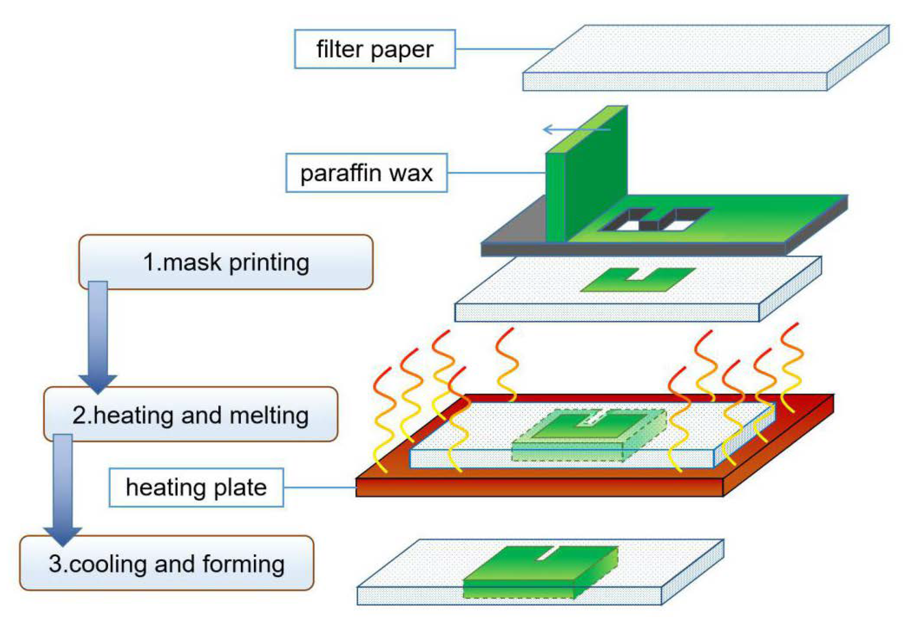
Upon repeated ionization, the material becomes a plasma state, exhibiting suitable flow, diffusion, and gas-like properties. Paper chips can also be prepared using plasma treatment technology, which uses high-energy plasma impacts to change the chemical structure of polymers and modify the surface of materials. Li et al. [21] soaked the filter paper sample in alkyl ketene dimmer (AKD) solution (0.6 g/L), and then heated it up to 100 °C for 45 min to make it hydrophobic. After clamping the filter paper between two metal plates with a specific hollow shape, it was subjected to plasma treatment to restore its hydrophilicity. According to Hecht et al. [22] plasma etching and plasma polymerization were two methods of preparing paper chips in which makes the substrate selectively hydrophobic and hydrophilic, respectively. For the preparation of paper chips, plasma etching is more efficient and cheaper, making it more suitable to use. These methods are low cost and do not destroy the flexibility of the filter paper. The disadvantage is that the preparation is less flexible due to the dependence on the template.

3.1.3. Wax Printing

In wax printing, paraffin is attached to paper-based materials in a prescribed shape, then heated to melt the paraffin and allow it to penetrate the paper to form a hydrophobic barrier so that a channel is created (Figure 2). Zhong et al. [41] compared the sample transport functions of A4 printing paper, napkins, and channels with different waxes and concluded that the experimental paper has the best sample transport function. Using the wax printing method, Lu et al. [25] explored the specific conditions for heating the paraffin on the surface of the filter paper: when the heating temperature was 110 °C, it took 5 min for the paraffin to penetrate the filter paper completely, whereas when the temperature was 130 °C, it took only 30 s to achieve the same effect. Martins et al. [24] fabricated a paper-based electrochemical sensor for the detection of 3-nitrotyrosine (3-NT) by printing a well-designed hydrophobic barrier on cut paper using a wax jet printer. Although the paraffin could diffuse horizontally after heating, the degree of diffusion is limited, and the effect can be reduced by reserving space for the wax to diffuse. A millimeter chip can be prepared using this method without chemical reagents, which makes it easy to prepare, safe to use, and widely applicable.

3.1.4. Inkjet Method

Similar to the wax printing method, the paper chip channels are manufactured by printing specific hydrophobic ink on the filter paper using an inkjet printer. For instance, Li et al. [42] printed the hydrophobic reagent (2% solution of octadecyl trimethoxysilane in N-heptane) on filter paper and heated the filter paper at 100 °C for 90 min using a heating plate, then cooled at room temperature for 3 h. Finally, the filter paper was treated with an oxygen plasma cleaner for 4 min to prepare inkjet-printed paper chips. By directly printing the AKD solution onto the paper base, Shen et al. [27] created a hydrophobic area without heating it. Moreover, Hamidon et al. [43] investigated an AKD ink with better device compatibility and material penetration. Hydrophobic ink does not spread by heat such as paraffin because it works on a different principle. Therefore, the channels drawn by the inkjet method are more accurate than the wax printing method.
Graphic design is performed on computers for both wax printing and inkjet printing, so specialized printing equipment is needed for efficient production. The high cost is one of the disadvantages of these devices. Recently, wax jet printers have been widely used in laboratories, leading to significant research on them. The hydrophobic ink used in inkjet is a liquid that has the characteristics of regular ink and can be loaded into the water-based pen to draw channels, thus eliminating the need for inkjet printing equipment. However, manual drawing inevitably brings the problem of precision. Additionally, hydrophobic inks such as siloxane can be printed with home-type printers, which solves the problems of production cost and preparation accuracy.

3.1.5. Screen Printing

Screen-printing technology is the process of injecting ink into the printing plate and applying pressure to print the pattern on the substrate, such as a screen-printing plate with graphics and text on it (Figure 3). Dunchai et al. [31] used screen-printing technology to print paraffin on the surface of filter paper in a specific pattern. By heating and cooling paraffin to room temperature, a hydrophobic channel wall structure can be constructed. This method does not require complicated and expensive equipment. It is suitable for low-cost production but needs different screen-printing plates for different chips, which limits its flexibility.

3.1.6. Laser Processing Technology

CO2 laser etching is used in laser processing to create a hydrophilic channel with a specific shape and depth on a hydrophobic substrate. In order to increase the hydrophilicity of these channels, nano-SiO2 particles can be applied inside them. Mahmud et al. [32] lined the lower layer of filter paper with aluminum foil. Laser was used to cut the filter paper with the hydrophobic barrier to prepare a paper chip. This method produces high precision paper chips, but expensive laser equipment makes it difficult to popularize, despite the theory of making such chips already being discussed.

3.2. Three-Dimensional Paper Chips Preparation

In fact, three-dimensional paper chips are just a superposition of two-dimensional paper chips, but they achieve the goal of “1 + 1 > 2”. By providing a multilayer structure and vertical flow channels, the flux of the paper chip is increased and the layers of the paper chip are enriched, allowing for a more controlled detection reaction in time and space, one of the new directions for paper chip development [44,45]. In comparison to 2D paper chips, 3D paper chips are more difficult to fabricate at a higher cost, but they are more convenient, accurate, and sensitive [46].

3.2.1. Origami Method

The origami method is a way to make a three-dimensional paper chip by cutting a piece of paper into different parts and making channels with different shapes for each part. The paper is then folded in a certain order. Cai et al. [35] made a paper chip with specific hydrophilic and hydrophobic structures. The chip was split into two equal parts, yellow and green, by heating the filter paper at 120 °C for 5 min after printing wax on it with a wax spray printer. Then, a Taqman probe and double-strand specific nuclease (DSN) were added to the hydrophilic regions of the yellow and green parts, respectively. The two parts were folded after the reagents were dried at room temperature (25 °C). For instance, a paper-based microfluidic chip based on the DSN amplification principle was fabricated for detecting cancer biomarkers microRNA (miRNA). Since the paper core has only two layers, it achieved structural stability without fixing. Xiao et al. [36] folded filter paper into five layers and fixed them with a phoenix clip to prepare a highly sensitive silver ion detection sensor. Although the origami paper chip has obtained better performance, the layers of the three-dimensional paper chip need to be fixed by reasonable means. Thus, the origami method is simple and easy to prepare. It takes the structural design of multilayer channels as its core. In addition, the fixing method can ensure a close connection between layers. While the smooth channel is also very important, it may break the continuity of vertical hydrophilic channels compared with double-sided adhesive tape. Clips and return pins can avoid this trouble with a little increasing cost.

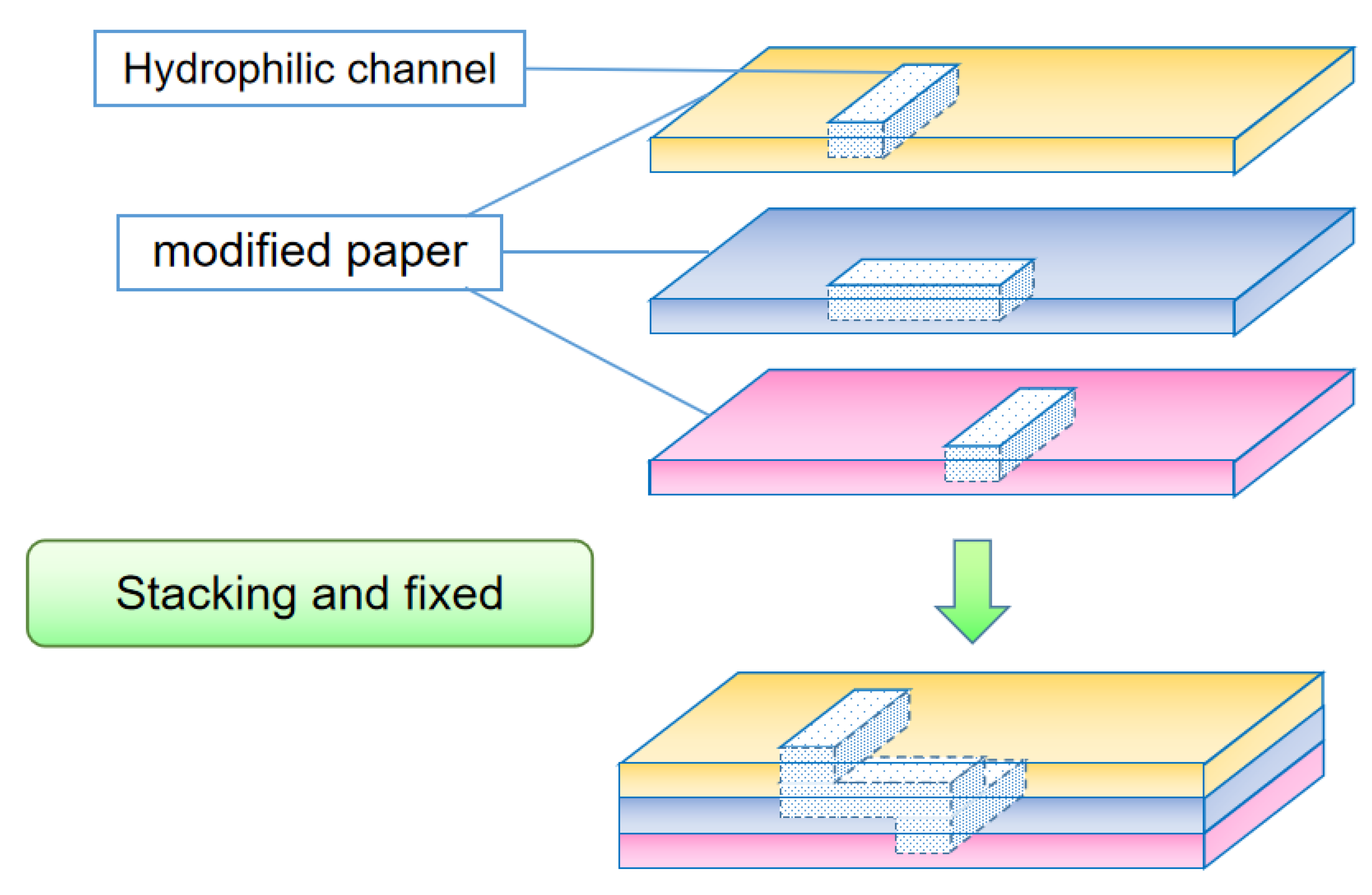
3.2.2. Lamination Method

Unlike the origami method, which prepares three-dimensional paper chips on the same piece of paper, the lamination method prepares different levels of channel structures on multiple sheets of paper with the same shape and size. Then, a double-sided tape, clip, or any other device is used to fix them into a whole paper chip (Figure 4). Wang et al. [47] constructed a paper-based microfluidic analysis device composed of four layers of different structures superimposed on each other. They made a hydrophobic barrier on the filter paper by the wax printing method and prepared a colorimetric analysis system for carcinoembryonic antigen. Hao et al. [38] used a wax jet printer to print out the hydrophobic barrier and hydrophilic channel structures on the paper. Three printed papers were assembled top to bottom to produce a general fast ratio fluorescence sensing platform for paper-based microfluidic chips; the top layer with a guiding fluid channel, a middle layer with a reagent-treated polyester fiber membrane, and a bottom layer capable of storing liquid. The lamination method is especially suitable for three-dimensional paper chips with different materials in each layer. Since there is not a clear fold line to fix the relative positions of theses layers, there is also the problem of how to line up the channel structure of each layer.

3.2.3. Other Methods

Researchers are trying to find new methods to prepare three-dimensional paper chips better than the classic origami and lamination methods. Jeong [39] et al. printed different patterns by spraying wax on both sides of the filter paper. Then, the paraffin on both sides was heated precisely so that it penetrated to an ideal depth and connected to each other in three-dimensional channels. The pinwheel-shaped 3D paper chip designed and prepared by Wang et al. [40] is fixed by a plastic shell. Through the process of rotation, four kinds of heavy metal ions (Cu2+, Cd2+, Pb2+, and Hg2+) can be sensitively detected at one time. Due to the high cost and difficult operation, the application of these methods in the preparation of 3D paper chips is relatively rare, but they help 3D paper chips achieve multiple functions, propose unprecedented innovations, and have broad potential for development.

4. Analysis Method of Paper-Based Microfluidic Chip

The paper-based microfluidic chip focuses on the results being presented in a visual way. Using colorimetry, electrochemistry, and fluorescence, the paper chip can find out both qualitative and quantitative information about different substances based on their physical, biological, and chemical properties [48,49,50] (described in Table 3).

4.1. Colorimetric Method

The chromogenic method is a well-known way to observe the results by looking at the change of color of a certain area before and after the reaction. This method uses the physical deposition of colored substances to make the color. These colored substances are often used to mark the reactants to obtain a visualization of the reaction result, which would not produce color change by itself. The lateral flow analysis is based on the chromogenic method. In contrast, colloidal gold is a classic reagent used for chromometric detections [62,63]. For instance, the well-known lateral flow analysis detection device “pregnancy test paper” employs colloidal gold immunochromatography for human chorionic gonadotropin detection [51]. By using an antibody fixed on a nitrocellulose membrane, it deposits a red gold-labeled antibody complex and shows red strips that can be recognized by the naked eye for detection of results [64,65]. In 2007, Gao’s research team [66] found that Fe3O4 nanoparticles are similar to colloidal gold particles, whose peroxidase properties can cascade and amplify the detection signal through enzyme-catalyzed reaction. The sensitivity of immunochromatography based on Fe3O4 is about 100 times higher than that of colloidal gold [67]; thus, work on Fe3O4 nanoparticles is becoming one of the new landscapes of chromogenic paper chips.
In contrast to chromogenic methods based on physics, the methods based on chemical reactions to produce color changes are color-changing methods. Hossain et al. [68] developed a multi-channel heavy metal detection paper chip with suitable sensitivity. The chip’s functioning depends on the property of heavy metal ions so that they can change the activity of β-galactosidase in conjunction with the color reaction of other metal ions. Based on the chemical properties of benzoic acid, Liu et al. [69] developed a paper chip that can quantitatively detect the concentration of benzoic acid in food by analyzing color development through a smartphone program. Wei et al. [52] prepared the paper chip using urea/phenol red impregnation technology. The chip employed an H2O2-treated Fe3O4@Ag multifunctional hybrid nanoprobe (APT-Fe3O4@AgNP) solution to generate Ag+. The Ag+ in the supernatant inhibited urease activity. As a result, when salmonella typhimurium was specifically captured in the solution, the bacteria were able to adsorb Ag+ in the supernatant. So, the supernatant no longer inhibited urease activity on paper, which caused urease to catalyze urea into a large amount of [NH₄]+. Briefly, [NH₄]+ reacts with the reagents on the paper chip to produce a significant pink color change for rapid and sensitive colorimetric detection. Since paper chip substrates are usually white, qualitative test results are easily readable by the naked eye. It is supposed to use smartphones, digital cameras, and other equipment [53]. Some image processing software can also be used for the quantitative analysis of parameters such as grayscale [70] and color rendering area [71]. In particular, colorimetry is a classic and commonly used for paper-based microfluidic chips for the detection of different substances (shown in Figure 5).

4.2. Electrochemical Method

Therefore, it measures the potential change through electrodes mounted on paper sheets, which are usually made of special inks such as Ag+/AgCl inks, carbon black, and more recently developed boron-doped diamond (BDD) electrodes printed [73,74,75] or metal sputtered [56] (mainly gold electrodes). Jemmeli et al. [54] used carbon black, a nano material, to print electrodes on the filter paper to prepare a sensor with high sensitivity but low cost, which can detect bisphenol A (BPA) in water by adding samples only. Yang et al. [55] used screen-printed electrodes to measure the impedance change caused by enzyme inhibition reaction on a five-layered paper chip, and successfully distinguished three different pesticides. Recently, some researchers combined nanoparticles with electrochemical paper chips to increase detection sensitivity. In a study, Zhou et al., introduced CuCo-CeO2 nano-spheres that are spherical in shape and have a particle size of 200 nm, a rough surface, and can effectively combine with DNA [76]. As a metal oxide nanomaterial with mixed surface valence, its unique electrocatalytic performance can enhance the electrochemical signal, providing ultra-sensitive detection of miRNA-155 in serum samples. However, electrochemical detection needs the support of professional equipment such as an electrochemical workstation. Recent research has designed a paper chip system that can realize self-power generation with the help of capacitance and read the results only with a multimeter [77]. Moreover, these chips can achieve accurate quantification of results within their sensitive range. However, the use of electrochemical paper chips is still a relatively complex process and needs further research in this field.

4.3. Fluorescence Method

Fluorophores absorb energy in specific wavelength areas, then reemit it in other wavelength areas. This causes fluorescence of different colors to be produced to visualize the results of the analysis. For instance, the chemiluminescence method uses chemical reactions to produce luminescence, while the electrochemical luminescence method uses voltage to drive the chemical reaction. It includes the chemiluminescence method, which makes the reagent glow through a chemical reaction, and the electrochemical luminescence method, which applies voltage to make the chemical reaction proceed. Ali et al. [59] prepared a new three-peak system based on paper-based microfluidic devices, which can help police grab ketamine as long as samples are added to the detection area. Delaney et al. [57] used the electrochemiluminescence method to screen-print the electrode on the paper chip with inkjet-printed channels and then applied voltage to achieve quantitative detection of 2-(dibutylamino) ethanol (DBAE) and nicotinamide adenine dinucleotide (NADH) based on the luminous intensity. Recently, Ezequiel’s team [78] optimized electrochemiluminescence to develop a single-electrode electrochemical detection paper chip using dye contained in commercial glow sticks with carbon paint electrodes. The system not only reduced detection cost but also increased detection flexibility by detecting H2O2. The advantage of the fluorescence method is that it is more sensitive. This is because even very weak fluorescence can be seen with fluorescence detection equipment, such as a fluorescence microscope, which can detect targets at micro-scale levels. However, fluorescence detection equipment must be used to read the results, which can be easily affected by the fluorescence emitted from the basic background of the paper.

4.4. Combining with Electronic Equipment

Accurate analysis results often require professional equipment and laboratory professionals. However, research has gradually improved paper chip analysis and detection capabilities, resulting in more accurate analysis results. The popularity of smart phones has led some researchers to develop paper chips that can be used with mobile phones. They can achieve sensitive insights into fluorescence intensity [58] or precise color-grayscale analysis [4,61,79] with mobile phone cameras and apps such as ImageJ, etc. Furthermore, some electrochemical workstations are gradually becoming home testing equipment, such as blood glucose meters [60], which have gradually reduced in size and become more portable. Carbon-13 labeling is also being used in hospitals for the detection of Helicobacter pylori breath. In this method, subjects blow air into the paper chip to determine the number of colonies in the body. They insert it into the electrochemical workstation, which not only greatly reduces the cost of detection but also avoids the physical trauma caused by traditional gastroscopic sampling methods. The combination with electronic equipment may reduce the lightweight advantage of paper chips, but medical equipment development and popularization could lead to better development prospects.

5. Application of Paper-Based Microfluidic Chip

5.1. Biochemical Marker Detection

The most classic and common use of paper-based microfluidic chips is to detect various biochemical markers in fields of food safety [80,81], environmental protection [82], and medical health [83,84], especially the detection of disease-related markers [85,86]. Using the above-mentioned methods, the paper chip can be prepared to detect various biochemical markers such as inorganic substances [71], proteins [87], and nucleic acids [88,89,90]. Ye et al. [91] combined paper-based devices with smart phones to enhance the sensitivity of quantitative detection of the Hg2+ ions in water samples. Boonkaew et al. [92] used the wax printing method to prepare paper chips and screen printing to obtain printed graphite electrodes for the electrochemical detection of C-reactive protein in human serum samples. Noviana et al. [93] prepared a paper chip based on a nuclease protection assay. The change in color of the reaction area can be used to find the target nucleic acid. Li et al. [94] developed a fluorescent paper-based sensor that can detect folate in solution.
From one to two channels and then to more, it is a leap from quantitative change to qualitative change. For more accurate results, many analyses and tests need to consider multiple markers, especially in disease detection. It is becoming increasingly critical to research and design multiplexing, which significantly improves the detection accuracy and efficiency of detection. This is performed through the preparation of additional channel structures and the addition of hydrophobic materials between the existing layers of channel structures [74,95,96].
Besides the detection of collected samples in the laboratory, the lightweight and compact characteristics of paper chips make them suitable for being used as wearable sensors for non-invasive monitoring of human health in daily life. Xiao et al. [97] and Castro et al. [98] developed wearable colorimetric sensors that can detect glucose in sweat and saliva, respectively. So, these sensors can monitor the physical condition of users at any time. The exploration of outdoor equipment has always been a hot topic for inventors. Even though facing more difficulties, it needs to be further developed and improved.

5.2. Nucleic Acid Preparation

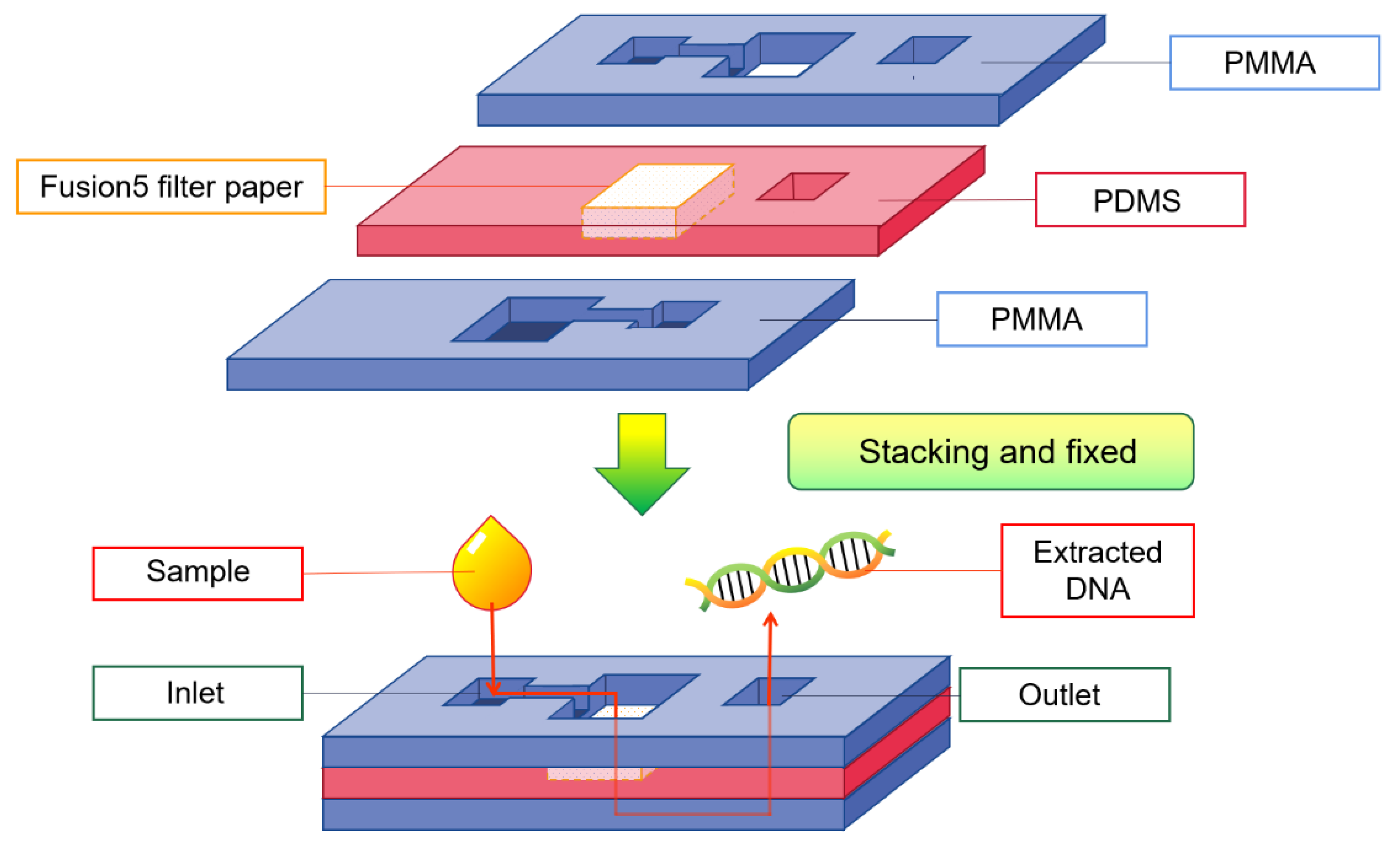
5.2.1. DNA Extraction

The extraction of target DNA is particularly important in a variety of scientific research fields. However, the conventional method of extracting DNA requires complex processes such as destroying cells, centrifuging and layering, heating, or adding reagents. These processes rely on professional equipment and must be carried out in the laboratory. Gan et al. [99] assembled the filter paper in the middle of the plastic shell to make a DNA extraction paper-based apparatus (shown in Figure 6). They successfully extracted biological DNA from the original samples, including dried blood stains and cigarette butts. Through primer-mediated and enzymatic catalysis, the obtained DNA can be amplified by polymerase chain reaction (PCR) for subsequent detection, which operates on the principle of DNA semiconservative replication to exponentially amplify target DNA. In 2017, Tang et al. [100] prepared a paper-based device with sponge storage modules and paper valves for the rapid extraction of DNA from biological samples. It takes only 2 min to obtain target DNA from samples such as serum, saliva, and bacterial suspension. It has great potential to complete the whole process of DNA extraction outside the laboratory and even in the absence of specific conditions.

5.2.2. Nucleic Acid Amplification
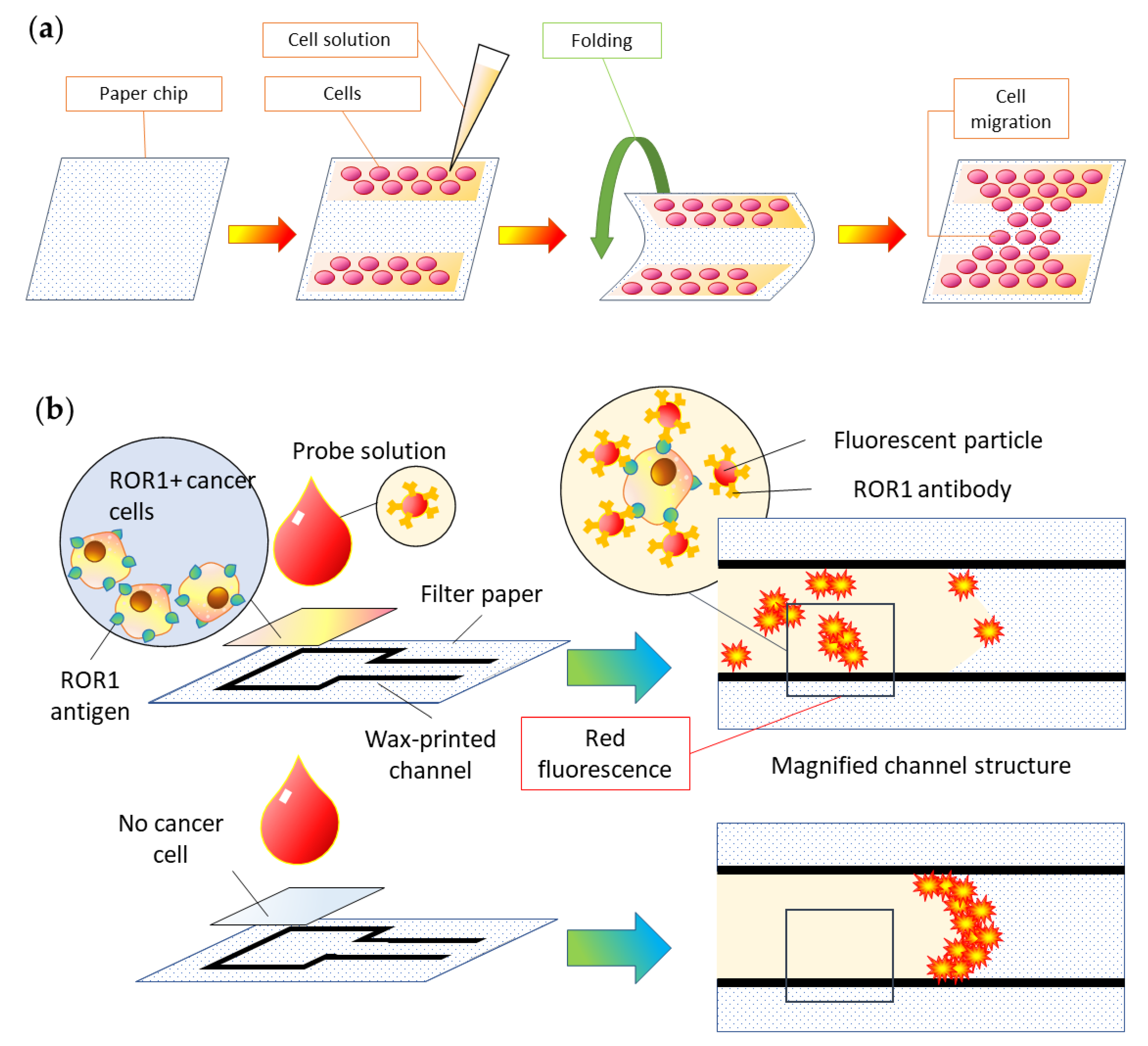
The amount of nucleic acid directly extracted from various biological samples is usually only trace amounts, which makes it difficult to reach the level used for detection. So, specific DNA fragments need to be amplified. PCR is the most common and typical method for DNA amplification in the lab. Conventional PCR requires sample processing, and the amplified DNA also needs to be detected by gel electrophoresis. Patil-Joshi et al. [101] used filter paper as the carrier of PCR samples, eliminating the need for sample processing. They not only successfully achieved the amplification of target DNA but also improved the efficiency of DNA replication. This method can be used for many different samples, and it makes the experimentation process much easier. The paper chip prepared by Chen et al. [102] integrates nucleic acid extraction, amplification, and detection functions. It can identify the EGFR mutations in lung cancer cells within 90 min and carry out the sensitive detection of lung adenocarcinoma, which is very powerful and worth popularizing.
In recent years, thermostatic amplification technology based on recombinase polymerase amplification (RPA) and recombinase-mediated amplification (RAA) has developed rapidly. Using a specific recombinant enzyme, the amplification of the target DNA can be achieved at 37–40 °C in 10 to 30 min. Therefore, it can cooperate with the paper chip. Colloidal gold immunochromatography can be used to find the target DNA by adding only a probe with a FAM fluorescent group to the amplification process and using an antibody against FAM. Heeseop et al. [103] used a paper chip made of glass fiber paper to carry out the RPA process on it. They performed simultaneous detection of a variety of foodborne pathogens by fluorescence method. Therefore, the combination of thermostatic amplification technology and paper chips is becoming the latest hotspot for nucleic acid detection. In addition, the latest research shows that the reverse transcription of mRNA samples can also be performed on paper-based equipment [104], further expanding the application of paper-based microfluidic chips in the field of nucleic acid detection.

5.3. Cell Analysis

In addition to analyzing and detecting biochemical markers at the molecular level, paper-based microfluidic chips can also be used for cell-level research, including cell separation, culture, stimulation, detection, and so on. The paper-based in vitro tissue chip developed by Kaarj et al. [105] can be knocked and bent through a program to provide local compression and shear flow mechanical stimulation to rat vascular endothelial cells fixed on it. As a result, it could provide insight into the specific impact of mechanical stimulation on angiogenesis as well as the possibility of using it for the treatment of malignant tumors that inhibit the growth of blood vessels (shown in Figure 7a). Ulep et al. [58] conjugated anti-ROR1 (receptor tyrosine-like orphan receptor one) and ethanolamine to highly carboxylated red fluorescent polystyrene particles having a diameter of 1 μm, forming a double-layer paper chip that can directly identify ROR1+ cancer cells from blood samples. Even ROR1+ cancer cells can be captured by observing the red fluorescence intensity with a special fluorescence microscope based on a smart phone (shown in Figure 7b). It is evident that paper-based cell analysis offers a new perspective on simplifying existing cell research methods as well as providing a wider prospect for developing paper chips.

5.4. Other Applications

Paper-based microfluidic chips can not only be directly applied to analyze and detect various biomarkers, but they can also help with other types of analyses. Samae et al. [106] used paraffin-treated paper to accurately prepare multilayer paper-based passive microfluidic micromixers. Comparatively, its zig-zag channel structure was studied for efficient mixing, enabling it to be docked with a variety of experimental equipment to play its role. Hong et al. [107] used photolithography to prepare the concentration gradient generator on the paper-based microfluidic chip. They successfully separated five concentration gradients of adriamycin. Then, they added different concentrations of adriamycin to the three-dimensional cell culture arrays to observe the effect of drug toxicity on tumor cells. Son et al. [108] made a cation-selective membrane by using cellulose paper and prepared a paper-based ion concentration polarization device that can achieve selective preconcentration of human breast cancer marker MUC1 gene fragment and Danon disease marker LAMP-2 gene fragment. Saha et al. [109] outlined the method of developing paper-based blood/plasma separation equipment by using the pore characteristics of different kinds of papers without adding additional power. With its low cost, high efficiency, and development potential, this equipment has several advantages. Niu et al. [110] used the principle of rapid isoelectric focusing to separate proteins. In paper fluid channels, they saw proteins in complex matrices become concentrated and separated at the same time. Paper-based microfluidic chips can not only do the analysis and detection function but also be used together with other experimental equipment, thereby broadening their applicability.

6. Summary and Outlook

The microfluidic chips made from paper would have profound effects on physics, biotechnology, and medicine. Rapid progress in microfluidic chips over the past decade makes us optimistic that this ambition is within reach. Among microfluidic chips of various materials, paper-based microfluidic chips are distinct for their small size, light weight, low cost, no additional driving force, easy processing, and environmental friendliness. Moreover, the excellent performance of paper chips makes them widely applicable for the detection of biochemical markers, nucleic acid preparation, cell analysis, and a variety of auxiliary analyses. Such extensive laboratory evolution has been used to promote biomedical legends. The advancement of science and the popularity of related equipment have made the preparation of paper chips a mature technology. Interestingly, mass production based on computer pattern design has further reduced the preparation cost of paper chips [111,112].
Currently, paper chip research focuses on efficiency, accuracy, integration, and innovation. Rather than making paper chips that can only detect one substance and only have one channel, it is becoming more common to make multi-channel paper chips that can detect and distinguish several substances at the same time [113,114,115]. Additionally, qualitative detection is being replaced by quantitative detection. This non-invasive technique not only detects the existence of substances but also determines their content in the tested products through parameters such as the depth of color development, the area of color change, the intensity of fluorescence, the size of the current, and so on [116,117,118,119]. It is common for the detection of a single substance to decrease the lower limit of detection and to continuously improve the sensitivity of detection. In order to enable non-professional users to obtain accurate detection results, the combination of paper chips and electronic devices is also gaining popularity. With the proliferation of multi-function paper chips, broader applications are possible besides just reading and detecting [120,121,122]. In summary, although there are considerable challenges to overcome, we remain cautiously optimistic that paper-based microfluidic chips—which can predict and trace biomarkers from home-grown samples to deliver efficient devices with desired functions—will eventually become a reality.

Author Contributions

Conceptualization, B.L. and Z.Z.; methodology, H.Z. and N.L.; writing—original draft preparation, Y.J.; writing—review and editing, A.u.R.A., B.W. and Y.L.; visualization, Y.J. All authors have read and agreed to the published version of the manuscript.

Funding

This work was funded by Major Research Plan Funds for Dalian, grant number 2021YF18SN023, Support Plan of Liaoning Provincial Natural Foundation, grant number 2021-MS-121 and Basic Research Funds for Central Universities, grant number DUT22YG239.

Institutional Review Board Statement

Not applicable.

Informed Consent Statement

Not applicable.

Data Availability Statement

Not applicable.

Conflicts of Interest

The authors declare no conflict of interest.

References

  1. Manz, A.; Graber, N.; Widmer, H.M. Miniaturized total chemical-analysis systems—A novel concept for chemical sensing. Sens. Actuators B-Chem. 1990, 1, 244–248. [Google Scholar] [CrossRef]
  2. Whitesides, G.M. The origins and the future of microfluidics. Nature 2006, 442, 368–373. [Google Scholar] [CrossRef] [PubMed]
  3. Liu, J.; Kong, X.; Wang, H.; Zhang, Y.; Fan, Y. Roll-to-roll wax transfer for rapid and batch fabrication of paper-based microfluidics. Microfluid. Nanofluidics 2019, 24, 6. [Google Scholar] [CrossRef]
  4. Trofimchuk, E.; Nilghaz, A.; Sun, S.; Lu, X. Determination of norfloxacin residues in foods by exploiting the coffee-ring effect and paper-based microfluidics device coupling with smartphone-based detection. J. Food Sci. 2020, 85, 736–743. [Google Scholar] [CrossRef]
  5. Kaewjua, K.; Nakthong, P.; Chailapakul, O.; Siangproh, W. Flow-based System: A Highly Efficient Tool Speeds Up Data Production and Improves Analytical Performance. Anal. Sci. 2021, 37, 79–92. [Google Scholar] [CrossRef]
  6. Yoon, J.-Y. Lab-on-a-Chip Biosensors. In Introduction to Biosensors: From Electric Circuits to Immunosensors; Yoon, J.-Y., Ed.; Springer International Publishing: Cham, Switzerland, 2016; pp. 257–297. [Google Scholar] [CrossRef]
  7. Lisowski, P.; Zarzycki, P.K. Microfluidic Paper-Based Analytical Devices (μPADs) and Micro Total Analysis Systems (μTAS): Development, Applications and Future Trends. Chromatographia 2013, 76, 1201–1214. [Google Scholar] [CrossRef] [Green Version]
  8. Martinez, A.W.; Phillips, S.T.; Butte, M.J.; Whitesides, G.M. Patterned Paper as a Platform for Inexpensive, Low-Volume, Portable Bioassays. Angew. Chem. Int. Ed. 2007, 46, 1318–1320. [Google Scholar] [CrossRef] [Green Version]
  9. Almeida, M.I.G.S.; Jayawardane, B.M.; Kolev, S.D.; McKelvie, I.D. Developments of microfluidic paper-based analytical devices (μPADs) for water analysis: A review. Talanta 2018, 177, 176–190. [Google Scholar] [CrossRef]
  10. Dou, M.; Sanjay, S.T.; Benhabib, M.; Xu, F.; Li, X. Low-cost bioanalysis on paper-based and its hybrid microfluidic platforms. Talanta 2015, 145, 43–54. [Google Scholar] [CrossRef]
  11. Mabbott, S.; Fernandes, S.C.; Schechinger, M.; Cote, G.L.; Faulds, K.; Mace, C.R.; Graham, D. Detection of cardiovascular disease associated miR-29a using paper-based microfluidics and surface enhanced Raman scattering. Analyst 2020, 145, 983–991. [Google Scholar] [CrossRef]
  12. Zeng, Y.; Liang, D.; Zheng, P.; Zhang, Y.; Wang, Z.; Mari, G.M.; Jiang, H. A simple and rapid immunochromatography test based on readily available filter paper modified with chitosan to screen for 13 sulfonamides in milk. J. Dairy Sci. 2021, 104, 126–133. [Google Scholar] [CrossRef] [PubMed]
  13. Wang, C.-H.; Wu, J.-J.; Lee, G.-B. Screening of highly-specific aptamers and their applications in paper-based microfluidic chips for rapid diagnosis of multiple bacteria. Sens. Actuators B Chem. 2019, 284, 395–402. [Google Scholar] [CrossRef]
  14. Deng, D.; Lin, Q.; Li, H.; Huang, Z.; Kuang, Y.; Chen, H.; Kong, J. Rapid detection of malachite green residues in fish using a surface-enhanced Raman scattering-active glass fiber paper prepared by in situ reduction method. Talanta 2019, 200, 272–278. [Google Scholar] [CrossRef] [PubMed]
  15. Evans, E.; Gabriel, E.F.M.; Coltro, W.K.T.; Garcia, C.D. Rational selection of substrates to improve color intensity and uniformity on microfluidic paper-based analytical devices. Analyst 2014, 139, 2127–2132. [Google Scholar] [CrossRef] [Green Version]
  16. Noviana, E.; Ozer, T.; Carrell, C.S.; Link, J.S.; McMahon, C.; Jang, I.; Henry, C.S. Microfluidic Paper-Based Analytical Devices: From Design to Applications. Chem. Rev. 2021, 121, 11835–11885. [Google Scholar] [CrossRef]
  17. Brooks, J.C.; Mace, C.R. Scalable Methods for Device Patterning as an Outstanding Challenge in Translating Paper-Based Microfluidics from the Academic Benchtop to the Point-of-Care. J. Anal. Test. 2019, 3, 50–60. [Google Scholar] [CrossRef]
  18. Mahato, K.; Purohit, B.; Kumar, A.; Srivastava, A.; Chandra, P. Next-Generation Immunosensing Technologies Based on Nano-Bio-Engineered Paper Matrices. In Immunodiagnostic Technologies from Laboratory to Point-of-Care Testing; Suman, P., Chandra, P., Eds.; Springer: Singapore, 2021; pp. 93–110. [Google Scholar] [CrossRef]
  19. Wang, C.-M.; Chen, C.-Y.; Liao, W.-S. Enclosed paper-based analytical devices: Concept, variety, and outlook. Anal. Chim. Acta 2021, 1144, 158–174. [Google Scholar] [CrossRef]
  20. Busa, L.S.A.; Maeki, M.; Ishida, A.; Tani, H.; Tokeshi, M. Simple and sensitive colorimetric assay system for horseradish peroxidase using microfluidic paper-based devices. Sens. Actuators B Chem. 2016, 236, 433–441. [Google Scholar] [CrossRef]
  21. Li, X.; Tian, J.; Garnier, G.; Shen, W. Fabrication of paper-based microfluidic sensors by printing. Colloids Surf. B Biointerfaces 2010, 76, 564–570. [Google Scholar] [CrossRef]
  22. Hecht, L.; Philipp, J.; Mattern, K.; Dietzel, A.; Klages, C.-P. Controlling wettability in paper by atmospheric-pressure microplasma processes to be used in µPAD fabrication. Microfluid. Nanofluidics 2016, 20, 25. [Google Scholar] [CrossRef]
  23. Li, H.; Cui, J.; Yan, Z.; Jin, M.; Zheng, Y.; Zhou, G.; Shui, L. Paper-based electrowetting devices fabricated with cellulose paper and paraffin wax. Results Phys. 2021, 31, 105042. [Google Scholar] [CrossRef]
  24. Martins, G.V.; Marques, A.C.; Fortunato, E.; Sales, M.G.F. Wax-printed paper-based device for direct electrochemical detection of 3-nitrotyrosine. Electrochim. Acta 2018, 284, 60–68. [Google Scholar] [CrossRef]
  25. Lu, Y.; Shi, W.; Jiang, L.; Qin, J.; Lin, B. Rapid prototyping of paper-based microfluidics with wax for low-cost, portable bioassay. Electrophoresis 2009, 30, 1497–1500. [Google Scholar] [CrossRef] [PubMed]
  26. Lv, P.; Song, L.; Li, Y.; Pang, H.; Liu, W. Hybrid ternary rice paper/polypyrrole ink/pen ink nanocomposites as components of flexible supercapacitors. Int. J. Hydrog. Energy 2021, 46, 13219–13229. [Google Scholar] [CrossRef]
  27. Shen, W.; Filonanko, Y.; Truong, Y.; Parker, I.H.; Brack, N.; Pigram, P.; Liesegang, J. Contact angle measurement and surface energetics of sized and unsized paper. Colloids Surfaces. A Physicochem. Eng. Asp. 2000, 173, 117–126. [Google Scholar] [CrossRef]
  28. Rahbar, M.; Nesterenko, P.N.; Paull, B.; Macka, M. High-throughput deposition of chemical reagents via pen-plotting technique for microfluidic paper-based analytical devices. Anal. Chim. Acta 2019, 1047, 115–123. [Google Scholar] [CrossRef]
  29. Sitanurak, J.; Fukana, N.; Wongpakdee, T.; Thepchuay, Y.; Ratanawimarnwong, N.; Amornsakchai, T.; Nacapricha, D. T-shirt ink for one-step screen-printing of hydrophobic barriers for 2D- and 3D-microfluidic paper-based analytical devices. Talanta 2019, 205, 120113. [Google Scholar] [CrossRef]
  30. Yao, Y.; Zhang, C. A novel screen-printed microfluidic paper-based electrochemical device for detection of glucose and uric acid in urine. Biomed. Microdevices 2016, 18, 92. [Google Scholar] [CrossRef]
  31. Dungchai, W.; Chailapakul, O.; Henry, C.S. A low-cost, simple, and rapid fabrication method for paper-based microfluidics using wax screen-printing. Analyst 2010, 136, 77–82. [Google Scholar] [CrossRef]
  32. Mahmud, M.A.; Blondeel, E.J.M.; Kaddoura, M.; MacDonald, B.D. Creating compact and microscale features in paper-based devices by laser cutting. Analyst 2016, 141, 6449–6454. [Google Scholar] [CrossRef]
  33. Ghosh, R.; Gopalakrishnan, S.; Savitha, R.; Renganathan, T.; Pushpavanam, S. Fabrication of laser printed microfluidic paper-based analytical devices (LP-µPADs) for point-of-care applications. Sci. Rep. 2019, 9, 7896. [Google Scholar] [CrossRef] [Green Version]
  34. Sharifi, H.; Tashkhourian, J.; Hemmateenejad, B. A 3D origami paper-based analytical device combined with PVC membrane for colorimetric assay of heavy metal ions: Application to determination of Cu(II) in water samples. Anal. Chim. Acta 2020, 1126, 114–123. [Google Scholar] [CrossRef] [PubMed]
  35. Cai, X.; Zhang, H.; Yu, X.; Wang, W. A microfluidic paper-based laser-induced fluorescence sensor based on duplex-specific nuclease amplification for selective and sensitive detection of miRNAs in cancer cells. Talanta 2020, 216, 120996. [Google Scholar] [CrossRef] [PubMed]
  36. Xiao, W.; Gao, Y.; Zhang, Y.; Li, J.; Liu, Z.; Nie, J.; Li, J. Enhanced 3D paper-based devices with a personal glucose meter for highly sensitive and portable biosensing of silver ion. Biosens. Bioelectron. 2019, 137, 154–160. [Google Scholar] [CrossRef] [PubMed]
  37. Park, J.; Park, J.-K. Pressed region integrated 3D paper-based microfluidic device that enables vertical flow multistep assays for the detection of C-reactive protein based on programmed reagent loading. Sens. Actuators B Chem. 2017, 246, 1049–1055. [Google Scholar] [CrossRef]
  38. Hao, G.; Zhang, Z.; Ma, X.; Zhang, R.; Qin, X.; Sun, H.; Yang, X.; Rong, J. A versatile microfluidic paper chip platform based on MIPs for rapid ratiometric sensing of dual fluorescence signals. Microchem. J. 2020, 157, 105050. [Google Scholar] [CrossRef]
  39. Jeong, S.-G.; Lee, S.-H.; Choi, C.-H.; Kim, J.; Lee, C.-S. Toward instrument-free digital measurements: A three-dimensional microfluidic device fabricated in a single sheet of paper by double-sided printing and lamination. Lab Chip 2015, 15, 1188–1194. [Google Scholar] [CrossRef]
  40. Wang, M.; Song, Z.; Jiang, Y.; Zhang, X.; Wang, L.; Zhao, H.; Cui, Y.; Gu, F.; Wang, Y.; Zheng, G. A three-dimensional pinwheel-shaped paper-based microfluidic analytical device for fluorescence detection of multiple heavy metals in coastal waters by rational device design. Anal. Bioanal. Chem. 2021, 413, 3299–3313. [Google Scholar] [CrossRef]
  41. Zhong, Z.W.; Wang, Z.P.; Huang, G.X.D. Investigation of wax and paper materials for the fabrication of paper-based microfluidic devices. Microsyst. Technol. 2012, 18, 649–659. [Google Scholar] [CrossRef]
  42. Jiahao, L.; Xin, W.; Yanke, S.; Huachuan, H.; Dan, J.; Liang, X.; Shouyu, W.; Fei, L. Handheld Inkjet Printing Paper Chip Based Smart Tetracycline Detector. Micromachines 2019, 10, 27. [Google Scholar] [CrossRef] [Green Version]
  43. Hamidon, N.N.; Hong, Y.; Salentijn, G.I.J.; Verpoorte, E. Water-based alkyl ketene dimer ink for user-friendly patterning in paper microfluidics. Anal. Chim. Acta 2018, 1000, 180–190. [Google Scholar] [CrossRef] [PubMed]
  44. Cao, L.; Han, G.-C.; Xiao, H.; Chen, Z.; Fang, C. A novel 3D paper-based microfluidic electrochemical glucose biosensor based on rGO-TEPA/PB sensitive film. Anal. Chim. Acta 2020, 1096, 34–43. [Google Scholar] [CrossRef] [PubMed]
  45. Yukird, J.; Soum, V.; Kwon, O.-S.; Shin, K.; Chailapakul, O.; Rodthongkum, N. 3D paper-based microfluidic device: A novel dual-detection platform of bisphenol A. Analyst 2020, 145, 1491–1498. [Google Scholar] [CrossRef] [PubMed]
  46. Yang, N.; Chen, C.; Wang, P.; Sun, J.; Mao, H. Structure optimization method of microfluidic paper chip based on image grey-level statistics for chromogenic reaction. Chem. Eng. Process. 2019, 143, 107627. [Google Scholar] [CrossRef]
  47. Wang, K.; Yang, J.; Xu, H.; Cao, B.; Qin, Q.; Liao, X.; Wo, Y.; Jin, Q.; Cui, D. Smartphone-imaged multilayered paper-based analytical device for colorimetric analysis of carcinoembryonic antigen. Anal. Bioanal. Chem. 2020, 412, 2517–2528. [Google Scholar] [CrossRef]
  48. Ruecha, N.; Yamada, K.; Suzuki, K.; Citterio, D. (Bio)Chemical Sensors Based on Paper. In Materials for Chemical Sensing; Cesar Paixão, T.R.L., Reddy, S.M., Eds.; Springer International Publishing: Cham, Switzerland, 2017; pp. 29–74. [Google Scholar] [CrossRef]
  49. Zheng, W.; Wang, K.; Xu, H.; Zheng, C.; Cao, B.; Qin, Q.; Jin, Q.; Cui, D. Strategies for the detection of target analytes using microfluidic paper-based analytical devices. Anal. Bioanal. Chem. 2021, 413, 2429–2445. [Google Scholar] [CrossRef]
  50. Fu, L.-M.; Wang, Y.-N. Detection methods and applications of microfluidic paper-based analytical devices. TrAC Trends Anal. Chem. 2018, 107, 196–211. [Google Scholar] [CrossRef]
  51. Anderson, C.E.; Shah, K.G.; Yager, P. Sensitive Protein Detection and Quantification in Paper-Based Microfluidics for the Point of Care. Methods Enzymol. 2017, 589, 383. [Google Scholar] [CrossRef]
  52. Wei, S.; Li, J.; He, J.; Zhao, W.; Wang, F.; Song, X.; Xu, K.; Wang, J.; Zhao, C. Paper chip-based colorimetric assay for detection of Salmonella typhimurium by combining aptamer-modified FeO@Ag nanoprobes and urease activity inhibition. Mikrochim. Acta 2020, 187, 554. [Google Scholar] [CrossRef]
  53. Calabria, D.; Caliceti, C.; Zangheri, M.; Mirasoli, M.; Simoni, P.; Roda, A. Smartphone–based enzymatic biosensor for oral fluid L-lactate detection in one minute using confined multilayer paper reflectometry. Biosens. Bioelectron. 2017, 94, 124–130. [Google Scholar] [CrossRef]
  54. Jemmeli, D.; Marcoccio, E.; Moscone, D.; Dridi, C.; Arduini, F. Highly sensitive paper-based electrochemical sensor for reagent free detection of bisphenol A. Talanta 2020, 216, 120924. [Google Scholar] [CrossRef] [PubMed]
  55. Yang, N.; Zhou, X.; Yu, D.; Jiao, S.; Han, X.; Zhang, S.; Yin, H.; Mao, H. Pesticide residues identification by impedance time-sequence spectrum of enzyme inhibition on multilayer paper-based microfluidic chip. J. Food Process Eng. 2020, 43, e13544. [Google Scholar] [CrossRef]
  56. Wang, W.; Ding, S.-N.; Chen, F.-F.; Zhang, Q. Electrochemical paper-based analytical device for flow injection analysis based on locally enhanced evaporation. Sens. Actuators B Chem. 2022, 371, 132517. [Google Scholar] [CrossRef]
  57. Delaney, J.L.; Hogan, C.F.; Tian, J.; Shen, W. Electrogenerated chemiluminescence detection in paper-based microfluidic sensors.(Author abstract)(Report). Anal. Chem. 2011, 83, 1300. [Google Scholar] [CrossRef] [PubMed]
  58. Ulep, T.-H.; Zenhausern, R.; Gonzales, A.; Knoff, D.S.; Lengerke Diaz, P.A.; Castro, J.E.; Yoon, J.-Y. Smartphone based on-chip fluorescence imaging and capillary flow velocity measurement for detecting ROR1+ cancer cells from buffy coat blood samples on dual-layer paper microfluidic chip. Biosens. Bioelectron. 2020, 153, 112042. [Google Scholar] [CrossRef] [PubMed]
  59. Yehia, A.M.; Farag, M.A.; Tantawy, M.A. A novel trimodal system on a paper-based microfluidic device for on-site detection of the date rape drug “ketamine”. Anal. Chim. Acta 2020, 1104, 95–104. [Google Scholar] [CrossRef]
  60. Lan, T.; Zhang, J.; Lu, Y. Transforming the blood glucose meter into a general healthcare meter for in vitro diagnostics in mobile health. Biotechnol. Adv. 2016, 34, 331–341. [Google Scholar] [CrossRef]
  61. Shrivas, K.; Patel, S.; Sinha, D.; Thakur, S.S.; Patle, T.K.; Kant, T.; Dewangan, K.; Satnami, M.L.; Nirmalkar, J.; Kumar, S. Colorimetric and smartphone-integrated paper device for on-site determination of arsenic (III) using sucrose modified gold nanoparticles as a nanoprobe. Microchim. Acta 2020, 187, 173. [Google Scholar] [CrossRef]
  62. Khurana, S.; Kukreti, S.; Kaushik, M. Designing a two-stage colorimetric sensing strategy based on citrate reduced gold nanoparticles: Sequential detection of Sanguinarine (anticancer drug) and visual sensing of DNA. Spectrochim. Acta. Part A Mol. Biomol. Spectrosc. 2021, 246, 119039. [Google Scholar] [CrossRef]
  63. Yan, L.; Dou, L.; Bu, T.; Huang, Q.; Wang, R.; Yang, Q.; Huang, L.; Wang, J.; Zhang, D. Highly sensitive furazolidone monitoring in milk by a signal amplified lateral flow assay based on magnetite nanoparticles labeled dual-probe. Food Chem. 2018, 261, 131–138. [Google Scholar] [CrossRef]
  64. Xu, M.; Lu, F.; Lyu, C.; Wu, Q.; Zhang, J.; Tian, P.; Xue, L.; Xu, T.; Wang, D. Broad-range and effective detection of human noroviruses by colloidal gold immunochromatographic assay based on the shell domain of the major capsid protein. BMC Microbiol. 2021, 21, 22. [Google Scholar] [CrossRef] [PubMed]
  65. Zhang, X.; Liu, X.; Wu, X.; Ren, W.; Zou, Y.; Xia, X.; Sun, H. A colloidal gold test strip assay for the detection of African swine fever virus based on two monoclonal antibodies against P30.(Report). Arch. Virol. 2021, 166, 871. [Google Scholar] [CrossRef] [PubMed]
  66. Lizeng, G.; Jie, Z.; Leng, N.; Jinbin, Z.; Yu, Z.; Ning, G.; Taihong, W.; Jing, F.; Dongling, Y.; Sarah, P.; et al. Intrinsic peroxidase-like activity of ferromagnetic nanoparticles. Nat. Nanotechnol. 2007, 2, 577. [Google Scholar] [CrossRef]
  67. Jiang, B.; Duan, D.; Gao, L.; Zhou, M.; Fan, K.; Tang, Y.; Xi, J. Standardized assays for determining the catalytic activity and kinetics of peroxidase-like nanozymes. Nat. Protoc. 2018, 13, 1506. [Google Scholar] [CrossRef]
  68. Hossain, S.; Brennan, J. [Beta]-Galactosidase-Based Colorimetric Paper Sensor for Determination of Heavy Metals. Anal. Chem. 2011, 83, 8772. [Google Scholar] [CrossRef] [PubMed]
  69. Liu, C.-C.; Wang, Y.-N.; Fu, L.-M.; Chen, K.-L. Microfluidic paper-based chip platform for benzoic acid detection in food. Food Chem. 2018, 249, 162–167. [Google Scholar] [CrossRef]
  70. Singh, H.; Singh, G.; Mahajan, D.K.; Kaur, N.; Singh, N. A low-cost device for rapid ‘color to concentration’ quantification of cyanide in real samples using paper-based sensing chip. Sens. Actuators B Chem. 2020, 322, 128622. [Google Scholar] [CrossRef]
  71. Wu, C.; Gao, G.; Zhai, K.; Xu, L.; Zhang, D. A visual Hg2+ detection strategy based on distance as readout by G-quadruplex DNAzyme on microfluidic paper. Food Chem. 2020, 331, 127208. [Google Scholar] [CrossRef]
  72. Tran, T.D.; Nguyen, P.T.; Le, T.N.; Kim, M.I. DNA-copper hybrid nanoflowers as efficient laccase mimics for colorimetric detection of phenolic compounds in paper microfluidic devices. Biosens. Bioelectron. 2021, 182, 113187. [Google Scholar] [CrossRef]
  73. Eduardo da Silva Ferreira, M.; Canhete de Moraes, N.; Souza Ferreira, V.; Amorim Bezerra da Silva, R.; Marques Petroni, J.; Gabriel Lucca, B. A novel 3D-printed batch injection analysis (BIA) cell coupled to paper-based electrochemical devices: A cheap and reliable analytical system for fast on-site analysis. Microchem. J. 2022, 179, 107663. [Google Scholar] [CrossRef]
  74. Nantaphol, S.; Kava, A.A.; Channon, R.B.; Kondo, T.; Siangproh, W.; Chailapakul, O.; Henry, C.S. Janus electrochemistry: Simultaneous electrochemical detection at multiple working conditions in a paper-based analytical device. Anal. Chim. Acta 2019, 1056, 88–95. [Google Scholar] [CrossRef] [PubMed]
  75. Xu, G.; Díez-Pérez, I.; Paolucci, F. Editorial overview: Organic and molecular electrochemistry section (2022) electrosynthesis, electrocatalysis, electroanalysis, and molecular device. Curr. Opin. Electrochem. 2022, 101156. [Google Scholar] [CrossRef]
  76. Zhou, C.; Cui, K.; Liu, Y.; Li, L.; Zhang, L.; Xu, M.; Ge, S.; Wang, Y.; Yu, J. Ultrasensitive lab-on-paper device via Cu/Co double-doped CeO2 nanospheres as signal amplifiers for electrochemical/visual sensing of miRNA-155. Sens. Actuators B Chem. 2020, 321, 128499. [Google Scholar] [CrossRef]
  77. Li, Q.; Xu, Y.; Qi, J.; Zheng, X.; Liu, S.; Lin, D.; Zhang, L.; Liu, P.; Li, B.; Chen, L. A self-powered rotating paper-based analytical device for sensing of thrombin. Sens. Actuators B Chem. 2022, 351, 130917. [Google Scholar] [CrossRef]
  78. Vidal, E.; Domini, C.E.; Whitehead, D.C.; Garcia, C.D. From glow-sticks to sensors: Single-electrode electrochemical detection for paper-based devices. Sens. Diagn. 2022, 1, 496–503. [Google Scholar] [CrossRef]
  79. Agarwal, R.; Chakraborty, S. Analytics with blood on hybrid paper-rotating disc device. Sens. Actuators Rep. 2022, 4, 100122. [Google Scholar] [CrossRef]
  80. Fu, L.-M.; Liu, C.-C.; Yang, C.-E.; Wang, Y.-N.; Ko, C.-H. A PET/paper chip platform for high resolution sulphur dioxide detection in foods. Food Chem. 2019, 286, 316–321. [Google Scholar] [CrossRef]
  81. Muthukumar, R.; Kapoor, A.; Balasubramanian, S.; Vaishampayan, V.; Gabhane, M. Detection of adulteration in sunflower oil using paper-based microfluidic lab-on-a-chip devices. Mater. Today Proc. 2021, 34, 496–501. [Google Scholar] [CrossRef]
  82. Tavares, M.C.; Oliveira, K.A.; de Fátima, Â.; Coltro, W.K.T.; Santos, J.C.C. Paper-based analytical device with colorimetric detection for urease activity determination in soils and evaluation of potential inhibitors. Talanta 2021, 230, 122301. [Google Scholar] [CrossRef]
  83. Shi, Z.Z.; Lu, Y.; Yu, L. Microfluidic Paper-Based Analytical Devices for Point-of-Care Diagnosis. In Next Generation Point-of-Care Biomedical Sensors Technologies for Cancer Diagnosis; Chandra, P., Tan, Y.N., Singh, S.P., Eds.; Springer: Singapore, 2017; pp. 365–396. [Google Scholar] [CrossRef]
  84. Trinh, K.T.L.; Chae, W.R.; Lee, N.Y. Recent advances in the fabrication strategies of paper-based microfluidic devices for rapid detection of bacteria and viruses. Microchem. J. 2022, 180, 107548. [Google Scholar] [CrossRef]
  85. Suntornsuk, W.; Suntornsuk, L. Recent applications of paper-based point-of-care devices for biomarker detection. Electrophoresis 2020, 41, 287–305. [Google Scholar] [CrossRef] [PubMed]
  86. Han, T.; Jin, Y.; Geng, C.; Aziz, A.u.R.; Zhang, Y.; Deng, S.; Ren, H.; Liu, B. Microfluidic Paper-based Analytical Devices in Clinical Applications. Chromatographia 2020, 83, 693–701. [Google Scholar] [CrossRef]
  87. Jang, I.; Ko, H.; You, G.; Lee, H.; Paek, S.; Chae, H.; Lee, J.; Choi, S.; Kwon, O.-S.; Shin, K.; et al. Application of paper EWOD (electrowetting-on-dielectrics) chip: Protein tryptic digestion and its detection using MALDI-TOF mass spectrometry. BioChip J. 2017, 11, 146–152. [Google Scholar] [CrossRef]
  88. Batule, B.S.; Seok, Y.; Kim, M.-G. An innovative paper-based device for DNA extraction from processed meat products. Food Chem. 2020, 321, 126708. [Google Scholar] [CrossRef] [PubMed]
  89. Batule, B.S.; Seok, Y.; Kim, M.-G. Paper-Based Molecular Diagnostics. In Paper-Based Medical Diagnostic Devices: As a Part of Bioanalysis-Advanced Materials, Methods, and Devices; Lee, J.H., Ed.; Springer: Singapore, 2021; pp. 155–181. [Google Scholar] [CrossRef]
  90. Dong, T.; Wang, G.; Li, F. Shaping up field-deployable nucleic acid testing using microfluidic paper-based analytical devices. Anal. Bioanal. Chem. 2019, 411, 4401–4414. [Google Scholar] [CrossRef]
  91. Ye, J.; Geng, Y.; Cao, F.; Sun, D.; Xu, S.; Chang, J.; Xu, W.; Chen, Q. A Smartphone-assisted Paper-based Analytical Device for Fluorescence Assay of Hg2+. Chem. Res. Chin. Univ. 2019, 35, 972–977. [Google Scholar] [CrossRef]
  92. Boonkaew, S.; Chaiyo, S.; Jampasa, S.; Rengpipat, S.; Siangproh, W.; Chailapakul, O. An origami paper-based electrochemical immunoassay for the C-reactive protein using a screen-printed carbon electrode modified with graphene and gold nanoparticles. Microchim. Acta 2019, 186, 1–10. [Google Scholar] [CrossRef]
  93. Noviana, E.; Jain, S.; Hofstetter, J.; Geiss, B.J.; Dandy, D.S.; Henry, C.S. Paper-based nuclease protection assay with on-chip sample pretreatment for point-of-need nucleic acid detection.(Paper in Forefront). Anal. Bioanal. Chem. 2020, 412, 3051. [Google Scholar] [CrossRef] [Green Version]
  94. Li, W.; Zhang, X.; Miao, C.; Li, R.; Ji, Y. Fluorescent paper-based sensor based on carbon dots for detection of folic acid. Anal. Bioanal. Chem. 2020, 412, 2805. [Google Scholar] [CrossRef]
  95. Han, D.K.; Oh, J.; Lee, J.; Cho, Y.G.; Park, J.S.; Choi, J.S.; Kim, D.S.; Kwon, J. Paper-based multiplex analytical device for simultaneous detection of Clostridioides difficile toxins and glutamate dehydrogenase. Biosens. Bioelectron. 2021, 176, 112894. [Google Scholar] [CrossRef]
  96. Chauhan, A.; Toley, B.J. Barrier-Free Microfluidic Paper Analytical Devices for Multiplex Colorimetric Detection of Analytes. Anal. Chem 2021, 93, 8954–8961. [Google Scholar] [CrossRef] [PubMed]
  97. Xiao, G.; He, J.; Chen, X.; Qiao, Y.; Wang, F.; Xia, Q.; Yu, L.; Lu, Z. A wearable, cotton thread/paper-based microfluidic device coupled with smartphone for sweat glucose sensing. Cellulose 2019, 26, 4553–4562. [Google Scholar] [CrossRef]
  98. Castro, L.; Freitas, S.; Duarte, L.; Souza, J.; Paixão, T.; Coltro, W. Salivary diagnostics on paper microfluidic devices and their use as wearable sensors for glucose monitoring. Anal. Bioanal. Chem. 2019, 411, 4919–4928. [Google Scholar] [CrossRef] [PubMed]
  99. Gan, W.; Zhuang, B.; Zhang, P.; Han, J.; Li, C.-X.; Liu, P. A filter paper-based microdevice for low-cost, rapid, and automated DNA extraction and amplification from diverse sample types. Lab A Chip 2014, 14, 3719. [Google Scholar] [CrossRef]
  100. Tang, R.; Yang, H.; Choi, J.R.; Gong, Y.; Hu, J.; Wen, T.; Li, X.; Xu, B.; Mei, Q.; Xu, F. Paper-based device with on-chip reagent storage for rapid extraction of DNA from biological samples. (Report). Microchim. Acta 2017, 184, 2141. [Google Scholar] [CrossRef]
  101. Patil-Joshi, A.; Rangaswamy, B.E.; Apte-Deshpande, A. Paper-based PCR method development, validation and application for microbial detection. J. Genet. Eng. Biotechnol. 2021, 19, 37. [Google Scholar] [CrossRef] [PubMed]
  102. Chen, P.; Chen, C.; Liu, Y.; Du, W.; Feng, X.; Liu, B.-F. Fully integrated nucleic acid pretreatment, amplification, and detection on a paper chip for identifying EGFR mutations in lung cancer cells. Sens. Actuators B Chem. 2019, 283, 472–477. [Google Scholar] [CrossRef]
  103. Ahn, H.; Batule, B.S.; Seok, Y.; Kim, M.-G. Single-Step Recombinase Polymerase Amplification Assay Based on a Paper Chip for Simultaneous Detection of Multiple Foodborne Pathogens. Anal. Chem. 2018, 90, 10211–10216. [Google Scholar] [CrossRef]
  104. Shiju, T.M.; Tripura, C.; Saha, P.; Mansingh, A.; Challa, V.; Bhatnagar, I.; Nagesh, N.; Asthana, A. Ready-to-use vertical flow paper device for instrument-free room temperature reverse transcription. New Biotechnol. 2022, 68, 77–86. [Google Scholar] [CrossRef]
  105. Kaarj, K.; Madias, M.; Akarapipad, P.; Cho, S.; Yoon, J.-Y. Paper-based in vitro tissue chip for delivering programmed mechanical stimuli of local compression and shear flow. J. Biol. Eng. 2020, 14, 1–11. [Google Scholar] [CrossRef]
  106. Samae, M.; Ritmetee, P.; Chirasatitsin, S.; Kojić, S.; Kojić, T.; Jevremov, J.; Stojanović, G.; Al Salami, H. Precise Manufacturing and Performance Validation of Paper-Based Passive Microfluidic Micromixers. Int. J. Precis. Eng. Manuf. 2020, 21, 499–508. [Google Scholar] [CrossRef]
  107. Hong, B.; Xue, P.; Wu, Y.; Bao, J.; Chuah, Y.; Kang, Y. A concentration gradient generator on a paper-based microfluidic chip coupled with cell culture microarray for high-throughput drug screening. Biomed. Microdevices 2016, 18, 1–8. [Google Scholar] [CrossRef] [PubMed]
  108. Son, S.Y.; Lee, H.; Kim, S.J. Paper-based ion concentration polarization device for selective preconcentration of muc1 and lamp-2 genes. Micro Nano Syst. Lett. 2017, 5, 8. [Google Scholar] [CrossRef] [Green Version]
  109. Saha, A.; Bhattacharya, S. Paper Microfluidic Based Device for Blood/Plasma Separation. In Paper Microfluidics: Theory and Applications; Bhattacharya, S., Kumar, S., Agarwal, A.K., Eds.; Springer: Singapore, 2019; pp. 67–81. [Google Scholar] [CrossRef]
  110. Niu, J.-C.; Zhou, T.; Niu, L.-L.; Xie, Z.-S.; Fang, F.; Yang, F.-Q.; Wu, Z.-Y. Simultaneous pre-concentration and separation on simple paper-based analytical device for protein analysis. Anal. Bioanal. Chem. 2018, 410, 1689–1695. [Google Scholar] [CrossRef] [PubMed]
  111. Atabakhsh, S.; Jafarabadi Ashtiani, S. Programmable paper-based microfluidics device with prefabricated patterns for prototyping of µPADs. Microfluid. Nanofluid 2019, 23, 1–11. [Google Scholar] [CrossRef]
  112. Sun, H.; Dong, H.; Zheng, J. An Ultra-Fast and Large-Scale Fabrication Method for Paper-Based Microfluidic Chips; Advances in Mechanical Design, Singapore, 2018; Tan, J., Gao, F., Xiang, C., Eds.; Springer: Singapore, 2018; pp. 1561–1572. [Google Scholar] [CrossRef]
  113. Chen, Y.; Guo, X.; Liu, W.; Zhang, L. Paper-based fluorometric immunodevice with quantum-dot labeled antibodies for simultaneous detection of carcinoembryonic antigen and prostate specific antigen. Microchim. Acta 2019, 186, 1–9. [Google Scholar] [CrossRef]
  114. Wang, Y.; Luo, J.; Liu, J.; Sun, S.; Xiong, Y.; Ma, Y.; Yan, S.; Yang, Y.; Yin, H.; Cai, X. Label-free microfluidic paper-based electrochemical aptasensor for ultrasensitive and simultaneous multiplexed detection of cancer biomarkers. Biosens. Bioelectron. 2019, 136, 84–90. [Google Scholar] [CrossRef]
  115. Yue, J.; Lv, Q.; Wang, W.; Zhang, Q. Quantum-dot-functionalized paper-based device for simultaneous visual detection of Cu(II), Mn(II), and Hg(II). Talanta Open 2022, 5, 100099. [Google Scholar] [CrossRef]
  116. Chutvirasakul, B.; Nuchtavorn, N.; Macka, M.; Suntornsuk, L. Distance-based paper device using polydiacetylene liposome as a chromogenic substance for rapid and in-field analysis of quaternary ammonium compounds.(Research Paper). Anal. Bioanal. Chem. 2020, 412, 3221. [Google Scholar] [CrossRef]
  117. Hao, Z.; Zheng, Q.; Jin, L.; Zhou, S.; Chen, H.; Liu, X.; Lu, C. Rapid measurement of total polyphenol content in tea by kinetic matching approach on microfluidic paper-based analytical devices. Food Chem. 2021, 342, 128368. [Google Scholar] [CrossRef]
  118. Wang, T.; Xu, G.; Wu, W.; Wang, X.; Chen, X.; Zhou, S.; You, F. A novel combination of quick response code and microfluidic paper-based analytical devices for rapid and quantitative detection. Biomed. Microdevices 2018, 20, 1–8. [Google Scholar] [CrossRef] [PubMed]
  119. Zhang, D.; Wu, C.; Luan, C.; Gao, P.; Wang, H.; Chi, J.; Kong, T. Distance-based quantification of miRNA-21 by the coffee-ring effect using paper devices. Mikrochim. Acta 2020, 187, 513. [Google Scholar] [CrossRef]
  120. Fu, S.-x.; Zuo, P.; Ye, B.-C. A Novel Wick-Like Paper-Based Microfluidic Device for 3D Cell Culture and Anti-Cancer Drugs Screening. Biotechnol. J. 2021, 16, 2000126. [Google Scholar] [CrossRef] [PubMed]
  121. Lee, D.; Kim, S.J. Spontaneous diffusiophoretic separation in paper-based microfluidic device. Micro Nano Syst. Lett. 2020, 8, 6. [Google Scholar] [CrossRef]
  122. Wu, Z.-Y.; Wang, Y.-H.; Niu, B.-S.; Yang, Y.; Fang, F.; Song, Y.-Y. Simultaneous enrichment and separation based on ion concentration polarization effect on a paper based analytical device. Anal. Chim. Acta 2022, 1208, 339844. [Google Scholar] [CrossRef] [PubMed]
Figure 1. Schematic diagram of paper chip preparation using photoresist.
Micromachines 13 01835 g001
Figure 2. Schematic diagram of paper chip prepared by wax printing.
Micromachines 13 01835 g002
Figure 3. Schematic diagram of paper chip prepared by screen-printing method.
Micromachines 13 01835 g003
Figure 4. Schematic diagram of 3D paper chip prepared by lamination method.
Micromachines 13 01835 g004
Figure 5. Paper chip for marker detection by colorimetry. (a) Chromogenic method in lateral flow analysis to achieve colloidal gold immunochromatography [64]; (b) the principle of color-changing methods of the analyte [72].
Micromachines 13 01835 g005
Figure 6. DNA extraction device designed by Gan et al. [99].
Micromachines 13 01835 g006
Figure 7. Paper chip-based cell analysis. (a) Cell stress stimulation device developed by Kaarj et al. [105]; (b) ROR1+ cancer cell capture paper chip developed by Ulep et al. [58].
Micromachines 13 01835 g007
Table 1. Materials of paper-based microfluidic chips [12,13,14].
MaterialCharacteristicsApplications
Filter paperSuitable water absorption, easy to obtain, low cost, poor strengthSuitable for all kinds of paper chips, the most widely used paper-based materials
Nitrocellulose paperIt can bind and fix protein, and high costDetection based on Western blot reaction, colloidal gold test paper reaction zone
Glass fiber paperStable properties, not easy to break, high temperature resistance, corrosion resistanceDetection based on chemical reactions
Table 2. Preparation method of paper-based microfluidic chips.
MethodsAdvantagesDisadvantages
PhotolithographyThe earliest method for making paper chips, precise channel structure [20]The process is complicated
The resulting paper chips are not suitable for bending [8]
Plasma treatment technologyMore suitable for mass production and have low cost [21]Depends on templates, reducing flexibility [22]
Wax printingSimple processing, environmentally friendly materials [23,24]Rely on wax spray printers, heating-induced horizontal diffusion reduces structure accuracy [25]
Inkjet methodSimple processing can be drawn with ink pen, no heating diffusion, more precise structure [26,27]Hydrophobic inks can be toxic, ink pens are inaccurate for hand drawing, still rely on inkjet printers [28]
Screen printingIdeal for mass production, simple process, and low cost [29,30]Rely on templates, greatly reducing flexibility during research [31]
Laser processing technologyVery precise structures can be prepared [32]Rely on expensive laser equipment and difficult to popularize [33]
3D origami method 3D structure has more functions, direct registration of each layer [34,35]Means of fixing are required between layers, only single material can be used [36]
3D lamination method3D structure has more functions, can use a variety of materials [37]Fixed means are required between layers, registration methods are required [38]
Other 3D methodsHighly innovative and has huge development potential [39]Special uses, difficult to promote [40]
Table 3. Analysis Method of paper-based microfluidic chips.
MethodsAdvantagesDisadvantages
Colorimetric methodIntuitive results, easy to read with the naked eye, low cost [51,52]Unable to achieve accurate quantitative detection [53]
Electrochemical methodQuantitatively accurate, fast reading [54,55]Rely on electrochemical workstation, increase cost and reduce flexibility [56]
Fluorescence methodLow detection limit, very sensitive [57,58]Relying on fluorescence detection equipment, easily affected by the signal of paper fluorescent agent [59]
United electronicsCombining the aforementioned methods enables non-professionals to obtain accurate results [60,61]Need to install a mobile APP or even larger devices, reducing flexibility [4]
Publisher’s Note: MDPI stays neutral with regard to jurisdictional claims in published maps and institutional affiliations.

