Article

Citrate Functionalized Zirconium-Based Metal Organic Framework for the Fluorescent Detection of Ciprofloxacin in Aqueous Media

by
Bo-Tau Liu
1,
Dillirani Nagarajan
2,
Selvam Kaliyamoorthy
3 and
Balamurugan Rathinam
1,*
1
Department of Chemical and Materials, Engineering National Yunlin University of Science and Technology, Yunlin 64002, Taiwan
2
Department of Chemical Engineering, National Cheng Kung University, Tainan 70101, Taiwan
3
The Noyori Laboratory, Graduate School of Science and Research Center for Materials Science Nagoya University, Furo-Cho, Chikusa-ku, Nagoya 464-8602, Japan
*
Author to whom correspondence should be addressed.
Micromachines 2022, 13(12), 2097; https://doi.org/10.3390/mi13122097
Submission received: 14 November 2022 / Revised: 24 November 2022 / Accepted: 26 November 2022 / Published: 28 November 2022
(This article belongs to the Special Issue Metal-Organic Frameworks-Based Devices)

Abstract

:
Ciprofloxacin (CIP) is a commonly used antibiotic for the treatment of infectious diseases in humans and as a prophylactic agent in the livestock industry, leading to the environmental discharge of significant amounts of CIP. CIP is stable in aquatic systems leading to its pseudo-persistence. Constant exposure to these antibiotics results in the generation of antibiotic-resistant pathogens and potential toxicity/hypersensitivity in humans. Therefore, it is necessary to develop a convenient, rapid, and cost-effective method for the monitoring of ciprofloxacin in environmental samples. Rhodamine-based fluorescent receptors have the limitation of aqueous solubility. Therefore, in order to overcome this drawback, we designed a novel fluorescent receptor based on a zirconium-based metal organic framework (MOF-808). The precursor, MOF-808, was synthesized and functionalized by using sodium citrate to obtain a receptor called C-MOF-808. The C-MOF-808 was structurally characterized by XRD and spectroscopic analyses. Thus, this synthesized receptor can be used for the fluorescent detection of CIP in aqueous media with a detection limit of 9.4 µM. The detection phenomena of the receptor were studied by absorption as well as fluorescent spectra. The binding behavior of CIP with the receptor was studied by FT-IR and 1H-NMR analyses, and a binding mechanism is proposed.

1. Introduction

Metal–organic frameworks (MOFs) are class of coordination polymers built from organic–inorganic hybrid materials, in which organic ligands and metal ion/clusters act as struts and nodes, respectively [1,2,3,4,5,6]. Owing to their unique features of diverse compositions, adjustable porosity/tunable pore size, large surface area, facile synthesis, enhanced catalytic activity, easy surface functionalization, and biocompatibility, MOFs have gained significantly increasing attention especially for chemical and bio-sensing applications [7]. Among the MOFs, zirconium-based MOFs are increasing in attention as the consequence of their exceptional chemical, hydrothermal, and mechanical stabilities [8], which alleviate the limitations to their uses in industrial processes [9,10,11,12,13,14,15]. For example, a zirconium-based MOF called MOF-808, possessing the structure of [Zr6O4(µ3-OH)4(OH)6(BTC)2]. nH2O exhibits excellent catalytic activity under both acidic and neutral pH reaction conditions, which has increased its use in biological applications [8,16]. Due to its peroxidase-like activity at neutral pH, MOF-808can be used for colorimetric biosensing, for example, the sensing of hydrogen peroxide (H2O2) when used as an oxidant for 3,3’,5,5’-tretramethylbenzidine [8] and fluorescent sensors [17,18,19,20,21].
For the treatment of respiratory and digestive infections in humans and in livestock excrement, soil, and even water, a fluoroquinolone antibiotic called ciprofloxacin (CIP), with long residence times due to the unparalleled stability and fabulous germ-proof effects, is extensively used [22,23,24,25]. However, this antibiotic also exhibits adverse effects, especially inducing and engendering poisonousness in aquatic organisms [25]. This is also expressed in the meat and milk of animals that are treated with CIP, which leads to potential toxicity [26]. Therefore, there is an urgent need to develop a novel method for the detection/monitoring of CIP in samples. Due to expensive/sophisticated instrumentation, requirement of laborious sample-preparation steps, rigorous experimental conditions, and highly skilled operational for the available electrochemical [27,28] and liquid chromatography techniques [29,30], the finding of an alternative method is highly demanded. Fluorescent methods offer the advantages of simplicity, a rapid response, sensitivity, a low cost, a non-destructive methodology, real-time monitoring, and a high spatial resolution via microscopic imaging, which can overcome the abovementioned limitations [22,31,32,33,34,35,36,37,38,39,40,41,42]. Hence, fluorophore based on an organic molecule such as rhodamine dye has the drawback of aqueous solubility, which lime the biological applications. Therefore, in this work, we designed a citrate-functionalized zirconium-based metal organic framework (MOF-808) as a receptor for the fluorescent detection of ciprofloxacin (CIP) in aqueous media at a physiological pH. The receptor exhibited high fluorescence in the presence of CIP, and its detection behavior was studied by using absorption and emission profiles, and the results are discussed in detail.

2. Materials, Methods, and Experiments

Zirconyl chloride octahydrate (ZrOCl2.8H2O), benzene tricarboxylic acid (H3BTC), and all the other chemicals were purchased from Aldrich Chemicals and used without further purification. All the solvents including DMF and formic acid used for the syntheses were spectroscopic grade and were used as received. Fourier-transform infrared spectra (FTIR) was recorded on a Jasco VALOR III Fourier-transform infrared spectrophotometer. Nuclear magnetic resonance (NMR) spectra were obtained on a Bruker AMX-500 high-resolution NMR spectrometer in deuterated solvents. UV/Vis absorption spectra were measured with a Jasco V-550 spectrophotometer. The fluorescence spectra were measured with a Hitachi F-4500 fluorescence spectrophotometer at λex = 310 nm. To measure the optical responses of the receptor (C-MOF-808), 1 micromolar solution of the receptor was titrated against CIP.

2.1. Synthesis of MOF-808

The synthesis of MOF-808 and the receptor C-MOF-808 were synthesized according to the reported procedures in the literature [12]. The typical synthesis of MOF-808 is as follows: About 2.58 g of ZrOCl2.8H2O and 0.84 g of H3BTC were taken in a 500 mL glass flask and dissolved in the mixture of DMF/Formic acid (100 mL/100 mL) by ultrasonic in a 40 KHZ ultrasonic cleaner. The obtained solution was continuously stirred and heated at 150 °C for 6 h, and then allowed to cool to room temperature. The solution was subjected to centrifugation and washed three times with absolute alcohol, followed by drying in an oven at 60 °C for 24 h, which resulted in MOF-808 as white powder (Figure 1).

2.2. Synthesis of Receptor (C-MOF-808)

The receptor called C-MOF-808 was obtained by functionalization of MOF-808 as follows. About 1 g of the above-synthesized MOF-808 was added to 20 mL of water, and then 3 g of sodium citrate was added to the solution; the resulting mixture was allowed to stir for 30 min at room temperature and then heated at 85 °C for 4 h. The milky solution become homogeneous transparent liquid, indicating the formation of C-MOF-808. Then, it was allowed to cool down to room temperature. The resulting solution was lyophilized/freeze-dried to obtain C-MOF-808 as white powder (Figure 2).

3. Results and Discussion

3.1. Characterization of the Synthesized MOF and C-MOF-808

The structure of the synthesized precursor and receptor are shown in Figure 1 and Figure 2, respectively. The receptor was synthesized starting from zirconyl chloride octahydrate (ZrOCl2.8H2O), which is reacted with benzene tricarboxylic acid in the presence of formic acid and DMF. Thus, the obtained MOF-808 was further functionalized with trisodium citrate to form C-MOF-808. The successful formation of C-MOF-808 was characterized by XRD and 1H-NMR spectroscopy analysis. The XRD analysis of both MOF and C-MOF-808 are shown in Figure 3. It can be seen that the prepared MOFs-808 displayed peaks at 2θ = 8.6° and 9.9°, which were assigned to diffraction from the planes (311) and (222) of MOF-808, respectively, and as are in accordance with reports in the literature, indicating the successful synthesis of MOF-808 [43,44,45]. From Figure 3, it can be observed that the peak corresponding to MOF-808 was found to be slightly affected after functionalization with sodium citrate, indicating the successful functionalization of MOF-808. Moreover, after functionalization with sodium citrate, the resulting receptor, C-MOF-808, is found to be soluble in water. However, MOF-808 is not soluble in water. This also further confirmed the formation of C-MOF-808.
In addition, both MOF-808 and C-MOF-808 were subjected to morphological analysis by SEM, and the results are merged in Figure 4. As seen in Figure 4, MOF-808 exhibited nanoparticles’ morphology with the size of 50–100 nm, which is similar to the reported morphology in the literature [10]. However, C-MOF-808 exhibited a highly crystalline morphology, indicating the successful functionalization of MOF-808 by citrate.
Moreover, the 1H-NMR spectra of both MOF and C-MOF-808 were carried out, and the results are given in Figure 5. From the proton NMR of MOF-808, the presence of peaks at 8. 43 and 8.58 ppm was observed, corresponding to the aromatic protons and formate, respectively. All the peaks are in accordance with the reported references [47]. Peaks around 3 ppm are reported as the peak for the DMF solvent used during synthesis [47]. After modification by sodium citrate, the spectra showed new peaks at 1.3 and 1.8 ppm, corresponding to the -CH3 and –CH2 groups of citric acid, indicating the successful functionalization of MOF-808.

3.2. Detection of Ciprofloxacin (CIP) by C-MOF-808

The detection of ciprofloxacin was carried out by using the absorption and fluorescent spectroscopy methods. Aqueous solution of both the receptor (1 µM) and CIP (1 mM) was prepared. From the real images, it was observed that the aqueous solution of the receptor was colorless and had little or no fluorescence (Figure 6b). However, after the addition of CIP into the receptor, an obvious fluoresce was observed, and the fluorescent intensity was increased by increasing the concentration of CIP. The fluorescent spectral changes of the receptor with CIP are shown in Figure 6a. From the spectra, it was observed that the receptor exhibits a fluorescence signal around 350 nm and 660 nm, whereas CIP showed fluorescence peak around 350, 450, and 650 nm. Once CIP was added into the receptor, which enhanced the intensity of the fluorescence (Figure 6b), a bathochromic shift was observed (Figure 6a). This shift may be due to the addition of CIP, which affects or reduces the energy gap between the HOMO (highly occupied molecular orbital) and LUMO (lowest unoccupied molecular orbital) [48].
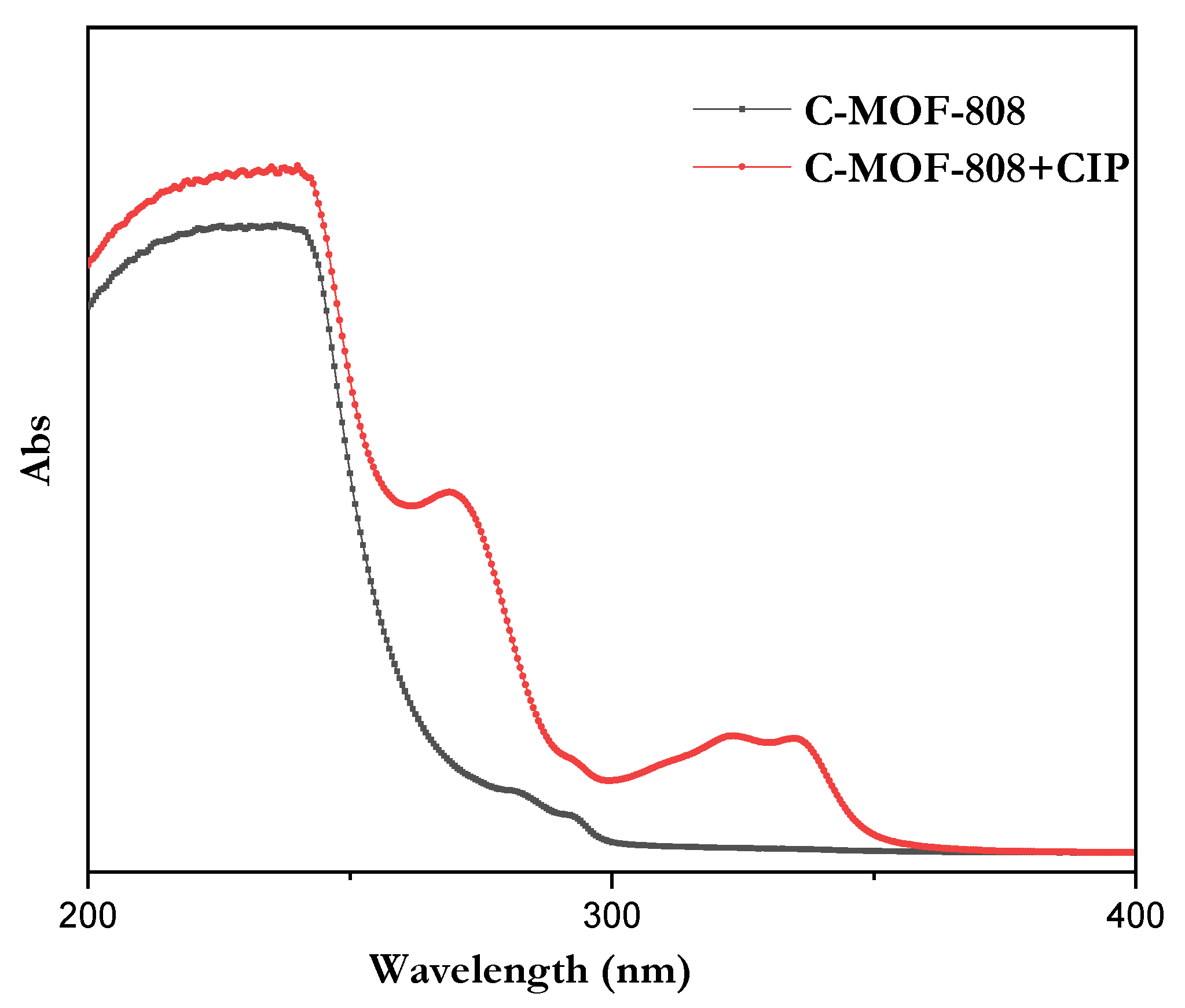
From the absorption spectra, there was no obvious absorption peak observed for a receptor within the range of 300 to 400 nm. However, after the addition of CIP, the absorption intensity was found to be increased at 323 and 335 nm, as shown in Figure 7.

3.3. Absorption and Emission–Kinetic Studies

Therefore, absorption and fluorescence titrations were carried out for the binding of the receptor with CIP. The titrations revealed that the intensity of the absorption (Figure 8a) and emission (Figure 8b) increased at regular intervals as CIP was progressively added (up to 1000 equivalents), resulting in a brilliant blue fluorescence under UV light. An emission band centered at 345 nm in the fluorescence spectra of the receptor red shifted linearly when increasing the concentration of CIP (Figure 8b). The kinetics study demonstrated that the absorption changes were proportional to the CIP concentration (Figure 8c). In the case of the emission spectra, ratiometric detection was observed. The emission peak at 345 nm is found to be lowered (Figure 8d) and the peak at 450 nm is found to be enhanced, when increasing the concentration of CIP up to 30 equivalents, which then starts to decrease when further increasing the concentration of CIP, indicating that CIP undergoes an aggregation-induced quenching phenomena that occurs at a higher concentration.
The detection limit (DL) can be determined from the following equation [49]: DL = K (Sb1/S); where K = 3, Sb1 is the standard deviation of the blank solution (only receptor), and S denotes the slope of the calibration curve (fluorescent spectra). In our experiments, Sb1 is calculated to 18,310 and the slope was determined to be 5835. As a result, the DL for the detection of CIP reaches to 9.4 µM, which is comparable with that reported in the literature [22]. Most of the reported sensors for the fluorescent detection of CIPs are lanthanides (La/Eu/Tb)-based coordination polymer nanoparticles, and their detection limits are varied, in the range of 0.0136 µM–780 nM [39,41,42]. Our designed receptor consists of MOF nanoparticles without doping of the lanthanides; however, it exhibited the detection limit in micromolar concentrations. Moreover, our receptor is simple, cost-effective, and easy to synthesize with an enhanced aqueous solubility over the other reported receptors.

3.4. Binding Mechanism of the Receptors towards CIP

To understand the binding mechanism of the receptor with CIP, it was subjected to FTIR, and the results are merged in Figure 9a. From the spectrum of the receptor (C-MOF-808), the broad peak around 1650–1550 cm−1 corresponds to the –COO stretching mode of the receptor. Moreover, the bands in the 1413 cm−1 and 1378 cm−1 regions are related to the C = C bond and the stretching mode of O–H in the aromatic compound of the receptor, suggesting the formation of C-MOF-808. From the spectra of the receptor with CIP, it can be observed that the crystalline CIP had many characteristic peaks at the fingerprint (1200–1800 cm−1) [50]. There was a peak that appeared in 1711 cm−1 due to the fact that the vibration of the carboxylic acid (C=O) and the vibration of the ketone (C=O) were located at 1618 cm−1 [51,52]. In addition, two relatively strong peaks appeared at 1279 cm−1 and 1485 cm−1, corresponding to the O–H deformation vibration and C-O vibration of CIP, indicating the interaction of CIP with the receptor. The characteristic peaks of the receptor such as 1413 cm−1 and 1378 cm−1 were found to be slightly shifted to 1440 cm−1 and 1382 cm−1, which further confirmed the interaction of CIP with the receptor. Moreover, these compounds were subjected to 1H-NMR spectral analysis, and their results are merged in Figure 9b.
As seen in Figure 9b, the presence of a peak at 1.8 ppm for C-MOF-808, which corresponds to the methyl group, indicates the successful functionalization of MOF-808 with acetic acid (C-MOF-808). The abovementioned peaks were also observed in C-MOF-808+CIP, indicating the presence of C-MOF-808 in the mixture. The peaks at around 8.3–8.4 ppm correspond to the aromatic protons of C-MOF-808. The peaks at 1.15, 1.35, and 3.65 ppm for the CIP corresponding to the –CH2 and -CH groups of the cyclopropyl unit were found to be shifted to 1.3 and 1.4 (only for the CH2 protons) in the mixture, indicating the interaction of CIP with C-MOF-808. The peak at 4.8 ppm corresponds to the solvent peak of D2O. Moreover, the peaks at 7.4 and 7.5 ppm for CIP corresponding to the aromatic protons of the CIP were found to be shifted and merged with the aromatic protons of C-MOF-808 around 8.5-8.7 ppm in the mixture, indicating the interaction of CIP with the receptor.
Based on this evidence, it was concluded that the CIP can interact with the receptor. The receptor structurally possessing the carboxylic acid group (-COOH) and CIP has a piperazine –NH group. Therefore, there is the possibility of H-bonding between the –NH group of the CIP and the –COOH group of the receptor, which altered the absorption and fluorescent properties of the receptor. This strongly suggests that the fluorescent enhancement by CIP is due to the H-bonding induced emission (HBIE) in water, as reported by Haifang Liu and coworkers [53]. Based on the structures of the receptor and analyte, the schematic representation of the proposed mechanism for the possible binding of CIP with the receptor is given in Figure 10.

4. Findings and Discussion

Generally, in fluorescent sensors, the non-aqueous solubility of the receptor is the main drawback that limits their utilization in both environmental and biological applications. In order to overcome the abovementioned limitations, the development of water-soluble fluorescent nanomaterials has received great attention in recent years. Water-soluble fluorescent nanomaterials can be achieved by proper functionalization of the nanomaterials. Here, we achieved aqueous solubility by the functionalization of MOF-808 using citrate, and, thus, the developed receptor can detect CIP in aqueous media with the detection limit of micromolar solutions. Very interestingly, the CIP-induced sensitized emission band is fully separated from both receptor and analyte, which would enable us to detect Cipro by using a common fluorimeter. Hydrogen bonding induced emission (HBIE) is the cause of the changes in the emission spectra when CIP was added. Hence, improving the detection limit to the picomolar level is a still challenging task, one which is in progress.

5. Conclusions

In conclusion, we have designed and synthesized a novel fluorescent receptor based on a citrate-functionalized metal–organic framework consisting of zirconium metal ions (C-MOF-808), which was characterized by XRD and 1H-NMR spectroscopy analysis. Our synthesized receptor is colorless and less fluorescent. It shows high sensitivity towards ciprofloxacin (CIP). The sensing behavior was studied by using absorption and fluorescent spectroscopy. Increasing the concentration of CIP increased the absorption intensity and the red-shifting of the fluorescent intensity when compared to that of neat CIP or C-MOF-808, indicating the detection of CIP. The receptor, before and after binding with CIP, was studied by using FTIR and 1H-NMR spectra and compared with neat CIP. The results showed that CIP has a strong interaction/binding with C-MOF-808. Based on the structures of C-MOF-808 and CIP, a possible detection mechanism was proposed. The hydrogen bonding between the carboxylic group (-COOH) of the C-MOF-808 with the –NH group of the CIP may alter the absorption as well as the fluorescent profiles of the receptor, indicating the phenomena of hydrogen bonding induced emission (HBIE). Due to the aqueous solubility of C-MOF-808, it can be utilized for rapid and in situ imaging of the nucleus in living cells in a fluorescence turn-on mode, which has a great practicability to be used for the nucleus imaging in bioanalytical studies, especially detection of CIP in urinary samples and clinical applications.

Author Contributions

B.-T.L.: resources, validation, and project administration; D.N.: writing, validation, and review and editing; S.K.: partial writing and review and editing; B.R.: conceptualization, funding acquisition, methodology, data curation, and writing. All authors have read and agreed to the published version of the manuscript.

Funding

This study was supported financially by the National Science and Technology Council, Republic of China, Taiwan (MOST 110-2221-E-224-034 and MOST 111-2221-E-224-040).

Data Availability Statement

Not applicable.

Conflicts of Interest

The authors declare no conflict of interest.

References

  1. Yang, J.; Yang, Y.-W. Metal–Organic Frameworks for Biomedical Applications. Small 2020, 16, 1906846. [Google Scholar] [CrossRef] [PubMed]
  2. Chen, L.; Ye, J.W.; Wang, H.P.; Pan, M.; Yin, S.Y.; Wei, Z.W.; Zhang, L.Y.; Wu, K.; Fan, Y.N.; Su, C.Y. Ultrafast water sensing and thermal imaging by a metal-organic framework with switchable luminescence. Nat. Commun. 2017, 8, 15985. [Google Scholar] [CrossRef] [PubMed] [Green Version]
  3. Lin, Z.-J.; Zheng, H.-Q.; Zheng, H.-Y.; Lin, L.-P.; Xin, Q.; Cao, R. Efficient Capture and Effective Sensing of Cr2O72− From Water Using a Zirconium Metal–Organic Framework. Inorg. Chem. 2017, 56, 14178–14188. [Google Scholar] [CrossRef] [PubMed]
  4. Zhu, X.D.; Zhang, K.; Wang, Y.; Long, W.W.; Sa, R.J.; Liu, T.F.; Lu, J. Fluorescent Metal–Organic Framework (MOF) as a Highly Sensitive and Quickly Responsive Chemical Sensor for the Detection of Antibiotics in Simulated Wastewater. Inorg. Chem. 2018, 57, 1060–1065. [Google Scholar] [CrossRef] [PubMed]
  5. Zhou, J.; Li, H.; Zhang, H.; Li, H.; Shi, W.; Cheng, P. A Bimetallic Lanthanide Metal–Organic Material as a Self-Calibrating Color-Gradient Luminescent Sensor. Adv. Mater. 2015, 27, 7072–7077. [Google Scholar] [CrossRef]
  6. Li, H.; Shi, W.; Zhao, K.; Niu, Z.; Li, H.; Cheng, P. Highly Selective Sorption and Luminescent Sensing of Small Molecules Demonstrated in a Multifunctional Lanthanide Microporous Metal–Organic Framework Containing 1D Honeycomb-Type Channels. Chem.—Eur. J. 2013, 19, 3358–3365. [Google Scholar] [CrossRef]
  7. Giménez-Marqués, M.; Hidalgo, T.; Serre, C.; Horcajada, P. Nanostructured metal–organic frameworks and their bio-related applications. Coord. Chem. Rev. 2016, 307, 342–360. [Google Scholar] [CrossRef]
  8. Zheng, H.-Q.; Liu, C.-Y.; Zeng, X.-Y.; Chen, J.; Lü, J.; Lin, R.-G.; Cao, R.; Lin, Z.-J.; Su, J.-W. MOF-808: A Metal–Organic Framework with Intrinsic Peroxidase-Like Catalytic Activity at Neutral pH for Colorimetric Biosensing. Inorg. Chem. 2018, 57, 9096–9104. [Google Scholar] [CrossRef] [PubMed]
  9. Liang, W.; Chevreau, H.; Ragon, F.; Southon, P.D.; Peterson, V.K.; D’Alessandro, D.M. Tuning pore size in a zirconium–tricarboxylate metal–organic framework. CrystEngComm 2014, 16, 6530–6533. [Google Scholar] [CrossRef]
  10. Zheng, H.-Q.; Zeng, Y.-N.; Chen, J.; Lin, R.-G.; Zhuang, W.-E.; Cao, R.; Lin, Z.-J. Zr-Based Metal–Organic Frameworks with Intrinsic Peroxidase-Like Activity for Ultradeep Oxidative Desulfurization: Mechanism of H2O2 Decomposition. Inorg. Chem. 2019, 58, 6983–6992. [Google Scholar] [CrossRef]
  11. Jun, H.J.; Yoo, D.K.; Jhung, S.H. Metal-organic framework (MOF-808) functionalized with ethyleneamines: Selective adsorbent to capture CO2 under low pressure. J. CO2 Util. 2022, 58, 101932. [Google Scholar] [CrossRef]
  12. Jiang, J.; Gándara, F.; Zhang, Y.-B.; Na, K.; Yaghi, O.M.; Klemperer, W.G. Superacidity in Sulfated Metal–Organic Framework-808. J. Am. Chem. Soc. 2014, 136, 12844–12847. [Google Scholar] [CrossRef] [PubMed]
  13. Peng, Y.; Huang, H.; Zhang, Y.; Kang, C.; Chen, S.; Song, L.; Liu, D.; Zhong, C. A versatile MOF-based trap for heavy metal ion capture and dispersion. Nat. Commun. 2018, 9, 187. [Google Scholar] [CrossRef] [Green Version]
  14. Wang, F.; Xue, R.; Ma, Y.; Ge, Y.; Wang, Z.; Qiao, X.; Zhou, P. Study on the performance of a MOF-808-based photocatalyst prepared by a microwave-assisted method for the degradation of antibiotics. RSC Adv. 2021, 11, 32955–32964. [Google Scholar] [CrossRef] [PubMed]
  15. Rojas-Buzo, S.; Bohigues, B.; Lopes, C.W.; Meira, D.M.; Boronat, M.; Moliner, M.; Corma, A. Tailoring Lewis/Brønsted acid properties of MOF nodes via hydrothermal and solvothermal synthesis: Simple approach with exceptional catalytic implications. Chem. Sci. 2021, 12, 10106–10115. [Google Scholar] [CrossRef] [PubMed]
  16. Yi, K.; Li, H.; Zhang, X.; Zhang, L. Designed Tb(III)-Functionalized MOF-808 as Visible Fluorescent Probes for Monitoring Bilirubin and Identifying Fingerprints. Inorg. Chem. 2021, 60, 3172–3180. [Google Scholar] [CrossRef] [PubMed]
  17. Nandi, S.; Sk, M.; Biswas, S. Rapid switch-on fluorescent detection of nanomolar-level hydrazine in water by a diacetoxy-functionalized MOF: Application in paper strips and environmental samples. Dalton Trans. 2020, 49, 12565–12573. [Google Scholar] [CrossRef] [PubMed]
  18. Zhang, X.; Ma, Q.; Liu, X.; Niu, H.; Luo, L.; Li, R.; Feng, X. A turn-off Eu-MOF@Fe2+ sensor for the selective and sensitive fluorescence detection of bromate in wheat flour. Food Chem. 2022, 382, 132379. [Google Scholar] [CrossRef]
  19. Wang, X.-Y.; Yin, H.-Q.; Yin, X.-B. MOF@COFs with Strong Multiemission for Differentiation and Ratiometric Fluorescence Detection. ACS Appl. Mater. Interfaces 2020, 12, 20973–20981. [Google Scholar] [CrossRef]
  20. Liu, X.; Zhang, X.; Li, R.; Du, L.; Feng, X.; Ding, Y. A highly sensitive and selective “turn off-on” fluorescent sensor based on Sm-MOF for the detection of tertiary butylhydroquinone. Dyes Pigments 2020, 178, 108347. [Google Scholar] [CrossRef]
  21. Qin, G.; Wang, J.; Li, L.; Yuan, F.; Zha, Q.; Bai, W.; Ni, Y. Highly water-stable Cd-MOF/Tb3+ ultrathin fluorescence nanosheets for ultrasensitive and selective detection of Cefixime. Talanta 2021, 221, 121421. [Google Scholar] [CrossRef]
  22. Yuphintharakun, N.; Nurerk, P.; Chullasat, K.; Kanatharana, P.; Davis, F.; Sooksawat, D.; Bunkoed, O. A nanocomposite optosensor containing carboxylic functionalized multiwall carbon nanotubes and quantum dots incorporated into a molecularly imprinted polymer for highly selective and sensitive detection of ciprofloxacin. Spectrochim. Acta Part A Mol. Biomol. Spectrosc. 2018, 201, 382–391. [Google Scholar] [CrossRef] [PubMed]
  23. Gayen, P.; Chaplin, B.P. Selective Electrochemical Detection of Ciprofloxacin with a Porous Nafion/Multiwalled Carbon Nanotube Composite Film Electrode. ACS Appl. Mater. Interfaces 2016, 8, 1615–1626. [Google Scholar] [CrossRef] [PubMed]
  24. Zhang, F.; Lin, X.; Jia, R.; Liu, Z.; Liu, Z.; Hong, M.; Jia, A.; Li, Y.; Zhang, H. Fabrication of three-component hydrogen-bonded covalent-organic polymers for ciprofloxacin decontamination from water: Adsorption mechanism and modeling. Mater. Today Chem. 2021, 20, 100463. [Google Scholar] [CrossRef]
  25. Fan, H.; Ma, Y.; Wan, J.; Wang, Y.; Li, Z.; Chen, Y. Adsorption properties and mechanisms of novel biomaterials from banyan aerial roots via simple modification for ciprofloxacin removal. Sci. Total Environ. 2020, 708, 134630. [Google Scholar] [CrossRef] [PubMed]
  26. Velissariou, I.M. The use of fluoroquinolones in children: Recent advances. Expert Rev. Anti-Infect. Ther. 2006, 4, 853–860. [Google Scholar] [CrossRef]
  27. Shan, J.; Li, R.; Yan, K.; Zhu, Y.; Zhang, J. In situ anodic stripping of Cd(II) from CdS quantum dots for electrochemical sensing of ciprofloxacin. Sens. Actuators B Chem. 2016, 237, 75–80. [Google Scholar] [CrossRef]
  28. Bagheri, H.; Khoshsafar, H.; Amidi, S.; Hosseinzadeh Ardakani, Y. Fabrication of an electrochemical sensor based on magnetic multi-walled carbon nanotubes for the determination of ciprofloxacin. Anal. Methods 2016, 8, 3383–3390. [Google Scholar] [CrossRef]
  29. Locatelli, M.; Ciavarella, M.T.; Paolino, D.; Celia, C.; Fiscarelli, E.; Ricciotti, G.; Pompilio, A.; Di Bonaventura, G.; Grande, R.; Zengin, G.; et al. Determination of ciprofloxacin and levofloxacin in human sputum collected from cystic fibrosis patients using microextraction by packed sorbent-high performance liquid chromatography photodiode array detector. J. Chromatogr. A 2015, 1419, 58–66. [Google Scholar] [CrossRef]
  30. Chen, B.; Han, J.; Wang, Y.; Sheng, C.; Liu, Y.; Zhang, G.; Yan, Y. Separation, enrichment and determination of ciprofloxacin using thermoseparating polymer aqueous two-phase system combined with high performance liquid chromatography in milk, egg, and shrimp samples. Food Chem. 2014, 148, 105–111. [Google Scholar] [CrossRef]
  31. Balamurugan, R.; Liu, J.-H.; Liu, B.-T. A review of recent developments in fluorescent sensors for the selective detection of palladium ions. Coord. Chem. Rev. 2018, 376, 196–224. [Google Scholar] [CrossRef]
  32. Rathinam, B.; Liu, B.-T. Highly efficient probe of dinuclear zinc complex for selective detection of oxalic acid. J. Taiwan Inst. Chem. Eng. 2021, 127, 349–356. [Google Scholar] [CrossRef]
  33. Lu, Z.; Hu, Y.; Li, G.; Xia, L. Adamantane Three-Dimensional Porous Organic Framework as a Fluorescence Sensor for Rapid Determination of Tetracycline in Aquatic Products. Chemosensors 2022, 10, 457. [Google Scholar] [CrossRef]
  34. Santonocito, R.; Tuccitto, N.; Pappalardo, A.; Trusso Sfrazzetto, G. Smartphone-Based Dopamine Detection by Fluorescent Supramolecular Sensor. Molecules 2022, 27, 7503. [Google Scholar] [CrossRef]
  35. Li, Y.; Gu, X.; Zhao, J.; Xi, F. Fabrication of a Ratiometric Fluorescence Sensor Based on Carbon Dots as Both Luminophores and Nanozymes for the Sensitive Detection of Hydrogen Peroxide. Molecules 2022, 27, 7379. [Google Scholar] [CrossRef]
  36. Noushija, M.K.; Shanmughan, A.; Mohan, B.; Shanmugaraju, S. Selective Recognition and Reversible “Turn-Off” Fluorescence Sensing of Acetate (CH3COO−) Anion at Ppb Level Using a Simple Quinizarin Fluorescent Dye. Chemistry 2022, 4, 1407–1416. [Google Scholar]
  37. Muchohi, S.N.; Thuo, N.; Karisa, J.; Muturi, A.; Kokwaro, G.O.; Maitland, K. Determination of ciprofloxacin in human plasma using high-performance liquid chromatography coupled with fluorescence detection: Application to a population pharmacokinetics study in children with severe malnutrition. J. Chromatogr. B 2011, 879, 146–152. [Google Scholar] [CrossRef] [Green Version]
  38. El Kojok, H.; El Darra, N.; Khalil, M.; Capo, A.; Pennacchio, A.; Staiano, M.; Camarca, A.; D’Auria, S.; Varriale, A. Fluorescence polarization assay to detect the presence of traces of ciprofloxacin. Sci. Rep. 2020, 10, 4550. [Google Scholar] [CrossRef] [Green Version]
  39. An, X.; Zhu, X.; Liu, J.; Zou, L.; Li, G.; Ye, B. Ratiometric fluorescence detection of ciprofloxacin using the terbium-based coordination polymers. Spectrochim. Acta Part A Mol. Biomol. Spectrosc. 2022, 269, 120775. [Google Scholar] [CrossRef]
  40. Singha, S.; Ahn, K.H. Detection of Ciprofloxacin in Urine through Sensitized Lanthanide Luminescence. Sensors 2016, 16, 2065. [Google Scholar] [CrossRef] [Green Version]
  41. Yue, X.; Wu, C.; Zhou, Z.; Fu, L.; Bai, Y. Fluorescent Sensing of Ciprofloxacin and Chloramphenicol in Milk Samples via Inner Filter Effect and Photoinduced Electron Transfer Based on Nanosized Rod-Shaped Eu-MOF. Foods 2022, 11, 3138. [Google Scholar] [CrossRef]
  42. Liu, B.; Huang, Y.; Shen, Q.; Zhu, X.; Hao, Y.; Qu, P.; Xu, M. Turn-on fluorescence detection of ciprofloxacin in tablets based on lanthanide coordination polymer nanoparticles. RSC Adv. 2016, 6, 100743–100747. [Google Scholar] [CrossRef]
  43. Furukawa, H.; Gándara, F.; Zhang, Y.-B.; Jiang, J.; Queen, W.L.; Hudson, M.R.; Yaghi, O.M. Water Adsorption in Porous Metal–Organic Frameworks and Related Materials. J. Am. Chem. Soc. 2014, 136, 4369–4381. [Google Scholar] [CrossRef]
  44. Feng, J.; Zhong, Y.; Xie, M.; Li, M.; Jiang, S. Using MOF-808 as a Promising Support to Immobilize Ru for Selective Hydrogenation of Levulinic Acid to γ-Valerolactone. Catal. Lett. 2021, 151, 86–94. [Google Scholar] [CrossRef]
  45. Xu, J.; Liu, J.; Li, Z.; Wang, X.; Xu, Y.; Chen, S.; Wang, Z. Optimized synthesis of Zr(iv) metal organic frameworks (MOFs-808) for efficient hydrogen storage. New J. Chem. 2019, 43, 4092–4099. [Google Scholar] [CrossRef]
  46. Liu, Y.; Chen, G.; Chen, J.; Niu, H. Excellent Catalytic Performance of Ce–MOF with Abundant Oxygen Vacancies Supported Noble Metal Pt in the Oxidation of Toluene. Catalysts 2022, 12, 775. [Google Scholar]
  47. Healey, K.; Liang, W.; Southon, P.D.; Church, T.L.; D’Alessandro, D.M. Photoresponsive spiropyran-functionalised MOF-808: Postsynthetic incorporation and light dependent gas adsorption properties. J. Mater. Chem. A 2016, 4, 10816–10819. [Google Scholar] [CrossRef]
  48. Hadi Al-Kadhemy, M.F.; Abbas, K.N.; Abdalmuhdi, W.B. Physical Properties of Rhodamine 6G Laser Dye Combined in Polyvinyl Alcohol films as Heat Sensor. IOP Conf. Ser. Mater. Sci. Eng. 2020, 928, 072126. [Google Scholar] [CrossRef]
  49. Goswami, S.; Chakraborty, S.; Paul, S.; Halder, S.; Panja, S.; Mukhopadhyay, S.K. A new pyrene based highly sensitive fluorescence probe for copper(ii) and fluoride with living cell application. Org. Biomol. Chem. 2014, 12, 3037–3044. [Google Scholar] [CrossRef]
  50. Xing, X.; Feng, J.; Lv, G.; Song, K.; Mei, L.; Liao, L.; Wang, X.; Xu, B. Adsorption Mechanism of Ciprofloxacin from Water by Synthesized Birnessite. Adv. Mater. Sci. Eng. 2015, 2015, 148423. [Google Scholar] [CrossRef] [Green Version]
  51. Gu, C.; Karthikeyan, K.G. Sorption of the Antimicrobial Ciprofloxacin To Aluminum and Iron Hydrous Oxides. Environ. Sci. Technol. 2005, 39, 9166–9173. [Google Scholar] [CrossRef] [PubMed]
  52. Trivedi, P.; Vasudevan, D. Spectroscopic Investigation of Ciprofloxacin Speciation at the Goethite−Water Interface. Environ. Sci. Technol. 2007, 41, 3153–3158. [Google Scholar] [CrossRef] [PubMed]
  53. Liu, H.; Yang, J.; Li, Z.; Xiao, L.; Aryee, A.A.; Sun, Y.; Yang, R.; Meng, H.; Qu, L.; Lin, Y.; et al. Hydrogen-Bond-Induced Emission of Carbon Dots for Wash-Free Nucleus Imaging. Anal. Chem. 2019, 91, 9259–9265. [Google Scholar] [CrossRef] [PubMed]
Figure 1. Synthesis of MOF-808 (structure is reprinted with permission from J. Am. Chem. Soc. 2014, 136, 37, 12844–12847; copyright 2022 American Chemical Society) [12].
Micromachines 13 02097 g001
Figure 2. Synthesis of receptor C-MOF-808.
Micromachines 13 02097 g002
Figure 3. XRD patterns of (a) MOF and C-MOF-808; (b) reported XRD pattern of MOF-808 for comparison [46].
Micromachines 13 02097 g003
Figure 4. The SEM images of (a) MOF-808 and (b) C-MOF-808.
Micromachines 13 02097 g004
Figure 5. 1H-NMR spectrum of (a) MOF-808 (deutriated TFA) and (b) C-MOF-808 (D2O).
Micromachines 13 02097 g005
Figure 6. (a) Fluorescence spectra of receptor, CIP, and their mixture (λex = 310 nm) and (b) real images of the aqueous solution of receptor with increasing concentration of CIP under UV-light.
Micromachines 13 02097 g006
Figure 7. The absorption spectra of receptor and with CIP.
Micromachines 13 02097 g007
Figure 8. The changes in (a) the absorption and (b) emission spectra of the receptor at pH 6.9 upon titration with CIP (λex = 310 nm; inset: contour plot). The absorbance (c) and emission (d) intensities of the receptor as a function of the equivalents of CIP added.
Micromachines 13 02097 g008
Figure 9. (a) FTIR spectra of the receptor (C-MOF-808) and the analyte (CIP) and (b) 1H-NMR spectra of receptor, CIP, and receptor with CIP in D2O.
Micromachines 13 02097 g009
Figure 10. The schematic representation of the proposed mechanism of binding of CIP with the receptor.
Micromachines 13 02097 g010
Publisher’s Note: MDPI stays neutral with regard to jurisdictional claims in published maps and institutional affiliations.