Gastrointestinal Stromal Tumours (GIST) in Young Adult (18–40 Years) Patients: A Report from the Dutch GIST Registry

 , , , , ,
Abstract
:1. Introduction
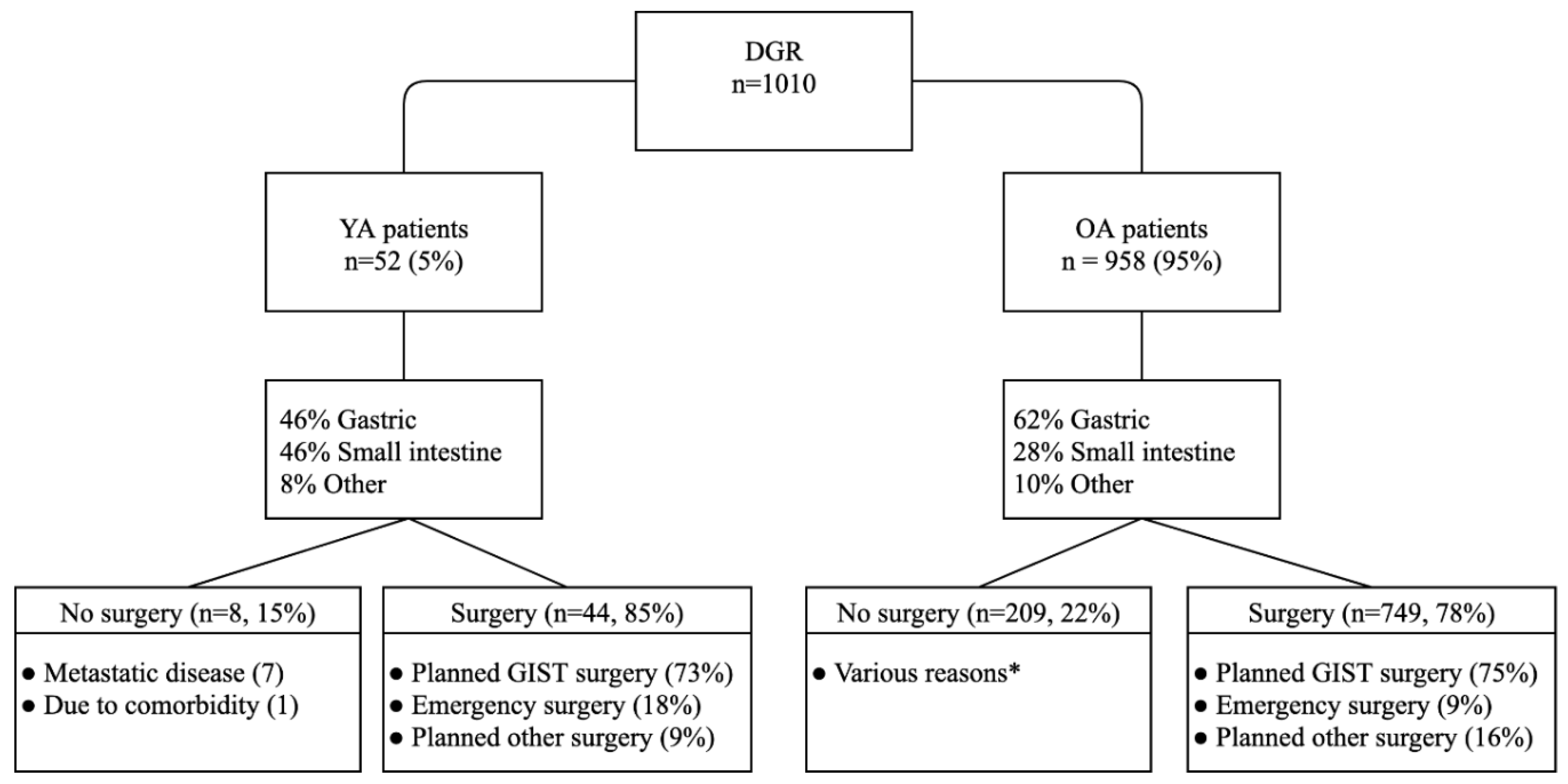
2. Results
2.1. Patient Characteristics
2.2. Pathology
2.3. Treatment
2.4. Outcome
2.5. Impact of GIST on Life
2.6. Comparison of Age Groups
2.6.1. Comparison within the YA Cohort
2.6.2. Comparison between the YA and OA Patients
3. Discussion
4. Materials and Methods
4.1. Patient Selection and Variables
4.2. Pathology
4.3. Quality of Life
4.4. Statistical Analyses and Outcome
5. Conclusions
Supplementary Materials
Author Contributions
Funding
Conflicts of Interest
References
- Joensuu, H. Gastrointestinal stromal tumor (GIST). Ann. Oncol. 2006, 17 (Suppl. S10), x280–x286. [Google Scholar] [CrossRef] [PubMed]
- Soreide, K.; Sandvik, O.M.; Soreide, J.A.; Giljaca, V.; Jureckova, A.; Bulusu, V.R. Global epidemiology of gastrointestinal stromal tumours (GIST): A systematic review of population-based cohort studies. Cancer Epidemiol. 2016, 40, 39–46. [Google Scholar] [CrossRef] [PubMed] [Green Version]
- Miettinen, M.; Sobin, L.H.; Lasota, J. Gastrointestinal stromal tumors of the stomach: A clinicopathologic, immunohistochemical, and molecular genetic study of 1765 cases with long-term follow-up. Am. J. Surg. Pathol. 2005, 29, 52–68. [Google Scholar] [CrossRef] [PubMed]
- Nilsson, B.; Bumming, P.; Meis-Kindblom, J.M.; Oden, A.; Dortok, A.; Gustavsson, B.; Sablinska, K.; Kindblom, L.G. Gastrointestinal stromal tumors: The incidence, prevalence, clinical course, and prognostication in the preimatinib mesylate era—A population-based study in western Sweden. Cancer 2005, 103, 821–829. [Google Scholar] [CrossRef] [PubMed]
- Tryggvason, G.; Gislason, H.G.; Magnusson, M.K.; Jonasson, J.G. Gastrointestinal stromal tumors in Iceland, 1990–2003: The icelandic GIST study, a population-based incidence and pathologic risk stratification study. Int. J. Cancer 2005, 117, 289–293. [Google Scholar] [CrossRef]
- Kaemmer, D.A.; Otto, J.; Lassay, L.; Steinau, G.; Klink, C.; Junge, K.; Klinge, U.; Schumpelick, V. The Gist of literature on pediatric GIST: Review of clinical presentation. J. Pediatr. Hematol. Oncol. 2009, 31, 108–112. [Google Scholar] [CrossRef]
- Rink, L.; Godwin, A.K. Clinical and molecular characteristics of gastrointestinal stromal tumors in the pediatric and young adult population. Curr. Oncol. Rep. 2009, 11, 314–321. [Google Scholar] [CrossRef]
- Quiroz, H.J.; Willobee, B.A.; Sussman, M.S.; Fox, B.R.; Thorson, C.M.; Sola, J.E.; Perez, E.A. Pediatric gastrointestinal stromal tumors-a review of diagnostic modalities. Transl. Gastroenterol. Hepatol. 2018, 3, 54. [Google Scholar] [CrossRef]
- Diment, J.; Tamborini, E.; Casali, P.; Gronchi, A.; Carney, J.A.; Colecchia, M. Carney triad: Case report and molecular analysis of gastric tumor. Hum. Pathol. 2005, 36, 112–116. [Google Scholar] [CrossRef]
- Pasini, B.; McWhinney, S.R.; Bei, T.; Matyakhina, L.; Stergiopoulos, S.; Muchow, M.; Boikos, S.A.; Ferrando, B.; Pacak, K.; Assie, G.; et al. Clinical and molecular genetics of patients with the Carney-Stratakis syndrome and germline mutations of the genes coding for the succinate dehydrogenase subunits SDHB, SDHC, and SDHD. Eur. J. Hum. Genet. 2008, 16, 79–88. [Google Scholar] [CrossRef]
- Gasparotto, D.; Rossi, S.; Polano, M.; Tamborini, E.; Lorenzetto, E.; Sbaraglia, M.; Mondello, A.; Massani, M.; Lamon, S.; Bracci, R.; et al. Quadruple-negative GIST is a sentinel for unrecognized neurofibromatosis type 1 syndrome. Clin. Cancer Res. 2017, 23, 273–282. [Google Scholar] [CrossRef] [PubMed] [Green Version]
- Heinrich, M.C.; Corless, C.L.; Duensing, A.; McGreevey, L.; Chen, C.J.; Joseph, N.; Singer, S.; Griffith, D.J.; Haley, A.; Town, A.; et al. PDGFRA activating mutations in gastrointestinal stromal tumors. Science 2003, 299, 708–710. [Google Scholar] [CrossRef] [PubMed]
- Hirota, S.; Isozaki, K.; Moriyama, Y.; Hashimoto, K.; Nishida, T.; Ishiguro, S.; Kawano, K.; Hanada, M.; Kurata, A.; Takeda, M.; et al. Gain-of-function mutations of c-kit in human gastrointestinal stromal tumors. Science 1998, 279, 577–580. [Google Scholar] [CrossRef] [PubMed]
- Corless, C.L.; Barnett, C.M.; Heinrich, M.C. Gastrointestinal stromal tumours: Origin and molecular oncology. Nat. Rev. Cancer 2011, 11, 865–878. [Google Scholar] [CrossRef] [PubMed]
- Pantaleo, M.A.; Astolfi, A.; Urbini, M.; Nannini, M.; Paterini, P.; Indio, V.; Saponara, M.; Formica, S.; Ceccarelli, C.; Casadio, R.; et al. Analysis of all subunits, SDHA, SDHB, SDHC, SDHD, of the succinate dehydrogenase complex in KIT/PDGFRA wild-type GIST. Eur. J. Hum. Genet. 2014, 22, 32–39. [Google Scholar] [CrossRef] [PubMed] [Green Version]
- Daniels, M.; Lurkin, I.; Pauli, R.; Erbstosser, E.; Hildebrandt, U.; Hellwig, K.; Zschille, U.; Luders, P.; Kruger, G.; Knolle, J.; et al. Spectrum of KIT/PDGFRA/BRAF mutations and Phosphatidylinositol-3-Kinase pathway gene alterations in gastrointestinal stromal tumors (GIST). Cancer Lett. 2011, 312, 43–54. [Google Scholar] [CrossRef]
- Flavahan, W.A.; Drier, Y.; Johnstone, S.E.; Hemming, M.L.; Tarjan, D.R.; Hegazi, E.; Shareef, S.J.; Javed, N.M.; Raut, C.P.; Eschle, B.K.; et al. Altered chromosomal topology drives oncogenic programs in SDH-deficient GISTs. Nature 2019, 575, 229–233. [Google Scholar] [CrossRef]
- Casali, P.G.; Abecassis, N.; Aro, H.T.; Bauer, S.; Biagini, R.; Bielack, S.; Bonvalot, S.; Boukovinas, I.; Bovee, J.; Brodowicz, T.; et al. Gastrointestinal stromal tumours: ESMO-EURACAN clinical practice guidelines for diagnosis, treatment and follow-up. Ann. Oncol. 2018, 29, iv267. [Google Scholar] [CrossRef]
- Prakash, S.; Sarran, L.; Socci, N.; DeMatteo, R.P.; Eisenstat, J.; Greco, A.M.; Maki, R.G.; Wexler, L.H.; LaQuaglia, M.P.; Besmer, P.; et al. Gastrointestinal stromal tumors in children and young adults: A clinicopathologic, molecular, and genomic study of 15 cases and review of the literature. J. Pediatr. Hematol. Oncol. 2005, 27, 179–187. [Google Scholar] [CrossRef]
- Miettinen, M.; Lasota, J.; Sobin, L.H. Gastrointestinal stromal tumors of the stomach in children and young adults: A clinicopathologic, immunohistochemical, and molecular genetic study of 44 cases with long-term follow-up and review of the literature. Am. J. Surg. Pathol. 2005, 29, 1373–1381. [Google Scholar] [CrossRef]
- Rutkowski, P.; Magnan, H.; Chou, A.J.; Benson, C. Treatment of gastrointestinal stromal tumours in paediatric and young adult patients with sunitinib: A multicentre case series. BMC Cancer 2017, 17, 717. [Google Scholar] [CrossRef] [PubMed] [Green Version]
- Benesch, M.; Leuschner, I.; Wardelmann, E.; Thielen, M.; Schmid, I.; Kontny, U.; Ebetsberger, G.; Frey, E.; Graf, N.; Schneider, D.T.; et al. Gastrointestinal stromal tumours in children and young adults: A clinicopathologic series with long-term follow-up from the database of the Cooperative Weichteilsarkom Studiengruppe (CWS). Eur. J. Cancer 2011, 47, 1692–1698. [Google Scholar] [CrossRef] [PubMed]
- Rubin, B.P.; Heinrich, M.C.; Corless, C.L. Gastrointestinal stromal tumour. Lancet 2007, 369, 1731–1741. [Google Scholar] [CrossRef]
- Agaimy, A.; Wunsch, P.H. Lymph node metastasis in gastrointestinal stromal tumours (GIST) occurs preferentially in young patients < or = 40 years: An overview based on our case material and the literature. Langenbecks Arch. Surg. 2009, 394, 375–381. [Google Scholar] [CrossRef]
- Rege, T.A.; Wagner, A.J.; Corless, C.L.; Heinrich, M.C.; Hornick, J.L. “Pediatric-type” gastrointestinal stromal tumors in adults: Distinctive histology predicts genotype and clinical behavior. Am. J. Surg. Pathol. 2011, 35, 495–504. [Google Scholar] [CrossRef]
- Younger, E.; Husson, O.; Bennister, L.; Whelan, J.; Wilson, R.; Roast, A.; Jones, R.L.; van der Graaf, W.T. Age-related sarcoma patient experience: Results from a national survey in England. BMC Cancer 2018, 18, 991. [Google Scholar] [CrossRef] [Green Version]
- De Rojas, T.; Kasper, B.; Van der Graaf, W.; Pfister, S.M.; Bielle, F.; Ribalta, T.; Shenjere, P.; Preusser, M.; Frohling, S.; Golfinopoulos, V.; et al. EORTC SPECTA-AYA: A unique molecular profiling platform for adolescents and young adults with cancer in Europe. Int. J. Cancer 2019. [Google Scholar] [CrossRef]
- Ferrari, A.; Thomas, D.; Franklin, A.R.; Hayes-Lattin, B.M.; Mascarin, M.; van der Graaf, W.; Albritton, K.H. Starting an adolescent and young adult program: Some success stories and some obstacles to overcome. J. Clin. Oncol. 2010, 28, 4850–4857. [Google Scholar] [CrossRef] [Green Version]
- Kang, G.; Park, Y.S.; Jung, E.S.; Joo, M.; Kang, M.S.; Ahn, S.; Kang, G.H.; Kim, K.M. Gastrointestinal stromal tumors in children and young adults: A clinicopathologic and molecular genetic study of 22 Korean cases. APMIS 2013, 121, 938–944. [Google Scholar] [CrossRef]
- Fero, K.E.; Coe, T.M.; Fanta, P.T.; Tang, C.M.; Murphy, J.D.; Sicklick, J.K. Surgical management of adolescents and young adults with gastrointestinal stromal tumors: A US population-based analysis. JAMA Surg. 2017, 152, 443–451. [Google Scholar] [CrossRef]
- Farag, S.; van Coevorden, F.; Sneekes, E.; Grunhagen, D.J.; Reyners, A.K.L.; Boonstra, P.A.; van der Graaf, W.T.; Gelderblom, H.J.; Steeghs, N. Elderly patients with gastrointestinal stromal tumour (GIST) receive less treatment irrespective of performance score or comorbidity—A retrospective multicentre study in a large cohort of GIST patients. Eur. J. Cancer 2017, 86, 318–325. [Google Scholar] [CrossRef] [PubMed]
- Boikos, S.A.; Pappo, A.S.; Killian, J.K.; LaQuaglia, M.P.; Weldon, C.B.; George, S.; Trent, J.C.; von Mehren, M.; Wright, J.A.; Schiffman, J.D.; et al. Molecular subtypes of KIT/PDGFRA wild-type gastrointestinal stromal tumors: A report from the national institutes of health gastrointestinal stromal tumor clinic. JAMA Oncol. 2016, 2, 922–928. [Google Scholar] [CrossRef] [PubMed] [Green Version]
- Weldon, C.B.; Madenci, A.L.; Boikos, S.A.; Janeway, K.A.; George, S.; von Mehren, M.; Pappo, A.S.; Schiffman, J.D.; Wright, J.; Trent, J.C.; et al. Surgical management of wild-type gastrointestinal stromal tumors: A report from the national institutes of health pediatric and wildtype gist clinic. J. Clin. Oncol. 2017, 35, 523–528. [Google Scholar] [CrossRef] [PubMed] [Green Version]
- Boonstra, P.A.; Steeghs, N.; Farag, S.; van Coevorden, F.; Gelderblom, H.; Grunhagen, D.J.; Desar, I.M.E.; van der Graaf, W.T.A.; Bonenkamp, J.J.; Reyners, A.K.L.; et al. Surgical and medical management of small bowel gastrointestinal stromal tumors: A report of the Dutch GIST registry. Eur. J. Surg. Oncol. 2019, 45, 410–415. [Google Scholar] [CrossRef] [PubMed]
- Soomers, V.L.M.N.; Husson, O.; Young, R.J.; Desar, I.M.E.; Van der Graaf, W.T.A. The sarcoma diagnostic interval: A systematic review on length, contributing factors and patient outcomes. ESMO Open 2020, 5. [Google Scholar] [CrossRef] [PubMed] [Green Version]
- Van Nederveen, F.H.; Gaal, J.; Favier, J.; Korpershoek, E.; Oldenburg, R.A.; de Bruyn, E.M.; Sleddens, H.F.; Derkx, P.; Riviere, J.; Dannenberg, H.; et al. An immunohistochemical procedure to detect patients with paraganglioma and phaeochromocytoma with germline SDHB, SDHC, or SDHD gene mutations: A retrospective and prospective analysis. Lancet Oncol. 2009, 10, 764–771. [Google Scholar] [CrossRef] [Green Version]
- Miettinen, M.; Lasota, J. Gastrointestinal stromal tumors: Pathology and prognosis at different sites. Semin. Diagn. Pathol. 2006, 23, 70–83. [Google Scholar] [CrossRef]


| Characteristic | Total (%) |
|---|---|
| No. of YA Patients | 52 (100) |
| Median age at Diagnosis (Range) | 35 years (18–40) |
| Gender | |
| Male | 28 (53.8) |
| Female | 24 (46.2) |
| Median Baseline Tumour Size in mm (Range) | 60 (6–370) |
| Location | |
| Gastric | 24 (46.2) |
| Small intestine | 24 (46.2) |
| Rectum | 3 (5.8) |
| Oesophageal–cardiac junction | 1 (1.9) |
| Syndromic Presentation | |
| No Yes | 39 (90.7) 4 (9.3) |
| Neurofibromatosis 1 | 2 |
| Carney triad | 1 |
| KIT exon 11 germline mutation | 1 |
| Unknown | 9 |
| Symptoms at Diagnosis* | |
| Active gastrointestinal bleeding | 11 (22.4) |
| Abdominal pain | 9 (18.4) |
| Weight loss | 9 (18.4) |
| Mass related symptoms (e.g., reflux, palpable mass, distended abdomen) | 7 (14.3) |
| Anaemia related symptoms | 6 (12.2) |
| Acute abdomen | 6 (12.2) |
| Nausea/vomiting | 4 (8.2) |
| Change in bowel habits | 3 (6.1) |
| Analysis vitamin B12 deficiency | 1 (2.0) |
| None (GIST coincidental finding) | 8 (16.3) |
| Unknown | 3 |
| Second Malignancies | |
| No | 49 (94.2) |
| Yes | 3 (5.8) |
| Tumour Status at Diagnosis | |
| Local disease | 45 (86.5) |
| Metastasized | 7 (13.5) |
| Risk Classification | |
| None Low | 2 (3.9) 19 (37.3) |
| Moderate | 9 (17.6) |
| High Unknown | 21 (41.2) 1 |
| Characteristic | No. (%) |
|---|---|
| Histology | |
| Spindle cell | 37 (75.5) |
| Epithelioid | 3 (6.1) |
| Mixed type | 9 (18.4) |
| Not reported | 3 |
| Baseline Mitotic Rate | |
| Low (≤ 5/5 mm2) | 35 (68.6) |
| High (> 5/5 mm2) | 16 (31.4) |
| Unknown | 1 |
| Mutation Status | |
| KIT | 34 (69.4) |
| Exon 11 * | 32 |
| Exon 9 | 3 |
| PDGFRA | 3 (6.1) |
| D842V | 2 |
| Non-D842V | 1 |
| SDH deficiency | 4 (8.2) |
| NF1 associated | 2 (4.1) |
| ETV6-NTRK3 gene fusion | 1 (2.0) |
| KIT/PDGFRA WT (BRAF and/or SDH not performed) | 1 (2.0) |
| Triple WT (SDH not performed) | 1 (2.0) |
| Quadruple WT | 3 (6.1) |
| No mutational analysis performed | 3 |
| Characteristic | 18–29 Years (n = 15) | 30–40 Years (n = 37) | p-Value |
|---|---|---|---|
| Median Age at Diagnosis (Range) | 26 (18–29) | 38 (30–40) | |
| Gender | 0.07 | ||
| Male | 5 (33.3) | 23 (62.2) | |
| Female | 10 (66.6) | 14 (37.8) | |
| Median Baseline Tumour Size in mm (Range) | 45 (14–170) | 60 (6–370) | 0.23 |
| Primary Site Grouped | 0.48 | ||
| Gastric | 5 (33.3) | 19 (51.4) | |
| Small intestine | 9 (60.0) | 15 (40.5) | |
| Other | 1 (6.7) | 3 (8.1) | |
| Histology | 0.74 | ||
| Spindle cell | 12 (80.0) | 24 (73.5) | |
| Epithelioid | 0 (0.0) | 3 (8.8) | |
| Mixed type | 3 (20.0) | 6 (17.6) | |
| Unknown | 0 | 3 | |
| Tumour Status at Diagnosis | 1.00 | ||
| Local disease | 13 (87.7) | 32 (86.5) | |
| Metastasized | 2 (13.3) | 5 (13.5) | |
| Risk Category | 0.53 | ||
| Low/moderate | 7 (46.7) | 23 (62.2) | |
| High | 7 (53.3) | 14 (37.8) | |
| Unknown | 1 | 0 | |
| Mutation Status | 0.72 | ||
| KIT | 10 (66.7) | 24 (70.6) | |
| PDGFRA | 0 (0.0) | 3 (8.8) | |
| SDH deficient | 2 (13.3) | 2 (5.9) | |
| NF1 associated | 1 (6.7) | 1 (2.9) | |
| ETV6-NTRK3 gene fusion | 0 (0.0) | 1 (2.9) | |
| WT (min. KIT and PDGFRA) Unknown | 2 (13.3) 0 | 3 (8.8) 3 | |
| Baseline Mitotic Rate | 0.32 | ||
| Low (≤ 5/5 mm2) | 8 (57.1) | 27 (73.0) | |
| High (> 5/5 mm2) Unknown | 6 (42.9) 1 | 10 (27.0) 0 | |
| Surgery | 0.41 | ||
| Yes No | 14 (93.3) 1 (6.7) | 30 (81.1) 7 (18.9) | |
| Patients who Received Surgery | 14 (93.3) | 30 (81.1) | 0.41 |
| Resection Margin | 0.26 | ||
| Negative (R0) | 12 (85.7) | 27 (96.4) | |
| Positive (R1/R2) | 2 (14.3) | 1 (3.6) | |
| Unknown | 0 | 2 | |
| Tumour Rupture | 1.00 | ||
| No | 11 (84.6) | 24 (80.0) | |
| Yes | 2 (15.4) | 5 (20.0) | |
| Unknown | 1 | 1 |
| Characteristic | Total (%) n = 1010 | YA (%) n = 52 | OA (%) n = 958 | p-Value |
|---|---|---|---|---|
| Median Age at Diagnosis (range) | 64 (18–95) | 35 (18–40) | 65 (41–95) | |
| Gender | 0.89 | |||
| Male | 533 (52.8) | 28 (53.8) | 505 (52.7) | |
| Female | 477 (47.2) | 24 (46.2) | 453 (47.3) | |
| Median Baseline Tumour Size in mm (Range) | 72 (1–1000) | 60 (6–370) | 73 (1–1000) | 0.09 |
| Primary Site Grouped | 0.03 * | |||
| Gastric Small intestine Other | 604 (60.7) 290 (29.1) 101 (10.2) | 24 (46.2) 24 (46.2) 4 (7.7) | 580 (61.5) 266 (28.2) 91 (10.3) | |
| Syndromic Presentation | 0.047 * | |||
| No | 799 (96.7) | 39 (90.7) | 760 (97.1) | |
| Yes | 27 (3.3) | 4 (9.3) | 23 (2.9) | |
| Unknown | 184 | 9 | 175 | |
| Tumour Status at Diagnosis | 0.09 | |||
| Local disease | 767 (76.0) | 45 (86.5) | 722 (75.4) | |
| Metastasized | 242 (24.0) | 7 (13.5) | 235 (24.6) | |
| Unknown | 1 | 0 | 1 | |
| Histology | 0.26 | |||
| Spindle cell | 739 (78.3) | 37 (75.5) | 702 (78.4) | |
| Epithelioid | 94 (10.0) | 3 (6.1) | 91 (10.2) | |
| Mixed type | 111 (11.8) | 9 (18.4) | 102 (11.4) | |
| Unknown | 66 | 3 | 63 | |
| Baseline Mitotic Rate | 0.76 | |||
| Low (≤5/5 mm2) | 491 (66.4) | 35 (68.6) | 462 (66.2) | |
| High (>5/5 mm2) | 252 (33.6) | 16 (31.4) | 236 (33.8) | |
| Unknown | 261 | 1 | 260 | |
| Mutation Status | 0.001 * | |||
| KIT | 630 (75.9) | 34 (69.4) | 596 (76.3) | |
| PDGFRA | 106 (12.8) | 3 (6.1) | 103 (13.2) | |
| BRAF | 1 (0.1) | 0 | 1 (0.1) | |
| SDH deficiency | 13 (1.6) | 4 (8.2) | 9 (1.2) | |
| NF1 associated | 16 (1.9) | 2 (4.1) | 14 (1.8) | |
| ETV6-NTRK3 gene fusion | 1 (0.1) | 1 (2.0) | 0 | |
| Wildtype (at least KIT and PDGFRA) | 63 (7.6) | 5 (10.2) | 58 (7.4) | |
| Unknown | 180 | 3 | 177 | |
| KIT/PDGFRA vs. non-KIT/PDGFRA | 0.008 * | |||
| KIT/PDGFRA | 736 (88.7) | 37 (75.5) | 699 (89.5) | |
| Non-KIT/PDGFRA | 94 (11.3) | 12 (24.5) | 82 (10.5) | |
| Patients who Received Surgery | 793 (78.5) | 44 (84.6) | 749 (78.2) | 0.30 |
| Surgery Setting | 0.06 | |||
| Planned operation | 712 (90.7) | 36 (81.8) | 676 (91.2) | |
| Emergency setting | 73 (9.3) | 8 (18.2) | 65 (8.8) | |
| Unknown | 8 | 0 | 8 |
© 2020 by the authors. Licensee MDPI, Basel, Switzerland. This article is an open access article distributed under the terms and conditions of the Creative Commons Attribution (CC BY) license (http://creativecommons.org/licenses/by/4.0/).
Share and Cite
IJzerman, N.S.; Drabbe, C.; den Hollander, D.; Mohammadi, M.; van Boven, H.; Desar, I.M.E.; Gelderblom, H.; Grünhagen, D.J.; Reyners, A.K.L.; van Noesel, M.M.; et al. Gastrointestinal Stromal Tumours (GIST) in Young Adult (18–40 Years) Patients: A Report from the Dutch GIST Registry. Cancers 2020, 12, 730. https://doi.org/10.3390/cancers12030730
IJzerman NS, Drabbe C, den Hollander D, Mohammadi M, van Boven H, Desar IME, Gelderblom H, Grünhagen DJ, Reyners AKL, van Noesel MM, et al. Gastrointestinal Stromal Tumours (GIST) in Young Adult (18–40 Years) Patients: A Report from the Dutch GIST Registry. Cancers. 2020; 12(3):730. https://doi.org/10.3390/cancers12030730
Chicago/Turabian StyleIJzerman, Nikki S., Cas Drabbe, Dide den Hollander, Mahmoud Mohammadi, Hester van Boven, Ingrid M.E. Desar, Hans Gelderblom, Dirk J. Grünhagen, An K.L. Reyners, Max M. van Noesel, and et al. 2020. "Gastrointestinal Stromal Tumours (GIST) in Young Adult (18–40 Years) Patients: A Report from the Dutch GIST Registry" Cancers 12, no. 3: 730. https://doi.org/10.3390/cancers12030730