Article

Oligoprogressive Non-Small-Cell Lung Cancer under Treatment with PD-(L)1 Inhibitors

1
Translational Lung Research Center Heidelberg (TLRC-H), Member of the German Center for Lung Research (DZL), 69120 Heidelberg, Germany
2
Department for Diagnostic and Interventional Radiology, Heidelberg University Hospital, 69120 Heidelberg, Germany
3
Diagnostic and Interventional Radiology with Nuclear Medicine, Thoraxklinik at Heidelberg University Hospital, 69126 Heidelberg, Germany
4
Department of Thoracic Oncology, Thoraxklinik at Heidelberg University Hospital, 69126 Heidelberg, Germany
5
Department of Thoracic Surgery, Thoraxklinik at Heidelberg University Hospital, 69126 Heidelberg, Germany
6
Department of Pneumology, Thoraxklinik at Heidelberg University Hospital, 69126 Heidelberg, Germany
7
Translational Research Unit, Thoraxklinik at Heidelberg University Hospital, 69126 Heidelberg, Germany
8
Pharmacy, Thoraxklinik at Heidelberg University Hospital, 69126 Heidelberg, Germany
9
Institute of Pathology, Heidelberg University Hospital, 69120 Heidelberg, Germany
10
Department of Radiation Oncology, Heidelberg University Hospital, 69120 Heidelberg, Germany
*
Authors to whom correspondence should be addressed.
Cancers 2020, 12(4), 1046; https://doi.org/10.3390/cancers12041046
Submission received: 25 March 2020 / Revised: 16 April 2020 / Accepted: 20 April 2020 / Published: 23 April 2020

Abstract

:
Oligoprogression (OPD) of non-small-cell lung cancer (NSCLC) occurs in approximately half of patients under targeted compounds (TKI) and facilitates use of regional therapies that can prolong survival. In order to characterize OPD in immunotherapy (IO)-treated NSCLC, we analyzed the failure pattern under PD-1/PD-L1 inhibitors (n = 297) or chemoimmunotherapy (n = 75). Under IO monotherapy, OPD was more frequent (20% vs. 10%, p < 0.05), occurred later (median 11 vs. 5 months, p < 0.01), affected fewer sites (mean 1.1 vs. 1.5, p < 0.05), and involved fewer lesions (1.4 vs. 2.3, p < 0.05) in the first compared to later lines. Lymph nodes (42%, mainly mediastinal) and the brain (39%) were mostly affected, followed by the lung (24%) and other organs. Compared to multifocal progression, OPD occurred later (11 vs. 4 months, p < 0.001) and was associated with longer survival (26 vs. 13 months, p < 0.001) and higher tumor PD-L1 expression (p < 0.001). Chemoimmunotherapy showed a similar incidence of OPD as IO monotherapy (13% vs. 11% at 2 years). Local treatments were applied regularly for brain but only in 50% for extracranial lesions. Thus, NSCLC oligoprogression is less common under IO than under TKI, but also favorable. Since its frequency drops later in the disease, regular restaging and multidisciplinary evaluation are essential in order to exploit the full therapeutic potential.

1. Introduction

Oligoprogression (OPD) is a relatively new concept that emerged as more effective systemic therapies became available in oncology and denotes anatomically restricted tumor progression in patients with otherwise controlled widespread disease [1]. Such an asynchronous behavior of metastatic disease has been observed in several tumor types, including melanoma, renal-cell, prostatic, and non-small-cell lung cancer (NSCLC) [1,2]. Its pathogenesis is considered to be complex, as numerous parameters, including molecular evolution of cancer cells, changes in the tumor microenvironment, hemodynamic parameters, and previous application of local therapies can potentially modulate the anatomical pattern of treatment failure. Notwithstanding, from a therapeutic point of view, OPD carries a very simple and important implication: the opportunity to regain control of disseminated tumors by use of local treatments, which can thereby prolong benefit from systemic therapies and patient survival [3]. Oncogene-driven NSCLC has been a model disease for the study of OPD, proper management of which has resulted in a median time-to-next-treatment gain of 5–10 months [3,4,5,6,7,8] and substantial overall survival (OS) improvement in several series [6,7]. In the particular context of immunotherapy (IO)-treated NSCLC, which includes the majority of lung cancer patients currently, smoldering tumor escape is not uncommon, but studies of OPD are presently lacking. The aim of this study was to characterize the frequency, radiologic characteristics, and potential clinical relevance of OPD in NSCLC patients under immunotherapy.

2. Results

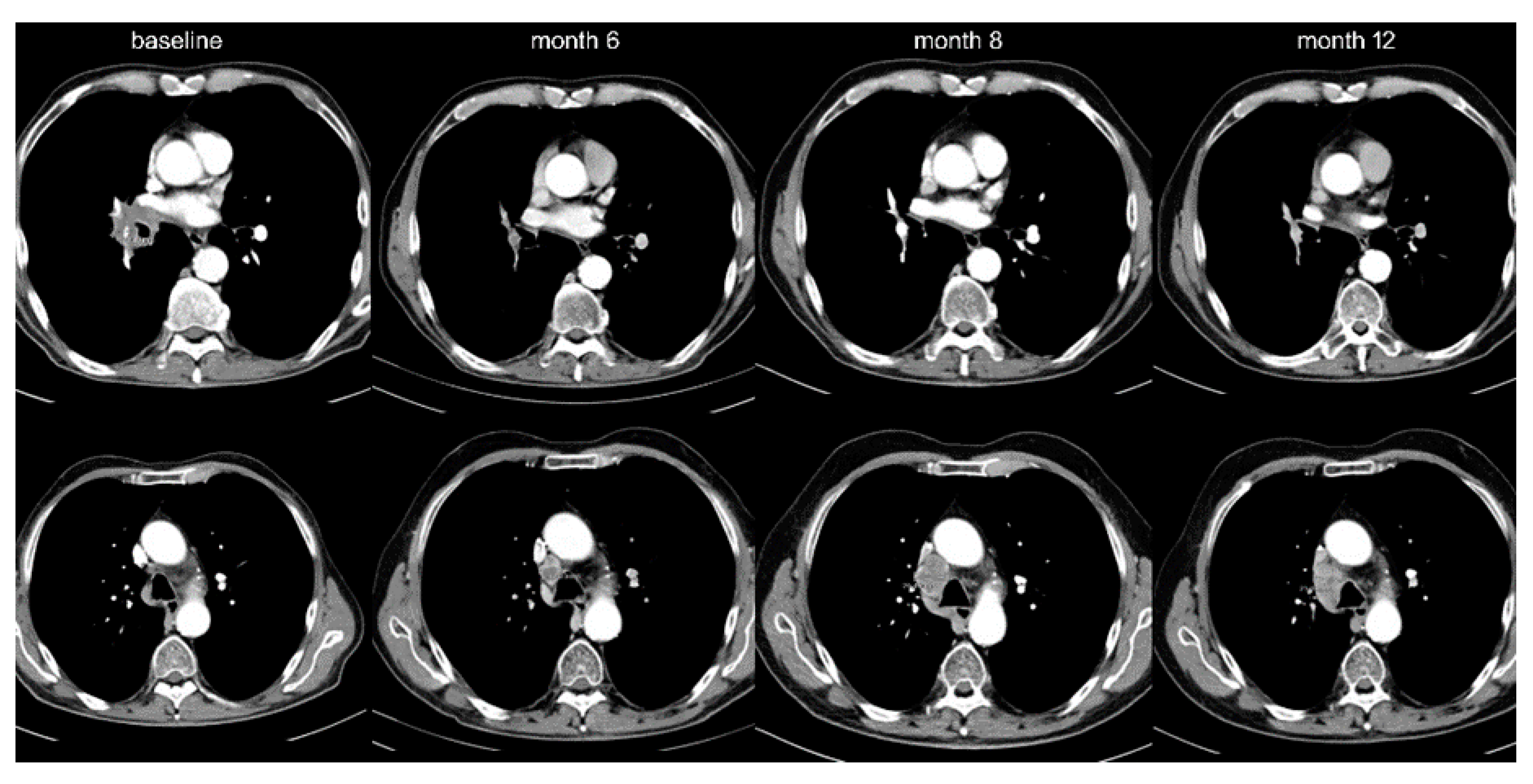
Among IO-monotherapy patients, 38 (13%) showed OPD with decreasing frequency beyond the first line (20% vs. 10%, p < 0.05, Table 1, Figure 1). Furthermore, OPD in patients treated with IO monotherapy in the first line occurred later (after 11 vs. 2 months in median, p < 0.001), involved fewer anatomical sites (mean 1.1 vs. 1.5, p < 0.05), and affected fewer lesions (mean 1.4 vs. 2.3, p < 0.05) compared to OPD in patients receiving IO monotherapy in later lines (Table 2). Lymph nodes (42% of OPD cases, mainly mediastinal, Figure 2 and Table 2) and brain (39%) were affected most frequently, but OPD was also observed in other organs typically affected by NSCLC, namely lung (24%, Figure 3), adrenal glands (16%), bone (8%), liver (5%), skin and soft tissues (3%).
The anatomic distribution of OPD was roughly similar across treatment lines, and the time to progression (TTP) for development of OPD did not differ significantly according to the organ involved (Table 2). NSCLC patients treated with first-line chemoimmunotherapy showed a similar incidence of oligoprogression as patients treated with first-line IO monotherapy (Figure 4A). Of note, the follow-up of chemoimmunotherapy patients in our study is shorter than that of IO monotherapy patients (7 vs. 15 months in median, Table 2), because chemoimmunotherapy was approved more recently for the treatment of non-squamous (September 2018) and squamous (March 2019) NSCLC in Europe.
OPD among IO-monotherapy patients was associated with a higher tumor PD-L1 expression (mean 65% vs. 41%, p < 0.001) than multifocal progression and was not encountered in any never-smokers in our cohort (p < 0.05), but other clinical characteristics of OPD and diffusely progressive patients were very similar (Table 1). No factor associated with OPD could be identified in chemoimmunotherapy-treated patients (Table 1). The characteristics of all OPD cases in our study are given in Table S1. The association between PD-L1 expression and OPD remained significant also in a multivariable analysis including all patient characteristics (Table S2).
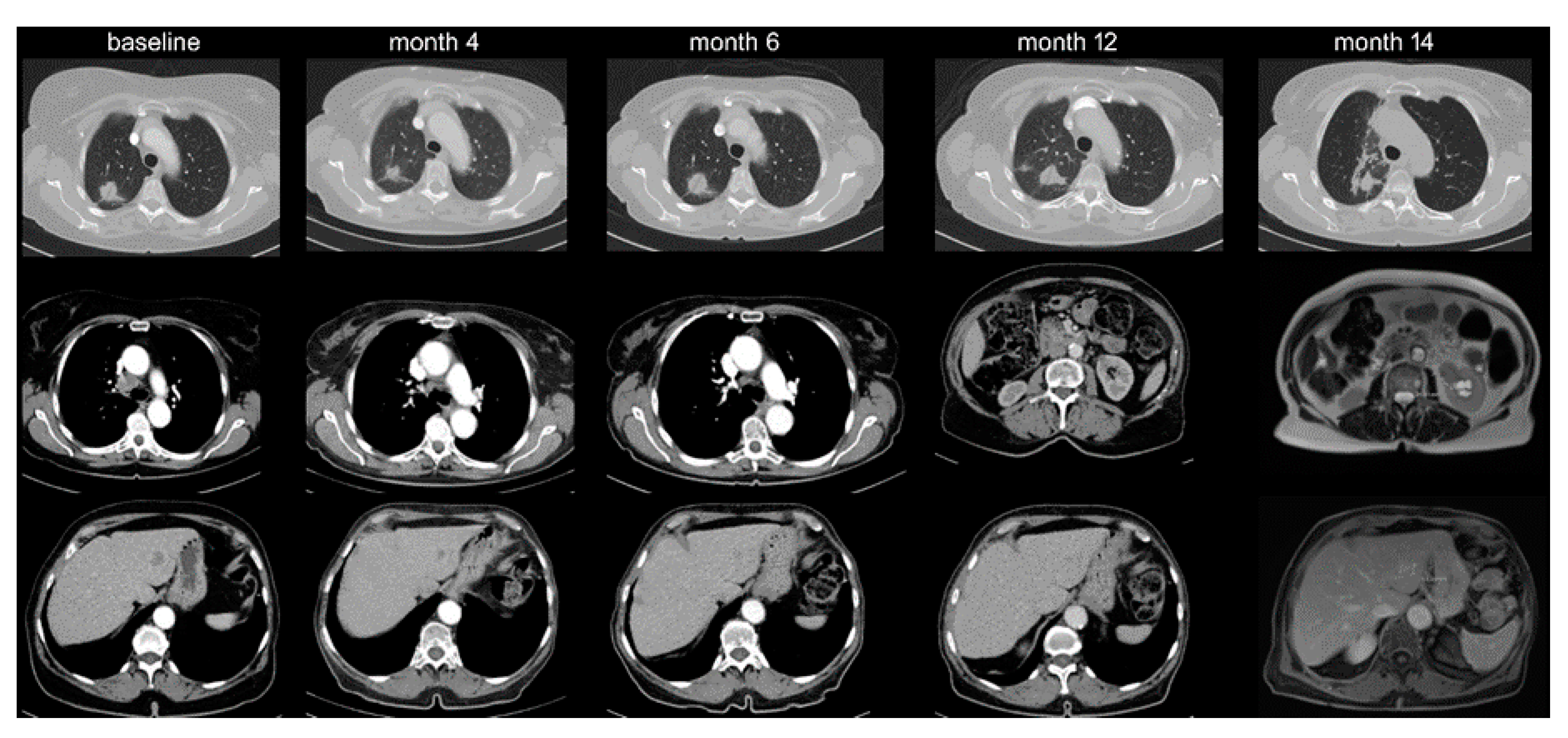
Upon histologic verification, two patients (5%) with suspected OPD were found to harbor a second primary tumor, namely a hepatocellular carcinoma and a transitional cell carcinoma of the kidney (Figure 3).

2.1. Association of OPD with Patient Survival

OPD occurred later after start of IO monotherapy (median TTP 9 vs. 2 months, p < 0.001, Table 1 and Figure 4B) and was associated with longer overall survival than multifocal progression (mean 32 vs. 16 months, p < 0.001, Table 1 and Figure 4C). In a multivariable analysis based on all patient characteristics included in this study, the pattern of disease progression, i.e., OPD vs. diffuse, showed the strongest association with overall survival from IO treatment start (Table S3). Median time to first subsequent progression (TFSP, observed in 28/38 OPD cases) was 14 months (range 1–26 months), with comparable frequencies of multifocal tumor growth (15/28 = 54%) and “tandem OPD” (13/28 = 46%). For chemoimmunotherapy-treated patients, analysis of TTP according to the pattern of initial progression, as well as analysis of TFSP, were limited by the relatively short follow-up and low number of subsequent progression events (only 1/10 chemoimmunotherapy-treated OPD patients had developed subsequent progression up to the time of this study, and this was a “tandem OPD”).

2.2. Therapeutic Relevance of Oligoprogression

Local treatments were offered to 19/38 (50%) of IO-monotherapy- and 9/10 (90%) of chemoimmunotherapy-treated patients in our study. Across both patient subgroups, local therapies were more frequently applied to patients with cerebral OPD, 11/16 (72%) of which received brain radiotherapy, with the exceptions being either asymptomatic (n = 2) or having already been cerebrally irradiated in the past (n = 3). In contrast, the rate of local therapies was lower in case of extracerebral OPD (18/37 or 49%, 16/18 with radiotherapy and 2/18 with surgery). Based on radiological and clinical features, an additional 12/37 (32%) cases with extracerebral OPD could also have received local therapies, resulting in a total rate of eligibility comparable to that of brain OPD (30/37 or 81%).

3. Discussion

The clinical features of oligoprogression in oncogene-driven NSCLC have been well characterized in several studies [3,4,5,6,7,8,9,10]. For example, Yu et al. analyzed a group of 184 EGFR patients failing erlotinib or gefitinib and found an OPD rate of 33% with an intra- to extracranial ratio of about 2:1 (23% vs. 10%) [8]. Similar observations were made by several other investigators, for example, Weickhardt et al., in a cohort of 51 EGFR or ALK-mutated patients under erlotinib or crizotinib, 35% and 14% of which developed brain and extracranial OPD, respectively [5]. In contrast, brain was outnumbered by other OPD sites among 50 EGFR T790M-positive patients treated with osimertinib, even though the overall OPD rate was even higher at 72% [4]. Apparently, the brain efficacy of the administered compound, which is very high and similar to the systemic efficacy for osimertinib, but lower for erlotinib, gefitinib, and crizotinib, influences the balance between intra- and extracranial oligoprogression [11]. Immunotherapy is characterized by comparable intra- and extracranial efficacies [12], which is consistent with the predominance of non-brain OPD observed in our patients (Table 2), similar to the osimertinib study [4]. Another conclusion is that the OPD rate in NSCLC under targeted therapies is high and varies between 30%–70%, depending on operational definition, baseline patient status, type of therapy, frequency, and duration of follow-up [1,13].
In our study, the frequency of OPD among NSCLC patients receiving either IO monotherapy or chemoimmunotherapy was lower, at about 10%–20% (Table 2). This is in line with the result of a smaller series including 81 immunotherapy-treated patients that detected a rate of 11% (9/81) for OPD using slightly different criteria [14]. The lower rate of OPD in IO-treated NSCLC could be related to its presumably higher genetic instability, as inferred by the higher tumor mutational burden (TMB) [15,16], and to the lower efficacy of current immunotherapies compared to molecularly targeted drugs [17,18,19,20]. Interestingly, our results suggest similar OPD rates for first-line IO monotherapy compared to first-line chemoimmunotherapy (Figure 4A), which parallels the roughly similar efficacy of these treatments in terms of response rates and overall survival in the respective clinical studies, especially for patients with a higher PD-L1 expression, who are also more likely to develop OPD [19,20]. However, a special effect of chemoimmunotherapy is that it blunts the predictive significance of biomarkers relevant for IO monotherapy; for example, a higher PD-L1 expression, a higher TMB, and presence of KRAS mutations predict a better outcome of NSCLC patients treated with IO monotherapy but not chemoimmunotherapy [21,22,23,24]. This is likely due to an increased benefit of IO-unfavorable, i.e., PD-L1-negative, TMB-low, and KRAS-wild-type tumors from the additional chemotherapy. Along the same lines, in our study, PD-L1 expression was associated with OPD under IO monotherapy but showed no predictive value in case of chemoimmunotherapy (Table 1). Of note, the association of PD-L1 expression with OPD under IO monotherapy does not mean that PD-L1 is an accurate OPD biomarker, because we observed several (7/33 evaluable or 21%) IO-monotherapy-treated OPD cases with low (0–10%) PD-L1 expression (Table S1), and, conversely, many patients with high PD-L1 expression developed diffuse progression instead (Table 1). Furthermore, the blood lymphocyte-to-neutrophil ratio, a biomarker associated with efficacy of immunotherapy and chemotherapy in NSCLC [25,26], did not correlate with the pattern of NSCLC progression in our patients (Table 1 and Table S2). Overall, our results show that the development of OPD in IO-treated NSCLC cannot be reliably predicted based on commonly available clinical, laboratory, and pathologic parameters, which underlines the importance of radiologic surveillance for its detection. This is not surprising, since reliable biomarkers are currently not available for IO efficacy in general, and the development of OPD under PD-(L)1 inhibitors in a way represents greater benefit from immunotherapy in a special patient subset.
Indeed, the later occurrence of OPD compared to multifocal progression, its association with longer overall survival, and the occurrence of “tandem OPD” in about 50% of cases suggest a better effect of treatment and a more favorable biology of oligoprogressive NSCLC tumors. This changes during later disease stages, as tumors acquire more mutations, increase their heterogeneity, and become more aggressive [27,28]. This biologic deterioration of the tumors along with the decreased efficacy of immunotherapy in pretreated patients [29] is consistent with the lower frequency of OPD (Table 2), with the shorter time-to-OPD (Table 1), and with the shorter survival of OPD patients vs. patients with multifocal progression (Table 1) observed beyond the first line in our study. In keeping with this, the association of OPD with a higher PD-L1 expression (Table 1) suggests that immunologic tumor control is important for spatial containment of treatment failure, since PD-L1 expression in non-EGFR/ALK mutated NSCLC is associated with the presence of tumor-infiltrating lymphocytes and active anti-cancer immunity [30,31].
Enhanced immune activity in lymph nodes and lung has been proposed as the cause for better tumor control in these tissues compared to liver, adrenal glands, and bone in a study analyzing organ-specific response patterns in IO-treated NSCLC [32]. Since lymph nodes and lung are also very frequent sites of metastatic involvement in NSCLC, it is not surprising that they showed a higher frequency of OPD in our series (Table 2). On the other hand, liver OPD was rare in our patients (Table 2), which parallels the rarity of hepatic oligoprogression in EGFR-mutated NSCLC [4,5] and is in line with the lower immune reactivity and worse prognosis of hepatic metastases and hepatic oligoprogression in NSCLC [33,34,35].
From a therapeutic point of view, OPD is very important because it offers the opportunity to eradicate resistant tumor cell clones using local therapies [36], such as irradiation, surgery, or any percutaneous ablation, which can prolong survival [4]. In our patients, local treatments were generally offered to almost all brain OPD, but only half of extracranial OPD cases. Our retrospective analysis shows that the great majority of patients with extracranial OPD are also eligible for local therapies. Precise knowledge about the clinical and radiologic characteristics of OPD under immunotherapy, as elaborated by the present study, could enhance awareness for this special constellation in IO-treated NSCLC. Since the frequency of OPD drops later in the disease course, regular complete restaging under immunotherapy and multidisciplinary evaluation of new or enlarging lesions are of key importance in order to exploit the full therapeutic potential. The fact that up to half of oligoprogressive patients subsequently develop a second OPD underscores the importance of proper initial management, since such cases could potentially derive an even larger benefit through tandem use of local therapies. On the other hand, it is also imperative to verify ambiguous cases histologically, as some lesions might turn out to represent inflammatory reactions to immunotherapy [37,38] or even second primary tumors, as noted for two of our patients.
The main limitation of our study is its retrospective nature. In addition, histologic verification was not obtained for all OPD lesions, and their classification as such in many cases relied on clinical criteria including further progression of the respective lesions in follow-up imaging and clinical deterioration of the patient during the subsequent clinical course. Furthermore, due to the relatively recent approval of chemoimmunotherapy for NSCLC in Europe, the follow-up of our chemoimmunotherapy-treated patients is not very long.

4. Materials and Methods

The main study population encompassed all 372 stage IV NSCLC patients with radiologically documented disease progression under PD-1/PD-L1 inhibitors among all 636 IO-treated patients in our hospital from March 2013 until September 2019. Treatment consisted of either checkpoint inhibitor monotherapy in various treatment lines (454 IO-monotherapy patients, of which 297 had radiologic evidence of disease progression at the time of study inclusion) or upfront combined chemoimmunotherapy (106 patients, 75 of which with radiologic progression, Figure 1). For analysis, the study population was divided in three groups: patients with IO monotherapy in the first line (n = 89), who were contrasted to patients with IO monotherapy in subsequent treatment lines (n = 208) and to patients with chemoimmunotherapy in the first line (n = 75, Table 1). Imaging studies were performed according to the standard in our institution with chest/abdomen CT and brain MRI scans before treatment start and every 6–12 weeks thereafter (Figure 2). Cases with disease progression suitable for inclusion in this study were identified by a retrospective review of medical records, followed by a review of radiological images in order to characterize the progression pattern as diffuse or OPD. Other clinical parameters were collected from the patients’ records (Table 1). Patients with early death before the first restaging, and patients who died during the stable phase of the disease after the initial response due to other reasons, were non-evaluable and excluded from this analysis (n = 66). Patients with chemoimmunotherapy after pretreatment with tyrosine kinase inhibitors for actionable genetic alterations were also excluded from this study because of potential biologic differences (n = 10, Figure 1). Testing for actionable EGFR/ALK/ROS1/BRAF mutations had been performed as indicated (e.g., in case of adenocarcinomatous histology or never/light-smoker status) for all patients at initial diagnosis, followed by administration of targeted therapies if positive, so that practically no cases with treatable genetic alterations were included in this analysis.
Image analyses were performed at a routine PACS station by expert radiologists in cooperation with thoracic oncologists. OPD was defined as localized treatment failure at one or two anatomic sites, with one to five progressive measurable (according to RECIST 1.1) lesions, either new or with ≥ 20% growth of their longest diameter (short-axis in lymph nodes), while other tumor manifestations could shrink or grow less than 20% in diameter [39]. Growth of non-measurable tumor manifestations and primary refractory disease were classified as diffuse progression. OPD lesions were retrospectively characterized as eligible for local treatment when they were radiologically amenable to surgery, radiotherapy, and/or percutaneous ablation and when the clinical condition of the patient permitted such a treatment.
For cases with clinical suspicion of alternative diagnoses, e.g., radiation pneumonitis or a second primary neoplasm, transbronchial or CT-guided percutaneous biopsies were performed for histological verification by the treating physicians. Cases without evidence of malignancy (or with diagnosis of a second primary tumor) in these biopsies were analyzed as not having disease progression. TTP was calculated from start of immunotherapy and compared between patient groups with the log-rank test. The cumulative incidence of OPD was calculated by considering OPD and diffuse progression as competing risks and compared among patient subgroups with Gray’s test. Time from OPD to first subsequent progression and overall survival from the start of immunotherapy were calculated according to Kaplan–Meier and compared with the log-rank test. Cox regression was performed for multivariable analysis of overall survival, and logistic regression was performed for multivariable analysis of factors associated with the development of OPD vs. diffuse progression. Categorical variables (like the presence of OPD) were compared among patient groups with the chi-squared test, and continuous variables were compared with the Student’s t-test. Duration of follow-up was calculated using the reverse Kaplan–Meier method. Statistical analyses were performed with SPSS (IBM, Armonk, NY, USA) and NCSS (Kaysville, UT, USA), while most figures were created with GraphPad Prism (La Jolla, CA, USA). The study was approved by the ethics committee of Heidelberg University (S-145/2017).

5. Conclusions

Our study provides real-world evidence that OPD occurs with a considerable frequency of 10%–20% in lung cancer under immunotherapy and is readily detectable, prognostically favorable, and amenable to local therapies. Our findings provide an additional opportunity to further improve patient outcome in NSCLC.

Supplementary Materials

The following are available online at https://www.mdpi.com/2072-6694/12/4/1046/s1, Table S1: Characteristics of oligoprogressive cases in this study, Table S2: Occurrence of oligoprogression in immunotherapy-treated NSCLC according to patient characteristics, Table S3: Overall survival of immunotherapy-treated NSCLC according to the pattern of progression and other patient characteristics.

Author Contributions

Conceptualization: S.R. and P.C; methodology: S.R. and P.C; validation: H.W., F.J.H., T.M., S.L., A.S., H.B.; investigation: S.R., P.M., L.G., F.B., M.K., R.A.E.S., P.C.; data curation: S.R., L.G., P.C., writing—original draft preparation: S.R. and P.C.; visualization: S.R. and P.C.; writing—review and editing: all authors; supervision: C.-P.H., M.T., H.-U.K., P.C. All authors have read and agreed to the published version of the manuscript.

Funding

This study was supported by the German Center for Lung Research (DZL).

Acknowledgments

We would like to thank Petra Kettenring of our Clinical Trial Unit for assistance with the collection of patient data.

Conflicts of Interest

Dr. Heussel reports consultation, lecture and other fees from Novartis, Basilea, Bayer, Grifols, Boehringer, Pierre Fabre, Covidien, Siemens, Chiesi, Intermune, MEDA Pharma, Bracco, Pfizer, MSD, Roche, Lilly, AstraZeneca, Schering-Plough, Essex, Gilead, MeVis, Fresenius, Astellas as well as ownership of GSK stocks; Dr. Herth reports advisory board fees and honoraria from Lilly, Roche, AstraZeneca, Novartis, Boehringer, Chiesi, Teva, Pulmonx BTG and Olympus as well as research funding from Lilly, Roche, AstraZeneca, Novartis, Boehringer, Chiesi and Teva; Dr. Stenzinger reports advisory board honoraria and/or speaker fees: Astra Zeneca, Bayer, Eli Lilly, Roche, BMS, Illumina, MSD, Novartis, Pfizer, Seattle Genetics, Takeda, Thermo Fisher, and research grants from BMS, Bayer and Chugai; Dr. Thomas reports advisory board honoraria from Novartis, Lilly, BMS, MSD, Roche, Celgene, Takeda, AbbVie, Boehringer, speaker’s honoraria from Lilly, MSD, Takeda, research funding from AstraZeneca, BMS, Celgene, Novartis, Roche and travel grants from BMS, MSD, Novartis, Boehringer. The remaining authors declare no conflict of interest.

References

  1. Patel, P.H.; Palma, D.; McDonald, F.; Tree, A.C. The Dandelion Dilemma Revisited for Oligoprogression: Treat the Whole Lawn or Weed Selectively? Clin. Oncol. 2019, 31, 824–833. [Google Scholar] [CrossRef] [PubMed] [Green Version]
  2. Guida, M.; Bartolomeo, N.; De Risi, I.; Fucci, L.; Armenio, A.; Filannino, R.; Ruggieri, E.; Macina, F.; Traversa, M.; Nardone, A.; et al. The Management of Oligoprogression in the Landscape of New Therapies for Metastatic Melanoma. Cancers 2019, 11, 1559. [Google Scholar] [CrossRef] [PubMed] [Green Version]
  3. Tumati, V.; Iyengar, P. The current state of oligometastatic and oligoprogressive non-small cell lung cancer. J. Thorac. Dis. 2018, 10, S2537–S2544. [Google Scholar] [CrossRef]
  4. Schmid, S.; Klingbiel, D.; Aeppli, S.; Britschgi, C.; Gautschi, O.; Pless, M.; Rothschild, S.I.; Wannesson, L.; Janthur, W.; Foerbs, D.; et al. Patterns of progression on osimertinib in EGFR T790M positive NSCLC: A Swiss cohort study. Lung Cancer 2019, 130, 149–155. [Google Scholar] [CrossRef]
  5. Weickhardt, A.J.; Scheier, B.; Burke, J.M.; Gan, G.; Lu, X.; Bunn, P.A.; Aisner, D.L.; Gaspar, L.E.; Kavanagh, B.D.; Doebele, R.C.; et al. Local ablative therapy of oligoprogressive disease prolongs disease control by tyrosine kinase inhibitors in oncogene-addicted non-small-cell lung cancer. J. Thorac. Oncol. 2012, 7, 1807–1814. [Google Scholar] [CrossRef] [Green Version]
  6. Gan, G.N.; Weickhardt, A.J.; Scheier, B.; Doebele, R.C.; Gaspar, L.E.; Kavanagh, B.D.; Camidge, D.R. Stereotactic radiation therapy can safely and durably control sites of extra-central nervous system oligoprogressive disease in anaplastic lymphoma kinase-positive lung cancer patients receiving crizotinib. Int. J. Radiat. Oncol. 2014, 88, 892–898. [Google Scholar] [CrossRef] [PubMed] [Green Version]
  7. Xu, Q.; Liu, H.; Meng, S.; Jiang, T.; Li, X.; Liang, S.; Ren, S.; Zhou, C. First-line continual EGFR-TKI plus local ablative therapy demonstrated survival benefit in EGFR-mutant NSCLC patients with oligoprogressive disease. J. Cancer 2019, 10, 522–529. [Google Scholar] [CrossRef]
  8. Yu, H.A.; Sima, C.S.; Huang, J.; Solomon, S.B.; Rimner, A.; Paik, P.; Pietanza, M.C.; Azzoli, C.G.; Rizvi, N.A.; Krug, L.M.; et al. Local therapy with continued EGFR tyrosine kinase inhibitor therapy as a treatment strategy in EGFR-mutant advanced lung cancers that have developed acquired resistance to EGFR tyrosine kinase inhibitors. J. Thorac. Oncol. 2013, 8, 346–351. [Google Scholar] [CrossRef] [Green Version]
  9. Shukuya, T.; Takahashi, T.; Naito, T.; Kaira, R.; Ono, A.; Nakamura, Y.; Tsuya, A.; Kenmotsu, H.; Murakami, H.; Harada, H.; et al. Continuous EGFR-TKI administration following radiotherapy for non-small cell lung cancer patients with isolated CNS failure. Lung Cancer 2011, 74, 457–461. [Google Scholar] [CrossRef]
  10. Ashworth, A.B.; Senan, S.; Palma, D.A.; Riquet, M.; Ahn, Y.C.; Ricardi, U.; Congedo, M.T.; Gomez, D.R.; Wright, G.M.; Melloni, G.; et al. An individual patient data metaanalysis of outcomes and prognostic factors after treatment of oligometastatic non-small-cell lung cancer. Clin. Lung Cancer 2014, 15, 346–355. [Google Scholar] [CrossRef]
  11. Morgan, R.L.; Camidge, D.R. Reviewing RECIST in the Era of Prolonged and Targeted Therapy. J. Thorac. Oncol. 2018, 13, 154–164. [Google Scholar] [CrossRef] [PubMed] [Green Version]
  12. Harary, M.; Reardon, D.A.; Iorgulescu, J.B. Efficacy and safety of immune checkpoint blockade for brain metastases. CNS Oncol. 2019, 8, CNS33. [Google Scholar] [CrossRef] [PubMed]
  13. Kim, C.; Hoang, C.D.; Kesarwala, A.H.; Schrump, D.S.; Guha, U.; Rajan, A. Role of Local Ablative Therapy in Patients with Oligometastatic and Oligoprogressive Non–Small Cell Lung Cancer. J. Thorac. Oncol. 2017, 12, 179–193. [Google Scholar] [CrossRef] [PubMed] [Green Version]
  14. Mersiades, A.; Crumbaker, M.; Gao, B.; Nagrial, A.; Hui, R. P3.02c-033 Patterns of Progression and Management of Acquired Resistance to Anti-PD-1 Antibodies in Advanced Non-Small Cell Lung Cancer. J. Thorac. Oncol. 2017, 12, S1293. [Google Scholar] [CrossRef]
  15. Christopoulos, P.; Budczies, J.; Kirchner, M.; Dietz, S.; Sultmann, H.; Thomas, M.; Stenzinger, A. Defining molecular risk in ALK(+) NSCLC. Oncotarget 2019, 10, 3093–3103. [Google Scholar] [CrossRef]
  16. Berland, L.; Heeke, S.; Humbert, O.; Macocco, A.; Long-Mira, E.; Lassalle, S.; Lespinet-Fabre, V.; Lalvée, S.; Bordone, O.; Cohen, C.; et al. Current views on tumor mutational burden in patients with non-small cell lung cancer treated by immune checkpoint inhibitors. J. Thorac. Dis. 2019, 11, S71–S80. [Google Scholar] [CrossRef]
  17. Peters, S.; Camidge, D.R.; Shaw, A.T.; Gadgeel, S.; Ahn, J.S.; Kim, N.-W.; Coudert, B.; Perol, M.; Dziadziuszko, R.; Rosell, R.; et al. Alectinib versus Crizotinib in Untreated ALK-Positive Non-Small-Cell Lung Cancer. N. Engl. J. Med. 2017, 377, 829–838. [Google Scholar] [CrossRef]
  18. Camidge, D.R.; Kim, H.R.; Ahn, M.-J.; Yang, J.; Han, J.-Y.; Lee, J.; Hochmair, M.; Li, J.Y.-C.; Chang, G.-C.; Lee, K.H.; et al. Brigatinib versus Crizotinib in ALK-Positive Non-Small-Cell Lung Cancer. N. Engl. J. Med. 2018, 379, 2027–2039. [Google Scholar] [CrossRef]
  19. Reck, M.; Rodríguez-Abreu, D.; Robinson, A.G.; Hui, R.; Csőszi, T.; Fülöp, A.; Gottfried, M.; Peled, N.; Tafreshi, A.; Cuffe, S.; et al. Updated Analysis of KEYNOTE-024: Pembrolizumab Versus Platinum-Based Chemotherapy for Advanced Non–Small-Cell Lung Cancer With PD-L1 Tumor Proportion Score of 50% or Greater. J. Clin. Oncol. 2019, 37, 537–546. [Google Scholar] [CrossRef]
  20. Gandhi, L.; Rodríguez-Abreu, D.; Gadgeel, S.; Esteban, E.; Felip, E.; De Angelis, F.; Dómine, M.; Clingan, P.; Hochmair, M.J.; Powell, S.F.; et al. Pembrolizumab plus Chemotherapy in Metastatic Non–Small-Cell Lung Cancer. N. Engl. J. Med. 2018, 378, 2078–2092. [Google Scholar] [CrossRef]
  21. Herbst, R.; Lopes, G.; Kowalski, D.; Nishio, M.; Wu, Y.-L.; Junior, G.D.C.; Baas, P.; Kim, D.-W.; Gubens, M.; Cristescu, R.; et al. Association between tissue TMB (tTMB) and clinical outcomes with pembrolizumab monotherapy (pembro) in PD-L1-positive advanced NSCLC in the KEYNOTE-010 and -042 trials. Ann. Oncol. 2019, 30, v916–v917. [Google Scholar] [CrossRef]
  22. Paz-Ares, L.; Langer, C.; Novello, S.; Halmos, B.; Cheng, Y.; Gadgeel, S.; Hui, R.; Sugawara, S.; Borghaei, H.; Cristescu, R.; et al. Pembrolizumab (PEMBRO) plus platinium-based chemotherapy (CHEMO) for metastatic NSCLC: Tissue TMB (tTMB) and outcomes in keynote-021, 189, and 407. Ann. Oncol. 2019, 30 (Suppl. 5), v851–v934. [Google Scholar] [CrossRef]
  23. Herbst, R.; Lopes, G.; Kowalski, D.; Kasahara, K.; Wu, Y.-L.; De Castro, G.; Cho, B.; Turna, H.; Cristescu, R.; Aurora-Garg, D.; et al. LBA4 Association of KRAS mutational status with response to pembrolizumab monotherapy given as first-line therapy for PD-L1-positive advanced non-squamous NSCLC in Keynote-042. Ann. Oncol. 2019, 30, xi63–xi64. [Google Scholar] [CrossRef]
  24. Gadgeel, S.; Rodriguez-Abreu, D.; Felip, E.; Esteban, E.; Speranza, G.; Reck, M.; Hui, R.; Boyer, M.; Garon, E.; Horinouchi, H.; et al. KRAS mutational status and efficacy in KEYNOTE-189: Pembrolizumab (pembro) plus chemotherapy (chemo) vs. placebo plus chemo as first-line therapy for metastatic non-squamous NSCLC. Ann. Oncol. 2019, 30, xi64–xi65. [Google Scholar] [CrossRef]
  25. Wang, Z.; Zhan, P.; Lv, Y.; Shen, K.; Wei, Y.; Liu, H.; Song, Y. Prognostic role of pretreatment neutrophil-to-lymphocyte ratio in non-small cell lung cancer patients treated with systemic therapy: A meta-analysis. Transl. Lung Cancer Res. 2019, 8, 214–226. [Google Scholar] [CrossRef]
  26. Christopoulos, P.; Schneider, M.A.; Bozorgmehr, F.; Kuon, J.; Engel-Riedel, W.; Kollmeier, J.; Baum, V.; Muley, T.R.; Schnabel, P.A.; Bischoff, H.; et al. Large cell neuroendocrine lung carcinoma induces peripheral T-cell repertoire alterations with predictive and prognostic significance. Lung Cancer 2018, 119, 48–55. [Google Scholar] [CrossRef]
  27. Stanta, G.; Bonin, S. Overview on Clinical Relevance of Intra-Tumor Heterogeneity. Front. Med. 2018, 5. [Google Scholar] [CrossRef] [Green Version]
  28. Christopoulos, P.; Dietz, S.; Kirchner, M.; Volckmar, A.-L.; Endris, V.; Neumann, O.; Ogrodnik, S.; Heussel, C.P.; Herth, F.J.; Eichhorn, M.E.; et al. Detection of TP53 Mutations in Tissue or Liquid Rebiopsies at Progression Identifies ALK+ Lung Cancer Patients with Poor Survival. Cancers 2019, 11, 124. [Google Scholar] [CrossRef] [Green Version]
  29. Christopoulos, P.; Kohlhäufl, J.; Bozorgmehr, F.; Kuon, J.; Schneider, M.; Neumann, O.; Liersch, S.; Heussel, C.; Winter, H.; Herth, F.; et al. Clinical and laboratory predictors of immune checkpoint inhibitor efficacy in non-small cell lung cancer. Ann. Oncol. 2018, 29, x3–x4. [Google Scholar] [CrossRef]
  30. Velcheti, V.; Schalper, K.A.; Carvajal, D.E.; Anagnostou, V.K.; Syrigos, K.N.; Sznol, M.; Herbst, R.S.; Gettinger, S.N.; Chen, L.; Rimm, D.L. Programmed death ligand-1 expression in non-small cell lung cancer. Lab. Investig. 2013, 94, 107–116. [Google Scholar] [CrossRef]
  31. Fridman, W.H.; Zitvogel, L.; Sautès-Fridman, C.; Kroemer, G. The immune contexture in cancer prognosis and treatment. Nat. Rev. Clin. Oncol. 2017, 14, 717–734. [Google Scholar] [CrossRef] [PubMed]
  32. Schmid, S.; Diem, S.; Li, Q.; Krapf, M.; Flatz, L.; Leschka, S.; Desbiolles, L.; Klingbiel, D.; Jochum, W.; Früh, M. Organ-specific response to nivolumab in patients with non-small cell lung cancer (NSCLC). Cancer Immunol. Immunother. 2018, 67, 1825–1832. [Google Scholar] [CrossRef] [PubMed]
  33. Ren, Y.; Dai, C.; Zheng, H.; Zhou, F.; She, Y.; Jiang, G.; Fei, K.; Yang, P.; Xie, N.; Chen, C. Prognostic effect of liver metastasis in lung cancer patients with distant metastasis. Oncotarget 2016, 7, 53245–53253. [Google Scholar] [CrossRef]
  34. Tumeh, P.C.; Hellmann, M.D.; Hamid, O.; Tsai, K.K.; Loo, K.L.; Gubens, M.A.; Rosenblum, M.; Harview, C.; Taube, J.M.; Handley, N.; et al. Liver Metastasis and Treatment Outcome with Anti-PD-1 Monoclonal Antibody in Patients with Melanoma and NSCLC. Cancer Immunol. Res. 2017, 5, 417–424. [Google Scholar] [CrossRef] [Green Version]
  35. Kissel, M.; Martel-Lafay, I.; LeQuesne, J.; Faivre, J.-C.; Le Pechoux, C.; Stefan, D.; Barraux, V.; Loiseau, C.; Grellard, J.-M.; Danhier, S.; et al. Stereotactic ablative radiotherapy and systemic treatments for extracerebral oligometastases, oligorecurrence, oligopersistence and oligoprogression from lung cancer. BMC Cancer 2019, 19, 1–9. [Google Scholar] [CrossRef] [PubMed]
  36. Gainor, J.F.; Dardaei, L.; Yoda, S.; Friboulet, L.; Leshchiner, I.; Katayama, R.; Dagogo-Jack, I.; Gadgeel, S.; Schultz, K.; Singh, M.; et al. Molecular Mechanisms of Resistance to First- and Second-Generation ALK Inhibitors in ALK-Rearranged Lung Cancer. Cancer Discov. 2016, 6, 1118–1133. [Google Scholar] [CrossRef] [PubMed] [Green Version]
  37. Tetzlaff, M.T.; Nelson, K.; Diab, A.; Staerkel, G.A.; Nagarajan, P.; Torres-Cabala, C.A.; Chasen, B.A.; Wargo, J.A.; Prieto, V.G.; Amaria, R.N.; et al. Granulomatous/sarcoid-like lesions associated with checkpoint inhibitors: A marker of therapy response in a subset of melanoma patients. J. Immunother. Cancer 2018, 6, 14. [Google Scholar] [CrossRef] [PubMed]
  38. Gkiozos, I.; Kopitopoulou, A.; Kalkanis, A.; Vamvakaris, I.N.; Judson, M.A.; Syrigos, K.N.; Ioannis, V.N.; Syrigos, K.N. Sarcoidosis-Like Reactions Induced by Checkpoint Inhibitors. J. Thorac. Oncol. 2018, 13, 1076–1082. [Google Scholar] [CrossRef]
  39. Eisenhauer, E.A.; Therasse, P.; Bogaerts, J.; Schwartz, L.; Sargent, D.; Ford, R.; Dancey, J.; Arbuck, S.; Gwyther, S.; Mooney, M.; et al. New response evaluation criteria in solid tumours: Revised RECIST guideline (version 1.1). Eur. J. Cancer 2009, 45, 228–247. [Google Scholar] [CrossRef]
Figure 1. CONSORT diagram of the study.
Cancers 12 01046 g001
Figure 2. Lymph node oligoprogression. A 66-year-old male patient with adeno-NSCLC (PD-L1 90%) was started on pembrolizumab in November 2017. Nodal progression on the right side was noted in June 2018, which appeared stable in a subsequent restaging in October 2018, even though no change in therapy occurred.
Cancers 12 01046 g002
Figure 3. Lung oligoprogression and transitional cell carcinoma of the kidney. A 75-year-old female with adeno-NSCLC (PD-L1 90%) was started on pembrolizumab in September 2017 with response of the primary tumor, mediastinal lymph nodes, and liver metastases. Upon oligoprogression of the primary tumor in March 2018, thoracic radiotherapy was administered. In August 2018, a new kidney lesion was noted that grew oligoprogressive-like. At biopsy, this lesion turned out to be a transitional-cell carcinoma.
Cancers 12 01046 g003
Figure 4. Incidence and prognosis of oligoprogression during first-line immunotherapy (to the right). (A) Cumulative incidence of oligoprogression (OPD) in stage IV NSCLC under first-line IO monotherapy (n = 163) vs. first-line chemoimmunotherapy (n = 106) in the entire study population (Figure 1). Patients without disease progression were censored, while OPD and diffuse progression were considered as competing risks. Cumulative incidence at 2 years was 12.6% for chemoimmunotherapy-treated vs. 11.0% for IO-monotherapy-treated patients (Gray’s p = 0.99). (B) Time-to-progression for NSCLC patients developing OPD (n = 18) vs. diffuse disease progression (n = 71) under first-line IO monotherapy (Table 1, log-rank p < 0.001). (C) Overall survival (OS) of NSCLC patients developing OPD (n = 18) vs. diffuse disease progression (n = 71), under first-line IO monotherapy (Table 1, log-rank p < 0.001).
Cancers 12 01046 g004
Table 1. Characteristics of patients with disease progression in this study.
IO Monotherapy-Treated Stage IV NSCLC PatientsIO+CHT-Treated NSCLC Patientsp-Value 1
OPD (13%, n = 38)Diffuse PD (n = 259)p-Value 1OPD (13%, n = 10)Diffuse PD (n = 65)
Age (Median; SD) 63 (10)64 (9)ns64 (11)65 (10)ns
Gender (% male) 5858ns4058ns
Smoking Status (%) 2never smokers010p < 0.052022ns
ex-smokers6153ns4048ns
current smokers3937ns4030ns
ECOG PS (%) 304741ns5040ns
14758ns5059ns
252ns01ns
Histology (%)adenocarcinoma6863ns9088ns
squamous cell carcinoma2931ns17ns
other (LCNEC, NOS, mixed)36ns06ns
No. of Metastatic Sites at IO Start (Mean; SD)2.4 (1.2)2.5 (1.4)ns1.1 (2.5)1.8 (2.7)ns
PD-L1 IHC 4 (Average % of Positive Cells; SD)65 (33)41 (36)p < 0.00117 (22)18 (30)ns
LNR (Mean; SD) 0.24 (0.11)0.23 (0.46)ns0.21 (0.09)0.17 (0.14)ns
IO Treatmentfirst line1871p < 0.051065
second-and-beyond line20188p < 0.05
TTP from IO Treatment Start in Months, Median92p < 0.001
first-line patients112p < 0.00144ns
second-and-beyond-line patients52p = 0.015
OS from IO Treatment Start in Months, Median (Mean)n.r. (26)10 (13)p < 0.001
first-line patientsn.r. (39)14 (15)p < 0.001n.r.n.r.ns
second-and-beyond-line patients1610p < 0.05
(O)PD: (oligo) progressive disease; SD: standard deviation; ns: not statistically significant; PS: performance status; LNR (lymphocyte-to-neutrophil ratio); no.: number; nr: not reached; TTP: time-to-progression; OS: overall survival. 1 Statistical comparisons were performed with a chi-squared test for categorical, with a t-test for numerical, and with the log-rank test for time-to-event data. 2 Data available for 293/297 cases. 3 Data available for 259/297 cases. 4 Data available for 226/297 cases.
Table 2. Frequency and anatomical distribution of oligoprogression in IO-treated NSCLC.
Location of OPD, no. (%) 1IO monotherapy1L IO + CHT 1TTP of OPD
All OPD 1
(n = 38, 13%)
1L IO OPD
(n = 18, 20%)
2+L IO OPD
(n = 20, 10%)
All OPD Cases
(n = 10, 13%)
in Months,
Median (IQR)
Lymph NodesAll16 (5%)8 (9%) 8 (4%)0 (0%)8 (4–14)
mediastinal1064
axillary, cervical523
abdominal212
Brain 15 (5%)3 (3%)10 (5%)3 (4%)4 (2–11)
LungAll9 (3%)5 (6%)5 (2%)2 (2%)7 (3–15)
primary tumor4132
lung metastasis5420
Adrenal Gland 6 (2%)1 (1%)4 (2%)2 (2%)8 (5–13)
Bone 3 (1%)1 (1%)03 (4%)4 (3–27)
Liver 2 (< 1%)1 (1%)1 (< 1%)1 (2%)12 (7–15)
Skin/Soft Tissue 1 (< 1%)01 (< 1%)09 (n/a)
No. of Anatomic Sites, Average (SD)21.4 (0.5)1.1 (0.3) *1.5 (0.6)1.1 (0.3)
No. of Lesions, Average (SD)1.8 (1.2)1.4 (0.8) *2.3 (1.4)1.6 (0.8)
TTP in Months, Median1111 **56
IO monotherapy1L IO + CHT
All Patients with PD (no.)all (297)1L (89)2+L (208)all (85)
FU, Median (IQR)15 (9–21)13 (7–21)15 (9–22)7 (3–10)
(O)PD: (oligo) progressive disease; 1L: first line; IO: immunotherapy; CHT: chemotherapy; no.: number; TTP: time-to-progression; IQR: interquartile range; SD: standard deviation; 2+L: second line and beyond; FU: follow-up from IO start; n/a: not applicable. 1 Percentages refer to the parent populations of 297 IO-treated (1L 89, 2+L 208) and 75 IO + CHT-treated patients (Table 1); in 10 IO-treated patients and 2 IO + CHT-treated patients, OPD occurred in 2 organs, therefore the percentages sum to >100%. 2 Each lymph-node station (e.g., right supraclavicular lymph nodes) and each organ (e.g., brain, left adrenal gland) was considered as one anatomic site. * p < 0.05 vs. 2+L IO-treated patients. ** p < 0.01 vs. 2+L OPD patients.