Abstract
Reactive oxygen species (ROS) are produced predominantly by the mitochondrial electron transport chain and by NADPH oxidases in peroxisomes and in the endoplasmic reticulum. The antioxidative defense counters overproduction of ROS with detoxifying enzymes and molecular scavengers, for instance, superoxide dismutase and glutathione, in order to restore redox homeostasis. Mutations in the redox landscape can induce carcinogenesis, whereas increased ROS production can perpetuate cancer development. Moreover, cancer cells can increase production of antioxidants, leading to resistance against chemo- or radiotherapy. Research has been developing pharmaceuticals to target the redox landscape in cancer. For instance, inhibition of key players in the redox landscape aims to modulate ROS production in order to prevent tumor development or to sensitize cancer cells in radiotherapy. Besides the redox landscape of a single cell, alternative strategies take aim at the multi-cellular level. Extracellular vesicles, such as exosomes, are crucial for the development of the hypoxic tumor microenvironment, and hence are explored as target and as drug delivery systems in cancer therapy. This review summarizes the current pharmaceutical and experimental interventions of the cancer redox landscape.
1. Introduction
Redox biology is a vastly complex and heterogeneous field that has drawn increasing attention in research due to its fundamental implications for our understanding of physiological function [1]. The concept of redox biology usually operates with a set of defined oxidants and antioxidants, which can lead to redox stress if the equilibrium of both classes of redox molecules becomes imbalanced [2,3]. In the case of archetypical oxidative stress, this balance is tilted towards the class of oxidants, which usually comprise reactive oxygen species (ROS) [4]. The cell aims to restore the redox balance by employing an antioxidant defense system. If the balance cannot be restored, the relative elevation of ROS levels leads to the development of diseases, including cancer [5,6,7].
The detailed role of ROS in cancer development remains unclear since the role of ROS varies greatly among different cancer types, tissues, and stages [8,9]. The general consensus in research suggests that high ROS production induces carcinogenesis by impairing the DNA repair system, which subsequently leads to an accumulation of DNA damage, such as base modifications, inter- and intrastrand bindings and DNA–protein crosslinks [10]. In addition, increased H2O2 and O2•− is associated with increased cancer cell proliferation [11]. Cancer cells show an altered metabolism, which demonstrates a substantially increased production of ROS [12,13,14]. A comprehensive review of the metabolic regulation of redox balance in cancer was published earlier by Purohit et al. [15]. With continuous growth, cancer cells consolidate into a tumor that faces cycles of hypoxia and reoxygenation [16]. Hypoxic conditions stimulate remodeling of the tissue microenvironment in order to ensure influx of nutrition, efflux of waste products, and establishment of a suitable redox microenvironment [12]. Adaption to hypoxia is a crucial step in the transformation of a cell to a malignant state [17]. This process is accompanied by blood vessel development, which is often devoid of coordinated structure and organization, and induces oxidative stress through periods of changing redox environment [16,18]. Studies suggest that cancer cells engage in intercellular communication with the tumor and the surrounding tissue via extracellular vesicles (EVs), such as exosomes, in order to create a metabolic microenvironment that fosters tumor growth and metastasis [19].
Since redox biology is linked tightly to cancer, current pharmaceutical developments include the design of compounds that target key players and processes within the oxidative and antioxidative landscape in the cancer cell. For instance, chemo- and radiotherapy are employed to induce overproduction of ROS and, hence, apoptosis of tumor cells [20]. The application of such interventions, however, depends on the genetic polymorphism of the patient, on cancer type and stage, and on the affected tissue [8,21]. Other approaches aim to utilize redox-driven strategies to modulate immunotherapy in cancer therapy [22,23]. In this review, we provide an overview of the major components of both the oxidative and the antioxidative landscape and their connection to the development of cancer drugs. In addition, we present examples of current efforts that aim to modulate key proteins of the redox landscape in cancer, which is summarized in Table 1.

Table 1.
Overview of compounds presented in this review for targeting the redox landscape in cancer.
2. The Oxidative Landscape in Cancer
A cornerstone of the cellular redox landscape is the interplay of three organelles: mitochondria, the endoplasmic reticulum (ER), and peroxisomes [95]. The contribution of mitochondria, peroxisomes and the ER to the intracellular production of ROS varies among cells, tissues, and the general redox environment. Studies on perfused liver tissue indicate that peroxisomes produce the largest absolute amount of ROS [96]. Mitochondria can contribute substantially to general ROS production as well [97]. In comparison however, the ER provides the highest relative amount of cytosolic ROS due to the lack of antioxidative systems in the ER [95]. The predominant sources of ROS in both normal and cancer cells comprise NADPH oxidases (NOXs) and the electron transport chains (ETC) in the mitochondria, whereas the ER can also serve as a substantial source of ROS due to ER oxidoreductases and NOXs [15,95,98,99]. Both NOXs and mitochondrial ETC reduce oxygen to the highly reactive superoxide anion (O2•−). O2•− subsequently undergoes a complex series of conversion reactions, giving rise to more stable hydrogen peroxide (H2O2) but also to more toxic ROS, e.g., hydroxyl radical (−OH), or reactive nitrogen species (RNS), e.g., nitric oxide (NO−). An overview of the cellular oxidative landscape is presented in Figure 1. Since increased ROS production is associated with cancer development, pharmaceutical research aims to modulate the oxidative landscape in cancer therapy.
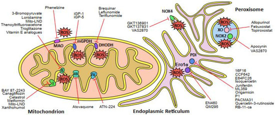
Figure 1.
Schematic overview of the major sources of reactive oxygen species (ROS) in the cell and the corresponding inhibitors of those sites. The mitochondrial electron transport chain (ETC) complexes I, II and III generate ROS directly, whereas complex IV is the rate-determing step of the ETC. Other enzymes that produce ROS in the mitochondria are dihydroorotate dehydrogenase (DHODH), glycerol-3-phosphate dehydrogenase 2 (mGPDH or GPDH2) and monoamine oxidase (MAO). The endoplasmic reticulum (ER) comprises several sites of ROS production, such as NADPH oxidase 4 (NOX4) and the Ero1α-PDI oxidative protein folding pathway (Ero1α, ER oxidoreductin 1α; PDI, protein disulfide isomerase). Peroxisomes are another major source of cellular ROS production due to the activity of xanthine oxidase (XO) and NADPH oxidase 2 (NOX2). Names of pharmaceutical and experimental inhibitors are presented and the corresponding target sites of ROS production are indicated by red lines.
2.1. The Mitochondrial Electron Transport Chain
The ETC in the inner mitochondrial membrane comprises four multi-enzyme complexes, termed complexes I, II, III, and IV. They drive an electrochemical proton gradient across the membrane, which can be used for ATP generation via ATP synthase (also called complex V) or heat generation via protein uncoupling. Electrons, leaked from the ETC at complexes I, II and III, are the major source of O2•− and other ROS in the mitochondria [100,101]. Mutations in the ETC complexes can disturb the electron chain reaction, leading to elevated ROS levels, and hence contribute to cancer proliferation and metastasis [102]. Hence, ETC complex inhibitors are actively pursued in drug discovery and development of novel anticancer drugs [103].
Mutations in complex I, the largest of all complexes, occur frequently in many different tumors and are considered essential for the glycolytic switch, known as the Warburg effect [104], and for ROS-driven metastasis [105,106]. However, some mutations appear to have tumor-suppressor effects, suggesting that pharmaceutical targeting needs to consider type and stage of the cancer prior to the start of the intervention [107]. As of today, a number of approved complex I inhibitors are available, including the well-known and established diabetic drugs canagliflozin [27,28] and metformin [30], which are also being investigated as anticancer drugs. In addition, some recently approved compounds, e.g., celastrol [29], BAY 87-2243 [24,25,26], and xanthohumol [32,33], also target complex I, resulting in mitochondrial ROS production and anticancer effects.
Complex II, the smallest of the respiratory complexes, has also drawn considerable attention since it is positioned at the intersection between the ETC and the TCA cycle [37]. Many compounds have been identified as potent complex II inhibitors, but clinical application is hampered by the high degree of toxicity, for instance, in the cases of troglitazone and thenoyltrifluoroacetone. Nonetheless, vitamin E analogues, such as tocopherols and tocotrienols, have shown promising results in preclinical trials [38,39]. In addition, 3-Bromopyruvate derivatives have also produced promising results in preclinical trials and have advanced to clinical trials [34]. Notably, the anticancer drug lonidamine (LND) has been reported to inhibit complex II, and to increase the overall treatment response in cancer patients in combination with standard-of-care drugs, e.g., doxorubicin [35,36]. However, LND showed limited efficiency in clinical phase 3 trials but was recently modified into mito-lonidamine (Mito-LND), which is 100-fold more potent in cell culture and mouse models, and inhibits complex I and II [31].
Complex III can also contribute to cancer development. Mutations in this complex are associated with increased ROS production and apoptotic resistance, which is linked to accelerated growth in cancer cells [108]. A study showed that the long-established antimalarial drug, atovaquone, targets complex III and has anticancer properties [40]. This eventually led to two clinical trials, one of which investigated the effect of atovaquone on the tumor microenvironment of solid tumors [Atovaquone as a Tumour hypOxia Modifier (ATOM), NCT02628080] and the other on the outcome of chemotherapy in acute myeloid leukemia [Atovaquone (Mepron®) Combined With Conventional Chemotherapy for de Novo Acute Myeloid Leukemia (AML) (ATACC AML), NCT03568994].
The last of the four ETC complexes is the copper-dependent complex IV (or cytochrome c oxidase), which is the rate-determining enzyme of the ETC and crucial for cellular energy production [109]. It has been shown that tumors often need more copper [110] and that the copper chelator bis-choline tetrathiomolybdate (also known as SOD1 inhibitor ATN-224) inhibits complex IV activity in cancer cells [43]. Taken together, this indicates that inhibitors of ETC complex IV possess intriguing translational potential in the treatment of cancer.
2.2. ROS-Generating Enzymes of the Mitochondria
In addition to the ETC, mitochondria harbor enzymes that are also a source of ROS. One notable example at the inner mitochondrial membrane is dihydroorotate dehydrogenase (DHODH). DHODH is associated with complex III, and generates O2•− and H2O2 [44]. Interestingly, DHODH was unknown until researchers identified the target of the anticancer drug brequinar [111]. Despite successful initial clinical trials, brequinar eventually failed due to inconsistencies in patient response [44]. Recent studies, however, show beneficial effects for administration of brequinar in chemotherapy, suggesting that brequinar can still be used in combination therapy for treatment of cancer patients [112]. Another established DHODH inhibitor is teriflunomide, which is approved for the treatment of multiple sclerosis [113]. Furthermore, teriflunomide, the prodrug of leflunomide, is approved for the treatment of rheumatoid and psoriatic arthritis [45]. Both drugs have been evaluated for various diseases and showed anticancer properties [44,46,47,48]; however, these compounds causes considerable adverse reactions due to significant off-target effects. This led to the termination of a clinical trial in which melanoma cancer was treated with a combination of leflunomide and the anticancer drug vemurafenib [Leflunomide+Vemurafenib in V600 Mutant Met. Melanoma, NCT01611675]. Despite these setbacks, leflunomide and teriflunomide remain the only FDA-approved DHODH inhibitors for clinical application, and are currently explored for other types of cancers [44].
Another ROS-producing enzyme in the inner mitochondrial membrane is glycerol-3-phosphate dehydrogenase 2 (mGPDH or GPDH2), which produces ROS through reverse electron transport from flavin adenine dinucleotide (FAD) to the ETC [114]. Much of mGPDH’s function remains to be established, but recent findings demonstrated that mGDPH inhibition impedes prostate cancer, indicating a pharmaceutically relevant role of this enzyme in cancer development [115]. Nevertheless, available inhibitors lack selectivity, and reports of two potent and selective mGPDH inhibitors, i.e., iGP-1 and iGP-5, were never followed up [49].
In contrast to DHODH and mGPDH, mitochondrial monoamine oxidase (MAO) is located at the outer membrane. Human cells express two variants, MAO-A and MAO-B, which contribute to ROS production in mitochondria by oxidative deamination of serotonins or catecholamines [116]. MAO-A is found in several tissues, for example, in the prostate, whereas MAO-B expression is limited to platelets; however, both variants are also expressed in the brain and contribute to neurological disorders [117]. While MAO-B is currently explored as a drug target in various neuropathologies [118], only MAO-A has been associated with cancer. For instance, MAO-A is overexpressed in prostate cancer and contributes to tumorigenesis [51]. Inhibitors for MAO-A have been in use since the 1950s for the treatment of major depression [119], but they are now studied as anticancer drugs as well. Phenelzine, a notable example of a potent non-selective and irreversible MAO-A inhibitor, is currently under investigation in clinical trials for the treatment of prostate cancer [50] [Phenelzine Sulfate in Treating Patients With Non-metastatic Recurrent Prostate Cancer, NCT02217709].
2.3. The Endoplasmic Reticulum
The ER is an intracellular network of membranes that is involved in a variety of basic physiological processes: in particular, protein synthesis, posttranslational processing, protein folding and transportation, as well as Ca2+ signaling and bioenergetics at mitochondria–ER contact sites [120]. The ER is an important player in the redox environment of the cell, which comprises two major sources of ROS. One of the sources of ROS is NOX4, a member of NADPH oxidase family, which we will discuss subsequently. The second major source of ROS is the Ero1α–PDI protein folding pathway [121]. One component of this pathway is protein disulfide isomerase (PDI). PDI, an abundant protein in the ER, is the founding member of a family of 20 related proteins in the ER, and essential for protein folding in the ER [122,123]. PDI-family proteins share at least one thioredoxin-like domain that contains a catalytic cysteine pair as part of a canonical CXXC motif [124]. The catalytically active domains of PDI catalyze a thiol-disulfide exchange reaction with cysteines of nascent client proteins, resulting in breakage, formation or isomerization of disulfide bonds [125,126]. This oxidoreductase-catalyzed reaction also requires the activity of the membrane-bound ER oxidoreductin 1α (Ero1α), a FAD-dependent oxidoreductase [127,128,129,130]. A byproduct of the Ero1α–PDI oxidative protein folding pathway is H2O2, which contributes to a slightly oxidative environment within the ER, characterized by a high GSSG:GSH ratio [131]. Excess H2O2 is compensated by peroxiredoxin 4 (PRDX4), a member of the peroxiredoxin protein family, which is described in a later chapter [132]. Alternative models of oxidative protein folding involve quiescin–sulfhydryl oxidase 1 (QSOX1), which produces H2O2 and facilitates the formation of disulfide bridges in client proteins independently of Ero1α–PDI activity [133].
PDI upregulation correlates with cancer metastasis and invasion in various cancer types, and has drawn a lot of attention as a drug target in cancer therapy [134,135,136]. As of today, a large and growing number of PDI inhibitors have been discovered and characterized, such as RB-11-ca [67], 16F16 [56], origamicin [62,63], the phenyl vinyl sulfonate compound P1 [64], juniferdin [60], quercetin-3-rutinoside [66], ML359 [61] and PACMA31 [65], but none of them have progressed beyond preclinical studies. Nonetheless, two recent PDI inhibitors, CCF642 [57] and E64FC26 [58], demonstrated favorable results in preclinical studies in terms of potency, selectivity, and anticancer effects, and, hence, are promising candidates for clinical translation. Notably, isoquercetin is a PDI inhibitor that advanced the furthest in clinical use, and entered phase 2 trials in cancer patients a few years ago (Cancer Associated Thrombosis and Isoquercetin (CATIQ), NCT02195232). However, its primary application is not aimed at targeting cancer but at the inhibition of PDI activity in platelets in order to reduce the risk of thrombosis [59,137]. In contrast to this large pool of compounds targeting PDI inhibitors, only two inhibitors of Ero1α have been identified in a screen, i.e., EN460 and QM295, and no subsequent study was reported [55]. A lot of research is still needed, but recent developments of PDI inhibitors, as outlined above, indicate the potential of targeting the Ero1α–PDI protein folding pathway in cancer.
2.4. Peroxisomes
Peroxisomes, formerly known as “microbodies”, are small organelles with a single membrane located in the cytoplasm of almost all eukaryotic cells [138]. The biogenesis of peroxisomes is still under debate as one model suggests growth and division whereas another model promotes de novo synthesis; regardless, both models agree on the contribution of the ER to the compartmentalization of the organelle [139,140]. A panel of transcription factors that are termed peroxisome proliferator-activated receptors (PPARs) regulate proliferation of peroxisomes [141]. Among many biological functions, peroxisomes are crucial for lipid homeostasis and cellular ROS metabolism [142]. Peroxisomes contain several flavin-dependent oxidoreductases, most notably xanthine oxidase (XO), which generates ROS [143]. In addition, peroxisomes also contain nitric oxide synthase (NOS2), which generates NO. To counterbalance ROS production, peroxisomes possess several antioxidant enzymes, such as catalase (CAT), superoxide dismutase 1 (SOD1), peroxiredoxin 5 (PRDX5), glutathione S-transferase kappa 1 (GSTK1) and glutathione peroxidase (GPx) [144,145]. The role of peroxisomes within the cellular redox landscape is not fully understood, but it has been suggested that their function as a source or sink for H2O2 is tissue-specific [144]. Nevertheless, peroxisomal function and redox metabolism are important for metabolic reprogramming and are crucially involved in cancer development [146]. In fact, various cancer types show decreased peroxisome levels, which is associated with overexpression of the negative regulator of peroxisome abundance and metabolism, termed hypoxia-inducible factor 2a (HIF2α) [147,148]. Current research efforts aim at peroxisomal ROS production, for instance, by targeting XO [149]. Inhibitors of XO, such as the purine analogue allopurinol, and the non-purine analogues febuxostat and topiroxostat, are approved for the treatment of gout and hyperuricemia, indicating that XO is a suitable therapeutic target [68,150]. Furthermore, there are several compounds that target PPARs in the treatment of cancer, demonstrating that modulation of peroxisomal function is a promising approach in disease management [151].
2.5. NADPH Oxidases
NADPH oxidases (NOXs) play a key role in a wide range of physiological processes, such as gene expression regulation, cell signaling and differentiation. They are also crucially involved in many pathological processes, including cancer. The seven human NOX isoforms (NOX1 to NOX5, and the dual oxidases DUOX1 and DUOX2) are transmembrane proteins that transport electrons across the cytoplasm via FAD or across the extracellular membrane, using two heme groups, in order to generate O2•− or H2O2 [152]. NOXs require various membrane and cytosolic protein subunits for their activity. For instance, the stability and activation of NOX1 to NOX4 and DUOX1 and DUOX2 depend on the stabilization partner p22phox and the maturation partners DUOXA1 and DUOXA2. The cytosolic subunits p47phox and NOXO1, and the activator subunits p67phox and NOXA1, are essential for NOX1 and NOX2 function [152,153]. NOX2, in particular, is an important member of the NADPH oxidase family. NOX2 was originally discovered in phagocytes as a source of ROS, which are employed in the defense against bacterial infection [153]. p47phox, the organizing component of NOX2, is phosphorylated and then translocated to the plasma membrane by p67phox, which activates the NOX2 complex [153].
Several studies show that cancer cells accumulate mutations, which increase ROS generation from NOX enzymes, eventually inducing tumorigenesis [153,154]. A particular type of mutation involves the GTPase KRAS, a member of the Ras oncogene family. KRAS mutations affect phosphorylation and membrane translocation of p47phox, and, thus, induce NOX1-mediated ROS formation and metastasis [154,155]. For instance, KRAS mutation increases NOX1 expression in colon adenocarcinoma and lung cancer cells [156].
Other NOX isoforms are also involved in cancer development. For example, upregulation of NOX4, the major ER NADPH oxidase, plays a key role in ovarian, pancreatic, kidney, and glioblastoma cells [157,158,159,160]. Recent in vivo studies reported that the inhibition of NOXs hinders tumor growth, indicating the pharmaceutical relevance of NOXs; however, current NOX inhibitors lack selectivity among NOX isoforms [161,162]. For instance, the natural organic compound apocynin inhibits p47phox membrane translocation, and thus activates NOX2, but it also inhibits Rho kinases, thus leading to cell cycle arrest [69,70,108,163]. In a recent study, Solbak et al. used fragment-based drug discovery to develop dimeric NOX2 inhibitors that target p47phox–p22phox protein–protein interaction [164]. The pan-NOX inhibitor VAS2870 interferes with NOX binding proteins, and hence inhibits NOX complex formation [54,165,166]. The two pyrazolopyridine compounds GKT136901 and GKT137831 show 10-fold selectivity towards NOX1 and NOX4 over NOX2 [52,53,167,168]. Notably, the NOX1/NOX4 dual inhibitor GKT137831 is the only NOX inhibitor that has entered clinical trials [GKT137831 in IPF Patients with Idiopathic Pulmonary Fibrosis (GKT137831), NCT03865927]. Nevertheless, recent clinical data failed to reproduce any pharmacological effects in humans, causing a decline in interest in pursuing NOXs as drug targets [169].
3. The Antioxidative Landscape in Cancer
The antioxidative defense plays a crucial role in maintaining an adequate redox environment for physiological cell function and survival in oxidative stress. For example, the production of O2•− in mitochondria, peroxisomes and the ER is countered by different types of superoxide dismutases (SODs) that catalyze the disproportionation of O2•− to H2O2 and O2. Eventually, H2O2 disproportionates to water and O2, completing the detoxification process of O2•−. Detoxification of H2O2 is thoroughly facilitated by various mechanisms, comprising the activity of the enzyme catalase (CAT) and oxidation of cysteine residues either in glutathione (GSH) or in peroxiredoxins (PRDXs). All of these antioxidative processes are under tight regulation by transcription factors and are upregulated during the oxidative stress response. One important transcription factor is Nrf2, which in turn is under the control of the negative regulator Keap1. An overview of the cellular antioxidative landscape is presented in Figure 2.
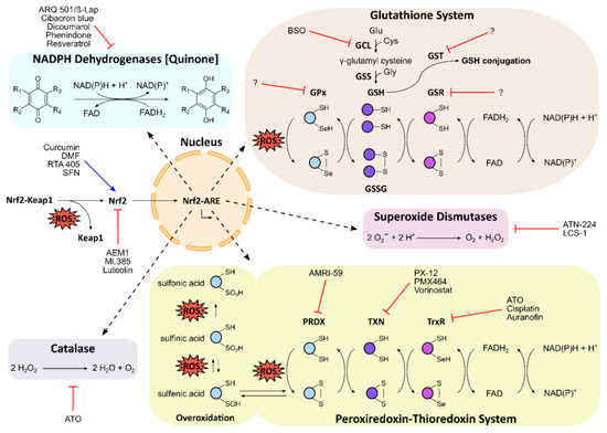
Figure 2.
Schematic overview of the antioxidative landscape in the cell, and the corresponding modulators of key players. Reactive oxygen species (ROS) activate the Nrf2–Keap1 signalling pathway, resulting in induction of the antioxidant response elements (ARE) by Nrf2 in the nucleus. ARE comprise the glutathione (GSH) system, the peroxiredoxin–thioredoxin system, and antioxidative enzymes, such as NAD(P)H dehydrogenases [quinone], superoxide dismutases, and catalase. In the GSH system, the sequential activity of glutamate cysteine ligase (GCL) and glutathione synthetase (GSS) produces the tripeptide GSH. Glutathione peroxidases (GPx), which are often selenoproteins, use GSH to scavenge ROS, resulting in glutathione disulfide (GSSG). Glutathione reductase (GSR) regenerates GSH using FAD and NAD(P)H. GSH is also used for conjugation by glutathione S-transferase (GST) in cellular detoxification processes. In the peroxiredoxin–thioredoxin system, ROS are scavenged by peroxiredoxin (PRDX), resulting in oxidation of PRDX’s peroxidatic cysteine to sulfenic acid or disulfide bonds. Overoxidation yields sulfinic or irreversible sulfonic acid. Thioredoxin (TXN) and the selenoprotein thioredoxin reductase (TxR) regenerate PRDX using FAD and NAD(P)H. Names of pharmaceutical and experimental inhibitors and activators are presented, and the corresponding target sites are indicated by red lines or blue arrows, respectively.
3.1. The Nrf2–Keap1 Signaling Pathway
The transcription factor, nuclear factor erythroid 2-related factor 2 (Nrf2), plays a central role in the antioxidative landscape. Upon oxidative stress, Nrf2 translocates to the nucleus and induces antioxidant response elements (ARE), a large array of various antioxidative factors that comprises antioxidative and cytoprotective enzymes, e.g., NQO1, GSH and thioredoxin [170] (Figure 2). Under basal conditions, however, the repressor protein Kelch-like ECH-associated protein 1 (Keap1) promotes polyubiquitinylation and subsequent proteasomal degradation of Nrf2, thus maintaining a low cellular concentration of Nrf2 [171].
The exact role of Nrf2 in carcinogenesis remains unclear. On the one hand, it has been shown that Nrf2 is upregulated in various cancers [172,173], which is caused either by DNA methylation in the promoter region of Keap1, constitutive Nrf2 activation, or mutations in the Keap1 domain [174,175]. Furthermore, basal Nrf2 levels can increase during chemo- or radiotherapy, which correlates with therapy resistance [173]. On the other hand, Nrf2 plays a protective role and prevents cancer development by reducing ROS levels [172,173]. This implies a dual role for Nrf2 in cancer development and suggests that optimal therapy likely depend on cancer stage or cancer type, as summarized in Milkovic et al. [176]. Consequently, there are two pharmaceutical approaches for targeting the Nrf2–Keap1 signaling pathway in cancer cells.
One approach employs Nrf2 inhibitors in order to counter the effects of Nrf2 upregulation and reduce the oxidative stress response [71]. As of today, many different Nrf2 inhibitors have been described, for instance, the flavonoid luteolin, and some synthetic compounds, e.g., AEM1 and ML385, have shown promising results in cell lines; however, none are in clinical trials. An overview of available Nrf2 inhibitors can be found here [71].
The other pharmaceutical approach targets the cytosolic protein–protein interaction between Nrf2 and Keap1 in order to activate Nrf2 and to boost the oxidative stress response [173,177,178]. An earlier study demonstrated Nrf2 activation by genetic knockout of Keap1 in vivo as well as inhibition of the Nrf2–Keap1 interaction with covalent electrophilic modifiers like dimethyl fumarate (DMF) or peptides [179]. Due to insufficient specificity of these covalent modifiers and low bioavailability and cell permeability of the employed peptides, current efforts focus on alternative classes of compounds. Recent studies show successful induction of Nrf2 through targeting of the Nrf2–Keap1 protein–protein interaction with non-covalently interacting small molecules [180]. One example is the synthetic oleanane triterpenoid compound RTA 405, which showed antitumor activity in cell culture [76,77,181]. There are also a number of natural compounds, for instance, sulforaphane (SFN) and curcumin, which act as Nrf2 activators and show anticancer effects [72,75,78,177,178]. A comprehensive overview of current modulators of Nrf2–Keap1 protein–protein interaction is presented by Robledinos-Anton et al. [178]. In summary, several Nrf2 activators and inhibitors are in development and in different stages of clinical trials, but, so far, the only Nrf2 modulator in the clinic is DMF, which is approved for the treatment of multiple sclerosis and psoriasis [73,74,75,177,178].
3.2. Glutathione Homeostasis
Glutathione (GSH) is a ubiquitous antioxidant and the most abundant thiol in animal cells, with a local concentration of up to 10 mM [182]. GSH also occurs as an oxidized glutathione disulfide (GSSG) in the cytosol and organelles; hence, the GSSG:GSH ratio is an indicator of the cellular redox state [183]. GSH is synthesized by a two-step reaction (Figure 2): (1) glutamate cysteine ligase (GCL) conjugates the amino acids glutamate and cysteine to γ-glutamyl cysteine, followed by (2) the addition of glycine to the cysteine carboxyl by glutathione synthetase (GSS) [182].
Under normal conditions, the overwhelming majority of the cellular GSH pool is present in the reduced form, but during oxidative stress, the ratio shifts towards GSSG. In response to oxidative stress, cancer cells upregulate the GSH level, which correlates with cancer progression and resistance towards chemotherapy [184]. As of today, attempts to modulate the GSH pool in cancer have failed due to insufficient selectivity of the available compounds [185]. Current strategies include inhibition of GSH synthesis by targeting GCL or by interfering with uptake of cystine, the oxidized version of cysteine, through inhibition of the XC− antiporter system [186]. Notably, buthionine sulfoximine (BSO) is a GCL inhibitor that has been shown to decrease the GSH level in cancer cells but failed to deliver translatable clinical benefits [79,187]; however, recent studies attempt to identify sensitive patients and cancer types for treatment with BSO [188].
Alleviating the effects of oxidative stress can be achieved by increasing the GSH level but also by reducing GSSG to GSH. This reaction is facilitated by glutathione reductase (glutathione–disulfide reductase; GSR) (Figure 2). GSR uses an FAD prosthetic group to transfer the reductive equivalent of NADPH to GSSG [189]. One study showed that GSR is associated with decreased ROS levels and anticancer drug resistance in glioblastoma cells, indicating a novel drug target [190].
In contrast, the oxidation of the cysteine thiol in GSH with H2O2 to GSSG is catalyzed by glutathione peroxidases (GPx). The GPx family, which has been summarized comprehensively by Brigelius-Flohe et al. [191], plays a crucial role in the protection against oxidative stress. There are eight human GPx isoforms that contain either a selenocysteine (GPx1-4 and GPx6) or a cysteine (GPx5, GPx7 and GPx8) as the active residue. All GPx isoforms vary in location and biological function. GPx1, located in the cytoplasm, is the most abundant isoform and uses mainly H2O2 as the substrate [192]. GPx4 is predominantly located in mitochondria and has a high affinity for lipid hydroperoxides [193], whereas GPx7 and GPx8 play an important role in the ER [194]. A number of published studies show the involvement of GPx proteins in tumorigenesis and chemotherapy resistance, which is summarized in these reviews [195,196]. Currently, there is no selective inhibitor for GPx proteins for therapeutic application; however, recent developments in medicinal chemistry show promising advancements for targeting GPx1 [197] and GPx4 [198].
In addition to its role as an ROS scavenger, GSH is involved in cellular detoxification processes. The diverse family of glutathione S-transferases (GSTs) conjugates GSH to biological substrates, e.g., xenobiotics or lipid peroxides, in order to promote further processing or excretion [199]. Lipid peroxides are often generated in peroxisomes and are detoxified by GSTK1, which is located in mitochondria, peroxisomes and the ER [200]. Notably, GSTs detoxify anticancer drugs in cancer cells and, according to several studies, GSTs play additional roles in cancer development, particularly glutathione S-transferase pi (GSTP) [201,202]. However, low selectivity has hindered the translation of compounds to a clinical setting [203].
3.3. The Peroxiredoxin–Thioredoxin System
Another important branch of thiol metabolism involves the highly conserved family of peroxiredoxins (PRDXs). PRDXs are key players in the antioxidant system because they play an important role in the detoxification of H2O2. There are six PRDX isoforms (PRDX1 to PRDX6) in the human genome that are abundantly expressed, highlighting their importance for redox balance and signaling [204,205]. The isoforms are located in different compartments of the cell. For instance, PRDX4 is found in lysosomes and the ER, whereas PRDX5 is also present in the mitochondria. Notably, PRDX6 is the only isoform that has been found in the extracellular environment. The active site of PRDX proteins contains a redox-active cysteine, known as the peroxidatic cysteine, which oxidizes to form sulfenic acid or engages in disulfide bond formation upon conversion of H2O2 to water. Overoxidation of the peroxidatic cysteine yields sulfinic or irreversible sulfonic acid, rendering PRDX inactive [206] (Figure 2). Sulfinic acid in PRDX can be reduced to sulfenic acid by sulfiredoxin, an antioxidative enzyme that is explored as a potential drug target in cancer therapy [207,208,209]. Reversible sulfenic acid in PRDXs is reduced by the thiol-containing thioredoxin, which exists as a cytosolic (TXN1) and a mitochondrial version (TXN2). It also maintains the redox state of its interaction partners [210,211]. Oxidized thioredoxin itself is reduced by the selenocysteine-containing active sites of the FAD- and NADPH-dependent thioredoxin reductases (TrxR1, TrxR2 and TrxR3), which play a central role in the thioredoxin system and in cell survival and DNA replication [212,213] (Figure 2).
Multiple reports show upregulation of PRDXs in cancer and involvement in resistance to radiation therapy [214,215]. Currently, no approved inhibitors of PRDXs are available, but there are ongoing research efforts in the development of several compounds in preclinical development. One notable example is adenanthin, a natural diterpenoid that exhibits potent anticancer effects [216]; however, it was shown that adenanthin targets several redox pathways, and is hence not a selective PRDX inhibitor [217]. Another compound, AMRI-59, is a derivative of the natural antibiotic thiostrepton, which targets PRDX1 in cancer cells and shows radiosensitizing effects in cell culture [80,81].
Similar to PRDXs, a large number of studies have shown that thioredoxin and TrxR1 are overexpressed in various cancers and are associated with resistance to anticancer drugs [82,218,219,220]. Consequently, substantial drug development efforts target the thioredoxin system, in particular TrxR. These endeavors have yielded a large pool of TrxR inhibitors, including gold- or selenium-containing compounds, nitroaromatic compounds, polyphenolic compounds like curcumin derivatives, and, notably, the standard-of-care compounds cisplatin and arsenic trioxide (ATO) [84,85]. An overview of TrX inhibitors is presented in a review from Urig and Becker [86]. Other compounds are used for treatment of different diseases but are currently studied as anticancer drugs, e.g., the antirheumatic agent drug auranofin [221].
In addition to TrxR, researchers have developed three different types of inhibitors of thioredoxin. One compound is the small molecule inhibitor 1-Methylpropyl 2-imidazolyl disulfide (PX-12), which advanced into phase 1 clinical trials against solid tumors and phase 2 clinical trials against pancreatic cancer, but PX-12 eventually failed to deliver sufficient results [222,223]. The second compound 4-Benzothiazole-substituted quinol (PMX464) also showed anticancer properties but never advanced to clinical trials [83]. One report demonstrated efforts to repurpose both thioredoxin inhibitors, PX-12 and PMX464, as antiplatelet agents [224]. The third compound is the histone deacetylase inhibitor suberoylanilide, commonly known as Vorinostat (Zolinza). This first-in-class anticancer drug was approved by the FDA for the treatment of cutaneous T-cell lymphoma [225], and is currently under investigation in numerous clinical trials against many different types of cancer [82,218].
3.4. Superoxide Dismutase
A major player in the detoxification of ROS is the ubiquitous metalloenzyme SOD [226]. SOD catalyzes a two-step reaction converting two molecules of O2•− into one molecule of O2 and one molecule of H2O2 [227] (Figure 2). The human genome encodes several types of SODs, which are all strictly compartmentalized [228]. The dimeric copper zinc SOD (CuZnSOD, SOD1) is located in the cytosol, nucleus, peroxisomes, and intermembrane space of the mitochondria [229]. The tetrameric manganese SOD (MnSOD, SOD2) is present in mitochondria and executes important functions in cell signaling [230]. The extracellular SOD (EcSOD, SOD3) is cell-type specific and mainly secreted in the cardiovascular endothelium, lungs, and placenta [231]. It also modulates the redox state of the extracellular environment. EcSOD contains a heparin-binding domain (HBD) enabling binding to heparin sulfate proteoglycans on the cell surface and the extracellular matrix [232].
The contribution of the individual SOD isoforms to cancer development is not fully understood, as summarized in a review by Che et al. [233]. The review describes that SOD1 is a known disease-causing gene, whereas the role of SOD2 is less clear, but the general consensus is that overexpression of SOD2 correlates with invasive and metastatic cancer. The contribution of EcSOD to cancer development is even less clear, but a growing body of research suggests that EcSOD is pro-oncogenic [233]. One study demonstrated that overexpression of EcSOD mediates tumorigenesis through modulation of the tumor microenvironment (TME) [234]. As of today, there are only a few SOD inhibitors available, and they all target SOD1. The most advanced SOD1 inhibitor is the copper chelator ATN-224, which has also been identified as an inhibitor of ETC complex IV (as mentioned above). Two clinical trials were launched to examine ATN-224 in solid tumors and in prostate cancer [41,42], but ATN-224 did not match the expectations set by preclinical studies and failed to show clinical significance. Another SOD1 inhibitor is the estrogen derivative 2-methoxyoestradiol (2-ME), which induces ROS production and selectively kills human leukemia cells while sparing normal lymphocytes [235]; however, 2-ME does not bind SOD1, as initially suggested, but interfered with the assay read-out [236]. The SOD1 inhibitor LCS-1 (4,5-Dichloro-2-m-tolylpyridazin-3(2H)-one) was discovered in a high-throughput screen, but no follow-up studies were reported to date [88]. A different approach is the use of SOD mimics during and after cancer radiotherapy in order to increase ROS detoxification and to mitigate damage of healthy tissue [237].
3.5. Catalase
The enzyme CAT is present in almost all cells exposed to oxygen and catalyzes the detoxification step of H2O2 [238]. CAT converts H2O2 to water and shows one of the highest turnover numbers of any known enzyme [239] (Figure 2). Human CAT contains four iron-containing heme groups and is mainly located in peroxisomes but is also present in the cytoplasm [240]. In cancer cells, CAT is often found in high concentrations at the plasma membrane [144,241] and sometimes released in the extracellular matrix [242,243,244]. There is conflicting data on the overall intracellular CAT concentration in cancer cells, likely due to tissue-specific effects [245]; nonetheless, it has been reported that CAT upregulation in cancer cells impairs chemotherapy [246]. Currently, there are several approaches to inhibit CAT in cancer therapy, aiming to elevate cellular ROS levels and thus inducing apoptosis in cancer cells. Current approaches focus on targeting membrane-associated CAT in cancer cells using antibodies [247] or exogenous singlet oxygen [248,249]. A recent study suggested that ATO causes down-regulation of CAT, indicating that CAT is a suitable anticancer drug [87,250].
3.6. NADPH Dehydrogenases (Quinone)
The cytosolic NAD(P)H dehydrogenase [quinone] 1 (NQO1), also known as DT-diaphorase, is an important player in the oxidative stress response [251,252]. NQO1 maintains the redox barrier between the organism and its environment, and is predominantly localized in the epithelial and endothelial tissues of mammalians [253]. NQO1 forms a homodimer and detoxifies ROS-generating quinones to hydroquinones. NQO1 follows a ping-pong mechanism. First, it uses NAD(P)H to reduce FAD and then catalyzes a two-electron reduction, regenerating FAD and yielding hydroquinone [254] (Figure 2).
Studies show that NQO1 is upregulated in certain types of cancer and associated with resistance towards anticancer drugs [255]. Furthermore, NQO1 polymorphism is associated with the development of certain cancer types [256,257]. These observations led to the interrogation of NQO1 as a cancer target. One notable example is the prodrug ß-lapachone (ß-Lap, ARQ 501), which consumes NAD(P)H and concomitantly generates O2•−; this was tested in numerous clinical trials including phase 2 but was not successful. Nonetheless, current studies are still exploring NQO1 as a direct target in cancer therapy [89,90]. Interestingly, recent findings suggest a novel approach of targeting NQO1 to modulate the TME in immunotherapy [258].
Besides NQO1, the human genome encodes the paralog NQO2, which is not as well studied as NQO1, and its role remains elusive [251,259]. NQO2 shows a different substrate specificity than NQO1, likely indicating a different biological role.
NQO2 is not affected by typical NQO1 inhibitors, such as dicoumarol, cibacron blue or phenindone, but is inhibited instead by the natural phenol resveratrol [91,259,260,261]. Current research focuses on furan-amidines as inhibitors of NQO2, but there is currently no selective inhibitor used in the clinic [93].
4. Exosomes in the Tumor Redox Microenvironment
Redox pathways govern fundamental physiological processes within the cell, but the redox landscape extends beyond the single cell—it is also crucial for multi-cellular systems, such as the tumor microenvironment (TME). In fact, the TME is characterized by oxygen depletion resulting in hypoxic conditions, which is associated with increased tumor aggressiveness and metastasis [262]. Hypoxia affects intercellular communication, for instance, by altering the release and uptake of extracellular vesicles, such as exosomes [263]. Several studies have shown that hypoxia-derived tumor exosomes are implicated in breast cancer [264], prostate cancer [265], pancreatic cancer [266], lung cancer [267], glioblastoma [268,269] and ovarian cancer [270]. In all of these instances, exosomes contribute to tumor growth, progression, and treatment resistance, which resulted in poor patient outcomes in some cases [271,272]. Therefore, current research seeks to understand the mechanisms behind exosomes in redox TME in order to improve current therapeutic strategies and develop novel ones, especially for the treatment of aggressive tumors. An overview of the role of exosomes in the redox TME is presented in Figure 3.
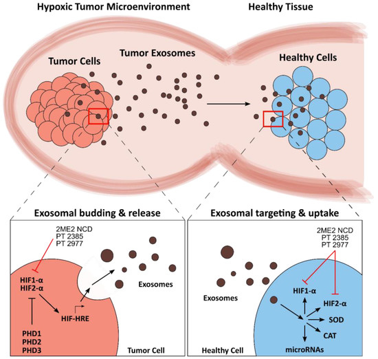
Figure 3.
Schematic overview of exosomal modulation of the redox tumor microenvironment (TME). Tumor-derived exosomes maintain and propagate the TME by invading healthy cells (top). Prolyl hydroxylases (PHDs) negatively regulate hypoxia-inducible factors HIF1-α and HIF2-α. Hypoxic conditions activate HIFs, resulting in induction of hypoxia response elements (HREs) and an increase in exosome production (bottom left). The exosomal cargo contains active proteins, such as HIFs, but also microRNAs and RNA transcripts of redox proteins, such as superoxide dismutase (SOD) and catalase (CAT). Exosome uptake leads to the release of this cargo, which alters the redox landscape of the receiving cell (bottom right). The names of pharmaceutical and experimental inhibitors are presented, and the corresponding target sites are indicated by red lines.
4.1. Redox Mechanisms of Tumor Exosomes
Exosomes are extracellular carriers that transport cytosolic biomolecules, such as miRNAs and proteins, from virtually all cells in the body to neighboring and distal cells via the endocytic pathway. Correspondingly, tumor cells generate distinct exosomes and other extracellular vesicles (EVs), such as microvesicles (MVs), to communicate and to invade other cells with their own tumorigenic-specific cargo. Thus, EVs are able to perpetuate and sustain the TME and modulate the redox environment [273,274,275].
Understanding the underlying mechanisms behind the regulation of exosome formation and release is an emerging field of research, which aims to reveal potential and novel cancer targets. The hypoxia-inducible factor (HIF) family are transcription factors that mediate expression of genes under hypoxic conditions, including genes that are associated with tumor growth and progression. According to several studies, HIFs are also involved in the formation and release of tumor-derived exosomes and other EVs in hypoxic conditions [263,266,268,276,277]. These studies showed that overexpression of HIFs correlates with increased release of tumor exosomes. Upon suppression and inhibition of HIF-1α and HIF-2α, exosome release levels reverted to those in normoxic conditions. Similar results were obtained via promotion of the negative regulators of HIF, i.e., prolyl hydroxylases (PHD1, PHD2, and PHD3). In addition, both HIF-1α and HIF-2α have been shown to bind to hypoxia response elements (HREs), which regulate a large array of genes that are also associated with hypoxia-derived exosomes. For instance, hypoxia in oral squamous cell carcinoma (OSCC) activates HREs in the promoter regions of exosomal microRNA-21 (miR-21), leading to miR-21 upregulation, which is linked to tumor growth [278]. Besides modulating exosomes, HIFs are a requirement in MV shedding of breast cancer cells [279]. These studies suggest that HIFs play an important and diverse role in the regulation of EVs, showing promise as drug targets in cancer therapy. In addition, redox pathways directly affect exosomal release via post-translational modification of exosomal surface proteins [280]. Specifically, redox-sensitive thiol groups can influence protein folding, acting as switches in the regulation of exosomal release [281].
Redox imbalance in the TME also alters the abundance of exosomal cargo proteins, and, subsequently, affects the redox states of cells that receive the exosomal cargo. For instance, the redox-sensitive signaling pathway PI3K/Akt/eNOS regulates the exosomal release of angpoietin-2 (Ang2), an important player in vascular remodeling of tumors [282,283]. Another example is the elevated exosomal release of a mutant SOD1 variant to neurons, which fosters disease spreading and has been described for motor neuron pathology in amyotrophic lateral sclerosis (ALS) [284]. Remarkably, exosomes also deliver increased levels of active HIF1-α and HIF2-α to healthy cells, transferring tumorigenic properties to the new host cell [285].
Redox imbalance also affects exosomal RNA cargo, which serves as a key mechanism in advancing tumorigenic stages [286,287]. An earlier study showed that exosomes laterally transfer RNA transcripts for CAT and SOD2, which promotes chemoresistance to pancreatic cancer cells; however, this study was conducted under very specific in vitro conditions, which are not necessarily physiologically relevant [288].
4.2. Leveraging Exosomes in Cancer Therapy
Exosomes are an important contributor to the redox TME, and hence have potential to be exploited in the development of cancer therapy. This is illustrated by a study on prostate cancer cells that showed inhibition of cancer cell growth upon treatment with exosome biogenesis inhibitors [289]. Current efforts in the development of novel therapeutic strategies against cancer also explore exosomal pathways that modulate the redox TME.
The HIF family, particularly the isoforms HIF1-α and HIF2-α, are important targets, especially in aggressive forms of cancer where drug resistance interferes with therapy [290,291,292]. Selective inhibitors of HIF1-α and HIF2-α, such as the compound 2ME2 NCD (panzem), showed promise in phase 2 clinical trials when used in combination with bevacizumab for carcinoid neuroendocrine tumor [94,293]. The first-in-class HIF2-α inhibitor—PT2385—and the more potent second-generation variant, PT2977, are both in phase 2 clinical trials [HIF-2 Alpha Inhibitor PT2385 in Treating Patients With Recurrent Glioblastoma (PT2385), NCT03216499; A Trial of PT2977 in Combination With Cabozantinib in Patients With Clear Cell Renal Cell Carcinoma (ccRCC), NCT03634540]. Besides HIF antagonists, researchers have also explored HIF regulatory pathways as pharmaceutical targets in order to enhance HIF degradation. One notable example is the known thioredoxin inhibitor Vorinostat (Zolinza), which inhibits HIF1-α and is approved for cancer treatment [294].
Interestingly, exosomes are also exploited as innovative drug delivery systems that target diseased cells with high selectivity [295,296]. Current promising developments in exosomal engineering include delivery of therapeutic cargo, e.g., the enzyme CAT, to neuronal cells in the treatment of Parkinson’s disease [297]. Another example is the delivery of miRNAs in the treatment of lung cancer [267]. Many challenges need to be addressed in developing exosomes as drug delivery systems for wide-scale clinical use, such as increasing the scale of production to meet expected market needs. However, exosomal biocompatibility and ability to incorporate many different therapeutically relevant payloads enable broad and well-adjusted application in cancer therapy.
5. Conclusions and Outlook
A growing body of research on the emerging field of redox biology illustrates the vast complexity of this discipline, and its implication for a variety of biological process. It is becoming clear that generalization of redox biology into oxidants and antioxidants, which are either “good” or “bad” for cancer, is difficult, if not impossible [9]. Nevertheless, our growing knowledge enables us to isolate specific pathways, enzyme kinetics, redox gradients and compartments as well as key players within a narrowly defined cellular environment, which can be exploited to modulate biological processes for disease management. It is well established that redox metabolism is involved in cancer development, progression and resistance to therapy. Specific and carefully adjusted intervention opens up the opportunity to tip the balance and disrupt the redox landscape in cancer cells. The ongoing advancements in cancer biology, for instance, the role of exosomal vesicles in the creation and maintenance of tumor microenvironments, will provide us with future targets and new therapeutic platforms. In addition, continuous developments in medicinal chemistry will provide us with novel tools, e.g. Nrf2–Keap1 modulators, to target key players in the redox landscape. Redox biology has the potential to bring novel therapeutic approaches and improve patient outcomes in the future.
Author Contributions
D.Ö. conceptualized, planned and supervised the work; D.N., S.M. and D.Ö. retrieved information, designed figures, wrote and edited the draft. All authors have read and agreed to the published version of the manuscript.
Information Retrieval
Literature was retrieved by searching the main databases PubMed and Scopus as well as Google Scholar and Google Search; references were managed using Endnote X8.2 and Zotero 5.0.87; figures were created using Inkscape 0.92, Adobe Illustrator CC 2018 (22.1), and ChemDraw Profession 16.0.1.4.
Funding
This work received no external funding.
Conflicts of Interest
The authors declare no conflict of interest.
References
- Santolini, J.; Wootton, S.A.; Jackson, A.A.; Feelisch, M. The Redox architecture of physiological function. Curr. Opin. Physiol. 2019, 9, 34–47. [Google Scholar] [CrossRef] [PubMed]
- Jones, D.P.; Sies, H. The Redox Code. Antioxid. Redox Signal. 2015, 23, 734–746. [Google Scholar] [CrossRef] [PubMed]
- Sies, H.; Berndt, C.; Jones, D.P. Oxidative Stress. Annu. Rev. Biochem. 2017, 86, 715–748. [Google Scholar] [CrossRef]
- Sies, H. Oxidative stress: A concept in redox biology and medicine. Redox Biol. 2015, 4, 180–183. [Google Scholar] [CrossRef]
- Du, C.; Gao, Z.; Venkatesha, V.A.; Kalen, A.L.; Chaudhuri, L.; Spitz, D.R.; Cullen, J.J.; Oberley, L.W.; Goswami, P.C. Mitochondrial ROS and radiation induced transformation in mouse embryonic fibroblasts. Cancer Biol. 2009, 8, 1962–1971. [Google Scholar] [CrossRef] [PubMed]
- Srinivas, U.S.; Tan, B.W.Q.; Vellayappan, B.A.; Jeyasekharan, A.D. ROS and the DNA damage response in cancer. Redox Biol. 2019, 25, 101084. [Google Scholar] [CrossRef] [PubMed]
- Liou, G.Y.; Storz, P. Reactive oxygen species in cancer. Free. Radic. Res. 2010, 44, 479–496. [Google Scholar] [CrossRef]
- de Sa Junior, P.L.; Camara, D.A.D.; Porcacchia, A.S.; Fonseca, P.M.M.; Jorge, S.D.; Araldi, R.P.; Ferreira, A.K. The Roles of ROS in Cancer Heterogeneity and Therapy. Oxid. Med. Cell. Longev. 2017, 2017, 2467940. [Google Scholar] [CrossRef]
- Harris, I.S.; DeNicola, G.M. The Complex Interplay between Antioxidants and ROS in Cancer. Trends Cell. Biol. 2020. [Google Scholar] [CrossRef]
- Jena, N.R. DNA damage by reactive species: Mechanisms, mutation and repair. J. Biosci. 2012, 37, 503–517. [Google Scholar] [CrossRef]
- Storz, P. Reactive oxygen species in tumor progression. Front. Biosci. 2005, 10, 1881–1896. [Google Scholar] [CrossRef] [PubMed]
- DeBerardinis, R.J.; Chandel, N.S. Fundamentals of cancer metabolism. Sci. Adv. 2016, 2, e1600200. [Google Scholar] [CrossRef] [PubMed]
- Glasauer, A.; Chandel, N.S. Ros. Curr. Biol. 2013, 23, R100–R102. [Google Scholar] [CrossRef] [PubMed]
- Hsu, P.P.; Sabatini, D.M. Cancer cell metabolism: Warburg and beyond. Cell 2008, 134, 703–707. [Google Scholar] [CrossRef] [PubMed]
- Purohit, V.; Simeone, D.M.; Lyssiotis, C.A. Metabolic Regulation of Redox Balance in Cancer. Cancers 2019, 11, 955. [Google Scholar] [CrossRef] [PubMed]
- Dewhirst, M.W.; Cao, Y.; Moeller, B. Cycling hypoxia and free radicals regulate angiogenesis and radiotherapy response. Nat. Rev. Cancer 2008, 8, 425–437. [Google Scholar] [CrossRef]
- Harris, A.L. Hypoxia—A key regulatory factor in tumour growth. Nat. Rev. Cancer 2002, 2, 38–47. [Google Scholar] [CrossRef]
- Brown, N.S.; Bicknell, R. Hypoxia and oxidative stress in breast cancer. Oxidative stress: Its effects on the growth, metastatic potential and response to therapy of breast cancer. Breast Cancer Res. 2001, 3, 323–327. [Google Scholar] [CrossRef]
- Xu, R.; Rai, A.; Chen, M.; Suwakulsiri, W.; Greening, D.W.; Simpson, R.J. Extracellular vesicles in cancer—Implications for future improvements in cancer care. Nat. Rev. Clin. Oncol. 2018, 15, 617–638. [Google Scholar] [CrossRef]
- Trachootham, D.; Alexandre, J.; Huang, P. Targeting cancer cells by ROS-mediated mechanisms: A radical therapeutic approach? Nat. Rev. Drug Discov. 2009, 8, 579–591. [Google Scholar] [CrossRef]
- Perillo, B.; Di Donato, M.; Pezone, A.; Di Zazzo, E.; Giovannelli, P.; Galasso, G.; Castoria, G.; Migliaccio, A. ROS in cancer therapy: The bright side of the moon. Exp. Mol. Med. 2020, 52, 192–203. [Google Scholar] [CrossRef] [PubMed]
- Gun, S.Y.; Lee, S.W.L.; Sieow, J.L.; Wong, S.C. Targeting immune cells for cancer therapy. Redox Biol. 2019, 25, 101174. [Google Scholar] [CrossRef] [PubMed]
- Ni, J.; Song, J.; Wang, B.; Hua, H.; Zhu, H.; Guo, X.; Xiong, S.; Zhao, Y. Dendritic cell vaccine for the effective immunotherapy of breast cancer. Biomed. Pharmacother. 2020, 126, 110046. [Google Scholar] [CrossRef] [PubMed]
- Schockel, L.; Glasauer, A.; Basit, F.; Bitschar, K.; Truong, H.; Erdmann, G.; Algire, C.; Hagebarth, A.; Willems, P.H.; Kopitz, C.; et al. Targeting mitochondrial complex I using BAY 87-2243 reduces melanoma tumor growth. Cancer Metab. 2015, 3, 11. [Google Scholar] [CrossRef] [PubMed]
- Ellinghaus, P.; Heisler, I.; Unterschemmann, K.; Haerter, M.; Beck, H.; Greschat, S.; Ehrmann, A.; Summer, H.; Flamme, I.; Oehme, F.; et al. BAY 87-2243, a highly potent and selective inhibitor of hypoxia-induced gene activation has antitumor activities by inhibition of mitochondrial complex I. Cancer Med. 2013, 2, 611–624. [Google Scholar] [CrossRef]
- Helbig, L.; Koi, L.; Bruchner, K.; Gurtner, K.; Hess-Stumpp, H.; Unterschemmann, K.; Baumann, M.; Zips, D.; Yaromina, A. BAY 87-2243, a novel inhibitor of hypoxia-induced gene activation, improves local tumor control after fractionated irradiation in a schedule-dependent manner in head and neck human xenografts. Radiat Oncol. 2014, 9, 207. [Google Scholar] [CrossRef]
- Villani, L.A.; Smith, B.K.; Marcinko, K.; Ford, R.J.; Broadfield, L.A.; Green, A.E.; Houde, V.P.; Muti, P.; Tsakiridis, T.; Steinberg, G.R. The diabetes medication Canagliflozin reduces cancer cell proliferation by inhibiting mitochondrial complex-I supported respiration. Mol. Metab. 2016, 5, 1048–1056. [Google Scholar] [CrossRef]
- Hung, M.H.; Chen, Y.L.; Chen, L.J.; Chu, P.Y.; Hsieh, F.S.; Tsai, M.H.; Shih, C.T.; Chao, T.I.; Huang, C.Y.; Chen, K.F. Canagliflozin inhibits growth of hepatocellular carcinoma via blocking glucose-influx-induced beta-catenin activation. Cell Death Dis. 2019, 10, 420. [Google Scholar] [CrossRef]
- Chen, G.; Zhang, X.; Zhao, M.; Wang, Y.; Cheng, X.; Wang, D.; Xu, Y.; Du, Z.; Yu, X. Celastrol targets mitochondrial respiratory chain complex I to induce reactive oxygen species-dependent cytotoxicity in tumor cells. Bmc Cancer 2011, 11, 170. [Google Scholar] [CrossRef]
- Wheaton, W.W.; Weinberg, S.E.; Hamanaka, R.B.; Soberanes, S.; Sullivan, L.B.; Anso, E.; Glasauer, A.; Dufour, E.; Mutlu, G.M.; Budigner, G.S.; et al. Metformin inhibits mitochondrial complex I of cancer cells to reduce tumorigenesis. Elife 2014, 3, e02242. [Google Scholar] [CrossRef]
- Cheng, G.; Zhang, Q.; Pan, J.; Lee, Y.; Ouari, O.; Hardy, M.; Zielonka, M.; Myers, C.R.; Zielonka, J.; Weh, K.; et al. Targeting lonidamine to mitochondria mitigates lung tumorigenesis and brain metastasis. Nat. Commun. 2019, 10, 2205. [Google Scholar] [CrossRef] [PubMed]
- Zhang, B.; Chu, W.; Wei, P.; Liu, Y.; Wei, T. Xanthohumol induces generation of reactive oxygen species and triggers apoptosis through inhibition of mitochondrial electron transfer chain complex I. Free Radic. Biol. Med. 2015, 89, 486–497. [Google Scholar] [CrossRef] [PubMed]
- Jiang, C.H.; Sun, T.L.; Xiang, D.X.; Wei, S.S.; Li, W.Q. Anticancer Activity and Mechanism of Xanthohumol: A Prenylated Flavonoid From Hops (Humulus lupulus L.). Front. Pharmacol. 2018, 9, 530. [Google Scholar] [CrossRef] [PubMed]
- Azevedo-Silva, J.; Queiros, O.; Baltazar, F.; Ulaszewski, S.; Goffeau, A.; Ko, Y.H.; Pedersen, P.L.; Preto, A.; Casal, M. The anticancer agent 3-bromopyruvate: A simple but powerful molecule taken from the lab to the bedside. J. Bioenerg. Biomembr. 2016, 48, 349–362. [Google Scholar] [CrossRef]
- Buccheri, G.; Ferrigno, D.; Rosso, A. A phase II study of methotrexate, doxorubicin, cyclophosphamide, and lomustine chemotherapy and lonidamine in advanced non-small cell lung cancer. Cancer 1993, 72, 1564–1572. [Google Scholar] [CrossRef]
- Guo, L.; Shestov, A.A.; Worth, A.J.; Nath, K.; Nelson, D.S.; Leeper, D.B.; Glickson, J.D.; Blair, I.A. Inhibition of Mitochondrial Complex II by the Anticancer Agent Lonidamine. J. Biol. Chem. 2016, 291, 42–57. [Google Scholar] [CrossRef]
- Kluckova, K.; Bezawork-Geleta, A.; Rohlena, J.; Dong, L.; Neuzil, J. Mitochondrial complex II, a novel target for anti-cancer agents. Biochim. Biophys. Acta 2013, 1827, 552–564. [Google Scholar] [CrossRef]
- Constantinou, C.; Charalambous, C.; Kanakis, D. Vitamin E and cancer: An update on the emerging role of gamma and delta tocotrienols. Eur. J. Nutr. 2020, 59, 845–857. [Google Scholar] [CrossRef]
- Montagnani Marelli, M.; Marzagalli, M.; Fontana, F.; Raimondi, M.; Moretti, R.M.; Limonta, P. Anticancer properties of tocotrienols: A review of cellular mechanisms and molecular targets. J. Cell. Physiol. 2019, 234, 1147–1164. [Google Scholar] [CrossRef]
- Fiorillo, M.; Lamb, R.; Tanowitz, H.B.; Mutti, L.; Krstic-Demonacos, M.; Cappello, A.R.; Martinez-Outschoorn, U.E.; Sotgia, F.; Lisanti, M.P. Repurposing atovaquone: Targeting mitochondrial complex III and OXPHOS to eradicate cancer stem cells. Oncotarget 2016, 7, 34084–34099. [Google Scholar] [CrossRef]
- Lin, J.; Zahurak, M.; Beer, T.M.; Ryan, C.J.; Wilding, G.; Mathew, P.; Morris, M.; Callahan, J.A.; Gordon, G.; Reich, S.D.; et al. A non-comparative randomized phase II study of 2 doses of ATN-224, a copper/zinc superoxide dismutase inhibitor, in patients with biochemically recurrent hormone-naive prostate cancer. Urol. Oncol. 2013, 31, 581–588. [Google Scholar] [CrossRef]
- Lowndes, S.A.; Adams, A.; Timms, A.; Fisher, N.; Smythe, J.; Watt, S.M.; Joel, S.; Donate, F.; Hayward, C.; Reich, S.; et al. Phase I study of copper-binding agent ATN-224 in patients with advanced solid tumors. Clin. Cancer Res. 2008, 14, 7526–7534. [Google Scholar] [CrossRef] [PubMed]
- Kim, K.K.; Abelman, S.; Yano, N.; Ribeiro, J.R.; Singh, R.K.; Tipping, M.; Moore, R.G. Tetrathiomolybdate inhibits mitochondrial complex IV and mediates degradation of hypoxia-inducible factor-1alpha in cancer cells. Sci. Rep. 2015, 5, 14296. [Google Scholar] [CrossRef] [PubMed]
- Madak, J.T.; Bankhead, A., 3rd; Cuthbertson, C.R.; Showalter, H.D.; Neamati, N. Revisiting the role of dihydroorotate dehydrogenase as a therapeutic target for cancer. Pharmacol. Ther. 2019, 195, 111–131. [Google Scholar] [CrossRef] [PubMed]
- Teschner, S.; Burst, V. Leflunomide: A drug with a potential beyond rheumatology. Immunotherapy 2010, 2, 637–650. [Google Scholar] [CrossRef]
- Hail, N., Jr.; Chen, P.; Bushman, L.R. Teriflunomide (leflunomide) promotes cytostatic, antioxidant, and apoptotic effects in transformed prostate epithelial cells: Evidence supporting a role for teriflunomide in prostate cancer chemoprevention. Neoplasia 2010, 12, 464–475. [Google Scholar] [CrossRef]
- Huang, O.; Zhang, W.; Zhi, Q.; Xue, X.; Liu, H.; Shen, D.; Geng, M.; Xie, Z.; Jiang, M. Teriflunomide, an immunomodulatory drug, exerts anticancer activity in triple negative breast cancer cells. Exp. Biol. Med. (Maywood) 2015, 240, 426–437. [Google Scholar] [CrossRef]
- Jiang, L.; Zhang, W.; Li, W.; Ling, C.; Jiang, M. Anti-inflammatory drug, leflunomide and its metabolite teriflunomide inhibit NSCLC proliferation in vivo and in vitro. Toxicol. Lett. 2018, 282, 154–165. [Google Scholar] [CrossRef]
- Orr, A.L.; Ashok, D.; Sarantos, M.R.; Ng, R.; Shi, T.; Gerencser, A.A.; Hughes, R.E.; Brand, M.D. Novel inhibitors of mitochondrial sn-glycerol 3-phosphate dehydrogenase. PLoS ONE 2014, 9, e89938. [Google Scholar] [CrossRef] [PubMed]
- Gross, M.E.; Agus, D.B.; Dorff, T.B.; Pinski, J.K.; Quinn, D.I.; Castellanos, O.; Gilmore, P.; Shih, J.C. Phase 2 trial of monoamine oxidase inhibitor phenelzine in biochemical recurrent prostate cancer. Prostate Cancer Prostatic Dis. 2020. [Google Scholar] [CrossRef]
- Wu, J.B.; Shao, C.; Li, X.; Li, Q.; Hu, P.; Shi, C.; Li, Y.; Chen, Y.T.; Yin, F.; Liao, C.P.; et al. Monoamine oxidase A mediates prostate tumorigenesis and cancer metastasis. J. Clin. Investig. 2014, 124, 2891–2908. [Google Scholar] [CrossRef]
- Sampson, N.; Brunner, E.; Weber, A.; Puhr, M.; Schafer, G.; Szyndralewiez, C.; Klocker, H. Inhibition of Nox4-dependent ROS signaling attenuates prostate fibroblast activation and abrogates stromal-mediated protumorigenic interactions. Int. J. Cancer 2018, 143, 383–395. [Google Scholar] [CrossRef] [PubMed]
- Teixeira, G.; Szyndralewiez, C.; Molango, S.; Carnesecchi, S.; Heitz, F.; Wiesel, P.; Wood, J.M. Therapeutic potential of NADPH oxidase 1/4 inhibitors. Br. J. pharmacol. 2017, 174, 1647–1669. [Google Scholar] [CrossRef]
- Sancho, P.; Fabregat, I. The NADPH oxidase inhibitor VAS2870 impairs cell growth and enhances TGF-beta-induced apoptosis of liver tumor cells. Biochem. Pharmacol. 2011, 81, 917–924. [Google Scholar] [CrossRef] [PubMed]
- Blais, J.D.; Chin, K.T.; Zito, E.; Zhang, Y.; Heldman, N.; Harding, H.P.; Fass, D.; Thorpe, C.; Ron, D. A small molecule inhibitor of endoplasmic reticulum oxidation 1 (ERO1) with selectively reversible thiol reactivity. J. Biol. Chem. 2010, 285, 20993–21003. [Google Scholar] [CrossRef]
- Hoffstrom, B.G.; Kaplan, A.; Letso, R.; Schmid, R.S.; Turmel, G.J.; Lo, D.C.; Stockwell, B.R. Inhibitors of protein disulfide isomerase suppress apoptosis induced by misfolded proteins. Nat. Chem. Biol. 2010, 6, 900–906. [Google Scholar] [CrossRef] [PubMed]
- Vatolin, S.; Phillips, J.G.; Jha, B.K.; Govindgari, S.; Hu, J.; Grabowski, D.; Parker, Y.; Lindner, D.J.; Zhong, F.; Distelhorst, C.W.; et al. Novel Protein Disulfide Isomerase Inhibitor with Anticancer Activity in Multiple Myeloma. Cancer Res. 2016, 76, 3340–3350. [Google Scholar] [CrossRef] [PubMed]
- Robinson, R.M.; Reyes, L.; Duncan, R.M.; Bian, H.; Reitz, A.B.; Manevich, Y.; McClure, J.J.; Champion, M.M.; Chou, C.J.; Sharik, M.E.; et al. Inhibitors of the protein disulfide isomerase family for the treatment of multiple myeloma. Leukemia 2019, 33, 1011–1022. [Google Scholar] [CrossRef]
- Jasuja, R.; Passam, F.H.; Kennedy, D.R.; Kim, S.H.; van Hessem, L.; Lin, L.; Bowley, S.R.; Joshi, S.S.; Dilks, J.R.; Furie, B.; et al. Protein disulfide isomerase inhibitors constitute a new class of antithrombotic agents. J. Clin. Investig. 2012, 122, 2104–2113. [Google Scholar] [CrossRef]
- Khan, M.M.; Simizu, S.; Lai, N.S.; Kawatani, M.; Shimizu, T.; Osada, H. Discovery of a small molecule PDI inhibitor that inhibits reduction of HIV-1 envelope glycoprotein gp120. ACS Chem. Biol. 2011, 6, 245–251. [Google Scholar] [CrossRef]
- Khodier, C.; VerPlank, L.; Nag, P.P.; Pu, J.; Wurst, J.; Pilyugina, T.; Dockendorff, C.; Galinski, C.N.; Scalise, A.A.; Passam, F.; et al. Identification of ML359 as a Small Molecule Inhibitor of Protein Disulfide Isomerase. In Probe Reports from the NIH Molecular Libraries Program; Bethseda (MD): Rockville, MD, USA, 2010. [Google Scholar]
- Ozcelik, D.; Pezacki, J.P. Small Molecule Inhibition of Protein Disulfide Isomerase in Neuroblastoma Cells Induces an Oxidative Stress Response and Apoptosis Pathways. ACS Chem. Neurosci. 2019, 10, 4068–4075. [Google Scholar] [CrossRef]
- Rakic, B.; Clarke, J.; Tremblay, T.L.; Taylor, J.; Schreiber, K.; Nelson, K.M.; Abrams, S.R.; Pezacki, J.P. A small-molecule probe for hepatitis C virus replication that blocks protein folding. Chem. Biol. 2006, 13, 1051–1060. [Google Scholar] [CrossRef] [PubMed]
- Ge, J.; Zhang, C.J.; Li, L.; Chong, L.M.; Wu, X.; Hao, P.; Sze, S.K.; Yao, S.Q. Small molecule probe suitable for in situ profiling and inhibition of protein disulfide isomerase. ACS Chem. Biol. 2013, 8, 2577–2585. [Google Scholar] [CrossRef] [PubMed]
- Xu, S.; Butkevich, A.N.; Yamada, R.; Zhou, Y.; Debnath, B.; Duncan, R.; Zandi, E.; Petasis, N.A.; Neamati, N. Discovery of an orally active small-molecule irreversible inhibitor of protein disulfide isomerase for ovarian cancer treatment. Proc. Natl. Acad. Sci. USA 2012, 109, 16348–16353. [Google Scholar] [CrossRef] [PubMed]
- Flaumenhaft, R.; Furie, B.; Zwicker, J.I. Therapeutic implications of protein disulfide isomerase inhibition in thrombotic disease. Arter. Thromb. Vasc. Biol. 2015, 35, 16–23. [Google Scholar] [CrossRef]
- Banerjee, R.; Pace, N.J.; Brown, D.R.; Weerapana, E. 1,3,5-Triazine as a modular scaffold for covalent inhibitors with streamlined target identification. J. Am. Chem. Soc. 2013, 135, 2497–2500. [Google Scholar] [CrossRef] [PubMed]
- Luna, G.; Dolzhenko, A.V.; Mancera, R.L. Inhibitors of Xanthine Oxidase: Scaffold Diversity and Structure-Based Drug Design. Chem. Med. Chem. 2019, 14, 714–743. [Google Scholar] [CrossRef]
- Suzuki, S.; Pitchakarn, P.; Sato, S.; Shirai, T.; Takahashi, S. Apocynin, an NADPH oxidase inhibitor, suppresses progression of prostate cancer via Rac1 dephosphorylation. Exp. Toxicol. Pathol. 2013, 65, 1035–1041. [Google Scholar] [CrossRef]
- Yang, T.; Zang, D.W.; Shan, W.; Guo, A.C.; Wu, J.P.; Wang, Y.J.; Wang, Q. Synthesis and Evaluations of Novel Apocynin Derivatives as Anti-Glioma Agents. Front. Pharmacol. 2019, 10, 951. [Google Scholar] [CrossRef]
- Panieri, E.; Saso, L. Potential Applications of NRF2 Inhibitors in Cancer Therapy. Oxid. Med. Cell. Longev. 2019, 2019, 8592348. [Google Scholar] [CrossRef]
- Wang, Y.; Yu, J.; Cui, R.; Lin, J.; Ding, X. Curcumin in Treating Breast Cancer: A Review. J. Lab. Autom. 2016, 21, 723–731. [Google Scholar] [CrossRef]
- Loewe, R.; Valero, T.; Kremling, S.; Pratscher, B.; Kunstfeld, R.; Pehamberger, H.; Petzelbauer, P. Dimethylfumarate impairs melanoma growth and metastasis. Cancer Res. 2006, 66, 11888–11896. [Google Scholar] [CrossRef] [PubMed]
- Xie, X.; Zhao, Y.; Ma, C.Y.; Xu, X.M.; Zhang, Y.Q.; Wang, C.G.; Jin, J.; Shen, X.; Gao, J.L.; Li, N.; et al. Dimethyl fumarate induces necroptosis in colon cancer cells through GSH depletion/ROS increase/MAPKs activation pathway. Br. J. Pharmacol. 2015, 172, 3929–3943. [Google Scholar] [CrossRef]
- Cuadrado, A.; Rojo, A.I.; Wells, G.; Hayes, J.D.; Cousin, S.P.; Rumsey, W.L.; Attucks, O.C.; Franklin, S.; Levonen, A.L.; Kensler, T.W.; et al. Therapeutic targeting of the NRF2 and KEAP1 partnership in chronic diseases. Nat. Rev. Drug Discov. 2019, 18, 295–317. [Google Scholar] [CrossRef]
- Liby, K.; Risingsong, R.; Royce, D.B.; Williams, C.R.; Ma, T.; Yore, M.M.; Sporn, M.B. Triterpenoids CDDO-methyl ester or CDDO-ethyl amide and rexinoids LG100268 or NRX194204 for prevention and treatment of lung cancer in mice. Cancer Prev. Res. (Phila.) 2009, 2, 1050–1058. [Google Scholar] [CrossRef] [PubMed]
- Liby, K.T.; Royce, D.B.; Risingsong, R.; Williams, C.R.; Maitra, A.; Hruban, R.H.; Sporn, M.B. Synthetic triterpenoids prolong survival in a transgenic mouse model of pancreatic cancer. Cancer Prev. Res. (Phila.) 2010, 3, 1427–1434. [Google Scholar] [CrossRef]
- Alumkal, J.J.; Slottke, R.; Schwartzman, J.; Cherala, G.; Munar, M.; Graff, J.N.; Beer, T.M.; Ryan, C.W.; Koop, D.R.; Gibbs, A.; et al. A phase II study of sulforaphane-rich broccoli sprout extracts in men with recurrent prostate cancer. Investig. New Drugs 2015, 33, 480–489. [Google Scholar] [CrossRef] [PubMed]
- Tagde, A.; Singh, H.; Kang, M.H.; Reynolds, C.P. The glutathione synthesis inhibitor buthionine sulfoximine synergistically enhanced melphalan activity against preclinical models of multiple myeloma. Blood Cancer J. 2014, 4, e229. [Google Scholar] [CrossRef] [PubMed]
- Hong, W.G.; Kim, J.Y.; Cho, J.H.; Hwang, S.G.; Song, J.Y.; Lee, E.; Chang, T.S.; Um, H.D.; Park, J.K. AMRI-59 functions as a radiosensitizer via peroxiredoxin I-targeted ROS accumulation and apoptotic cell death induction. Oncotarget 2017, 8, 114050–114064. [Google Scholar] [CrossRef][Green Version]
- Yang, Y.J.; Baek, J.Y.; Goo, J.; Shin, Y.; Park, J.K.; Jang, J.Y.; Wang, S.B.; Jeong, W.; Lee, H.J.; Um, H.D.; et al. Effective Killing of Cancer Cells Through ROS-Mediated Mechanisms by AMRI-59 Targeting Peroxiredoxin I. Antioxid. Redox Signal. 2016, 24, 453–469. [Google Scholar] [CrossRef]
- Jia, J.J.; Geng, W.S.; Wang, Z.Q.; Chen, L.; Zeng, X.S. The role of thioredoxin system in cancer: Strategy for cancer therapy. Cancer Chemother. Pharmacol. 2019, 84, 453–470. [Google Scholar] [CrossRef] [PubMed]
- Mukherjee, A.; Huber, K.; Evans, H.; Lakhani, N.; Martin, S. A cellular and molecular investigation of the action of PMX464, a putative thioredoxin inhibitor, in normal and colorectal cancer cell lines. Br. J. Pharmacol. 2007, 151, 1167–1175. [Google Scholar] [CrossRef]
- Lu, J.; Chew, E.H.; Holmgren, A. Targeting thioredoxin reductase is a basis for cancer therapy by arsenic trioxide. Proc. Natl. Acad. Sci. USA 2007, 104, 12288–12293. [Google Scholar] [CrossRef]
- Ghosh, S. Cisplatin: The first metal based anticancer drug. Bioorg. Chem. 2019, 88, 102925. [Google Scholar] [CrossRef] [PubMed]
- Urig, S.; Becker, K. On the potential of thioredoxin reductase inhibitors for cancer therapy. Semin. Cancer. Biol. 2006, 16, 452–465. [Google Scholar] [CrossRef] [PubMed]
- Dugo, E.B.; Yedjou, C.G.; Stevens, J.J.; Tchounwou, P.B. Therapeutic Potential of Arsenic Trioxide (ATO) in Treatment of Hepatocellular Carcinoma: Role of Oxidative Stress in ATO-Induced Apoptosis. Ann. Clin. Pathol. 2017, 5. [Google Scholar]
- Somwar, R.; Erdjument-Bromage, H.; Larsson, E.; Shum, D.; Lockwood, W.W.; Yang, G.; Sander, C.; Ouerfelli, O.; Tempst, P.J.; Djaballah, H.; et al. Superoxide dismutase 1 (SOD1) is a target for a small molecule identified in a screen for inhibitors of the growth of lung adenocarcinoma cell lines. Proc. Natl. Acad. Sci. USA 2011, 108, 16375–16380. [Google Scholar] [CrossRef]
- Beg, M.S.; Huang, X.; Silvers, M.A.; Gerber, D.E.; Bolluyt, J.; Sarode, V.; Fattah, F.; Deberardinis, R.J.; Merritt, M.E.; Xie, X.J.; et al. Using a novel NQO1 bioactivatable drug, beta-lapachone (ARQ761), to enhance chemotherapeutic effects by metabolic modulation in pancreatic cancer. J. Surg. Oncol. 2017, 116, 83–88. [Google Scholar] [CrossRef]
- Silvers, M.A.; Deja, S.; Singh, N.; Egnatchik, R.A.; Sudderth, J.; Luo, X.; Beg, M.S.; Burgess, S.C.; DeBerardinis, R.J.; Boothman, D.A.; et al. The NQO1 bioactivatable drug, beta-lapachone, alters the redox state of NQO1+ pancreatic cancer cells, causing perturbation in central carbon metabolism. J. Biol. Chem. 2017, 292, 18203–18216. [Google Scholar] [CrossRef]
- Bianchet, M.A.; Faig, M.; Amzel, L.M. Structure and mechanism of NAD[P]H:quinone acceptor oxidoreductases (NQO). Methods Enzym. 2004, 382, 144–174. [Google Scholar] [CrossRef]
- Singh, B.; Shoulson, R.; Chatterjee, A.; Ronghe, A.; Bhat, N.K.; Dim, D.C.; Bhat, H.K. Resveratrol inhibits estrogen-induced breast carcinogenesis through induction of NRF2-mediated protective pathways. Carcinogenesis 2014, 35, 1872–1880. [Google Scholar] [CrossRef]
- Alnabulsi, S.; Hussein, B.; Santina, E.; Alsalahat, I.; Kadirvel, M.; Magwaza, R.N.; Bryce, R.A.; Schwalbe, C.H.; Baldwin, A.G.; Russo, I.; et al. Evaluation of analogues of furan-amidines as inhibitors of NQO2. Bioorg. Med. Chem. Lett. 2018, 28, 1292–1297. [Google Scholar] [CrossRef] [PubMed]
- Fallah, J.; Rini, B.I. HIF Inhibitors: Status of Current Clinical Development. Curr. Oncol. Rep. 2019, 21, 6. [Google Scholar] [CrossRef] [PubMed]
- Yoboue, E.D.; Sitia, R.; Simmen, T. Redox crosstalk at endoplasmic reticulum (ER) membrane contact sites (MCS) uses toxic waste to deliver messages. Cell. Death Dis. 2018, 9, 331. [Google Scholar] [CrossRef] [PubMed]
- Boveris, A.; Oshino, N.; Chance, B. The cellular production of hydrogen peroxide. Biochem. J. 1972, 128, 617–630. [Google Scholar] [CrossRef] [PubMed]
- Bak, D.W.; Weerapana, E. Cysteine-mediated redox signalling in the mitochondria. Mol. Biosyst. 2015, 11, 678–697. [Google Scholar] [CrossRef] [PubMed]
- Vyas, S.; Zaganjor, E.; Haigis, M.C. Mitochondria and Cancer. Cell 2016, 166, 555–566. [Google Scholar] [CrossRef] [PubMed]
- Reczek, C.R.; Chandel, N.S. ROS-dependent signal transduction. Curr. Opin. Cell. Biol. 2015, 33, 8–13. [Google Scholar] [CrossRef]
- Brand, M.D. Mitochondrial generation of superoxide and hydrogen peroxide as the source of mitochondrial redox signaling. Free Radic. Biol. Med. 2016, 100, 14–31. [Google Scholar] [CrossRef] [PubMed]
- Sullivan, L.B.; Chandel, N.S. Mitochondrial reactive oxygen species and cancer. Cancer Metab. 2014, 2, 17. [Google Scholar] [CrossRef]
- Hahn, A.; Zuryn, S. Mitochondrial Genome (mtDNA) Mutations that Generate Reactive Oxygen Species. Antioxidants 2019, 8, 392. [Google Scholar] [CrossRef] [PubMed]
- Rohlena, J.; Dong, L.F.; Ralph, S.J.; Neuzil, J. Anticancer drugs targeting the mitochondrial electron transport chain. Antioxid. Redox Signal. 2011, 15, 2951–2974. [Google Scholar] [CrossRef] [PubMed]
- Calabrese, C.; Iommarini, L.; Kurelac, I.; Calvaruso, M.A.; Capristo, M.; Lollini, P.L.; Nanni, P.; Bergamini, C.; Nicoletti, G.; Giovanni, C.D.; et al. Respiratory complex I is essential to induce a Warburg profile in mitochondria-defective tumor cells. Cancer Metab. 2013, 1, 11. [Google Scholar] [CrossRef] [PubMed]
- He, X.; Zhou, A.; Lu, H.; Chen, Y.; Huang, G.; Yue, X.; Zhao, P.; Wu, Y. Suppression of mitochondrial complex I influences cell metastatic properties. PLoS ONE 2013, 8, e61677. [Google Scholar] [CrossRef]
- Ishikawa, K.; Takenaga, K.; Akimoto, M.; Koshikawa, N.; Yamaguchi, A.; Imanishi, H.; Nakada, K.; Honma, Y.; Hayashi, J. ROS-generating mitochondrial DNA mutations can regulate tumor cell metastasis. Science 2008, 320, 661–664. [Google Scholar] [CrossRef] [PubMed]
- Urra, F.A.; Munoz, F.; Lovy, A.; Cardenas, C. The Mitochondrial Complex(I)ty of Cancer. Front. Oncol. 2017, 7, 118. [Google Scholar] [CrossRef]
- Dasgupta, S.; Hoque, M.O.; Upadhyay, S.; Sidransky, D. Forced cytochrome B gene mutation expression induces mitochondrial proliferation and prevents apoptosis in human uroepithelial SV-HUC-1 cells. Int. J. Cancer. 2009, 125, 2829–2835. [Google Scholar] [CrossRef]
- Arnold, S. The power of life—Cytochrome c oxidase takes center stage in metabolic control, cell signalling and survival. Mitochondrion 2012, 12, 46–56. [Google Scholar] [CrossRef]
- Ishida, S.; Andreux, P.; Poitry-Yamate, C.; Auwerx, J.; Hanahan, D. Bioavailable copper modulates oxidative phosphorylation and growth of tumors. Proc. Natl. Acad. Sci. USA 2013, 110, 19507–19512. [Google Scholar] [CrossRef]
- Chen, S.F.; Ruben, R.L.; Dexter, D.L. Mechanism of action of the novel anticancer agent 6-fluoro-2-(2’-fluoro-1,1’-biphenyl-4-yl)-3-methyl-4-quinolinecarbo xylic acid sodium salt (NSC 368390): Inhibition of de novo pyrimidine nucleotide biosynthesis. Cancer Res. 1986, 46, 5014–5019. [Google Scholar]
- Koundinya, M.; Sudhalter, J.; Courjaud, A.; Lionne, B.; Touyer, G.; Bonnet, L.; Menguy, I.; Schreiber, I.; Perrault, C.; Vougier, S.; et al. Dependence on the Pyrimidine Biosynthetic Enzyme DHODH Is a Synthetic Lethal Vulnerability in Mutant KRAS-Driven Cancers. Cell Chem. Biol. 2018, 25, 705–717.e11. [Google Scholar] [CrossRef] [PubMed]
- Oh, J.; O’Connor, P.W. Teriflunomide in the treatment of multiple sclerosis: Current evidence and future prospects. Adv. Neurol. Disord. 2014, 7, 239–252. [Google Scholar] [CrossRef] [PubMed]
- Mracek, T.; Drahota, Z.; Houstek, J. The function and the role of the mitochondrial glycerol-3-phosphate dehydrogenase in mammalian tissues. Biochim. Biophys. Acta 2013, 1827, 401–410. [Google Scholar] [CrossRef] [PubMed]
- Singh, G. Mitochondrial FAD-linked Glycerol-3-phosphate Dehydrogenase: A Target for Cancer Therapeutics. Pharmaceuticals 2014, 7, 192–206. [Google Scholar] [CrossRef]
- Bortolato, M.; Chen, K.; Shih, J.C. Monoamine oxidase inactivation: From pathophysiology to therapeutics. Adv. Drug Deliv. Rev. 2008, 60, 1527–1533. [Google Scholar] [CrossRef]
- Shih, J.C. Monoamine oxidase isoenzymes: Genes, functions and targets for behavior and cancer therapy. J. Neural. Transm. (Vienna) 2018, 125, 1553–1566. [Google Scholar] [CrossRef]
- Tripathi, R.K.P.; Ayyannan, S.R. Monoamine oxidase-B inhibitors as potential neurotherapeutic agents: An overview and update. Med. Res. Rev. 2019, 39, 1603–1706. [Google Scholar] [CrossRef]
- Wimbiscus, M.; Kostenko, O.; Malone, D. MAO inhibitors: Risks, benefits, and lore. Clevel. Clin. J. Med. 2010, 77, 859–882. [Google Scholar] [CrossRef]
- Fan, Y.; Simmen, T. Mechanistic Connections between Endoplasmic Reticulum (ER) Redox Control and Mitochondrial Metabolism. Cells 2019, 8, 1071. [Google Scholar] [CrossRef]
- Zeeshan, H.M.; Lee, G.H.; Kim, H.R.; Chae, H.J. Endoplasmic Reticulum Stress and Associated ROS. Int. J. Mol. Sci. 2016, 17, 327. [Google Scholar] [CrossRef]
- Ali Khan, H.; Mutus, B. Protein disulfide isomerase a multifunctional protein with multiple physiological roles. Front. Chem. 2014, 2, 70. [Google Scholar] [CrossRef] [PubMed]
- Ellgaard, L.; Ruddock, L.W. The human protein disulphide isomerase family: Substrate interactions and functional properties. Embo. Rep. 2005, 6, 28–32. [Google Scholar] [CrossRef] [PubMed]
- Darby, N.J.; Creighton, T.E. Functional properties of the individual thioredoxin-like domains of protein disulfide isomerase. Biochemistry 1995, 34, 11725–11735. [Google Scholar] [CrossRef] [PubMed]
- Kersteen, E.A.; Raines, R.T. Catalysis of protein folding by protein disulfide isomerase and small-molecule mimics. Antioxid. Redox Signal. 2003, 5, 413–424. [Google Scholar] [CrossRef] [PubMed]
- Hatahet, F.; Ruddock, L.W. Protein disulfide isomerase: A critical evaluation of its function in disulfide bond formation. Antioxid. Redox Signal. 2009, 11, 2807–2850. [Google Scholar] [CrossRef]
- Benham, A.M.; van Lith, M.; Sitia, R.; Braakman, I. Ero1-PDI interactions, the response to redox flux and the implications for disulfide bond formation in the mammalian endoplasmic reticulum. Philos. Trans. R. Soc. Lond. B Biol. Sci. 2013, 368, 20110403. [Google Scholar] [CrossRef]
- Sevier, C.S.; Kaiser, C.A. Ero1 and redox homeostasis in the endoplasmic reticulum. Biochim. Biophys. Acta 2008, 1783, 549–556. [Google Scholar] [CrossRef]
- Tu, B.P.; Weissman, J.S. The FAD- and O(2)-dependent reaction cycle of Ero1-mediated oxidative protein folding in the endoplasmic reticulum. Mol. Cell 2002, 10, 983–994. [Google Scholar] [CrossRef]
- Wang, L.; Li, S.J.; Sidhu, A.; Zhu, L.; Liang, Y.; Freedman, R.B.; Wang, C.C. Reconstitution of human Ero1-Lalpha/protein-disulfide isomerase oxidative folding pathway in vitro. Position-dependent differences in role between the a and a domains of protein-disulfide isomerase. J. Biol. Chem. 2009, 284, 199–206. [Google Scholar] [CrossRef]
- Appenzeller-Herzog, C. Glutathione- and non-glutathione-based oxidant control in the endoplasmic reticulum. J. Cell. Sci. 2011, 124, 847–855. [Google Scholar] [CrossRef]
- Zito, E. PRDX4, an endoplasmic reticulum-localized peroxiredoxin at the crossroads between enzymatic oxidative protein folding and nonenzymatic protein oxidation. Antioxid. Redox Signal. 2013, 18, 1666–1674. [Google Scholar] [CrossRef]
- Kodali, V.K.; Thorpe, C. Oxidative protein folding and the Quiescin-sulfhydryl oxidase family of flavoproteins. Antioxid. Redox Signal. 2010, 13, 1217–1230. [Google Scholar] [CrossRef] [PubMed]
- Khan, M.M.; Simizu, S.; Kawatani, M.; Osada, H. The potential of protein disulfide isomerase as a therapeutic drug target. Oncol. Res. 2011, 19, 445–453. [Google Scholar] [CrossRef] [PubMed]
- Lee, E.; Lee, D.H. Emerging roles of protein disulfide isomerase in cancer. BMB Rep. 2017, 50, 401–410. [Google Scholar] [CrossRef] [PubMed]
- Xu, S.; Sankar, S.; Neamati, N. Protein disulfide isomerase: A promising target for cancer therapy. Drug Discov. Today 2014, 19, 222–240. [Google Scholar] [CrossRef]
- Zwicker, J.I.; Schlechter, B.L.; Stopa, J.D.; Liebman, H.A.; Aggarwal, A.; Puligandla, M.; Caughey, T.; Bauer, K.A.; Kuemmerle, N.; Wong, E.; et al. Targeting protein disulfide isomerase with the flavonoid isoquercetin to improve hypercoagulability in advanced cancer. Jci. Insight. 2019, 4. [Google Scholar] [CrossRef] [PubMed]
- Islinger, M.; Voelkl, A.; Fahimi, H.D.; Schrader, M. The peroxisome: An update on mysteries 2.0. Histochem. Cell. Biol. 2018, 150, 443–471. [Google Scholar] [CrossRef] [PubMed]
- Farre, J.C.; Mahalingam, S.S.; Proietto, M.; Subramani, S. Peroxisome biogenesis, membrane contact sites, and quality control. Embo. Rep. 2019, 20. [Google Scholar] [CrossRef]
- Schrader, M.; Costello, J.L.; Godinho, L.F.; Azadi, A.S.; Islinger, M. Proliferation and fission of peroxisomes - An update. Biochim. Biophys. Acta 2016, 1863, 971–983. [Google Scholar] [CrossRef]
- Dreyer, C.; Krey, G.; Keller, H.; Givel, F.; Helftenbein, G.; Wahli, W. Control of the peroxisomal beta-oxidation pathway by a novel family of nuclear hormone receptors. Cell 1992, 68, 879–887. [Google Scholar] [CrossRef]
- Bonekamp, N.A.; Volkl, A.; Fahimi, H.D.; Schrader, M. Reactive oxygen species and peroxisomes: Struggling for balance. Biofactors 2009, 35, 346–355. [Google Scholar] [CrossRef] [PubMed]
- Fransen, M.; Nordgren, M.; Wang, B.; Apanasets, O. Role of peroxisomes in ROS/RNS-metabolism: Implications for human disease. Biochim. Biophys. Acta 2012, 1822, 1363–1373. [Google Scholar] [CrossRef] [PubMed]
- Fransen, M.; Lismont, C. Redox Signaling from and to Peroxisomes: Progress, Challenges, and Prospects. Antioxid. Redox Signal. 2019, 30, 95–112. [Google Scholar] [CrossRef] [PubMed]
- Lismont, C.; Nordgren, M.; Van Veldhoven, P.P.; Fransen, M. Redox interplay between mitochondria and peroxisomes. Front. Cell. Dev. Biol. 2015, 3, 35. [Google Scholar] [CrossRef] [PubMed]
- Dahabieh, M.S.; Di Pietro, E.; Jangal, M.; Goncalves, C.; Witcher, M.; Braverman, N.E.; Del Rincon, S.V. Peroxisomes and cancer: The role of a metabolic specialist in a disease of aberrant metabolism. Biochim. Biophys. Acta Rev. Cancer 2018, 1870, 103–121. [Google Scholar] [CrossRef] [PubMed]
- Robey, R.B.; Weisz, J.; Kuemmerle, N.B.; Salzberg, A.C.; Berg, A.; Brown, D.G.; Kubik, L.; Palorini, R.; Al-Mulla, F.; Al-Temaimi, R.; et al. Metabolic reprogramming and dysregulated metabolism: Cause, consequence and/or enabler of environmental carcinogenesis? Carcinogenesis 2015, 36 (Suppl. 1), S203–S231. [Google Scholar] [CrossRef]
- Walter, K.M.; Schonenberger, M.J.; Trotzmuller, M.; Horn, M.; Elsasser, H.P.; Moser, A.B.; Lucas, M.S.; Schwarz, T.; Gerber, P.A.; Faust, P.L.; et al. Hif-2alpha promotes degradation of mammalian peroxisomes by selective autophagy. Cell. Metab. 2014, 20, 882–897. [Google Scholar] [CrossRef]
- Oh, S.H.; Choi, S.Y.; Choi, H.J.; Ryu, H.M.; Kim, Y.J.; Jung, H.Y.; Cho, J.H.; Kim, C.D.; Park, S.H.; Kwon, T.H.; et al. The emerging role of xanthine oxidase inhibition for suppression of breast cancer cell migration and metastasis associated with hypercholesterolemia. Faseb. J. 2019, 33, 7301–7314. [Google Scholar] [CrossRef]
- Battelli, M.G.; Polito, L.; Bortolotti, M.; Bolognesi, A. Xanthine Oxidoreductase in Drug Metabolism: Beyond a Role as a Detoxifying Enzyme. Curr. Med. Chem. 2016, 23, 4027–4036. [Google Scholar] [CrossRef]
- Gou, Q.; Gong, X.; Jin, J.; Shi, J.; Hou, Y. Peroxisome proliferator-activated receptors (PPARs) are potential drug targets for cancer therapy. Oncotarget 2017, 8, 60704–60709. [Google Scholar] [CrossRef]
- Bedard, K.; Krause, K.H. The NOX family of ROS-generating NADPH oxidases: Physiology and pathophysiology. Physiol. Rev. 2007, 87, 245–313. [Google Scholar] [CrossRef] [PubMed]
- Drummond, G.R.; Selemidis, S.; Griendling, K.K.; Sobey, C.G. Combating oxidative stress in vascular disease: NADPH oxidases as therapeutic targets. Nat. Rev. Drug. Discov. 2011, 10, 453–471. [Google Scholar] [CrossRef] [PubMed]
- Park, M.T.; Kim, M.J.; Suh, Y.; Kim, R.K.; Kim, H.; Lim, E.J.; Yoo, K.C.; Lee, G.H.; Kim, Y.H.; Hwang, S.G.; et al. Novel signaling axis for ROS generation during K-Ras-induced cellular transformation. Cell. Death Differ. 2014, 21, 1185–1197. [Google Scholar] [CrossRef] [PubMed]
- Suh, Y.; Lee, S.J. KRAS-driven ROS promote malignant transformation. Mol. Cell Oncol. 2015, 2, e968059. [Google Scholar] [CrossRef][Green Version]
- Laurent, E.; McCoy, J.W., 3rd; Macina, R.A.; Liu, W.; Cheng, G.; Robine, S.; Papkoff, J.; Lambeth, J.D. Nox1 is over-expressed in human colon cancers and correlates with activating mutations in K-Ras. Int J. Cancer 2008, 123, 100–107. [Google Scholar] [CrossRef]
- Block, K.; Gorin, Y.; Hoover, P.; Williams, P.; Chelmicki, T.; Clark, R.A.; Yoneda, T.; Abboud, H.E. NAD(P)H oxidases regulate HIF-2alpha protein expression. J. Biol. Chem. 2007, 282, 8019–8026. [Google Scholar] [CrossRef]
- Diaz, B.; Shani, G.; Pass, I.; Anderson, D.; Quintavalle, M.; Courtneidge, S.A. Tks5-dependent, nox-mediated generation of reactive oxygen species is necessary for invadopodia formation. Sci. Signal. 2009, 2, ra53. [Google Scholar] [CrossRef]
- Ogrunc, M.; Di Micco, R.; Liontos, M.; Bombardelli, L.; Mione, M.; Fumagalli, M.; Gorgoulis, V.G.; d’Adda di Fagagna, F. Oncogene-induced reactive oxygen species fuel hyperproliferation and DNA damage response activation. Cell. Death Differ. 2014, 21, 998–1012. [Google Scholar] [CrossRef]
- Xia, C.; Meng, Q.; Liu, L.Z.; Rojanasakul, Y.; Wang, X.R.; Jiang, B.H. Reactive oxygen species regulate angiogenesis and tumor growth through vascular endothelial growth factor. Cancer Res. 2007, 67, 10823–10830. [Google Scholar] [CrossRef]
- Altenhofer, S.; Radermacher, K.A.; Kleikers, P.W.; Wingler, K.; Schmidt, H.H. Evolution of NADPH Oxidase Inhibitors: Selectivity and Mechanisms for Target Engagement. Antioxid. Redox Signal. 2015, 23, 406–427. [Google Scholar] [CrossRef]
- Cifuentes-Pagano, E.; Meijles, D.N.; Pagano, P.J. The quest for selective nox inhibitors and therapeutics: Challenges, triumphs and pitfalls. Antioxid. Redox Signal. 2014, 20, 2741–2754. [Google Scholar] [CrossRef] [PubMed]
- Schluter, T.; Steinbach, A.C.; Steffen, A.; Rettig, R.; Grisk, O. Apocynin-induced vasodilation involves Rho kinase inhibition but not NADPH oxidase inhibition. Cardiovasc. Res. 2008, 80, 271–279. [Google Scholar] [CrossRef] [PubMed]
- Solbak, S.M.O.; Zang, J.; Narayanan, D.; Hoj, L.J.; Bucciarelli, S.; Softley, C.; Meier, S.; Langkilde, A.E.; Gotfredsen, C.H.; Sattler, M.; et al. Developing Inhibitors of the p47phox-p22phox Protein-Protein Interaction by Fragment-Based Drug Discovery. J. Med. Chem. 2020, 63, 1156–1177. [Google Scholar] [CrossRef]
- Wingler, K.; Altenhoefer, S.A.; Kleikers, P.W.; Radermacher, K.A.; Kleinschnitz, C.; Schmidt, H.H. VAS2870 is a pan-NADPH oxidase inhibitor. Cell Mol. Life Sci. 2012, 69, 3159–3160. [Google Scholar] [CrossRef]
- Altenhofer, S.; Kleikers, P.W.; Radermacher, K.A.; Scheurer, P.; Rob Hermans, J.J.; Schiffers, P.; Ho, H.; Wingler, K.; Schmidt, H.H. The NOX toolbox: Validating the role of NADPH oxidases in physiology and disease. Cell Mol. Life Sci. 2012, 69, 2327–2343. [Google Scholar] [CrossRef]
- Aoyama, T.; Paik, Y.H.; Watanabe, S.; Laleu, B.; Gaggini, F.; Fioraso-Cartier, L.; Molango, S.; Heitz, F.; Merlot, C.; Szyndralewiez, C.; et al. Nicotinamide adenine dinucleotide phosphate oxidase in experimental liver fibrosis: GKT137831 as a novel potential therapeutic agent. Hepatology 2012, 56, 2316–2327. [Google Scholar] [CrossRef]
- Laleu, B.; Gaggini, F.; Orchard, M.; Fioraso-Cartier, L.; Cagnon, L.; Houngninou-Molango, S.; Gradia, A.; Duboux, G.; Merlot, C.; Heitz, F.; et al. First in class, potent, and orally bioavailable NADPH oxidase isoform 4 (Nox4) inhibitors for the treatment of idiopathic pulmonary fibrosis. J. Med. Chem. 2010, 53, 7715–7730. [Google Scholar] [CrossRef]
- Urner, S.; Ho, F.; Jha, J.C.; Ziegler, D.; Jandeleit-Dahm, K. NADPH Oxidase Inhibition: Preclinical and Clinical Studies in Diabetic Complications. Antioxid. Redox Signal. 2020. [Google Scholar] [CrossRef]
- Tonelli, C.; Chio, I.I.C.; Tuveson, D.A. Transcriptional Regulation by Nrf2. Antioxid. Redox Signal. 2018, 29, 1727–1745. [Google Scholar] [CrossRef]
- Pallesen, J.S.; Tran, K.T.; Bach, A. Non-covalent Small-Molecule Kelch-like ECH-Associated Protein 1-Nuclear Factor Erythroid 2-Related Factor 2 (Keap1-Nrf2) Inhibitors and Their Potential for Targeting Central Nervous System Diseases. J. Med. Chem. 2018, 61, 8088–8103. [Google Scholar] [CrossRef]
- Zimta, A.A.; Cenariu, D.; Irimie, A.; Magdo, L.; Nabavi, S.M.; Atanasov, A.G.; Berindan-Neagoe, I. The Role of Nrf2 Activity in Cancer Development and Progression. Cancers 2019, 11, 1715. [Google Scholar] [CrossRef] [PubMed]
- Sporn, M.B.; Liby, K.T. NRF2 and cancer: The good, the bad and the importance of context. Nat. Rev. Cancer 2012, 12, 564–571. [Google Scholar] [CrossRef] [PubMed]
- Hayes, A.J.; Skouras, C.; Haugk, B.; Charnley, R.M. Keap1-Nrf2 signalling in pancreatic cancer. Int. J. Biochem. Cell. Biol. 2015, 65, 288–299. [Google Scholar] [CrossRef] [PubMed]
- Kansanen, E.; Kuosmanen, S.M.; Leinonen, H.; Levonen, A.L. The Keap1-Nrf2 pathway: Mechanisms of activation and dysregulation in cancer. Redox Biol. 2013, 1, 45–49. [Google Scholar] [CrossRef]
- Milkovic, L.; Zarkovic, N.; Saso, L. Controversy about pharmacological modulation of Nrf2 for cancer therapy. Redox Biol. 2017, 12, 727–732. [Google Scholar] [CrossRef]
- Panieri, E.; Buha, A.; Telkoparan-Akillilar, P.; Cevik, D.; Kouretas, D.; Veskoukis, A.; Skaperda, Z.; Tsatsakis, A.; Wallace, D.; Suzen, S.; et al. Potential Applications of NRF2 Modulators in Cancer Therapy. Antioxidants 2020, 9, 193. [Google Scholar] [CrossRef]
- Robledinos-Anton, N.; Fernandez-Gines, R.; Manda, G.; Cuadrado, A. Activators and Inhibitors of NRF2: A Review of Their Potential for Clinical Development. Oxid. Med. Cell. Longev. 2019, 2019, 9372182. [Google Scholar] [CrossRef]
- Bach, A. Targeting Oxidative Stress in Stroke. In Neuroprotective Therapy for Stroke and Ischemic Disease; Springer: Berlin, Germany, 2017; pp. 203–250. [Google Scholar] [CrossRef]
- Tran, K.T.; Pallesen, J.S.; Solbak, S.M.O.; Narayanan, D.; Baig, A.; Zang, J.; Aguayo-Orozco, A.; Carmona, R.M.C.; Garcia, A.D.; Bach, A. A Comparative Assessment Study of Known Small-Molecule Keap1-Nrf2 Protein-Protein Interaction Inhibitors: Chemical Synthesis, Binding Properties, and Cellular Activity. J. Med. Chem. 2019, 62, 8028–8052. [Google Scholar] [CrossRef]
- Probst, B.L.; McCauley, L.; Trevino, I.; Wigley, W.C.; Ferguson, D.A. Cancer Cell Growth Is Differentially Affected by Constitutive Activation of NRF2 by KEAP1 Deletion and Pharmacological Activation of NRF2 by the Synthetic Triterpenoid, RTA 405. PLoS ONE 2015, 10, e0135257. [Google Scholar] [CrossRef]
- Lu, S.C. Glutathione synthesis. Biochim. Biophys. Acta 2013, 1830, 3143–3153. [Google Scholar] [CrossRef]
- Kalinina, E.V.; Chernov, N.N.; Novichkova, M.D. Role of glutathione, glutathione transferase, and glutaredoxin in regulation of redox-dependent processes. Biochemistry (Moscow) 2014, 79, 1562–1583. [Google Scholar] [CrossRef] [PubMed]
- Bansal, A.; Simon, M.C. Glutathione metabolism in cancer progression and treatment resistance. J. Cell. Biol. 2018, 217, 2291–2298. [Google Scholar] [CrossRef] [PubMed]
- Lv, H.; Zhen, C.; Liu, J.; Yang, P.; Hu, L.; Shang, P. Unraveling the Potential Role of Glutathione in Multiple Forms of Cell Death in Cancer Therapy. Oxid. Med. Cell. Longev. 2019, 2019, 3150145. [Google Scholar] [CrossRef] [PubMed]
- Desideri, E.; Ciccarone, F.; Ciriolo, M.R. Targeting Glutathione Metabolism: Partner in Crime in Anticancer Therapy. Nutrients 2019, 11, 1926. [Google Scholar] [CrossRef]
- Bailey, H.H.; Mulcahy, R.T.; Tutsch, K.D.; Arzoomanian, R.Z.; Alberti, D.; Tombes, M.B.; Wilding, G.; Pomplun, M.; Spriggs, D.R. Phase I clinical trial of intravenous L-buthionine sulfoximine and melphalan: An attempt at modulation of glutathione. J. Clin. Oncol. 1994, 12, 194–205. [Google Scholar] [CrossRef]
- Nishizawa, S.; Araki, H.; Ishikawa, Y.; Kitazawa, S.; Hata, A.; Soga, T.; Hara, T. Low tumor glutathione level as a sensitivity marker for glutamate-cysteine ligase inhibitors. Oncol. Lett. 2018, 15, 8735–8743. [Google Scholar] [CrossRef]
- Couto, N.; Wood, J.; Barber, J. The role of glutathione reductase and related enzymes on cellular redox homoeostasis network. Free Radic. Biol. Med. 2016, 95, 27–42. [Google Scholar] [CrossRef]
- Zhu, Z.; Du, S.; Du, Y.; Ren, J.; Ying, G.; Yan, Z. Glutathione reductase mediates drug resistance in glioblastoma cells by regulating redox homeostasis. J. Neurochem. 2018, 144, 93–104. [Google Scholar] [CrossRef]
- Brigelius-Flohe, R.; Maiorino, M. Glutathione peroxidases. Biochim. Biophys. Acta 2013, 1830, 3289–3303. [Google Scholar] [CrossRef]
- Lubos, E.; Loscalzo, J.; Handy, D.E. Glutathione peroxidase-1 in health and disease: From molecular mechanisms to therapeutic opportunities. Antioxid. Redox Signal. 2011, 15, 1957–1997. [Google Scholar] [CrossRef]
- Maiorino, M.; Conrad, M.; Ursini, F. GPx4, Lipid Peroxidation, and Cell Death: Discoveries, Rediscoveries, and Open Issues. Antioxid. Redox Signal. 2018, 29, 61–74. [Google Scholar] [CrossRef]
- Chen, Y.I.; Wei, P.C.; Hsu, J.L.; Su, F.Y.; Lee, W.H. NPGPx (GPx7): A novel oxidative stress sensor/transmitter with multiple roles in redox homeostasis. Am. J. Transl. Res. 2016, 8, 1626–1640. [Google Scholar]
- Kipp, A.P. Selenium-Dependent Glutathione Peroxidases During Tumor Development. Adv. Cancer Res. 2017, 136, 109–138. [Google Scholar] [CrossRef]
- Short, S.P.; Williams, C.S. Selenoproteins in Tumorigenesis and Cancer Progression. Adv. Cancer Res. 2017, 136, 49–83. [Google Scholar] [CrossRef] [PubMed]
- Behnisch-Cornwell, S.; Bandarub, S.S.M.; Napierkowski, M.; Wolff, L.; Zubair, M.; Urbainsky, C.; Lillig, C.; Schulzke, C.; Bednarski, P. Pentathiepins: A novel class of glutathione peroxidase 1 inhibitors that induce oxidative stress, loss of mitochondrial membrane potential and apoptosis in human cancer cells. Chem. Med. Chem. 2020. [Google Scholar] [CrossRef] [PubMed]
- Eaton, J.K.; Furst, L.; Ruberto, R.A.; Moosmayer, D.; Hilpmann, A.; Ryan, M.J.; Zimmermann, K.; Cai, L.L.; Niehues, M.; Badock, V.; et al. Selective covalent targeting of GPX4 using masked nitrile-oxide electrophiles. Nat. Chem. Biol. 2020, 16, 497–506. [Google Scholar] [CrossRef]
- Hayes, J.D.; Flanagan, J.U.; Jowsey, I.R. Glutathione transferases. Annu. Rev. Pharm. Toxicol. 2005, 45, 51–88. [Google Scholar] [CrossRef]
- Morel, F.; Aninat, C. The glutathione transferase kappa family. Drug. Metab. Rev. 2011, 43, 281–291. [Google Scholar] [CrossRef]
- Chatterjee, A.; Gupta, S. The multifaceted role of glutathione S-transferases in cancer. Cancer Lett. 2018, 433, 33–42. [Google Scholar] [CrossRef]
- Singh, S. Cytoprotective and regulatory functions of glutathione S-transferases in cancer cell proliferation and cell death. Cancer Chemother. Pharmacol. 2015, 75, 1–15. [Google Scholar] [CrossRef] [PubMed]
- Allocati, N.; Masulli, M.; Di Ilio, C.; Federici, L. Glutathione transferases: Substrates, inihibitors and pro-drugs in cancer and neurodegenerative diseases. Oncogenesis 2018, 7, 8. [Google Scholar] [CrossRef]
- Detienne, G.; De Haes, W.; Mergan, L.; Edwards, S.L.; Temmerman, L.; Van Bael, S. Beyond ROS clearance: Peroxiredoxins in stress signaling and aging. Ageing. Res. Rev. 2018, 44, 33–48. [Google Scholar] [CrossRef]
- Rhee, S.G.; Kil, I.S. Multiple Functions and Regulation of Mammalian Peroxiredoxins. Annu. Rev. Biochem. 2017, 86, 749–775. [Google Scholar] [CrossRef] [PubMed]
- Veal, E.A.; Underwood, Z.E.; Tomalin, L.E.; Morgan, B.A.; Pillay, C.S. Hyperoxidation of Peroxiredoxins: Gain or Loss of Function? Antioxid. Redox Signal. 2018, 28, 574–590. [Google Scholar] [CrossRef] [PubMed]
- Kim, M.; Kwon, J.; Goo, J.I.; Choi, Y.; Cho, A.E. Elucidation of the inhibition mechanism of sulfiredoxin using molecular modeling and development of its inhibitors. J. Mol. Graph. Model. 2019, 92, 208–215. [Google Scholar] [CrossRef] [PubMed]
- Barquilha, C.N.; Santos, N.J.; Moncao, C.C.D.; Barbosa, I.C.; Lima, F.O.; Justulin, L.A.; Pertega-Gomes, N.; Felisbino, S.L. Sulfiredoxin as a Potential Therapeutic Target for Advanced and Metastatic Prostate Cancer. Oxid. Med. Cell. Longev. 2020, 2020, 2148562. [Google Scholar] [CrossRef]
- Ramesh, A.; Varghese, S.S.; Doraiswamy, J.; Malaiappan, S. Role of sulfiredoxin in systemic diseases influenced by oxidative stress. Redox Biol. 2014, 2, 1023–1028. [Google Scholar] [CrossRef]
- Lu, J.; Holmgren, A. The thioredoxin superfamily in oxidative protein folding. Antioxid. Redox Signal. 2014, 21, 457–470. [Google Scholar] [CrossRef]
- Netto, L.E.; Antunes, F. The Roles of Peroxiredoxin and Thioredoxin in Hydrogen Peroxide Sensing and in Signal Transduction. Mol. Cells 2016, 39, 65–71. [Google Scholar] [CrossRef]
- Mustacich, D.; Powis, G. Thioredoxin reductase. Biochem. J. 2000, 346 Pt. 1, 1–8. [Google Scholar] [CrossRef]
- Dagnell, M.; Schmidt, E.E.; Arner, E.S.J. The A to Z of modulated cell patterning by mammalian thioredoxin reductases. Free Radic. Biol. Med. 2018, 115, 484–496. [Google Scholar] [CrossRef] [PubMed]
- Forshaw, T.E.; Holmila, R.; Nelson, K.J.; Lewis, J.E.; Kemp, M.L.; Tsang, A.W.; Poole, L.B.; Lowther, W.T.; Furdui, C.M. Peroxiredoxins in Cancer and Response to Radiation Therapies. Antioxidants 2019, 8, 11. [Google Scholar] [CrossRef] [PubMed]
- Kim, Y.; Jang, H.H. The Role of Peroxiredoxin Family in Cancer Signaling. J. Cancer Prev. 2019, 24, 65–71. [Google Scholar] [CrossRef] [PubMed]
- Hou, J.K.; Huang, Y.; He, W.; Yan, Z.W.; Fan, L.; Liu, M.H.; Xiao, W.L.; Sun, H.D.; Chen, G.Q. Adenanthin targets peroxiredoxin I/II to kill hepatocellular carcinoma cells. Cell. Death Dis. 2014, 5, e1400. [Google Scholar] [CrossRef] [PubMed]
- Soethoudt, M.; Peskin, A.V.; Dickerhof, N.; Paton, L.N.; Pace, P.E.; Winterbourn, C.C. Interaction of adenanthin with glutathione and thiol enzymes: Selectivity for thioredoxin reductase and inhibition of peroxiredoxin recycling. Free Radic. Biol. Med. 2014, 77, 331–339. [Google Scholar] [CrossRef]
- Mohammadi, F.; Soltani, A.; Ghahremanloo, A.; Javid, H.; Hashemy, S.I. The thioredoxin system and cancer therapy: A review. Cancer Chemother. Pharmacol. 2019, 84, 925–935. [Google Scholar] [CrossRef]
- Zhang, J.; Li, X.; Han, X.; Liu, R.; Fang, J. Targeting the Thioredoxin System for Cancer Therapy. Trends Pharmacol. Sci. 2017, 38, 794–808. [Google Scholar] [CrossRef]
- Bian, M.; Fan, R.; Zhao, S.; Liu, W. Targeting the Thioredoxin System as a Strategy for Cancer Therapy. J. Med. Chem. 2019, 62, 7309–7321. [Google Scholar] [CrossRef]
- Onodera, T.; Momose, I.; Kawada, M. Potential Anticancer Activity of Auranofin. Chem. Pharm. Bull. (Tokyo) 2019, 67, 186–191. [Google Scholar] [CrossRef]
- Ramanathan, R.K.; Abbruzzese, J.; Dragovich, T.; Kirkpatrick, L.; Guillen, J.M.; Baker, A.F.; Pestano, L.A.; Green, S.; Von Hoff, D.D. A randomized phase II study of PX-12, an inhibitor of thioredoxin in patients with advanced cancer of the pancreas following progression after a gemcitabine-containing combination. Cancer Chemother. Pharmacol. 2011, 67, 503–509. [Google Scholar] [CrossRef]
- Ramanathan, R.K.; Kirkpatrick, D.L.; Belani, C.P.; Friedland, D.; Green, S.B.; Chow, H.H.; Cordova, C.A.; Stratton, S.P.; Sharlow, E.R.; Baker, A.; et al. A Phase I pharmacokinetic and pharmacodynamic study of PX-12, a novel inhibitor of thioredoxin-1, in patients with advanced solid tumors. Clin. Cancer Res. 2007, 13, 2109–2114. [Google Scholar] [CrossRef]
- Metcalfe, C.; Ramasubramoni, A.; Pula, G.; Harper, M.T.; Mundell, S.J.; Coxon, C.H. Thioredoxin Inhibitors Attenuate Platelet Function and Thrombus Formation. PLoS ONE 2016, 11, e0163006. [Google Scholar] [CrossRef]
- Mann, B.S.; Johnson, J.R.; Cohen, M.H.; Justice, R.; Pazdur, R. FDA approval summary: Vorinostat for treatment of advanced primary cutaneous T-cell lymphoma. Oncologist 2007, 12, 1247–1252. [Google Scholar] [CrossRef]
- Case, A.J. On the Origin of Superoxide Dismutase: An Evolutionary Perspective of Superoxide-Mediated Redox Signaling. Antioxidants 2017, 6, 82. [Google Scholar] [CrossRef]
- Azadmanesh, J.; Borgstahl, G.E.O. A Review of the Catalytic Mechanism of Human Manganese Superoxide Dismutase. Antioxidants 2018, 7, 25. [Google Scholar] [CrossRef]
- Wang, Y.; Branicky, R.; Noe, A.; Hekimi, S. Superoxide dismutases: Dual roles in controlling ROS damage and regulating ROS signaling. J. Cell. Biol. 2018, 217, 1915–1928. [Google Scholar] [CrossRef]
- Fetherolf, M.M.; Boyd, S.D.; Taylor, A.B.; Kim, H.J.; Wohlschlegel, J.A.; Blackburn, N.J.; Hart, P.J.; Winge, D.R.; Winkler, D.D. Copper-zinc superoxide dismutase is activated through a sulfenic acid intermediate at a copper ion entry site. J. Biol. Chem. 2017, 292, 12025–12040. [Google Scholar] [CrossRef]
- Palma, F.R.; He, C.; Danes, J.M.; Paviani, V.; Coelho, D.R.; Gantner, B.N.; Bonini, M.G. Mitochondrial Superoxide Dismutase: What the Established, the Intriguing, and the Novel Reveal About a Key Cellular Redox Switch. Antioxid. Redox Signal. 2020, 32, 701–714. [Google Scholar] [CrossRef]
- Marklund, S.L. Extracellular superoxide dismutase in human tissues and human cell lines. J. Clin. Investig. 1984, 74, 1398–1403. [Google Scholar] [CrossRef]
- Sandstrom, J.; Carlsson, L.; Marklund, S.L.; Edlund, T. The heparin-binding domain of extracellular superoxide dismutase C and formation of variants with reduced heparin affinity. J. Biol. Chem. 1992, 267, 18205–18209. [Google Scholar]
- Che, M.; Wang, R.; Li, X.; Wang, H.Y.; Zheng, X.F.S. Expanding roles of superoxide dismutases in cell regulation and cancer. Drug Discov. Today 2016, 21, 143–149. [Google Scholar] [CrossRef]
- Sibenaller, Z.A.; Welsh, J.L.; Du, C.; Witmer, J.R.; Schrock, H.E.; Du, J.; Buettner, G.R.; Goswami, P.C.; Cieslak, J.A., 3rd; Cullen, J.J. Extracellular superoxide dismutase suppresses hypoxia-inducible factor-1alpha in pancreatic cancer. Free Radic. Biol. Med. 2014, 69, 357–366. [Google Scholar] [CrossRef] [PubMed]
- Huang, P.; Feng, L.; Oldham, E.A.; Keating, M.J.; Plunkett, W. Superoxide dismutase as a target for the selective killing of cancer cells. Nature 2000, 407, 390–395. [Google Scholar] [CrossRef] [PubMed]
- Kachadourian, R.; Liochev, S.I.; Cabelli, D.E.; Patel, M.N.; Fridovich, I.; Day, B.J. 2-methoxyestradiol does not inhibit superoxide dismutase. Arch. Biochem. Biophys. 2001, 392, 349–353. [Google Scholar] [CrossRef]
- Mapuskar, K.A.; Anderson, C.M.; Spitz, D.R.; Batinic-Haberle, I.; Allen, B.G.; Oberley-Deegan, R.E. Utilizing Superoxide Dismutase Mimetics to Enhance Radiation Therapy Response While Protecting Normal Tissues. Semin. Radiat. Oncol. 2019, 29, 72–80. [Google Scholar] [CrossRef]
- Sepasi Tehrani, H.; Moosavi-Movahedi, A.A. Catalase and its mysteries. Prog. Biophys. Mol. Biol. 2018, 140, 5–12. [Google Scholar] [CrossRef] [PubMed]
- Heck, D.E.; Shakarjian, M.; Kim, H.D.; Laskin, J.D.; Vetrano, A.M. Mechanisms of oxidant generation by catalase. Ann. N. Y. Acad. Sci. 2010, 1203, 120–125. [Google Scholar] [CrossRef]
- Glorieux, C.; Zamocky, M.; Sandoval, J.M.; Verrax, J.; Calderon, P.B. Regulation of catalase expression in healthy and cancerous cells. Free Radic. Biol. Med. 2015, 87, 84–97. [Google Scholar] [CrossRef]
- Bauer, G. Tumor cell-protective catalase as a novel target for rational therapeutic approaches based on specific intercellular ROS signaling. Anticancer Res. 2012, 32, 2599–2624. [Google Scholar]
- Bohm, B.; Heinzelmann, S.; Motz, M.; Bauer, G. Extracellular localization of catalase is associated with the transformed state of malignant cells. Biol. Chem. 2015, 396, 1339–1356. [Google Scholar] [CrossRef]
- Moran, E.C.; Kamiguti, A.S.; Cawley, J.C.; Pettitt, A.R. Cytoprotective antioxidant activity of serum albumin and autocrine catalase in chronic lymphocytic leukaemia. Br. J. Haematol. 2002, 116, 316–328. [Google Scholar] [CrossRef] [PubMed]
- Sandstrom, P.A.; Buttke, T.M. Autocrine production of extracellular catalase prevents apoptosis of the human CEM T-cell line in serum-free medium. Proc. Natl. Acad. Sci. USA 1993, 90, 4708–4712. [Google Scholar] [CrossRef] [PubMed]
- Glorieux, C.; Calderon, P.B. Catalase, a remarkable enzyme: Targeting the oldest antioxidant enzyme to find a new cancer treatment approach. Biol. Chem. 2017, 398, 1095–1108. [Google Scholar] [CrossRef]
- Glorieux, C.; Dejeans, N.; Sid, B.; Beck, R.; Calderon, P.B.; Verrax, J. Catalase overexpression in mammary cancer cells leads to a less aggressive phenotype and an altered response to chemotherapy. Biochem. Pharmacol. 2011, 82, 1384–1390. [Google Scholar] [CrossRef] [PubMed]
- Bauer, G.; Motz, M. The Antitumor Effect of Single-domain Antibodies Directed Towards Membrane-associated Catalase and Superoxide Dismutase. Anticancer. Res. 2016, 36, 5945–5956. [Google Scholar] [CrossRef]
- Bauer, G.; Sersenova, D.; Graves, D.B.; Machala, Z. Cold Atmospheric Plasma and Plasma-Activated Medium Trigger RONS-Based Tumor Cell Apoptosis. Sci. Rep. 2019, 9, 14210. [Google Scholar] [CrossRef] [PubMed]
- Riethmuller, M.; Burger, N.; Bauer, G. Singlet oxygen treatment of tumor cells triggers extracellular singlet oxygen generation, catalase inactivation and reactivation of intercellular apoptosis-inducing signaling. Redox Biol. 2015, 6, 157–168. [Google Scholar] [CrossRef] [PubMed]
- Glorieux, C.; Calderon, P.B. Catalase down-regulation in cancer cells exposed to arsenic trioxide is involved in their increased sensitivity to a pro-oxidant treatment. Cancer Cell Int. 2018, 18, 24. [Google Scholar] [CrossRef]
- Vasiliou, V.; Ross, D.; Nebert, D.W. Update of the NAD(P)H:quinone oxidoreductase (NQO) gene family. Hum. Genom. 2006, 2, 329–335. [Google Scholar] [CrossRef]
- Ernster, L.; Danielson, L.; Ljunggren, M. DT diaphorase. I. Purification from the soluble fraction of rat-liver cytoplasm, and properties. Biochim. Biophys. Acta 1962, 58, 171–188. [Google Scholar] [CrossRef]
- Siegel, D.; Ross, D. Immunodetection of NAD(P)H:quinone oxidoreductase 1 (NQO1) in human tissues. Free Radic. Biol. Med. 2000, 29, 246–253. [Google Scholar] [CrossRef]
- Hosoda, S.; Nakamura, W.; Hayashi, K. Properties and reaction mechanism of DT diaphorase from rat liver. J. Biol. Chem. 1974, 249, 6416–6423. [Google Scholar] [PubMed]
- Ross, D. Quinone reductases multitasking in the metabolic world. Drug Metab. Rev. 2004, 36, 639–654. [Google Scholar] [CrossRef] [PubMed]
- Lajin, B.; Alachkar, A. The NQO1 polymorphism C609T (Pro187Ser) and cancer susceptibility: A comprehensive meta-analysis. Br. J. Cancer 2013, 109, 1325–1337. [Google Scholar] [CrossRef] [PubMed]
- Siegel, D.; Yan, C.; Ross, D. NAD(P)H:quinone oxidoreductase 1 (NQO1) in the sensitivity and resistance to antitumor quinones. Biochem. Pharmacol. 2012, 83, 1033–1040. [Google Scholar] [CrossRef] [PubMed]
- Li, X.; Liu, Z.; Zhang, A.; Han, C.; Shen, A.; Jiang, L.; Boothman, D.A.; Qiao, J.; Wang, Y.; Huang, X.; et al. NQO1 targeting prodrug triggers innate sensing to overcome checkpoint blockade resistance. Nat. Commun. 2019, 10, 3251. [Google Scholar] [CrossRef]
- Vella, F.; Ferry, G.; Delagrange, P.; Boutin, J.A. NRH:quinone reductase 2: An enzyme of surprises and mysteries. Biochem. Pharmacol. 2005, 71, 1–12. [Google Scholar] [CrossRef] [PubMed]
- Megarity, C.F.; Gill, J.R.; Caraher, M.C.; Stratford, I.J.; Nolan, K.A.; Timson, D.J. The two common polymorphic forms of human NRH-quinone oxidoreductase 2 (NQO2) have different biochemical properties. Febs. Lett. 2014, 588, 1666–1672. [Google Scholar] [CrossRef] [PubMed]
- Buryanovskyy, L.; Fu, Y.; Boyd, M.; Ma, Y.; Hsieh, T.C.; Wu, J.M.; Zhang, Z. Crystal structure of quinone reductase 2 in complex with resveratrol. Biochemistry 2004, 43, 11417–11426. [Google Scholar] [CrossRef] [PubMed]
- Petrova, V.; Annicchiarico-Petruzzelli, M.; Melino, G.; Amelio, I. The hypoxic tumour microenvironment. Oncogenesis 2018, 7, 10. [Google Scholar] [CrossRef] [PubMed]
- Meng, W.; Hao, Y.; He, C.; Li, L.; Zhu, G. Exosome-orchestrated hypoxic tumor microenvironment. Mol. Cancer. 2019, 18, 57. [Google Scholar] [CrossRef] [PubMed]
- Hannafon, B.N.; Gin, A.L.; Xu, Y.F.; Bruns, M.; Calloway, C.L.; Ding, W.Q. Metastasis-associated protein 1 (MTA1) is transferred by exosomes and contributes to the regulation of hypoxia and estrogen signaling in breast cancer cells. Cell Commun. Signal. 2019, 17, 13. [Google Scholar] [CrossRef] [PubMed]
- Deep, G.; Panigrahi, G.K. Hypoxia-Induced Signaling Promotes Prostate Cancer Progression: Exosomes Role as Messenger of Hypoxic Response in Tumor Microenvironment. Crit. Rev. Oncog. 2015, 20, 419–434. [Google Scholar] [CrossRef]
- Wang, X.; Luo, G.; Zhang, K.; Cao, J.; Huang, C.; Jiang, T.; Liu, B.; Su, L.; Qiu, Z. Hypoxic Tumor-Derived Exosomal miR-301a Mediates M2 Macrophage Polarization via PTEN/PI3Kgamma to Promote Pancreatic Cancer Metastasis. Cancer Res. 2018, 78, 4586–4598. [Google Scholar] [CrossRef]
- Hsu, Y.L.; Hung, J.Y.; Chang, W.A.; Lin, Y.S.; Pan, Y.C.; Tsai, P.H.; Wu, C.Y.; Kuo, P.L. Hypoxic lung cancer-secreted exosomal miR-23a increased angiogenesis and vascular permeability by targeting prolyl hydroxylase and tight junction protein ZO-1. Oncogene 2017, 36, 4929–4942. [Google Scholar] [CrossRef]
- Kore, R.A.; Edmondson, J.L.; Jenkins, S.V.; Jamshidi-Parsian, A.; Dings, R.P.M.; Reyna, N.S.; Griffin, R.J. Hypoxia-derived exosomes induce putative altered pathways in biosynthesis and ion regulatory channels in glioblastoma cells. Biochem. Biophys. Rep. 2018, 14, 104–113. [Google Scholar] [CrossRef] [PubMed]
- Kucharzewska, P.; Christianson, H.C.; Welch, J.E.; Svensson, K.J.; Fredlund, E.; Ringner, M.; Morgelin, M.; Bourseau-Guilmain, E.; Bengzon, J.; Belting, M. Exosomes reflect the hypoxic status of glioma cells and mediate hypoxia-dependent activation of vascular cells during tumor development. Proc. Natl. Acad. Sci. USA 2013, 110, 7312–7317. [Google Scholar] [CrossRef] [PubMed]
- Dorayappan, K.D.P.; Wanner, R.; Wallbillich, J.J.; Saini, U.; Zingarelli, R.; Suarez, A.A.; Cohn, D.E.; Selvendiran, K. Hypoxia-induced exosomes contribute to a more aggressive and chemoresistant ovarian cancer phenotype: A novel mechanism linking STAT3/Rab proteins. Oncogene 2018, 37, 3806–3821. [Google Scholar] [CrossRef]
- Azmi, A.S.; Bao, B.; Sarkar, F.H. Exosomes in cancer development, metastasis, and drug resistance: A comprehensive review. Cancer Metastasis Rev. 2013, 32, 623–642. [Google Scholar] [CrossRef]
- Mashouri, L.; Yousefi, H.; Aref, A.R.; Ahadi, A.M.; Molaei, F.; Alahari, S.K. Exosomes: Composition, biogenesis, and mechanisms in cancer metastasis and drug resistance. Mol. Cancer 2019, 18, 75. [Google Scholar] [CrossRef]
- Hu, C.; Chen, M.; Jiang, R.; Guo, Y.; Wu, M.; Zhang, X. Exosome-related tumor microenvironment. J. Cancer 2018, 9, 3084–3092. [Google Scholar] [CrossRef] [PubMed]
- Othman, N.; Jamal, R.; Abu, N. Cancer-Derived Exosomes as Effectors of Key Inflammation-Related Players. Front. Immunol. 2019, 10, 2103. [Google Scholar] [CrossRef] [PubMed]
- Wu, Q.; Zhou, L.; Lv, D.; Zhu, X.; Tang, H. Exosome-mediated communication in the tumor microenvironment contributes to hepatocellular carcinoma development and progression. J. Hematol. Oncol. 2019, 12, 53. [Google Scholar] [CrossRef] [PubMed]
- King, H.W.; Michael, M.Z.; Gleadle, J.M. Hypoxic enhancement of exosome release by breast cancer cells. Bmc Cancer 2012, 12, 421. [Google Scholar] [CrossRef]
- Zhang, W.; Zhou, X.; Yao, Q.; Liu, Y.; Zhang, H.; Dong, Z. HIF-1-mediated production of exosomes during hypoxia is protective in renal tubular cells. Am. J. Physiol. Ren. Physiol. 2017, 313, F906–F913. [Google Scholar] [CrossRef]
- Li, L.; Li, C.; Wang, S.; Wang, Z.; Jiang, J.; Wang, W.; Li, X.; Chen, J.; Liu, K.; Li, C.; et al. Exosomes Derived from Hypoxic Oral Squamous Cell Carcinoma Cells Deliver miR-21 to Normoxic Cells to Elicit a Prometastatic Phenotype. Cancer Res. 2016, 76, 1770–1780. [Google Scholar] [CrossRef] [PubMed]
- Wang, T.; Gilkes, D.M.; Takano, N.; Xiang, L.; Luo, W.; Bishop, C.J.; Chaturvedi, P.; Green, J.J.; Semenza, G.L. Hypoxia-inducible factors and RAB22A mediate formation of microvesicles that stimulate breast cancer invasion and metastasis. Proc. Natl. Acad. Sci. USA 2014, 111, E3234–E3242. [Google Scholar] [CrossRef]
- Szabo-Taylor, K.; Ryan, B.; Osteikoetxea, X.; Szabo, T.G.; Sodar, B.; Holub, M.; Nemeth, A.; Paloczi, K.; Pallinger, E.; Winyard, P.; et al. Oxidative and other posttranslational modifications in extracellular vesicle biology. Semin. Cell. Dev. Biol 2015, 40, 8–16. [Google Scholar] [CrossRef]
- Benedikter, B.J.; Weseler, A.R.; Wouters, E.F.M.; Savelkoul, P.H.M.; Rohde, G.G.U.; Stassen, F.R.M. Redox-dependent thiol modifications: Implications for the release of extracellular vesicles. Cell Mol. Life Sci. 2018, 75, 2321–2337. [Google Scholar] [CrossRef]
- Ju, R.; Zhuang, Z.W.; Zhang, J.; Lanahan, A.A.; Kyriakides, T.; Sessa, W.C.; Simons, M. Angiopoietin-2 secretion by endothelial cell exosomes: Regulation by the phosphatidylinositol 3-kinase (PI3K)/Akt/endothelial nitric oxide synthase (eNOS) and syndecan-4/syntenin pathways. J. Biol. Chem. 2014, 289, 510–519. [Google Scholar] [CrossRef]
- Koundouros, N.; Poulogiannis, G. Phosphoinositide 3-Kinase/Akt Signaling and Redox Metabolism in Cancer. Front. Oncol. 2018, 8, 160. [Google Scholar] [CrossRef] [PubMed]
- Basso, M.; Pozzi, S.; Tortarolo, M.; Fiordaliso, F.; Bisighini, C.; Pasetto, L.; Spaltro, G.; Lidonnici, D.; Gensano, F.; Battaglia, E.; et al. Mutant copper-zinc superoxide dismutase (SOD1) induces protein secretion pathway alterations and exosome release in astrocytes: Implications for disease spreading and motor neuron pathology in amyotrophic lateral sclerosis. J. Biol. Chem. 2013, 288, 15699–15711. [Google Scholar] [CrossRef] [PubMed]
- Aga, M.; Bentz, G.L.; Raffa, S.; Torrisi, M.R.; Kondo, S.; Wakisaka, N.; Yoshizaki, T.; Pagano, J.S.; Shackelford, J. Exosomal HIF1alpha supports invasive potential of nasopharyngeal carcinoma-associated LMP1-positive exosomes. Oncogene 2014, 33, 4613–4622. [Google Scholar] [CrossRef] [PubMed]
- Eldh, M.; Ekstrom, K.; Valadi, H.; Sjostrand, M.; Olsson, B.; Jernas, M.; Lotvall, J. Exosomes communicate protective messages during oxidative stress; possible role of exosomal shuttle RNA. PLoS ONE 2010, 5, e15353. [Google Scholar] [CrossRef]
- Park, J.E.; Dutta, B.; Tse, S.W.; Gupta, N.; Tan, C.F.; Low, J.K.; Yeoh, K.W.; Kon, O.L.; Tam, J.P.; Sze, S.K. Hypoxia-induced tumor exosomes promote M2-like macrophage polarization of infiltrating myeloid cells and microRNA-mediated metabolic shift. Oncogene 2019, 38, 5158–5173. [Google Scholar] [CrossRef]
- Patel, G.K.; Khan, M.A.; Bhardwaj, A.; Srivastava, S.K.; Zubair, H.; Patton, M.C.; Singh, S.; Khushman, M.; Singh, A.P. Exosomes confer chemoresistance to pancreatic cancer cells by promoting ROS detoxification and miR-155-mediated suppression of key gemcitabine-metabolising enzyme, DCK. Br. J. Cancer 2017, 116, 609–619. [Google Scholar] [CrossRef]
- Panigrahi, G.K.; Praharaj, P.P.; Peak, T.C.; Long, J.; Singh, R.; Rhim, J.S.; Abd Elmageed, Z.Y.; Deep, G. Hypoxia-induced exosome secretion promotes survival of African-American and Caucasian prostate cancer cells. Sci. Rep. 2018, 8, 3853. [Google Scholar] [CrossRef]
- Melillo, G. Targeting hypoxia cell signaling for cancer therapy. Cancer Metastasis Rev. 2007, 26, 341–352. [Google Scholar] [CrossRef]
- Rapisarda, A.; Melillo, G. Overcoming disappointing results with antiangiogenic therapy by targeting hypoxia. Nat. Rev. Clin. Oncol. 2012, 9, 378–390. [Google Scholar] [CrossRef]
- Rey, S.; Schito, L.; Wouters, B.G.; Eliasof, S.; Kerbel, R.S. Targeting Hypoxia-Inducible Factors for Antiangiogenic Cancer Therapy. Trends Cancer 2017, 3, 529–541. [Google Scholar] [CrossRef]
- Hsu, C.W.; Huang, R.; Khuc, T.; Shou, D.; Bullock, J.; Grooby, S.; Griffin, S.; Zou, C.; Little, A.; Astley, H.; et al. Identification of approved and investigational drugs that inhibit hypoxia-inducible factor-1 signaling. Oncotarget 2016, 7, 8172–8183. [Google Scholar] [CrossRef]
- Kim, M.S.; Kwon, H.J.; Lee, Y.M.; Baek, J.H.; Jang, J.E.; Lee, S.W.; Moon, E.J.; Kim, H.S.; Lee, S.K.; Chung, H.Y.; et al. Histone deacetylases induce angiogenesis by negative regulation of tumor suppressor genes. Nat. Med. 2001, 7, 437–443. [Google Scholar] [CrossRef]
- Bunggulawa, E.J.; Wang, W.; Yin, T.; Wang, N.; Durkan, C.; Wang, Y.; Wang, G. Recent advancements in the use of exosomes as drug delivery systems. J. Nanobiotech. 2018, 16, 81. [Google Scholar] [CrossRef] [PubMed]
- Luan, X.; Sansanaphongpricha, K.; Myers, I.; Chen, H.; Yuan, H.; Sun, D. Engineering exosomes as refined biological nanoplatforms for drug delivery. Acta Pharmacol. Sin. 2017, 38, 754–763. [Google Scholar] [CrossRef] [PubMed]
- Haney, M.J.; Klyachko, N.L.; Zhao, Y.; Gupta, R.; Plotnikova, E.G.; He, Z.; Patel, T.; Piroyan, A.; Sokolsky, M.; Kabanov, A.V.; et al. Exosomes as drug delivery vehicles for Parkinson’s disease therapy. J. Control. Release 2015, 207, 18–30. [Google Scholar] [CrossRef] [PubMed]


© 2020 by the authors. Licensee MDPI, Basel, Switzerland. This article is an open access article distributed under the terms and conditions of the Creative Commons Attribution (CC BY) license (http://creativecommons.org/licenses/by/4.0/).