Article

Survival in Cytologically Proven Node-Positive Breast Cancer Patients with Nodal Pathological Complete Response after Neoadjuvant Chemotherapy

1
Breast Oncology Center, Cancer Institute Hospital, Japanese Foundation for Cancer Research, 3-8-31 Ariake, Koto-ku, Tokyo 135-8550, Japan
2
Department of Surgery, Saiseikai Yokohamashi Nanbu Hospital, 3-2-10 Konandai, Konan-ku, Yokohama 234-8503, Japan
3
Department of Pathology, Saitama Cancer Center, 780 Ina-machi, Saitama 362-0806, Japan
4
Department of Pathology, Cancer Institute, Japanese Foundation for Cancer Research, 3-8-31 Ariake, Koto-ku, Tokyo 135-8550, Japan
5
Department of Medical Oncology, Cancer Institute Hospital, Japanese Foundation for Cancer Research, 3-8-31 Ariake, Koto-ku, Tokyo 135-8550, Japan
6
Breast Center, Japanese Red Cross Nagoya Daiichi Hospital, 3-35 Michishitacho, Nakamura-ku, Nagoya 453-8511, Japan
*
Author to whom correspondence should be addressed.
Cancers 2020, 12(9), 2633; https://doi.org/10.3390/cancers12092633
Submission received: 27 August 2020 / Revised: 8 September 2020 / Accepted: 11 September 2020 / Published: 15 September 2020
(This article belongs to the Special Issue Neoadjuvant Systemic Therapy in Early Breast Cancer)

Abstract

:

Simple Summary

It is unknown whether patients with cytologically proven axillary node-positive breast cancer, who achieve axillary pathological complete response (pCR) after neoadjuvant chemotherapy (NAC) have a comparable prognosis to patients with axillary pathological node-negative disease (pN-) without NAC. The aim of this retrospective study was to clarify the clinical impact of axillary pCR after NAC on survival and to compare survival outcomes between breast cancer patients with axillary pCR, and those with axillary pN- without NAC, using propensity score matching to adjust for baseline characteristics other than nodal status. Axillary pCR after NAC was associated with improved prognosis in patients with axillary node-positive disease, and patients with axillary pCR and matched pairs with axillary pN- without NAC had comparable outcomes.

Abstract

Background: It is unknown whether patients with cytologically proven axillary node-positive breast cancer who achieve axillary pathological complete response (pCR) after neoadjuvant chemotherapy (NAC) have comparable prognosis to patients with axillary pathological node-negative disease (pN-) without NAC. Methods: We retrospectively reviewed the data of patients with cytologically proven axillary node-positive disease who received NAC and those with axillary pN- without NAC for control between January 2007 and December 2012. We compared outcomes according to response in the axilla to NAC and between patients with axillary pCR and matched pairs with axillary pN- without NAC using propensity scores. Results: We included 596 patients with node-positive breast cancer who received NAC. The median follow-up period was 64 months. Patients with axillary pCR showed significantly better distant disease-free survival (DDFS) and overall survival (OS) than patients with residual axillary disease (both p < 0.01). There was no significant difference in DDFS and OS between patients with axillary pCR and matched pairs with axillary pN- without NAC. Conclusion: Axillary pCR was associated with improved prognosis. Patients with axillary pCR and matched pairs with axillary pN- without NAC had comparable outcomes. This information will be useful when considering the intensity of follow-up and adjuvant therapy.

Graphical Abstract

1. Introduction

There are at least four major subtypes of breast cancer based on pathological examination: luminal, luminal-human epidermal growth factor receptor (HER)2, HER2-type, and triple negative [1]. Axillary lymph node (LN) involvement at diagnosis is considered the most critical determinant of long-term poor prognosis for patients with all subtypes of breast cancer [2,3,4,5,6,7,8]. The 20-year mortality risk for estrogen receptor (ER)-positive breast cancer is 15% for N0 disease, 28% for N1–3, and 49% for N4–9 [9]. Thus, it is critical to improve survival in patients with nodal involvement regardless of the subtype of breast cancer.
Neoadjuvant chemotherapy (NAC) is one of the standard treatment strategies for operable invasive breast cancer, especially for patients with node-positive breast cancer in all subtypes [10,11,12,13,14,15,16,17,18,19]. Various studies have demonstrated that axillary LN pathological complete response (pCR) after NAC is associated with improved prognosis in patients with clinically node-positive disease [18,19,20,21,22,23]. However, it is unknown whether patients with cytologically proven axillary node-positive breast cancer, who achieve axillary pCR after NAC have comparable prognosis to those with pathologically axillary node-negative disease who did not receive NAC. This study aimed to clarify the clinical impact of axillary pCR after NAC on survival, and to compare survival outcomes between cytologically proven node-positive breast cancer patients with axillary pCR and those with axillary pathological node-negative disease (pN-) without NAC using propensity score matching to adjust for baseline characteristics other than nodal status.

2. Results

2.1. Clinicopathological Characteristics and Survival Outcomes According to Pathological LN Response to NAC

A total of 2772 patients were enrolled (Figure 1). Of the 780 patients with cytologically proven node-positive disease, 596 received NAC. Of these, 211 achieved axillary pCR, and 385 had residual axillary disease (Figure 1). The clinicopathological characteristics of the 596 patients are shown in Table 1. T stage, nuclear grade (NG), ER and HER2 status, breast-pCR, and lymphatic invasion (LI) status differed significantly between the two groups but clinical N stage did not (Table 1).
We examined distant disease-free survival (DDFS) and overall survival (OS) according to the pathological response in the LNs (Figure 2a,b). The median follow-up time after primary surgery was 64 months (range, 9–145 months). Patients with axillary pCR showed significantly better DDFS and OS than patients with residual axillary disease (p < 0.01 for both DDFS and OS). The 5 year DDFS rate was 90% in patients with axillary pCR, and 70% in those with residual axillary disease. Multivariate analysis adjusted for other confounding factors related to survival outcome was performed (Table 2). Axillary pCR remained an independent prognostic factor for DDFS after adjustment for clinical stage, ER, HER2, and NG (p < 0.01) (Table 2).

2.2. Clinicopathological Factors Associated with Axillary pCR

To investigate factors associated with axillary pCR, a logistic regression analysis that includes menopausal status, cT, cN, ER, HER2 status, NG, and breast-pCR was performed (Table 3). The analysis showed that ER, HER2 status, NG, and breast-pCR showed an independent association with axillary pCR, whereas clinical T and N stages did not (Table 3).

2.3. Survival Outcomes between Patients with Axillary pCR and Matched Pairs with Axillary pN- without NAC Using Propensity Score Matching

To examine the clinical relevance of axillary pCR, we compared survival outcomes between patients with axillary pCR and those with axillary pN- without NAC, using propensity score matching. For the control, we enrolled patients with pT1-4 pN0 M0 breast cancer who underwent surgery without NAC (Figure 1). The variables used for propensity score matching included menopausal status, clinical T stage, ER status, HER2 status, and NG. Propensity score matching resulted in 133 patients in each group. The baseline characteristics of each group showed no significant difference between the two groups except for radiotherapy and systemic therapy (Table 4), which was expected because nodal status is an important factor for the decision of radiotherapy and systemic therapy. We compared DDFS and OS between the matched groups (Figure 3a,b). The 5 year DDFS rate was 91% for both patients with axillary pCR and those with axillary pN- without NAC. The 5 year OS rate was 95% for patients with axillary pCR, and 92% for those with axillary pN- without NAC. The log-rank test showed no significant difference in DDFS and OS between the two groups.

3. Discussion

Once patients with cytologically proven node-positive disease achieved axillary pCR, they showed improved DDFS and OS, compared with patients with residual axillary disease. This is in agreement with previous reports [18,19,20,21,22,23]. Axillary pCR remained prognostic for DDFS after adjustment for other prognostic factors such as clinical stage, ER, HER2, and NG. Axillary pCR after NAC can be used as an independent predictor of long-term favorable outcomes in patients with clinically node-positive disease [19].
In this study, in order to match the background characteristics other than nodal status, we used propensity score matching. After matching baseline clinicopathological characteristics between patients with axillary pCR and those with axillary pN- without NAC, we demonstrated that OS and DDFS were similar between the two groups. Patients with axillary pCR in clinically node-positive disease had a favorable prognosis, which was comparable to the prognosis of those with axillary pN- without NAC. This information will be useful to predict prognosis more practically and to decide upon the intensity of follow-up and the administration of additional therapies such as pertuzumab and capecitabine after surgery.
In this study, axillary pCR was shown to be associated with subtypes, including ER and HER2 status, high NG, and breast-response to NAC (pCR), which agrees with previous reports [19,23]. Although axillary pCR may indicate no need for axillary surgery after NAC, axillary dissection is currently the standard treatment strategy in patients with originally node-positive disease [23,24]. Accurate prediction of axillary pCR would help to avoid axillary dissection [19,23,24,25]. Application of sentinel lymph nodes biopsy (SLNB) for patients with clinically negative conversion of lymph node would be one strategy, although the false negative rate of SLNB for such patients was reported to be high at more than 10% [26,27,28,29,30]. However, consideration of factors relevant to axillary pCR, such as subtype and breast response to NAC would help in reducing the false negative rate and, consequently, to avoid unnecessary axillary dissection [25,31,32,33].
We enrolled patients who underwent surgery between 2007 and 2012, because they had a consistent strategy for systemic therapy. In particular, the chemotherapy and endocrine therapy regimens were identical to the current regimens, and the criteria for perioperative chemotherapy were similar to the current criteria, which will make the results more practically useful.
This study has several limitations. First, the treatment between the two groups was different. Baseline nodal status is an important determinant for radiotherapy and perioperative systemic therapy, especially chemotherapy, so it is understandable that some patients with node-negative disease do not need radiotherapy or chemotherapy. Nevertheless, it is clinically important that patients with axillary pCR after NAC have a prognosis comparable to those with axillary pN- without NAC, who do not necessarily need radiotherapy or chemotherapy. In order to match the background characteristics other than nodal status between the two groups, propensity score matching was used. Propensity score matching is being used with increasing frequency to account for treatment selection bias when estimating causal treatment effects using observational data [34]. We utilized this statistical method to match the background characteristics that affect prognosis except for nodal status. Another limitation was that pertuzumab was not used in patients with HER2-positive disease. Most of the patients with node-positive HER2-positive breast cancer currently receive pertuzumab, so it is not clear whether our results can be applied to patients receiving pertuzumab. However, considering that favorable prognosis in patients with axillary pCR is consistent across different chemotherapy regimens [18,19,20,21,22,23], it is likely that our results will be applicable even to those who receive pertuzumab for HER2-positive disease. Another limitation was the short follow-up period. The median follow-up time after the primary surgery was 64 months. It is important to follow the patients for a longer period to examine the long-term outcomes of patients with axillary pCR.

4. Materials and Methods

4.1. Patients

We enrolled patients with stage IIA–IIIC breast cancer who underwent surgery at the Breast Oncology Center, Cancer Institute Hospital, Japanese Foundation for Cancer Research, Tokyo, between January 2007 and December 2012. Patients with clinically node-negative disease, bilateral breast cancer, those who received preoperative hormone therapy, and male patients were excluded. Of the 780 patients with cytologically proven node-positive disease, 596 received NAC. The data of the 596 patients are shown in Table S1. For the control as patients with axillary pN- without NAC, we enrolled patients with pT1-4pN0M0 breast cancer who underwent surgery without NAC at our institution during the same time period (Figure 1). The data of patients for the control are shown in Table S2. The Ethics Committees of Cancer Institute Hospital (# 2018-1100) approved the study protocol on 19 September 2018.

4.2. Definition of Clinical LN Status at Diagnosis

All patients underwent preoperative LN evaluation by palpation and ultrasonography. LN metastasis was confirmed using aspiration cytology when ultrasonography suspected LN metastasis. LN metastasis was suspected by ultrasonography in case of hypoechoic round shape, focally thickened cortex, or absent fatty hilum [35,36].

4.3. NAC

Regimens for NAC were based on guidelines provided by the Japanese Breast Cancer Society [37]. The regimens included anthracycline-based or anthracycline followed by taxane chemotherapy. Anthracycline-based regimens inclued 4–6 cycles of CAF (cyclophosphamide 500 mg/m2, adriamycin 50 mg/m2, and fluorouracil 500 mg/m2, q3w); AC (adriamycin 60 mg/m2 and cyclophosphamide 600 mg/m2, q3w); and CEF (cyclophosphamide 500 mg/m2, epirubicin 100 mg/m2, and fluorouracil 500 mg/m2, q3w). Taxane regimens included 12 cycles of weekly paclitaxel at 80 mg/m2 or four cycles of tri-weekly docetaxel at 75 mg/m2. Trastuzumab was administered together with and after taxane totally for one year in HER2-positive patients.

4.4. Adjuvant Therapy

Adjuvant therapy was based on the guidelines provided by the Japanese Breast Cancer Society [37]. Anthracycline and/or taxane regimens were administered depending on the risk factors, such as tumor size, nodal involvement, hormone receptor status, HER2 status, NG, and Ki-67 status, if not administered at NAC. Endocrine and anti-HER2 therapy were based on hormone receptor and HER2 status, respectively. Post-mastectomy radiotherapy was administered to patients with ≥ 4 positive LNs, 1–3 positive LNs with extensive LI, internal mammary and/or supraclavicular LN metastasis, or inflammatory breast cancer.

4.5. Nodal Surgery and LN Pathology

All patients with cytologically proven node-positive breast cancer patients at baseline underwent LN dissection, even without lymphadenopathy after NAC. Patients who achieved axillary pCR were confirmed to be negative for metastasis in all dissected LNs. In patients with axillary pN- without NAC, LN negativity was confirmed by SLNB with radioisotope and/or dye methods.

4.6. Definition of Breast-pCR

Pathological tumor response was based on the general rules for clinical and pathological recording of breast cancer (18th edition) [38]. In our study, breast-pCR was defined as no residual invasive cancer cells in the breast regardless of in situ lesions.

4.7. Immunohistochemical (IHC) Analysis

Samples were considered positive for ER and progesterone receptor if staining of the tumor cell nuclei was observed in ≥ 10% of the cancer cells. HER2 protein expression was indicated by IHC scores 0, 1+, 2+ and 3+. Samples with IHC score 2+ were further analyzed by in situ hybridization to identify gene amplification. HER2 positivity was defined as HER2 IHC score 3+ or HER2 gene amplification according to the ASCO/CAP guidelines [39].

4.8. Follow-Up Data

Follow-up data until 31 October 2018 were collected using the institutional database. No patient was lost to follow-up during the study period. We retrospectively reviewed the clinicopathological characteristics (including age, menopausal status, clinical T stage, clinical N stage, clinical stage, hormone receptor status, HER2 status, and LI status of the operative specimens), treatment modality (surgery, chemotherapy, endocrine therapy, trastuzumab, and radiotherapy), and DDFS and OS. TNM classification was based on the Union for International Cancer Control Staging System (eighth edition) [40]. T described the size of the primary tumor and whether it had invaded nearby tissue. N described regional lymph nodes that were involved. M described distant metastasis. DDFS was defined as the period from the day of primary surgery until the day of diagnosis of distant metastasis or death from any cause. OS was defined as the period from the day of primary surgery until the day of death from any cause.

4.9. Propensity Score Matching

Propensity score matching can be used to adjust for baseline characteristics and reduce the effect of selection bias [34,41,42]. Each patient in the study was assessed by a score calculated by potential confounders, and the two cohorts were matched based on these scores. The comparison of outcomes between the two groups (patients with axillary pCR and axillary node-negative disease without NAC) can be fair and avoid bias to some extent. The variables for propensity score matching were selected as follows: menopausal status, cT, ER status, HER2 status, NG. We used a ratio of 1:1 for nearest neighbor matching within 0.2 standard deviations of the logit of the propensity score.

4.10. Statistical Analysis

Independent t-tests and χ2 tests were used to analyze the differences in clinicopathological characteristics between the two groups. The Kaplan–Meier method was used to determine DDFS and OS, and survival curves were analyzed using the log-rank test. All p values were two tailed, and p < 0.05 was considered statistically significant. All statistical analysis were performed with EZR (Saitama Medical Center, Jichi Medical, Saitama, Japan), which is a graphical user interface for R (The R Foundation for Statistical Computing, Vienna, Austria) [43].

5. Conclusions

We showed that axillary pCR after NAC was associated with improved prognosis in patients with cytologically proven axillary node-positive disease. Furthermore, we showed that patients with axillary pCR and matched pairs with axillary pN- without NAC had comparable DDFS and OS. These results will be useful for patients and oncologists when considering the intensity of follow-up and adjuvant therapy in patients with clinically node-positive disease.

Supplementary Materials

The following are available online at https://www.mdpi.com/2072-6694/12/9/2633/s1, Table S1: The data of patients with cytologically proven node-positive breast cancer after NAC, Table S2: The data of patients with pathological node-negative breast cancer without NAC.

Author Contributions

Conceptualization, H.I., N.T., M.K. and T.U.; Data curation, H.I.; Formal analysis, H.I.; Investigation, H.I., R.H. and F.A.; Methodology, H.I. and T.U.; Project administration, T.U.; Supervision, S.T., Y.I., T.I. and S.O.; Writing—original draft, H.I.; Writing—review & editing, T.U. All authors have read and agreed to the published version of the manuscript.

Funding

This research received no external funding.

Acknowledgments

We thank Rie Gokan of the Breast Oncology Center for data management. We would like to thank Editage (www.editage.com) for English language editing.

Conflicts of Interest

The authors declare no conflict of interest.

References

  1. Wiechmann, L.; Sampson, M.; Stempel, M.; Jacks, L.M.; Patil, S.M.; King, T.; Morrow, M. Presenting features of breast cancer differ by molecular subtype. Ann. Surg Oncol. 2009, 16, 2705–2710. [Google Scholar] [CrossRef] [PubMed]
  2. Giuliano, A.E.; Dale, P.S.; Turner, R.R.; Morton, D.L.; Evans, S.W.; Krasne, D.L. Improved axillary staging of breast cancer with sentinel lymphadenectomy. Ann. Surg. 1995, 222, 394–399. [Google Scholar] [CrossRef] [PubMed]
  3. Yoshihara, E.; Smeets, A.; Laenen, A.; Reynders, A.; Soens, J.; Van Ongeval, C.; Moerman, P.; Paridaens, R.; Wildiers, H.; Neven, P.; et al. Predictors of axillary lymph node metastases in early breast cancer and their applicability in clinical practice. Breast 2013, 22, 357–361. [Google Scholar] [CrossRef] [PubMed]
  4. Viale, G.; Zurrida, S.; Maiorano, E.; Mazzarol, G.; Pruneri, G.; Paganelli, G.; Maisonneuve, P.; Veronesi, U. Predicting the status of axillary sentinel lymph nodes in 4351 patients with invasive breast carcinoma treated in a single institution. Cancer 2005, 103, 492–500. [Google Scholar] [CrossRef] [PubMed]
  5. Crabb, S.J.; Cheang, M.C.; Leung, S.; Immonen, T.; Nielsen, T.O.; Huntsman, D.D.; Bajdik, C.D.; Chia, S.K. Basal breast cancer molecular subtype predicts for lower incidence of axillary lymph node metastases in primary breast cancer. Clin. Breast Cancer 2008, 8, 249–256. [Google Scholar] [CrossRef] [PubMed]
  6. Liu, N.; Yang, Z.; Liu, X.; Niu, Y. Lymph node status in different molecular subtype of breast cancer: Triple negative tumours are more likely lymph node negative. Oncotarget 2017, 8, 55534–55543. [Google Scholar] [CrossRef] [Green Version]
  7. Yin, L.; Shuang, H.; Sheng, C.; Liang, H.; Sun, X.J.; Yang, W.T.; Shao, Z.M. The Prognostic Value of Nodal Staging in Triple-Negative Breast Cancer-A Cohort from China. Sci. Rep. 2018, 8, 9007. [Google Scholar] [CrossRef]
  8. Hernandez-Aya, L.F.; Chavez-Macgregor, M.; Lei, X.; Meric-Bernstam, F.; Buchholz, T.A.; Hsu, L.; Sahin, A.A.; Do, K.A.; Valero, V.; Hortobagyi, G.N.; et al. Nodal status and clinical outcomes in a large cohort of patients with triple-negative breast cancer. J. Clin. Oncol. 2011, 29, 2628–2634. [Google Scholar] [CrossRef] [Green Version]
  9. Pan, H.; Gray, R.; Braybrooke, J.; Davies, C.; Taylor, C.; McGale, P.; Peto, R.; Pritchard, K.I.; Bergh, J.; Dowsett, M.; et al. 20-Year Risks of Breast-Cancer Recurrence after Stopping Endocrine Therapy at 5 Years. N. Engl. J. Med. 2017, 377, 1836–1846. [Google Scholar] [CrossRef] [Green Version]
  10. Fisher, B.; Bryant, J.; Wolmark, N.; Mamounas, E.; Brown, A.; Fisher, E.R.; Wickerham, D.L.; Begovic, M.; DeCillis, A.; Robidoux, A.; et al. Effect of preoperative chemotherapy on the outcome of women with operable breast cancer. J. Clin. Oncol. 1998, 16, 2672–2685. [Google Scholar] [CrossRef]
  11. Van Nes, J.G.; Putter, H.; Julien, J.P.; Tubiana-Hulin, M.; van de Vijver, M.; Bogaerts, J.; de Vos, M.; van de Velde, C.J.; Cooperating Investigators of the EORTC. Preoperative chemotherapy is safe in early breast cancer, even after 10 years of follow-up; clinical and translational results from the EORTC trial 10902. Breast Cancer Res. Treat. 2009, 115, 101–113. [Google Scholar] [CrossRef]
  12. Wolmark, N.; Wang, J.; Mamounas, E.; Bryant, J.; Fisher, B. Preoperative chemotherapy in patients with operable breast cancer: Nine-year results from National Surgical Adjuvant Breast and Bowel Project B-18. J. Natl. Cancer Inst. Monogr. 2001, 2001, 96–102. [Google Scholar] [CrossRef] [PubMed]
  13. Rastogi, P.; Anderson, S.J.; Bear, H.D.; Geyer, C.E.; Kahlenberg, M.S.; Robidoux, A.; Margolese, R.G.; Hoehn, J.L.; Vogel, V.G.; Dakhil, S.R.; et al. Preoperative chemotherapy: Updates of National Surgical Adjuvant Breast and Bowel Project Protocols B-18 and B-27. J. Clin. Oncol. 2008, 26, 778–785. [Google Scholar] [CrossRef] [PubMed] [Green Version]
  14. Fukada, I.; Araki, K.; Kobayashi, K.; Shibayama, T.; Takahashi, S.; Horii, R.; Akiyama, F.; Iwase, T.; Ohno, S.; Hatake, K.; et al. Predictive Factors and Value of ypN+ after Neoadjuvant Chemotherapy in Clinically Lymph Node-Negative Breast Cancer. PLoS ONE 2016, 11, e0162616. [Google Scholar] [CrossRef] [Green Version]
  15. Fukuda, T.; Horii, R.; Gomi, N.; Miyagi, Y.; Takahashi, S.; Ito, Y.; Akiyama, F.; Ohno, S.; Iwase, T. Accuracy of magnetic resonance imaging for predicting pathological complete response of breast cancer after neoadjuvant chemotherapy: Association with breast cancer subtype. Springerplus 2016, 5, 152. [Google Scholar] [CrossRef] [Green Version]
  16. Fukada, I.; Araki, K.; Kobayashi, K.; Shibayama, T.; Takahashi, S.; Gomi, N.; Kokubu, Y.; Oikado, K.; Horii, R.; Akiyama, F.; et al. Pattern of Tumor Shrinkage during Neoadjuvant Chemotherapy Is Associated with Prognosis in Low-Grade Luminal Early Breast Cancer. Radiology 2018, 286, 49–57. [Google Scholar] [CrossRef] [PubMed] [Green Version]
  17. Fayanju, O.M.; Ren, Y.; Thomas, S.M.; Greenup, R.A.; Plichta, J.K.; Rosenberger, L.H.; Tamirisa, N.; Force, J.; Boughey, J.C.; Hyslop, T.; et al. The Clinical Significance of Breast-only and Node-only Pathologic Complete Response (pCR) After Neoadjuvant Chemotherapy (NACT): A Review of 20,000 Breast Cancer Patients in the National Cancer Data Base (NCDB). Ann. Surg. 2018, 268, 591–601. [Google Scholar] [CrossRef] [PubMed]
  18. Zhang, G.C.; Zhang, Y.F.; Xu, F.P.; Qian, X.K.; Guo, Z.B.; Ren, C.Y.; Yao, M. Axillary lymph node status, adjusted for pathologic complete response in breast and axilla after neoadjuvant chemotherapy, predicts differential disease-free survival in breast cancer. Curr. Oncol. 2013, 20, e180–e192. [Google Scholar] [CrossRef] [PubMed] [Green Version]
  19. Mougalian, S.S.; Hernandez, M.; Lei, X.; Lynch, S.; Kuerer, H.M.; Symmans, W.F.; Theriault, R.L.; Fornage, B.D.; Hsu, L.; Buchholz, T.A.; et al. Ten-Year Outcomes of Patients with Breast Cancer with Cytologically Confirmed Axillary Lymph Node Metastases and Pathologic Complete Response After Primary Systemic Chemotherapy. JAMA Oncol. 2016, 2, 508–516. [Google Scholar] [CrossRef]
  20. Hennessy, B.T.; Hortobagyi, G.N.; Rouzier, R.; Kuerer, H.; Sneige, N.; Buzdar, A.U.; Kau, S.W.; Fornage, B.; Sahin, A.; Broglio, K.; et al. Outcome after pathologic complete eradication of cytologically proven breast cancer axillary node metastases following primary chemotherapy. J. Clin. Oncol. 2005, 23, 9304–9311. [Google Scholar] [CrossRef] [PubMed]
  21. Meric, F.; Mirza, N.Q.; Buzdar, A.U.; Hunt, K.K.; Ames, F.C.; Ross, M.I.; Pollock, R.E.; Newman, L.A.; Feig, B.W.; Strom, E.A.; et al. Prognostic implications of pathological lymph node status after preoperative chemotherapy for operable T3N0M0 breast cancer. Ann. Surg. Oncol. 2000, 7, 435–440. [Google Scholar] [CrossRef] [PubMed]
  22. van Nijnatten, T.J.; Simons, J.M.; Moossdorff, M.; de Munck, L.; Lobbes, M.B.; van der Pol, C.C.; Koppert, L.B.; Luiten, E.J.; Smidt, M.L. Prognosis of residual axillary disease after neoadjuvant chemotherapy in clinically node-positive breast cancer patients: Isolated tumor cells and micrometastases carry a better prognosis than macrometastases. Breast Cancer Res. Treat. 2017, 163, 159–166. [Google Scholar] [CrossRef] [PubMed] [Green Version]
  23. Samiei, S.; van Nijnatten, T.J.A.; de Munck, L.; Keymeulen, K.B.M.I.; Simons, J.M.; Kooreman, L.F.S.; Siesling, S.; Lobbes, M.B.I.; Smidt, M.L. Correlation Between Pathologic Complete Response in the Breast and Absence of Axillary Lymph Node Metastases After Neoadjuvant Systemic Therapy. Ann. Surg. 2020, 271, 574–580. [Google Scholar] [CrossRef] [PubMed]
  24. Carter, S.; Neuman, H.; Mamounas, E.P.; Bedrosian, I.; Moulder, S.; Montero, A.J.; Jagsi, R. Debating the Optimal Approach to Nodal Management After Pathologic Complete Response to Neoadjuvant Chemotherapy in Patients with Breast Cancer. Am. Soc. Clin. Oncol. Educ. Book 2019, 39, 42–48. [Google Scholar] [CrossRef]
  25. Barron, A.U.; Hoskin, T.L.; Day, C.N.; Hwang, E.S.; Kuerer, H.M.; Boughey, J.C. Association of Low Nodal Positivity Rate Among Patients with ERBB2-Positive or Triple-Negative Breast Cancer and Breast Pathologic Complete Response to Neoadjuvant Chemotherapy. JAMA Surg. 2018, 153, 1120–1126. [Google Scholar] [CrossRef] [Green Version]
  26. Boughey, J.C.; Suman, V.J.; Mittendorf, E.A.; Ahrendt, G.M.; Wilke, L.G.; Taback, B.; Leitch, A.M.; Kuerer, H.M.; Bowling, M.; Flippo-Morton, T.S.; et al. Sentinel lymph node surgery after neoadjuvant chemotherapy in patients with node-positive breast cancer: The ACOSOG Z1071 (Alliance) clinical trial. JAMA 2013, 310, 1455–1461. [Google Scholar] [CrossRef] [Green Version]
  27. Classe, J.M.; Loaec, C.; Gimbergues, P.; Alran, S.; de Lara, C.T.; Dupre, P.F.; Rouzier, R.; Faure, C.; Paillocher, N.; Chauvet, M.P.; et al. Sentinel lymph node biopsy without axillary lymphadenectomy after neoadjuvant chemotherapy is accurate and safe for selected patients: The GANEA 2 study. Breast Cancer Res. Treat 2019, 173, 343–352. [Google Scholar] [CrossRef]
  28. Kuehn, T.; Bauerfeind, I.; Fehm, T.; Fleige, B.; Hausschild, M.; Helms, G.; Lebeau, A.; Liedtke, C.; von Minckwitz, G.; Nekljudova, V.; et al. Sentinel-lymph-node biopsy in patients with breast cancer before and after neoadjuvant chemotherapy (SENTINA): A prospective, multicentre cohort study. Lancet Oncol. 2013, 14, 609–618. [Google Scholar] [CrossRef]
  29. Boileau, J.F.; Poirier, B.; Basik, M.; Holloway, C.M.; Gaboury, L.; Sideris, L.; Meterissian, S.; Arnaout, A.; Brackstone, M.; McCready, D.R.; et al. Sentinel node biopsy after neoadjuvant chemotherapy in biopsy-proven node-positive breast cancer: The SN FNAC study. J. Clin. Oncol. 2015, 33, 258–264. [Google Scholar] [CrossRef]
  30. Caudle, A.S.; Yang, W.T.; Krishnamurthy, S.; Mittendorf, E.A.; Black, D.M.; Gilcrease, M.Z.; Bedrosian, I.; Hobbs, B.P.; DeSnyder, S.M.; Hwang, R.F.; et al. Improved axillary evaluation following neoadjuvant therapy for patients with node-positive breast cancer using selective evaluation of clipped nodes: Implementation of targeted axillary dissection. J. Clin. Oncol. 2016, 34, 1072–1078. [Google Scholar] [CrossRef] [Green Version]
  31. Mamtani, A.; Barrio, A.V.; King, T.A.; Van Zee, K.J.; Plitas, G.; Pilewskie, M.; El-Tamer, M.; Gemignani, M.L.; Heerdt, A.S.; Sclafani, L.M.; et al. How Often Does Neoadjuvant Chemotherapy Avoid Axillary Dissection in Patients with Histologically Confirmed Nodal Metastases? Results of a Prospective Study. Ann. Surg. Oncol. 2016, 23, 3467–3474. [Google Scholar] [CrossRef] [PubMed] [Green Version]
  32. Li, J.W.; Mo, M.; Yu, K.D.; Chen, C.M.; Hu, Z.; Hou, Y.F.; Di, G.H.; Wu, J.; Shen, Z.Z.; Shao, Z.M.; et al. ER-poor and HER2-positive: A potential subtype of breast cancer to avoid axillary dissection in node positive patients after neoadjuvant chemo-trastuzumab therapy. PLoS ONE. 2014, 9, e114646. [Google Scholar] [CrossRef]
  33. Tadros, A.B.; Yang, W.T.; Krishnamurthy, S.; Rauch, G.M.; Smith, B.D.; Valero, V.; Black, D.M.; Lucci, A., Jr.; Caudle, A.S.; DeSnyder, S.M.; et al. Identification of patients with documented pathologic complete response in the breast after neoadjuvant chemotherapy for omission of axillary surgery. JAMA Surg. 2017, 152, 665–670. [Google Scholar] [CrossRef] [PubMed]
  34. Austin, P.C. Optimal caliper widths for propensity-score matching when estimating differences in means and differences in proportions in observational studies. Pharm. Stat. 2011, 10, 150–161. [Google Scholar] [CrossRef] [PubMed] [Green Version]
  35. Ogiya, A.; Kimura, K.; Nakashima, E.; Sakai, T.; Miyagi, Y.; Iijima, K.; Morizono, H.; Makita, M.; Horii, R.; Akiyama, F.; et al. Long-term prognoses and outcomes of axillary lymph node recurrence in 2578 sentinel lymph node-negative patients for whom axillary lymph node dissection was omitted: Results from one Japanese hospital. Breast Cancer 2016, 23, 318–322. [Google Scholar] [CrossRef]
  36. Stachs, A.; Thi, A.T.; Dieterich, M.; Stubert, J.; Hartmann, S.; Glass, Ä.; Reimer, T.; Gerber, B. Assessment of Ultrasound Features Predicting Axillary Nodal Metastasis in Breast Cancer: The Impact of Cortical Thickness. Ultrasound Int. Open. 2015, 1, E19–E24. [Google Scholar] [CrossRef] [Green Version]
  37. The Japanese Breast Cancer Society Clinical Practice Guidelines for Breast Cancer 2018. Available online: http://jbcs.gr.jp/guidline/2018/index/ (accessed on 4 January 2019).
  38. Japanese Breast Cancer Society. General Rules for Clinical and Pathological Recording of Breast Cancer, 18th ed.; Japanese Breast Cancer Society: Kanahara, Japan, 2018; p. 94. [Google Scholar]
  39. Wolff, A.C.; Hammond, M.E.; Hicks, D.G.; Dowsett, M.; McShane, L.M.; Allison, K.H.; Allred, D.C.; Bartlett, J.M.; Bilous, M.; Fitzgibbons, P.; et al. Recommendations for human epidermal growth factor receptor 2 testing in breast cancer: American Society of Clinical Oncology/College of American Pathologists clinical practice guideline update. J. Clin. Oncol. 2013, 31, 3997–4013. [Google Scholar] [CrossRef]
  40. Brierley, J.D.; Gospodarowicz, M.K.; Wittekind, C. TNM Classification of Malignant Tumours, 8th ed.; Wiley: New York, NY, USA, 2017; pp. 151–159. [Google Scholar]
  41. Hao, S.; Zhao, Y.Y.; Peng, J.J.; Ren, F.; Yang, W.T.; Yu, K.D.; Shao, Z.M. Invasive micropapillary carcinoma of the breast had no difference in prognosis compared with invasive ductal carcinoma: A propensity-matched analysis. Sci. Rep. 2019, 9, 286. [Google Scholar] [CrossRef]
  42. Li, F.; Zaslavsky, A.M.; Landrum, M.B. Propensity score weighting with multilevel data. Stat. Med. 2013, 32, 3373–3387. [Google Scholar] [CrossRef]
  43. Kanda, Y. Investigation of the freely available easy-to-use software ‘EZR’ for medical statistics. Bone Marrow Transpl. 2013, 48, 452–458. [Google Scholar] [CrossRef] [Green Version]
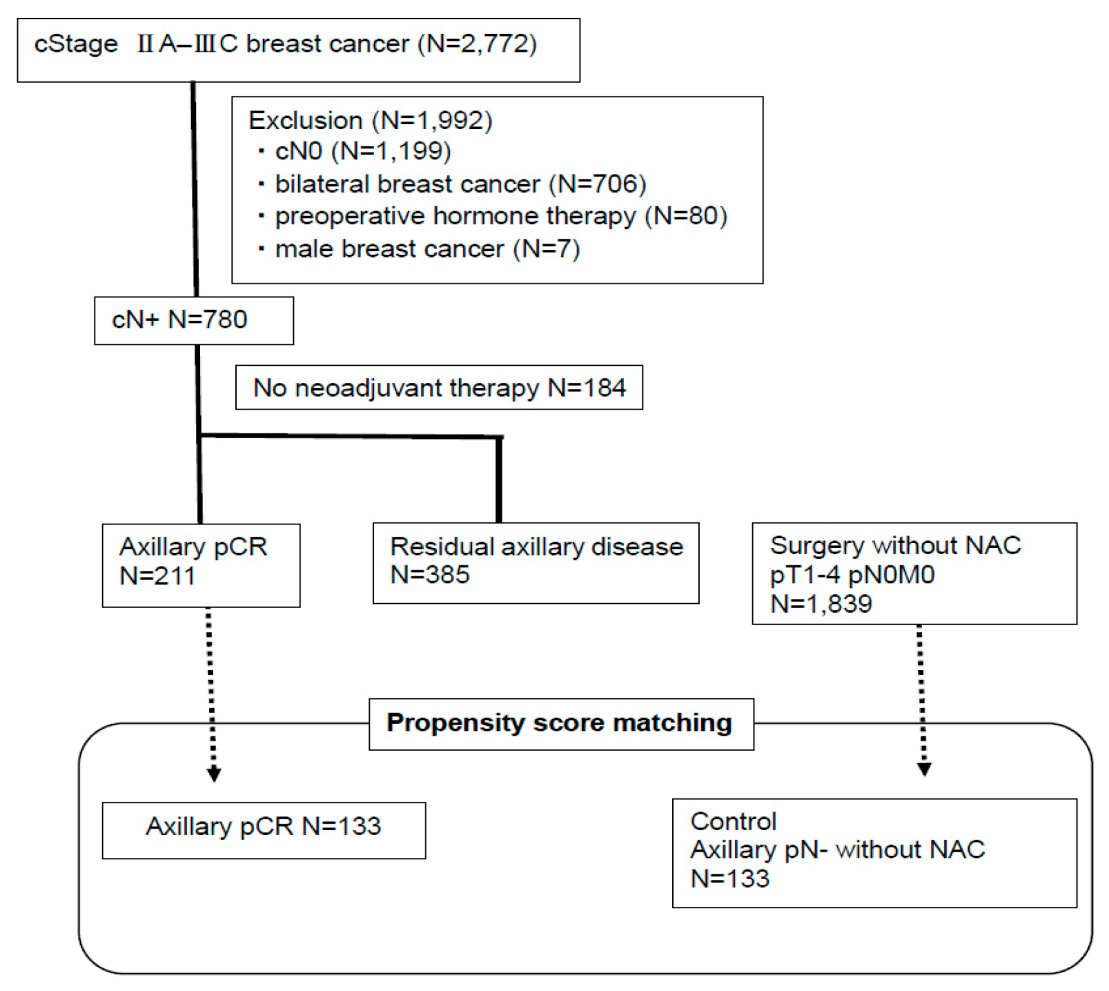
Figure 1. Flow diagram of the study.
Cancers 12 02633 g001
Figure 2. Survival outcomes according to pathological LN response to NAC. Kaplan–Meier curves for distant disease-free survival (DDFS) (a) and overall survival (OS) (b) in all patients with cytologically proven node-positive breast cancer after NAC. The 5-year DDFS and OS rates were 90% and 94%, respectively, in patients with axillary pCR; 70% and 80%, respectively, in patients with residual axillary disease (p < 0.01 for both DDFS and OS). DDFS—distant disease-free survival, NAC—neoadjuvant chemotherapy, OS—overall survival, pCR—pathological complete response.
Cancers 12 02633 g002
Figure 3. Survival outcomes of patients with axillary pCR and matched pairs with axillary pN- without NAC. Kaplan–Meier curves for DDFS (a) and OS (b) in patients with cytologically proven node-positive breast cancer with axillary pCR after NAC and matched pairs with axillary pN- without NAC. The log-rank test showed that there was no significant difference in DDFS and OS between patients with axillary pCR and matched pairs with axillary pN- without NAC (p = 0.88 for DDFS; p = 0.67 for OS).
Cancers 12 02633 g003aCancers 12 02633 g003b
Table 1. Clinicopathological characteristics of patients after neoadjuvant chemotherapy (NAC).
CharacteristicsPatients with Axillary pCR (n = 211)Patients with Residual Axillary Disease (n = 385)p-Value
Age, years (mean ± SD)50.7 ± 10.951.1 ± 10.80.65
Menopausal status
Pre-106 (50%)195 (51%)0.93
Post-105 (50%)190 (49%)
Clinical T stage a
T131 (14.5%)48 (13%)0.02
T2139 (66%)221 (57%)
T324(11%)67 (17%)
T416 (8%)49 (13%)
TX1 (0.5%)0
Clinical N stage a
N1162 (77%)306 (80%)0.65
N29 (4%)18 (4%)
N340 (19%)61 (16%)
Clinical stage a
II137 (65%)224 (58%)0.12
III74 (35%)161 (42%)
ER status
Negative115 (55%)70 (18%)<0.01
Positive83 (39%)295 (77%)
Unknown13 (6%)20 (5%)
HER2 status
Negative90 (43%)288 (75%)<0.01
Positive100 (47%)52 (13%)
Unknown21 (10%)45 (12%)
NG
147 (22%)170 (44%)<0.01
258 (28%)111 (29%)
382 (38%)56 (15%)
Unknown24 (12%)48 (12%)
Type of surgery
Partial mastectomy86 (40%)70 (18%)<0.01
Mastectomy125 (60%)315 (82%)
Breast pCR
No120 (57%)372 (97%)<0.01
Yes91 (43%)13 (3%)
LI status of the surgical specimens
Negative179 (85%)211 (55%)<0.01
Positive32 (15%)174 (45%)
Pre and Postoperative Chemotherapy
Anthracycline6 (3%)11 (3%)0.29
Taxane3 (2%)1 (0/3%)
Anthracycline followed by taxane202 (95%)373 (96.7%)
Pre and Postoperative Trastuzumab for HER2-positive disease
Yes95 (95%)48 (92%)0.90
No5 (5%)4 (8%)
Hormone therapy after surgery for ER-positive disease
Yes81 (98%)289 (98%)0.90
No2 (2%)6 (2%)
Radiotherapy after surgery
Yes141 (67%)281 (73%)0.13
No70 (33%)104 (27%)
a TNM classification is shown based on the eighth edition of the Union for International Cancer Control staging system. The bar indicates values that are statistically significant (p < 0.05). ER—estrogen receptor; HER2—human epidermal growth factor receptor 2; LI—lymphatic invasion; NAC—neoadjuvant chemotherapy; NG—nuclear grade; SD—standard deviation; pCR-—pathological complete response.
Table 2. Multivariate analysis of prognostic factors related to DDFS in patients with NAC.
CharacteristicsHazard Ratio95%CIp-Value
Pathological lymph node status after NAC
Axillary pCR1
Residual axillary disease4.552.48–8.35<0.01
Clinical stage a
II1
III2.921.96–4.35<0.01
ER status
Negative1
Positive0.600.37–0.960.03
HER2 status
Negative1
Positive0.920.77–1.090.35
NG
1,21
31.230.96–1.570.10
a TNM classification is shown based on the eighth edition of the Union for International Cancer Control staging system. The bar indicates values that are statistically significant (p < 0.05). CI—confidence interval; DDFS—distant disease-free survival; ER—estrogen receptor; HER2—human epidermal growth factor receptor 2; NAC—neoadjuvant chemotherapy; NG—nuclear grade; pCR—pathological complete response.
Table 3. Factors associated with axillary pCR using multivariate logistic regression analysis.
CharacteristicsOdds Ratio95%CIp-Value
Menopausal status
Pre-1
Post-0.630.37–1.040.07
Clinical T a
11
2–40.910.40–1.290.79
Clinical N a
11
2,30.720.40–1.290.27
ER status
Negative1
Positive0.400.23–0.70<0.01
HER2 status
Negative1
Positive1.491.24–1.78<0.01
NG
1,21
31.511.15–2.00<0.01
Breast-pCR
No1
Yes3.812.52–5.76<0.01
a TNM classification is shown based on the eighth edition of the Union for International Cancer Control staging system. The bar indicates values that are statistically significant (p < 0.05). CI—confidence interval; ER—estrogen receptor; HER2—human epidermal growth factor receptor 2; NG—nuclear grade; pCR—pathological complete response.
Table 4. Baseline characteristics at diagnosis of patients with axillary pCR and axillary pN- without NAC, using propensity score matching.
CharacteristicsAxillary pCR (n = 133)Axillary pN- without NAC (n = 133)p-Value
Menopausal status
Pre-68691.00
Post-6564
Clinical T a
123270.78
210299
375
412
ER status
Negative71670.62
Positive6266
HER2 status
Negative73810.38
Positive6052
NG
131330.18
23547
36753
Type of surgery
Partial mastectomy61700.33
Mastectomy7263
Pre and Postoperative Chemotherapy
No061<0.01
Anthracycline563
Taxane23
Anthracycline followed by taxane1263
Others03
Pre and Postoperative Trastuzumab for HER2-positive disease
Yes58380.02
No7595
Hormone therapy after surgery for ER-positive disease
Yes61520.39
No7281
Radiotherapy after surgery
Yes8838<0.01
No4595
a TNM classification is shown based on the eighth edition of the Union for International Cancer Control staging system. The bar indicates values that are statistically significant (p < 0.05). ER—estrogen receptor; HER2—human epidermal growth factor receptor 2; NG—nuclear grade; pCR—pathological complete response.