Article

Molecular Changes in Retinoblastoma beyond RB1: Findings from Next-Generation Sequencing

by
Jasmine H. Francis
1,2,*,
Allison L. Richards
3,
Diana L. Mandelker
4,
Michael F. Berger
4,
Michael F. Walsh
5,6,
Ira J. Dunkel
6,7,
Mark T. A. Donoghue
3,† and
David H. Abramson
1,2,†
1
Ophthalmic Oncology Service, Department of Surgery, Memorial Sloan Kettering Cancer Center, New York, NY 10065, USA
2
Department of Ophthalmology, Weill Cornell Medical Center, New York, NY 10065, USA
3
Marie-Josee and Henry R. Kravis Center for Molecular Oncology, Memorial Sloan Kettering Cancer Center, New York, NY 10065, USA
4
Department of Pathology, Memorial Sloan Kettering Cancer Center, New York, NY 10065, USA
5
Department of Medicine, Memorial Sloan Kettering Cancer Center, New York, NY 10065, USA
6
Department of Pediatrics, Memorial Sloan Kettering Cancer Center, New York, NY 10065, USA
7
Department of Pediatrics, Weill Cornell Medical Center, New York, NY 10065, USA
*
Author to whom correspondence should be addressed.
These authors contributed equally to this paper as senior authors.
Cancers 2021, 13(1), 149; https://doi.org/10.3390/cancers13010149
Submission received: 3 November 2020 / Revised: 25 December 2020 / Accepted: 30 December 2020 / Published: 5 January 2021
(This article belongs to the Section Methods and Technologies Development)

Abstract

:

Simple Summary

The gene causing retinoblastoma was the first tumor suppressor cloned (1986) and because retinoblastoma is the classic example of autosomal dominant inheritance, there has been little research on non-RB1 alterations in tumors and the impact these alterations have on growth patterns in the eye, metastases and predilection for non-ocular cancers. This study interrogated enucleated retinoblastoma specimens using a MSK-IMPACT clinical next-generation sequencing panel with the aim to correlate them with clinicopathologic characteristics. We found that vitreous seeding (the main reason for eye removal) correlates with copy number variations, specifically 1q gains and 16q loss. We also found that somatic BCOR mutations correlate with propensity for metastasis and this offers a molecular pathway for monitoring high risk tumors. In addition, the finding that 11% of these retinoblastoma patients have additional germline mutations (on other chromosomes) that predispose them to a different host of cancers throughout their lives enables more targeted and specific screening strategies.

Abstract

This investigation uses hybridization capture-based next-generation sequencing to deepen our understanding of genetics that underlie retinoblastoma. Eighty-three enucleated retinoblastoma specimens were evaluated using a MSK-IMPACT clinical next-generation sequencing panel to evaluate both somatic and germline alterations. Somatic copy number variations (CNVs) were also identified. Genetic profiles were correlated to clinicopathologic characteristics. RB1 inactivation was found in 79 (97.5%) patients. All specimens had additional molecular alterations. The most common non-RB1 gene alteration was BCOR in 19 (22.9%). Five (11.0%) had pathogenic germline mutations in other non-RB1 cancer predisposition genes. Significant clinicopathologic correlations included: vitreous seeds associated with 1q gains and 16q loss of heterozygosity (BH-corrected p-value = 0.008, 0.004; OR = 12.6, 26.7, respectively). BCOR mutations were associated with poor prognosis, specifically metastases-free survival (MFS) (nominal p-value 0.03). Furthermore, retinoblastoma patients can have non-RB1 germline mutations in other cancer-associated genes. No two specimens had the identical genetic profile, emphasizing the individuality of tumors with the same clinical diagnosis.

1. Introduction

Retinoblastoma is the most common primary pediatric intraocular tumor [1]. More than 100 years ago, a common genetic basis for many retinoblastomas was uncovered. While the pattern of inheritance was found to be a classical autosomal dominant pattern, it was not until the 1970s that it was recognized to be caused by a loss of function of a gene, specifically RB1. RB1 was the first tumor suppressor gene to be cloned (in 1986). Since then, the knowledge of RB1 alterations has been used as a part of genetic counseling for patients and families, with commercial germline RB1 testing now available worldwide. Tumors are initiated by biallelic loss of the RB1 gene, with 90% penetrance [2]. Patients can present with bilateral disease derived from germline RB1 mutations or with unilateral disease, which commonly have somatic mutations but can be germline in approximately 15% of patients [1].
The importance of the biallelic loss of RB1 is an example of Knudson’s “two-hit” hypothesis of cancer [3]. RB1 inactivation is recognized to occur by a variety of mechanisms including single nucleotide variants (SNVs), insertions-deletions (indels), loss of heterozygosity (LOH), large deletions, translocations, promoter hypermethylation and chromothripsis [4,5,6,7,8,9,10]. Unlike other cancers that have one or a small number of hotspot mutations, the suspected hotspots for retinoblastoma account for only 40% of mutations and the remaining are dispersed throughout the gene [11]. Although MYCN amplification in association with RB1 inactivation is known to occur, approximately 2% of retinoblastoma tumors have no derangement in RB1, and reportedly exhibit only MYCN amplification [12].
Besides tumor initiation by biallelic loss of the RB1 gene, cytogenetic analysis has identified recurrent copy number variants (CNVs) in retinoblastoma tumors, leading to the proposal of several candidate driver genes other than RB1 such as KIF14, MYCN, DEK, E2F3, RBL2/p130, and NGFR [4,13,14,15,16]. Recently, whole exome sequencing has identified additional putative drivers in BCOR and CREBBP [7,8,17]. However, the clinical significance of these additional gene mutations, along with the specific mechanism of RB1 loss, remains to be fully elucidated. While next-generation sequencing (NGS) has been used to analyze retinoblastoma, most studies are either limited to detecting germline mutations, constrained to only a handful of known mutations to occur in retinoblastoma or without cliniopathological correlations [7,12,17,18,19,20]. This study investigated retinoblastoma tumors with hybridization capture-based NGS, analyzed using a MSK-IMPACT platform [21,22]. This platform analyzes 468 genes for somatic analysis, 88 genes for germline analysis, and can identify DNA sequence variants, copy-number alterations, and select rearrangements. This genetic data was analyzed for correlations with clinicopathological features.

2. Results

2.1. Genomic Landscape of RB1 Alterations in Retinoblastoma

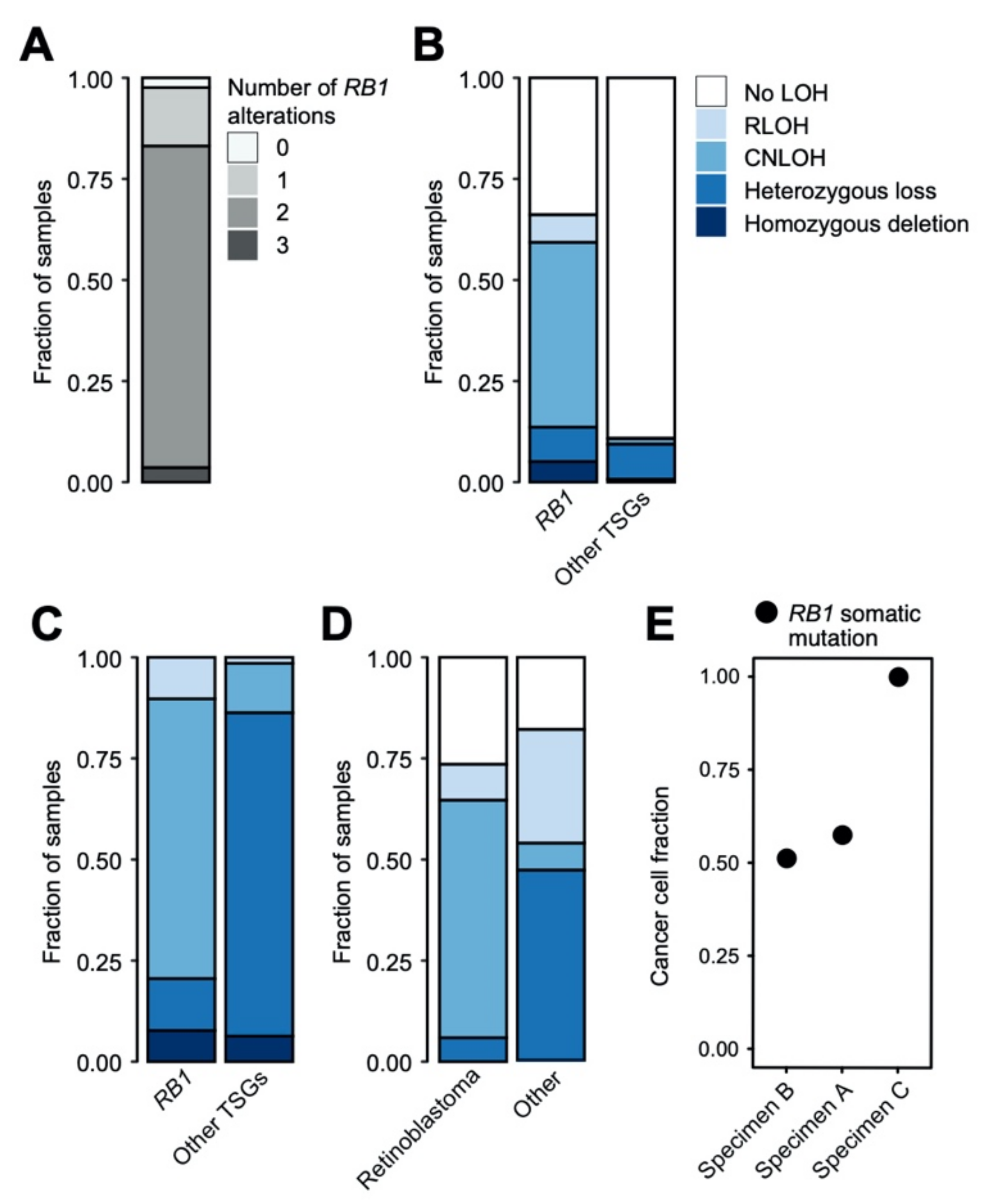
As expected, pathognomonic RB1 alterations were identified in 97.5% (79/81) of patients (Figure 1A), of which ten were found in specimens from multiple patients in this cohort (Figure 1B).
Of these, seven are shared exclusively in the somatic context, while three can be found in the germline and soma (c.1072C>T, c.1654C>T and c.1981C>T). Furthermore, of the forty-six somatic RB1 mutations that could be assessed for clonality, only five were subclonal. Two additional specimens did not have detectable RB1 alterations in either somatic or germline and no other putative drivers were identified in these specimens. One specimen had two additional subclonal mutations in BIRC3 (p.A385D), a variant of unknown significance, and PBRM1 (p.G176*), a truncating mutation. The other specimen did not have any identifiable genomic alterations except for a gain of 6p. The lack of RB1 alterations in two samples is consistent with either RB1 negative tumorigenesis of retinoblastoma or a mechanism of RB1 inactivation not detectable by the methods used in this paper.
Biallelic loss of RB1 was identified in 79.5% (66/83) of specimens (Figure 2A).
Of the 66 specimens with two hits to RB1, ten had somatic homozygous deletion of RB1, while one had a germline heterozygous loss of exons 7–11 in RB1 followed by a somatic LOH event leading to two dysfunctional copies of RB1. Of the remaining 53, 38 (71.6%) had a mutation followed by an LOH event to lose the wild-type copy of RB1 while 15 had two mutations in RB1. Two specimens (with additional methylation testing) had hypermethylated promotors and LOH over the RB1 locus. Twelve specimens had apparent heterozygous alterations to RB1 as detected by MSK-IMPACT. This suggests other modes of silencing RB1 may be occurring in these patients that are not detectable by MSK-IMPACT. Evidence of this possibility is highlighted by two patients for which RB1 methylation testing was performed. However, not all samples were tested for methylation status, and as such the prevalence of RB1 silencing via this mechanism in patients with a single genomic alteration is unknown. These results demonstrate the variety of different mechanisms leading to RB1 loss, though none of the mechanisms were associated with any particular clinical outcome.
Of the 42 matched specimens with LOH over the RB1 locus, 31 (73.8%) were copy-neutral LOH (CN-LOH), where two identical alleles are present, in contrast to LOH via the loss of an allele. In the case of RB1, this means that two dysfunctional mutant alleles are present but for the rest of the genes on 13q, two supposedly wild-type alleles can be expressed. Compared to other tumor suppressor genes, our specimens showed much higher CN-LOH at RB1 (p-value < 2.2 × 10⁻¹⁶, OR 19.8) (Figure 2B,C). Additionally, when compared to other cancer types, RB1 is subject to CN-LOH at much higher rates in retinoblastoma than in other cancers that also contain mutations in RB1 (p-value < 2.2 × 10⁻¹⁶, OR 44.4) (Figure 2D). This preference for CN-LOH of RB1 in retinoblastoma may reflect the necessity of a gene or genes on 13q to be expressed from two alleles or there may be genes essential to the fitness of the cell type that would be left vulnerable by the presence of a single allele.

2.2. Evidence of Intertumoral Genetic Heterogeneity

Two patients with bilateral disease and germline RB1 alterations had two specimens collected, one from each eye. As expected, within each patient the germline mutations were consistent across the specimens. However, each specimen had different somatic RB1 second hits. In the first patient, there was a germline loss in RB1 found in both eyes but different oncogenic somatic variants in each eye, i.e., splice site alteration p.X566_splice and truncating p.V654Cfs*4. In the second patient, the germline alteration was a splice mutation (RB1 X775_splice). In one eye, this was followed by a somatic LOH while the other eye had a somatic truncating mutation (RB1 p.S807*). The samples from these patients show that the germline event is not predictive of a particular somatic event and also demonstrates the importance of considering the specimens separately as they often have different etiologies even in the same patient.
In our cohort, three patients had evidence of more than two potential loss of function alterations in RB1. All three patients had a germline mutation, an LOH event leading to the somatic loss of the WT copy of RB1 and an additional somatic mutation. The presence of the second somatic hit in the RB1 germline background is suggestive of two distinct molecular alterations within the specimen. In two of the patients, the additional somatic mutation is at very low allele frequency (Figure 2E) in comparison to the purity of the sample, consistent with the presence of two genetic alterations within the same tumor sample. Under this scenario, the LOH would also be expected to be heterogeneous, however, it appears clonal in both cases and may represent differences between mutation and CNA methods in terms of sensitivity in detecting subclonal events. In the third individual, the germline mutation is silent but previous work suggests that it may impact splicing [25]. However, both somatic events (LOH and mutation) appear clonal (Figure 2E), indicating they must occur in the same tumor cells. In this case, the role of the germline mutation is less clear and its presence in the patient may be coincidental.

2.3. Non-RB1 Alterations in Retinoblastoma

We assessed all alterations and identified genes that were frequently altered in our patient population or were identified in other publications about retinoblastoma [7,8,17] (Figure 1A). Aside from RB1, the most commonly somatically mutated genes among the specimens were BCOR in 19 (22.9%), RPTOR in 3 (3.6%), TERT in 3 (3.6%), MSH3 in 3 (3.6%), ARID1A in 3 (3.6%), MYCN in 3 (3.6%), TSC2 in 2 (2.4%), and CREBBP in 1 (1.2%) (Figure 1A). All BCOR mutations detected are putative loss of function mutations and therefore likely oncogenic. BCOR has been previously proposed to have an association with worse prognosis [7,17]. Here, BCOR mutations were associated with a poor prognosis for metastasis-free survival from date of enucleation (nominal p-value = 0.03) (Figure 3A), but not with “higher risk” histopathological features. Besides BCOR, the ten most frequent non-RB1 gene alterations were found three times, each, in our cohort of 83 (3.6%) patients. Among these ten genes, only four (TERT, MSH3, ARID1A, and HLA-A) had any mutations that were considered putative drivers [26]. Eleven additional genes have singleton, somatic alterations that were considered putative drivers. Only BCOR appeared in high enough frequency in our population to assess association with clinical features.
In contrast, none of the mutations in FAT1 and RPTOR are truncating or fall at any hotspot location, as such they are considered variants of unknown significance (Figure 1A). In addition to mutations, three patients exhibited MYCN amplification, in the presence of an RB1 mutation, which has been shown previously to be prevalent in retinoblastoma [27]. Overall, the mutation burden was generally low in these retinoblastoma samples (0–3 mutations per megabase).
In addition to germline pathogenic variants in RB1, five of 42 patients (12.0%) consented for germline MSK-IMPACT testing had pathogenic or likely pathogenic germline mutations in non-RB1 cancer predisposition genes. Mutations were detected in base excision repair: NTHL1 c.268C>T (p.Gln90*); nucleotide excision repair: ERCC3 c.1354C>T (p.Arg452*), mis-match repair: MSH3 c.1341-1G>T (p.X447_splice) splice site mutation and cell cycle: CHEK2 c.470T>C (p.Ile157Thr) and transcription factor: MITF c.1255G>A (p.Glu419Lys) genes. Notably, two of these patients harbored heterozygous alterations in cancer predisposing genes associated with autosomal recessive cancer predisposition syndromes (NTHL1 and MSH3). There was no evidence of microsatellite instability in the patient with germline MSH3.

2.4. Copy Number Alterations and Loss of Heterozygosity in Retinoblastoma

Several recurrent arm-level CNAs were identified, including 1q gain (67.0%), 2p gain (32.0%), 6p gain (59.0%), LOH 13q (51.0%), LOH 16q (43.0%) (Figure 3B). Specifically, gains on 1q and loss of heterozygosity on 16q were associated with vitreous seeds (BH-corrected p-value = 0.008, 0.004; OR = 12.6, 26.7, respectively) (Figure 3C). Gains on 1q and losses on 16q were not significantly correlated with each other (p-value = 0.37) (Figure 3D) suggesting that they do not need to co-occur to present with vitreous seeds. A model of vitreous seed presence including both 1q gains and 16q losses showed significant association suggesting that they both play an independent role in vitreous seeds (model p-value = 8.2 × 10⁻⁸) with both being predictive (1q p-value = 1.3 × 10⁻⁴, 16q p-value = 2.3 × 10⁻⁴). No other CNAs were associated with any other clinical features.

3. Discussion

The genetic basis of retinoblastoma was long believed to be predominantly limited to biallelic loss of RB1. Other non-RB1 genes have previously been identified [7,8,17] and their clinical relevance sparingly elucidated. However, the present findings based on 83 enucleation retinoblastoma specimens demonstrate that the genetic landscape is more complex and extends our current knowledge. While we detected mutations, loss of heterozygosity, and homozygous deletions leading to biallelic inactivation of RB1, we also identified large-scale CNVs and somatic and germline mutations in other genes not on chromosome 13.
The majority of series investigating next-generation sequencing (NGS) in retinoblastoma have used this technology for germline RB1 detection, and copy number alteration details are not consistently available [18]. Few studies focus on using NGS for tumor specimens, in detecting mutations other than RB1 and no more than a dozen other genes known to occur in retinoblastoma, or in correlating these findings with clinic-histopathologic features [7,12,17,19,20]. One study used NGS of 500 oncogenes with clinical correlation, but in only 30 tumor samples of retinoblastoma [28]. Our study investigated 468 genes in 83 retinoblastoma tumor specimens with cliniopathological correlations.
Previous work has shown recurrent chromosomal abnormalities in retinoblastoma including gains of 1q, 2p, 6p and loss of 13q and 16q [4,13,14,15]. Despite extensive knowledge on the presence of CNVs in retinoblastoma, clinicopathologic correlates are limited to correlations with age, differentiation, pathological features and disease laterality [6,15,27]. Loss of 16q has been associated with diffuse vitreous seeding with CDH11 being proposed as the candidate gene [5,29]. In the present study, both 1p gain and 16q loss were independently associated with the presence of vitreous seeds. However, in our cohort, the role of CDH11 is unclear in the absence of mutations. Although 6p was recurrently gained in our cohort, contrary to a previous report, it was not associated with aggressive histopathological features or a poor prognosticator of ocular survival [30,31]. This difference may reflect the presence of enucleated-only specimens. Intriguingly, one unilateral specimen with confirmed wildtype RB1 did possess 6p gain. Indicating either a deficiency in testing sensitivity or strategy, or alternatively, 6p gain may represent an independent biomarker for retinoblastoma in the presence of wildtype RB1.
We found that, compared to other cancers, RB1 in retinoblastoma is significantly and disproportionately more likely to display copy-neutral LOH rather than heterozygous loss LOH. This unique characteristic is previously unreported in retinoblastoma. Other studies have shown the presence of CN-LOH in different cancers, some of which highlight CN-LOH of 13q [32,33,34,35,36]. In our data, we focused on tumors with mutations in RB1 but CN-LOH is a mechanism that may affect different tumor suppressor genes in other tumor types. While we do not currently have an explanation for this phenomenon, it may suggest that some gene (or genes) on chromosome 13 are essential for cell survival and are needed at two copies.
An additional clinical correlation is observed in the three specimens with three hits to RB1. These may represent multiple clones in the same eye and explain the well-recognized observation that multifocal tumors in the same eye have distinct features.
BCOR is located at Xp11.4, encodes the BCL6 corepressor and as such is a transcription regulatory factor. BCOR mutations are present in a number of malignancies; and correlates with poor cancer prognosis [37,38]. Furthermore, BCOR expression aids in eye development and is highly expressed in the lens and retina [39]; and this latter property may explain why retinoblastomas are prone to mutated BCOR. In our cohort, it was most common non-RB1 gene abnormality, occurring in 23% of specimens (higher than previously published series (13%) [7,17]) and was associated with poor prognosis, specifically worse metastases-free survival, but not with high-risk pathological features. Identification of a BCOR mutation, either in an enucleated specimen or through cell free DNA (cfDNA) analysis, may influence surveillance strategies or preventative measures for metastatic retinoblastoma. It could mark minimal residual disease and/or be informative of treatment response through quantitative cfDNA.
Future studies may expand upon these results by considering more downstream analytes, such as DNA methylation or RNA expression, to assess biological differences between patients either dependent or independent of DNA mutations. For example, recent studies have considered genome-wide methylation and found expression patterns unique to retinoblastoma, suggesting their importance to oncogenesis [40,41]. Further clinical assessment of patient phenotypes alongside various biological measurements may yield important insight into the progression of retinoblastoma from DNA mutation to unique clinicopathological features of retinoblastoma.
Five patients had non-RB1 cancer-associated germline mutations in keeping with other cancer cohorts in which 12–17.5% patient had germline mutations [42]. However, whereas germline cancer-associated mutations are associated with metastatic disease, and therapy-specific prognosis in other cancers [43,44], to date our five patients have demonstrated intraocular disease only. This raises the question whether germline variants influence tumorigenesis, the clinical course or subsequent disease risks (metastatic disease or second cancers).
Of the non-RB1 gene alterations in our cohort, only MYCN on 2p24.3 and TSC2 on 16p13.3 correspond with common CNVs that characterize retinoblastoma [4,13,14,15,16]. Previous studies have reported mutations in CREBBP and we found one example in our patients [7,8,17]. We identified additional variants that are recognized as oncogenic in other cancer types [26]. However, in our cohort, each of these occurs at low frequency, limiting our ability to assess their functionality in retinoblastoma, as none of the genes are commonly associated with retinoblastoma. The remaining gene alterations do not correspond with typical oncogenic variants associated with retinoblastoma or other cancer types. For example, FAT1 acts as both a tumor suppressor and oncogene and occurs in bladder cancer, head and neck cancers, breast and colorectal cancers. Published literature suggests it increases invasiveness of tumors [45]. RPTOR, along with TSC2, is a part of the mTOR signaling pathway known to affect many different types of cancer [46]. However, neither FAT1 nor RPTOR have putative drivers in our dataset.

4. Materials and Methods

An institutional review board approval was obtained from Memorial Sloan Kettering Cancer Center (#17-049). Eligible patients included those with retinoblastoma who had undergone a primary or secondary enucleation, and those included were enucleated between May 2006 and August 2019. Patients were consented for either somatic or somatic and germline testing via MSK-IMPACT. Prior commercial germline testing for RB1 was also available for some patients.
Clinicopathologic data was collected for each patient and retinoblastoma tumor. This data included gender, laterality, prior treatment, ocular hypertension, rubeosis, vitreous seeds (including seed class: dust, spheres, or clouds) or subretinal seeds, histopathologic features (invasion into the choroid (massive or not), sclera, ciliary body, anterior chamber, optic nerve pass the lamina cribrosa (yes or no) optic nerve to the cut section), second cancers, occurrence and site of metastatic disease (defined as orbital or other extraocular metastasis). “Higher risk” eyes were defined as those with high risk pathological features defined as massive choroidal invasion, ciliary body/iris invasion or optic nerve invasion past the laminar cribrosa.

4.1. Isolation and Purification of DNA

Microdissection was performed on 83 formalin-fixed and paraffin-embedded (FFPE) samples on 10 µm-thick unstained sections, using hematoxylin and eosin-stained (H&E) sections as a guide. The DNeasy Tissue Kit (Qiagen, Hilden, Germany) was used for DNA extraction according to the manufacturer’s recommendations. The Nano-Drop 8000 (Thermo Scientific, Waltham, MA, USA) and Qubit (Life Technologies, Carlsbad, CA, USA) were employed to quantify the extracted DNA. The minimum concentration of formalin fixed paraffin embedded DNA was 250 ng.

4.2. Exon-Capture Sequencing

Targeted next-generation sequencing via MSK-IMPACT was performed on DNA extracted from tumor and matched normal DNA samples, with somatic mutations (substitutions and small insertions and deletions), gene-level focal copy number alterations, and structural rearrangements detected [21,22]. Germline variants were evaluated in 88 cancer predisposition genes in 42 patients [47].

4.3. Outside Testing of RB1 Alterations

Outside commercial testing was performed by multiple companies as routine clinical care on 23 specimens. Each company performed their individual assay, which identifies mutations, methylation, and copy number alterations at the RB1 locus. In this work, we used methylation status and germline RB1 mutations for patients not consented to germline testing via MSK-IMPACT. In addition, germline CNVs in RB1 and somatic CNAs from outside testing are reported for cases sequenced via MSK-IMPACT unmatched (without a normal tissue comparator from the patient). MSK-IMPACT identified all RB1 mutations found by outside testing in specimens that had both outside germline testing and germline testing via MSK-IMPACT.

4.4. Biostatistics

Total, allele-specific and integer copy number as well as tumor purity and ploidy were identified in the 74 specimens with matched normal tissue using FACETS (Fraction and Allele-Specific Copy Number Estimates from Tumor Sequencing) v0.5.14 [48] and the clonality of mutations was calculated as described [49]. For the nine cases sequenced without matching normal tissue, copy number alterations were identified using the GATK DepthOfCoverage tool as previously described [49] Microsatellite instability was estimated using MSIsensor [50]. Kaplan–Meier estimates of event-free survival were calculated starting from the date of enucleation to the detection of metastasis, and correlated to genetic alterations. Four individuals were removed from the metastasis-free survival estimates because they presented with metastasis at, or before, the time of enucleation. When multiple tests were performed, Benjamini–Hochberg (BH) multiple testing correction was used [51].
For the purposes of this manuscript, LOH is the state of total copy number 1 and lower copy number 0 (1,0). Copy-neutral LOH (CN-LOH) is 2,0, while reciprocal LOH (RLOH) [52] is total copy number greater than two, while lower copy number is 0. Tumor suppressor genes were defined from OncoKB [26]. LOH rates at the RB1 locus across the MSK cohort (frozen in Jan, 2019 with 39,846 tumor specimens) used default FACETS fits (not manually reviewed) that passed QC.

5. Conclusions

In conclusion, there is true genetic diversity among retinoblastoma tumors. Based on an analysis of 468 genes, no two specimens analyzed had identical pattern of mutations (or type of RB1 loss), LOH or CNV, emphasizing the individuality of tumors with the same clinical diagnosis. We identified clinicopathologic correlations with genetic aberrations including the association of vitreous seeds with 1q gains and 16q loss. Our discovery of BCOR mutations being correlated with the development of metastatic disease may guide future systemic surveillance or management of these patients. Insights gained on small numbers of specimens include the observation that a single eye with multifocal tumors may include more than two identifiable forms of RB1 loss, suggesting intertumoral heterogeneity, and that in wildtype RB1, 6p gain may be sufficient for retinoblastoma tumorigenesis. Finally, retinoblastoma patients can have non-RB1 germline mutations in other cancer-associated genes. In the future, we hope to discover how these alternate non-RB1 germline mutations may influence the primary ocular retinoblastoma, the metastatic potential or additional primary malignancies.

Author Contributions

Conceptualization, J.H.F., M.T.A.D., I.J.D., D.H.A.; methodology, J.H.F., A.L.R., M.F.B., M.T.A.D., D.H.A.; software, D.L.M., M.F.B.; validation, D.L.M., M.F.B.; formal analysis, J.H.F., A.L.R., M.T.A.D.; data curation, J.H.F., A.L.R.; writing—original draft preparation, J.H.F., A.L.R.; writing—review and editing, D.L.M., M.F.B., M.F.W., I.J.D., M.T.A.D., D.H.A.; funding acquisition, D.H.A. All authors have read and agreed to the published version of the manuscript.

Funding

This work was supported by philanthropic funding from The Fund for Ophthalmic Knowledge (J.H.F. and D.H.A.), and the Cancer Center Support Grant (grant number P30 CA008748) (all authors), the Robert and Kate Niehaus Center for Inherited Cancer Genomics (M.F.W.). The sponsors were not involved in the design, collection, analysis, interpretation of data, the writing of the manuscript nor the decision to submit the manuscript for publication.

Institutional Review Board Statement

The study was conducted according to the guidelines of the Declaration of Helsinki, and approved by the Institutional Review Board (or Ethics Committee) of Memorial Sloan Kettering Cancer Center (protocol code 12-245 in 2012).

Informed Consent Statement

Informed consent was obtained from all subjects (or their legal guardian) involved in the study.

Data Availability Statement

IMPACT mutation calls were deposited via the cBioPortal for Cancer Genomics (https://cbioportal.mskcc.org/study/summary?id=rbl_mskcc_2020).

Acknowledgments

We thank Ritika Kundra and Ramyasree Madupuri for preparing the cBioPortal instance.

Conflicts of Interest

The authors declare no conflict of interest.

References

  1. Abramson, D.H. Retinoblastoma: Saving life with vision. Annu. Rev. Med. 2014, 65, 171–184. [Google Scholar] [CrossRef]
  2. Lohmann, D.R.; Brandt, B.; Höpping, W.; Passarge, E.; Horsthemke, B. Distinct RB1 gene mutations with low penetrance in hereditary retinoblastoma. Hum. Genet. 1994, 94, 349–354. [Google Scholar] [CrossRef] [PubMed]
  3. Knudson, A.G. Mutation and cancer: Statistical study of retinoblastoma. Proc. Natl. Acad. Sci. USA 1971, 68, 820–823. [Google Scholar] [CrossRef] [PubMed] [Green Version]
  4. Corson, T.W.; Gallie, B.L. One hit, two hits, three hits, more? Genomic changes in the development of retinoblastoma. Genes Chromosom. Cancer 2007, 46, 617–634. [Google Scholar] [CrossRef] [PubMed]
  5. Gratias, S.; Rieder, H.; Ullmann, R.; Klein-Hitpass, L.; Schneider, S.; Bölöni, R.; Kappler, M.; Lohmann, D.R. Allelic Loss in a Minimal Region on Chromosome 16q24 Is Associated with Vitreous Seeding of Retinoblastoma. Cancer Res. 2007, 67, 408–416. [Google Scholar] [CrossRef] [Green Version]
  6. Herzog, S.; Lohmann, D.; Buiting, K.; Schüler, A.; Horsthemke, B.; Rehder, H.; Rieder, H. Marked differences in unilateral isolated retinoblastomas from young and older children studied by comparative genomic hybridization. Hum. Genet. 2001, 108, 98–104. [Google Scholar] [CrossRef]
  7. Kooi, I.E.; Mol, B.M.; Massink, M.P.G.; Ameziane, N.; Meijers-Heijboer, H.; Dommering, C.J.; van Mil, S.E.; de Vries, Y.; van der Hout, A.H.; Kaspers, G.J.L.; et al. Somatic genomic alterations in retinoblastoma beyond RB1 are rare and limited to copy number changes. Sci. Rep. 2016, 6, 25264. [Google Scholar] [CrossRef] [Green Version]
  8. McEvoy, J.; Nagahawatte, P.; Finkelstein, D.; Richards-Yutz, J.; Valentine, M.; Ma, J.; Mullighan, C.; Song, G.; Chen, X.; Wilson, M.; et al. RB1 gene inactivation by chromothripsis in human retinoblastoma. Oncotarget 2014, 5, 438–450. [Google Scholar] [CrossRef] [Green Version]
  9. Lee, W.H.; Murphree, A.L.; Benedict, W.F. Expression and amplification of the N-myc gene in primary retinoblastoma. Nature 1984, 309, 458–460. [Google Scholar] [CrossRef]
  10. Ohtani-Fujita, N.; Dryja, T.P.; Rapaport, J.M.; Fujita, T.; Matsumura, S.; Ozasa, K.; Watanabe, Y.; Hayashi, K.; Maeda, K.; Kinoshita, S.; et al. Hypermethylation in the retinoblastoma gene is associated with unilateral, sporadic retinoblastoma. Cancer Genet. Cytogenet. 1997, 98, 43–49. [Google Scholar] [CrossRef]
  11. Mallipatna, A.; Marino, M.; Singh, A.D. Genetics of Retinoblastoma. Asia Pac. J. Ophthal. 2016, 5, 260–264. [Google Scholar] [CrossRef] [PubMed]
  12. Rushlow, D.E.; Mol, B.M.; Kennett, J.Y.; Yee, S.; Pajovic, S.; Thériault, B.L.; Prigoda-Lee, N.L.; Spencer, C.; Dimaras, H.; Corson, T.W.; et al. Characterisation of retinoblastomas without RB1 mutations: Genomic, gene expression, and clinical studies. Lancet Oncol. 2013, 14, 327–334. [Google Scholar] [CrossRef] [Green Version]
  13. Bowles, E.; Corson, T.W.; Bayani, J.; Squire, J.A.; Wong, N.; Lai, P.B.-S.; Gallie, B.L. Profiling genomic copy number changes in retinoblastoma beyond loss of RB1. Genes Chromosom. Cancer 2007, 46, 118–129. [Google Scholar] [CrossRef] [PubMed]
  14. Marchong, M.N.; Chen, D.; Corson, T.W.; Lee, C.; Harmandayan, M.; Bowles, E.; Chen, N.; Gallie, B.L. Minimal 16q genomic loss implicates cadherin-11 in retinoblastoma. Mol. Cancer Res. 2004, 2, 495–503. [Google Scholar]
  15. Van der Wal, J.E.; Hermsen, M.A.J.A.; Gille, H.J.P.; Schouten-Van Meeteren, N.Y.N.; Moll, A.C.; Imhof, S.M.; Meijer, G.A.; Baak, J.P.A.; van der Valk, P. Comparative genomic hybridisation divides retinoblastomas into a high and a low level chromosomal instability group. J. Clin. Pathol. 2003, 56, 26–30. [Google Scholar] [CrossRef] [PubMed] [Green Version]
  16. Qi, D.-L.; Cobrinik, D. MDM2 but not MDM4 promotes retinoblastoma cell proliferation through p53-independent regulation of MYCN translation. Oncogene 2017, 36, 1760–1769. [Google Scholar] [CrossRef] [PubMed] [Green Version]
  17. Zhang, J.; Benavente, C.A.; McEvoy, J.; Flores-Otero, J.; Ding, L.; Chen, X.; Ulyanov, A.; Wu, G.; Wilson, M.; Wang, J. A novel retinoblastoma therapy from genomic and epigenetic analyses. Nature 2012, 481, 329–334. [Google Scholar] [CrossRef]
  18. Grotta, S.; D’Elia, G.; Scavelli, R.; Genovese, S.; Surace, C.; Sirleto, P.; Cozza, R.; Romanzo, A.; De Ioris, M.A.; Valente, P. Advantages of a next generation sequencing targeted approach for the molecular diagnosis of retinoblastoma. BMC Cancer 2015, 15, 841. [Google Scholar] [CrossRef] [PubMed] [Green Version]
  19. Amitrano, S.; Marozza, A.; Somma, S.; Imperatore, V.; Hadjistilianou, T.; De Francesco, S.; Toti, P.; Galimberti, D.; Meloni, I.; Cetta, F. Next generation sequencing in sporadic retinoblastoma patients reveals somatic mosaicism. Eur. J. Hum. Genet. 2015, 23, 1523–1530. [Google Scholar] [CrossRef]
  20. Devarajan, B.; Prakash, L.; Kannan, T.R.; Abraham, A.A.; Kim, U.; Muthukkaruppan, V.; Vanniarajan, A. Targeted next generation sequencing of RB1 gene for the molecular diagnosis of Retinoblastoma. BMC Cancer 2015, 15, 320. [Google Scholar] [CrossRef] [Green Version]
  21. Cheng, D.T.; Mitchell, T.N.; Zehir, A.; Shah, R.H.; Benayed, R.; Syed, A.; Chandramohan, R.; Liu, Z.Y.; Won, H.H.; Scot, N.S. Memorial Sloan Kettering-Integrated Mutation Profiling of Actionable Cancer Targets (MSK-IMPACT): A Hybridization Capture-Based Next-Generation Sequencing Clinical Assay for Solid Tumor Molecular Oncology. J. Mol. Diagn. 2015, 17, 251–264. [Google Scholar] [CrossRef] [PubMed]
  22. Zehir, A.; Benayed, R.; Shah, R.H.; Syed, A.; Middha, S.; Kim, H.R.; Srinivasan, P.; Gao, J.; Chakravarty, D.; Devlin, S.M. Mutational landscape of metastatic cancer revealed from prospective clinical sequencing of 10,000 patients. Nat. Med. 2017, 23, 703. [Google Scholar] [CrossRef] [PubMed]
  23. Cerami, E.; Gao, J.; Dogrusoz, U.; Gross, B.E.; Sumer, S.O.; Aksoy, B.A.; Jacobsen, A.; Byrne, C.J.; Heuer, M.L.; Larsson, E. The cBio cancer genomics portal: An open platform for exploring multidimensional cancer genomics data. Cancer Discov. 2012, 2, 401–404. [Google Scholar] [CrossRef] [PubMed] [Green Version]
  24. Gao, J.; Aksoy, B.A.; Dogrusoz, U.; Dresdner, G.; Gross, B.; Sumer, S.O.; Sun, Y.; Jacobsen, A.; Sinha, R.; Larsson, E. Integrative analysis of complex cancer genomics and clinical profiles using the cBioPortal. Sci. Signal. 2013, 6, pl1. [Google Scholar] [CrossRef] [Green Version]
  25. Ahani, A.; Behnam, B.; Khorshid, H.R.K.; Akbari, M.T. RB1 gene mutations in Iranian patients with retinoblastoma: Report of four novel mutations. Cancer Genet. 2011, 204, 316–322. [Google Scholar] [CrossRef]
  26. Chakravarty, D.; Gao, J.; Phillips, S.M.; Kundra, R.; Zhang, H.; Wang, J.; Rudolph, J.E.; Yaeger, R.; Soumerai, T.; Nissan, M.H. OncoKB: A Precision Oncology Knowledge Base. JCO Precis. Oncol. 2017, 2017, 1–16. [Google Scholar] [CrossRef]
  27. Lillington, D.M.; Kingston, J.E.; Coen, P.G.; Price, E.; Hungerford, J.; Domizio, P.; Young, B.D.; Onadim, Z. Comparative genomic hybridization of 49 primary retinoblastoma tumors identifies chromosomal regions associated with histopathology, progression, and patient outcome. Genes Chromosom. Cancer 2002, 36, 121–128. [Google Scholar] [CrossRef]
  28. Afshar, A.R.; Pekmezci, M.; Bloomer, M.M.; Cadenas, N.J.; Stevers, M.; Banerjee, A.; Roy, R.; Olshen, A.B.; Van Ziffle, J.; Onodera, C. Next-Generation Sequencing of Retinoblastoma Identifies Pathogenic Alterations beyond RB1 Inactivation That Correlate with Aggressive Histopathologic Features. Ophthalmology 2020, 127, 804–813. [Google Scholar] [CrossRef]
  29. Gustmann, S.; Klein-Hitpass, L.; Stephan, H.; Weber, S.; Bornfeld, N.; Kaulisch, M.; Lohmann, D.R.; Dünker, N. Loss at chromosome arm 16q in retinoblastoma: Confirmation of the association with diffuse vitreous seeding and refinement of the recurrently deleted region. Genes Chromosom. Cancer 2011, 50, 327–337. [Google Scholar] [CrossRef]
  30. Xu, L.; Polski, A.; Prabakar, R.K.; Reid, M.W.; Chévez-Barrios, P.; Jubran, R.; Kim, J.W.; Kuhn, P.; Cobrinik, D.; Hicks, J.; et al. Chromosome 6p amplification in aqueous humor cell-free DNA is a prognostic biomarker for retinoblastoma ocular survival. Mol. Cancer Res. 2020, 18, 1166–1675. [Google Scholar] [CrossRef]
  31. Berry, J.L.; Xu, L.; Kooi, I.; Murphree, A.L.; Prabakar, R.K.; Reid, M.; Stachelek, K.; Le, B.H.A.; Welter, L.; Reiser, B.J.; et al. Genomic cfDNA Analysis of Aqueous Humor in Retinoblastoma Predicts Eye Salvage: The Surrogate Tumor Biopsy for Retinoblastoma. Mol. Cancer Res. 2018, 16, 1701–1712. [Google Scholar] [CrossRef] [Green Version]
  32. Andersen, C.L.; Wiuf, C.; Kruhøffer, M.; Korsgaard, M.; Laurberg, S.; Ørntoft, T.F. Frequent occurrence of uniparental disomy in colorectal cancer. Carcinogenesis 2007, 28, 38–48. [Google Scholar] [CrossRef] [PubMed] [Green Version]
  33. O’Keefe, C.; McDevitt, M.A.; Maciejewski, J.P. Copy neutral loss of heterozygosity: A novel chromosomal lesion in myeloid malignancies. Blood 2010, 115, 2731–2739. [Google Scholar] [CrossRef] [PubMed] [Green Version]
  34. Raghavan, M.; Smith, L.-L.; Lillington, D.M.; Chaplin, T.; Kakkas, I.; Molloy, G.; Chelala, C.; Cazier, J.-B.; Cavenagh, J.D.; Fitzgibbon, J.; et al. Segmental uniparental disomy is a commonly acquired genetic event in relapsed acute myeloid leukemia. Blood 2008, 112, 814–821. [Google Scholar] [CrossRef] [PubMed] [Green Version]
  35. Murthy, S.K.; DiFrancesco, L.M.; Ogilvie, R.T.; Demetrick, D.J. Loss of heterozygosity associated with uniparental disomy in breast carcinoma. Mod. Pathol. 2002, 15, 1241–1250. [Google Scholar] [CrossRef] [Green Version]
  36. Torabi, K.; Miró, R.; Fernández-Jiménez, N.; Quintanilla, I.; Ramos, L.; Prat, E.; del Rey, J.; Pujol, N.; Killian, J.K.; Meltzer, P.; et al. Patterns of somatic uniparental disomy identify novel tumor suppressor genes in colorectal cancer. Carcinogenesis 2015, 36, 1103–1110. [Google Scholar] [CrossRef] [PubMed] [Green Version]
  37. Astolfi, A.; Fiore, M.; Melchionda, F.; Indio, V.; Bertuccio, S.N.; Pession, A. BCOR involvement in cancer. Epigenomics 2019, 11, 835–855. [Google Scholar] [CrossRef] [Green Version]
  38. Terada, K.; Yamaguchi, H.; Ueki, T.; Usuki, K.; Kobayashi, Y.; Tajika, K.; Gomi, S.; Kurosawa, S.; Saito, R.; Furuta, Y.; et al. Usefulness of BCOR gene mutation as a prognostic factor in acute myeloid leukemia with intermediate cytogenetic prognosis. Genes Chromosom. Cancer 2018, 57, 401–408. [Google Scholar] [CrossRef]
  39. Zhou, Y.; Wojcik, A.; Sanders, V.R.; Rahmani, B.; Kurup, S.P. Ocular findings in a patient with oculofaciocardiodental (OFCD) syndrome and a novel BCOR pathogenic variant. Int. Ophthalmol. 2018, 38, 2677–2682. [Google Scholar] [CrossRef]
  40. Zeng, Y.; He, T.; Liu, J.; Li, Z.; Xie, F.; Chen, C.; Xing, Y. Bioinformatics analysis of multi-omics data identifying molecular biomarker candidates and epigenetically regulatory targets associated with retinoblastoma. Medicine 2020, 99, e23314. [Google Scholar] [CrossRef]
  41. Yazici, H.; Wu, H.-C.; Tigli, H.; Yilmaz, E.Z.; Kebudi, R.; Santella, R.M. High levels of global genome methylation in patients with retinoblastoma. Oncol. Lett. 2020, 20, 715–723. [Google Scholar] [CrossRef] [PubMed]
  42. Zhang, J.; Walsh, M.F.; Wu, G.; Edmonson, M.N.; Gruber, T.A.; Easton, J.; Hedges, D.; Ma, X.; Zhou, X.; Yergeau, D.A. Germline Mutations in Predisposition Genes in Pediatric Cancer. N. Engl. J. Med. 2015, 373, 2336–2346. [Google Scholar] [CrossRef] [PubMed] [Green Version]
  43. Mandelker, D.; Zhang, L.; Kemel, Y.; Stadler, Z.K.; Joseph, V.; Zehir, A.; Pradhan, N.; Arnold, A.; Walsh, M.F.; Li, Y. Mutation Detection in Patients With Advanced Cancer by Universal Sequencing of Cancer-Related Genes in Tumor and Normal DNA vs. Guideline-Based Germline Testing. JAMA 2017, 318, 825–835. [Google Scholar] [CrossRef] [PubMed]
  44. Qin, N.; Wang, Z.; Liu, Q.; Song, N.; Wilson, C.L.; Ehrhardt, M.J.; Shelton, K.; Easton, J.; Mulder, H.; Kennetz, D. Pathogenic Germline Mutations in DNA Repair Genes in Combination With Cancer Treatment Exposures and Risk of Subsequent Neoplasms Among Long-Term Survivors of Childhood Cancer. J. Clin. Oncol. 2020, 38, 2728–2740. [Google Scholar] [CrossRef] [PubMed]
  45. Hu, X.; Zhai, Y.; Shi, R.; Qian, Y.; Cui, H.; Yang, J.; Bi, Y.; Yan, T.; Yang, J.; Ma, Y. FAT1 inhibits cell migration and invasion by affecting cellular mechanical properties in esophageal squamous cell carcinoma. Oncol. Rep. 2018, 39, 2136–2146. [Google Scholar] [CrossRef] [PubMed] [Green Version]
  46. Guertin, D.A.; Sabatini, D.M. Defining the role of mTOR in cancer. Cancer Cell 2007, 12, 9–22. [Google Scholar] [CrossRef] [PubMed] [Green Version]
  47. Carlo, M.I.; Mukherjee, S.; Mandelker, D.; Vijai, J.; Kemel, Y.; Zhang, L.; Knezevic, A.; Patil, S.; Ceyhan-Birsoy, O.; Huang, K.-C. Prevalence of Germline Mutations in Cancer Susceptibility Genes in Patients with Advanced Renal Cell Carcinoma. JAMA Oncol. 2018, 4, 1228–1235. [Google Scholar] [CrossRef] [Green Version]
  48. Shen, R.; Seshan, V.E. FACETS: Allele-specific copy number and clonal heterogeneity analysis tool for high-throughput DNA sequencing. Nucleic Acids Res. 2016, 44, e131. [Google Scholar] [CrossRef] [Green Version]
  49. Jonsson, P.; Bandlamudi, C.; Cheng, M.L.; Srinivasan, P.; Chavan, S.S.; Friedman, N.D.; Rosen, E.Y.; Richards, A.L.; Bouvier, N.; Selcuklu, S.D. Tumour lineage shapes BRCA-mediated phenotypes. Nature 2019, 571, 576–579. [Google Scholar] [CrossRef]
  50. Niu, B.; Ye, K.; Zhang, Q.; Lu, C.; Xie, M.; McLellan, M.D.; Wendl, M.C.; Ding, L. MSIsensor: Microsatellite instability detection using paired tumor-normal sequence data. Bioinformatics 2014, 30, 1015–1016. [Google Scholar] [CrossRef] [Green Version]
  51. Benjamini, Y.; Hochberg, Y. Controlling the false discovery rate: A practical and powerful approach to multiple testing. J. R. Stat. Soc. Ser. 1995, 57, 289–300. [Google Scholar] [CrossRef]
  52. De Toni, L.; Šabovic, I.; Cosci, I.; Ghezzi, M.; Foresta, C.; Garolla, A. Testicular Cancer: Genes, Environment, Hormones. Front. Endocrinol. 2019, 10, 408. [Google Scholar] [CrossRef] [PubMed]
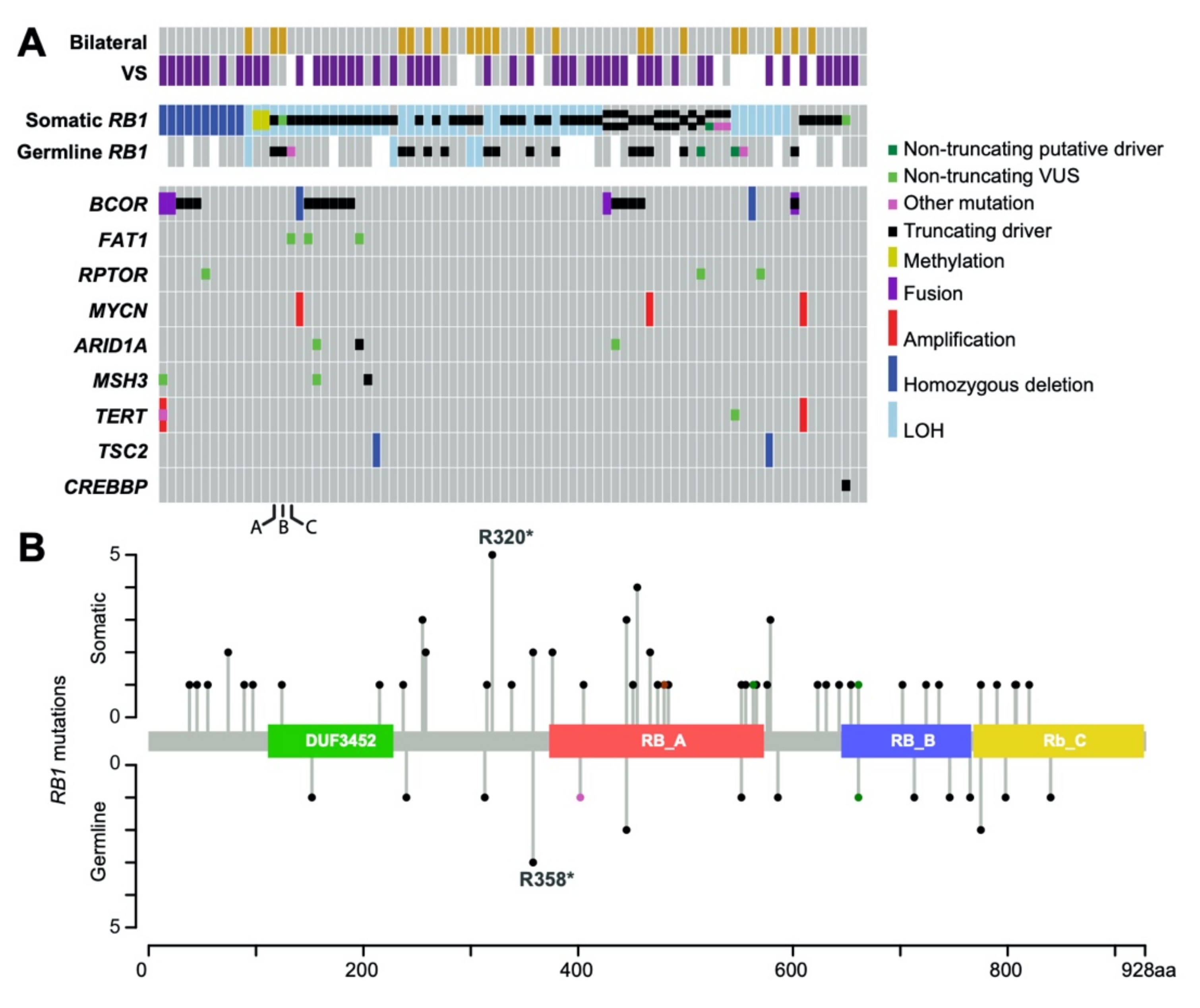
Figure 1. Mutational landscape of retinoblastoma. (A) Oncoprint of genetic, clinical, histopathologic features of 83 retinoblastoma eyes. Top section depicts clinicopathologic features of the specimens including presence of vitreous seeds (VS) and laterality. Because of the importance of RB1, we annotated germline and somatic RB1 hits separately and also included loss of heterozygosity and methylation status, which we display when multiple mutations occur. The rest of the genes are annotated, combining germline, somatic and multiple alterations. Missing data is white. A–C annotate three samples with three hits to RB1. (B) Plot of the somatic (top) and germline (bottom) alterations of RB1, known RB1 domains marked along the axis. The height of each line indicates the number of alterations that occur at that position within the protein, with the most frequent mutation labeled in both somatic and germline.
Cancers 13 00149 g001
Figure 2. Mechanisms of inactivation of RB1. (A) Stacked bar graph depicting number of hits to RB1 in 83 eyes. (B,C) Bar plot showing the types of loss of heterozygosity that occurs in retinoblastoma samples at the RB1 locus vs. loci of other tumor suppressor genes as annotated by OncoKb [23,24]. (C) Bar plot of the proportion of each type of loss of heterozygosity across the specimens, excluding those without any type of loss. There is a significantly higher proportion of CN-LOH at the RB1 locus (p < 2.2 × 10⁻¹⁶, OR = 19.8). (D) Bar plots comparing the types of loss of heterozygosity at the RB1 locus in malignancies with RB1 mutations. Retinoblastoma samples are compared to all other types of malignancies. There is significantly higher proportion of CN-LOH at the RB1 locus in retinoblastoma compared to other malignancies (p < 2.2 × 10⁻¹⁶, OR = 44.4). (E) For the somatic variants in the specimens with three detectable hits to RB1, we calculated the variant allele frequency corrected for purity of the sample and ploidy of the region (cancer cell fraction). Each column is the variant from a different specimen. Specimens are labeled based on Figure 1A.
Cancers 13 00149 g002
Figure 3. Correlation of genomic features with clinical characteristics. (A) Kaplan–Meier graph comparing metastases-free survival between specimens with a BCOR alteration and those without. BCOR is nominally correlated with metastases-free survival (p-value = 0.03). (B) Copy number alterations of 69 eyes. Each row is an individual and each column is a different chromosome. White space shows where no gains or losses occur. Losses are shown in shades of blue while gains are shown in shades of red. (C) Plot showing the genomic copy number changes in individuals with vitreous seeds vs. without. The genome was binned into 100,000 bp regions and the height of each region shows how many specimens had a gain or loss in a given bin. Any gain is shown in red on the top of each plot while any sort of loss of heterozygosity is shown in blue at the bottom of each plot. There is a significant enrichment for 1q gain and 16q loss in individuals with vitreous seeds (BH-corrected p-value = 0.008 and 0.004, respectively). (D) Venn diagram of specimens that contain 1q gain, 16q loss and vitreous seeds. Limited to 43 samples that had annotation of vitreous seeds and FACETS fits to call alterations on 1q and 16q.
Cancers 13 00149 g003
Publisher’s Note: MDPI stays neutral with regard to jurisdictional claims in published maps and institutional affiliations.