Review

Predictive and Prognostic Value of Microsatellite Instability in Gynecologic Cancer (Endometrial and Ovarian)

by
Camille Evrard
1,* and
Jérôme Alexandre
2
1
Service d’Oncologie Médicale, CHU de Poitiers, 86021 Poitiers, France
2
Service d’Oncologie Médicale, AP-HP, CARPEM, Cochin-Hospital, Université de Paris, 75014 Paris, France
*
Author to whom correspondence should be addressed.
Cancers 2021, 13(10), 2434; https://doi.org/10.3390/cancers13102434
Submission received: 31 March 2021 / Revised: 10 May 2021 / Accepted: 11 May 2021 / Published: 18 May 2021
(This article belongs to the Special Issue Microsatellite Instability and Cancers)

Abstract

:

Simple Summary

Endometrial cancers are the cancers most affected by microsatellite instability. This phenotype confers a demonstrated sensitivity to immunotherapy and in this sense is a major parameter to know in order to manage patients. Molecular biology, therefore, has an essential role in the better knowledge of these endometrial tumors. Moreover, the microsatellite instability phenotype is very poorly understood in ovarian cancer, yet it does exist. We therefore present here a review of the literature concerning microsatellite instability in gynecological cancers (endometrium and ovaries): its diagnosis, its clinical characteristics, and its therapeutic and prognostic impact.

Abstract

For endometrial cancer, a new classification is now available from ESMO, ESGO, and ESTRO based on clinical and molecular characteristics to determine adjuvant therapy. The contribution of molecular biology is major for this pathology mainly by the intermediary of deficient mismatch repair/microsatellite instability. Detection techniques for this phenotype have many peculiarities in gynecologic cancers (endometrial and ovarian) because it has been initially validated in colorectal cancer only. Endometrial cancer is the most common tumor with deficient mismatch repair, which is an important prognostic factor and a predictor of the benefit of adjuvant treatments. Concerning advanced stages, this phenotype is a theragnostic marker for using immunotherapy. Among ovarian cancer, microsatellite instability is less described in literature but exists, particularly in endometrioid type ovarian cancer. This review aims to provide an overview of the publications concerning deficient mismatch repair/microsatellite instability in endometrial and ovarian cancers, detection techniques, and clinical implications of these molecular characteristics.

1. Introduction

Microsatellites are small DNA sequences, coding or non-coding, with many repetitive single nucleotide or di-, tri-, or tetra-nucleotides; they are also called “short tandem repeats”. Because of this kind of repetitive structure, it is very often a carrier of replication error especially in the case of deficiency of the MisMatch Repair system, also called dMMR. These microsatellite instabilities (MSI), with an accumulation of errors, allowed to highlight the MMR dysfunction.
When cancer carries this kind of phenotype, this indicates microsatellite instable (MSI) or MMR deficient (dMMR) as opposed to microsatellite stable (MSS) and MMR-proficient (pMMR).
Tumor dMMR phenotype is related to the loss of one (or more) of the four main proteins involved in MMR process: MLH1, MSH2, MSH6, and PMS2. In most cases, this primary cause is the hypermethylation of MLH1 gene promotor in the tumor leading to its loss of expression.
A less common cause is the inherited transmission of a mutated allele of one of the MMR genes. It is responsible for the Hereditary Non-Polyposis Colorectal Cancer (HNPCC) or Lynch syndrome that is associated with a high risk of colon cancer but also of endometrial and ovarian cancer.
Endometrial cancer is the neoplasm most commonly associated with phenotype dMMR [1]. In recent years, this phenotype has emerged as an important prognostic factor but also as a predictor of the benefit of adjuvant treatments. In advanced stages, it is a theragnostic marker for the use of immunotherapy.

2. Clinical Classifications of Endometrial and Ovarian Cancers

2.1. Endometrial Cancer

Most endometrial cancers (EC) are diagnosed at early stage (80% in stage I), with 5-year event-free and survival rates of 80 and 95%, respectively. However, the 5-year survival dramatically decreases in locally advanced (Federation of Gynecology and Obstetrics [FIGO] stages III–IVA) or metastatic (FIGO stage IVB) diseases (68% and 17% respectively) [2].
Endometrial carcinoma can broadly be divided into two types according to histology: endometrioid, affecting approximately 80% of patients, and non-endometrioid, associated with poorer prognosis. This latter type includes serous carcinoma, the most common, clear-cell carcinoma, and carcinosarcoma.
In non-metastatic stages (I–III with complete surgery), the indications of adjuvant treatment (external radiotherapy, brachytherapy, chemotherapy) were until very recently based only on clinical prognostic factors according to the recommendations of the ESMO (European Society for Medical Oncology) (Table 1, first column). These factors are the FIGO stage; the histological type (endometrioid versus non-endometrioid); and the grade and the presence or absence of vascular or lymphatic tumor emboli [2].
As outlined below, the molecular classification including MMR status may improve the relapse prediction and has been recently added to guidelines for post-surgical treatment in early stages (Table 1, second column) [3].

2.2. Ovarian Cancer

Among ovarian carcinoma, there are five histological sub-types: high-grade serous, low-grade serous, endometrial, clear cell, and mucinous carcinomas.
High-grade serous carcinoma (HGSC) is the most common (70% of cases), it is consistently associated with a TP53 mutation.
Around half of these tumors harbor defects in homologous recombination (HRD) due to BRCA 1–2 defect (25% of HGSC) or other mechanisms partially unknown [4]. These HRD tumors are especially sensitive to platinum and to the poly ADP ribose polymerase (PARP) inhibitors who have revolutionized the prognosis of patients. PARP inhibitors in maintenance as a first or second line of treatment have allowed longer progression-free survival (PFS) after platinum-based chemotherapy and surgery [4].

3. Detection of dMMR/MSI Gynecologic Tumors

3.1. Detection Techniques

There are two types of techniques to detect the dMMR phenotype in daily practice: immunohistochemistry (IHC) or polymerase chain reaction (PCR) assay; these two techniques have been mainly developed in colorectal cancer (CRC).
The first technique, IHC, is based on the protein expression on tissue samples. There are four proteins in MMR system: MLH1, MSH2, MSH6, and PMS2 that assemble two by two to spot and repair DNA replication errors [5]. The MutLα complex is formed with MLH1 and PMS2, and the MutSa complex is formed with MSH2 and MSH6. First the MutSa locates a single base pair mismatch and forms a sleeve around the DNA at the site of the error and then allows the attachment of the second complex MutLα. This repair step involves many other proteins and enzymes, including DNA polymerase, which allows the excision of the defective DNA base and then the re-synthesis of DNA. The loss of expression of one of the four proteins of the system MMR reflects its loss of function, resulting in poor DNA replication with many “uncorrected” errors, which leads to the formation of ultra-muted cancers called dMMR/MSI.
If the four proteins of MMR are staining with IHC in the nucleus of tumor cells, the tumor is pMMR, and if at least one or more proteins is lost, the tumor is called dMMR (Figure 1).
The second technique is PCR assay; the purpose is the analysis of microsatellite loci according to recommendations in CRC with mono- or di-nucletotides repeats [6,7].
There are two referenced panels for PCR assay, Bethesda panel with two mononucleotide loci (Big Adenine Tract BAT-25 and BAT-26) and three dinucleotide loci (D2S123, D5S346, and D17S250). As this technique is less sensitive, its interpretation requires comparison with healthy tissue. A tumor is considered an MSI if there is instability on at least two out of five loci, if no loci is unstable, the tumor is said to be MSS.
The pentaplex panel is composed of five mononucleotide repeats quasi monomorphic: BAT-25, BAT-26, NR-21, NR-24, and NR-27 (MONO-27). Its interpretation is easier because of several very homogeneous repetitions: with three markers out of five or more being instable, the tumor is MSI, and in the case of no instable marker or one, the tumor is MSS. With the pentaplex, panel healthy tissue is recommended to be used when two markers are unstable. Thanks to its characteristics, this panel is the new standard in international recommendations for MSI testing in CRC [8].
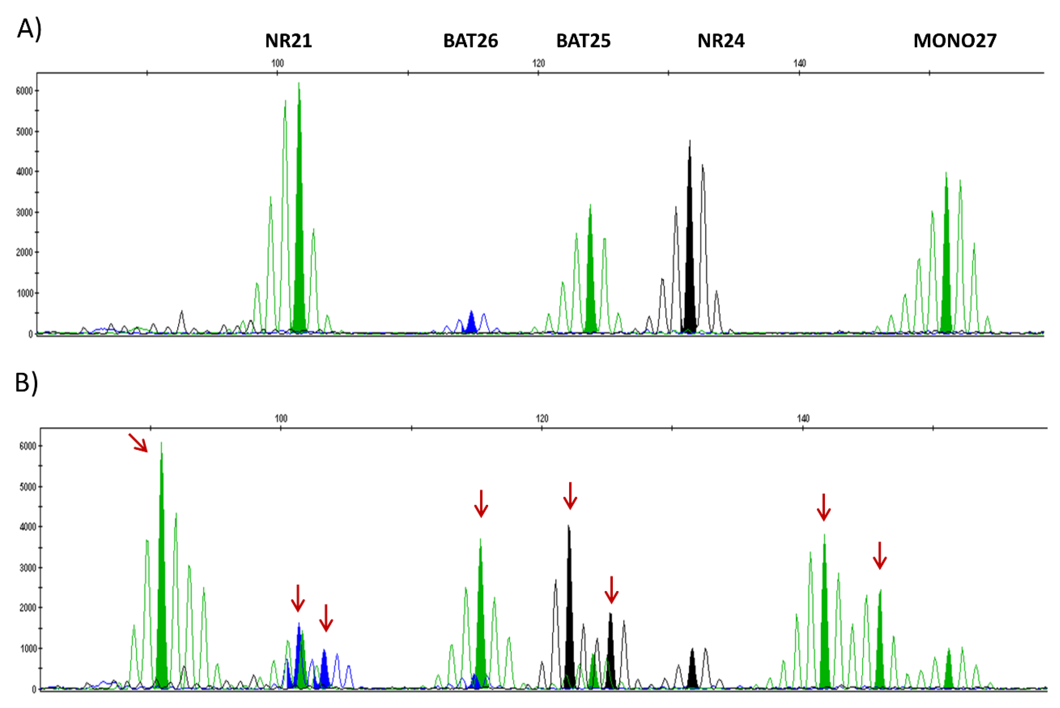
These two panels are based on capillary electrophoresis with intervention of Taq polymerase carrying out an amplification leading to a result in the form of multiple peaks, the markers being distributed according to their sizes (Figure 2). The higher peak is the reference size for one marker and is compared to its size in the general population: If the size of the peak significantly differs from the reference, it has instability [6]. All these recommendations are built for CRC, that is why for non-colorectal cancers (and gynecologic cancers) it is essential to compare tumor tissue and non-tumor tissue for MSI testing. In this case, a tumor is called “MSI” if there are two instable markers out of five [9].

3.2. Specificity of Detection Techniques in Endometrial Cancers and Discrepancies

The detection of microsatellite instability has mainly been studied in CRC because of a higher incidence of this type of cancer compared to gynecological cancers (endometrial and ovarian). In 2018, in the world there were more than: 1.8 million, 380,000 and 290,000 new cases of colorectal cancers, endometrial cancers and ovarian cancers, respectively [11].
The dMMR tumor rate is higher in EC compared to CRC as found in Wang’s publication in 2017, with analysis of 91 EC and 311 CRC [12]. For PCR analysis, they found 22% and 14.8% of MSI respectively for EC and CRC. Regardless of the type of tumor (EC or CRC), instability was found most of the time in all five mononucleotide loci (up to 85%), but being more often MONO27 instable in ECs and BAT26 instable in CRCs [12].
The main difficulty for detection of MSI in EC is that they exhibited smaller repeat number changes than CRC: shift in the reading frame caused by the repetition of unstable microsatellites is more marked in CRC than in EC with, on average, in this study, a shift to the left of −6.3 nucleotides (nt) for CRC against −2.9 nt for EC (Figure 3) [12].
Because of this characteristic, the detection sensitivity of the different tests is lower in EC, it is therefore necessary to use tests with mononucleotide markers (pentaplex type), to compare the results with those of non-tumor DNA and use a tissue sample with at least 30% tumor cells.
Very recently, Rafonne et al. published a meta-analysis with 10 studies involving a total of 3097 patients with excellent agreement (95%) between the evaluation of the MMR phenotype in IHC (use of four antibodies) and the results of the PCR in EC [13]. An ancillary analysis of Portec 1 and 2 clinical trials shows the same results with 696 patients [14]. In most cases, when a discrepancy is observed with loss of expression of MMR proteins and MSS phenotype, methylation of the MLH1 promoter or an MSI subclone is returned. It remains to be determined whether these discordant cases have the same clinical behavior as concordant dMMR cases with MSI.
Concerning the rare reverse cases of microsatellite instability (MSI) without loss of expression of proteins in IHC (pMMR), two cases (<1%) of this type were found in this series with each time a mutation of the POLE gene to explain this discrepancy [14].
The first-line of use of two antibodies for the detection of PMS2 and MSH6 seems to have identical performance to the use of four antibodies [13,14]. This attitude could be sufficient when MMR status is evaluated for prognostic purposes. However, for the detection of Lynch syndrome or theragnostic purpose, four antibodies are certainly still necessary [15,16].
In total, in endometrial cancers, the search for a deficiency in the MMR system is primarily based on IHC, which is a technique more readily available than molecular biology but also less expensive and which makes it possible to specify the affected gene. In addition, this technique does not require normal reference tissue and the response time is shorter.
The evaluation of microsatellite status by PCR remains necessary as a confirmation test in case of dMMR status by IHC or equivocal results.
Finally, in the event of loss of expression of MLH1 (generally associated with a loss of PMS2), it is necessary to test for methylation of the MLH1 promoter.
Oncogenetic counselling must be proposed to all patients with dMMR tumors in relation to MSH2, MSH6 or PMS2 loss or MLH1 loss without hypermethylation of its promotor.

3.3. Prevalence of dMMR Phenotype among Gynecologic Cancers

With a prevalence between 20 to 30% in the early stage, the dMMR phenotype is found especially in endometrial cancers (Table 2) [1,16]. It is this localization that will be the subject of the main part of this review, with a smaller second part concerning the knowledge on dMMR ovarian tumors, in which prevalence of dMMR tumors is below 10%.

4. Characteristics of dMMR Endometrial Cancer

Data from The Cancer Genome Atlas (TCGA) identified four distinct molecular subclasses based on mutational load and somatic copy-number alterations (SCNAs):
  • ultramuted associated with an inactivating mutation of the exonuclease domain of POLE (5%) and 232 × 10−6 mutations per Megabase (Mb) average.
  • hypermuted dMMR (30%) with 18 × 10−6 mutations per Mb and most with MLH1 promoter methylation.
  • “serous-like” (20%) characterized by a mutation of TP53 and a high number of alterations in the number of copies of genes (“copy number high”).
  • “copy number low” with few mutations and a low number of alterations in the number of copy, MSS, and without mutation of TP53 or POLE (45%) [18].
This classification had a prognostic impact: The tumors with the best prognosis were those mutated with POLE mutation, while a poorer prognosis was associated with “serous-like” tumors. MSI and “copy number low” tumors had an intermediate prognosis.
These TCGA data were the basis of the Proactive Molecular Risk Classifier for Endometrial Cancer (ProMisE) classification, which makes it possible to distinguish the four molecular groups by sequencing the exonuclease domain of POLE and the analysis by IHC of the expression of the proteins of MMR and TP53 [23].

4.1. Molecular Alterations Associated with dMMR Phenotype

About 95% of dMMR EC are sporadic, meaning that the MMR defect occurred primarily in the endometrial epithelium. The most common cause (75% of all dMMR EC) is the loss of MLH1 expression related to methylation of its promoter. In 5% of cases, a constitutional mutation in one of the genes of the MMR system is observed (Lynch syndrome) [1,24,25].
The constitutional mutations of MSH2 are the most frequently found in patients with endometrial cancer in the context of Lynch syndrome (40% of cases), followed by those of MLH1 and MSH6 (around 30% each). PMS2 mutations are much rarer [16].

4.2. Age of Women

Lynch syndrome-related endometrial cancers occur more in younger patients than in those without a constitutional mutation (median age: 54.3 to 62.3 years respectively) [26]. The prevalence of Lynch syndrome is 9% for cases occurring before age 50. However, in 25% of Lynch syndrome cases, endometrial cancer occurs after age 60 [24,27].
Tumors with methylation of the MLH1 promoter occur more in older women (median age at diagnosis: 65 years) than dMMR tumors with mutation (median age: 59 years) but also than MSS tumors (median age 60 years) [24,28].
In the majority of EC associated with Lynch syndrome, it would be the first cancer of the spectrum (between 51 and 80% of cases). In 20% of cases, there is a synchronous ovarian cancer [16,29].

4.3. Family History

A history of cancer related to Lynch syndrome in a first-degree relative is associated with the risk of Lynch syndrome in a patient with EC. However, in over a third of cases of endometrial cancer linked to Lynch syndrome, no family history is found [24].
These observations have led to the proposal of universal screening for Lynch syndrome by IHC in endometrial cancer [30,31]. In the event of loss of MLH1, a test for methylation of the promoter should be performed before considering an oncogenetic consultation.

4.4. Body Mass Index

The prevalence of obesity was found to be lower in patients with a mutation in the MMR system compared to those with epigenetic loss of MLH1 or pMMR status (50% versus 67% and 70%, respectively) [26,28].

4.5. Tumor Characteristics

The histological type of dMMR endometrial carcinoma is most often endometrioid (over 90% of cases) [23]. If 30% of endometrioid carcinomas are dMMR, the proportion would be around 20% for clear cell carcinomas, 7% for carcinosarcomas, 44% for undifferentiated and dedifferentiated carcinomas [20,21,22], and much lesser in serous subtype (0–6%) [18] (Table 3).
Endometrial cancers associated with Lynch syndrome are thought to be preferentially located in the lower segment of the uterus, but this does not appear to be the case for tumors with methylation of MLH1 [16,32].
Deficient MMR tumors present specific histological characteristics: significant intra-tumor lymphocytic infiltrate, presence of an undifferentiated tumor contingent coexisting with a low grade contingent, and increased frequency of high grade (47%) and vascular tumor emboli [15,23,33,34].
Methylation of MLH1, but not MMR genes mutations, is associated with a higher FIGO stage than pMMR tumors [28]. In a meta-analysis, dMMR tumors more often exhibits lymph node involvement (stage IIIC) than low-copy number endometrioid tumors (unmutated TP53 and pMMR) [34].
In total, dMMR tumors are associated with poor prognostic factors (high grade, vascular tumor emboli, and high FIGO stage) and are more often classified at high risk of relapse according to the clinical classification of ESMO compared to low-copy number (34 versus 14%) [23].

4.6. Prognostic Value of dMMR Status

In the meta-analysis of studies evaluating the ProMisE classification, dMMR tumors were associated in univariate analysis with a poor prognosis in terms of overall survival compared to “copy number low” tumors (unmutated pMMR TP53 and POLE). However, this difference disappeared in multivariate analysis considering other prognostic factors (grade, stage, presence of emboli, degree of myometrium invasion, age) [34].
Thus, considering the clinical prognostic factors, dMMR tumors have a prognosis equivalent to “copy number low” tumors and have a better prognosis than mutated TP53 tumors.

4.7. Effectiveness of Adjuvant Treatments and MMR Status

  • Pelvic radiotherapy in high intermediate risk tumors
The Portec 2 study compared post-operative pelvic radiotherapy to vaginal brachytherapy in tumors with a high intermediate prognosis according to the ESMO classification (Table 2). Overall, the two arms were equivalent in terms of vaginal recurrence rate, but the pelvic recurrence rate was significantly higher in the “brachytherapy” arm. In a post-hoc analysis, the increased pelvic recurrence in this arm was found to be limited to TP53-mutated tumors or in those with numerous vascular tumor emboli vascular or overexpressing L1 cell adhesion molecule (L1CAM).
Thus, these data suggested that the indication of pelvic radiotherapy in tumors with a high intermediate prognosis may be limited to TP53-mutated tumors or with numerous vascular tumor emboli or overexpressing L1CAM. In dMMR tumors without embolus, brachytherapy would be sufficient [14].
  • Adjuvant chemotherapy in high-risk tumors
The Portec 3 study evaluated the contribution of adjuvant chemotherapy in high-risk tumors according to the ESMO classification in addition to pelvic radiotherapy (Table 2): There was a modest but statistically significant benefit of adjuvant chemotherapy in terms of recurrence-free survival and overall survival [35].
A post-hoc analysis was carried out to evaluate these results according to the molecular classification: The TP53-mutated tumors in IHC presented an absolute benefit of adjuvant chemotherapy of 25% in terms of recurrence-free survival, whereas there was no benefit for dMMR tumors [36].

5. Use of MMR Status in Current Practice in Endometrial Cancers

The so-called high-risk tumors according to the classification proposed by ESMO for which there is an indication for pelvic radiotherapy and chemotherapy are in particular defined by a “aggressive” histology that covers grade 3 endometrioid carcinomas; serous, clear cell, and undifferentiated carcinomas; as well as carcinosarcomas [2].
However, several studies have reported suboptimal inter-observer reproducibility even among pathologists who are experts in identifying these tumors, both for the definition of high grade and for the endometrioid/serous distinction [37].
In addition, high-grade endometrioid carcinomas are molecularly heterogeneous: in one study, 36.2% were classified as dMMR, 12.9% POLE mutated, 20.7% TP53 mutated, and 30.2% “low copy number” (pMMR, POLE and TP53 unmutated). In multivariate analysis including FIGO stage and age, POLE mutation and dMMR status were independent prognostic factors associated with better relapse-free survival of high grade tumors, while the presence of a p53 mutation was associated with a poor prognosis [38]. More rarely, low grade endometrioid carcinomas could also present with TP53 mutations, but their prognosis is less clearly defined [39].
Finally, the identification of “serous-like” tumors cannot be based only on IHC TP53 since some POLE or dMMR mutated tumors can present a TP53 mutation that has no negative prognostic value [15].
For these multiple reasons, the International Society of Gyneco-Pathology in 2019 recommended the routine use of the ProMisE classification (POLE exonuclease domain sequencing, IHC TP 53, MMR) for high-grade endometrioid subgroup to better identify serous-like tumors with a worse prognosis and which require adjuvant treatment [15]. Conversely, “non-serous like” tumors could be reclassified as “low grade” and no longer be considered for chemotherapy or external radiotherapy if there is no extension beyond the uterus [15].
In 2021, ESGO-ESTRO-ESP jointly recommended to evaluate a molecular subgroup of all endometrial carcinoma and proposed new prognostic classification integrating clinical and molecular tumor characteristics (Table 2). Thus, all TP53 mutated tumors are considered at high risk and justify adjuvant chemotherapy (except in case of superficial tumor with no myometrium invasion), while this treatment can be avoided in dMMR stage I–II tumors [3].
However, data are still lacking to support therapeutic de-escalation for dMMR tumors with III–IVA stage or aggressive histology such as undifferentiated carcinoma or carcinosarcoma [22].

Predictive Benefit of Response to Immunotherapy in Advanced/Metastatic Stages

In metastatic endometrial cancers, the frequency of dMMR status has been estimated to be 15–20% [40]. No study has reported the prognosis of metastatic cancers based on MMR status.
The high rate of mutations observed in dMMR tumors or mutated for POLE is responsible for the continued formation of neoantigens. In vivo, these are responsible for an immune reaction that helps slow tumor growth [41]. In fact, dMMR or POLE mutated endometrial cancers usually present with an inflammatory lymphocytic infiltrate [42,43].
It is now well established that dMMR tumors have a particular sensitivity to immune checkpoint inhibitors, including anti-program death 1 (anti-PD-1), anti-program death ligand 1 (anti-PDL1), and anti-cytotoxic T-lymphocyte-associated protein 4 (anti-CTLA4) [44,45,46,47]. Specifically, in endometrial cancer, several phase II trials carried out in patients pretreated with chemotherapy have found response rates with immunotherapy ranging from 27 to 57% while they are less than 10% for pMMR tumors [48,49].
In the largest published phase II study evaluating Pembrolizumab in 49 patients, the response rate was 57% and the PFS was 25 months. The median duration of response was not reached [47]. For comparison, in a second-line phase III study, chemotherapy with doxorubicin or weekly paclitaxel was associated with an 11% response rate and a PFS of 4 months [28] (Table 3).
As a theragnostic objective, it remains an issue to define which test should be used to determine MMR status: IHC, PCR or both. In metastatic colorectal carcinoma, a high rate of primary failure of immunotherapy has been observed in cases of tumors with discordant results between IHC and PCR [52]. However, data are lacking in EC. In the case of dMMR status by IHC, confirmation by PCR is desirable before considering immunotherapy.

6. Ovarian Cancer and MSI: Description and Prognostic Value

Overall, in the literature, the percentage of ovarian cancer (OC) with microsatellite instability is between 2 and 10% of cases but reaches 20% in the endometrioid subgroup (Table 4). Most often, it is related to methylation of the MLH1 promoter [53].
The synchronous diagnosis of an endometrial cancer (endometrioid type) and an endometrioid ovarian cancer is a relatively common situation that could be associated with dMMR. Soliman and al reported 20% (12/59) of patients with loss of MSH2, MSH6, or MLH1 associated with an MSI-high tumor among 59 cases [54]. Another study of 32 of these patients found discordant results with a dMMR phenotype in 53.1% (17/32) of the endometrial tumors and in 31.3% (10/32) of ovarian tumors [55].
The dMMR status is much rarer in other histological types: 2–10% of clear cell cancers, <2% of serous carcinomas, and even more exceptional in mucinous carcinomas [53,61].
Deficient MMR status in endometrioid cancers did not appear to be associated with particular clinical features [53]. In a series of 36 cases of endometrioid carcinoma of the ovary, the four molecular subtypes described in endometrial cancer were found in similar proportions [62]. A larger study by Kramer and al. with 511 cases was able to find the four molecular subtypes of TCGA including 13.7% (70/511) of dMMR tumors [60]. As in endometrial cancer, dMMR ovarian endometrioid carcinomas were associated with a better prognosis than those with mutated TP53.
Among 80 patients with Lynch syndrome presenting with ovarian cancer, the most common histologic type was serous carcinoma followed by endometrioid type. The age of onset was younger than in the sporadic forms [63].
There are very few reports of immunotherapy in dMMR/MSI ovarian cancers. In the KEYNOTE-158 study, Pembrolizumab was associated with a response rate of 33% and median PFS of 2.3 months among 15 patients.

7. Conclusions

Beyond patients at risk of Lynch syndrome, evaluation of MMR status is becoming essential for all cases of EC. In routine clinical practice, the first step for the evaluation of MMR status is the immunohistochemistry. However, dMMR status should be confirmed by the evaluation of microsatellite instability by PCR.
In the early stage, the favorable prognosis value of dMMR is partially independent of clinical tumor characteristics and may allow therapeutic de-escalation in so-called “high risk” carcinoma. In addition, dMMR status is now recognized as the most promising therapeutic target in advanced EC, given the first results of immune checkpoint inhibitors. Clinical trials evaluating these drugs in early stage will start soon.
Available data suggest that MMR status should also be evaluated in endometrioid ovarian carcinoma from a prognosis and theragnostic perspective.

Author Contributions

Conceptualization, Validation, Investigation, Resources, Data curation and Writing—Review & Editing: C.E. and J.A. All authors have read and agreed to the published version of the manuscript.

Funding

This research received no external funding.

Conflicts of Interest

The authors have no relevant affiliations or financial involvement with any organization or entity with a financial interest in or financial conflict with the subject matter or materials discussed in the manuscript. This includes employment, consultancies, honoraria, stock ownership or options, expert testimony, grants or patents received or pending, or royalties. Peer reviewers on this manuscript have no relevant financial or other relationships to disclose.

Abbreviations

ADPadenosine diphosphate
CTLA4cytotoxic T-lymphocyte-associated protein 4
CRCcolorectal cancer
dMMRdeficient mismatch repair
ECendometrial cancers
ESGOEuropean Society of Gynaecological Oncology
ESMOEuropean Society for Medical Oncology
ESTROEuropean Society for Radiotherapy and Oncology
FIGOFederation of Gynecology and Obstetrics
HGSCHigh-grade serous carcinoma
HNPCCHereditary Non-Polyposis Colorectal Cancer
IHCimmunohistochemistry
LSLynch syndrome
LVSIlymphovascular space invasion
MMRmismatch repair
MSImicrosatellite instable
MSSmicrosatellite stable
NGSnext generation sequencing
NSMPnon specific molecular profile
OCovarian cancer
PARPpoly ADP ribose polymerase
PCRpolymerase chain reaction
PD-1program death 1
PD-L1program death ligand 1
pMMRproficient mismatch repair
POLEpolymerase ε
ProMisEProactive Molecular Risk Classifier for Endometrial Cancer

References

  1. Stelloo, E.; Jansen, A.M.L.; Osse, E.M.; Nout, R.A.; Creutzberg, C.L.; Ruano, D.; Church, D.N.; Morreau, H.; Smit, V.T.H.B.M.; van Wezel, T.; et al. Practical Guidance for Mismatch Repair-Deficiency Testing in Endometrial Cancer. Ann. Oncol. 2017, 28, 96–102. [Google Scholar] [CrossRef]
  2. Colombo, N.; Creutzberg, C.; Amant, F.; Bosse, T.; González-Martín, A.; Ledermann, J.; Marth, C.; Nout, R.; Querleu, D.; Mirza, M.R.; et al. ESMO-ESGO-ESTRO Consensus Conference on Endometrial Cancer: Diagnosis, Treatment and Follow-Up. Ann. Oncol. 2016, 27, 16–41. [Google Scholar] [CrossRef] [PubMed]
  3. Concin, N.; Matias-Guiu, X.; Vergote, I.; Cibula, D.; Mirza, M.R.; Marnitz, S.; Ledermann, J.; Bosse, T.; Chargari, C.; Fagotti, A.; et al. ESGO/ESTRO/ESP Guidelines for the Management of Patients with Endometrial Carcinoma. Int. J. Gynecol. Cancer 2021, 31, 12–39. [Google Scholar] [CrossRef]
  4. di Silvestro, P.; Colombo, N.; Scambia, G.; Kim, B.-G.; Oaknin, A.; Friedlander, M.; Lisyanskaya, A.; Floquet, A.; Leary, A.; Sonke, G.S.; et al. Efficacy of Maintenance Olaparib for Patients With Newly Diagnosed Advanced Ovarian Cancer With a BRCA Mutation: Subgroup Analysis Findings From the SOLO1 Trial. J. Clin. Oncol. 2020, 38, 3528–3537. [Google Scholar] [CrossRef] [PubMed]
  5. Sepulveda, A.R.; Hamilton, S.R.; Allegra, C.J.; Grody, W.; Cushman-Vokoun, A.M.; Funkhouser, W.K.; Kopetz, S.E.; Lieu, C.; Lindor, N.M.; Minsky, B.D.; et al. Molecular Biomarkers for the Evaluation of Colorectal Cancer: Guideline From the American Society for Clinical Pathology, College of American Pathologists, Association for Molecular Pathology, and American Society of Clinical Oncology. Arch. Pathol. Lab. Med. 2017, 141, 625–657. [Google Scholar] [CrossRef] [PubMed] [Green Version]
  6. Boland, C.R.; Thibodeau, S.N.; Hamilton, S.R.; Sidransky, D.; Eshleman, J.R.; Burt, R.W.; Meltzer, S.J.; Rodriguez-Bigas, M.A.; Fodde, R.; Ranzani, G.N.; et al. A National Cancer Institute Workshop on Microsatellite Instability for Cancer Detection and Familial Predisposition: Development of International Criteria for the Determination of Microsatellite Instability in Colorectal Cancer. Cancer Res. 1998, 58, 5248–5257. [Google Scholar]
  7. Siemanowski, J.; Schömig-Markiefka, B.; Buhl, T.; Haak, A.; Siebolts, U.; Dietmaier, W.; Arens, N.; Pauly, N.; Ataseven, B.; Büttner, R.; et al. Managing Difficulties of Microsatellite Instability Testing in Endometrial Cancer-Limitations and Advantages of Four Different PCR-Based Approaches. Cancers 2021, 13, 1268. [Google Scholar] [CrossRef]
  8. Suraweera, N.; Duval, A.; Reperant, M.; Vaury, C.; Furlan, D.; Leroy, K.; Seruca, R.; Iacopetta, B.; Hamelin, R. Evaluation of Tumor Microsatellite Instability Using Five Quasimonomorphic Mononucleotide Repeats and Pentaplex PCR. Gastroenterology 2002, 123, 1804–1811. [Google Scholar] [CrossRef]
  9. Wong, Y.F.; Cheung, T.H.; Lo, K.W.K.; Yim, S.F.; Chan, L.K.Y.; Buhard, O.; Duval, A.; Chung, T.K.H.; Hamelin, R. Detection of Microsatellite Instability in Endometrial Cancer: Advantages of a Panel of Five Mononucleotide Repeats over the National Cancer Institute Panel of Markers. Carcinogenesis 2006, 27, 951–955. [Google Scholar] [CrossRef] [PubMed] [Green Version]
  10. Evrard, C.; Tachon, G.; Randrian, V.; Karayan-Tapon, L.; Tougeron, D. Microsatellite Instability: Diagnosis, Heterogeneity, Discordance, and Clinical Impact in Colorectal Cancer. Cancers 2019, 11, 1567. [Google Scholar] [CrossRef] [Green Version]
  11. Cancer Today. Available online: http://gco.iarc.fr/today/home (accessed on 25 August 2020).
  12. Wang, Y.; Shi, C.; Eisenberg, R.; Vnencak-Jones, C.L. Differences in Microsatellite Instability Profiles between Endometrioid and Colorectal Cancers. J. Mol. Diagn. 2017, 19, 57–64. [Google Scholar] [CrossRef] [PubMed] [Green Version]
  13. Raffone, A.; Travaglino, A.; Cerbone, M.; Gencarelli, A.; Mollo, A.; Insabato, L.; Zullo, F. Diagnostic Accuracy of Immunohistochemistry for Mismatch Repair Proteins as Surrogate of Microsatellite Instability Molecular Testing in Endometrial Cancer. Pathol. Oncol. Res. 2020, 26, 1417–1427. [Google Scholar] [CrossRef]
  14. Wortman, B.G.; Creutzberg, C.L.; Putter, H.; Jürgenliemk-Schulz, I.M.; Jobsen, J.J.; Lutgens, L.C.H.W.; van der Steen-Banasik, E.M.; Mens, J.W.M.; Slot, A.; Kroese, M.C.S.; et al. Ten-Year Results of the PORTEC-2 Trial for High-Intermediate Risk Endometrial Carcinoma: Improving Patient Selection for Adjuvant Therapy. Br. J. Cancer 2018, 119, 1067–1074. [Google Scholar] [CrossRef] [Green Version]
  15. Soslow, R.A.; Tornos, C.; Park, K.J.; Malpica, A.; Matias-Guiu, X.; Oliva, E.; Parkash, V.; Carlson, J.; McCluggage, W.G.; Gilks, C.B. Endometrial Carcinoma Diagnosis: Use of FIGO Grading and Genomic Subcategories in Clinical Practice: Recommendations of the International Society of Gynecological Pathologists. Int. J. Gynecol. Pathol. 2019, 38, S64–S74. [Google Scholar] [CrossRef] [Green Version]
  16. Rossi, L.; Frere-Belda, M.-A.L.; Laurent-Puig, P.; Buecher, B.; Pauw, A.D.; Stoppa-Lyonnet, D.; Canlorbe, G.; Caron, O.; Borghese, B.; Colas, C.; et al. Clinicopathologic Characteristics of Endometrial Cancer in Lynch Syndrome: A French Multicenter Study. Int. J. Gynecol. Cancer 2017, 27, 953–960. [Google Scholar] [CrossRef] [PubMed]
  17. Bonneville, R.; Krook, M.A.; Kautto, E.A.; Miya, J.; Wing, M.R.; Chen, H.-Z.; Reeser, J.W.; Yu, L.; Roychowdhury, S. Landscape of Microsatellite Instability Across 39 Cancer Types. JCO Precis. Oncol. 2017, 2017, PO.17.00073. [Google Scholar] [CrossRef] [PubMed]
  18. Levine, D.A. Integrated Genomic Characterization of Endometrial Carcinoma. Nature 2013, 497, 67–73. [Google Scholar] [CrossRef] [PubMed] [Green Version]
  19. Jones, S.; Stransky, N.; McCord, C.L.; Cerami, E.; Lagowski, J.; Kelly, D.; Angiuoli, S.V.; Sausen, M.; Kann, L.; Shukla, M.; et al. Genomic Analyses of Gynaecologic Carcinosarcomas Reveal Frequent Mutations in Chromatin Remodelling Genes. Nat. Commun. 2014, 5, 5006. [Google Scholar] [CrossRef] [Green Version]
  20. Travaglino, A.; Raffone, A.; Gencarelli, A.; Mollo, A.; Guida, M.; Insabato, L.; Santoro, A.; Zannoni, G.F.; Zullo, F. TCGA Classification of Endometrial Cancer: The Place of Carcinosarcoma. Pathol. Oncol. Res. 2020, 26, 2067–2073. [Google Scholar] [CrossRef]
  21. de Lair, D.F.; Burke, K.A.; Selenica, P.; Lim, R.S.; Scott, S.N.; Middha, S.; Mohanty, A.S.; Cheng, D.T.; Berger, M.F.; Soslow, R.A.; et al. The Genetic Landscape of Endometrial Clear Cell Carcinomas. J. Pathol. 2017, 243, 230–241. [Google Scholar] [CrossRef]
  22. Travaglino, A.; Raffone, A.; Mascolo, M.; Guida, M.; Insabato, L.; Zannoni, G.F.; Zullo, F. TCGA Molecular Subgroups in Endometrial Undifferentiated/Dedifferentiated Carcinoma. Pathol. Oncol. Res. 2020, 26, 1411–1416. [Google Scholar] [CrossRef]
  23. Kommoss, S.; McConechy, M.K.; Kommoss, F.; Leung, S.; Bunz, A.; Magrill, J.; Britton, H.; Kommoss, F.; Grevenkamp, F.; Karnezis, A.; et al. Final Validation of the ProMisE Molecular Classifier for Endometrial Carcinoma in a Large Population-Based Case Series. Ann. Oncol. 2018, 29, 1180–1188. [Google Scholar] [CrossRef] [PubMed]
  24. Goodfellow, P.J.; Billingsley, C.C.; Lankes, H.A.; Ali, S.; Cohn, D.E.; Broaddus, R.J.; Ramirez, N.; Pritchard, C.C.; Hampel, H.; Chassen, A.S.; et al. Combined Microsatellite Instability, MLH1 Methylation Analysis, and Immunohistochemistry for Lynch Syndrome Screening in Endometrial Cancers From GOG210: An NRG Oncology and Gynecologic Oncology Group Study. J. Clin. Oncol. 2015, 33, 4301–4308. [Google Scholar] [CrossRef]
  25. Latham, A.; Srinivasan, P.; Kemel, Y.; Shia, J.; Bandlamudi, C.; Mandelker, D. Microsatellite Instability is Associated with the Presence of Lynch Syndrome Pan-Cancer. Available online: https://www-ncbi-nlm-nih-gov.proxy.insermbiblio.inist.fr/pmc/articles/PMC6553803/ (accessed on 6 October 2020).
  26. Dondi, G.; Coluccelli, S.; de Leo, A.; Ferrari, S.; Gruppioni, E.; Bovicelli, A.; Godino, L.; Coadă, C.A.; Morganti, A.G.; Giordano, A.; et al. An Analysis of Clinical, Surgical, Pathological and Molecular Characteristics of Endometrial Cancer According to Mismatch Repair Status. A Multidisciplinary Approach. Int. J. Mol. Sci. 2020, 21, 7188. [Google Scholar] [CrossRef]
  27. Kwon, J.S.; Scott, J.L.; Gilks, C.B.; Daniels, M.S.; Sun, C.C.; Lu, K.H. Testing Women With Endometrial Cancer to Detect Lynch Syndrome. J. Clin. Oncol. 2011, 29, 2247–2252. [Google Scholar] [CrossRef] [Green Version]
  28. McMeekin, D.S.; Tritchler, D.L.; Cohn, D.E.; Mutch, D.G.; Lankes, H.A.; Geller, M.A.; Powell, M.A.; Backes, F.J.; Landrum, L.M.; Zaino, R.; et al. Clinicopathologic Significance of Mismatch Repair Defects in Endometrial Cancer: An NRG Oncology/Gynecologic Oncology Group Study. J. Clin. Oncol. 2016, 34, 3062–3068. [Google Scholar] [CrossRef] [PubMed]
  29. Lu, H.K.; Broaddus, R.R. Gynecologic Cancers in Lynch Syndrome/HNPCC. Fam. Cancer 2005, 4, 249–254. [Google Scholar] [CrossRef] [PubMed]
  30. Rabban, J.T.; Calkins, S.M.; Karnezis, A.N.; Grenert, J.P.; Blanco, A.; Crawford, B.; Chen, L. Association of Tumor Morphology With Mismatch-Repair Protein Status in Older Endometrial Cancer Patients: Implications for Universal Versus Selective Screening Strategies for Lynch Syndrome. Am. J. Surg. Pathol. 2014, 38, 793–800. [Google Scholar] [CrossRef]
  31. Gallon, R.; Gawthorpe, P.; Phelps, R.L.; Hayes, C.; Borthwick, G.M.; Santibanez-Koref, M.; Jackson, M.S.; Burn, J. How Should We Test for Lynch Syndrome? A Review of Current Guidelines and Future Strategies. Cancers 2021, 13, 406. [Google Scholar] [CrossRef]
  32. Westin, S.N.; Lacour, R.A.; Urbauer, D.L.; Luthra, R.; Bodurka, D.C.; Lu, K.H.; Broaddus, R.R. Carcinoma of the Lower Uterine Segment: A Newly Described Association With Lynch Syndrome. J. Clin. Oncol. 2008, 26, 5965–5971. [Google Scholar] [CrossRef]
  33. Garg, K.; Soslow, R.A. Lynch Syndrome (Hereditary Non-Polyposis Colorectal Cancer) and Endometrial Carcinoma. J. Clin. Pathol. 2009, 62, 679–684. [Google Scholar] [CrossRef] [PubMed] [Green Version]
  34. Raffone, A.; Travaglino, A.; Mascolo, M.; Carotenuto, C.; Guida, M.; Mollo, A.; Insabato, L.; Zullo, F. Histopathological Characterization of ProMisE Molecular Groups of Endometrial Cancer. Gynecol. Oncol. 2020, 157, 252–259. [Google Scholar] [CrossRef]
  35. de Boer, S.M.; Powell, M.E.; Mileshkin, L.; Katsaros, D.; Bessette, P.; Haie-Meder, C.; Ottevanger, P.B.; Ledermann, J.A.; Khaw, P.; D’Amico, R.; et al. Adjuvant Chemoradiotherapy versus Radiotherapy Alone in Women with High-Risk Endometrial Cancer (PORTEC-3): Patterns of Recurrence and Post-Hoc Survival Analysis of a Randomised Phase 3 Trial. Lancet Oncol. 2019, 20, 1273–1285. [Google Scholar] [CrossRef] [Green Version]
  36. León-Castillo, A.; de Boer, S.M.; Powell, M.E.; Mileshkin, L.R.; Mackay, H.J.; Leary, A.; Nijman, H.W.; Singh, N.; Pollock, P.M.; Bessette, P.; et al. Molecular Classification of the PORTEC-3 Trial for High-Risk Endometrial Cancer: Impact on Prognosis and Benefit From Adjuvant Therapy. J. Clin. Oncol. 2020, 38, 3388–3397. [Google Scholar] [CrossRef]
  37. Murali, R.; Davidson, B.; Fadare, O.; Carlson, J.A.; Crum, C.P.; Gilks, C.B.; Irving, J.A.; Malpica, A.; Matias-Guiu, X.; McCluggage, W.G.; et al. High-Grade Endometrial Carcinomas: Morphologic and Immunohistochemical Features, Diagnostic Challenges and Recommendations. Int. J. Gynecol. Pathol. 2019, 38, S40–S63. [Google Scholar] [CrossRef]
  38. Bosse, T.; Nout, R.A.; McAlpine, J.N.; McConechy, M.K.; Britton, H.; Hussein, Y.; Gonzalez, C.; Ganesan, R.; Steele, J.C.; Harrison, B.T.; et al. Molecular Classification of Grade 3 Endometrioid Endometrial Cancers Identifies Distinct Prognostic Subgroups. Am. J. Surg. Pathol. 2018, 42, 561–568. [Google Scholar] [CrossRef]
  39. Beinse, G.; Rance, B.; Just, P.-A.; Izac, B.; Letourneur, F.; Saidu, N.E.B.; Chouzenoux, S.; Nicco, C.; Goldwasser, F.; Batteux, F.; et al. Identification of TP53 Mutated Group Using a Molecular and Immunohistochemical Classification of Endometrial Carcinoma to Improve Prognostic Evaluation for Adjuvant Treatments. Int. J. Gynecol. Cancer 2020, 30, 640–647. [Google Scholar] [CrossRef] [PubMed]
  40. Soumerai, T.E.; Donoghue, M.T.A.; Bandlamudi, C.; Srinivasan, P.; Chang, M.T.; Zamarin, D.; Cadoo, K.A.; Grisham, R.N.; O’Cearbhaill, R.E.; Tew, W.P.; et al. Clinical Utility of Prospective Molecular Characterization in Advanced Endometrial Cancer. Clin. Cancer Res. 2018, 24, 5939–5947. [Google Scholar] [CrossRef] [Green Version]
  41. Germano, G.; Lamba, S.; Rospo, G.; Barault, L.; Magrì, A.; Maione, F.; Russo, M.; Crisafulli, G.; Bartolini, A.; Lerda, G.; et al. Inactivation of DNA Repair Triggers Neoantigen Generation and Impairs Tumour Growth. Nature 2017, 552, 116–120. [Google Scholar] [CrossRef]
  42. Pakish, J.B.; Zhang, Q.; Chen, Z.; Liang, H.; Chisholm, G.B.; Yuan, Y.; Mok, S.C.; Broaddus, R.R.; Lu, K.H.; Yates, M.S. Immune Microenvironment in Microsatellite-Instable Endometrial Cancers: Hereditary or Sporadic Origin Matters. Clin. Cancer Res. 2017, 23, 4473–4481. [Google Scholar] [CrossRef] [Green Version]
  43. Talhouk, A.; Derocher, H.; Schmidt, P.; Leung, S.; Milne, K.; Gilks, C.B.; Anglesio, M.S.; Nelson, B.H.; McAlpine, J.N. Molecular Subtype Not Immune Response Drives Outcomes in Endometrial Carcinoma. Clin. Cancer Res. 2019, 25, 2537–2548. [Google Scholar] [CrossRef] [PubMed] [Green Version]
  44. Le, D.T.; Durham, J.N.; Smith, K.N.; Wang, H.; Bartlett, B.R.; Aulakh, L.K.; Lu, S.; Kemberling, H.; Wilt, C.; Luber, B.S.; et al. Mismatch Repair Deficiency Predicts Response of Solid Tumors to PD-1 Blockade. Science 2017, 357, 409–413. [Google Scholar] [CrossRef] [Green Version]
  45. Le, D.T.; Uram, J.N.; Wang, H.; Bartlett, B.R.; Kemberling, H.; Eyring, A.D.; Skora, A.D.; Luber, B.S.; Azad, N.S.; Laheru, D.; et al. PD-1 Blockade in Tumors with Mismatch-Repair Deficiency. Available online: https://www-nejm-org.gate2.inist.fr/doi/10.1056/NEJMoa1500596?url_ver=Z39.88-2003&rfr_id=ori%3Arid%3Acrossref.org&rfr_dat=cr_pub%3Dwww.ncbi.nlm.nih.gov (accessed on 22 June 2019).
  46. Overman, M.J.; Lonardi, S.; Wong, K.Y.M.; Lenz, H.-J.; Gelsomino, F.; Aglietta, M.; Morse, M.A.; Van Cutsem, E.; McDermott, R.; Hill, A.; et al. Durable Clinical Benefit With Nivolumab Plus Ipilimumab in DNA Mismatch Repair-Deficient/Microsatellite Instability-High Metastatic Colorectal Cancer. J. Clin. Oncol. Off. J. Am. Soc. Clin. Oncol. 2018, 36, 773–779. [Google Scholar] [CrossRef]
  47. Marabelle, A.; Le, D.T.; Ascierto, P.A.; di Giacomo, A.M.; de Jesus-Acosta, A.; Delord, J.-P.; Geva, R.; Gottfried, M.; Penel, N.; Hansen, A.R.; et al. Efficacy of Pembrolizumab in Patients With Noncolorectal High Microsatellite Instability/Mismatch Repair–Deficient Cancer: Results From the Phase II KEYNOTE-158 Study. J. Clin. Oncol. 2019, 38, 1–10. [Google Scholar] [CrossRef] [PubMed]
  48. Ott, P.A.; Bang, Y.-J.; Berton-Rigaud, D.; Elez, E.; Pishvaian, M.J.; Rugo, H.S.; Puzanov, I.; Mehnert, J.M.; Aung, K.L.; Lopez, J.; et al. Safety and Antitumor Activity of Pembrolizumab in Advanced Programmed Death Ligand 1–Positive Endometrial Cancer: Results From the KEYNOTE-028 Study. J. Clin. Oncol. 2017, 35, 2535–2541. [Google Scholar] [CrossRef]
  49. Oaknin, A.; Duska, L.R.; Sullivan, R.J.; Pothuri, B.; Ellard, S.L.; Leath, C.A.; Moreno, V.; Kristeleit, R.S.; Guo, W.; Danaee, H.; et al. Preliminary Safety, Efficacy, and Pharmacokinetic/Pharmacodynamic Characterization from GARNET, a Phase I/II Clinical Trial of the Anti–PD-1 Monoclonal Antibody, TSR-042, in Patients with Recurrent or Advanced MSI-h and MSS Endometrial Cancer. Gynecol. Oncol. 2019, 154, 17. [Google Scholar] [CrossRef]
  50. Oaknin, A.; Gilbert, L.; Tinker, A.V.; Sabatier, R.; Boni, V.; O’Malley, D.M.; Ghamande, S.; Duska, L.; Ghatage, P.; Guo, W.; et al. LBA36 Safety and Antitumor Activity of Dostarlimab in Patients (Pts) with Advanced or Recurrent DNA Mismatch Repair Deficient (DMMR) or Proficient (MMRp) Endometrial Cancer (EC): Results from GARNET. Ann. Oncol. 2020, 31, S1166. [Google Scholar] [CrossRef]
  51. Antill, Y.C.; Kok, P.S.; Robledo, K.; Barnes, E.; Friedlander, M.; Baron-Hay, S.E.; Shannon, C.M.; Coward, J.; Beale, P.J.; Goss, G.; et al. Activity of Durvalumab in Advanced Endometrial Cancer (AEC) According to Mismatch Repair (MMR) Status: The Phase II PHAEDRA Trial (ANZGOG1601). J. Clin. Oncol. 2019, 37, 5501. [Google Scholar] [CrossRef]
  52. Cohen, R.; Hain, E.; Buhard, O.; Guilloux, A.; Bardier, A.; Kaci, R.; Bertheau, P.; Renaud, F.; Bibeau, F.; Fléjou, J.-F.; et al. Association of Primary Resistance to Immune Checkpoint Inhibitors in Metastatic Colorectal Cancer With Misdiagnosis of Microsatellite Instability or Mismatch Repair Deficiency Status. JAMA Oncol. 2019, 5, 551–555. [Google Scholar] [CrossRef] [PubMed]
  53. Fraune, C.; Rosebrock, J.; Simon, R.; Hube-Magg, C.; Makrypidi-Fraune, G.; Kluth, M.; Büscheck, F.; Höflmayer, D.; Schmalfeldt, B.; Müller, V.; et al. High Homogeneity of MMR Deficiency in Ovarian Cancer. Gynecol. Oncol. 2020, 156, 669–675. [Google Scholar] [CrossRef]
  54. Soliman, P.T.; Broaddus, R.R.; Schmeler, K.M.; Daniels, M.S.; Gonzalez, D.; Slomovitz, B.M.; Gershenson, D.M.; Lu, K.H. Women With Synchronous Primary Cancers of the Endometrium and Ovary: Do They Have Lynch Syndrome? J. Clin. Oncol. 2005, 23, 9344–9350. [Google Scholar] [CrossRef] [PubMed]
  55. Kobayashi, Y.; Nakamura, K.; Nomura, H.; Banno, K.; Irie, H.; Adachi, M.; Iida, M.; Umene, K.; Nogami, Y.; Masuda, K.; et al. Clinicopathologic Analysis With Immunohistochemistry for DNA Mismatch Repair Protein Expression in Synchronous Primary Endometrial and Ovarian Cancers. Int. J. Gynecol. Cancer 2015, 25, 440–446. [Google Scholar] [CrossRef] [PubMed]
  56. Xiao, X.; Dong, D.; He, W.; Song, L.; Wang, Q.; Yue, J.; Xie, L. Mismatch Repair Deficiency Is Associated with MSI Phenotype, Increased Tumor-Infiltrating Lymphocytes and PD-L1 Expression in Immune Cells in Ovarian Cancer. Gynecol. Oncol. 2018, 149, 146–154. [Google Scholar] [CrossRef]
  57. Aysal, A.; Karnezis, A.; Medhi, I.; Grenert, J.P.; Zaloudek, C.J.; Rabban, J.T. Ovarian Endometrioid Adenocarcinoma: Incidence and Clinical Significance of the Morphologic and Immunohistochemical Markers of Mismatch Repair Protein Defects and Tumor Microsatellite Instability. Am. J. Surg. Pathol. 2012, 36, 163–172. [Google Scholar] [CrossRef] [PubMed]
  58. Rambau, P.F.; Duggan, M.A.; Ghatage, P.; Warfa, K.; Steed, H.; Perrier, R.; Kelemen, L.E.; Köbel, M. Significant Frequency of MSH2/MSH6 Abnormality in Ovarian Endometrioid Carcinoma Supports Histotype-Specific Lynch Syndrome Screening in Ovarian Carcinomas. Histopathology 2016, 69, 288–297. [Google Scholar] [CrossRef]
  59. Hollis, R.L.; Thomson, J.P.; Stanley, B.; Churchman, M.; Meynert, A.M.; Rye, T.; Bartos, C.; Iida, Y.; Croy, I.; Mackean, M.; et al. Molecular Stratification of Endometrioid Ovarian Carcinoma Predicts Clinical Outcome. Nat. Commun. 2020, 11, 4995. [Google Scholar] [CrossRef]
  60. Kramer, P.; Talhouk, A.; Brett, M.A. Endometrial Cancer Molecular Risk Stratification is Equally Prognostic for Endometrioid Ovarian Carcinoma|Clinical Cancer Research. Available online: https://clincancerres-aacrjournals-org.proxy.insermbiblio.inist.fr/content/26/20/5400.long (accessed on 16 November 2020).
  61. Willis, B.C.; Sloan, E.A.; Atkins, K.A.; Stoler, M.H.; Mills, A.M. Mismatch Repair Status and PD-L1 Expression in Clear Cell Carcinomas of the Ovary and Endometrium. Mod. Pathol. 2017, 30, 1622–1632. [Google Scholar] [CrossRef] [PubMed]
  62. Cybulska, P.; Paula, A.D.C.; Tseng, J.; Leitao, M.M.; Bashashati, A.; Huntsman, D.G.; Nazeran, T.M.; Aghajanian, C.; Abu-Rustum, N.R.; DeLair, D.F.; et al. Molecular Profiling and Molecular Classification of Endometrioid Ovarian Carcinomas. Gynecol. Oncol. 2019, 154, 516–523. [Google Scholar] [CrossRef]
  63. Akbari, M.R.; Zhang, S.; Cragun, D.; Lee, J.-H.; Coppola, D.; McLaughlin, J.; Risch, H.A.; Rosen, B.; Shaw, P.; Sellers, T.A.; et al. Correlation between Germline Mutations in MMR Genes and Microsatellite Instability in Ovarian Cancer Specimens. Fam. Cancer 2017, 16, 351–355. [Google Scholar] [CrossRef]
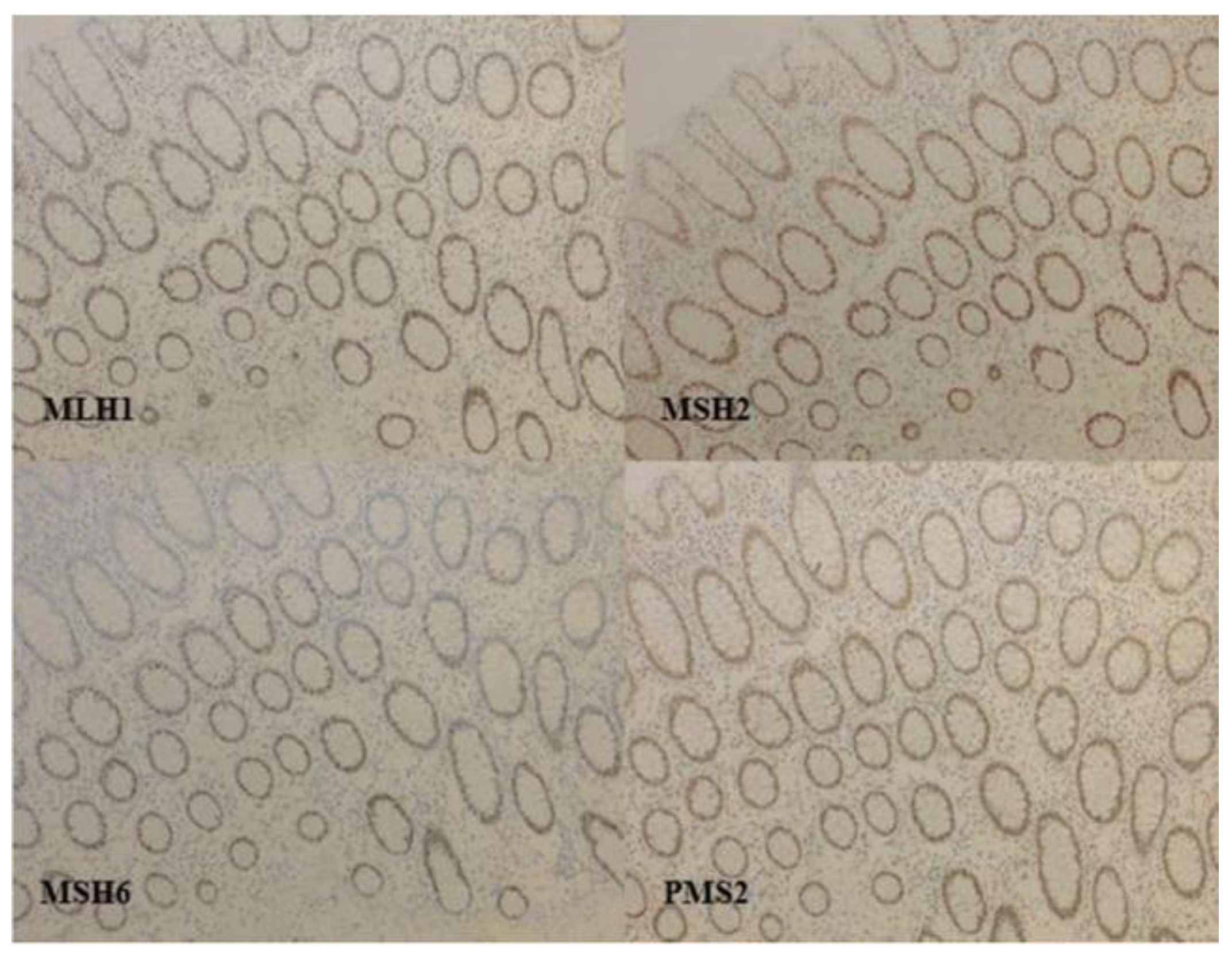
Figure 1. Immunochemistry of MMR proteins in colorectal cancer: a proficient MMR tumor, normal colonic mucosa, no loss of expression of MMR proteins. Antibodies used: anti-MLH1 (clone M1 Ventana® Oro Valley, AZ, USA, kit Optiview® for revelation, Roche Diagnostics, Meylan, France); anti-MSH2 (clone G219-1129 Ventana®, kit Optiview® for revelation); anti-MSH6 (clone 44BD Biosciences®, dilution 1/500, Franklin Lakes, NJ, USA; kit Ultraview® for revelation, Berkeley, CA, USA); anti-PMS2 (clone EPR 3947 Ventana®, ready for use; kit Optiview® for revelation with amplification).
Cancers 13 02434 g001
Figure 2. MSS/MSI profiles using the pentaplex panel [10]. Legend: (A): MSS profile of the five-consensus mononucleotide repeats. (B): MSI profile with five unstable mononucleotide repeats. Red arrows indicate unstable microsatellites.
Cancers 13 02434 g002
Figure 3. Differences in microsatellite instability (MSI) profiles between CRC and EMC [12]. Legend: MSI: microsatellite instability; CRC: colorectal cancer; EC: endometrioid cancer. (A): MSI profile of a representative MSI high (MSI-H) CRC with its paired normal control. Shifts in microsatellite repeat lengths are labeled at the bottom (e.g., gene NR21/SLC7A8, −7 nt). (B): MSI profile of a representative MSI-H EMC compared with its paired normal control. Shifts in microsatellite repeat lengths are labeled at the bottom (e.g., gene NR21/SLC7A8, −2 nt).
Cancers 13 02434 g003
Table 1. New risk groups to guide adjuvant therapy use.
Risk Group2016 ESMO Classification Based on Clinical Characteristics [2]2021 ESGO ESTRO ESP Classification Based on Clinical and Molecular Characteristics [3]
Low
  • Stage IA endometrioid + low grade + LVSI negative
  • Stage I–II POLEmut endometrial carcinoma, no residual disease
  • Stage IA dMMR/NSMP endometrioid carcinoma + grade 1–2 + LVSI negative or focal
Intermediate
  • Stage IB endometrioid + low-grade + LVSI negative or focal
  • Stage IB dMMR/NSMP endometrioid carcinoma + grade 1–2 + LVSI negative or focal
  • Stage IA dMMR/NSMP endometrioid carcinoma + grade 3 + LVSI negative or focal
  • Stage IA p53abn and/or non-endometrioid (serous, clear cell, undifferentiated carcinoma, carcinosarcoma, mixed) without myometrial invasion
High-intermediate
  • Stage I A-B endometrioid low grade + substantial LVSI,
  • Stage I dMMR/NSMP endometrioid carcinoma + substantial LVSI, regardless of grade and depth of invasion
  • Stage IA endometrioid high-grade, regardless of LVSI status
  • Stage IB dMMR/NSMP endometrioid carcinoma grade 3, regardless of LVSI status
  • Stage II dMMR/NSMP endometrioid carcinoma
High
  • Stage II–IVA with no residual disease
  • Stage III–IVA dMMR/NSMP endometrioid carcinoma with no residual disease
  • Non-endometrioid (serous, clear cell, undifferentiated carcinoma, carcinosarcoma, mixed) with myometrial invasion, and with no residual disease
  • Stage I–IVA p53abn endometrial carcinoma with myometrial invasion, with no residual disease
  • Stage IB endometrioid G3
  • Stage I–IVA NSMP/dMMR serous, undifferentiated carcinoma, carcinosarcoma with myometrial invasion, with no residual disease
Advanced Metastatic
  • Stage III–IVA with residual disease
  • Stage III–IVA with residual disease of any molecular type
  • Stage IVB
  • Stage IVB of any molecular type
Legend: ESMO, European Society of Medical Oncology; ESGO, European Society of Gynaecological Oncology; ESTRO, European Society for Radiotherapy and Oncology; ESP, European Society of Pathology; LVSI, lymphovascular space invasion; p53abn: p53 abnormal; dMMR: Mismatch Repair Deficient; NSMP: non-specific molecular profile; POLEmut: polymerase ε mutated.
Table 2. dMMR status in endometrial cancers.
Tumoral Type% of dMMR Tumor
NGS [17]PCR (MSI)IHC
Endometrial carcinoma32% (n = 542)24% (n = 696) [1]28% (n = 696) [1]
Endometrioid 25% (n = 679) [1]
Serous0 (n = 53) [18]6% (n = 17) [1]
Carcinosarcoma3.5% (n = 57) 18% (n = 22) [19]
7% (n = 231) [20]
Clear cells 19% (n = 32) [21]
Un- and dedifferenciated 44% (n = 73) [22]
Legend: dMMR, deficient MisMatch Repair; NGS, next generation sequencing, PCR, polymerase chain reaction; IHC, immunohistochemistry.
Table 3. Main trials evaluating immunotherapy in MSI endometrial cancer.
TrialsLine of TreatmentEvaluated TreatmentsPopulationNumber of PatientsObjective Response Rate (%) (95% CI)Duration of Response (Months)PFS (Months)OS (Months)
Marabelle et al., 2020 [47]≥2PembrolizumabdMMR4957.1% (42.2 to 71.2)NR (2.9 to 27.0+)25.7 (4.9 to NR)NR (27.2 to NR)
Oaknin et al., 2020 [50]≥2DostarlimabdMMR17944.7% (34.9–54.8)NR (2.6–28.9)/NR
pMMR16113.4% (8.3–20.1)NR (1.5–30.4)/NR
Antill et al., 2019 [51]≥1DurvalumabdMMR3540% (26–56)///
pMMR363% (1–14)///
Legend: dMMR, deficient MisMatch Repair; pMMR, proficient MisMatch Repair; NR, not reached; OS, overall survival; PFS, progression-free survival.
Table 4. Main references concerning dMMR/MSI status in ovarian cancers.
TrialEvaluable PatientsAll ComersEndometrioid OC
Fraune et al., 2020 [53]47810/478 (2.1%) (IHC)8/35 (22.8%) (IHC)
9/478 (1.8%) (PCR)8/35 (22.8%) (PCR)
Xiao et al., 2018 [56] *41929/419 (6.9%) (IHC)15/98 (15.3%) (IHC)
Aysal et al., 2012 [57]71/7/71 (10.0%) (IHC)
7/71 (10.0%) (PCR)
Rambau et al., 2016 [58]61229/612 (4.7%) (IHC)25/181 (13.8%) (IHC)
Hollis et al., 2020 [59]112/20/112 (17.5%) (NGS)
Kramer P et al., 2020 [60]511/13.7% (IHC)
Legend: IHC was immunohistochemistry with Bethesda panel; PCR, polymerase chain reaction; NGS, next generation sequencing; OC, ovarian carcinoma. * Endometrioid and serous OC were combined.
Publisher’s Note: MDPI stays neutral with regard to jurisdictional claims in published maps and institutional affiliations.