1. Introduction
Soft tissue sarcomas (STS) are a group of rare, histologically diffuse neoplasms of mesenchymal origin. STS can originate anywhere in the musculoskeletal or peripheral nervous system, and local treatment typically includes wide resection and radiation. Even after appropriate management of the primary tumor, STS have the potential to metastasize, leading to a mortality rate of 60% in the first five years after treatment [
1]. Because of this, post-treatment surveillance is universally practiced, and prognostic factors such as histologic grade, location, size, and time since treatment are used to dictate the type and frequency of imaging based on the likelihood of metastases [
2,
3].
The lungs are the initial site of metastases in 80% of patients who develop distant disease [
4,
5,
6], with extrapulmonary metastases occurring less frequently, and often after pulmonary metastases have been identified [
4,
7]. Although pulmonary surveillance is well-accepted, the use of computed tomography (CT) of the abdomen and pelvis for routine sarcoma surveillance remains controversial, with little evidence to support this practice [
5,
6,
8,
9]. In addition to the relatively low incidence of extrapulmonary metastases, there are also concerns related to unnecessary diagnostic imaging, including radiation exposure, incidental findings requiring additional workup, and increased health care expenditures [
10]. Finally, while certain histologic subtypes of STS are linked with increased risk of extrapulmonary metastases, no other independent prognostic factors have been identified thus far [
6,
7]. As such, current guidelines for patients with STS are more variable and less specific regarding surveillance of the abdomen and pelvis [
7,
11,
12,
13,
14].
Given the uncertain indications for routine extrapulmonary surveillance in patients with STS, we sought to answer the following questions: (1) Of the patients who developed metastatic disease following initial treatment, what proportion presented with extrapulmonary metastases alone compared to pulmonary metastases or both? (2) Was time from surgery to initial metastasis different for pulmonary versus extrapulmonary metastases? (3) Can patient characteristics (age, body mass index [BMI] and gender) and pathologic factors (histologic type, grade, depth, location and size) be used to identify patients at increased risk for developing pulmonary versus extrapulmonary metastases?
2. Materials and Methods
We performed a retrospective review including all patients who underwent resection of a non-metastatic STS by one of two orthopedic oncologists at a single sarcoma center between August 1999 and December 2018. Approval was obtained from the institutional review board at Washington University School of Medicine, and informed consent was obtained from all patients. A total of 545 patients with STS received treatment at our institution during the study period. This included 36 patients with positive margins from a prior excision who were referred to our institution for definitive surgical treatment. Thirty-five patients with low grade liposarcomas and 21 patients with metastatic STS at presentation were excluded. Additionally, 107 patients were lost to follow up within 2 years of initial treatment and were not known to have died, leaving 382 patients available for study. (
Figure 1) Patients that were lost to follow up were compared with included patients on the basis of relevant patient and tumor characteristics. The two groups differed in terms of age, as younger patients were more likely to follow up, and surgical margin status, as patients with positive margins were more likely to follow up compared to patients with widely negative (≥5 mm) margins. (
Table 1)
Patients were managed according to standard sarcoma treatment and surveillance guidelines. All patients underwent resection of their primary STS. Adjuvant radiation therapy was used for high grade tumors and myxoid sarcomas, with the exception of those that were small and superficial with wide margins, as well as for patients that underwent amputation with wide margins. Radiation was used for patients with low grade sarcomas when widely negative margins could not be achieved surgically. Overall, 87% (333/382) of patients underwent radiation therapy, including 94% (303/323) of patients with high grade STS and 51% (30/59) of patients with low grade STS. Patients were subsequently followed by a multidisciplinary sarcoma team consisting of orthopedic, medical and radiation oncologists. High grade sarcomas were followed every 6 months for 3 to 5 years; while low grade sarcomas were followed every 6 months until 5 years, then annually thereafter. Routine surveillance included physical examination, local magnetic resonance imaging (MRI), and CT of the chest, abdomen and pelvis, with occasional use of positron emission tomography (PET) in conjunction with CT if indicated. Potential metastatic findings on surveillance imaging received shorter-interval follow up and/or biopsy as indicated.
2.1. Variables and Outcome Measures
Pulmonary metastases were confined to the lung parenchyma and pleura, while metastases occurring in any other visualized tissue were considered extrapulmonary. Diagnoses of metastases were confirmed with imaging or biopsy. Independent variables related to the primary tumor included depth, size, grade, histopathology, location and surgical margins. Tumor depth was characterized relative to investing fascia. Tumor sizes were based on their greatest dimension and categorized into three groups: ≤5 cm, 5 to 10 cm, and >10 cm. All STS were graded as low, intermediate, or high based on the French Federation of Cancer Centers Sarcoma Group (FNCLCC) system, and tumors were grouped according to histologic type (
Table 2). Finally, surgical margins were categorized into four groups that represented: positive margins, malignancy at <0.1 cm from the nearest margin, malignancy at <0.5 cm from the nearest margin, and malignancy at ≥0.5 cm from the nearest margin.
2.2. Demographics and Description of Distant Metastases
Thirty-three percent (126/382) of patients developed distant metastases. The distribution of location for initial metastases was: 72% (90 patients) pulmonary, 22% (28 patients) extrapulmonary, and 6% (8 patients) with simultaneous pulmonary and extrapulmonary metastases. The median age of patients at the time of diagnosis with extrapulmonary metastases was 59 years (IQR, 44 to 73) versus 60 years (IQR, 40 to 71) for pulmonary metastases. Eighteen patients with extrapulmonary metastases died of their disease, seven within 24 months of initial treatment. The median length of follow-up for patients with extrapulmonary metastases was 38 months (IQR, 22 to 79). (
Table 3).
Twelve out of 15 histologic subtypes had development of initial extrapulmonary metastases. Eighty-nine percent (25/28) of tumors were high grade and 82% (23/28) were deep to the investing fascia. The most common locations for initial extrapulmonary metastases were the lymph nodes and appendicular bone, followed by the liver. (
Table 4) Out of the 36 initial extrapulmonary metastases that were detected, 19 were found with abdominopelvic CT and 12 were found with routine chest CT. The remaining 5 extrapulmonary metastases were in the head and distal extremities, and were detected using extremity MRI, whole body PET or head CT.
2.3. Statistical Analysis
To answer our first study question, the proportion of patients who developed initial extrapulmonary metastases was determined. A binomial proportion test was performed to compare the proportion of patients with metastatic STS who developed initial extrapulmonary versus initial pulmonary metastases.
To answer our second study question, whether there was a difference in time to extrapulmonary versus pulmonary metastases following primary tumor resection, a Kaplan–Meier curve was created, and a log rank test was used to compare metastases-free survival between these groups. Median time to initial metastases for each group was also calculated and compared using a Kruskal–Wallis test.
To answer our third study question, whether the development of initial extrapulmonary metastases was associated with clinically relevant factors (patient age, BMI, and gender, tumor grade, depth, location, histologic type and size) or a combination of these variables, a t-test or Kruskal–Wallis test (as appropriate) was performed for continuous variables, and a chi-square test (or Fischer’s exact test for comparisons with inadequately populated cases) was performed for categorical variables.
To address transfer bias in our study, included patients were compared to those who were lost to follow up in terms of tumor (size, location, grade, depth, surgical margins) and patient (age, gender, BMI) characteristics. This analysis was performed using t-tests or Shapiro–Wilk tests for continuous variables, and chi-square or Fisher exact tests for categorical variables. Median age was greater in patients who were lost to follow up than those who were included in the study 63 (IQR, 50 to 76) and 58 (IQR, 42 to 70), respectively;
p = 0.002)), and, patients with widely negative margins were more likely to be lost to follow up than patients with positive margins [Odds Ratio (OR) = 1.96, 95% confidence interval (CI): 1.11–3.46,
p < 0.01] (
Table 1).
SPSS statistical software (IBM Corp, Armonk, NY, USA) was used to perform all statistical analyses. A significance threshold of 0.05 was considered to be statistically significant.
3. Results
3.1. Incidence of Initial Extrapulmonary Metastases
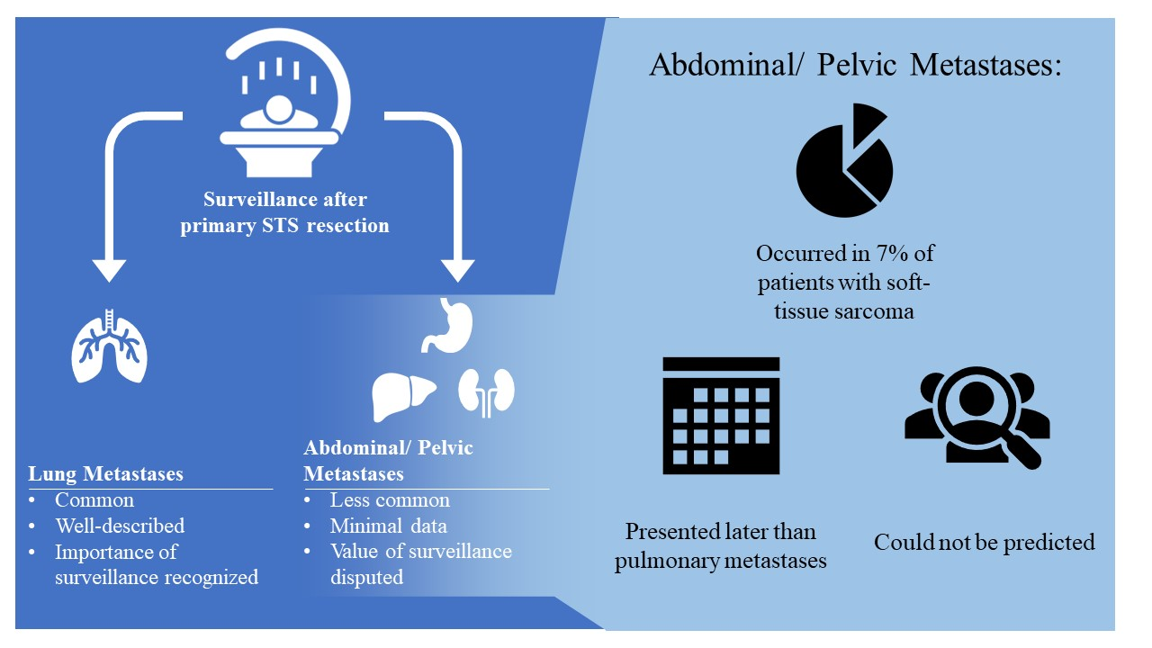
Thirty-three percent (126/382) of patients developed distant metastases. Of these, 72% (90/126) had initial metastatic disease located in the lungs, while 22% (28/126) had extrapulmonary metastases. The remaining 6% (8/126) of patients were found to have simultaneous pulmonary and extrapulmonary metastases. These results show that initial extrapulmonary metastases occur less commonly than initial pulmonary metastases (binomial proportion 0.22 [95% CI 0.15 to 0.30]; p < 0.001).
3.2. Time to Development of Initial Extrapulmonary Metastases
Initial extrapulmonary metastases occurred later than initial pulmonary metastases (log rank
p = 0.049) (
Figure 2). Median time from surgical treatment to development of metastases was 11 months (IQR, 5 to 19) for isolated pulmonary and 22 months (IQR, 6 to 45) for isolated extrapulmonary disease (
p = 0.08) (
Table 3).
3.3. Association of Relevant Patient and Tumor Characteristics with Extrapulmonary Metastases
Our multivariate regression was unable to detect independent prognostic factors that would be useful in identifying patients at an increased risk of developing new extrapulmonary metastases. Patients with high grade tumors were at a greater risk of developing pulmonary metastases compared to those with low grade tumors [OR = 2.916, 95% CI: 1.183–7.191,
p = 0.0201]. In addition, increasing tumor size was found to correlate with an increased incidence of new pulmonary metastases (OR = 4.414, 95% CI: 2.245–8.680,
p < 0.0001 for tumor size <5 cm versus 5–10 cm; OR = 3.530, 95% CI: 1.778–7.008,
p = 0.0003 for tumor size <5 cm versus >10 cm) (
Table 5).
4. Discussion
Surveillance following initial treatment is considered an essential component of sarcoma management. The goal is to detect recurrent or metastatic disease in order to provide patients with prognostic information and treatment. Sarcomas most commonly metastasize to the lungs, so the importance of pulmonary surveillance is universally accepted; however, extrapulmonary surveillance remains controversial. This knowledge gap has led to differences in surveillance recommendations and practices between geographic regions and among individual providers. We sought to build on the existing literature by studying a large cohort of STS patients to better define the incidence and timing of extrapulmonary metastases, as well as any factors that could identify patients more likely to develop extrapulmonary metastases and therefore benefit from abdominopelvic imaging.
4.1. Limitations
There are several limitations to this work. First, while our study found that routine abdominopelvic imaging may lead to earlier diagnosis of metastases, it was not designed to assess potential morbidity and mortality benefits of extrapulmonary imaging. While metastatic STS was historically considered untreatable, strategies for managing systemic disease have emerged over recent decades with novel immunotherapy modalities being particularly exciting [
15]. Research on pulmonary metastasectomy has shown an association between fewer pulmonary nodules, greater resectability, extended disease-free interval, and improved survival [
8,
16]. Studies evaluating locally ablative modalities, such as stereotactic body radiotherapy and cryoablation, have also produced promising results in improving progression-free survival and local control of pulmonary metastases. These modalities may play a future role in treatment for extrapulmonary metastases due to their quicker recovery times and greater accessibility to lesions in the bone, liver, and visceral sites compared to surgical intervention [
17,
18]. However, we are not aware of any data demonstrating clear morbidity and mortality benefits to early treatment of extrapulmonary disease. Other relevant issues, such as the time and cost associated with closer surveillance, are also beyond the scope of this study—as such, we cannot draw definitive conclusions or recommendations about whether abdominopelvic imaging should be included in routine surveillance.
Second, because sarcoma is an uncommon disease and extrapulmonary metastases are even more rare, our sample size was limited. This may have caused our analysis to be underpowered with respect to detecting associations between the clinicopathological variables in our multivariate model and the development of new extrapulmonary metastases.
Our study was also limited by its retrospective design in several respects. Each follow-up visit included physical examination, chest CT, and local MRI, in accordance with current guidelines. The lack of clear recommendations for extrapulmonary surveillance may have led to more variable acquisition of abdominal and pelvic CT. This may have resulted in a sampling bias, with some patients developing extrapulmonary metastases that were not imaged before their pulmonary metastases were diagnosed; in this case, our results would underestimate the frequency of initial extrapulmonary disease. Transfer bias limited our study as well, as 22% of patients received less than 2 years of follow-up and were excluded from the analysis. Older patients were more likely to be lost to follow up, although the difference of median 63 compared to 58 years is not likely to be clinically significant in this context. Patients with widely negative (>0.5 cm) surgical margins were also more likely to be lost to follow up compared to those with positive margins. A potential explanation for this could be that patients with positive surgical margins were aware of their higher risk for recurrence, causing them to be more consistent with follow up. As we did not identify any risk factors for extrapulmonary metastases, we have no indication of how this may have changed our findings. Finally, the evolution of sarcoma treatment over the course of our 20-year study period may have led to co-treatment bias; however, given the complexity of these factors and their intersectionality, we are unable to determine their effect on our results.
4.2. Existing Literature
4.2.1. Proportion of Initial Extrapulmonary Metastases
Our study found that 33% of patients developed distant metastases after resection of their primary STS. Within this group, 22% were initially diagnosed with extrapulmonary metastases, while an additional 6% of patients were found to have simultaneous pulmonary and extrapulmonary metastases. The overall frequency of distant metastases in our patient population is comparable with that reported in the literature, which ranges between 20% and 40% [
2,
4,
5,
7,
9,
19,
20,
21,
22]. The rarity of this condition has limited prior studies, thus contributing to the lack of evidence-based guidelines. The majority of existing studies report 20 or fewer incidences of isolated extrapulmonary metastases, averaging approximately 5% of all STS patients [
2,
4,
5,
6,
7,
22,
23,
24]. Because of these low numbers, authors have generally refrained from providing recommendations regarding extrapulmonary surveillance. Some have concluded that routine imaging of the abdomen and pelvis is difficult to support given the low incidence of initial extrapulmonary metastases and the interplay of other variables such as cost of imaging, as well as complications associated with false positives and related work-up [
7,
24]; others have recommended that all patients be given the option to receive CT of the abdomen and pelvis as part of their regular follow-up.
Studies on STS patients followed with CT of the chest, abdomen, and pelvis have found that extrapulmonary metastases are rare relative to pulmonary metastases, and they tend to occur after pulmonary metastases have been detected. One study reported that 10% (14/140) of patients had abdominal or pelvic metastases; however, only four of these patients first developed isolated extrapulmonary metastases, while the other 10 patients developed extrapulmonary metastases after pulmonary metastases had already been diagnosed [
7]. The other study reported that 16% (20/124) of patients developed extrapulmonary metastases, but only seven patients had initial extrapulmonary metastases. These studies found the incidence of initial extrapulmonary metastases to be 3% and 6%, respectively, compared to the 7% incidence we observed. An additional eight patients in our study were initially diagnosed with pulmonary and extrapulmonary metastases, making it unclear which developed first. Also of note, 12 of the 36 initial extrapulmonary metastases were detected on chest CT, so it would have been identified without abdominopelvic imaging.
Although low compared to the frequency of pulmonary metastases in STS patients, a 7% incidence has warranted surveillance in other malignancies. For example, asynchronous lung metastases occur in 1.7% to 7.2% of patients with colorectal cancer [
25], and surveillance guidelines for this population generally include routine CT of the chest every 6 to 12 months for the first 3 to 5 years after surgery [
26,
27]. In patients with stage IV colorectal cancer, resection of pulmonary metastases is not curative but has been shown to significantly improve 5-year survival (13.8% in all patients with pulmonary metastases, but 32–61% in patients able to undergo pulmonary metastectomy) [
25]. A similar improvement in survival has not been clearly defined for resection of extrapulmonary STS metastases, although new advances in treatment have shown promising preliminary results [
15,
17,
18].
Overall, our findings suggest that abdominal and pelvic imaging in routine surveillance may lead to earlier diagnosis of metastases in a substantial minority of patients. Of note, the incidence of initial extrapulmonary metastases in STS is comparable to that of metastases routinely screened for in other cancers; however, a clear survival benefit of early detection of extrapulmonary metastases in STS has not yet been demonstrated.
4.2.2. Time to Development of Initial Extrapulmonary Metastases
Our study found a longer interval of metastases-free survival between patients who developed initial extrapulmonary compared to pulmonary metastases. The median time from surgical treatment to diagnosis of initial extrapulmonary metastases was twice that of initial pulmonary metastases (22 and 11 months, respectively); moreover, the IQR was over twice as broad for median time to extrapulmonary metastases (6 to 45) compared to pulmonary metastases (5 to 19).
Prior literature on the timing of metastatic diagnosis in STS has focused on pulmonary disease. Most of these metastases are identified within the first two years of STS diagnosis, especially in patients with high grade tumors [
14,
22,
24,
28]; hence, the NCCN and ESMO surveillance guidelines recommend more frequent imaging in the first 2–3 years for patients with high and intermediate grade STS. [
12,
13]. However, these intervals may not accurately reflect the timing for extrapulmonary metastases. Thompson et al. reported an average time to isolated extrapulmonary metastases of 12 months among four patients; this is only half the time we observed, but the discrepancy may be related to the small number in their study and the large range of time to extrapulmonary metastases in ours. Among the 28 patients with isolated extrapulmonary metastases in our study, the median time to diagnosis was 22 months after surgery; moreover, 25% of these initial metastases were not diagnosed until ≥45 months. Thus, while extrapulmonary metastases were less common, they were also less predictable in terms of timing, which limits the development of targeted surveillance strategies.
4.2.3. Factors Associated with Extrapulmonary versus Pulmonary Metastases
Previous studies have found increased depth, higher grade, larger size, and shorter length of the disease-free interval to be associated with distant metastases in general [
5,
9,
29], but not specifically to extrapulmonary metastases. To our knowledge, there are no prior studies which have investigated prognostic factors for the development of extrapulmonary metastases in STS. While our data confirmed that patients with larger, higher grade tumors experienced an increased incidence of new pulmonary metastases, we were unable to identify patient or tumor variables that could be used to identify patients with an increased risk of developing extrapulmonary metastases. Specifically, our multivariate analysis did not detect an association between patient age, gender, BMI, tumor size, depth, grade, location, or histologic subtype with development of initial extrapulmonary metastases. Thus, although higher grade and larger size have been widely linked to increased risk of metastases in STS [
6,
19,
20,
21,
29], we did not observe either of these variables to be associated with initial extrapulmonary metastases.
Prior studies have demonstrated a relatively high incidence of extrapulmonary metastases in patients with myxoid liposarcomas. Estourgie et al. and Muratori et al. found that 55% of metastases in patients with myxoid liposarcomas were extrapulmonary, and both groups of authors emphasized the importance of routine extrapulmonary imaging for surveillance [
30,
31]. Current NCCN guidelines name angiosarcomas, epithelioid sarcomas, leiomyosarcomas, and myxoid liposarcomas as histologic subtypes with increased frequency of extrapulmonary metastases [
13] but do not cite evidence supporting this recommendation. While we did not detect increased risk associated with histologic subtype in our multivariate analysis, 67% (2/3) of angiosarcoma patients, 17% (1/6) of epithelioid sarcoma patients, and 17% (3/18) of myxoid liposarcomas patients developed initial extrapulmonary metastases; by contrast, only 3% (2/66) of patients with metastatic leiomyosarcomas had initial extrapulmonary metastases. While histologic diagnosis did not reach significance in our regression model, the high incidence of initial extrapulmonary metastases among patients with certain sarcoma subtypes warrants further investigation in a larger patient population.
5. Conclusions
Of the 33% of patients with primary STS that developed metastatic disease after resection, 22% were initially diagnosed with extrapulmonary metastases, and an additional 6% with simultaneous pulmonary and extrapulmonary metastases. Initial extrapulmonary metastases occurred later and over a broader time range than pulmonary metastases. While patients with larger, higher grade tumors were more likely to develop pulmonary metastases, we were unable to identify patient or tumor variables that were associated with an increased risk of extrapulmonary metastases. These findings suggest that routine abdominal and pelvic imaging may detect new metastatic disease in a substantial minority of patients. Future studies with larger numbers may identify risk factors for extrapulmonary disease, but at this time we are unable to predict which, if any, patients are more or less likely to benefit from routine abdominopelvic imaging. Further research is needed to evaluate the potential advantages of early detection of extrapulmonary metastases on treatment and survival. Additionally, better understanding the psychosocial impact of increased imaging, the potential morbidity of investigating false positive findings, and the overall cost-effectiveness of advanced imaging will be necessary to develop definitive surveillance recommendations.
Author Contributions
Conceptualization, D.M. and C.A.C.; Data curation, Z.H. and P.E.; Formal analysis, Z.H. and L.R.; Investigation, P.E. and C.A.C.; Methodology, Z.H., P.E., and C.A.C.; Supervision, C.A.C.; Validation, A.H., D.M., and C.A.C.; Visualization, Z.H. and C.A.C.; Writing—original draft, Z.H. and P.E.; Writing—review and editing, Z.H., A.H., D.M., and C.A.C. All authors have read and agreed to the published version of the manuscript.
Funding
This research received no external funding.
Institutional Review Board Statement
The study was conducted according to the guidelines of the Declaration of Helsinki, and approved by the Institutional Review Board of Washington University in St. Louis (protocol code 201105450, 28 September 2020).
Informed Consent Statement
Informed consent was obtained from all subjects involved in the study.
Data Availability Statement
The data presented in this study are available on request from the corresponding author. The data are not publicly available due to inclusion of protected health information.
Conflicts of Interest
The authors declare no conflict of interest.
References
- Abbas, J.S.; Holyoke, E.D.; Moore, R.; Karakousis, C.P. The Surgical Treatment and Outcome of Soft-Tissue Sarcoma. Arch. Surg. 1981, 116, 765–769. [Google Scholar] [CrossRef]
- King, D.M.; Hackbarth, D.A.; Ba, C.M.K.; Carrera, G.F. Soft-Tissue Sarcoma Metastases Identified on Abdomen and Pelvis CT Imaging. Clin. Orthop. Relat. Res. 2009, 467, 2838–2844. [Google Scholar] [CrossRef] [PubMed] [Green Version]
- Brennan, M.F.; Antonescu, C.R.; Moraco, N.; Singer, S. Lessons Learned from the Study of 10,000 Patients with Soft Tissue Sarcoma. Ann. Surg. 2014, 260, 416–422. [Google Scholar] [CrossRef] [Green Version]
- Potter, D.A.; Glenn, J.; Kinsella, T.; Glatstein, E.; Lack, E.E.; Restrepo, C.; White, D.E.; Seipp, C.A.; Wesley, R.; Rosenberg, S.A. Patterns of Recurrence in Patients with High-Grade Soft-Tissue Sarcomas. J. Clin. Oncol. 1985, 3, 353–366. [Google Scholar] [CrossRef] [PubMed]
- Gronchi, A.; Lo Vullo, S.; Colombo, C.; Collini, P.; Stacchiotti, S.; Mariani, L.; Fiore, M.; Giovanni Casali, P. Extremity Soft Tissue Sarcoma in a Series of Patients Treated at a Single Institution: Local Control Directly Impacts Survival. Ann. Surg. 2010, 251, 506–511. [Google Scholar] [CrossRef] [PubMed]
- Smith, H.G.; Memos, N.; Thomas, J.M.; Smith, M.J.F.; Strauss, D.C.; Hayes, A.J. Patterns of Disease Relapse in Primary Extremity Soft-Tissue Sarcoma. Br. J. Surg. 2016, 103, 1487–1496. [Google Scholar] [CrossRef]
- Thompson, M.J.; Ross, J.; Domson, G.; Foster, W. Screening and Surveillance CT Abdomen/Pelvis for Metastases in Patients with Soft-Tissue Sarcoma of the Extremity. Bone Jt. Res. 2015, 4, 45–49. [Google Scholar] [CrossRef] [Green Version]
- Billingsley, K.G.; Burt, M.E.; Jara, E.; Ginsberg, R.J.; Woodruff, J.M.; Leung, D.H.Y.; Brennan, M.F. Pulmonary Metastases From Soft Tissue Sarcoma. Ann. Surg. 1999, 229, 602. [Google Scholar] [CrossRef]
- Whooley, B.P.; Gibbs, J.F.; Mooney, M.M.; Mcgrath, B.E.; Kraybill, W.G. Primary Extremity Sarcoma: What Is the Appropriate Follow-Up? Ann. Surg. Oncol. 2000, 7, 9–14. [Google Scholar] [CrossRef]
- Brenner, D.J.; Hall, E.J. Current Concepts-Computed Tomography—An Increasing Source of Radiation Exposure. N. Engl. J. Med. 2007, 357, 2277–2284. [Google Scholar] [CrossRef] [Green Version]
- Kane, J.M. Surveillance Strategies for Patients Following Surgical Resection of Soft Tissue Sarcomas. Curr. Opin. Oncol. 2004, 16, 328–332. [Google Scholar] [CrossRef]
- Casali, P.G.; Abecassis, N.; Bauer, S.; Biagini, R.; Bielack, S.; Bonvalot, S.; Boukovinas, I.; Bovee, J.V.M.G.; Brodowicz, T.; Broto, J.M.; et al. Soft Tissue and Visceral Sarcomas: ESMO-EURACAN Clinical Practice Guidelines for Diagnosis, Treatment and Follow-Up. Ann. Oncol. 2018, 29, iv51–iv67. [Google Scholar] [CrossRef]
- Von Mehren, M.; Randall, R.L.; Benjamin, R.S.; Boles, S.; Bui, M.M.; Ganjoo, K.N.; George, S.; Gonzalez, R.J.; Heslin, M.J.; Kane, J.M.; et al. Soft Tissue Sarcoma, Version 2.2018: Clinical Practice Guidelines in Oncology. J. Natl. Compr. Canc. Netw. 2018, 16, 536–563. [Google Scholar] [CrossRef]
- Cipriano, C.A.; Jang, E.; Tyler, W. Sarcoma Surveillance: A Review of Current Evidence and Guidelines. J. Am. Acad. Orthop. Surg. 2020, 28, 145–156. [Google Scholar] [CrossRef]
- Van Tine, B.A.; Butler, M.O.; Araujo, D.; Johnson, M.L.; Clarke, J.; Liebner, D.; Odunsi, K.; Olszanski, A.J.; Basu, S.; Brophy, F.; et al. ADP-A2M4 (MAGE-A4) in Patients with Synovial Sarcoma. Ann. Oncol. 2019, 30, v684–v685. [Google Scholar] [CrossRef]
- Garcı, C.E.; San-julia, M.; Minda, J.P.; Buxalleu, W.T. Long-Term Results after Resection for Soft Tissue Sarcoma Pulmonary Metastases. Interact. Cardiovasc. Thorac. Surg. 2009, 9, 223–226. [Google Scholar] [CrossRef] [Green Version]
- Baumann, B.C.; Bernstein, K.D.A.; Delaney, T.F.; Charles, F.; Ii, B.S.; Kolker, J.D.; Choy, E.; Levin, W.P.; Weber, K.L.; Muniappan, A.; et al. Multi-Institutional Analysis of Stereotactic Body Radiotherapy for Sarcoma Pulmonary Metastases: High Rates of Local Control with Favorable Toxicity. J. Surg. Oncol. 2020, 122, 877–883, No. May. [Google Scholar] [CrossRef] [PubMed]
- Hirbe, A.C.; Jennings, J.; Saad, N.; Giardina, J.D.; Tao, Y.; Luo, J.; Berry, S.; Toeniskoetter, J.; Van Tine, B.A. A Phase II Study of Tumor Ablation in Patients with Metastatic Sarcoma Stable on Chemotherapy. Oncologist 2018, 23, 760. [Google Scholar] [CrossRef] [PubMed] [Green Version]
- Sabolch, A.; Feng, M.; Griffith, K.; Rzasa, C.; Gadzala, L.; Feng, F.; Biermann, S.; Chugh, R.; Ray, M.; Ben-josef, E. Risk Factors for Local Recurrence and Metastasis in Soft Tissue Sarcomas of the Extremity. Am. J. Clin. Oncol. 2012, 35, 151–157. [Google Scholar] [CrossRef]
- Ballo, M.T.; Pisters, P.W.T.; Pollock, R.E.; Ph, D.; Patel, S.R.; Benjamin, R.S.; Evans, H.L. Prognostic Factors for Patients with Localized Soft- Tissue Sarcoma Treated with Conservation Surgery An Analysis of 1225 Patients. Cancer 2003, 97, 2530–2543. [Google Scholar] [CrossRef]
- Terrier, P.; Guillou, L.; Le Doussal, V.; Vilain, M. Predictive Value of Grade for Metastasis Development in the Main Histologic Types of Adult Soft Tissue Sarcomas A Study of 1240 Patients from the French Federation of Cancer Centers Sarcoma Group. Cancer 2001, 91, 1914–1926. [Google Scholar]
- De Angelis, F.; Guy, F.; Varbedian, O.; Hervieu, A.; Truc, G.; Thibouw, D.; Fraisse, J.; Burnier, P.; Isambert, N.; Causeret, S. Limbs and Trunk Soft Tissue Sarcoma Systematic Local and Remote Monitoring by MRI and Thoraco-Abdomino-Pelvic Scanner: A Single-Centre Retrospective Study. Eur. J. Surg. Oncol. 2019, 45, 1274–1280. [Google Scholar] [CrossRef]
- Alldinger, I.; Yang, Q.; Pilarsky, C.; Saeger, H.D.; Knoefel, W.T.; Peiper, M. Retroperitoneal Soft Tissue Sarcomas: Prognosis and Treatment of Primary and Recurrent Disease in 117 Patients. Anticancer Res. 2006, 26, 1577–1581. [Google Scholar] [PubMed]
- Patel, S.A.; Royce, T.J.; Barysauskas, C.M.; Thornton, K.A.; Raut, C.P.; Baldini, E.H. Surveillance Imaging Patterns and Outcomes Following Radiation Therapy and Radical Resection for Localized Extremity and Trunk Soft Tissue Sarcoma. Ann. Surg. Oncol. 2017, 24, 1588–1595. [Google Scholar] [CrossRef]
- Stewart, C.L.; Warner, S.; Ito, K.; Raoof, M.; Wu, G.X.; Kessler, J.; Kim, J.Y.; Fong, Y. Cytoreduction for Colorectal Metastases: Liver, Lung, Peritoneum, Lymph Nodes, Bone, Brain. When Does It Palliate, Prolong Survival, and Potentially Cure? Curr. Probl. Surg. 2018, 55, 330–379. [Google Scholar] [CrossRef] [PubMed]
- Argilés, G.; Tabernero, J.; Labianca, R.; Hochhauser, D.; Salazar, R.; Iveson, T.; Quirke, P.; Yoshino, T.; Taieb, J.; Martinelli, E.; et al. Localised Colon Cancer: ESMO Clinical Practice Guidelines for Diagnosis, Treatment and Follow-Up. Ann. Oncol. 2020, 31, 1291–1305. [Google Scholar] [CrossRef]
- Iii, A.B.B.; Venook, A.P.; Al-hawary, M.M.; Cederquist, L.; Chen, Y.; Ciombor, K.K.; Cohen, S.; Cooper, H.S.; Deming, D.; Engstrom, P.F.; et al. NCCN Guidelines Insights: Colon Cancer, Version 2.2018. J. Natl. Compr. Cancer Netw. 2018, 16, 359–369. [Google Scholar] [CrossRef] [Green Version]
- Sawamura, C.; Matsumoto, S.; Shimoji, T.; Okawa, A.; Ae, K. How Long Should We Follow Patients with Soft Tissue Sarcomas? Clin. Orthop. Relat. Res. 2014, 472, 842–848. [Google Scholar] [CrossRef] [PubMed] [Green Version]
- Stojadinovic, B.A.; Leung, D.H.Y.; Allen, P.; Lewis, J.J.; Jaques, D.P.; Brennan, M.F. Primary Adult Soft Tissue Sarcoma: Time-Dependent Influence of Prognostic Variables. J. Clin. Oncol. 2002, 20, 4344–4352. [Google Scholar] [CrossRef]
- Estourgie, S.H.; Nielsen, G.P.; Ott, M.J. Metastatic Patterns of Extremity Myxoid Liposarcoma and Their Outcome. J. Surg. Oncol. 2002, 80, 89–93. [Google Scholar] [CrossRef]
- Muratori, F.; Bettini, L.; Frenos, F.; Mondanelli, N.; Greto, D.; Livi, L.; Franchi, A.; Roselli, G.; Scorianz, M.; Capanna, R.; et al. Myxoid Liposarcoma: Prognostic Factors and Metastatic Pattern in a Series of 148 Patients Treated at a Single Institution. Int. J. Surg. Oncol. 2018, 2018, 1–9. [Google Scholar] [CrossRef] [PubMed] [Green Version]
| Publisher’s Note: MDPI stays neutral with regard to jurisdictional claims in published maps and institutional affiliations. |
© 2021 by the authors. Licensee MDPI, Basel, Switzerland. This article is an open access article distributed under the terms and conditions of the Creative Commons Attribution (CC BY) license (https://creativecommons.org/licenses/by/4.0/).


 ,
, 


