
State-of-the-Art Challenges and Perspectives in Multi-Organ Cancer Diagnosis via Deep Learning-Based Methods

 , , and
Abstract
:Simple Summary
Abstract
1. Introduction
- 1.
- What is the commonly employed imaging modality in each of the four cancers?
- 2.
- What kind of databases is utilized for medical image analysis?
- 3.
- Which kind of AI technology is in trend for the early diagnosis of these cancers?
- 4.
- Why is CNN architecture is a trend in breast, brain, lung, and skin cancer diagnosis?
- 5.
- What performance evaluation metrics are employed to evaluate the models’ efficiency?
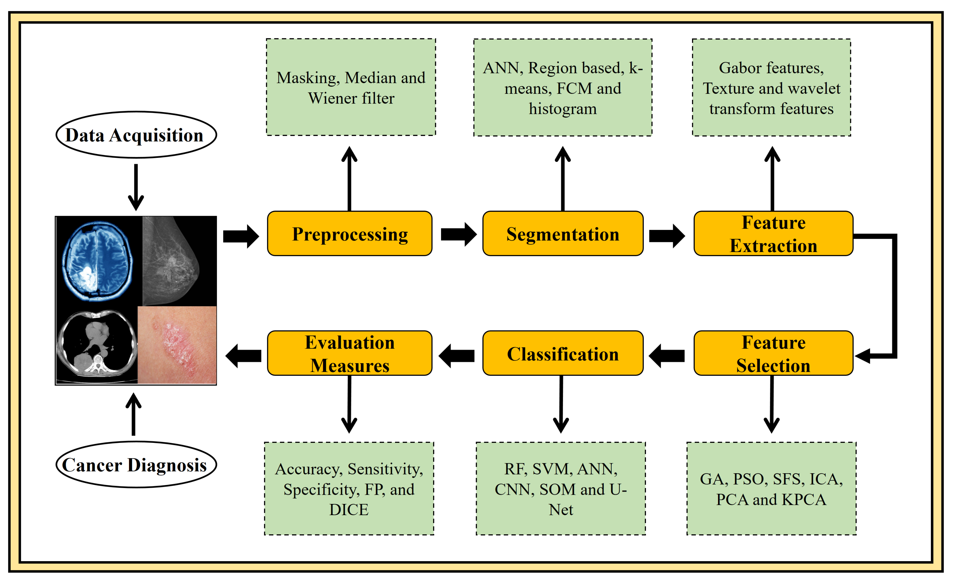
2. Material and Methods
2.1. Search Strategy and Selection Criteria
2.2. Most Popular Publicly Available Datasets
2.2.1. Multimodal Brain Tumor Image Segmentation Benchmark(BraTS) Database
2.2.2. Lung Image Database Consortium image collection(LIDC/IDRI) Database
2.2.3. Digital Database for Screening Mammography(DDSM) Database
2.2.4. Wisconsin Breast Cancer Database(WBCD) Database
2.2.5. International Skin Imaging Collaboration(ISIC) Database
2.2.6. PH2 Database
2.3. Performance Evaluation Metrics
3. Brain Tumor
4. Breast Cancer
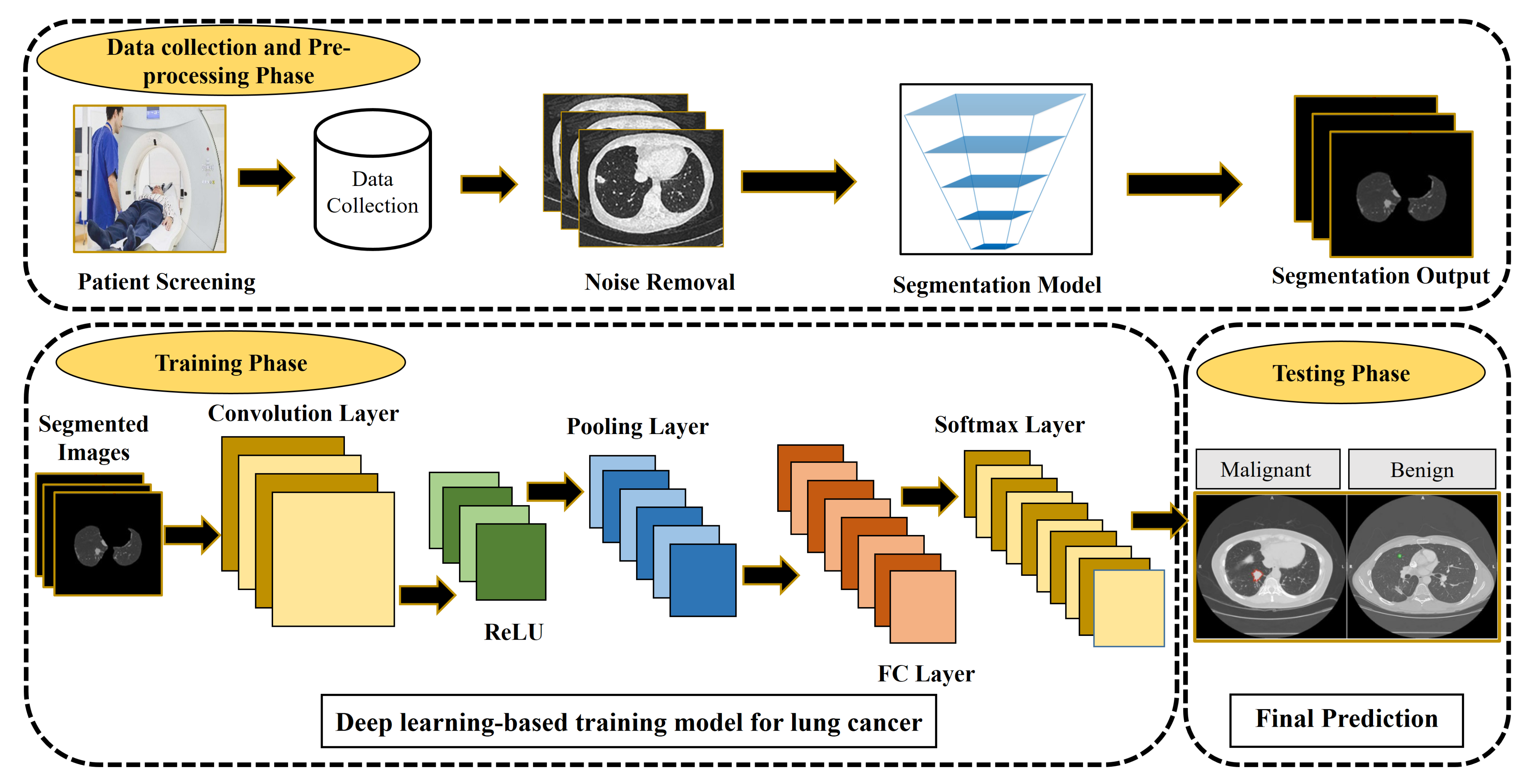
5. Lung Cancer
6. Skin Cancer
7. Discussion
7.1. Primary Observations
7.2. Open Research Challenges, Possible Solutions and Future Prospects
- The most generally implemented technique to extend the range of the training dataset is named data augmentation. It is the application in which different offline changes are done, including affine transformation, cropping, flip, rotation, padding, saturation to the examples [146], and colour augmentation [147];
- Transfer learning from the popular networks [86] employed in the same field or even another area is considered another solution to surpass limited data. It has been established that transfer learning by pre-trained networks produced superior results even when the source and target networks are not the same, transferring the weights of different tasks [148];
- The morphological variation in the cancerous cell is one of the significant issues in medical cancer image detection. The cancerous organ/lesion may differ significantly in dimension, outline, and position from patient to patient [149]. Using deeper architectures can be an effective solution to this issue, as reported in [115]. The unclear border with an imperfect contrast among targeting organs and the nearby tissues in tumor images is an inherent challenge typically produced via attenuation coefficient [150,151]. The use of multi-modality-based methods can solve this issue [152,153];
- The computational complexity of the network is another challenge in DL-based techniques, owing to variability in image dimensions, network construction, or the heavily over-parameterized networks. To evade the powerful GPU hardware constraint and accelerate the segmentation task, one can decrease the number of hidden layers or parameters of the proposed network and emphasize algorithms that artificially generate training data for example GAN [154,155] rather than altering the network;
- The appearance of mostly AI-based architectures seems like a black box. Thus, researchers have no idea about the internal representations of the network and the perfect approach to realize the system completely. Hence, DL approaches are greatly affected by the inherent snags of medical images, that is, noise and illumination. Complete knowledge and understanding of such black box issues in the future would be a revolution in the DL field [156];
- During training time, the ground truth outlines are manually delineated by expert physicians. If manual delineation would be done by a different individual or even the same one at the distinct circumstance, there must be a possibility that the proposed model can be biased and favor expert ground truths as a system error. However, this drawback could be expected to occur in all supervised learning CAD techniques;
- The amalgamation of the robust individual approaches by utilizing their benefits is suitable in further improving the CAD performance. Develop novel CAD systems using hybrid ML-based approaches like SegNet [74], U-Net-Vnet-Fast-R-CNN [157], AgileNet [158] to overcome the complication of overfitting that happens in the training time; this could help in the early diagnosis of multi-organ cancers;
- It is observed that the DL-based unsupervised clustering techniques include; deep auto-encoders, regularized information maximization (RIM), Deep InfoMax (DIM), deep adaptive clustering (DAC), and so forth, have not been engaged widely in comparison with supervised learning techniques [159]. It could avoid the costly training process. These techniques could also be employed to improve the performance of CAD systems in the medical imaging domain.
8. Conclusions
Author Contributions
Funding
Acknowledgments
Conflicts of Interest
References
- Zhang, N.; Cai, Y.X.; Wang, Y.Y.; Tian, Y.T.; Wang, X.L.; Badami, B. Skin cancer diagnosis based on optimized convolutional neural network. Artif. Intell. Med. 2020, 102, 1–7. [Google Scholar] [CrossRef] [PubMed]
- Mambou, S.J.; Maresova, P.; Krejcar, O.; Selamat, A.; Kuca, K. Breast cancer detection using infrared thermal imaging and a deep learning model. Sensors 2018, 18, 2799. [Google Scholar] [CrossRef] [PubMed] [Green Version]
- Priya, S.S.; Ramamurthy, B. Lung cancer detection using image processing techniques. Res. J. Pharm. Technol. 2018, 11, 2045–2049. [Google Scholar] [CrossRef]
- Mohsen, H.; El-Dahshan, E.S.A.; El-Horbaty, E.S.M.; Salem, A.B.M. Classification using deep learning neural networks for brain tumors. Future Comput. Informatics J. 2018, 3, 68–71. [Google Scholar] [CrossRef]
- Kapoor, L.; Thakur, S. A survey on brain tumor detection using image processing techniques. In Proceedings of the 2017 7th International Conference on Cloud Computing, Data Science & Engineering-Confluence, Noida, India, 12–13 January 2017; pp. 582–585. [Google Scholar]
- Latha, T.S. Recognition of Blood Cancer Using Different Classification Techniques. Int. Trans. Electr. Eng. Comput. Sci. 2020, 1, 33–41. [Google Scholar]
- Fahad, H.; Ghani Khan, M.U.; Saba, T.; Rehman, A.; Iqbal, S. Microscopic abnormality classification of cardiac murmurs using ANFIS and HMM. Microsc. Res. Tech. 2018, 81, 449–457. [Google Scholar] [CrossRef]
- Sung, H.; Ferlay, J.; Siegel, R.L.; Laversanne, M.; Soerjomataram, I.; Jemal, A.; Bray, F. Global cancer statistics 2020: GLOBOCAN estimates of incidence and mortality worldwide for 36 cancers in 185 countries. CA Cancer J. Clin. 2021, 71, 209–249. [Google Scholar] [CrossRef]
- Sulzer, D.; Cassidy, C.; Horga, G.; Kang, U.J.; Fahn, S.; Casella, L.; Pezzoli, G.; Langley, J.; Hu, X.P.; Zucca, F.A.; et al. Neuromelanin detection by magnetic resonance imaging (MRI) and its promise as a biomarker for Parkinsons disease. NPJ Park. Dis. 2018, 4, 1–13. [Google Scholar]
- Lameka, K.; Farwell, M.D.; Ichise, M. Positron emission tomography. Handb. Clin. Neurol. 2016, 135, 209–227. [Google Scholar]
- Christe, A.; Peters, A.A.; Drakopoulos, D.; Heverhagen, J.T.; Geiser, T.; Stathopoulou, T.; Christodoulidis, S.; Anthimopoulos, M.; Mougiakakou, S.G.; Ebner, L. Computer-aided diagnosis of pulmonary fibrosis using deep learning and CT images. Investig. Radiol. 2019, 54, 627–632. [Google Scholar] [CrossRef] [Green Version]
- Katzen, J.; Dodelzon, K. A review of computer aided detection in mammography. Clin. Imaging 2018, 52, 305–309. [Google Scholar] [CrossRef]
- Brown, T.J.; Bota, D.A.; van Den Bent, M.J.; Brown, P.D.; Maher, E.; Aregawi, D.; Liau, L.M.; Buckner, J.C.; Weller, M.; Berger, M.S.; et al. Management of low-grade glioma: A systematic review and meta-analysis. Neuro-Oncol. Pract. 2019, 6, 249–258. [Google Scholar] [CrossRef] [Green Version]
- Cho, H.h.; Park, H. Classification of low-grade and high-grade glioma using multi-modal image radiomics features. In Proceedings of the 2017 39th Annual International Conference of the IEEE Engineering in Medicine and Biology Society (EMBC), Jeju Island, Korea, 11–15 July 2017; pp. 3081–3084. [Google Scholar]
- Ali, S.; Xia, Q.; Muhammad, T.; Liu, L.; Meng, X.; Bars-Cortina, D.; Khan, A.A.; Huang, Y.; Dong, L. Glioblastoma Therapy: Rationale for a Mesenchymal Stem Cell-based Vehicle to Carry Recombinant Viruses. Stem Cell Rev. Rep. 2021, in press. [Google Scholar] [CrossRef]
- Anaraki, A.K.; Ayati, M.; Kazemi, F. Magnetic resonance imaging-based brain tumor grades classification and grading via convolutional neural networks and genetic algorithms. Biocybern. Biomed. Eng. 2019, 39, 63–74. [Google Scholar] [CrossRef]
- Collins, O. Last Year’s Virus, This Year’s Cancer Treatment. BU Well 2018, 3, 1–4. [Google Scholar]
- Spanhol, F.A.; Oliveira, L.S.; Petitjean, C.; Heutte, L. Breast cancer histopathological image classification using convolutional neural networks. In Proceedings of the 2016 International Joint Conference on Neural Networks (IJCNN), Vancouver, BC, Canada, 24–29 July 2016; pp. 2560–2567. [Google Scholar]
- Li, J.; Pei, Y.; Yasin, A.; Ali, S.; Mahmood, T. Computer Vision-Based Microcalcification Detection in Digital Mammograms Using Fully Connected Depthwise Separable Convolutional Neural Network. Sensors 2021, 21, 4854. [Google Scholar]
- Husham, A.; Hazim Alkawaz, M.; Saba, T.; Rehman, A.; Saleh Alghamdi, J. Automated nuclei segmentation of malignant using level sets. Microsc. Res. Tech. 2016, 79, 993–997. [Google Scholar] [CrossRef]
- Shakeel, P.M.; Burhanuddin, M.; Desa, M.I. Automatic lung cancer detection from CT image using improved deep neural network and ensemble classifier. Neural Comput. Appl. 2020, in press. [Google Scholar] [CrossRef]
- Leiter, U.; Keim, U.; Garbe, C. Epidemiology of skin cancer: Update 2019. In Sunlight, Vitamin D and Skin Cancer; Springer: Berlin/Heidelberg, Germany, 2020; pp. 123–139. [Google Scholar]
- Pacheco, A.G.; Krohling, R.A. Recent advances in deep learning applied to skin cancer detection. arXiv 2019, arXiv:1912.03280. [Google Scholar]
- Ashraf, R.; Afzal, S.; Rehman, A.U.; Gul, S.; Baber, J.; Bakhtyar, M.; Mehmood, I.; Song, O.Y.; Maqsood, M. Region-of-Interest Based Transfer Learning Assisted Framework for Skin Cancer Detection. IEEE Access 2020, 8, 147858–147871. [Google Scholar] [CrossRef]
- Menze, B.H.; Jakab, A.; Bauer, S.; Kalpathy-Cramer, J.; Farahani, K.; Kirby, J.; Burren, Y.; Porz, N.; Slotboom, J.; Wiest, R.; et al. The multimodal brain tumor image segmentation benchmark (BRATS). IEEE Trans. Med. Imaging 2014, 34, 1993–2024. [Google Scholar] [CrossRef] [PubMed]
- Armato, S.G., III; McLennan, G.; Bidaut, L.; McNitt-Gray, M.F.; Meyer, C.R.; Reeves, A.P.; Zhao, B.; Aberle, D.R.; Henschke, C.I.; Hoffman, E.A.; et al. The lung image database consortium (LIDC) and image database resource initiative (IDRI): A completed reference database of lung nodules on CT scans. Med. Phys. 2011, 38, 915–931. [Google Scholar] [CrossRef]
- Heath, M.; Bowyer, K.; Kopans, D.; Kegelmeyer, P.; Moore, R.; Chang, K.; Munishkumaran, S. Current status of the digital database for screening mammography. In Digital mammography; Springer: Berlin/Heidelberg, Germany, 1998; pp. 457–460. [Google Scholar]
- Showrov, M.I.H.; Islam, M.T.; Hossain, M.D.; Ahmed, M.S. Performance Comparison of Three Classifiers for the Classification of Breast Cancer Dataset. In Proceedings of the 2019 4th International Conference on Electrical Information and Communication Technology (EICT), Khulna, Bangladesh, 20–22 December 2019; pp. 1–5. [Google Scholar]
- Milton, M.A.A. Automated skin lesion classification using ensemble of deep neural networks in ISIC 2018: Skin lesion analysis towards melanoma detection challenge. arXiv 2019, arXiv:1901.10802. [Google Scholar]
- Mendonça, T.; Ferreira, P.M.; Marques, J.S.; Marcal, A.R.; Rozeira, J. PH 2-A dermoscopic image database for research and benchmarking. In Proceedings of the 2013 35th Annual International Conference of the IEEE Engineering in Medicine and Biology Society (EMBC), Osaka, Japan, 3–7 July 2013; pp. 5437–5440. [Google Scholar]
- Louis, D.N.; Perry, A.; Reifenberger, G.; Von Deimling, A.; Figarella-Branger, D.; Cavenee, W.K.; Ohgaki, H.; Wiestler, O.D.; Kleihues, P.; Ellison, D.W. The 2016 World Health Organization classification of tumors of the central nervous system: A summary. Acta Neuropathol. 2016, 131, 803–820. [Google Scholar] [CrossRef] [Green Version]
- Tandel, G.S.; Biswas, M.; Kakde, O.G.; Tiwari, A.; Suri, H.S.; Turk, M.; Laird, J.R.; Asare, C.K.; Ankrah, A.A.; Khanna, N.; et al. A review on a deep learning perspective in brain cancer classification. Cancers 2019, 11, 111. [Google Scholar] [CrossRef] [Green Version]
- Ganau, L.; Paris, M.; Ligarotti, G.; Ganau, M. Management of gliomas: Overview of the latest technological advancements and related behavioral drawbacks. Behav. Neurol. 2015, 2015, 862634. [Google Scholar] [CrossRef]
- Jayadevappa, D.; Srinivas Kumar, S.; Murty, D. Medical image segmentation algorithms using deformable models: A review. IETE Tech. Rev. 2011, 28, 248–255. [Google Scholar] [CrossRef]
- Yazdani, S.; Yusof, R.; Karimian, A.; Pashna, M.; Hematian, A. Image segmentation methods and applications in MRI brain images. IETE Tech. Rev. 2015, 32, 413–427. [Google Scholar] [CrossRef]
- Bahadure, N.B.; Ray, A.K.; Thethi, H.P. Image analysis for MRI based brain tumor detection and feature extraction using biologically inspired BWT and SVM. Int. J. Biomed. Imaging 2017, 2017, 9749108. [Google Scholar] [CrossRef] [Green Version]
- Alfonse, M.; Salem, A.B.M. An automatic classification of brain tumors through MRI using support vector machine. Egy. Comp. Sci. J 2016, 40, 11–22. [Google Scholar]
- Wu, W.; Chen, A.Y.; Zhao, L.; Corso, J.J. Brain tumor detection and segmentation in a CRF (conditional random fields) framework with pixel-pairwise affinity and superpixel-level features. Int. J. Comput. Assist. Radiol. Surg. 2014, 9, 241–253. [Google Scholar] [CrossRef]
- Soltaninejad, M.; Ye, X.; Yang, G.; Allinson, N.; Lambrou, T. Brain tumor grading in different MRI protocols using SVM on statistical features. In Proceedings of the MIUA 2014 18th Annual Conference, Medical Image Understanding and Analysis, Egham, UK, 9–11 July 2014; pp. 259–264. [Google Scholar]
- Soltaninejad, M.; Ye, X.; Yang, G.; Allinson, N.; Lambrou, T. An image analysis approach to MRI brain tumor grading. Oncol. News 2015, 9, 204–207. [Google Scholar]
- Ren, T.; Wang, H.; Feng, H.; Xu, C.; Liu, G.; Ding, P. Study on the improved fuzzy clustering algorithm and its application in brain image segmentation. Appl. Soft Comput. 2019, 81, 1–9. [Google Scholar] [CrossRef]
- Sulaiman, S.N.; Non, N.A.; Isa, I.S.; Hamzah, N. Segmentation of brain MRI image based on clustering algorithm. In Proceedings of the 2014 IEEE Symposium on Industrial Electronics & Applications (ISIEA), Hangzhou, China, 9–11 June 2014; pp. 60–65. [Google Scholar]
- Li, J.; Yu, Z.L.; Gu, Z.; Liu, H.; Li, Y. MMAN: Multi-modality aggregation network for brain segmentation from MR images. Neurocomputing 2019, 358, 10–19. [Google Scholar] [CrossRef]
- Singh, A. Detection of brain tumor in MRI images, using combination of fuzzy c-means and SVM. In Proceedings of the 2015 2nd International Conference on Signal Processing and Integrated Networks (SPIN), New Delhi, India, 19–20 February 2015; pp. 98–102. [Google Scholar]
- Sujan, M.; Alam, N.; Noman, S.A.; Islam, M.J. A segmentation based automated system for brain tumor detection. Int. J. Comput. Appl. 2016, 153, 41–49. [Google Scholar] [CrossRef]
- Vishnuvarthanan, A.; Rajasekaran, M.P.; Govindaraj, V.; Zhang, Y.; Thiyagarajan, A. An automated hybrid approach using clustering and nature inspired optimization technique for improved tumor and tissue segmentation in magnetic resonance brain images. Applied Soft Computing 2017, 57, 399–426. [Google Scholar] [CrossRef]
- Gajhede, N.; Beck, O.; Purwins, H. Convolutional neural networks with batch normalization for classifying hi-hat, snare, and bass percussion sound samples. In Proceedings of the Audio Mostly (ACM), Norrköping, Sweden, 4–6 October 2016; pp. 111–115. [Google Scholar]
- Kaur, T.; Gandhi, T.K. Automated brain image classification based on VGG-16 and transfer learning. In Proceedings of the 2019 International Conference on Information Technology (ICIT), Bhubaneswar, India, 19–21 December 2019; pp. 94–98. [Google Scholar]
- Deepak, S.; Ameer, P. Brain tumor classification using deep CNN features via transfer learning. Comput. Biol. Med. 2019, 111, 103345. [Google Scholar] [CrossRef]
- Mittal, M.; Goyal, L.M.; Kaur, S.; Kaur, I.; Verma, A.; Hemanth, D.J. Deep learning based enhanced tumor segmentation approach for MR brain images. Appl. Soft Comput. 2019, 78, 346–354. [Google Scholar] [CrossRef]
- Amin, J.; Sharif, M.; Yasmin, M.; Saba, T.; Anjum, M.A.; Fernandes, S.L. A new approach for brain tumor segmentation and classification based on score level fusion using transfer learning. J. Med. Syst. 2019, 43, 1–16. [Google Scholar] [CrossRef]
- Rehman, M.U.; Cho, S.; Kim, J.; Chong, K.T. BrainSeg-Net: Brain Tumor MR Image Segmentation via Enhanced Encoder–Decoder Network. Diagnostics 2021, 11, 169. [Google Scholar] [CrossRef]
- Chandra, G.R.; Rao, K.R.H. Tumor detection in brain using genetic algorithm. Procedia Comput. Sci. 2016, 79, 449–457. [Google Scholar] [CrossRef] [Green Version]
- Arunachalam, M.; Royappan Savarimuthu, S. An efficient and automatic glioblastoma brain tumor detection using shift-invariant shearlet transform and neural networks. Int. J. Imaging Syst. Technol. 2017, 27, 216–226. [Google Scholar] [CrossRef]
- Pereira, S.; Pinto, A.; Alves, V.; Silva, C.A. Brain tumor segmentation using convolutional neural networks in MRI images. IEEE Trans. Med. Imaging 2016, 35, 1240–1251. [Google Scholar] [CrossRef] [PubMed]
- Perkuhn, M.; Stavrinou, P.; Thiele, F.; Shakirin, G.; Mohan, M.; Garmpis, D.; Kabbasch, C.; Borggrefe, J. Clinical evaluation of a multiparametric deep learning model for glioblastoma segmentation using heterogeneous magnetic resonance imaging data from clinical routine. Investig. Radiol. 2018, 53, 647–654. [Google Scholar] [CrossRef]
- Zhao, X.; Wu, Y.; Song, G.; Li, Z.; Zhang, Y.; Fan, Y. A deep learning model integrating FCNNs and CRFs for brain tumor segmentation. Med. Image Anal. 2018, 43, 98–111. [Google Scholar] [CrossRef]
- Vishnuvarthanan, G.; Rajasekaran, M.P.; Subbaraj, P.; Vishnuvarthanan, A. An unsupervised learning method with a clustering approach for tumor identification and tissue segmentation in magnetic resonance brain images. Appl. Soft Comput. 2016, 38, 190–212. [Google Scholar] [CrossRef]
- Amin, J.; Sharif, M.; Yasmin, M.; Saba, T.; Raza, M. Use of machine intelligence to conduct analysis of human brain data for detection of abnormalities in its cognitive functions. Multimed. Tools Appl. 2020, 79, 10955–10973. [Google Scholar] [CrossRef]
- Zhao, J.; Meng, Z.; Wei, L.; Sun, C.; Zou, Q.; Su, R. Supervised brain tumor segmentation based on gradient and context-sensitive features. Front. Neurosci. 2019, 13, 1–11. [Google Scholar] [CrossRef]
- Khan, M.A.; Lali, I.U.; Rehman, A.; Ishaq, M.; Sharif, M.; Saba, T.; Zahoor, S.; Akram, T. Brain tumor detection and classification: A framework of marker-based watershed algorithm and multilevel priority features selection. Microsc. Res. Tech. 2019, 82, 909–922. [Google Scholar] [CrossRef]
- Peng, S.; Chen, W.; Sun, J.; Liu, B. Multi-Scale 3D U-Nets: An approach to automatic segmentation of brain tumor. Int. J. Imaging Syst. Technol. 2020, 30, 5–17. [Google Scholar] [CrossRef]
- Li, Q.; Yu, Z.; Wang, Y.; Zheng, H. TumorGAN: A multi-modal data augmentation framework for brain tumor segmentation. Sensors 2020, 20, 4203. [Google Scholar] [CrossRef]
- Alqazzaz, S.; Sun, X.; Yang, X.; Nokes, L. Automated brain tumor segmentation on multi-modal MR image using SegNet. Comput. Vis. Media 2019, 5, 209–219. [Google Scholar] [CrossRef] [Green Version]
- Bodapati, J.D.; Shaik, N.S.; Naralasetti, V.; Mundukur, N.B. Joint training of two-channel deep neural network for brain tumor classification. Signal Image Video Process. 2021, 15, 753–760. [Google Scholar] [CrossRef]
- Abd El Kader, I.; Xu, G.; Shuai, Z.; Saminu, S.; Javaid, I.; Salim Ahmad, I. Differential deep convolutional neural network model for brain tumor classification. Brain Sci. 2021, 11, 352. [Google Scholar] [CrossRef]
- Shivaprasad, B. Bidirectional ConvLSTMXNet for Brain Tumor Segmentation of MR Images. Teh. Glas. 2021, 15, 37–42. [Google Scholar]
- Raja, N.; Rajinikanth, V.; Fernandes, S.L.; Satapathy, S.C. Segmentation of breast thermal images using Kapur’s entropy and hidden Markov random field. J. Med. Imaging Health Inf. 2017, 7, 1825–1829. [Google Scholar] [CrossRef]
- Mahmood, T.; Li, J.; Pei, Y.; Akhtar, F.; Imran, A.; Rehman, K.U. A Brief Survey on Breast Cancer Diagnostic With Deep Learning Schemes Using Multi-Image Modalities. IEEE Access 2020, 8, 165779–165809. [Google Scholar] [CrossRef]
- Zerouaoui, H.; Idri, A. Reviewing Machine Learning and Image Processing Based Decision-Making Systems for Breast Cancer Imaging. J. Med. Syst. 2021, 45, 1–20. [Google Scholar] [CrossRef]
- Nie, K.; Baltzer, P.; Preim, B.; Mistelbauer, G. Knowledge-Assisted Comparative Assessment of Breast Cancer using Dynamic Contrast-Enhanced Magnetic Resonance Imaging. Comput. Graph. Forum. 2020, 39, 13–23. [Google Scholar] [CrossRef]
- Makandar, A.; Halalli, B. Threshold based segmentation technique for mass detection in mammography. J. Comput. 2016, 11, 472–478. [Google Scholar] [CrossRef]
- Li, S.; Dong, M.; Du, G.; Mu, X. Attention dense-u-net for automatic breast mass segmentation in digital mammogram. IEEE Access 2019, 7, 59037–59047. [Google Scholar] [CrossRef]
- El Adoui, M.; Mahmoudi, S.A.; Larhmam, M.A.; Benjelloun, M. MRI breast tumor segmentation using different encoder and decoder CNN architectures. Computers 2019, 8, 52. [Google Scholar] [CrossRef] [Green Version]
- Sridhar, B. A quality representation of tumor in breast using hybrid model watershed transform and Markov random fields. In Proceedings of the 2020 International Conference on Computer Communication and Informatics (ICCCI), Coimbatore, India, 22–24 January 2020; pp. 1–5. [Google Scholar]
- Benzebouchi, N.E.; Azizi, N.; Ayadi, K. A computer-aided diagnosis system for breast cancer using deep convolutional neural networks. In Computational Intelligence in Data Mining; Springer: Berlin/Heidelberg, Germany, 2019; pp. 583–593. [Google Scholar]
- Wang, S.; Hu, J.; Zhang, Y.; Shen, J.; Dong, F.; Zhang, X.; Lu, C.; Shang, D. Presentation and survival by hormonal receptor status in metaplastic breast cancer: A propensity score-matched analysis. Breast 2021, 60, 168–176. [Google Scholar] [CrossRef]
- Duarte, M.A.; Pereira, W.C.; Alvarenga, A.V. Calculating texture features from mammograms and evaluating their performance in classifying clusters of microcalcifications. In Proceedings of the Mediterranean Conference on Medical and Biological Engineering and Computing, Seville, Spain, 25–28 September 2019; pp. 322–332. [Google Scholar]
- Byra, M.; Jarosik, P.; Szubert, A.; Galperin, M.; Ojeda-Fournier, H.; Olson, L.; O Boyle, M.; Comstock, C.; Andre, M. Breast mass segmentation in ultrasound with selective kernel U-Net convolutional neural network. Biomed. Signal Process. Control. 2020, 61, 1–10. [Google Scholar] [CrossRef]
- Ragab, D.A.; Sharkas, M.; Marshall, S.; Ren, J. Breast cancer detection using deep convolutional neural networks and support vector machines. PeerJ 2019, 7, 1–23. [Google Scholar] [CrossRef]
- Htay, T.T.; Maung, S.S. Early stage breast cancer detection system using glcm feature extraction and k-nearest neighbor (k-NN) on mammography image. In Proceedings of the 2018 18th International Symposium on Communications and Information Technologies (ISCIT), Bangkok, Thailand, 26–28 September 2018; pp. 171–175. [Google Scholar]
- Sun, W.; Tseng, T.L.B.; Zhang, J.; Qian, W. Enhancing deep convolutional neural network scheme for breast cancer diagnosis with unlabeled data. Comput. Med. Imaging Graph. 2017, 57, 4–9. [Google Scholar] [CrossRef] [Green Version]
- Zhang, Q.; Xiao, Y.; Dai, W.; Suo, J.; Wang, C.; Shi, J.; Zheng, H. Deep learning based classification of breast tumors with shear-wave elastography. Ultrasonics 2016, 72, 150–157. [Google Scholar] [CrossRef] [PubMed]
- Debelee, T.G.; Amirian, M.; Ibenthal, A.; Palm, G.; Schwenker, F. Classification of mammograms using convolutional neural network based feature extraction. In Proceedings of the International Conference on Information and Communication Technology for Develoment for Africa, Bahir Dar, Ethiopia, 22–24 November 2017; pp. 89–98. [Google Scholar]
- Han, S.; Kang, H.K.; Jeong, J.Y.; Park, M.H.; Kim, W.; Bang, W.C.; Seong, Y.K. A deep learning framework for supporting the classification of breast lesions in ultrasound images. Phys. Med. Biol. 2017, 62, 7714–7728. [Google Scholar] [CrossRef]
- Khamparia, A.; Bharati, S.; Podder, P.; Gupta, D.; Khanna, A.; Phung, T.K.; Thanh, D.N. Diagnosis of breast cancer based on modern mammography using hybrid transfer learning. Multidimens. Syst. Signal Process. 2021, 32, 747–765. [Google Scholar] [CrossRef]
- Jahangeer, G.S.B.; Rajkumar, T.D. Early detection of breast cancer using hybrid of series network and VGG-16. Multimed. Tools Appl. 2021, 80, 7853–7886. [Google Scholar] [CrossRef]
- Dora, L.; Agrawal, S.; Panda, R.; Abraham, A. Optimal breast cancer classification using Gauss–Newton representation based algorithm. Expert Syst. Appl. 2017, 85, 134–145. [Google Scholar] [CrossRef]
- Setio, A.A.A.; Ciompi, F.; Litjens, G.; Gerke, P.; Jacobs, C.; Van Riel, S.J.; Wille, M.M.W.; Naqibullah, M.; Sánchez, C.I.; Van Ginneken, B. Pulmonary nodule detection in CT images: False positive reduction using multi-view convolutional networks. IEEE Trans. Med. Imaging 2016, 35, 1160–1169. [Google Scholar] [CrossRef] [PubMed]
- Naqi, S.; Sharif, M.; Yasmin, M.; Fernandes, S.L. Lung nodule detection using polygon approximation and hybrid features from CT images. Curr. Med. Imaging 2018, 14, 108–117. [Google Scholar] [CrossRef]
- Paing, M.P.; Hamamoto, K.; Tungjitkusolmun, S.; Visitsattapongse, S.; Pintavirooj, C. Automatic detection of pulmonary nodules using three-dimensional chain coding and optimized random forest. Appl. Sci. 2020, 10, 2346. [Google Scholar] [CrossRef] [Green Version]
- Perez, G.; Arbelaez, P. Automated lung cancer diagnosis using three-dimensional convolutional neural networks. Med. Biol. Eng. Comput. 2020, 58, 1803–1815. [Google Scholar] [CrossRef]
- Hu, Z.; Tang, J.; Wang, Z.; Zhang, K.; Zhang, L.; Sun, Q. Deep learning for image-based cancer detection and diagnosis- A survey. Pattern Recognit. 2018, 83, 134–149. [Google Scholar] [CrossRef]
- Ali, S.; Shaukat, Z.; Azeem, M.; Sakhawat, Z.; Mahmood, T.; ur Rehman, K. An efficient and improved scheme for handwritten digit recognition based on convolutional neural network. SN Appl. Sci. 2019, 1, 1–9. [Google Scholar] [CrossRef] [Green Version]
- Ali, S.; Li, J.; Pei, Y.; Aslam, M.S.; Shaukat, Z.; Azeem, M. An Effective and Improved CNN-ELM Classifier for Handwritten Digits Recognition and Classification. Symmetry 2020, 12, 1742. [Google Scholar] [CrossRef]
- Suzuki, K.; Otsuka, Y.; Nomura, Y.; Kumamaru, K.K.; Kuwatsuru, R.; Aoki, S. Development and Validation of a Modified Three-Dimensional U-Net Deep-Learning Model for Automated Detection of Lung Nodules on Chest CT Images from the Lung Image Database Consortium and Japanese Datasets. Acad. Radiol. 2020. In Press. [Google Scholar] [CrossRef]
- Zhao, C.; Han, J.; Jia, Y.; Gou, F. Lung nodule detection via 3D U-Net and contextual convolutional neural network. In Proceedings of the 2018 International Conference on Networking and Network Applications (NaNA), Xi’an, China, 12–18 October 2018; pp. 356–361. [Google Scholar]
- Xie, H.; Yang, D.; Sun, N.; Chen, Z.; Zhang, Y. Automated pulmonary nodule detection in CT images using deep convolutional neural networks. Pattern Recognition 2019, 85, 109–119. [Google Scholar] [CrossRef]
- Kasinathan, G.; Jayakumar, S.; Gandomi, A.H.; Ramachandran, M.; Fong, S.J.; Patan, R. Automated 3-D lung tumor detection and classification by an active contour model and CNN classifier. Expert Syst. Appl. 2019, 134, 112–119. [Google Scholar] [CrossRef]
- Asuntha, A.; Brindha, A.; Indirani, S.; Srinivasan, A. Lung cancer detection using SVM algorithm and optimization techniques. J. Chem. Pharm. Sci 2016, 9, 3198–3203. [Google Scholar]
- Anirudh, R.; Thiagarajan, J.J.; Bremer, T.; Kim, H. Lung nodule detection using 3D convolutional neural networks trained on weakly labeled data. In Proceedings of the Medical Imaging 2016: Computer-Aided Diagnosis, International Society for Optics and Photonics, San Diego, CA, USA, 27 February–3 March 2016; Volume 9785, pp. 1–6. [Google Scholar]
- Chon, A.; Balachandar, N.; Lu, P. Deep Convolutional Neural Networks for Lung Cancer Detection; Standford University: Standford, CA, USA, 2017; pp. 1–9. [Google Scholar]
- Nibali, A.; He, Z.; Wollersheim, D. Pulmonary nodule classification with deep residual networks. Int. J. Comput. Assist. Radiol. Surg. 2017, 12, 1799–1808. [Google Scholar] [CrossRef]
- Alakwaa, W.; Nassef, M.; Badr, A. Lung cancer detection and classification with 3D convolutional neural network (3D-CNN). Lung Cancer 2017, 8, 409–417. [Google Scholar] [CrossRef] [Green Version]
- Naqi, S.M.; Sharif, M.; Jaffar, A. Lung nodule detection and classification based on geometric fit in parametric form and deep learning. Neural Comput. Appl. 2020, 32, 4629–4647. [Google Scholar] [CrossRef]
- Ali, I.; Hart, G.R.; Gunabushanam, G.; Liang, Y.; Muhammad, W.; Nartowt, B.; Kane, M.; Ma, X.; Deng, J. Lung nodule detection via deep reinforcement learning. Frontiers in oncology 2018, 8, 1–7. [Google Scholar] [CrossRef] [Green Version]
- Naqi, S.M.; Sharif, M.; Lali, I.U. A 3D nodule candidate detection method supported by hybrid features to reduce false positives in lung nodule detection. Multimed. Tools Appl. 2019, 78, 26287–26311. [Google Scholar] [CrossRef]
- El-Regaily, S.A.; Salem, M.A.M.; Aziz, M.H.A.; Roushdy, M.I. Multi-view Convolutional Neural Network for lung nodule false positive reduction. Expert Syst. Appl. 2020, 162, 1–17. [Google Scholar] [CrossRef]
- Onishi, Y.; Teramoto, A.; Tsujimoto, M.; Tsukamoto, T.; Saito, K.; Toyama, H.; Imaizumi, K.; Fujita, H. Multiplanar analysis for pulmonary nodule classification in CT images using deep convolutional neural network and generative adversarial networks. Int. J. Comput. Assist. Radiol. Surg. 2020, 15, 173–178. [Google Scholar] [CrossRef]
- Wang, Q.; Shen, F.; Shen, L.; Huang, J.; Sheng, W. Lung nodule detection in CT images using a raw patch-based convolutional neural network. J. Digit. Imaging 2019, 32, 971–979. [Google Scholar] [CrossRef]
- Chen, L.; Gu, D.; Chen, Y.; Shao, Y.; Cao, X.; Liu, G.; Gao, Y.; Wang, Q.; Shen, D. An artificial-intelligence lung imaging analysis system (ALIAS) for population-based nodule computing in CT scans. Comput. Med. Imaging Graph. 2021, 89, 1–9. [Google Scholar] [CrossRef] [PubMed]
- Chaunzwa, T.L.; Hosny, A.; Xu, Y.; Shafer, A.; Diao, N.; Lanuti, M.; Christiani, D.C.; Mak, R.H.; Aerts, H.J. Deep learning classification of lung cancer histology using CT images. Sci. Rep. 2021, 11, 1–12. [Google Scholar] [CrossRef] [PubMed]
- Masood, A.; Sheng, B.; Yang, P.; Li, P.; Li, H.; Kim, J.; Feng, D.D. Automated decision support system for lung cancer detection and classification via enhanced RFCN with multilayer fusion RPN. IEEE Trans. Ind. Inform. 2020, 16, 7791–7801. [Google Scholar] [CrossRef]
- Ding, J.; Li, A.; Hu, Z.; Wang, L. Accurate pulmonary nodule detection in computed tomography images using deep convolutional neural networks. In Proceedings of the International Conference on Medical Image Computing and Computer-Assisted Intervention, Cambridge, UK, 27 September–1 October 2017; pp. 559–567. [Google Scholar]
- Yu, L.; Chen, H.; Dou, Q.; Qin, J.; Heng, P.A. Automated melanoma recognition in dermoscopy images via very deep residual networks. IEEE Trans. Med. Imaging 2016, 36, 994–1004. [Google Scholar] [CrossRef] [PubMed]
- Adeyinka, A.A.; Viriri, S. Skin lesion images segmentation: A survey of the state-of-the-art. In Proceedings of the International Conference on Mining Intelligence and Knowledge Exploration, Cluj-Napoca, Romania, 20–22 December 2018; pp. 321–330. [Google Scholar]
- Naylor, P.; Laé, M.; Reyal, F.; Walter, T. Nuclei segmentation in histopathology images using deep neural networks. In Proceedings of the 2017 IEEE 14th International Symposium on Biomedical Imaging (ISBI 2017), Melbourne, Australia, 18–21 April 2017; pp. 933–936. [Google Scholar]
- Ratul, M.A.R.; Mozaffari, M.H.; Lee, W.; Parimbelli, E. Skin lesions classification using deep learning based on dilated convolution. BioRxiv 2020. [Google Scholar] [CrossRef] [Green Version]
- Vesal, S.; Ravikumar, N.; Maier, A. SkinNet: A deep learning framework for skin lesion segmentation. In Proceedings of the 2018 IEEE Nuclear Science Symposium and Medical Imaging Conference Proceedings (NSS/MIC), Sydney, NSW, Australia, 10–17 November 2018; pp. 1–3. [Google Scholar]
- Esteva, A.; Kuprel, B.; Novoa, R.A.; Ko, J.; Swetter, S.M.; Blau, H.M.; Thrun, S. Dermatologist-level classification of skin cancer with deep neural networks. Nature 2017, 542, 115–118. [Google Scholar] [CrossRef]
- Gessert, N.; Nielsen, M.; Shaikh, M.; Werner, R.; Schlaefer, A. Skin Lesion Classification Using Ensembles of Multi-Resolution EfficientNets with Meta Data. In MethodsX; 2020; pp. 1–10. Available online: https://arxiv.org/pdf/1910.03910.pdf (accessed on 30 June 2021).
- Hameed, N.; Shabut, A.; Hameed, F.; Cirstea, S.; Harriet, S.; Hossain, A. Mobile based Skin Lesions Classification Using Convolution Neural Network. Ann. Emerg. Technol. Comput. 2020, 4, 26–37. [Google Scholar] [CrossRef]
- Li, Y.; Shen, L. Skin lesion analysis towards melanoma detection using deep learning network. Sensors 2018, 18, 556. [Google Scholar] [CrossRef] [Green Version]
- Dash, M.; Londhe, N.D.; Ghosh, S.; Semwal, A.; Sonawane, R.S. PsLSNet: Automated psoriasis skin lesion segmentation using modified U-Net-based fully convolutional network. Biomed. Signal Process. Control. 2019, 52, 226–237. [Google Scholar] [CrossRef]
- Al-Masni, M.A.; Al-Antari, M.A.; Choi, M.T.; Han, S.M.; Kim, T.S. Skin lesion segmentation in dermoscopy images via deep full resolution convolutional networks. Comput. Methods Programs Biomed. 2018, 162, 221–231. [Google Scholar] [CrossRef]
- Suganya, R. An automated computer aided diagnosis of skin lesions detection and classification for dermoscopy images. In Proceedings of the 2016 International Conference on Recent Trends in Information Technology (ICRTIT), Chennai, India, 8–9 April 2016; pp. 1–5. [Google Scholar]
- Shoieb, D.A.; Youssef, S.M.; Aly, W.M. Computer-aided model for skin diagnosis using deep learning. J. Image Graph. 2016, 4, 122–129. [Google Scholar] [CrossRef] [Green Version]
- Jafari, M.H.; Karimi, N.; Nasr-Esfahani, E.; Samavi, S.; Soroushmehr, S.M.R.; Ward, K.; Najarian, K. Skin lesion segmentation in clinical images using deep learning. In Proceedings of the 2016 23rd International Conference on Pattern Recognition (ICPR), Cancun, Mexico, 4–8 December 2016; pp. 337–342. [Google Scholar]
- Bakheet, S. An svm framework for malignant melanoma detection based on optimized hog features. Computation 2017, 5, 4. [Google Scholar] [CrossRef] [Green Version]
- Qian, C.; Liu, T.; Jiang, H.; Wang, Z.; Wang, P.; Guan, M.; Sun, B. A detection and segmentation architecture for skin lesion segmentation on dermoscopy images. arXiv 2018, arXiv:1809.03917. [Google Scholar]
- Zhuang, J.; Li, W.; Manivannan, S.; Wang, R.; Zhang, J.J.G.; Pan, J.; Jiang, G.; Yin, Z. Skin lesion analysis towards melanoma detection using deep neural network ensemble. ISIC Challenge 2018 2018, 2, 1–6. [Google Scholar]
- Lee, Y.C.; Jung, S.H.; Won, H.H. WonDerM: Skin lesion classification with fine-tuned neural networks. arXiv 2018, arXiv:1808.03426. [Google Scholar]
- Zhang, G.; Shen, X.; Chen, S.; Liang, L.; Luo, Y.; Yu, J.; Lu, J. DSM: A deep supervised multi-scale network learning for skin cancer segmentation. IEEE Access 2019, 7, 140936–140945. [Google Scholar] [CrossRef]
- Seeja, R.; Suresh, A. Deep learning based skin lesion segmentation and classification of melanoma using support vector machine (SVM). Asian Pac. J. Cancer Prev. 2019, 20, 1555–1561. [Google Scholar]
- Jianu, S.R.S.; Ichim, L.; Popescu, D. Automatic diagnosis of skin cancer using neural networks. In Proceedings of the 2019 11th International Symposium on Advanced Topics in Electrical Engineering (ATEE), Bucharest, Romania, 28–30 March 2019; pp. 1–4. [Google Scholar]
- Srividhya, V.; Sujatha, K.; Ponmagal, R.; Durgadevi, G.; Madheshwaran, L. Vision based Detection and Categorization of Skin lesions using Deep Learning Neural networks. Procedia Comput. Sci. 2020, 171, 1726–1735. [Google Scholar] [CrossRef]
- Thomas, S.M.; Lefevre, J.G.; Baxter, G.; Hamilton, N.A. Interpretable deep learning systems for multi-class segmentation and classification of non-melanoma skin cancer. Med. Image Anal. 2021, 68, 1–26. [Google Scholar] [CrossRef]
- Afza, F.; Sharif, M.; Mittal, M.; Khan, M.A.; Hemanth, D.J. A hierarchical three-step superpixels and deep learning framework for skin lesion classification. Methods 2021, in press. [Google Scholar] [CrossRef]
- Adegun, A.; Viriri, S. Deep learning techniques for skin lesion analysis and melanoma cancer detection: A survey of state-of-the-art. Artif. Intell. Rev. 2021, 54, 811–841. [Google Scholar] [CrossRef]
- LeCun, Y.; Boser, B.; Denker, J.S.; Henderson, D.; Howard, R.E.; Hubbard, W.; Jackel, L.D. Backpropagation applied to handwritten zip code recognition. Neural Comput. 1989, 1, 541–551. [Google Scholar] [CrossRef]
- Krizhevsky, A.; Sutskever, I.; Hinton, G.E. Imagenet classification with deep convolutional neural networks. Adv. Neural Inf. Process. Syst. 2012, 25, 1097–1105. [Google Scholar] [CrossRef]
- Russakovsky, O.; Deng, J.; Su, H.; Krause, J.; Satheesh, S.; Ma, S.; Huang, Z.; Karpathy, A.; Khosla, A.; Bernstein, M.; et al. Imagenet large scale visual recognition challenge. Int. J. Comput. Vis. 2015, 115, 211–252. [Google Scholar] [CrossRef] [Green Version]
- Murphy, A.; Skalski, M.; Gaillard, F. The utilisation of convolutional neural networks in detecting pulmonary nodules: A review. Br. J. Radiol. 2018, 91, 1–6. [Google Scholar] [CrossRef] [PubMed]
- Khan, A.; Sohail, A.; Zahoora, U.; Qureshi, A.S. A survey of the recent architectures of deep convolutional neural networks. Artificial Intelligence Review 2020, 53, 5455–5516. [Google Scholar] [CrossRef] [Green Version]
- Suzuki, K. Overview of deep learning in medical imaging. Radiol. Phys. Technol. 2017, 10, 257–273. [Google Scholar] [CrossRef]
- Milletari, F.; Ahmadi, S.A.; Kroll, C.; Plate, A.; Rozanski, V.; Maiostre, J.; Levin, J.; Dietrich, O.; Ertl-Wagner, B.; Bötzel, K.; et al. Hough-CNN: Deep learning for segmentation of deep brain regions in MRI and ultrasound. Comput. Vis. Image Underst. 2017, 164, 92–102. [Google Scholar] [CrossRef] [Green Version]
- Golan, R.; Jacob, C.; Denzinger, J. Lung nodule detection in CT images using deep convolutional neural networks. In Proceedings of the 2016 International Joint Conference on Neural Networks (IJCNN), Vancouver, BC, Canada, 24–29 July 2016; pp. 243–250. [Google Scholar]
- Yosinski, J.; Clune, J.; Bengio, Y.; Lipson, H. How transferable are features in deep neural networks? arXiv 2014, arXiv:1411.1792. [Google Scholar]
- Kamnitsas, K.; Ledig, C.; Newcombe, V.F.; Simpson, J.P.; Kane, A.D.; Menon, D.K.; Rueckert, D.; Glocker, B. Efficient multi-scale 3D CNN with fully connected CRF for accurate brain lesion segmentation. Med. Image Anal. 2017, 36, 61–78. [Google Scholar] [CrossRef] [PubMed]
- Dou, Q.; Yu, L.; Chen, H.; Jin, Y.; Yang, X.; Qin, J.; Heng, P.A. 3D deeply supervised network for automated segmentation of volumetric medical images. Med. Image Anal. 2017, 41, 40–54. [Google Scholar] [CrossRef]
- Kronman, A.; Joskowicz, L. A geometric method for the detection and correction of segmentation leaks of anatomical structures in volumetric medical images. Int. J. Comput. Assist. Radiol. Surg. 2016, 11, 369–380. [Google Scholar] [CrossRef]
- Zeng, G.; Zheng, G. Multi-stream 3D FCN with multi-scale deep supervision for multi-modality isointense infant brain MR image segmentation. In Proceedings of the 2018 IEEE 15th International Symposium on Biomedical Imaging (ISBI 2018), Washington, DC, USA, 4–8 April 2018; pp. 136–140. [Google Scholar]
- Zhang, W.; Li, R.; Deng, H.; Wang, L.; Lin, W.; Ji, S.; Shen, D. Deep convolutional neural networks for multi-modality isointense infant brain image segmentation. NeuroImage 2015, 108, 214–224. [Google Scholar] [CrossRef] [Green Version]
- Goodfellow, I.J.; Pouget-Abadie, J.; Mirza, M.; Xu, B.; Warde-Farley, D.; Ozair, S.; Courville, A.; Bengio, Y. Generative adversarial networks. arXiv 2014, arXiv:1406.2661. [Google Scholar] [CrossRef]
- Yi, X.; Walia, E.; Babyn, P. Generative adversarial network in medical imaging: A review. Med. Image Anal. 2019, 58, 1–24. [Google Scholar] [CrossRef] [Green Version]
- Hussain, J. Deep Learning Black Box Problem. Master’s Thesis, Uppsala University, Uppsala, Sweden, 2019. [Google Scholar]
- Monkam, P.; Qi, S.; Ma, H.; Gao, W.; Yao, Y.; Qian, W. Detection and classification of pulmonary nodules using convolutional neural networks: A survey. IEEE Access 2019, 7, 78075–78091. [Google Scholar] [CrossRef]
- Zhao, X.; Qi, S.; Zhang, B.; Ma, H.; Qian, W.; Yao, Y.; Sun, J. Deep CNN models for pulmonary nodule classification: Model modification, model integration, and transfer learning. J. X-ray Sci. Technol. 2019, 27, 615–629. [Google Scholar] [CrossRef]
- Karim, M.R.; Beyan, O.; Zappa, A.; Costa, I.G.; Rebholz-Schuhmann, D.; Cochez, M.; Decker, S. Deep learning-based clustering approaches for bioinformatics. Briefings Bioinform. 2021, 22, 393–415. [Google Scholar] [CrossRef] [Green Version]


| Database | Modality | Images/Patients | Link to the Source |
|---|---|---|---|
| BRATS2012 | MRI-scans | 45 Patients | https://www.smir.ch/BRATS/Start2012 accessed on 30 June 2021 |
| BRATS2015 | MRI-scans | 274 Patients | https://www.smir.ch/BRATS/Start2015 accessed on 30 June 2021 |
| BRATS2017 | MRI-scans | 285 Patients | https://www.med.upenn.edu/sbia/brats2017/registration.html accessed on 30 June 2021 |
| BrainWeb | MRI-scans | 20 | http://www.bic.mni.mcgill.ca/brainweb/ accessed on 30 June 2021 |
| Harvard | MRI-scans | 13,000 brain MRIs | http://www.med.harvard.edu/aanlib/ accessed on 30 June 2021 |
| Mini-MIAS | Mammograms | 322 | http://peipa.essex.ac.uk/info/mias.html accessed on 30 June 2021 |
| DDSM | Mammograms | 2620 cases | http://www.eng.usf.edu/cvprg/Mammography/Database.html accessed on 30 June 2021 |
| WBCD | Biopsy | 683 Patients | https://archive.ics.uci.edu/ml/datasets/Breast+Cancer+Wisconsin+(Diagnostic) accessed on 30 June 2021 |
| LIDC/IDRI | CT-scans | 1018 cases | https://wiki.cancerimagingarchive.net/display/Public/LIDC-IDRI accessed on 30 June 2021 |
| ISIC-2016 | Dermoscopic | 1279 | https://challenge.isic-archive.com/data accessed on 30 June 2021 |
| ISIC-2017 | Dermoscopic | 2750 | https://challenge.isic-archive.com/data accessed on 30 June 2021 |
| HAM10000 | Dermoscopic | 10,015 | https://challenge.isic-archive.com/data accessed on 30 June 2021 |
| PH2 | Dermoscopic | 200 | https://www.fc.up.pt/addi/ph2%20database.html accessed on 30 June 2021 |
| SD-198 | Clinical | 6584 | http://xiaopingwu.cn/assets/projects/sd-198/ accessed on 30 June 2021 |
| SD-260 | Clinical | 20,660 | http://xiaopingwu.cn/assets/projects/sd-198/ accessed on 30 June 2021 |
| Methods | Task Performed | User Intervention | Dataset | Evaluation Matrix (%) | Year | Ref. |
|---|---|---|---|---|---|---|
| PCA+DNN | Segmentation | Fully-automatic | Harvard | SN = 0.97, Acc = 96.9, AUC = 0.98 | 2017 | [4] |
| GLCM + Logistic regression (LR) | Segmentation | Fully-automatic | Brats15 | SN = 0.88, SP = 0.90, Acc = 0.89, AUC = 0.88 | 2017 | [14] |
| DWT + Genetic algorithms | Detection | Semi-automatic | Private | Acc = 95.6 | 2016 | [53] |
| BWT + SVM | Detection | Fully-automatic | BrainWeb | SN = 97.7, SP = 94.2, Acc = 96.5 | 2017 | [36] |
| GLCM + Gabor + DWT + K-means | Detection | Fully-automatic | Brats15 | SN = 89.7, SP = 99.9, Acc = 99.8 | 2017 | [54] |
| CNN | Segmentation | Fully-automatic | Brats13 | WT = 0.78, TC = 0.65, ET = 0.75 | 2016 | [55] |
| DeepMedic | Segmentation | Fully-automatic | Public | WT = 0.86, ET = 0.78, TC = 0.62 | 2018 | [56] |
| Integration of FCNNs and CRFs | Segmentation | Fully-automatic | Brats15 | WT = 0.84, TC = 0.67, ET = 0.62 | 2018 | [57] |
| SOM + FKM | Segmentation | Fully-automatic | Harvard | Acc = 96.1, SN = 87.1 | 2016 | [58] |
| CNN | Segmentation | Fully-automatic | TCGA-GBM | Acc = 90.9 | 2019 | [16] |
| KNN | Segmentation | Fully-automatic | Brats15 | SN = 100, SP = 87.7, Acc = 96.6, AUC = 0.98 | 2020 | [59] |
| Random forest | Segmentation | Fully-automatic | Brats15 | SN = 0.84, SP = 0.71, Acc = 0.87 | 2019 | [60] |
| Watershed, Gamma Contrast stretching | Classification | - | Harvard | Acc = 0.98 | 2019 | [61] |
| Multi-Scale 3D U-Nets | Segmentation | Fully-automatic | Brats15 | SN = 0.86, SP = 0.86, Acc = 0.85 | 2020 | [62] |
| TumorGAN | Segmentation | Fully-automatic | Brats17 | WT = 0.85, TC = 0.79 | 2020 | [63] |
| SegNet | Segmentation | Fully-automatic | Brats17 | WT = 0.85, TC = 0.81, ET = 0.79 | 2019 | [64] |
| Two-Channel DNN | Classification | Fully-automatic | Brats18 | Acc = 93.69 | 2021 | [65] |
| DCNN | Classification | Fully-automatic | Private | Acc = 99.25 | 2021 | [66] |
| Convolutional LSTM XNet | Segmentation | Fully-automatic | Brats19 | SN = 0.91, SP = 0.98, Acc = 0.99 | 2021 | [67] |
| BrainSeg-Net | Segmentation | Fully-automatic | Brats18 | WT = 0.89, TC = 0.82, ET = 0.77 | 2021 | [52] |
| Methods | Task Performed | User Intervention | Dataset | Evaluation Matrix (%) | Year | Ref. |
|---|---|---|---|---|---|---|
| Morphological threshold | Mass detection | Automatic | Mini-MIAS | Acc = 94.54 | 2016 | [72] |
| SSL scheme using CNN | Mass detection | Automatic | Private | Acc = 0.82 | 2017 | [82] |
| DL | Classification | Automatic | Private | Acc = 93.4, SN = 88.6, SP = 97.1 | 2016 | [83] |
| CNN | Classification | Automatic | DDSM | Acc = 98.90 | 2018 | [84] |
| DCNN | Lesions classification | Automatic | - | Acc = 90, SN = 90, SP = 96 | 2017 | [85] |
| Attention Dense-U-Net | Segmentation | Automatic | DDSM | Acc = 78.3, SN = 77.8, SP = 84.6 | 2019 | [73] |
| SegNet and U-Net | Tumor Segmentation | Sami-automatic | Private institute | Acc = 68.88, 76.14 | 2019 | [80] |
| DCNN-SVM-AlexNet | Cancer detection | Sami-automatic | CBIS-DDSM | Acc = 87.2 | 2019 | [81] |
| CNN based selective kernel U-Net | Segmentation | Automatic | Medical centers | Dice score = 0.826 | 2020 | [79] |
| OPTICS clustering | Lesion classification | Automatic | DCE-MRI | Acc = 71.4 | 2020 | [71] |
| Hybrid transfer learning | Cancer detection | Automatic | DDSM | MVGG + ImageNet = 94.3, MVGG = 89.8 | 2021 | [86] |
| Hybrid VGG-16 and series network, GDDT | Classification | Automatic | - | VGG-16 = 96.45, GDDT = 95.15 | 2021 | [87] |
| GNRBA | Breast classification | Automatic | WDBC | Acc = 0.98 | 2017 | [88] |
| Methods | Task Performed | Dataset | Evaluation Matrix (%) | Year | Ref. |
|---|---|---|---|---|---|
| SVM algorithm | Segmentation | Private | Acc = 89.5 | 2016 | [100] |
| 3D CNN trained on weakly labeled data | Nodule Detection | SPIE-LUNGx | SN = 80 | 2016 | [101] |
| DCNN | Lung cancer detection | Kaggle, LUNA16 | Acc = 0.75, SN = 0.77, SP = 0.74 | 2017 | [102] |
| Deep residual networks | Nodule classification | LIDC/IDRI | Acc = 89.9, SN = 91, SP = 88.6 | 2017 | [103] |
| 3D-CNN | Detection and Classification | Bowl 2017 | Acc = 86.6 | 2017 | [104] |
| Polygon approximation with SVM | Nodule detection | LIDC | Acc = 98.8, SN = 97.7, SP = 96.2 | 2018 | [90] |
| Deep residual networks | Nodule classification | LIDC-IDRI | Acc = 0.89, SN = 0.91, SP = 0.88 | 2017 | [103] |
| Deep learning | Nodule detection | LIDC-IDRI | Acc = 0.96, SN = 0.95, SP = 0.97 | 2020 | [105] |
| Deep reinforcement learning | Nodule detection | LIDC-IDRI | Acc = 0.64, SN = 0.58, SP = 0.55 | 2018 | [106] |
| 3D nodule candidate | Nodule detection | LIDC | Acc = 0.99, SN = 0.98, SP = 0.98 | 2019 | [107] |
| Optimized Random Forest | Automatic detection | LIDC-IDRI | Acc = 93.1, SN = 94.8, SP = 91.3, FP = 0.086 | 2020 | [91] |
| CNN | Segments nodules | LIDC | Acc = 89.8, SN = 85.2, SP = 90.6 | 2020 | [108] |
| 2D DCNN | Nodule detection | LUNA16 | SN = 86.42, FP = 73.4 | 2019 | [98] |
| Generative adversarial networks with DCNN | Nodule classification | Private | SN = 93.9, SP = 77.8 | 2020 | [109] |
| Patch-Based CNN | Nodule detection | LIDC-IDRI | SN = 92.8 | 2019 | [110] |
| SVM | Detection and segmentation | Private | SN = 90.6, SP = 73.6 | 2021 | [111] |
| VGG-16 based CNN | Classifcation | Massachusetts General Hospital (MGH) | Acc = 68.6, SN = 37.5, SP = 82.9, AUC = 0.70 | 2021 | [112] |
| Methods | Task Performed | Dataset | Evaluation Matrix (%) | Year | Ref. |
|---|---|---|---|---|---|
| K-means clustering and SVM | Skin Lesions Detection | Dermweb | Acc = 95.4, SN = 96.8, SP = 89.3 | 2016 | [126] |
| CNN and SVM | Melanoma classification | DERMIS | Acc = 93.7, SN = 87.5, SP = 100 | 2016 | [127] |
| CNN | Melanoma lesion segmentation | Dermquest | Acc = 98.5, SN = 95.0, SP = 98.9 | 2016 | [128] |
| SVM Framework | Melanoma Detection | Public | Acc = 97.32, SN = 98.21, SP = 96.43 | 2017 | [129] |
| CNNs | Classification | Clinical Images | Acc = 72.0 | 2017 | [120] |
| Encoder-Decoder with DeepLab and PSPNet | Skin lesion segmentation | ISIC 2018 | Acc = 94.2, SN = 90.6, SP = 96.3, Dice = 89.8 | 2018 | [130] |
| Ensemble Classifiers | Classification | ISIC 2018 | Acc = 97.4, SN = 74.7, SP = 95.1, Dice = 97.4 | 2018 | [131] |
| Fine-tuned neural neworks | Classification | ISIC 2018 | Acc = 97.4, SN = 75.7, SP = 95.9, Dice = 97.2 | 2018 | [132] |
| Deeep Supervised Multi-Scale Network | Skin Cancer Segmentation | ISBI 2017 and PH2 | Acc = 94.3, SN = 85.9, Dice = 87.5 | 2019 | [133] |
| SVM | Skin lesion classification | ISBI 2016 | Dice = 77.5, Acc = 85.1 | 2019 | [134] |
| Neural Networks | Melanoma detection | PH2 | Acc = 0.81, SN = 0.72, SP = 0.89 | 2019 | [135] |
| Full resolution convolutional network (FRCN) | Segmentation | ISBI 2017 and PH2 | Acc = 94 | 2018 | [125] |
| CNN | Detection and Categorization | DermIS | Acc = 95, SN = 93.3 | 2020 | [136] |
| Deep Learning | Segmentation and classification | MyLab Pathology | Acc = 97.9 | 2021 | [137] |
| ResNet-50 based CNN and Naive Bayes classifier | Skin lesion classification | Ph2, ISBI2016, and HAM1000 | Acc = 95.40, 91.1, 85.50 | 2021 | [138] |
Publisher’s Note: MDPI stays neutral with regard to jurisdictional claims in published maps and institutional affiliations. |
© 2021 by the authors. Licensee MDPI, Basel, Switzerland. This article is an open access article distributed under the terms and conditions of the Creative Commons Attribution (CC BY) license (https://creativecommons.org/licenses/by/4.0/).
Share and Cite
Ali, S.; Li, J.; Pei, Y.; Khurram, R.; Rehman, K.u.; Rasool, A.B. State-of-the-Art Challenges and Perspectives in Multi-Organ Cancer Diagnosis via Deep Learning-Based Methods. Cancers 2021, 13, 5546. https://doi.org/10.3390/cancers13215546
Ali S, Li J, Pei Y, Khurram R, Rehman Ku, Rasool AB. State-of-the-Art Challenges and Perspectives in Multi-Organ Cancer Diagnosis via Deep Learning-Based Methods. Cancers. 2021; 13(21):5546. https://doi.org/10.3390/cancers13215546
Chicago/Turabian StyleAli, Saqib, Jianqiang Li, Yan Pei, Rooha Khurram, Khalil ur Rehman, and Abdul Basit Rasool. 2021. "State-of-the-Art Challenges and Perspectives in Multi-Organ Cancer Diagnosis via Deep Learning-Based Methods" Cancers 13, no. 21: 5546. https://doi.org/10.3390/cancers13215546