Review

Barriers to Immunotherapy in Ovarian Cancer: Metabolic, Genomic, and Immune Perturbations in the Tumour Microenvironment

by
Racheal Louise Johnson
1,*,
Michele Cummings
2,
Amudha Thangavelu
1,
Georgios Theophilou
1,
Diederick de Jong
1 and
Nicolas Michel Orsi
2
1
Department Gynaecological Oncology, St. James’s University Hospital, Leeds LS9 7TF, UK
2
Leeds Institute of Medical Research, St. James’s University Hospital, Leeds LS9 7TF, UK
*
Author to whom correspondence should be addressed.
Cancers 2021, 13(24), 6231; https://doi.org/10.3390/cancers13246231
Submission received: 26 October 2021 / Revised: 3 December 2021 / Accepted: 7 December 2021 / Published: 11 December 2021

Abstract

:

Simple Summary

Immunotherapy hinges on stimulating patients’ immune system to fight cancer. This treatment has led to improved survival in patients with malignancies such as melanoma and lung cancer but, disappointingly, its benefits have not been as forthcoming in ovarian cancer. This review summarises how the ovarian cancer tumour microenvironment hinders the efficacy of immunotherapy by modulating immunoregulatory pathways, reorchestrating metabolism and featuring specific cancer cell genomic aberrations. The impact of novel targeted drugs and combination therapies aiming to overcome these obstacles and improve the clinical success of immunotherapy in ovarian cancer are considered.

Abstract

A lack of explicit early clinical signs and effective screening measures mean that ovarian cancer (OC) often presents as advanced, incurable disease. While conventional treatment combines maximal cytoreductive surgery and platinum-based chemotherapy, patients frequently develop chemoresistance and disease recurrence. The clinical application of immune checkpoint blockade (ICB) aims to restore anti-cancer T-cell function in the tumour microenvironment (TME). Disappointingly, even though tumour infiltrating lymphocytes are associated with superior survival in OC, ICB has offered limited therapeutic benefits. Herein, we discuss specific TME features that prevent ICB from reaching its full potential, focussing in particular on the challenges created by immune, genomic and metabolic alterations. We explore both recent and current therapeutic strategies aiming to overcome these hurdles, including the synergistic effect of combination treatments with immune-based strategies and review the status quo of current clinical trials aiming to maximise the success of immunotherapy in OC.

Graphical Abstract

1. Introduction

Epithelial ovarian cancer (OC) is the most fatal gynaecological malignancy with around 7400 new diagnoses per year in the United Kingdom alone, and a 5-year overall survival (OS) rate of only 45% [1], prompting efforts towards developing novel treatments. One such area is the rise of immunotherapy as a new therapeutic strategy that is being deployed across a broad spectrum of malignancies.
The tumour microenvironment (TME) is composed of malignant cells, tumour vasculature, lymphatic vessels, fibroblasts and immune cells, including lymphocytes, neutrophils, macrophages, myeloid derived suppressor cells (MDSCs) and dendritic cells (DCs). Tumours can reorchestrate this arrangement to facilitate tumourigenesis, promote angiogenesis and enable metastasis [2]. Cancer cell interaction with host immune cells organises a supportive but exceedingly immunosuppressive TME, allowing cancer cells to evade detection and destruction by the immune system and assisting tumour survival and progression. The adaptive immune response is mainly comprised of CD8+ T-cells, which can target and kill cells altered by infection or cancer; it is this specific cytotoxic response that makes them an attractive target for immunotherapy. T-cells express checkpoint proteins that suppress their activation and function. Inhibiting these proteins with immune checkpoint blockade (ICB) can amplify the anti-tumour T-cell response within the TME to reduce tumour burden. The pioneering work supporting this novel therapy received recognition when awarded the Nobel prize for medicine in 2018 and has led to Food and Drug Administration (FDA) approval for ICB therapy in a variety of tumours, with impressive, robust responses particularly noted in melanoma and lung cancer [3,4].
Other immune strategies have also been deployed as anti-cancer therapy, notably in the context of antigen-presenting cells (APCs). Tumour antigens are processed by APCs, including DCs and macrophages, which present them to T-cells to stimulate a cytotoxic, anti-tumour T-cell reaction within the TME. The FDA approval of a therapeutic cancer vaccine, which enhanced tumour antigen presentation and improved survival in metastatic hormone refractory prostate cancer patients was hailed as a monumental achievement for immunotherapy in this regard [5]. Similarly, modified T-cell transfusions with chimaeric antigen receptor (CAR) have been shown to enhance the immune system’s recognition of tumour specific antigens and gained FDA approval for the treatment of B-cell lymphoma [6]. Anti-tumour vaccines and T-cell therapy are now also being investigated in OC as well as other treatment strategies involving other innate immune system cells.
Despite these early successes, the efficacy of immunotherapy is limited, and many patients develop resistance to therapy. The mechanisms of tumour immune resistance are complex and involve cell autonomous (genomic) mechanisms as well as the modulation of the TME via the suppression of anti-tumour immune response pathways, abnormal neovascularisation and altered metabolism. Restricted therapeutic responses to ICB are observed in OC; they are characterised by poor durability and yet are not without treatment toxicity [7]. Treatment failure in OC is multifactorial. Late diagnosis presents with a greater tumour burden that has fostered a more robust immunosuppressive TME. Metabolic perturbations also have a role to play. As immune cell differentiation and function require energy, the competing metabolic demands of both tumour and stromal cells concurrently impair effector T-cell function by overexposure to suppressive metabolites and essential nutrient starvation [8]. As a result, both innate and adaptive immune responses are blunted, which amplifies tumour tolerance, requiring novel approaches to circumvent these barriers by using alternative immune strategies or combinatorial approaches. Here, we review how genomic, metabolic, and immune factors within the ovarian cancer TME synergistically interact to weaken the anti-tumour response, impairing the effectiveness of ICB and other immunotherapies. We discuss novel strategies designed to overcome these hurdles and summarise ongoing clinical trials aiming to apply immunotherapy in the context of OC management.

2. Immunoregulatory Pathways within the TME

The evolution of the immune system is centred on pathogen surveillance and eradication whilst minimising host collateral damage. This is mediated by innate components which distinguish between self and non-self, in addition to immune checkpoints. Together, they regulate the magnitude of the adaptive immune response, thereby generating central tolerance [9]. The first line of host defence is the innate immune system which can rapidly identify cells altered by damage, infection or malignancy through pattern recognition receptors. Once identified, the innate system can deploy phagocytic cells to destroy these abnormal cells, producing a targeted cytokine-mediated inflammatory response and triggering the adaptive immune system. Adaptive immunity refers to a directed, antigen-specific defence mechanism. Major histocompatibility protein complexes I (MHC-I) and II (MHC-II) are responsible for presenting peptide antigens on their cell surface to T-cells; this enables ‘self’ recognition to prevent autoimmunity. An antigen that is recognised as ‘non-self’ via antigen-presenting cells (APC) and co-stimulatory signals (e.g., interleukin (IL)-2, IL-4 and IL-7) [10] triggers an immune response. While immunogenic MHC-I peptides are present on all nucleated cells and recognised by CD8+ T-cells, MHC-II peptides are found on cells such as macrophages and DCs and are recognised by CD4+ T helper (Th) cell subtypes (Th1, Th2, Th17), eliciting the production of cytokines to induce effector T-cell differentiation. This process is co-opted for cancer cell clearance. Tumour-associated antigens presented to T-cells stimulate proliferation and cytotoxic CD8+ T-cell killing of cancer cells within the TME. As this potent CD8+ T-cell reaction has been the principal target for cancer immunotherapy, this review will cover the adaptive immune response first.

2.1. The Adaptive Immune Response

The adaptive immune system governs a tailored pathogen response, with safeguards to regulate/suppress the immune response to prevent auto-reactivity. Co-inhibitory molecules such as cytotoxic T lymphocyte antigen 4 (CTLA-4) and programmed cell death protein 1 (PD-1) are induced on T-cell activation and act as physiological brakes on cytotoxic T-cell function to obviate the activity of potentially autoreactive T-cells. Given that CTLA-4 and PD-1/PD-L1 pathways create a mechanism through which cancer cells avoid anti-tumour responses, the blockade of these pathways aimed at reversing T-cell exhaustion, and reinvigorating the anti-cancer response has been central to the development of novel therapies.

2.1.1. CTLA-4

The CTLA-4 receptor ligands CD80/86 are expressed on the surface of APCs and also act as ligands for the co-stimulatory receptor CD28 [11]. Both CTLA-4 and CD28 are expressed on CD4+ and CD8+ T-cells but mediate opposing immunoregulatory functions. CD80/86 interaction with CD28 induces T-cell stimulation but, by contrast, inhibits T-cell responses with CTLA-4 [11]. Although the mechanism is not fully understood, it is thought that CTLA-4 competes with CD28 for ligand binding.
CTLA-4 participates in other aspects of immune control. The subset of CD4+ T-cells that co-express the transcription factor FoxP3 and IL-2 are described as regulatory T-cells (Tregs). Murine models have indicated that their absence causes profound autoimmunity, highlighting their role in T-cell tolerance to self-antigens [12]. Tregs constitutively express CTLA-4, which has a high affinity for CD80/86 ligands. CTLA-4-expressing Tregs capture ligands from opposing APCs cells by a process of trans-endocytosis, thereby acting as a competitive binder for CD28 and thus reducing co-stimulation of CD28 in APCs [13]. In normal tissue, this ensures appropriate immune recognition, but in the TME, an abundance of Tregs leads to the suppression of T-cell mediated immune responses, thereby promoting cancer progression. Moreover, an increased effector CD8+ T-cell:Treg ratio within the tumour infiltrating lymphocyte (TIL) population has been shown to be a crucial predictor of anti-CTLA-4’s clinical efficacy in melanoma patients [14]. Disappointingly, the anti-CTLA-4 treatment’s effects on Treg cells remain inconclusive: While a reduction in tumour-infiltrating Tregs has been observed in some melanoma patients [15], other studies on melanoma, prostate and bladder tumours have reported no such depletion [16]. In OC, low levels of FoxP3+ Tregs and high levels of CD8+ TILs are beneficial to survival [17], suggesting that patients could benefit from immunomodulatory strategies. Indeed, targeted immunotherapy to block CTLA-4 signalling can enhance T-cell activation, shifting the balance away from an immunosuppressive TME. Ipilimumab is the first and only FDA approved CTLA-4 inhibitor after phase III trials demonstrated prolonged survival in metastatic melanoma [18]. However, as of yet there has been no clinical translation of anti-CTLA-4 monotherapy in the OC setting, as reflected by the fact that 95% of OC patients failed to complete a phase II trial due to toxicity, disease progression or death (NCT 01611558) [19].

2.1.2. PD-1

PD-1 is another immune checkpoint protein expressed on the surface of activated CD8+ T-cells, B-cells, macrophages and Tregs, so its selective targeting has broader clinical effects than that of CTLA-4. PD-1 binds to its ligand PD-L1, which is expressed on haemopoietic cells as well as placenta, pancreatic islets and cancer cells, underscoring its purported immunoregulatory role. The PD-1/PD-L1 pathway promotes a hyporesponsive T-cell response, resulting in anergy (diminished antigen response) and apoptosis of CD8+ T-cells, thereby protecting cancer cells from direct cytotoxic attack [20]. Tregs also express PD-1, with ligand binding inducing Treg proliferation, which exacerbates the suppression of TME anti-tumour responses [20]. In this regard, the PD-1 inhibitors nivolumab, pembrolizumab, cemiplimab, dostarlimab and the PD-L1 inhibitors atezolizumab, avelumab and durvalumab have been authorised by the FDA for the treatment of melanoma, non-small-cell lung cancer, renal cell carcinoma, Hodgkin’s lymphoma, head and neck squamous cell carcinoma, urothelial carcinoma and triple-negative breast cancer.

2.1.3. Predictors of ICB Response

PD-1 blockade with pembrolizumab has been approved for the management of any human carcinoma with high microsatellite instability (MSI)/mismatch repair-deficiency (MMR-d) due to its well-established value as a predictive biomarker of immunotherapy response [21]. High MSI/MMR-d causes an accumulation of somatic mutations leading to increased tumour mutational burden and expression of immunogenic tumour neoantigens. The presence of CD8+ TILs, the expression of tumour immunogenic neoantigens and PD-L1/PD-1 positivity are characteristic of the high-MSI/MMR-d phenotype, identifying patients who could benefit from immunotherapy [22]. In 2021, the FDA approved the anti-PD-1 antibody dostarlimab for the treatment of advanced MMR-d endometrial cancer following a successful clinical trial [23]. Unfortunately, MSI or MMR-d only occurs in around 12% of OC and is rare in high-grade serous OC, the commonest histological subtype [24]. Biomarkers such as TILs, immune checkpoint expression and mutational burden could instead be favoured for patient stratification in OC. Disappointingly, early phase clinical trials of anti-PD-1 and anti-PD-L1 monotherapy for OC had limited success with a 9.6–22.2% objective response rate (ORR) [25,26,27,28], prompting the consideration of dual ICB therapy. This has been successful in other malignancies: nivolumab and ipilimumab combination therapy is approved for advanced melanoma after superior survival outcomes compared to ipilimumab monotherapy (2-year OS 63.5% versus 53.6%, respectively), albeit with greater treatment-related grade 3–4 adverse events (55% versus 20%, respectively) [29]. In recurrent or refractory OC, dual ICB therapy offered minor prognostic benefits: Combining nivolumab and ipilimumab led to a limited improvement of median progression-free survival (PFS) compared to nivolumab alone (3.9 versus 2 months, respectively). It did, however, result in a noteworthy increased rate of serious adverse events (>grade 3) in the combination group (49% versus 33%) [30]. Another trial of patients with advanced solid tumours including triple-negative breast, lung, gastric, pancreatic and oesophageal cancers also reported a serious adverse event rate of 73.5% (NCT02658214) [31].
Modifying current ICB techniques to improve their therapeutic benefit has recently been explored in OC. In this regard, current anti-PD-1 antibodies are not optimised to prevent interaction between PD-1 and its alternative ligand PD-L2. The binding of PD-L2 with PD-1 inhibits T-cell CD28-mediated proliferation and CD4+ T-cell cytokine production, further contributing to immunosuppression [32]. In this vein, a retrospective analysis of tumour samples (oesophageal/gastric carcinomas and glioblastoma) from patients with a poor clinical response to anti-PD-1 antibody therapy demonstrated high tumour PD-L2 expression. By contrast, ICB-sensitive bladder cancers have reportedly low PD-L2 expression, implicating PD-L2 as a biomarker of anti-PD-1 clinical response. In OC, both PD-L2 and PD-L1 expression profiles are significantly elevated, making it essential to block both ligands in order to achieve a robust anti-tumour immune response [33]. Recent preclinical studies using an engineered PD-1 receptor with enhanced binding to both PD-L1 and PD-L2 ligands have demonstrated improved anti-PD-1 activity both in vitro in OC cell lines and in vivo in OC mouse models [33]. The potential of this novel approach remains to be fully validated in the context of OC clinical trials.
While much emphasis has been placed on patient hyporesponsiveness to ICB, reports of rapid tumour progression following ICB monotherapy have remained an ongoing clinical concern. Hyperprogression is defined as a ≥2-fold increase in tumour size within a 2-month period of commencing treatment and is associated with markedly worse survival. This pattern of hyperprogression has been reported in a subset of patients with 21 types of cancer who received anti-PD-1 therapy, including OC (12/131), with such events associating with older age (>65 years) [34]. The molecular profiling of another 6 patients (with bladder or lung cancer, endometrial sarcoma or triple-negative breast cancer) with hyperprogression in response to anti-PD-1 and anti-CTLA-4 therapy was associated with MDM2/MDM4 amplifications (all cases) and epidermal growth factor receptor (EGFR) mutations (2 patients) [35]. MDM proteins are negative regulators of the tumour-suppressor p53 protein (see later) [36], while EGFR activation is associated with the upregulation of the tumour PD-1/PD-L1 pathway, which can drive immunosuppression [37]. Both of these mechanisms can explain the role of MDM2/4 and EGFR in ICB resistance, but the link—if any—to their role in hyperprogression remains unclear. Plausibly, this may reflect the fact that disease progression may have occurred regardless of ICB intervention. However, urgent research is needed to identify the genetic alterations associated with hyperprogression so as to safely align patients with the most appropriate therapy.

2.1.4. ICB Combination Therapies

The lack of durable response with either ICB monotherapy or dual therapy has prompted investigations to focus on a combinatorial approach with conventional OC treatments and immunomodulators. Cytotoxic chemotherapy induces direct cancer cell autophagy, causing the release of immunostimulatory molecules such as lysosomal ATP, which promotes DC recruitment to the TME. Furthermore, chemotherapy-induced cancer cell DNA damage leads to the accumulation of aberrant nucleic acids. These stimulate innate immune signalling through the cyclic guanosine monophosphate-adenosine monophosphate synthase and stimulator of interferon genes (cGAS-STING) pathway and toll-like receptors (TLR 9 or 3), resulting in increased Type I interferon (IFN) production. Together, these mechanisms help to promote DC-mediated presentation of tumour antigens to CD8+ T-cells so as to eliminate residual cancer cells [38]. Additionally, lymphodepletion after chemotherapy mediates an acute state of lymphopenia-induced T-cell proliferation [39]. Furthermore, agents such as cyclophosphamide have been shown to deplete tumour-infiltrating Treg numbers and their suppressive function in mouse models, as demonstrated by a significantly decreased expansion of CD4+/CD8+ T-cells in the presence of untreated Tregs [40].
Evidence of the immunostimulatory effects of chemotherapy has thus provided a rationale for its combination with ICB therapy with a view to further enhance cytotoxic T-cell activity and improve clinical outcomes. Regrettably, phase III trials failed to demonstrate any survival benefit when combining the anti-PDL-1 avelumab with pegylated liposomal doxorubicin chemotherapy in recurrent OC [41]. Another trial was terminated due to lack of efficacy when combining ICB with carboplatin and paclitaxel in untreated, advanced OC (NCT02718417) [42]. However, a subgroup of these patients with tumours positive for PD-L1, CD8+ infiltrates or both exhibited a significant survival benefit with combination treatment [41]. Pembrolizumab monotherapy for advanced OC also resulted in a greater ORR in patients with PD-L1 expression, identifying a subpopulation for future ICB OC studies [43].
The combination of ICB and poly (ADP-ribose) polymerase (PARP) inhibitor (PARPi) therapy is under investigation. PARP is involved in the repair of single-strand DNA breaks through the base excision repair pathway. PARPis lead to the trapping of PARP proteins at sites of single-strand breaks, allowing them to persist unrepaired during DNA replication. Subsequently, this causes the accumulation of double-strand DNA breaks. BRCA1 and BRCA2 proteins help repair DNA double-strand breaks via homologous recombination repair (HRR) [44]. As such, the accumulation of DNA damage induced by PARPis selectively kills BRCA mutated/silenced OC cells. The effectiveness of PARPis has led to their approval as first [45] or second line [46] maintenance therapy in BRCA mutant OC. High-grade serous ovarian cancers (HGSOCs) which are BRCA mutant/HRR deficient show increased CD8+ TILs, elevated levels of PD-1/PD-L1 expression and greater neoantigen load, indicating that certain OC subtypes may benefit from PD-1/PD-L1 blockade. PARPi therapy could thus have a potential role in supplementing ICB therapy in BRCA mutated OC [47]. Preclinical murine models of BRCA deficient OC revealed a strong anti-tumour immune response to PARPi combined with anti-PD-1 [48] or anti-CTLA-4 therapy [49]. Early phase trials combining the anti-PD-1 pembrolizumab and PARPi niraparib in recurrent OC demonstrated a 45% ORR and 73% disease control rate (DCR) in BRCA mutated patients [50]. In line with these promising results, anti-PD-L1 durvalumab and PARPi olaparib combination therapy resulted in a 12-week DCR of 81% and 2-year OS of 87% [51]. In addition, the DNA damage induced by PARPi treatment triggers the innate immune response via the production of type 1 interferon via the cGAS-STING pathway [44]. STING signalling promotes TIL recruitment, thereby stimulating anti-tumour immunity. This response can occur independently of BRCA status. This was observed in vivo in murine OC models, extending the immunostimulatory value of PARPis beyond BRCA mutated patients [52]. In this respect, PARPis have been approved for maintenance therapy of platinum-sensitive OC patients, irrespective of BRCA status [53]. Further trials exploring the merits of PARPi combination therapy are ongoing. The phase III ATHENA trial will evaluate anti-PD-1 nivolumab and PARPi rucaparib as maintenance therapy following surgical cytoreduction and platinum-based chemotherapy in advanced platinum-sensitive OC (NCT03522246) [54]. Investigations have been extended to CTLA-4 blockade: An early phase trial recruiting BRCA mutated recurrent OC patients will assess the efficacy of the anti-CTLA-4 antibody tremelimumab and olaparib combination (NCT02571725) [55]. PARPis used in combination with ICB could enhance immunotherapy for OC patients and potentially improve survival outcomes.
Other OC treatments have been trialled in the context of immunotherapy combination strategies. The family of vascular endothelial growth factor (VEGF) receptors contains key neoangiogenic regulators whose stimulation promotes endothelial survival, migration, and permeability [56]. As such, VEGF expression is associated with aggressive tumour growth and poor survival in OC [57]. Inhibiting VEGF signalling with the recombinant humanised monoclonal anti-VEGF antibody bevacizumab has been shown to reduce tumour growth and is approved in OC in combination with standard carboplatin and paclitaxel chemotherapy in light of an increased PFS resulting from this regimen [58]. Abnormal tumour vasculature also promotes an immunosuppressive TME characterised by hypoxia and an allied low pH. In particular, VEGF-A promotes Treg proliferation [59] and enhances PD-1 expression on CD8+ T-cells within the TME in mouse models of colorectal cancer, providing a rationale for combining antiangiogenic treatment with ICB [60]. The synergistic effects of bevacizumab and cyclophosphamide with the anti-PD-1 pembrolizumab demonstrated a clinical benefit in 95% (47.5% partial and 47.5% complete response) of patients with recurrent OC in a single arm trial, and the therapy was well tolerated. Moreover, there was evidence of durable response (PFS > 12 months) in 25% of patients [61]. The most common grade 3 events of this triplet combination were hypertension (15%) and lymphopenia (7.5%). Although this was a single-arm study, this strong clinical response highlights the merit of adopting a multi-drug approach to complement ICB therapy. In this respect, the phase III DUO-O trial will investigate the benefit of anti-PD-1 durvalumab in combination with chemotherapy and bevacizumab, followed by maintenance durvalumab, bevacizumab and the PARPi olaparib in newly diagnosed advanced high grade OC after cytoreductive surgery (NCT03737643) [62]. If this trial demonstrates a good survival advantage, it may pave the way for immunotherapy to be included as part of routine OC treatment. Clinical trials using ICB alongside dual OC treatment combinations are summarised in Table 1.

2.1.5. Other Adaptive Immunotherapeutic Strategies

Unfortunately, the success of ICB in the cancer patient population as a whole is limited, with only an estimated 13% of patients eligible for, and responding to, ICB therapy [63]. Alternative immunotherapeutic strategies employing the adaptive immune response include adoptive T-cell therapy (ACT). This approach involves the infusion of autologous or allogeneic antigen-specific T-cells that have been modified ex vivo into patients to stimulate a targeted immune response. Its success can be amplified by lymphodepletion prior to treatment, as demonstrated by complete tumour regression in 22% (20/93) of patients with metastatic melanoma. Impressively, in this study, 98% of participants remained in complete remission at 3 years [64]. Evidence of the cost-effectiveness of ACT compared to ipilimumab for second-line treatment in melanoma also makes it an attractive option [65]. In OC, a small study of 13 patients with no detectable lesion post primary surgery and cisplatin chemotherapy had a 3-year OS of 100% and PFS of 82.1% in the T-cell transfused group, versus 67.5% and 54.5%, respectively, in the cisplatin monotherapy group [66]. Nevertheless, ACT is limited by the technical challenge of generating sufficient numbers of CD8+ T-cells in vitro for infusion. Moreover, when ACT is administered into a high antigen burden environment, infused CD8+ T-cells often do not persist beyond a few days and thus cannot establish a sufficiently effective anti-tumour response to achieve a durable clinical effect. While this hurdle could conceivably be overcome through repeated ACT transfusions, this strategy is both technically demanding and costly. Moreover, the expression of co-inhibitory molecules such as T-cell CTLA-4 can lead to the inactivation of CD8+ TILs. As a result, the use of anti-CTLA-4 antibodies during initial autologous OC TIL culture could favour expansion of tumour-reactive CD8+ T-cells in TIL populations and provide a more robust anti-tumour response [67]. An interventional trial combined ipilimumab with ACT in six patients with advanced, recurrent, platinum-resistant HGSOC. This dual therapy resulted in tumour regression in all 6 patients (8–32% reduction in size of metastatic lesions), with 1 patient exhibiting stable disease for 12 months [68].
Other approaches are also aimed at maintaining CD8+ T-cell populations. Administering IL-2 has been shown to prevent the loss of CD8+ T-cells in animal models and clinical trials. More specifically, IL-2 promotes effector T-cell differentiation into memory cells and increases natural killer (NK) and CD8+ T-cell-mediated cytotoxicity [69]. However, a pilot study of progressive platinum-resistant OC demonstrated minimal anti-tumour effects with ACT following lymphodepletion and IL-2 therapy, with four out of six patients showing stable disease for only three months and two out of six patients for five months. This study concluded that the poor efficacy of ACT was associated with a high frequency of markers associated with effector T-cell dysfunction, including PD-1 and lymphocyte-activation gene 3 (LAG3) expressed on TILs, in addition to substantial PD-L1 expression in the tumour tissue from which the TILs were harvested [70]. LAG3 is another immune checkpoint protein that negatively regulates CD8+ and CD4+ T-cell activity and promotes Treg function. Persistent tumour antigen presentation induces chronic LAG3 expression on T-cells within the TME, leading to the impairment of CD8+ T-cell function through a progressive loss of cytokine production and cancer cell killing ability, a phenomenon dubbed T-cell functional ‘exhaustion’ [71]. Dual antibody blockade of LAG3 and PD-1 has been shown to significantly improve T-cell effector function and delay tumour growth in vivo in OC murine models [72]. The fact that PD-1 expression has been identified as a selective marker of tumour-reactive CD8+ TILs in OC highlights a subgroup of patients who may have a better response to ACT TIL therapy [73]. An interventional clinical trial will investigate the effect of combining T-cell therapy with the anti-PD-1 nivolumab, anti-CTLA-4 ipilimumab and LAG3 blocking antibody relatlimab in patients with metastatic OC (NCT04611126) [74].
The clinical application of ACT TIL therapy in OC is restricted by the proportion of patients with sufficient tumour-reactive CD8+ T-cell numbers. Around 80% of HGSOCs exhibit CD8+ T-cell TME infiltrates, but only 22% of these are characterised by a high CD8+ T-cell count. Similarly, while some 50% of mucinous and clear cell OCs display CD8+ T-cells within the TME, only 12% and 4%, respectively, have a high CD8+ T-cell counts [75]. In addition, the immunosuppressive TME environment can further hinder T-cell responses, limiting the clinical application of ACT. Efforts to enhance antitumour activity include chimaeric antigen receptor (CAR) T-cell therapy. CAR is an engineered hybrid combining an antibody variable region with a T-cell receptor (TCR) to stimulate immune cell responses targeted to a specific tumour antigen. CAR T-cell therapies have already secured FDA approval for the management of B-cell lymphoma following the success of clinical trials [6]. However, a combination of the financial outlay to support drug development, the need for close outpatient follow-up, the preventative management/treatment of complications (e.g., cytokine storm) and its modest success in treating solid malignancies continue to present obstacles to the technique’s further development and adoption [76]. Antigen targets for OC include mesothelin, mucin 16 (MUC16) and folate receptor (FR)-α. Mesothelin is a membrane glycoprotein that is overexpressed in OC (circa 55% of high/low-grade serous tumours), making it an attractive target for immunotherapy [77]. In this regard, the administration of anti-mesothelin CAR T-cells in six patients with recurrent OC stimulated an immune response with a clearance of pleural effusion demonstrated in one participant, suggesting a direct anti-tumour effect [78]. There were no adverse side effects reported and all patients had stable disease on imaging by one month. FR-α is even more commonly overexpressed in OC cells (>90% in non-mucinous OCs), where it modulates folate uptake to facilitate DNA synthesis and tumour cell proliferation [79]. In vivo, FR-α-specific CAR T-cells have demonstrated anti-tumour activity in OC mouse models [80], and comparable results have also been obtained in a phase I trial [81]. A summary of ongoing CAR T-cell clinical trials in OC is given in Table 2.
In order to enhance priming and activation of antigen-specific T cells, various cancer vaccination strategies have been developed, with 126 OC vaccine trials being registered on clinicaltrials.gov. A phase II trial of the FR-α-derived peptide vaccine for patients in remission from OC and endometrial cancer demonstrated a superior 2-year OS compared to controls (55% versus 40%, respectively) and doubling of 2-year PFS (90% versus 42.9%, respectively), underscoring the merit of directing the adaptive immune response to improve outcomes in OC [82]. To date, however, the vast majority of vaccine research has involved the use of DC precursors which function as part of the innate immune system, which will be covered next.

2.2. The Innate Immune Response

Adaptive immunity is triggered by antigen presentation to T-cells via innate cells such as macrophages or DCs. This aids pathogen clearance through antigen-specific T-cell functions and develops immunological memory. As such, these characteristics offer new avenues for therapeutic developments in OC given that they can be manipulated to amplify anti-tumour responses, improve the efficacy of immunotherapy and overcome the development of therapeutic resistance.

2.2.1. Dendritic Cells

DCs are present in all tissues. Therein, they function as professional APCs to process and present tumour-associated antigens via MHC-I/II molecules and stimulate specific T-cell effector and memory cells. They thus provide a crucial link between innate and adaptive immune systems. OC cells secrete transforming growth factor (TGF)-β and prostaglandin (PG) E2 in the TME, leading to an increased expression of PD-L1 on DCs and inhibition of adequate CD8+ T-cells responses, thus driving immunosuppression [83]. High levels of DC infiltration are seen in the OC TME, but due to the downregulation of co-stimulatory molecules (e.g., CD80/86) and decreased expression of MHC-II, they are characterised by having a weak antigen presenting ability resulting in suppressed T-cell priming [84]. Moreover, the expansion and suppressive functions of Tregs are also dependent on inducible co-stimulator ligand (ICOS-L) stimulation by tumour plasmacytoid DCs and contribute to immunosuppression within the TME [85].
The APC function of DCs has been exploited in anti-cancer vaccine strategies, with the FDA’s approval of the first DC-based vaccine (DCV) for prostate cancer in 2010 [5]. A common strategy for DCV therapy involves harvesting peripheral blood monocytes differentiated ex vivo, which are then loaded with specific tumour peptides or whole tumour lysates prior to re-infusion [86]. A small trial of advanced OC patients in remission used DCV loaded with human epidermal growth factor receptor (HER) 2-derived peptide, which is overexpressed in OC and associated with poor prognosis [87]. No evidence of disease was observed in 6 of 11 patients by 36 months, and the 3-year OS was 90% [88]. Another DCV generated with antigenic FR-α peptides was administered to patients with advanced OC in remission following primary treatment. Patients who exhibited elevation of antigen-mediated cytotoxicity against FR-α had a significantly improved PFS of 39% over a median follow-up of 49 months. Importantly, no grade 3 toxicity was reported [89]. Whilst there was no comparative arm in this trial, PFS using this DCV was favourable compared to phase III trials in advanced OC incorporating bevacizumab with standard chemotherapy treatment for patients who had undergone debulking surgery (where appropriate): These women had a PFS of 27% at 4 years [90]. However, to put these figures into context, a study of olaparib treatment of platinum-sensitive relapsed BRCA1/2 mutant OCs achieved a 48% PFS rate at 5 years [91]. Thus, while DCVs continue to be a promising immunotherapeutic strategy in OC, further improvements in treatment success are warranted before these can be considered for routine management.
Chemotherapy such as gemcitabine has been shown to enhance antigen presentation by inducing tumour cell apoptosis in vitro in colon carcinoma cell lines and holds the potential to enhance vaccine-related immunity [92]. An early clinical trial of platinum-sensitive relapsed OC demonstrated improved OS when combining DCV with gemcitabine and carboplatin compared to chemotherapy alone, with a median OS of 35.5 versus 22.1 months, respectively, as well as presenting a favourable safety profile [93]. Similarly, cyclophosphamide has been shown to augment DCV effects in OC due to its ability to deplete Treg numbers. Moreover, cyclophosphamide, bevacizumab and DCV triple therapy demonstrated a superior OS to bevacizumab and DCV or DCV alone in advanced, recurrent platinum-treated OC, with a 60% remission rate and a median PFS of 15 months amongst patients with a reactive T-cell vaccine response [94].
Other strategies to improve vaccine efficacy include the use of immunostimulatory cytokines. A preclinical melanoma mouse model study systematically evaluated 10 types of vaccines modified to incorporate immunomodulators. It demonstrated that granulocyte-macrophage colony stimulating factor (GM-CSF) provided the most durable and specific anti-tumour immunity compared to other immunomodulatory cytokines (e.g., IL-2, IL-4, IL-6 and IFN-γ) [95]. GM-CSF used in combination with IL-2 local adjuvant treatment in mouse models of lung cancer enhanced antigen presentation to increase CD8+ T-cell responses and induced immunological memory. This protected mice from subsequent tumour challenge, highlighting the potential of GM-CSF and DCV combination therapy to enhance the efficacy of anti-tumour responses [96]. In the clinical setting, superior survival outcomes were noted in advanced OC patients when a p53 peptide-based DCV was combined with subcutaneous GM-CSF compared to those without supplementary GM-CSF (median OS 40.8 versus 29.6, median PFS 8.7 versus 4.2 months, respectively) [97]. There are currently eight active trials investigating DCVs for OC (see Table 3). The success of DCVs in early trials has led to the first phase III trial of 678 patients with relapsed platinum-sensitive OC (HGSOC or endometroid histology) randomised to receive whole tumour lysate-loaded DCs in combination with bevacizumab, PARPi and chemotherapy agents (NCT 03905902) [98]. If clinical benefit is confirmed in this trial, the first DCV approved for OC may be anticipated.

2.2.2. Natural Killer Cells

NK lymphocytes can detect and destroy virally infected or malignant cells without priming or prior sensitisation. They induce target cell apoptosis and produce anti-tumour cytokines such as IFN-γ (which is involved in priming the adaptive immune system by promoting maturation of DCs and inducing differentiation of CD4+ T-cells) [99]. NK function is governed by activating and inhibitory receptors that upon engagement with tumour cells can either induce or suppress their clearance. The immunosuppressive TME limits NK cell cytotoxicity by reducing the expression of activating receptors, such as NKp30. The expression of this receptor is substantially reduced on ascitic tumour-associated NK cells taken from HGSOC patients compared to their peripheral blood counterparts. Impaired NKp30 expression is associated with high levels of its ligand B7-H6 on the surface of OC cells, suggesting that defective expression is driven by chronic engagement of NK cells with B7-H6. This leads to NK cell hyporesponsiveness, characterised by impaired cytolytic activity and IFN-γ production [100]. Patient-derived NK treatment in vitro and in vivo in OC mouse models reduced tumour migration and invasion with the expansion of CD4+ and CD8+ T-cell populations, as well as IFN-γ production, leading to improved survival rates [101].
Inhibitory receptors impair NK cell cytokine secretion and cytotoxicity upon recognition of self-MHC-I ligands. While self-MHC-I molecules are downregulated in infected or malignant cells in order to avoid antigen-presentation to CD8+ T-cells, a key role of NK lymphocytes is to eliminate cells with low or absent MHC-I expression while maintaining tolerance to self-antigens. Cancer cells avoid this fate by overexpressing the non-classical MHC-I (mediator of inhibitory or activating NK cell stimuli) molecule human leukocyte antigen-E (HLA-E), which is a ligand of the inhibitory NKG2A receptor expressed by both NK and CD8+ T-cells. Chronic NK cell NKG2A expression therefore allows cancer cells to evade the anti-tumour immune response driven by these lymphocytes [102]. Early human trials of monalizumab, a NKG2A inhibitor, combined with an EGFR inhibitor in head and neck cancers previously treated with anti-PD-1/PD-L1 therapy showed a 31% ORR [103], leading to a phase III trial in this cohort (NCT04590963) [104]. Unfortunately, monalizumab monotherapy did not elicit any clinical effect in a study of 58 patients with recurrent gynaecological malignancies (HGSOC, squamous cervical and endometrial carcinoma) [105]. In mouse models of lymphoma, monalizumab and anti-PD-L1 therapy showed a 45% tumour growth control with expansion of CD8+ T-cells [103]. Regrettably, this combination had no comparable benefits in advanced HGSOC, with a 5.4% (2/32) ORR in a recent phase 1b clinical trial, prompting the consideration of alternative NK cell therapeutic approaches [106].
As previously discussed, there have been encouraging clinical results from adoptive T-cell therapy. However, a limitation of this treatment is the development of graft-versus-host disease (GVHD). NK cells provide a safer source of allogeneic cells for immunotherapy since their effector function is controlled by interaction with activating ligands on cellular targets, rather than by the expression of antigen-specific receptors on their surface, thus reducing the risk of GVHD. In this regard, adoptive NK cell transfer has demonstrated clinical effectiveness in early phase trials, notably by inducing remission in advanced myeloid leukaemia [107]. Tumour cells downregulate MHC-I molecule expression, which correlates with increased PD-1 expression on NK cells. This subsequently leads to defects in degranulation, cytotoxicity and INF-γ production [108]. This observation bore out in clinical trials, which demonstrated superior OS/PFS when combining pembrolizumab with allogeneic NK cells in advanced lung cancer patients with PD-L1 positive tumours, compared to pembrolizumab alone [109]. These findings lend support to the notion of using NK cell therapy to complement ICB for the treatment of solid tumours. However, although allogeneic NK cell therapy has shown clinical promise, there are no accessible ‘off the shelf’ NK cell products currently available. In the OC setting, intraperitoneal NK cell transfusions have the potential to bypass the depletion of transferred cells by the liver and spleen, thus maximising direct interaction with cancer cells. In this respect, early trials using intraperitoneal NK infusions, with or without preceding immunosuppressive conditioning [110] or combined with IL-2 and enoblituzumab (a monoclonal antibody that targets NK cell inhibitory ligand B7-H3) for OC patients are ongoing (NCT04630769) [111].
As intimated above, strategies to improve the anti-tumour NK cell therapy immune response include the addition of activating cytokines or their analogues. NK cells fall into two subsets: cytotoxic CD56dimCD16+ and immunoregulatory CD56brightCD16 NK cells. The levels of CD56bright NK cells are increased in OC ascites compared to those of peripheral blood [112]. Priming with the IL-15 receptor superagonist ALT-803 challenges this paradigm, as NK CD56bright cells show markedly enhanced anti-tumour function, improving NK cell cytotoxicity, degranulation and cytokine production [113]. The development of this agent ensued from the fact that significant toxicity was reported in clinical trials using IL-15 (which promotes NK cytotoxic activity by its trans-presentation on DCs and macrophages via IL-15Rα). ALT-803 mimics trans-presented IL-15 and enhances NK cell activity against OC cell lines in vitro [114]. ALT-803 used in murine models of OC showed an NK-cell-dependent tumour reduction [115]. Related approaches have also been the focus of recent study. Human cytomegalovirus (CMV) infection can induce long-lived NK cells that exhibit enhanced specific cytotoxicity. NK cells manufactured from CMV positive haploidentical donors cultured with IL-15 were transfused to platinum-resistant OC patients following lympho-conditioning. A clinical effect was observed in three out of nine patients, with one patient experiencing a 48% tumour reduction [116]. Such encouraging results have led to a phase I trial testing the efficacy of using NK cells isolated from CMV positive donors and transfused to OC patients (NCT03213964) [117].
The success of engineering immune cells with CAR to improve tumour-specific killing has been demonstrated by CAR T-cell therapy in haematological malignancies and has led to a resurgent interest in developing CAR NK cell therapy. CAR NK cells can deplete cancer cells both in a CAR-dependent manner as well as by utilising NK cells’ natural cytotoxicity. NK cells can be isolated from peripheral blood or from induced pluripotent stem cells (iPSCs). CAR constructs are engineered with a stimulatory domain to improve cytotoxic NK cell killing and a tumour-associated target. One such domain is that of NKG2D, a stimulatory receptor expressed by NK cells [118]. The overexpression of the NKG2D ligand ULBP2 in tumour cells leads to chronic engagement and impaired function and is associated with poor prognosis in OC [119,120]. As such, CAR NKs targeting NKG2D ligands may rescue the cytotoxic NK cell response. In this regard, peripheral blood NK cells transfected with mRNA constructs encoding the chemokine receptor CXCR1 demonstrated enhanced NK cell infiltrate to the TME, with superior tumour control in mouse models of peritoneal carcinoma. When NKG2D CAR NK cell therapy was modified to include CXCR1 gene transfer, it demonstrated superior in vivo tumour control compared to NKG2D CAR NK cell alone [121].
The favourable clinical effect and safety profiles of targeting mesothelin in CAR T-cell therapy also identified it as an attractive option for CAR NK therapy. NK cells differentiated from human iPSCs were transfected with a mesothelin-directed CAR construct. This contained anti-mesothelin single chain variable fragments, the NKG2D transmembrane domain, the CD244 NK co-stimulatory domain and the CD3ζ signalling domain. OC mouse models treated with these NK cells demonstrated significantly reduced tumour growth and improved survival compared to those treated with non-CAR transfected NK cells. Notably, the in vivo activity of these CAR-modified NK cells was similar to that of CAR expressing T-cell therapy, but with significantly less toxicity [122]. Another preclinical study validated the use of CAR-engineered NK cells targeting mesothelin and demonstrated robust anti-tumour NK cell-dependent activity in OC mouse models [123]. The merit of this therapy is currently being evaluated in an early human trial for advanced OC (NCT03692637) [124].

2.2.3. Macrophages

Monocyte-derived macrophages use pattern recognition receptors such as toll-like receptors (TLR) to identify pathogens. As phagocytes, they engulf these targets and present them to Th cells to engage the adaptive immune response, culminating in a vigorous anti-pathogen reaction. Circulating monocytes are recruited to the TME and differentiate to resemble classically activated M1 or alternatively activated M2 macrophages [125]. M1 phenotype polarization is driven in part by IFN-γ and exhibits anti-tumour properties characterised by a high capacity for antigen presentation. By contrast, M2 macrophages have poor antigen presenting ability, suppress CD4+ adaptive immunity, and promote the production of VEGF and immunosuppressive cytokines (IL-10 and TGF-β) [126,127]. Unsurprisingly, a high density of M2-like macrophages within the TME is associated with reduced PFS, [128] while a higher M1/M2 ratio correlates with improved 5-year survival in OC [129]. Macrophages also support tumour growth. Macrophage migration inhibitory factor (MIF) is a cytokine rapidly released by stimulated macrophages that promotes tumour cell migration, suppresses p53-mediated apoptosis and inhibits the anti-tumour immune response [130]. MIF is significantly overexpressed in OC (serous, mucinous and endometroid subtypes) compared to borderline or benign ovarian tissue and is implicated in the lack of NKG2D activation on NK cells [131]. Furthermore, MIF is associated with cancer invasiveness. Tumour-associated macrophages secrete matrix metalloproteases (MMPs) in an MIF-dependent manner, which promotes proliferation of tumour cells and TME neovascularisation. Moreover, MIF knockdown in OC cell lines has been shown to significantly inhibit invasiveness [132].
Preclinical work has focussed on strategies to reduce the number of immunosuppressive macrophages within the OC TME and/or suppress their function, such as by targeting the M2 macrophage pattern recognition receptor scavenger receptor A (SR-A). This receptor is upregulated in OC and promotes tumour cell invasion. Inhibiting SR-A in murine models of OC reduced tumour growth, revealing a potential therapeutic target for OC management [133]. Other studies have instead focussed on improving macrophage anti-tumour phagocytic activity. CD47 is a ligand for signal regulatory protein (SIRP) α, which is expressed on DCs and macrophages. The CD47/SIRPα axis suppresses the macrophage-mediated clearance of cancer cells. Importantly, CD47 is overexpressed in OC and is associated with adverse prognosis [134]. Anti-CD47 antibodies have been shown to stimulate macrophage tumour cell phagocytosis as well as antigen-specific CD8+ T-cell cytotoxicity, highlighting a potential synergy with ICB [135]. In this respect, CD47 antagonists and anti-PD-L1 reportedly elicit sustained anti-tumour effects and enhanced survival compared to anti-CD47 therapy alone in murine models of melanoma [136]. Moreover, anti-SIRPα antibody treatment increased NK cell and CD8+ T-cell infiltration and, when combined with anti-PD-1, yielded a more potent anti-tumour effect in mouse models of colon cancer [137]. A phase I/II trial is currently recruiting patients with advanced solid tumours for anti-CD47 or anti-PD-1 antibody and chemotherapy treatment (NCT04588324) [138].
CD24 interacts with the inhibitory receptor sialic acid binding Ig like lectin 10 (Siglec-10) on tumour-associated macrophages and promotes immune evasion by inhibiting phagocytosis. CD24 expression is also associated with enhanced cancer cell adhesion, invasion and metastasis [139]. It is commonly expressed in OCs (70–100% of cases), is rarely present in healthy tissue [140], and is an independent marker of poor prognosis in OC [141]. Anti-CD24 antibody therapy in both OC cell lines and murine OC models resulted in superior phagocytic clearance of tumour cells by macrophages, and correlating with reduced tumour growth in vivo [142]. Moreover, OC cell lines treated with anti-CD24-CAR NK cell therapy stimulated targeted cytotoxic activity against cancer cells, suggesting that anti-CD24 therapy may hold promise as a novel immunotherapeutic approach to be validated in future OC clinical trials [143].
Anexelekto (AXL) is a member of the Tyro3, AXL and Mer (TAM) receptor tyrosine kinase (RTK) family that orchestrates multiple processes, including cell survival, adhesion and migration. Tumour secretion of IL-10 promotes the upregulation and secretion of growth arrest specific 6 (GAS6; the ligand for AXL) in macrophages which, in turn, enhances cancer cell AXL signalling within the TME [144]. AXL is highly expressed in OC and intrinsically linked to epithelial–mesenchymal transition (EMT) and metastasis [145,146]. The inhibition of AXL in human OC cell lines suppressed tumour proliferation and increased sensitivity to cisplatin [147]. A phase Ib clinical trial comparing paclitaxel and AVB-S6-500 (which binds to GAS6 ligand, thus preventing AXL signalling) combination therapy with paclitaxel alone in platinum-resistant recurrent OC is ongoing (NCT03607955) [148].
TAM RTKs are also pleotropic inhibitors of both innate and adaptive immune responses. More specifically, they reduce the NK anti-tumour response, dampen innate cell-mediated inflammation, and regulate communication between dendritic and T-cells to control both the magnitude and quality of adaptive immune response [149]. AXL suppresses inflammatory TLR signalling in APCs by increasing suppressor of cytokine (SOCS)-1 and SOCS3 signalling [150]. Reversing TAM RTK-mediated immunosuppression may enhance ICB. Promising results from AXL-inhibitor and pembrolizumab combination therapy in AXL-positive advanced/metastatic lung cancer (NCT03184571) [151] led to its fast-track FDA approval in 2021. A phase I/II clinical trial of the GAS6-AXL signalling pathway blocker AVB-S6-500 in combination with anti-PD-L1 durvalumab in platinum-resistant, recurrent OC patients is ongoing (NCT04019288) [152] and, if successful, will justify phase III trials.

2.2.4. Myeloid-Derived Suppressor Cells

MDSCs are a heterogeneous population comprised of myeloid progenitors and immature myeloid cells whose numbers expand during infection, inflammation and malignancy. MDSCs suppress the innate immunity by impairing NK activity [153] and decreasing macrophage production of IL-12, which is a major determinant of immune activation. The crosstalk between MDSCs and macrophages polarises M1 macrophages towards an immunosuppressive M2 phenotype [154]. These cells also secrete IL-10, which suppresses DC maturation [155]. MDSCs also produce arginase 1, which, in turn, reduces arginine availability to CD8+ T-cells—this amino acid (AA) is otherwise conditionally essential for their proliferation and for functional cytokine production [156]. MDSCs also inhibit CD8+ T-cell activity through the arginine-derived free radical nitric oxide (NO), which, when combined with the superoxide anion (O2), produces peroxynitrites (PNTs). In turn, PNTs stimulate T-cell apoptosis, thus hindering adaptive immunity within the TME.
Compared to benign prostatic hyperplasia, prostate cancer displays increased MDSC numbers [157]. Their putative involvement in the pathophysiology of prostate cancer is supported by the observation that circulatory MDSC counts are significantly reduced following radical prostatectomy [158]. Analogously, when comparing the peripheral blood counts of patients with benign ovarian cysts, borderline or malignant tumours, MDSCs were noted to be independent predictors of ovarian malignancy, further suggesting that increased MDSC infiltration within the TME correlates with disease progression [159]. Importantly, melanoma studies have also reported that patients with high levels of circulating MDSCs have an inferior response to anti-PD-1 therapy, emphasising the potential of MDSC-targeted therapy to enhance the efficacy of ICB.
Selective MDSC depletion using an antibody targeted to the murine MDSC marker GR1 was shown to improve survival in a syngeneic OC mouse model [160]. However, no analogous human MDSC targets have yet been identified, focussing therapy towards the indirect suppression of MDSC function and reducing their recruitment to the TME instead. In this respect, breast cancer studies have shown that the chemotactic cytokine CCL2 drives MDSC migration to the tumour parenchyma [161]. Anti-CCL2 treatment has been shown to suppress intratumoural MSDC populations, inhibit both arginase 1 and NOS production and synergistically improve the effect of anti-PD-1 in murine models of lung cancer [162]. Tumour cells express ligands for the MDSC chemokine receptor CXCR2 which, upon binding, encourages MDSC infiltration into the TME. In this respect, anti-CXCR2 and anti-PD-1 combination therapy reportedly reduces tumour growth and improves the survival of mice inoculated with rhabdomyosarcoma compared to either agent alone. These mechanisms have been exploited in phase I/II trials combining anti-PD-1 with CXCR2 in melanoma (NCT03161431) [163] and with CXCR4 in metastatic pancreatic cancer (NCT02907099) [164] and could be a future approach for OC.
Other indirect strategies for manipulating MDSCs could further complement ICB. Liver-X receptor (LXR) is a nuclear hormone receptor that transcriptionally activates apolipoprotein E (ApoE). In melanoma, this has anti-tumour properties via LDL receptor related protein (LRP)1 receptor binding (which inhibits invasion) and endothelial LRP8 receptors (which suppress endothelial cell migration). In melanoma, the ApoE gene is positively regulated by the metastasis suppressor gene DNAJA4 and negatively regulated by metastasis-promoting miRNAs [165]. This allows melanoma cells to recruit endothelial cells and supports invasion. LRP8 receptors are also present on MDSCs and LXR agonism induces ApoE-mediated apoptosis of LRP8 positive MDSCs within the TME. Furthermore, LXR-mediated MDSC depletion with an LXR agonist was associated with a significant increase in IFN-γ and granzyme B positive TILs in vivo in mouse melanoma models, which enhanced the anti-tumour activity of anti-PD-1 therapy and significantly impaired tumour growth. This study also demonstrated the robust anti-tumour effects of LXR agonism in several in vivo murine cancer models, including OC. Consistent with observations in mouse models, the oral LXR agonist RGX-104 administered to patients with advanced solid malignancies or lymphoma in a phase I dose escalation trial reported a significant reduction in peripheral blood MDSCs. This also correlated with a substantial increase (216%) of peripheral tumour antigen specific CD8+ T-cells expressing glucocorticoid-induced TNFR-related protein (GIRT), a marker of T-cell activation [166]. Preclinical work on LXR agonist treatment led to validation of its use in a phase I trial, as a single agent or in combination with immunotherapy (nivolumab, ipilimumab or pembrolizumab) plus chemotherapy (carboplatin/pemetrexed) in advanced solid malignancies (NCT02922764) [167].
Since research developing innate immune system targets for OC management is in its infancy, there are no approved therapeutic applications based on targeting innate immune responses, either as monotherapy or in combination with other agents. The potential merit of combining innate and adaptive immune system targeting could maximise the efficacy of immunotherapy to enhance anti-tumour effects and generate durable results for OC patients.

3. Tumour Cell Intrinsic Pathways and the Immunosuppressive TME

There are several key oncogenic mutations in OC, including those affecting TP53, KRAS and PTEN. These mutations influence immunoregulatory pathways within the TME which both promote cancer cell immune evasion and support tumour development. TP53 gene mutations are almost universally observed in HGSOC [168], while KRAS and PTEN mutations occur in rarer OC subtypes. KRAS mutations are associated with low-grade serous OC (LGSOC) and mucinous tumours [169]. By contrast, functional PTEN loss is frequently detected in endometroid and clear cell OC morphological subtypes [170], and occurs only in around 7% of HGSOCs [168].

3.1. TP53

The transcription factor p53 plays an essential role as a tumour suppressor by regulating the cell cycle, DNA repair and apoptosis. CRISPR/Cas9-mediated TP53 deletion in a murine syngeneic graft model of HGSOC led to increased MDSC infiltrates in both solid tumours and ascites via amplified expression of the chemoattractant CCL2, highlighting its role in contributing to TME immunosuppression [171]. Restoring p53 expression in vivo in murine hepatocellular carcinoma models resulted in tumour cell senescence and clearance as well as increased cytokine production and innate immune cell recruitment to the TME [172]. The miR-34 tumour suppressor microRNA (miR) family is transcriptionally upregulated by p53. miR34a/c are deregulated in cancer and control ULBP2 translation, leading to its downregulation and limiting activation of its target, the NKG2D stimulatory NK cell receptor [173]. PD-L1 expression is also downregulated by p53 via miR-34a binding to its 3′ untranslated region but results in an immunostimulatory effect instead in this instance [174]. Preclinical work using synthetic miR-34a mimics in human melanoma cell lines in vitro and in vivo in mouse models established stable native miR-34a expression and restored its tumour suppressor function, as demonstrated by apoptosis and reduced tumour cell growth in vitro and improved survival in vivo [175]. A clinical trial using miR-34 also demonstrated its delivery within the TME of solid tumours, including OC, with promising pharmacodynamic activity. Unfortunately, the trial was terminated due to fatalities relating to immunotoxicity, highlighting the difficulties associated with restoring p53-mediated tumour suppressor functions [176].
Mutant p53 is frequently overexpressed in OC cells, whereas wild-type p53 in healthy cells is generally found at low concentrations; this makes p53 an attractive target for immunotherapy [177]. Although the induction of antigen-specific immunity with synthetic p53 peptide vaccines has demonstrated T-cell responses in OC, these encouraging findings have failed to translate into improved survival [178,179]. This apparent discrepancy may be attributable to the presence of Tregs. TME Treg numbers are reduced by the chemotherapy agents cyclophosphamide [40] and gemcitabine, ref. [180] providing a rationale for combining them with p53 vaccines. Accordingly, a phase I trial of recurrent, platinum-resistant OC patients combing gemcitabine with a recombinant modified vaccinia ankara virus (MVA) expressing wild-type p53 (p53MVA) vaccine reported improved PFS in those with p53-reactive expanded CD8+ T-cells (the so-called ‘immunological responders’), compared to those who failed to elicit an immune response (PFS 6.7 versus 2.4 months, respectively) [181]. Combining cyclophosphamide with a p53-based vaccine in a phase II trial of recurrent OC successfully improved anti-tumour response, with 90% of patients demonstrating p53-specific T-cell responses and 20% exhibiting stable disease [182]. However, this response gradually diminished with subsequent vaccinations. A similar trend was reported in another trial using the p53MVA vaccine in patients with refractory colon and pancreatic cancers. Analysis of peripheral blood T-cell populations revealed that patients with higher frequency of PD-1 positive CD8+ T-cells had a significantly reduced induction of p53-specific CD8+ T-cell responses, suggesting that PD-1 blockade could enhance immune responses [183]. In this regard, a higher frequency of PD-1 positive CD8+ T-cells was found in peripheral blood samples of OC patients compared with those of healthy controls [181]. This provides a rationale to support further clinical studies of p53MVA ICB combinations in OC. Subsequent phase I clinical trials in advanced solid tumours (breast, pancreatic and hepatocellular carcinomas) combining pembrolizumab and p53MVA reported a clinical benefit in 3 of 11 patients, as demonstrated by a persistent p53-specific CD8+ T-cell response with subsequent vaccines. This resulted in an improved PFS of 30–49 weeks compared to no clinical benefit (10 weeks) in those who had borderline or undetectable p53 immune responses [184]. Another phase I trial is currently recruiting recurrent, platinum-resistant OC for treatment with p53MVA vaccine and pembrolizumab (NCT03113487) and may reveal a new treatment strategy for achieving durable anti-tumour responses in OC patients [185].
The functional relationship between the oncogenic Wilms’ tumour gene (WT1) and p53 is well established [186]. In OC, WT1 expression is associated with prognostic indicators such as tumour grade and stage, and overexpression is consequently associated with poor OS [187]. Phase II trials of modified WT1 peptide vaccines have demonstrated a 25–40% stable disease rate for gynaecological malignancies (including OC, cervical cancer, uterine sarcoma and endometrioid endometrial adenocarcinoma) [188,189]. A phase I trial further determining the efficacy of WT1 vaccination as part of a nivolumab combination therapy in recurrent OC is ongoing (NCT02737787) [190]. Determining whether the efficacy of this intervention is matched in terms of safety profile will be key to determining the clinical viability of this approach.
While TGF-β has tumour-suppressive effects in early lesions, it becomes a key tumour promoter as cancers progress, promoting cell motility, invasion and immune evasion [191]. p53 is an essential contributor to TGF-β signalling. TGF-β type II receptors recruit, phosphorylate and activate Small Mothers Against Decapentaplegic (Smad) transcription factors [192], which promote EMT to support cancer cell survival and metastasis [193]. TGF-β is a potent immunosuppressor within the TME, as it promotes the expansion of Tregs, inhibits the generation and function of CD8+ T-cells and prevents the maturation of activated DCs [194]. This effect was observed in mouse models of colon cancer generated to be either TGF-β secreting or non-secreting. A DC-based vaccine in these models resulted in significantly lower, tumour-specific CD8+ T-cell responses, resulting in poorer survival in animals generated with a TGF-β secreting phenotype [195]. Moreover, using a nanoparticle-based delivery system to combine tumour antigen delivery to DCs with TGF-β small interfering RNA (siRNA) resulted in a 50% reduction of TGF-β within the TME. This was associated with the increased proliferation of CD8+ TILs and decreased Treg levels in the TME and featured a significant reduction in tumour growth compared to vaccine administered without TGF-β silencing in murine models of melanoma [196].
Furin is a proteolytic enzyme essential for activation of TGF-β1 and TGF-β2 pro-proteins. Vigil is an anti-tumour vaccine prepared from autologous tumour tissue, which is genetically modified to express short hairpin RNA to block furin, consequently downregulating TGF-β production. A phase II trial using Vigil in advanced OC in complete remission post cytoreductive surgery and chemotherapy reported a 27.6% 4-year PFS for the Vigil group versus 9.1% for the placebo control group [197]. These benefits have been replicated in other phase II OC trials, notably with a significant survival advantage noted in the subgroup of patients with wild-type BRCA status (see earlier) [198,199]. A phase I trial in advanced, recurrent OC (HGSOC, endometrioid and clear cell subtypes) also reported a survival advantage of BRCA wild-type OC when treated with Vigil and anti-PD-L1 atezolizumab combination therapy (NCT03073525) [200]. These promising results justify Vigil evaluation in later phase OC clinical trials.

3.2. KRAS and PTEN

The KRAS gene belongs to the family of RAS proto-oncogenes. RAS proteins are GTPases and operate a complex signalling network that regulates cellular functions such as proliferation, differentiation and apoptosis [201]. Murine models of lung carcinoma harbouring oncogenic KRAS mutations exhibited enhanced proliferation and immunosuppressive IL-6 production by myeloid cells within the TME [202]. The therapeutic inhibition of IL-6 in KRAS-mutated murine models of lung cancer reduced tumour progression and featured a decrease in tolerogenic macrophages, MDSCs and Tregs [203]. Post-transcriptional regulation of PD-L1 mRNA stability is induced by KRAS activation, causing an upregulation of PD-L1 expression on tumour cells [204]. Combining the KRAS G12C inhibitor AMG-510 and anti-PD-1 increased CD8+ TME infiltrates, causing marked tumour regression in vivo compared to either treatment alone [205]. A phase Ib trial recruiting over 1000 patients with KRAS mutated solid tumours to sotorasib monotherapy (a RAS GTPase inhibitor) or in combination with other treatments such as immunotherapy (pembrolizumab, atezolizumab), bevacizumab or chemotherapy (NCT04185883) [206] is ongoing and has potential clinical translational value for KRAS mutant OC (although this is typically confined to less common OC subtypes, such as LGSOC and mucinous OC).
Mutated KRAS is associated with phosphatidylinositol-3’-kinase (PI3K) signalling, which leads to the downstream activation of protein kinase b (AKT) and mammalian target of rapamycin (mTOR) [201]. This intracellular signalling pathway is responsible for regulating the cell cycle, metabolism and angiogenesis [207]. PTEN is an inhibitor of this pathway which limits cell proliferation when acting as a tumour suppressor. While PTEN loss in the fallopian tubal epithelium generates serous borderline tumours in mouse models [208], serous tubal intraepithelial carcinoma (STIC) remains the recognised precursor of HGSOC [209]. The PI3K/AKT/mTOR pathway is frequently deregulated in OC and is associated with an adverse outcome [210,211]. Activating PI3K mutations in mice-induced premalignant ovarian epithelial hyperplasia, when coupled with PTEN loss, led to the development of ovarian serous adenocarcinomas and granulosa cell tumours [212]. Both OC cell lines in vitro and OC mouse models showed enhanced anti-tumour effects and increased sensitivity to chemotherapy with PI3K and AKT inhibitors [213,214,215,216]. While approvals for PI3K inhibitors for the treatment of follicular lymphoma and mTOR inhibitors for renal cancer have been granted by the FDA, monotherapy targeting this pathway has provided underwhelming results in OC [217,218,219], such that no targeted therapies are currently available.
PTEN downregulates the expression of immunosuppressive cytokines and PD-L1 in a PI3K-dependent manner. As such, loss of PTEN control leads to the upregulation of PD-L1, as has been shown in advanced, triple-negative breast cancer cell lines [220]. PI3K inhibition combined with anti-PD-1 and anti-CTLA-4 significantly inhibited tumour growth and increased durable DC, NK cell and CD8+ T-cell responses compared to ICB alone in mouse models of this carcinoma subtype [221]. This preclinical research provides a rationale for combining PI3K inhibitors with ICB. In this respect, clinical trials are currently recruiting patients with metastatic or unresectable solid malignancies with PTEN or PI3K mutations for PI3K inhibitor (copanlisib) therapy alongside nivolumab or ipilimumab (NCT04317105) [222] or copanlisib and nivolumab for mismatch repair proficient colorectal cancer (NCT03711058) [223]. Therapeutic strategies targeting KRAS or PI3K/AKT/mTOR signalling pathways warrant further investigation, especially for rarer subtypes of OC such as LGSOC, which are notoriously chemoresistant.
The inactivation of tumour suppressor genes and/or activation of oncogenes also drives metabolic reprogramming of cancer cells. This plays an essential role in cancer progression to provide energy for rapid proliferation and survival (see section below). In particular, cancer cells’ high glycolytic activity needs to be met by markedly increased glucose uptake. Transmembrane glucose transport is the first rate-limiting step in glucose metabolism and is enabled by glucose transporters (GLUTs). In this regard, when acting as a tumour suppressor gene, TP53 directly reduces GLUT1 and GLUT4 transcription and indirectly reduces that of GLUT3 through the downregulation of NF-κB [224,225]. Furthermore, p53 transcriptionally activates parkin, which in turn promotes the degradation of hypoxia-inducible factor (HIF)-1α [226]. As HIF-1α activates the transcription of genes for both GLUTs and glycolytic enzymes [227], the resultant net effect is controlled glucose uptake. However, these metabolic controls are lost in mutated TP53, where metabolism is reorchestrated to support tumour growth. Loss of p53 functionality induces parkin deficiency. This subsequently leads to a downregulation of PTEN (a negative regulator of PI3K/AKT) [228]. The resulting activation of PI3K/AKT signalling promotes cancer cell glycolysis by directly regulating GLUT1 and glycolytic enzyme expression [229]. Thus, aberrant p53 and PTEN loss drive the Warburg effect to support cancer cell glucose metabolism and growth. Specific mutations also alter amino acid provision. For example, KRAS mutant tumours upregulate the amino acid transporter SLC7A5 in order to meet the increased demand for protein synthesis allied with rapid cancer cell proliferation [230]. As such, genomic aberrations also in part contribute to TME metabolic reorchestration.

4. Metabolic Profile of the TME

Immune cell differentiation and functional activity hinges on their ability to efficiently metabolise available substrates. The metabolic demands of rapidly dividing tumour cells and impaired vascularisation deplete the TME of nutrients and oxygen, resulting in competition between cancer and stromal cells for limited resources. Tumour cell-driven metabolic reprogramming of the TME interferes with immune surveillance and promotes cancer progression (Figure 1). Targeting these metabolic perturbations could restore anti-tumour defences and overcome resistance to ICB therapy.

4.1. Glucose and Lactate

Malignancy-associated metabolic reprogramming features accelerated aerobic glycolysis; a phenomenon dubbed the Warburg effect. The Warburg effect supplies intermediates for macromolecule biosynthesis as well as nicotinamide adenine dinucleotide phosphate (NADPH) for reductive biosynthetic reactions/redox homeostasis to support rapid cell proliferation and resilience to environmental stressors such as hypoxia [231]. Increased aerobic glycolysis leads to lactate accumulation in the TME. In ovarian and pancreatic cancer cell cultures, lactate exposure stimulates IL-8 expression, which promotes tumour cell proliferation and migration [232,233]. Moreover, elevated serum lactate dehydrogenase (LDH; which converts pyruvate to lactate) correlates with shorter survival in OC [234] and is a predictor of platinum resistance [235]. By contrast, downregulating LDH in vitro inhibits proliferation in OC cell lines [234]. Analogously, reversing the Warburg effect, through the knockdown of pyruvate kinase (which regulates the rate-limiting final step in glycolysis) expression has been shown to greatly compromise tumourigenicity in lung carcinoma cell lines [236].
Increased glycolytic activity and the allied lactate build-up impacts both adaptive and innate immune cells. Indeed, lactate reduces NK cell activation and cytolytic function, increases MDSC numbers [237], inhibits DC differentiation [238] and promotes macrophage polarisation to a pro-tumourigenic M2 phenotype [239]. Under acidic conditions, CD8+ T-cell production of IFN-γ and IL-2 is impaired, and the expression of CTLA-4 is upregulated, rendering CD8+ T-cells sensitive to inhibitory signals. Low extracellular pH also inhibits the surface expression of CD25 and CD71, which are involved with CD8+ T-cell activation, collectively illustrating how a low pH environment contributes to local immunosuppression [240].
T-cell effector function is dependent on aerobic glycolysis, but effector T-cell metabolic exhaustion is mediated by both cell-extrinsic competition for glucose due to the rapid consumption of dividing tumour cells and cell-intrinsic mTORC1-driven metabolic reprogramming. Increased glyceraldehyde 3-phosphate dehydrogenase (GAPDH), a glycolytic enzyme, inversely correlates with IFN-γ production and is associated with increased PD-1 expression on CD4+ T-cells, promoting TME immunosuppression [241]. Intratumoural CD8+ T-cell populations with high PD-1 expression have significantly higher glucose uptake than those without in lung cancer patients [242], indicating that increased glycolysis may, in part, impart intrinsic immune resistance to tumour cells. This is further illustrated by the work of Cascone and colleagues [243], who showed that reduced T-cell infiltration and clinical resistance to adoptive T-cell therapy were associated with an upregulation of glycolysis-related genes in both lung cancers and melanomas which negatively reflect TIL levels. In addition, inhibiting glycolysis in these patient-derived cell lines enhanced T-cell mediated killing in vitro [243].
While aerobic glycolysis is important in the early stages of T-cell activation, it has become increasingly clear that the tricarboxylic acid (TCA) cycle/oxidative phosphorylation (OXPHOS) are critical to T-cell function. TILs are characterised by a decrease in mitochondrial mass compared to peripheral T-cells and consequently have a reduced respiratory capacity [244]. A contributing factor to this phenomenon is a progressive loss of peroxisome proliferator-activated receptor (PPAR)-γ coactivator 1-α (PGC-1α) on TILs (which programs mitochondrial biogenesis) driven by chronic AKT signalling from tumour-specific T-cells [244]. This reduces OXPHOS capacity, leading to metabolic exhaustion. A loss of mitochondrial mass and increased glucose uptake correlate with the upregulation of co-inhibitory receptors such as PD-1 and LAG3. Moreover, PD-1 represses the expression of the key metabolic regulator PGC-1α in CD8+ T-cells, further exacerbating their exhaustion [245]. In this regard, metabolic reprogramming of T-cells by enforced PGC-1α expression rescued metabolic function and induced superior anti-tumour responses with increased cytokine production, resulting in reduced tumour growth and increased survival of murine models of melanoma [244]. Similarly, a PGC-1α activator enhanced CD8+ T-cell activity and augmented PD-1 blockade efficacy, thereby reducing tumour volume in mouse models of colon cancer [246], suggesting a potential druggable target of TME metabolism that could improve immunosuppression and complement ICB therapy.
The anti-hyperglycaemic drug metformin inhibits respiratory-chain complex I, decreasing NADH oxidation and leading to a reduction in adenosine triphosphate (ATP) synthesis by OXPHOS [247]. Declining ATP levels lead to a rise in adenosine diphosphate (ADP):ATP and adenosine monophosphate (AMP):ATP ratios. In turn, this activates AMP-activated protein kinase (AMPK) pathways, which inhibit ATP-consuming anabolic reactions and favour ATP-generating catabolic processes to maintain energy stores [248]. Furthermore, AMPK inhibits the rate-limiting step of lipogenesis by phosphorylation of acetyl-CoA carboxylase, making it a key regulator of glucose and lipid metabolism [249]. Accordingly, the modulation of the AMPK pathway by metformin inhibits OC cell proliferation, both in vitro [250] and in vivo in murine models of OC [251]. Metformin treatment correlates with improved survival rates in diabetic patients with OC [252], and has the potential to enhance platinum sensitivity in OC stem cells ex vivo [253]. Head and neck squamous cell carcinoma (SCC) patients had greater CD8+ effector T-cell tumour infiltration when taking metformin, suggesting an immunomodulatory effect [254]. Both preclinical studies on melanoma, breast, lung and colorectal cancers [255,256] and a retrospective analysis of metastatic melanoma clinical trial data showed reduced tumour growth, enhanced CD8+ T-cell function and improved OS/PFS/ORR in patients treated with metformin and ICB compared to ICB alone [257]. This provides proof-of-principle that metabolic reprogramming can bolster TIL effector function to enhance immunotherapy [256]. However, in leukaemia models, metformin suppressed the cytotoxic activity of CAR T-cell therapy in vivo, ref. [258] suggesting that metformin may impede effector T-cell function is some settings. In OC, metformin has been reported to increase IFN-γ, perforin and granzyme B production by CD8+ T-cells, in both in vitro and in vivo murine models. This was also associated with decreased MDSC activity due to the metformin-mediated suppression of HIF-1α, which is critical for the induction of CD39/CD73 ectoenzyme activity on MDSC subsets [259]. CD39/CD73 produce extracellular adenosine which accumulates in the TME and inhibits anti-tumour T-cell responses by reducing antigen-induced proliferation and IL-2 and IFN-γ production [260]. Early phase clinical trials are thus exploring metformin and anti-PD-1 combination therapy in advanced non-small-cell lung cancer (NCT03048500) [261], colorectal cancer (NCT03800602) [262] and metastatic head and neck cancer (NCT04414540, NCT03618654) [263,264] and may suggest whether this approach has a future role in OC management.

4.2. Lipids

In response to glucose starvation, fatty acid (FA) oxidation (FAO) and lipid metabolism more broadly is also utilised by tumours to fulfil their energetic requirements. Tregs rely on intrinsic FAO for differentiation as demonstrated in mouse models of colon cancer [265]. FAO also promotes macrophage-mediated tumour cell migration in vitro in hepatocellular carcinoma cell lines [266]. CD8+ T-cells starved of both glucose and oxygen showed both an increased triglyceride turnover and mitochondrial catabolism of FAs [267]. This suggests that metabolically stressed CD8+ T-cells are dependent on FA catabolism and that overcoming TME immunosuppression is reliant on FAs. In this regard, the PPAR transcription factor family also upregulate genes involved in FA transport and FAO [268]. Thiazolidinediones are potent binders of PPAR-γ and are used to treat diabetes as they promote adipogenesis and FA uptake. Treatment with PPAR/PGC-1α complex agonists in mouse models of melanoma [267] and lung cancer [269] promoted FA catabolism, correlating with improved CD8+ T-cell effector function and increased acyl-CoA dehydrogenase long chain (LCAD) expression, an enzyme involved in FAO. When this agonist was combined with PD-1 blockade, it yielded greater CD8+ IFN-γ positive T-cells than PD-1 blockade alone and synergistically reduced tumour growth in vivo in lung cancer murine models [269].
FA catabolism is required for CD8+ T-cell activation and is essential for the proliferation and generation of T-cell memory [270]. 4-1BB (CD137) is an activation-induced co-stimulatory molecule found on the surface of T-cells, NK cells and DCs. Agonistic 4-1BB therapy in vitro has been shown to increase glucose metabolism to enhance CD8+ T-cell proliferation. It also induces liver kinase (LK)B1, a primary upstream kinase of AMPK pathways, leading to phosphorylation of acetyl-CoA carboxylase (ACC) to enhance FA metabolism [271]. Combining PD-1 blockade and anti-4-1BB antibody resulted in reduced tumour growth and significantly increased CD8+ T-cells and NK cells within the TME in mouse models of lung cancer [272]. Similarly, melanoma ACT studies showed that 4-1BB co-stimulation may also improve TIL survival and cytolytic activity in vivo in mouse models [273]. A phase I/Ib study will review the clinical effects of IL-2-primed ACT of autologous CD8+ T-cells in combination with the anti-4-1BB antibody utomilumab in platinum-resistant OC patients (NCT03318900) [274]. The multicentre phase II JAVELIN Medley trial (NCT02554812) [275] of 620 patients with advanced stage solid tumours is also evaluating the effect of PD-1 blockade alongside anti-4-1BB antibody and OX40 agonist (see section below on hypoxia) to assess the potential of metabolic targeting to enhance ICB therapy efficacy.
Fatty acid synthase (FASN) is a key lipogenic enzyme. The overexpression of FASN is associated with poor survival in melanoma [276], breast [277], pancreatic [278] and OC [279,280]. Data from OC murine models indicate that FASN inhibitors can restore sensitivity to cisplatin and tumour proliferation [281]. DCs isolated from OC ascites are characterised by high FASN expression which correlates with defective antigen presenting abilities, resulting in inactivation of anti-tumour T-cells. In particular, high FASN expression correlated with lower intratumoural effector CD8+ memory T-cells. Treatment with the FASN inhibitor cerulenin correlated with increased CD8+ IFN-γ positive T-cell numbers, with enhanced granzyme B release within the OC TME. Moreover, cerulenin reduced the lipid content of tumour-infiltrating DCs, which correlated with an enhanced capacity to induce CD8+ T-cell proliferation in murine models of OC [282]. In this respect, the first human study of FASN inhibitor (TVB-2640) monotherapy in advanced tumours showed a disease control rate of 42% (29/69) compared to 70% (37/53) in combination with paclitaxel and exhibited an 11.8% partial response rate in the OC group. Importantly, it had a favourable tolerability profile [283]. FASN inhibition could thus provide a mechanism to restore anti-tumour immunity.
OC cell lines exhibit upregulation of the sterol regulatory element-binding protein gene (SREBF)-1. This is associated with increased expression of lipid synthesis genes and downregulation of SREBF-2, which suppresses regulation of cholesterogenic target genes [284]. Excess lipids in cancer cells are converted into triglycerides and free cholesterol is esterified to cholesteryl esters by acyl-CoA cholesterol acyltransferase 1 (ACAT1). In this regard, ACAT1 knockdown OC cell lines exhibit significantly reduced cell proliferation and migration, as well as an increased sensitivity to cisplatin [285]. Inhibiting ACAT1 activity enhanced proliferation of CD8+ T-cells and, when combined with anti-PD-1, displayed superior control of tumour progression and survival in mouse models of melanoma [286]. However, targeting cholesterol in the TME has yet to be explored in OC.
The mTOR signalling pathway is also involved in lipid metabolism. Herein, it controls FA uptake through activation of transcription factor sterol receptor element binding protein-1 (SREBP-1) on CD4+ T-cells, which promotes FA synthesis. mTOR signalling also induces PPAR-γ in adipocytes to facilitate FA uptake [287]. Studies of metastatic renal cell carcinoma patients using the mTOR signalling inhibitor everolimus reported increased CD8+ T-cell infiltrates and changes to Treg/anti-tumour Th1 balance, thus potentially improving responses to immunotherapies [288]. Such benefits have been demonstrated in murine models of oral SCC, which had improved survival with rapamycin-mediated mTOR inhibition combined with anti-PD-L1 treatment compared to either treatment alone [289]. Investigations have been extended to anti-CTLA-4 therapy which, when combined with rapamycin and an anti-cancer vaccine in vivo in mice, resulted in CD8+ T-cell expansion, IFN-γ production and differentiation towards a memory phenotype as well as featuring enhanced FA metabolism and an increased respiratory capacity [290]. An early phase trial of sirolimus (another mTOR inhibitor) with the anti-PD-L1 antibody durvalumab in lung cancer patients is ongoing (NCT04348292) [291]. This will establish the merit of mTOR inhibitors in cancer immunotherapy that may ultimately be translated in OC.

4.3. Hypoxia Is a Key Modulator of the TME

As solid tumours develop, large areas become deprived of oxygen and nutrients due to increased cancer cell metabolism and disorganised vasculature. Tumours compensate for hypoxia by promoting angiogenesis to support their growth. Endothelial cells coordinate vessel expansion under the control of Notch signalling, which arrests angiogenic proliferation [292]. PTEN is required for Notch function, and as such, PTEN deletion leads to loss of Notch-mediated control, resulting in constant pro-angiogenic signalling [293]. Many tumours, including OC [294], also overexpress VEGF within the TME, which stimulates neovascularisation and increases vascular permeability. This chaotic formation of pathological vessels maintains a hypoxic state within the TME.
Moreover, VEGFs also promote immune evasion by recruiting Tregs, MDSCs and impairing DC activation and differentiation (Figure 2) [295]. Anti-VEGF treatment of mouse models of melanoma significantly increased CD8+ T-cell numbers as well as their IFN-γ production, granzyme B release and perforin gene expression within the TME [296]. This improvement of effector TIL function provides a rationale for investigating the potential synergistic anti-tumour effect of anti-VEGF treatment with immunotherapy. As such, an early-stage clinical trial of OC patients with p53-mutated HGSOC or endometroid carcinoma is investigating the effect of the anti-VEGF bevacizumab combined with the anti-PD-L1 antibody atezolizumab (NCT04510584) [297]. Anti-VEGF treatment in mouse models of colon cancer and melanoma associated with the upregulation of the co-stimulatory T-cell receptor OX40 [296]. OX40 has an important role in both CD4+ and CD8+ T-cell activation, expansion and survival, as well as regulating CD8+ T-cell memory capacity, making it an appealing target for combination with immunotherapy [298]. Anti-PD-1 combined with an agonistic OX40 antibody in OC murine models increased CD8+ T-cell function and markedly reduced tumour growth, such that 60% of animals were tumour-free at 90 days [299]. In this regard, a phase II trial of relapsed OC managed with anti-PD-1 or anti-CTLA-4 antibody with OX40 agonist combination therapy is ongoing (NCT03267589) [300].
The most widely recognised pathway enabling tumour cell survival in the hypoxic TME is the HIF pathway. The HIF-1α subunit is stabilised under hypoxia and binds to HIF-1β to initiate the transcription of over 100 target genes [301], including VEGFs (to promote tumoural neovascularisation) and GLUT1 (to maintain glycolytic substrate provision). The HIF pathway also promotes EMT, encouraging cancer cell growth and metastasis [298]. Higher HIF-1α expression is observed in OC tissues and metastatic lesions than in benign fallopian tubes, suggesting that HIF-1α plays a role in OC progression [302]. HIF-1α is associated with worse survival outcomes in OC [303], and reduced expression is correlated with greater cisplatin sensitivity [304,305]. HIF-1α fosters an immunosuppressive TME by promoting Treg, MDSC and macrophage recruitment [306]. Hypoxia regulates the expression of inhibitory checkpoint proteins. As previously discussed, HIF-1α- mediates adenosine accumulation within the TME (due to upregulation of enzymes CD39 and CD73). This activates the adenosine receptor A2aR on tumour cells and CD8+ T-cells which subsequently upregulates their PD-1 and CTLA-4 expression [307]. CD8+ T-cells also upregulate LAG3, TIM3 and PD-1 expression in response to hypoxia in a HIF-1α-dependent manner [308,309]. Moreover, the upregulation of PD-L1 on tumour cells, MDSCs, Tregs, M2 macrophages and DCs is HIF-1α-dependent [310]. In this regard, PD-L1 blockade under hypoxic conditions induced CD8+ T-cell activation and was accompanied by the downregulation of IL-6 and IL-10 and reduced MDSC infiltrates in murine models of melanoma, colon, lung and breast carcinoma [311]. This suggests that synchronised PD-L1 blockade with HIF-1α inhibition could overcome ICB resistance and represent a novel approach for OC immunotherapy.
Topoisomerase I inhibitors such as camptothecin have also been shown to inhibit the HIF pathway. A phase II trial using the enhanced delivery camptothecin nanopharmaceutical CRLX101 combined with bevacizumab in 19 patients with platinum-resistant relapsed OC achieved durable partial responses in 3 patients (16%), tumour reduction in 14 (74%) and was well tolerated [312]. Moreover, this agent was well tolerated in combination with bevacizumab, resulting in an 18% ORR in phase II trials in patients with recurrent OC [313]. Decursin is a pyranocoumarin which has been shown to inhibit HIF-1α accumulation in human cancer cell lines grown under hypoxic conditions. When applied in vivo in mouse models of lung and colon cancer, it enhanced CD4+ and CD8+ T-cell expansion and attenuated both PD-1 expression and Treg accumulation, highlighting its potential synergy with ICB therapy [314]. Preclinical work on targeting the HIF pathway and anti-VEGF to enhance the efficacy of immunotherapy has led to a phase III trial currently recruiting 1,431 advanced renal cell carcinoma patients to evaluate pembrolizumab (anti-PD-1 antibody) and lenvatinib (VEGF receptor R1 (VEGFR1), VEGFR2 and VEGFR3 kinase inhibitor) with or without belzutifan (selective inhibitor of HIF-2α) versus quavonlimab (anti-CTLA-4 antibody) and lenvatinib (NCT04736706) [315]. If this trial successfully demonstrates good anti-tumour effects with low toxicity, this strategy could be replicated for future OC studies.

4.4. Amino Acids Metabolism and Immune Suppression in the TME

4.4.1. Glutamine

AA metabolism supports protein biosynthesis, the production of nitrogen-containing metabolites for nucleic acid synthesis and intermediates for the TCA, as well as maintains intracellular redox balance. Moreover, it supports the proliferation and survival of cancer cells under nutritional or oxidative stress. In this regard, glutamine is an essential AA for tumour survival [316]. Mitochondrial glutaminase catalyses the hydrolysis of glutamine to glutamate, which can be converted to α-ketoglutarate (α-KG) for the TCA cycle [317], or used to support citrate production to generate cytoplasmic acetyl-CoA, which is a precursor for de novo fatty acid synthesis [318]. Indeed, reductive glutamine metabolism is a major carbon source for fatty acid synthesis under hypoxic conditions or when mitochondrial respiration is impaired [319]. Glutamine metabolism is controlled by both tumour suppressor genes and oncogenes. Mitochondrial glutaminase, encoded by the GLS2 gene, can be transcriptionally induced by p53 [320]. In KRAS-mutated pancreatic cancer, the reprogramming of glutamine metabolism increased the NADPH/NADP+ ratio to maintain a stable redox state [321]. The overexpression of glutaminase in cancers helps to fulfil metabolic demand and confers platinum resistance in OC [322]. Inhibiting glutaminase has been shown to restrict tumour growth in both OC and other cancers [323,324,325], as well as sensitizing OC cells to paclitaxel chemotherapy [326,327]. Taken together, the exogenous manipulation of the glutamine metabolism within the TME may impact on the metabolic support of tumour growth.
AA metabolism also impacts tumour immunity. Blocking glutamine metabolism using the glutamine antagonist JHU08 in melanoma mouse models demonstrated how activated T-cells adapt to TME glutamine depletion by markedly upregulating OXPHOS and utilising acetate as a carbon source in the TCA cycle. This is reflected by an increase in acetyl-CoA levels in glutamine-restricted T-cells. These metabolic alterations allow T-cells to maintain their cellular AMP:ATP ratio and physiological function. By contrast, cancer cells are unable to compensate energetically when treated with a glutamine antagonist. This leads to increased AMP:ATP ratios, the activation of AMPK and the downregulation of the oncogene transcription factor c-MYC. Since AMPK and c-MYC are both crucial regulators of glucose influx [328,329], cancer cells’ ability to maintain Warburg metabolism is thus markedly reduced. Accordingly, JHU083 treatment in vivo in mouse models of melanoma combined with either ACT or anti-PD-1 antibody exhibited significant expansion of TIL numbers, resulting in improved tumour control and prolonged survival when compared to either agent alone [330]. Furthermore, targeting glutamine metabolism using the glutamine antagonist 6-diazo-5-oxo-L-norleucine (DON) inhibits TME infiltration by MDSCs and promotes the reprogramming of tumour-associated macrophages to the pro-inflammatory M1 phenotype in mouse models of breast cancer [331]. Intervention can also be achieved at the enzymatic level. In this vein, the glutaminase inhibitor CB-83 has been shown to synergise with anti-PD-1 treatment in mouse models of clear cell OC [332]. Collectively, these examples suggest that glutamine metabolic blockade could condition the TME to enhance its response to immunotherapy. Unfortunately, the use of DON in human trials was abandoned due to gastrointestinal toxicity. Constructing a prodrug to improve DON therapeutic index or testing low-dose regimes are potential strategies for redevelopment given the potential benefits of targeting TME glutamine metabolism as a complementary intervention for ICB or ACT immunotherapy.

4.4.2. Arginine

While tumours rely on extracellular arginine to support their metabolic requirements, it is also important for T-cell activation. Since arginine is crucial to support both cancer cell proliferation and immune responses, it has been explored as a potential anti-cancer therapy. Arginine is used as a substrate for nitric oxide (NO) synthase (NOS), which oxidises it to NO and citrulline. NO production by macrophages can stimulate macrophage activity and CD8+ T-cell-mediated cytotoxicity. However, arginine is also a substrate for arginase 1 (ARG1), which hydrolyses it into ornithine. In turn, this is used to generate polyamines, which support cancer cell proliferation. The contrasting potential anti-tumour effects of arginine metabolism are thus dependent on the balance of NOS and ARG1 activity [333,334]. The overexpression of ARG1 from M2 macrophages and Tregs within the TME enhances arginine degradation to ornithine, which subsequently promotes cancer cell growth, restricts NOS-mediated cytotoxicity and limits TME arginine availability for T-cells, thereby suppressing their activation [335]. Accordingly, early results of the ongoing clinical trial (NCT02903914) [336] using ARG1 inhibitor monotherapy or combined with anti-PD-1 for advanced solid tumours has shown good tolerability with ORR and disease control of 3%/28% and 6%/37%, respectively [337]. Furthermore, melanoma mouse model studies focussed on L-arginine supplementation showed that this intervention improved the survival of antigen-activated T-cells [338]. Moreover, L-arginine supplementation in combination with an anti-PD-L1 antibody amplified CD8+ T-cell infiltration and activity, resulting in significantly prolonged survival of mouse models of osteosarcoma [339].
OC cells are auxotrophic for arginine and depletion of this AA causes OC cell cycle arrest and leads to cell death by autophagy [340]. Under arginine restriction, this AA can be recycled from citrulline by argininosuccinate synthase (ASS). In contrast to T-cells, which can upregulate citrulline import and ASS in response to arginine depletion, OC cells do not express ASS. The result is that while T-cell proliferation can continue under arginine restriction, cancer cells remain reliant on extracellular sources [341]. In this regard, arginine depletion with PEGylated arginine deiminase (ADI-PEG20) in advanced melanoma patients resulted in stable disease in 38% (9/24) of patients and, when combined with pembrolizumab in advanced solid tumours, had a 24% (6/24) ORR, which was associated with increased TILs [342]. This combination therapy had a 40% rate of neutropenia but was otherwise well tolerated. Early results from a phase Ib trial using ADI-PEG20 in synergy with the anti-PD-1 pembrolizumab in advanced stage solid tumours demonstrated increased T-cell TME infiltrates (83%) and a 24% partial response rate, leading to this being extended to phase II trials [343]. ADI-PEG20 treatment in mouse models of small-cell OC inhibited tumour growth [344]. As this rare OC subtype has been reported to respond to anti-PD-L1 treatment, this raises the possibility of considering combination therapy for these patients [345].

4.4.3. Tryptophan

The essential amino acid tryptophan is catabolised by the rate-limiting enzymes indolamine-2,3-dioxygenase (IDO) and tryptophan-2,3-dioxgenase (TDO), resulting in tryptophan depletion and the production of kynurenine metabolites [346]. Kynurenine is either fed into the TCA cycle or converted to kynurenic acid. The fact that tryptophan metabolism plays a crucial role in immune tolerance was first identified at the foeto-maternal interface, where IDO expression prevents T-cell-mediated foetal rejection [347]. This concept is rooted in the fact that tryptophan depletion suppresses effector T-cell function [348]. Macrophages and DCs express IDO [349], which locally depletes tryptophan and arrests cytotoxic T-cell proliferation [350]. High levels of IDO in OC are correlated with poor survival outcomes [351] and preclinical work has pointed to a role for IDO in TME immunosuppression. Mouse OC models with IDO overexpressing cells displayed a significant increase of peritoneal disease and ascites compared to controls, suggesting that IDO is linked to peritoneal dissemination. This phenomenon was accompanied by reduced numbers of CD8+ T-cells and NK cells, as well as increased TGF-β levels in ascitic fluid. Treatment of these models with the IDO inhibitor 1-methyl-tryptophan suppressed tumour dissemination and reduced ascitic TGF-β concentrations [352]. Moreover, a mouse model of OC established with an IDO downregulated cell line increased NK cell infiltration to the TME and reduced tumour growth [353]. Similarly, IDO inhibition in mouse models of metastatic liver, bladder and lung cancers caused an increased TIL proliferation and reduced Treg infiltrate in the TME, which were associated with delayed tumour growth [354,355,356]. IDO expression on TILs has also been found to be higher in an anti-PD-1 resistant model of lung cancer compared to ICB-sensitive controls, suggesting that its blockade could overcome ICB failure [357]. Although initial clinical IDO inhibition data was promising for use in conjunction with ICB [358,359], the first phase III trial of metastatic melanoma patients failed to demonstrate a survival advantage of anti-PD-1 and the IDO inhibitor epacadostat combination therapy over anti-PD-1 monotherapy [360]. However, a murine breast cancer model treated with a DCV containing IDO-silenced DCs demonstrated enhanced CD8+ T-cell activity, reduced Treg infiltrates and decreased tumour size compared to antigen-loaded DC-based vaccine without concomitant IDO silencing [361]. These findings suggest that IDO inhibition may have a potential role in improving anti-tumour vaccine efficacy.
The lack of translational clinical effect with IDO inhibitors inspired recent work on the transcription factor aryl hydrocarbon receptor (AhR), whose high cytoplasmic expression in OC correlates with poor prognosis [362]. The tryptophan metabolites, kynurenine and kynurenic acid are both AhR receptor ligands. Kynurenine-AhR-dependent signalling is implicated in TME immunosuppression by promoting Treg differentiation [363] and driving the recruitment of immunosuppressive tumour-associated macrophages to the TME [364]. In the same vein, enzyme-mediated depletion of kynurenine has been shown to work synergistically with anti-CLTA-4 to reduce tumour volume and improve survival in murine models of breast cancer [365]. In addition, the selective blockade of AhR reduced the tumour growth of mouse models of IDO-expressing melanoma. This was associated with repolarisation of tumour-associated macrophages to an M1 phenotype and increased antigen-specific T-cell priming, demonstrating this approach’s potential in reversing TME immunosuppression. In this regard, AhR inhibition in combination with anti-PD-1 antibody significantly reduced tumour growth and improved survival compared to either treatment alone in these models [366]. The first human trial to review the safety profile and dose regimen of an AhR inhibitor, BAY 2416964, is currently recruiting patients with advanced lung, hepatic, colorectal and urothelial cancers (NCT04069026) [367]. There are another two ongoing clinical trials using AhR inhibitors with immunotherapy for the treatment of advanced solid tumours, one combining BAY 2416964 with pembrolizumab in hepatic, lung and urothelial carcinomas (NCT04999202) [368] and the other combining the AhR inhibitor IK-175 with nivolumab (NCT04200963) [369]. Although there have been no trials of AhR inhibition specifically targeting OC to date, the putative role of AhR in the disease warrants their consideration.
L-amino acid oxidase IL-4-induced-1 (IL4I1) is an enzyme found to be associated with AhR induced gene expression more than other tryptophan catabolic enzymes in human tumours [370]. IL4I1-mediated tryptophan metabolism yields AhR receptor ligands, including kynurenic acid and indoles. IL4I1 is overexpressed in OCs compared to benign ovarian tissues, indicating a likely role in carcinogenesis and/or metastasis. A large study of 32 tumour samples from the cancer genome atlas (including OC) comparing high versus low-IL4I1 expressing tumours demonstrated increased numbers of immunosuppressive cells (e.g., Tregs and MDSCs) in the TME and suppressed proliferation of CD4+ and CD8+ T-cells in high IL4I1 expressing lesions [370]. Studies were extended to mouse models of B-cell lymphoma generated with IL4I1 deficient B-cells. These models demonstrated a greater number of classical DCs within the TME compared to controls, suggesting a greater antigen-presenting capacity, in addition to higher CD8+ T-cell expression of killer cell lectin-like receptor (KLR)G1, a marker of activated CD8+ memory T-cells [371]. This has been replicated in vitro using a B-cell lymphoma model. In this context, tolerogenic DCs exhibited increased IL4I1 expression, which was associated with reduced CD8+ T-cell proliferation [372]. Furthermore, leukaemic cell lines overexpressing IL4I1 promote the M2 polarisation of tumour-associated macrophages in vitro [373]. The immunosuppressive effects of IL4I1 could be used to enhance ICB response. When studying IL4I1 levels in ICB-treated advanced melanoma patients, it was noted that nivolumab therapy correlated with the induction of both IL4I1 and IDO1 and resulted in AhR activation. Notably, IL4I1 was significantly elevated in ipilimumab-naïve patients who developed progressive disease, suggesting that IL4I1 is involved in ICB resistance [370]. A retrospective analysis of IL4I1 expression in melanoma patients will determine its predictive value in response to immunotherapy (NCT04253080) [374]. IDO-targeted drugs do not similarly inhibit IL4I1; this implicates IL4I1 expression as a resistance mechanism against IDO inhibitors given that this maintains AhR ligand production. This may explain their failure to improve the response to ICB in phase III trials. Overall, IL4I1 is implicated in diminishing anti-tumour CD8+ T-cell responses, thereby promoting resistance to ICB. Consequently, it is also associated with reduced patient survival. IL4I1 blockade is therefore a promising target with the potential to supplement immunotherapy, although it has yet to be considered as a potential metabolic target in the context of OC.

5. Concluding Remarks and the Future of Immunotherapy in OC

Despite the success of ICB in certain malignancies, both as monotherapy and combination therapy, it has only yielded modest benefits in the OC setting and not without significant toxicity. BRCA mutated OCs feature greater TIL levels (and mutational burden), which clinically translate in improved survival. This makes this subset of tumours a tantalising case for future treatment with ICB [50,51]. VEGF promotes intratumoural Treg recruitment and ineffectively attempts to resolve TME hypoxia, justifying pairing anti-VEGF targets with ICB. This strategy has shown promising survival outcomes in early phase trials [51]. Whether platinum-based chemoimmunotherapy regimes truly benefit from the addition of standard OC treatments such as anti-VEGF or PARPi remains unanswered but will be addressed by the ongoing phase III clinical trials discussed herein [54,62,375,376].
Overcoming cancer cells’ ability to evade and suppress antigen-mediated T-cell activity within the TME is crucial for the success of immunotherapy. Engineered CAR T or CAR NK-based therapies can enhance both specificity and affinity for tumour antigens, eliciting a stronger anti-tumour immune response [6]. However, the clinical utility of CAR T-cell therapies in OC is likely limited by the modest fraction of patients displaying reactive TILs (4–22% in OC) [75] combined with the impact of the systemic depletion of transferred T-cells by the liver and spleen. In this respect, it is hoped that a phase I trial determining the efficacy of intraperitoneal NK cell therapy in OC may result in more reliable immune cell delivery [111]. The robust antigen-presenting ability of DCs has also been co-opted for developing immunotherapy. The ability of DCVs to stimulate the crucial link between innate and adaptive immune systems has translated into improved survival in early OC trials [89,93,94,97], leading to the first phase III trial of the autologous DCVAC/OvCa vaccine in platinum-sensitive recurrent OC. If improved survival is observed, we may anticipate the first innate immune cell-based immunotherapy to be approved for OC [98].
Immunotherapies must be tailored to molecular tumour subtypes and individual TME immunogenic landscapes. Patient selection on the basis of biomarkers (PD-1/PD-L1/IL4I1 expression, TIL/Treg ratio) and mutational profiles (MSI, BRCA status, p53/KRAS/PTEN mutation) that correlate with improved immunotherapy response will help identify those patients most likely to achieve durable benefit. Mutant p53 is almost universally expressed in HGSOC and is both a driver of tumourigenesis and an accessory for immunosuppressive TGF-β signalling. Anti-tumour vaccines targeting p53 demonstrated significantly improved PFS when combined with chemotherapy among immunological OC responders [181]. An interventional trial is currently investigating the merit of a p53 vaccine in combination with pembrolizumab for recurrent OC patients [185].
Significantly improved survival outcomes of wild-type BRCA patients with the Vigil vaccine (an autologous tumour cell vaccine which downregulates TGF-β) justify phase III assessments and potential future FDA approval [197,198,199]. However, there are currently no immunotherapy-based clinical trials targeting either KRAS or PTEN mutated OC, which would be of value in the rarer OC subtypes. This is particularly pertinent given that these subtypes typically respond poorly to conventional platinum-based chemotherapy. Mutation of these genes is associated with activation of PI3K/AKT/mTOR pathway. Preclinical data support the use of PI3K inhibition to overcome ICB resistance [221]. In this regard, clinical trials investigating the PI3K inhibitor (copanlisib) alongside anti-PD-1 or anti-CTLA-4 therapies in solid tumours with PTEN/PI3K mutations are ongoing [222,223]. If successful, these could represent a novel combination for certain OC molecular subtypes.
Tumour-driven TME metabolic reprogramming restricts immune cell differentiation and function, such that targeting these pathways may prove key to enhance the efficacy of, and overcome resistance to, immunotherapy. Metformin reduces cellular respiration by inhibiting mitochondrial complex 1, limiting cancer cells’ metabolic plasticity as well as playing a central role in glucose and lipid metabolism. Preclinical studies on various solid tumour types and a retrospective analysis of melanoma clinical trial data suggest that metformin enhances CD8+ T-cell function and improves efficacy of ICB therapy [255,256,257]. Indeed, metformin treatment is associated with improved survival in diabetic patients with OC and is well-tolerated alongside standard chemotherapy (e.g., carboplatin/paclitaxel) in advanced OC [252,377]. Despite this, there are currently no OC clinical trials focussed on determining the efficacy of combining immunotherapy with metformin. As of yet, this notion is only being explored in the setting of both lung [261] and colorectal cancers [262].
Tumour cells escape hypoxia-induced apoptosis by activating the HIF pathway, which also contributes to establishing an immunosuppressive TME by recruiting intratumoural MDSCs and Tregs while upregulating tumour cell PD-L1 expression. Accordingly, targeting the HIF pathway may be one strategy to overcome ICB resistance. For example, a phase III trial investigating ICB in combination with VEGFR kinase inhibitors with or without a HIF-2α selective inhibitor is currently recruiting advanced renal cell cancer patients [315]. If results are encouraging, such an approach could potentially be translated to the OC setting.
Other immunosuppressive metabolic targets are currently being investigated. Recent preclinical research on the AhR receptor (which is activated by immunosuppressive metabolites of tryptophan produced by IDO1) demonstrated that AhR blockade can reverse TME immunosuppression and synergise with ICB to reduce tumour growth. The first trials to investigate the safety profile and dosing regimens of AhR inhibitors are ongoing [367], with two investigating the potential benefits of combination therapy with ICB in advanced solid tumours [368,369] although, disappointingly, they do not include OC. IL4I1 is a metabolic immune checkpoint that activates AhR through tryptophan metabolism and could thus constitute a resistance mechanism to IDO1-targeted therapies. IL4I1 is overexpressed in many cancers, including OC, and correlates with increased numbers of immunosuppressive cell TME infiltrates [370]. Elevated IL4I1 levels were noted in patients who received ICB therapy compared to ICB naïve patients, suggesting that it may have a role in the development of resistance to immunotherapy [370,378]. Thus, while IL4I1 represents a novel metabolic target with the potential to enhance the efficacy of immunotherapy, preclinical and clinical studies to understand its potential value in the OC setting remain wanting.
Many of the studies covered in this review highlight that the tantalising findings of preclinical research do not always bear out in clinical trials. In vitro tumour models are important tools for research and can be used for low-cost screening of drug therapies. However, they are often designed to replicate specific aspects of the TME rather than capturing its inherent complexity. Advances in three-dimensional tumour culture systems are increasingly focussed on replicating solid tumours’ complex in vivo histoarchitecture (e.g., involving tumour vasculature), as well as replicating the human immune system. This approach may allow future high-throughput drug screening and selection, thus accelerating candidate drug development programmes [379]. Moreover, most preclinical in vivo studies are conducted in mice, and efforts are currently under way to develop models that more closely reflect OC pathophysiology. For example, using oophorectomised mice better reflects the oestrogen levels of OC patients, most of whom are post-menopausal and/or have undergone oophorectomy as part of cytoreductive surgery. This is all the more relevant since oestrogen is known to impact immune cell function. In this regard, oophorectomised mouse models of HGSOC treated with PARPi in combination with anti-PD-1 therapy demonstrated a significant increase in memory CD8+ T-cells and CD4+/CD8+ T-cell IFN-γ production. This was associated with decreased tumour burden, thus emphasising the relevance of preclinical models accurately reflecting the hormonal TME established in OC [380]. Similarly, many mouse models are kept in a microbially defined environment, which may influence responses to therapy since the microbiome is known to affect immune behaviour. This is highlighted by evidence that co-housing laboratory mice with their pet shop counterparts upregulated both innate and adaptive pathways in the former, producing immune profiles closer to those of humans. Unfortunately, almost a quarter of the laboratory mice did not survive this exposure to an unscreened microbial environment [381]. Nevertheless, developing preclinical models to reflect the TME more accurately will enable the unlocking of the potential of immunotherapy regimens in OC.
Finally, most OC clinical trials of patients involving ICB therapy are conducted in women who have progressed despite conventional treatment. It is worth noting that these advanced tumours will likely have already established robust immune evasion strategies and consequently potentially have a lesser prognostic benefit in response to immunotherapy. Delayed adjuvant chemotherapy post cytoreductive surgery correlates with poorer survival, and it may be that earlier intervention with immunotherapy results in more favourable clinical outcomes. Developing a more detailed understanding of the integrated TME immunoregulatory, genomic and metabolic pathways involved in OC will be essential to enable us to develop, identify and target patients to effective, tailored treatments. In particular, targeted combination therapies addressing one or more of these areas may be needed to overcome the immunosuppressive TME in OC and maximise survival benefit. Patient stratification also has a role. Tailored treatment based on tumour genomic aberrations or PD-1/PD-L1 axis profiling will ascertain which OC patients have the greatest potential to derive survival advantages from immunotherapy. These efforts will highlight the value of immunotherapy in OC and increase clinicians’ support for its use and availability as part of a wider strategy to improve the durability of treatment response and survival.

Author Contributions

R.L.J., conceptualisation and writing—original draft preparation and revision; M.C. and N.M.O., supervision and writing—review and editing; A.T., G.T. and D.d.J., writing—review and editing. All authors have read and agreed to the published version of the manuscript.

Funding

No external funding was received.

Conflicts of Interest

The authors declare no conflict of interest.

References

  1. Ovarian Cancer Statistics. Cancer Research UK. Available online: https://www.cancerresearchuk.org/health-professional/cancer-statistics/statistics-by-cancer-type/ovarian-cancer (accessed on 26 August 2021).
  2. Giraldo, N.A.; Sanchez-Salas, R.; Peske, J.D.; Vano, Y.A.; Becht, E.; Petitprez, F.; Validire, P.; Ingels, A.; Cathelineau, X.; Fridman, W.H.; et al. The clinical role of the TME in solid cancer. Br. J. Cancer 2019, 120, 45–53. [Google Scholar] [CrossRef] [PubMed]
  3. Hamid, O.; Robert, C.; Daud, A.; Hodi, F.S.; Hwu, W.J.; Kefford, R.; Wolchok, J.D.; Hersey, P.; Joseph, R.; Weber, J.S.; et al. Five-year survival outcomes for patients with advanced melanoma treated with pembrolizumab in KEYNOTE-001. Ann. Oncol. 2019, 30, 582–588. [Google Scholar] [CrossRef] [PubMed]
  4. Garon, E.B.; Hellmann, M.D.; Rizvi, N.A.; Carcereny, E.; Leighl, N.B.; Ahn, M.-J.; Eder, J.P.; Balmanoukian, A.S.; Aggarwal, C.; Horn, L.; et al. Five-year overall survival for patients with advanced non-small-cell lung cancer treated with pembrolizumab: Results from the phase i KEYNOTE-001 study. J. Clin. Oncol. 2019, 37, 2518–2527. [Google Scholar] [CrossRef] [PubMed]
  5. Gardner, T.A.; Elzey, B.D.; Hahn, N.M. Sipuleucel-T (Provenge) autologous vaccine approved for treatment of men with asymptomatic or minimally symptomatic castrate-resistant metastatic prostate cancer. Hum. Vaccines Immunother. 2012, 8, 534–539. [Google Scholar] [CrossRef]
  6. Park, J.H.; Rivière, I.; Gonen, M.; Wang, X.; Sénéchal, B.; Curran, K.J.; Sauter, C.; Wang, Y.; Santomasso, B.; Mead, E.; et al. Long-Term Follow-up of CD19 CAR Therapy in Acute Lymphoblastic Leukemia. N. Engl. J. Med. 2018, 378, 449–459. [Google Scholar] [CrossRef]
  7. Pakish, J.B.; Jazaeri, A.A. Immunotherapy in Gynecologic Cancers: Are We There Yet? Curr. Treat. Options Oncol. 2017, 18, 1–13. [Google Scholar] [CrossRef] [Green Version]
  8. Chang, C.H.; Pearce, E.L. Emerging concepts of T cell metabolism as a target of immunotherapy. Nat. Immunol. 2016, 17, 364–368. [Google Scholar] [CrossRef] [Green Version]
  9. Waldman, A.D.; Fritz, J.M.; Lenardo, M.J. A guide to cancer immunotherapy: From T cell basic science to clinical practice. Nat. Rev. Immunol. 2020, 20, 651–668. [Google Scholar] [CrossRef]
  10. Lin, J.-X.; Leonard, W.J. The Common Cytokine Receptor γ Chain Family of Cytokines. Cold Spring Harb. Perspect. Biol. 2018, 10, a028449. [Google Scholar] [CrossRef]
  11. Rowshanravan, B.; Halliday, N.; Sansom, D.M. CTLA-4: A moving target in immunotherapy. Blood 2018, 131, 58–67. [Google Scholar] [CrossRef]
  12. Kalekar, L.A.; Rosenblum, M.D. Regulatory T cells in inflammatory skin disease: From mice to humans. Int. Immunol. 2019, 31, 457–463. [Google Scholar] [CrossRef] [Green Version]
  13. Sobhani, N.; Tardiel-Cyril, D.; Davtyan, A.; Generali, D.; Roudi, R.; Li, Y. CTLA-4 in Regulatory T Cells for Cancer Immunotherapy. Cancers 2021, 13, 1440. [Google Scholar] [CrossRef]
  14. Hamid, O.; Schmidt, H.; Nissan, A.; Ridolfi, L.; Aamdal, S.; Hansson, J.; Guida, M.; Hyams, D.M.; Gómez, H.; Bastholt, L.; et al. A prospective phase II trial exploring the association between tumor microenvironment biomarkers and clinical activity of ipilimumab in advanced melanoma. J. Transl. Med. 2011, 9, 1–16. [Google Scholar] [CrossRef] [Green Version]
  15. Romano, E.; Kusio-Kobialka, M.; Foukas, P.G.; Baumgaertner, P.; Meyer, C.; Ballabeni, P.; Michielin, O.; Weide, B.; Romero, P.; Speiser, D. Ipilimumab-dependent cell-mediated cytotoxicity of regulatory T cells ex vivo by nonclassical monocytes in melanoma patients. Proc. Natl. Acad. Sci. USA 2015, 112, 6140–6145. [Google Scholar] [CrossRef] [Green Version]
  16. Sharma, A.; Subudhi, S.K.; Blando, J.; Scutti, J.; Vence, L.; Wargo, J.; Allison, J.P.; Ribas, A.; Sharma, P. Anti-CTLA-4 Immunotherapy Does Not Deplete FOXP3 + Regulatory T Cells (Tregs) in Human Cancers. Clin. Cancer Res. 2019, 25, 1233–1238. [Google Scholar] [CrossRef] [Green Version]
  17. Hermans, C.; Anz, D.; Engel, J.; Kirchner, T.; Endres, S.; Mayr, D. Analysis of FoxP3+ T-regulatory cells and CD8+T-Cells in ovarian carcinoma: Location and tumor infiltration patterns are key prognostic markers. PLoS ONE 2014, 9, e111757. [Google Scholar] [CrossRef] [Green Version]
  18. Hanaizi, Z.; van Zwieten-Boot, B.; Calvo, G.; Lopez, A.S.; van Dartel, M.; Camarero, J.; Abadie, E.; Pignatti, F. The European Medicines Agency review of ipilimumab (Yervoy) for the treatment of advanced (unresectable or metastatic) melanoma in adults who have received prior therapy: Summary of the scientific assessment of the Committee for Medicinal Products for Human Use. Eur. J. Cancer 2012, 48, 237–242. [Google Scholar] [CrossRef]
  19. ClinicalTrials.gov. Phase II Study of Ipilimumab Monotherapy in Recurrent Platinum-Sensitive Ovarian Cancer. Available online: https://clinicaltrials.gov/ct2/show/NCT01611558 (accessed on 12 October 2021).
  20. Qin, W.; Hu, L.; Zhang, X.; Jiang, S.; Li, J.; Zhang, Z.; Wang, X. The Diverse Function of PD-1/PD-L Pathway beyond Cancer. Front. Immunol. 2019, 10, 2298. [Google Scholar] [CrossRef]
  21. Boyiadzis, M.M.; Kirkwood, J.M.; Marshall, J.L.; Pritchard, C.C.; Azad, N.S.; Gulley, J.L. Significance and implications of FDA approval of pembrolizumab for biomarker-defined disease. J. Immunother. Cancer 2018, 6, 35. [Google Scholar] [CrossRef] [Green Version]
  22. Chang, L.; Chang, M.; Chang, H.M.; Chang, F. Microsatellite Instability: A Predictive Biomarker for Cancer Immunotherapy. Appl. Immunohistochem. Mol. Morphol. AIMM 2018, 26, e15–e21. [Google Scholar] [CrossRef]
  23. Oaknin, A.; Tinker, A.V.; Gilbert, L.; Samouëlian, V.; Mathews, C.; Brown, J.; Barretina-Ginesta, M.-P.; Moreno, V.; Gravina, A.; Abdeddaim, C.; et al. Clinical Activity and Safety of the Anti–Programmed Death 1 Monoclonal Antibody Dostarlimab for Patients With Recurrent or Advanced Mismatch Repair–Deficient Endometrial Cancer: A Nonrandomized Phase 1 Clinical Trial. JAMA Oncol. 2020, 6, 1766–1772. [Google Scholar] [CrossRef]
  24. Pal, T.; Permuth-Wey, J.; Kumar, A.; Sellers, T.A. Systematic review and meta-analysis of ovarian cancers: Estimation of microsatellite-high frequency and characterization of mismatch repair deficient tumor histology. Clin. Cancer Res. 2008, 14, 6847–6854. [Google Scholar] [CrossRef] [Green Version]
  25. Liu, J.F.; Gordon, M.; Veneris, J.; Braiteh, F.; Balmanoukian, A.; Eder, J.P.; Oaknin, A.; Hamilton, E.; Wang, Y.; Sarkar, I.; et al. Safety, clinical activity and biomarker assessments of atezolizumab from a Phase I study in advanced/recurrent ovarian and uterine cancers. Gynecol. Oncol. 2019, 154, 314–322. [Google Scholar] [CrossRef]
  26. Disis, M.L.; Taylor, M.H.; Kelly, K.; Beck, J.T.; Gordon, M.; Moore, K.M.; Patel, M.R.; Chaves, J.; Park, H.; Mita, A.C.; et al. Efficacy and Safety of Avelumab for Patients with Recurrent or Refractory Ovarian Cancer: Phase 1b Results from the JAVELIN Solid Tumor Trial. JAMA Oncol. 2019, 154, 314–322. [Google Scholar] [CrossRef] [Green Version]
  27. Matulonis, U.A.; Shapira-Frommer, R.; Santin, A.D.; Lisyanskaya, A.S.; Pignata, S.; Vergote, I.; Raspagliesi, F.; Sonke, G.S.; Birrer, M.; Provencher, D.M.; et al. Antitumor activity and safety of pembrolizumab in patients with advanced recurrent ovarian cancer: Results from the phase II KEYNOTE-100 study. Ann. Oncol. 2019, 5, 393–401. [Google Scholar] [CrossRef]
  28. Varga, A.; Piha-Paul, S.; Ott, P.A.; Mehnert, J.M.; Berton-Rigaud, D.; Morosky, A.; Yang, P.; Ruman, J.; Matei, D. Pembrolizumab in patients with programmed death ligand 1–positive advanced ovarian cancer: Analysis of KEYNOTE-028. Gynecol. Oncol. 2019, 152, 243–250. [Google Scholar] [CrossRef] [PubMed]
  29. Hodi, F.S.; Chesney, J.; Pavlick, A.C.; Robert, C.; Grossmann, K.F.; McDermott, D.F.; Linette, G.P.; Meyer, N.; Giguere, J.K.; Agarwala, S.S.; et al. Combined nivolumab and ipilimumab versus ipilimumab alone in patients with advanced melanoma: 2-year overall survival outcomes in a multicentre, randomised, controlled, phase 2 trial. Lancet Oncol. 2016, 17, 1558–1568. [Google Scholar] [CrossRef] [Green Version]
  30. Zamarin, D.; Burger, R.A.; Sill, M.W.; Powell, D.J., Jr.; Lankes, H.A.; Feldman, M.D.; Zivanovic, O.; Gunderson, C.; Ko, E.; Mathews, C.; et al. Randomized phase II trial of nivolumab versus nivolumab and ipilimumab for recurrent or persistent ovarian cancer: An NRG oncology study. J. Clin. Oncol. 2020, 38, 1814–1823. [Google Scholar] [CrossRef] [PubMed]
  31. AstraZeneca Clinical Trials A Phase Ib Study to Evaluate the Safety and Tolerability of Durvalumab and Tremelimumab in Combination with First-Line Chemotherapy in Patients with Advanced Solid Tumors. Available online: https://astrazenecagrouptrials.pharmacm.com/ST/Submission/View?id=22730 (accessed on 28 September 2021).
  32. Latchman, Y.; Wood, C.R.; Chernova, T.; Chaudhary, D.; Borde, M.; Chernova, I.; Iwai, Y.; Long, A.J.; Brown, J.A.; Nunes, R.; et al. PD-L2 is a second ligand for PD-1 and inhibits T cell activation. Nat. Immunol. 2001, 2, 261–268. [Google Scholar] [CrossRef] [PubMed]
  33. Miao, Y.R.; Thakkar, K.N.; Qian, J.; Kariolis, M.S.; Huang, W.; Nandagopal, S.; Yang, T.T.C.; Diep, A.N.; Cherf, G.M.; Xu, Y.; et al. Neutralization of PD-L2 is Essential for Overcoming Immune Checkpoint Blockade Resistance in Ovarian Cancer. Clin. Cancer Res. 2021, 27, 4435–4448. [Google Scholar] [CrossRef]
  34. Champiat, S.; Dercle, L.; Ammari, S.; Massard, C.; Hollebecque, A.; Postel-Vinay, S.; Chaput, N.; Eggermont, A.M.; Marabelle, A.; Soria, J.-C.; et al. Hyperprogressive Disease Is a New Pattern of Progression in Cancer Patients Treated by Anti-PD-1/PD-L1. Clin. Cancer Res. 2017, 23, 1920–1928. [Google Scholar] [CrossRef] [Green Version]
  35. Kato, S.; Goodman, A.; Walavalkar, V.; Barkauskas, D.A.; Sharabi, A.; Kurzrock, R. Hyperprogressors after Immunotherapy: Analysis of Genomic Alterations Associated with Accelerated Growth Rate. Clin. Cancer Res. 2017, 23, 4242–4250. [Google Scholar] [CrossRef] [Green Version]
  36. Wade, M.; Li, Y.-C.; Wahl, G.M. MDM2, MDMX and p53 in oncogenesis and cancer therapy. Nat. Rev. Cancer 2013, 13, 83–96. [Google Scholar] [CrossRef] [Green Version]
  37. Akbay, E.; Koyama, S.; Carretero, J.; Altabef, A.; Tchaicha, J.H.; Christensen, C.L.; Mikse, O.R.; Cherniack, A.D.; Beauchamp, E.M.; Pugh, T.; et al. Activation of the PD-1 pathway contributes to immune escape in EGFR-driven lung tumors. Cancer Discov. 2013, 3, 1355–1363. [Google Scholar] [CrossRef] [Green Version]
  38. Galluzzi, L.; Humeau, J.; Buqué, A.; Zitvogel, L.; Kroemer, G. Immunostimulation with chemotherapy in the era of immune checkpoint inhibitors. Nat. Rev. Clin. Oncol. 2020, 17, 725–741. [Google Scholar] [CrossRef]
  39. Eldershaw, S.; Verma, K.; Croft, W.; Rai, T.; Kinsella, F.A.M.; Stephens, C.; Chen, L.; Nunnick, J.; Zuo, J.; Malladi, R.; et al. Lymphopenia-induced lymphoproliferation drives activation of naive T cells and expansion of regulatory populations. iScience 2021, 24, 102164. [Google Scholar] [CrossRef]
  40. Lutsiak, M.E.C.; Semnani, R.T.; De Pascalis, R.; Kashmiri, S.V.S.; Schlom, J.; Sabzevari, H. Inhibition of CD4+25+ T regulatory cell function implicated in enhanced immune response by low-dose cyclophosphamide. Blood 2005, 105, 2862–2868. [Google Scholar] [CrossRef]
  41. Pujade-Lauraine, E.; Fujiwara, K.; Ledermann, J.A.; Oza, A.M.; Kristeleit, R.; Ray-Coquard, I.-L.; Richardson, G.E.; Sessa, C.; Yonemori, K.; Banerjee, S.; et al. Avelumab alone or in combination with chemotherapy versus chemotherapy alone in platinum-resistant or platinum-refractory ovarian cancer (JAVELIN Ovarian 200): An open-label, three-arm, randomised, phase 3 study. Lancet Oncol. 2021, 22, 1034–1046. [Google Scholar] [CrossRef]
  42. Monk, B.J.; Colombo, N.; Oza, A.M.; Fujiwara, K.; Birrer, M.J.; Randall, L.; Poddubskaya, E.V.; Scambia, G.; Shparyk, Y.V.; Lim, M.C.; et al. Chemotherapy with or without avelumab followed by avelumab maintenance versus chemotherapy alone in patients with previously untreated epithelial ovarian cancer (JAVELIN Ovarian 100): An open-label, randomised, phase 3 trial. Lancet Oncol. 2021, 22, 1275–1289. [Google Scholar] [CrossRef]
  43. Nishio, S.; Matsumoto, K.; Takehara, K.; Kawamura, N.; Hasegawa, K.; Takeshima, N.; Aoki, D.; Kamiura, S.; Arakawa, A.; Kondo, E.; et al. Pembrolizumab monotherapy in Japanese patients with advanced ovarian cancer: Subgroup analysis from the KEYNOTE-100. Cancer Sci. 2020, 111, 1324–1332. [Google Scholar] [CrossRef]
  44. Kim, C.; Wang, X.-D.; Yu, Y. Parp1 inhibitors trigger innate immunity via parp1 trapping-induced DNA damage response. eLife 2020, 9, 1–47. [Google Scholar] [CrossRef]
  45. Pujade-Lauraine, E.; Ledermann, J.A.; Selle, F.; Gebski, V.; Penson, R.T.; Oza, A.M.; Korach, J.; Huzarski, T.; Poveda, A.; Pignata, S.; et al. Olaparib tablets as maintenance therapy in patients with platinum-sensitive, relapsed ovarian cancer and a BRCA1/2 mutation (SOLO2/ENGOT-Ov21): A double-blind, randomised, placebo-controlled, phase 3 trial. Lancet Oncol. 2017, 18, 1274–1284. [Google Scholar] [CrossRef] [Green Version]
  46. Kaufman, B.; Shapira-Frommer, R.; Schmutzler, R.K.; Audeh, M.W.; Friedlander, M.; Balmaña, J.; Mitchell, G.; Fried, G.; Stemmer, S.M.; Hubert, A.; et al. Olaparib monotherapy in patients with advanced cancer and a germline BRCA1/2 mutation. J. Clin. Oncol. 2015, 33, 244–250. [Google Scholar] [CrossRef]
  47. Strickland, K.C.; Howitt, B.E.; Shukla, S.A.; Rodig, S.; Ritterhouse, L.L.; Liu, J.F.; Garber, J.E.; Chowdhury, D.; Wu, C.J.; D’Andrea, A.D.; et al. Association and prognostic significance of BRCA1/2-mutation status with neoantigen load, number of tumor-infiltrating lymphocytes and expression of PD-1/PD-L1 in high grade serous ovarian cancer. Oncotarget 2016, 7, 13587–13598. [Google Scholar] [CrossRef] [Green Version]
  48. Ding, L.; Kim, H.-J.; Wang, Q.; Kearns, M.; Jiang, T.; Ohlson, C.E.; Li, B.B.; Xie, S.; Liu, J.F.; Stover, E.H.; et al. PARP Inhibition Elicits STING-Dependent Antitumor Immunity in Brca1-Deficient Ovarian Cancer. Cell Rep. 2018, 25, 2972–2980.e5. [Google Scholar] [CrossRef] [Green Version]
  49. Higuchi, T.; Flies, D.B.; Marjon, N.A.; Mantia-Smaldone, G.; Ronner, L.; Gimotty, P.A.; Adams, S.F. CTLA-4 Blockade Synergizes Therapeutically with PARP Inhibition in BRCA1-Deficient Ovarian Cancer. Cancer Immunol. Res. 2015, 3, 1257–1268. [Google Scholar] [CrossRef] [Green Version]
  50. Konstantinopoulos, P.A.; Waggoner, S.E.; Vidal, G.A.; Mita, M.M.; Fleming, G.F.; Holloway, R.W.; Van Le, L.; Sachdev, J.C.; Chapman-Davis, E.; Colon-Otero, G.; et al. TOPACIO/Keynote-162 (NCT02657889): A phase 1/2 study of niraparib + pembrolizumab in patients (pts) with advanced triple-negative breast cancer or recurrent ovarian cancer (ROC)—Results from ROC cohort. J. Clin. Oncol. 2018, 36, 106. [Google Scholar] [CrossRef]
  51. Drew, Y.; Kaufman, B.; Banerjee, S.; Lortholary, A.; Hong, S.H.; Park, Y.H.; Zimmermann, S.; Roxburgh, P.; Ferguson, M.; Alvarez, R.H.; et al. Phase II study of olaparib + durvalumab (MEDIOLA): Updated results in germline BRCA-mutated platinum-sensitive relapsed (PSR) ovarian cancer (OC). Ann. Oncol. 2019, 30, v485–v486. [Google Scholar] [CrossRef]
  52. Shen, J.; Zhao, W.; Ju, Z.; Wang, L.; Peng, Y.; Labrie, M.; Yap, T.A.; Mills, G.B.; Peng, G. PARPi triggers the STING-dependent immune response and enhances the therapeutic efficacy of immune checkpoint blockade independent of BRCAness. Cancer Res. 2019, 79, 311. [Google Scholar] [CrossRef] [Green Version]
  53. Coleman, R.L.; Oza, A.M.; Lorusso, D.; Aghajanian, C.; Oaknin, A.; Dean, A.; Colombo, N.; Weberpals, J.I.; Clamp, A.; Scambia, G.; et al. Rucaparib maintenance treatment for recurrent ovarian carcinoma after response to platinum therapy (ARIEL3): A randomised, double-blind, placebo-controlled, phase 3 trial. Lancet 2017, 390, 1949–1961. [Google Scholar] [CrossRef] [Green Version]
  54. Christopoulou, A.; Marmé, F.; Oaknin, A.; Lorusso, D.; Safra, T.; Lindahl, G.; Chudecka-Glaz, A.; Ciuleanu, T.; Collins, D.; Demirkiran, F.; et al. P116 ATHENA (GOG-3020/ENGOT-ov45; EudraCT 2017–004557–17; NCT03522246): A randomised, double-blind, placebo-controlled, phase 3 study of the poly(ADP-Ribose) polymerase (PARP) inhibitor rucaparib + the PD-1 inhibitor nivolumab following frontline platinum-based chemotherapy in ovarian cancer (OC). Int. J. Gynecol. Cancer 2019, 29, A130–A131. [Google Scholar] [CrossRef]
  55. ClinicalTrials.gov. A Phase 1-2 Study of the Combination of Olaparib and Tremelimumab, in BRCA1 and BRCA2 Mutation Carriers with Recurrent Ovarian Cancer. Available online: https://clinicaltrials.gov/ct2/show/NCT02571725 (accessed on 28 September 2021).
  56. Moghaddam, S.M.; Amini, A.; Morris, D.L.; Pourgholami, M.H. Significance of vascular endothelial growth factor in growth and peritoneal dissemination of ovarian cancer. Cancer Metastasis Rev. 2012, 31, 143–162. [Google Scholar] [CrossRef] [PubMed] [Green Version]
  57. Hartenbach, E.; Olson, T.; Goswitz, J.; Mohanraj, D.; Twiggs, L.; Carson, L.; Ramakrishnan, S. Vascular endothelial growth factor (VEGF) expression and survival in human epithelial ovarian carcinomas. Cancer Lett. 1997, 121, 169–175. [Google Scholar] [CrossRef]
  58. Perren, T.J.; Swart, A.M.; Pfisterer, J.; Ledermann, J.A.; Pujade-Lauraine, E.; Kristensen, G.; Carey, M.S.; Beale, P.; Cervantes, A.; Kurzeder, C.; et al. A phase 3 trial of bevacizumab in ovarian cancer. N. Engl. J. Med. 2011, 365, 2484–2496. [Google Scholar] [CrossRef] [Green Version]
  59. Terme, M.; Pernot, S.; Marcheteau, E.; Sandoval, F.; Benhamouda, N.; Colussi, O.; Dubreuil, O.; Carpentier, A.F.; Tartour, E.; Taieb, J. VEGFA-VEGFR pathway blockade inhibits tumor-induced regulatory T-cell proliferation in colorectal cancer. Cancer Res. 2013, 73, 539–549. [Google Scholar] [CrossRef] [Green Version]
  60. Voron, T.; Colussi, O.; Marcheteau, E.; Pernot, S.; Nizard, M.; Pointet, A.-L.; Latreche, S.; Bergaya, S.; Benhamouda, N.; Tanchot, C.; et al. VEGF-A modulates expression of inhibitory checkpoints on CD8+ T cells in tumors. J. Exp. Med. 2015, 212, 139–148. [Google Scholar] [CrossRef]
  61. Zsiros, E.; Lynam, S.; Attwood, K.M.; Wang, C.; Chilakapati, S.; Gomez, E.C.; Liu, S.; Akers, S.; Lele, S.; Frederick, P.J.; et al. Efficacy and Safety of Pembrolizumab in Combination With Bevacizumab and Oral Metronomic Cyclophosphamide in the Treatment of Recurrent Ovarian Cancer: A Phase 2 Nonrandomized Clinical Trial. JAMA Oncol. 2021, 7, 78–85. [Google Scholar] [CrossRef]
  62. Harter, P.; Bidziński, M.; Colombo, N.; Floquet, A.; Pérez, M.J.R.; Kim, J.-W.; Lheureux, S.; Marth, C.; Nyvang, G.-B.; Okamoto, A.; et al. DUO-O: A randomized phase III trial of durvalumab (durva) in combination with chemotherapy and bevacizumab (bev), followed by maintenance durva, bev and olaparib (olap), in newly diagnosed advanced ovarian cancer patients. J. Clin. Oncol. 2019, 37, TPS5598. [Google Scholar] [CrossRef]
  63. Haslam, A.; Prasad, V. Estimation of the Percentage of US Patients With Cancer Who Are Eligible for and Respond to Checkpoint Inhibitor Immunotherapy Drugs. JAMA Netw. Open 2019, 2, e192535. [Google Scholar] [CrossRef] [Green Version]
  64. Rosenberg, S.A.; Yang, J.C.; Sherry, R.M.; Kammula, U.S.; Hughes, M.S.; Phan, G.Q.; Citrin, D.E.; Restifo, N.P.; Robbins, P.F.; Wunderlich, J.R.; et al. Durable complete responses in heavily pretreated patients with metastatic melanoma using T-cell transfer immunotherapy. Clin. Cancer Res. 2011, 17, 4550–4557. [Google Scholar] [CrossRef] [Green Version]
  65. Retèl, V.P.; Steuten, L.M.G.; Foppen, M.H.G.; Mewes, J.C.; Lindenberg, M.A.; Haanen, J.B.A.G.; Van Harten, W.H. Early cost-effectiveness of tumor infiltrating lymphocytes (TIL) for second line treatment in advanced melanoma: A model-based economic evaluation. BMC Cancer 2018, 18, 895. [Google Scholar] [CrossRef] [Green Version]
  66. Fujita, K.; Ikarashi, H.; Takakuwa, K.; Kodama, S.; Tokunaga, A.; Takahashi, T. Prolonged Disease-Free Period in Patients with Advanced Epithelial Ovarian Cancer after Adoptive Transfer of Tumor-Infiltrating Lymphocytes. Clin. Cancer Res. 1995, 1, 501–507. [Google Scholar]
  67. Friese, C.; Harbst, K.; Borch, T.H.; Westergaard, M.C.W.; Pedersen, M.; Kverneland, A.; Jönsson, G.; Donia, M.; Svane, I.M.; Met, Ö. CTLA-4 blockade boosts the expansion of tumor-reactive CD8+ tumor-infiltrating lymphocytes in ovarian cancer. Sci. Rep. 2020, 10, 1–12. [Google Scholar] [CrossRef] [Green Version]
  68. Kverneland, A.H.; Pedersen, M.; Wulff Westergaard, M.C.; Nielsen, M.; Borch, T.H.; Olsen, L.R.; Aasbjerg, G.; Santegoets, S.J.; van der Burg, S.H.; Milne, K.; et al. Adoptive cell therapy in combination with checkpoint inhibitors in ovarian cancer. Oncotarget 2020, 11, 2092–2105. [Google Scholar] [CrossRef]
  69. Wrangle, J.M.; Patterson, A.; Johnson, C.B.; Neitzke, D.J.; Mehrotra, S.; Denlinger, C.E.; Paulos, C.M.; Li, Z.; Cole, D.J.; Rubinstein, M.P. IL-2 and Beyond in Cancer Immunotherapy. J. Interf. Cytokine Res. 2018, 38, 45. [Google Scholar] [CrossRef]
  70. Pedersen, M.; Westergaard, M.C.W.; Milne, K.; Nielsen, M.; Borch, T.H.; Poulsen, L.G.; Hendel, H.W.; Kennedy, M.; Briggs, G.; Ledoux, S.; et al. Adoptive cell therapy with tumor-infiltrating lymphocytes in patients with metastatic ovarian cancer: A pilot study. Oncoimmunology 2018, 7, e1502905. [Google Scholar] [CrossRef] [Green Version]
  71. Ruffo, E.; Wu, R.C.; Bruno, T.C.; Workman, C.J.; Vignali, D.A. Lymphocyte-activation gene 3 (LAG3): The next immune checkpoint receptor. Semin. Immunol. 2019, 42, 101305. [Google Scholar] [CrossRef]
  72. Huang, R.-Y.; Eppolito, C.; Lele, S.; Shrikant, P.; Matsuzaki, J.; Odunsi, K. LAG3 and PD1 co-inhibitory molecules collaborate to limit CD8+ T cell signaling and dampen antitumor immunity in a murine ovarian cancer model. Oncotarget 2015, 6, 27359–27377. [Google Scholar] [CrossRef]
  73. Salas-Benito, D.; Conde, E.; Tamayo-Uria, I.; Mancheño, U.; Elizalde, E.; Garcia-Ros, D.; Aramendia, J.M.; Muruzabal, J.C.; Alcaide, J.; Guillen-Grima, F.; et al. The mutational load and a T-cell inflamed tumour phenotype identify ovarian cancer patients rendering tumour-reactive T cells from PD-1+ tumour-infiltrating lymphocytes. Br. J. Cancer 2021, 124, 1138–1149. [Google Scholar] [CrossRef]
  74. ClinicaTrials.gov. T-cell Therapy in Combination with Nivolumab, Relatlimab and Ipilimumab for Patients with Advanced Ovarian-, Fallopian Tube- and Primary Peritoneal Cancer. Available online: https://www.clinicaltrials.gov/ct2/show/NCT04611126 (accessed on 28 September 2021).
  75. Osorio Ovarian Tumor Tissue Analysis (OTTA) Consortium; Goode, E.L.; Block, M.S.; Kalli, K.R.; Vierkant, R.; Chen, W.; Fogarty, Z.; Gentry-Maharaj, A.; Toloczko, A.; Hein, A.; et al. Dose-Response Association of CD8+ Tumor-Infiltrating Lymphocytes and Survival Time in High-Grade Serous Ovarian Cancer. JAMA Oncol. 2017, 3, e173290. [Google Scholar] [CrossRef] [Green Version]
  76. Lopes, G.D.L.; Nahas, G.R. Chimeric antigen receptor T cells, a savior with a high price. Chin. Clin. Oncol. 2018, 7, 21. [Google Scholar] [CrossRef]
  77. Yen, M.J.; Hsu, C.-Y.; Mao, T.-L.; Wu, T.-C.; Roden, R.; Wang, T.-L.; Shih, L.-M. Diffuse mesothelin expression correlates with prolonged patient survival in ovarian serous carcinoma. Clin. Cancer Res. 2006, 12, 827–831. [Google Scholar] [CrossRef] [Green Version]
  78. Tanyi, J.L.; Haas, A.R.; Beatty, G.L.; Stashwick, C.J.; O’Hara, M.H.; Morgan, M.A.; Porter, D.L.; Melenhorst, J.J.; Plesa, G.; Lacey, S.F.; et al. Anti-mesothelin chimeric antigen receptor T cells in patients with epithelial ovarian cancer. J. Clin. Oncol. 2016, 34, 5511. [Google Scholar] [CrossRef]
  79. Figini, M.; Ferri, R.; Mezzanzanica, D.; Bagnoli, M.; Luison, E.; Miotti, S.; Canevari, S. Reversion of transformed phenotype in ovarian cancer cells by intracellular expression of anti folate receptor antibodies. Gene Ther. 2003, 10, 1018–1025. [Google Scholar] [CrossRef] [Green Version]
  80. Song, D.-G.; Ye, Q.; Carpenito, C.; Poussin, M.; Wang, L.P.; Ji, C.; Figini, M.; June, C.H.; Coukos, G.; Powell, D.J., Jr. In vivo persistence, tumor localization, and antitumor activity of CAR-engineered T cells is enhanced by costimulatory signaling through CD137 (4-1BB). Cancer Res. 2011, 71, 4617–4627. [Google Scholar] [CrossRef] [Green Version]
  81. Kandalaft, L.E.; Powell, D.J.; Coukos, G. A phase I clinical trial of adoptive transfer of folate receptor-alpha redirected autologous T cells for recurrent ovarian cancer. J. Transl. Med. 2012, 10, 157. [Google Scholar] [CrossRef] [Green Version]
  82. Brown, T.A.; Byrd, K.; Vreeland, T.J.; Clifton, G.T.; Jackson, D.O.; Hale, D.F.; Herbert, G.S.; Myers, J.W.; Greene, J.M.; Berry, J.S.; et al. Final analysis of a phase I/IIa trial of the folate-binding protein-derived E39 peptide vaccine to prevent recurrence in ovarian and endometrial cancer patients. Cancer Med. 2019, 8, 4678–4687. [Google Scholar] [CrossRef]
  83. Scarlett, U.K.; Rutkowski, M.R.; Rauwerdink, A.M.; Fields, J.; Escovar-Fadul, X.; Baird, J.; Cubillos-Ruiz, J.R.; Jacobs, A.C.; Gonzalez, J.L.; Weaver, J.; et al. Ovarian cancer progression is controlled by phenotypic changes in dendritic cells. J. Exp. Med. 2012, 209, 495–506. [Google Scholar] [CrossRef]
  84. Harimoto, H.; Shimizu, M.; Nakagawa, Y.; Nakatsuka, K.; Wakabayashi, A.; Sakamoto, C.; Takahashi, H. Inactivation of tumor-specific CD8+CTLs by tumor-infiltrating tolerogenic dendritic cells. Immunol. Cell Biol. 2013, 91, 545–555. [Google Scholar] [CrossRef]
  85. Conrad, C.; Gregorio, J.; Wang, Y.-H.; Ito, T.; Meller, S.; Hanabuchi, S.; Anderson, S.; Atkinson, N.; Ramirez, P.T.; Liu, Y.-J.; et al. Plasmacytoid dendritic cells promote immunosuppression in ovarian cancer via ICOS costimulation of Foxp3+ T-regulatory cells. Cancer Res. 2012, 72, 5240–5249. [Google Scholar] [CrossRef] [Green Version]
  86. Harada, Y.; Yonemitsu, Y. Recent developments in patented DC-based immunotherapy for various malignancies. Recent Pat. Regen. Med. 2011, 1, 72–87. [Google Scholar] [CrossRef]
  87. Luo, H.; Xu, X.; Ye, M.; Sheng, B.; Zhu, X. The prognostic value of HER2 in ovarian cancer: A meta-analysis of observational studies. PLoS ONE 2018, 13, e0191972. [Google Scholar] [CrossRef] [Green Version]
  88. Chu, C.S.; Boyer, J.; Schullery, D.S.; Gimotty, P.A.; Gamerman, V.; Bender, J.; Levine, B.L.; Coukos, G.; Rubin, S.C.; Morgan, M.A.; et al. Phase I/II randomized trial of dendritic cell vaccination with or without cyclophosphamide for consolidation therapy of advanced ovarian cancer in first or second remission. Cancer Immunol. Immunother. 2012, 61, 629–641. [Google Scholar] [CrossRef] [PubMed]
  89. Block, M.S.; Dietz, A.B.; Gustafson, M.P.; Kalli, K.R.; Erskine, C.L.; Youssef, B.; Vijay, G.V.; Allred, J.B.; Pavelko, K.D.; Strausbauch, M.A.; et al. Th17-inducing autologous dendritic cell vaccination promotes antigen-specific cellular and humoral immunity in ovarian cancer patients. Nat. Commun. 2020, 11, 1–12. [Google Scholar] [CrossRef]
  90. Oza, A.M.; Cook, A.D.; Pfisterer, J.; Embleton, A.; Ledermann, J.A.; Pujade-Lauraine, E.; Kristensen, G.; Carey, M.S.; Beale, P.; Cervantes, A.; et al. Standard chemotherapy with or without bevacizumab for women with newly diagnosed ovarian cancer (ICON7): Overall survival results of a phase 3 randomised trial. Lancet Oncol. 2015, 16, 928–936. [Google Scholar] [CrossRef]
  91. Poveda, A.; Floquet, A.; Ledermann, J.A.; Asher, R.; Penson, R.T.; Oza, A.M.; Korach, J.; Huzarski, T.; Pignata, S.; Friedlander, M.; et al. Olaparib tablets as maintenance therapy in patients with platinum-sensitive relapsed ovarian cancer and a BRCA1/2 mutation (SOLO2/ENGOT-Ov21): A final analysis of a double-blind, randomised, placebo-controlled, phase 3 trial. Lancet Oncol. 2021, 22, 620–631. [Google Scholar] [CrossRef]
  92. Correale, P.; Cusi, M.G.; Del Vecchio, M.T.; Aquino, A.; Prete, S.; Tsang, K.Y.; Micheli, L.; Nencini, C.; La Placa, M.; Montagnani, F.; et al. Dendritic cell-mediated cross-presentation of antigens derived from colon carcinoma cells exposed to a highly cytotoxic multidrug regimen with gemcitabine, oxaliplatin, 5-fluorouracil, and leucovorin, elicits a powerful human antigen-specific CTL response with antitumor activity in vitro. J. Immunol. 2005, 175, 820–828. [Google Scholar] [CrossRef] [Green Version]
  93. Cibula, D.; Rob, L.; Mallmann, P.; Knapp, P.; Klat, J.; Chovanec, J.; Minar, L.; Melichar, B.; Hein, A.; Kieszko, D.; et al. Dendritic cell-based immunotherapy (DCVAC/OvCa) combined with second-line chemotherapy in platinum-sensitive ovarian cancer (SOV02): A randomized, open-label, phase 2 trial. Gynecol. Oncol. 2021, 162, 652–660. [Google Scholar] [CrossRef]
  94. Tanyi, J.L.; Bobisse, S.; Ophir, E.; Tuyaerts, S.; Roberti, A.; Genolet, R.; Baumgartner, P.; Stevenson, B.J.; Iseli, C.; Dangaj, D.; et al. Personalized cancer vaccine effectively mobilizes antitumor T cell immunity in ovarian cancer. Sci. Transl. Med. 2018, 10, eaao5931. [Google Scholar] [CrossRef] [Green Version]
  95. Dranoff, G.; Jaffee, E.; Lazenby, A.; Golumbek, P.; Levitsky, H.; Brose, K.; Jackson, V.; Hamada, H.; Pardoll, D.; Mulligan, R.C. Vaccination with irradiated tumor cells engineered to secrete murine granulocyte-macrophage colony-stimulating factor stimulates potent, specific, and long-lasting anti-tumor immunity. Proc. Natl. Acad. Sci. USA 1993, 90, 3539–3543. [Google Scholar] [CrossRef] [Green Version]
  96. Toubaji, A.; Hill, S.; Terabe, M.; Qian, J.; Floyd, T.; Simpson, R.M.; Berzofsky, J.A.; Khleif, S.N. The combination of GM-CSF and IL-2 as local adjuvant shows synergy in enhancing peptide vaccines and provides long term tumor protection. Vaccine 2007, 25, 5882–5891. [Google Scholar] [CrossRef]
  97. Rahma, O.E.; Ashtar, E.; Czystowska, M.; Szajnik, M.E.; Wieckowski, E.; Bernstein, S.; Herrin, V.E.; Shams, M.A.; Steinberg, S.M.; Merino, M.; et al. A gynecologic oncology group phase II trial of two p53 peptide vaccine approaches: Subcutaneous injection and intravenous pulsed dendritic cells in high recurrence risk ovarian cancer patients. Cancer Immunol. Immunother. 2012, 61, 373–384. [Google Scholar] [CrossRef] [Green Version]
  98. ClinicaTrials.gov. A Phase III, Multicenter, Randomized, Double-Blind, Placebo-Controlled Trial of DCVAC/OvCa Added to Standard of Care in Patients with Relapsed Platinum-sensitive Ovarian, Fallopian Tube, and Primary Peritoneal Carcinoma. Available online: https://clinicaltrials.gov/ct2/show/NCT03905902 (accessed on 28 September 2021).
  99. Pallmer, K.; Oxenius, A. Recognition and Regulation of T Cells by NK Cells. Front. Immunol. 2016, 7, 251. [Google Scholar] [CrossRef] [Green Version]
  100. Pesce, S.; Tabellini, G.; Cantoni, C.; Patrizi, O.; Coltrini, D.; Rampinelli, F.; Matta, J.; Vivier, E.; Moretta, A.; Parolini, S.; et al. B7-H6-mediated downregulation of NKp30 in NK cells contributes to ovarian carcinoma immune escape. Oncoimmunology 2015, 4, e1001224. [Google Scholar] [CrossRef] [Green Version]
  101. Sun, Y.; Yao, Z.; Zhao, Z.; Xiao, H.; Xia, M.; Zhu, X.; Jiang, X.; Sun, C. Natural killer cells inhibit metastasis of ovarian carcinoma cells and show therapeutic effects in a murine model of ovarian cancer. Exp. Ther. Med. 2018, 16, 1071–1078. [Google Scholar] [CrossRef]
  102. Kamiya, T.; Seow, S.V.; Wong, D.; Robinson, M.; Campana, D. Blocking expression of inhibitory receptor NKG2A overcomes tumor resistance to NK cells. J. Clin. Investig. 2019, 129, 2094–2106. [Google Scholar] [CrossRef] [Green Version]
  103. André, P.; Denis, C.; Soulas, C.; Bourbon-Caillet, C.; Lopez, J.; Arnoux, T.; Bléry, M.; Bonnafous, C.; Gauthier, L.; Morel, A.; et al. Anti-NKG2A mAb Is a Checkpoint Inhibitor that Promotes Anti-tumor Immunity by Unleashing Both T and NK Cells. Cell 2018, 175, 1731–1743.e13. [Google Scholar] [CrossRef] [Green Version]
  104. ClinicalTrials.gov. A Phase 3 Randomized, Double-Blind, Multicenter, Global Study of Monalizumab or Placebo in Combination with Cetuximab in Patients with Recurrent or Metastatic Squamous Cell Carcinoma of the Head and Neck Previously Treated with an Immune Checkpoint Inhibi. Available online: https://clinicaltrials.gov/ct2/show/NCT04590963 (accessed on 28 September 2021).
  105. Tinker, A.V.; Hirte, H.W.; Provencher, D.; Butler, M.; Ritter, H.; Tu, D.; Azim, H.A.; Paralejas, P.; Grenier, N.; Hahn, S.-A.; et al. Dose-Ranging and Cohort-Expansion Study of Monalizumab (IPH2201) in Patients with Advanced Gynecologic Malignancies: A Trial of the Canadian Cancer Trials Group (CCTG): IND221. Clin. Cancer Res. 2019, 25, 6052–6060. [Google Scholar] [CrossRef] [Green Version]
  106. Banerjee, S.; Oaknin, A.; Sanchez-Simon, I.; Salgado, A.C.; Patel, S.P.; Oza, A.; Das, M.; Kourtesis, P.; Ascierto, M.L.; Diamond, J.R. 518 Phase 1B trial of monalizumab (NKG2A inhibitor) plus durvalumab: Safety and efficacy in patients with metastatic ovarian, cervical, and microsatellite-stable endometrial cancers. Int. J. Gynecol. Cancer 2020, 30, A86–A87. [Google Scholar] [CrossRef]
  107. Cooley, S.; He, F.; Bachanova, V.; Vercellotti, G.M.; DeFor, T.E.; Curtsinger, J.M.; Robertson, P.; Grzywacz, B.; Conlon, K.C.; Waldmann, T.A.; et al. First-in-human trial of rhIL-15 and haploidentical natural killer cell therapy for advanced acute myeloid leukemia. Blood Adv. 2019, 3, 1970–1980. [Google Scholar] [CrossRef]
  108. Seo, H.; Jeon, I.; Kim, B.S.; Park, M.; Bae, E.A.; Song, B.; Koh, C.H.; Shin, K.S.; Kim, I.K.; Choi, K.; et al. IL-21-mediated reversal of NK cell exhaustion facilitates anti-Tumour immunity in MHC class I-deficient tumours. Nat. Commun. 2017, 8, 1–14. [Google Scholar] [CrossRef]
  109. Lin, M.; Luo, H.; Liang, S.; Chen, J.; Liu, A.; Niu, L.; Jiang, Y. Pembrolizumab plus allogeneic NK cells in advanced non-small cell lung cancer patients. J. Clin. Investig. 2020, 130, 2560–2569. [Google Scholar] [CrossRef]
  110. Hoogstad-van Evert, J.; Bekkers, R.; Ottevanger, N.; Schaap, N.; Hobo, W.; Jansen, J.H.; Massuger, L.; Dolstra, H. Intraperitoneal infusion of ex vivo-cultured allogeneic NK cells in recurrent ovarian carcinoma patients (a phase I study). Medicine 2019, 98, e14290. [Google Scholar] [CrossRef]
  111. ClinicalTrials.gov. Intraperitoneal FATE FT516 and Interleukin-2 (IL-2) with Intravenous Enoblituzumab in Recurrent Ovarian, Fallopian Tube, and Primary Peritoneal Cancer. Available online: https://www.clinicaltrials.gov/ct2/show/NCT04630769 (accessed on 28 September 2021).
  112. Tonetti, C.R.; de Souza-Araújo, C.N.; Yoshida, A.; da Silva, R.F.; Alves, P.C.M.; Mazzola, T.N.; Derchain, S.; Fernandes, L.G.R.; Guimarães, F. Ovarian Cancer-Associated Ascites Have High Proportions of Cytokine-Responsive CD56bright NK Cells. Cells 2021, 10, 1702. [Google Scholar] [CrossRef]
  113. Wagner, J.A.; Rosario, M.; Romee, R.; Berrien-Elliott, M.; Schneider, S.E.; Leong, J.W.; Sullivan, R.P.; Jewell, B.A.; Becker-Hapak, M.; Schappe, T.; et al. CD56bright NK cells exhibit potent antitumor responses following IL-15 priming. J. Clin. Investig. 2017, 127, 4042–4058. [Google Scholar] [CrossRef] [Green Version]
  114. Hoogstad-Van Evert, J.S.; Maas, R.J.; Van Der Meer, J.; Cany, J.; Van Der Steen, S.; Jansen, J.H.; Miller, J.S.; Bekkers, R.; Hobo, W.; Massuger, L.; et al. Peritoneal NK cells are responsive to IL-15 and percentages are correlated with outcome in advanced ovarian cancer patients. Oncotarget 2018, 9, 34810–34820. [Google Scholar] [CrossRef] [Green Version]
  115. Felices, M.; Chu, S.; Kodal, B.; Bendzick, L.; Ryan, C.; Lenvik, A.J.; Boylan, K.L.M.; Wong, H.C.; Skubitz, A.P.N.; Miller, J.S.; et al. IL-15 super-agonist (ALT-803) enhances natural killer (NK) cell function against ovarian cancer. Gynecol. Oncol. 2017, 145, 453–461. [Google Scholar] [CrossRef]
  116. Geller, M.A.; Cooley, S.A.; Wallet, M.; Valamehr, B.; Teoh, D.G.K.; DeFor, T.E.; Felices, M.; Miller, J. APOLLO: A phase I study of adaptive memory natural killer (NK) cells in recurrent ovarian cancer. J. Clin. Oncol. 2020, 38, 6044. [Google Scholar] [CrossRef]
  117. Cooley, S.; Geller, M.; Cichocki, F.; Curtsinger, J.; McKenna, D.H.; Storgard, C.; Valamehr, B.; Miller, J.S. In Vivo Persistence and Function of Adaptive NK Cell Infusions (FATE-NK100) from CMV Seropositive Haploidentical Related Donors. Biol. Blood Marrow Transplant. 2019, 25, S338. [Google Scholar] [CrossRef]
  118. Champsaur, M.; Lanier, L.L. Effect of NKG2D ligand expression on host immune responses. Immunol. Rev. 2010, 235, 267–285. [Google Scholar] [CrossRef] [Green Version]
  119. McGilvray, R.W.; Eagle, R.A.; Rolland, P.; Jafferji, I.; Trowsdale, J.; Durrant, L.G. ULBP2 and RAET1E NKG2D ligands are independent predictors of poor prognosis in ovarian cancer patients. Int. J. Cancer 2010, 127, 1412–1420. [Google Scholar] [CrossRef] [PubMed]
  120. Li, K.; Mandai, M.; Hamanishi, J.; Matsumura, N.; Suzuki, A.; Yagi, H.; Yamaguchi, K.; Baba, T.; Fujii, S.; Konishi, I. Clinical significance of the NKG2D ligands, MICA/B and ULBP2 in ovarian cancer: High expression of ULBP2 is an indicator of poor prognosis. Cancer Immunol. Immunother. 2009, 58, 641–652. [Google Scholar] [CrossRef] [PubMed]
  121. Ng, Y.Y.; Tay, J.C.K.; Wang, S. CXCR1 Expression to Improve Anti-Cancer Efficacy of Intravenously Injected CAR-NK Cells in Mice with Peritoneal Xenografts. Mol. Ther.-Oncolytics 2020, 16, 75–85. [Google Scholar] [CrossRef] [PubMed] [Green Version]
  122. Li, Y.; Hermanson, D.L.; Moriarity, B.S.; Kaufman, D.S. Human iPSC-Derived Natural Killer Cells Engineered with Chimeric Antigen Receptors Enhance Anti-tumor Activity. Cell Stem Cell 2018, 23, 181–192.e5. [Google Scholar] [CrossRef] [PubMed] [Green Version]
  123. Cao, B.; Liu, M.; Wang, L.; Liang, B.; Feng, Y.; Chen, X.; Shi, Y.; Zhang, J.; Ye, X.; Tian, Y.; et al. Use of chimeric antigen receptor NK-92 cells to target mesothelin in ovarian cancer. Biochem. Biophys. Res. Commun. 2020, 524, 96–102. [Google Scholar] [CrossRef] [PubMed]
  124. ClinicalTrials.gov. Clinical Study on the Safety and Efficacy of Anti-Mesothelin Car NK Cells with Epithelial Ovarian Cancer. Available online: https://clinicaltrials.gov/ct2/show/NCT03692637 (accessed on 28 September 2021).
  125. Baci, D.; Bosi, A.; Gallazzi, M.; Rizzi, M.; Noonan, D.M.; Poggi, A.; Bruno, A.; Mortara, L. The ovarian cancer tumor immune microenvironment (Time) as target for therapy: A focus on innate immunity cells as therapeutic effectors. Int. J. Mol. Sci. 2020, 21, 3125. [Google Scholar] [CrossRef]
  126. Schoppmann, S.F.; Birner, P.; Stöckl, J.; Kalt, R.; Ullrich, R.; Caucig, C.; Kriehuber, E.; Nagy, K.; Alitalo, K.; Kerjaschki, D. Tumor-associated macrophages express lymphatic endothelial growth factors and are related to peritumoral lymphangiogenesis. Am. J. Pathol. 2002, 161, 947–956. [Google Scholar] [CrossRef] [Green Version]
  127. Allavena, P.; Sica, A.; Solinas, G.; Porta, C.; Mantovani, A. The inflammatory micro-environment in tumor progression: The role of tumor-associated macrophages. Crit. Rev. Oncol. Hematol. 2008, 66, 1–9. [Google Scholar] [CrossRef]
  128. Zhang, Q.W.; Liu, L.; Gong, C.-y.; Shi, H.-s.; Zeng, Y.-h.; Wang, X.-z.; Zhao, Y.-w.; Wei, Y.-q. Prognostic Significance of Tumor-Associated Macrophages in Solid Tumor: A Meta-Analysis of the Literature. PLoS ONE 2012, 7, e50946. [Google Scholar] [CrossRef] [Green Version]
  129. Zhang, M.; He, Y.; Sun, X.; Li, Q.; Wang, W.; Zhao, A.; Di, W. A high M1/M2 ratio of tumor-associated macrophages is associated with extended survival in ovarian cancer patients. J. Ovarian Res. 2014, 7, 1–16. [Google Scholar] [CrossRef] [Green Version]
  130. Fingerle-Rowson, G.; Petrenko, O.; Metz, C.N.; Forsthuber, T.G.; Mitchell, R.; Huss, R.; Moll, U.; Muller, W.; Bucala, R. The p53-dependent effects of macrophage migration inhibitory factor revealed by gene targeting. Proc. Natl. Acad. Sci. USA 2003, 100, 9354–9359. [Google Scholar] [CrossRef] [Green Version]
  131. Krockenberger, M.; Dombrowski, Y.; Weidler, C.; Ossadnik, M.; Hönig, A.; Häusler, S.; Voigt, H.; Becker, J.C.; Leng, L.; Steinle, A.; et al. Macrophage migration inhibitory factor contributes to the immune escape of ovarian cancer by down-regulating NKG2D. J. Immunol. 2008, 180, 7338–7348. [Google Scholar] [CrossRef]
  132. Hagemann, T.; Wilson, J.; Kulbe, H.; Li, N.F.; Leinster, D.A.; Charles, K.; Klemm, F.; Pukrop, T.; Binder, C.; Balkwill, F.R. Macrophages Induce Invasiveness of Epithelial Cancer Cells Via NF-κB and JNK. J. Immunol. 2005, 175, 1197–1205. [Google Scholar] [CrossRef] [Green Version]
  133. Neyen, C.; Plüddemann, A.; Mukhopadhyay, S.; Maniati, E.; Bossard, M.; Gordon, S.; Hagemann, T. Macrophage Scavenger Receptor A Promotes Tumor Progression in Murine Models of Ovarian and Pancreatic Cancer. J. Immunol. 2013, 190, 3798–3805. [Google Scholar] [CrossRef] [Green Version]
  134. Willingham, S.B.; Volkmer, J.P.; Gentles, A.J.; Sahoo, D.; Dalerba, P.; Mitra, S.S.; Wang, J.; Contreras-Trujillo, H.; Martin, R.; Cohen, J.D.; et al. The CD47-signal regulatory protein alpha (SIRPa) interaction is a therapeutic target for human solid tumors. Proc. Natl. Acad. Sci. USA 2012, 109, 6662–6667. [Google Scholar] [CrossRef] [Green Version]
  135. Soto-Pantoja, D.R.; Terabe, M.; Ghosh, A.; Ridnour, L.A.; DeGraff, W.G.; Wink, D.A.; Berzofsky, J.A.; Roberts, D.D. Cd47 in the tumor microenvironment limits cooperation between antitumor t-cell immunity and radiotherapy. Cancer Res. 2014, 74, 6771–6783. [Google Scholar] [CrossRef] [Green Version]
  136. Sockolosky, J.T.; Dougan, M.; Ingram, J.R.; Ho, C.C.M.; Kauke, M.J.; Almo, S.C.; Ploegh, H.L.; Garciaa, K.C. Durable antitumor responses to CD47 blockade require adaptive immune stimulation. Proc. Natl. Acad. Sci. USA 2016, 113, E2646–E2654. [Google Scholar] [CrossRef] [Green Version]
  137. Yanagita, T.; Murata, Y.; Tanaka, D.; Motegi, S.; Arai, E.; Daniwijaya, E.W.; Hazama, D.; Washio, K.; Saito, Y.; Kotani, T.; et al. Anti-SIRPα antibodies as a potential new tool for cancer immunotherapy. JCI Insight 2017, 2, e89140. [Google Scholar] [CrossRef]
  138. ClinicalTrials.gov. Phase I/II Study Evaluating Safety and Clinical Efficacy of SHR2150 (TLR7 Agonist) in Combination with Chemotherapy Plus PD-1 or CD47 Antibody in Subjects with Unresectable/ Metastatic Solid Tumors. Available online: https://www.clinicaltrials.gov/ct2/show/NCT04588324 (accessed on 28 September 2021).
  139. Eyvazi, S.; Kazemi, B.; Dastmalchi, S.; Bandehpour, M. Involvement of CD24 in Multiple Cancer Related Pathways Makes It an Interesting New Target for Cancer Therapy. Curr. Cancer Drug Targets 2018, 18, 328–336. [Google Scholar] [CrossRef]
  140. Davidson, B. CD24 is highly useful in differentiating high-grade serous carcinoma from benign and malignant mesothelial cells. Hum. Pathol. 2016, 58, 123–127. [Google Scholar] [CrossRef]
  141. Kristiansen, G.; Denkert, C.; Schlüns, K.; Dahl, E.; Pilarsky, C.; Hauptmann, S. CD24 is expressed in ovarian cancer and is a new independent prognostic marker of patient survival. Am. J. Pathol. 2002, 161, 1215–1221. [Google Scholar] [CrossRef] [Green Version]
  142. Barkal, A.A.; Brewer, R.E.; Markovic, M.; Kowarsky, M.; Barkal, S.A.; Zaro, B.W.; Krishnan, V.; Hatakeyama, J.; Dorigo, O.; Barkal, L.J.; et al. CD24 signalling through macrophage Siglec-10 is a target for cancer immunotherapy. Nature 2019, 572, 392–396. [Google Scholar] [CrossRef]
  143. Klapdor, R.; Wang, S.; Morgan, M.; Dörk, T.; Hacker, U.; Hillemanns, P.; Büning, H.; Schambach, A. Characterization of a novel third-generation anti-CD24-CAR against ovarian cancer. Int. J. Mol. Sci. 2019, 20, 660. [Google Scholar] [CrossRef] [Green Version]
  144. Loges, S.; Schmidt, T.; Tjwa, M.; Van Geyte, K.; Lievens, D.; Lutgens, E.; Vanhoutte, D.; Borgel, D.; Plaisance, S.; Hoylaerts, M.; et al. Malignant cells fuel tumor growth by educating infiltrating leukocytes to produce the mitogen Gas6. Blood 2010, 115, 2264–2273. [Google Scholar] [CrossRef] [Green Version]
  145. Antony, J.; Tan, T.Z.; Kelly, Z.; Low, J.; Choolani, M.; Recchi, C.; Gabra, H.; Thiery, J.P.; Huang, R.Y.-J. The GAS6-AXL signaling network is a mesenchymal (Mes) molecular subtype-specific therapeutic target for ovarian cancer. Sci. Signal. 2016, 9, ra97. [Google Scholar] [CrossRef]
  146. Antony, J.; Huang, R.Y.J. AXL-driven EMT state as a targetable conduit in cancer. Cancer Res. 2017, 77, 3725–3732. [Google Scholar] [CrossRef] [Green Version]
  147. Tian, M.; Chen, X.S.; Li, L.Y.; Wu, H.Z.; Zeng, D.; Wang, X.L.; Zhang, Y.; Xiao, S.S.; Cheng, Y. Inhibition of AXL enhances chemosensitivity of human ovarian cancer cells to cisplatin via decreasing glycolysis. Acta Pharmacol. Sin. 2020, 42, 1180–1189. [Google Scholar] [CrossRef]
  148. ClinicalTrials.gov. Phase IB Study of Paclitaxel + Carboplatin with AVB-S6-500 in Women with Stage III or IV Epithelial Ovarian, Primary Peritoneal, or Fallopian Tube Cancer Receiving Neoadjuvant Chemotherapy. Available online: https://clinicaltrials.gov/ct2/show/NCT03607955 (accessed on 28 September 2021).
  149. Paolino, M.; Penninger, J.M. The Role of TAM Family Receptors in Immune Cell Function: Implications for Cancer Therapy. Cancers 2016, 8, 97. [Google Scholar] [CrossRef] [Green Version]
  150. Graham, D.K.; DeRyckere, D.; Davies, K.D.; Earp, H.S. The TAM family: Phosphatidylserine sensing receptor tyrosine kinases gone awry in cancer. Nat. Rev. Cancer 2014, 14, 769–785. [Google Scholar] [CrossRef]
  151. Spicer, J.; Helland, Å.; Carcereny, E.; Arriola, E.; Gomez, M.D.; Perez, J.M.T.; Thompson, J.; Strauss, J.; Granados, A.L.O.; Felip, E.; et al. 362A PhII study of bemcentinib, a first-in-class selective AXL kinase inhibitor, in combination with pembrolizumab in pts with previously-treated advanced NSCLC: Updated clinical & translational analysis. J. Immunother. Cancer 2020, 8, A387. [Google Scholar] [CrossRef]
  152. ClinicalTrials.gov. Randomized Phase I/II Study of AVB-S6-500 in Combination with Durvalumab (MEDI4736) in Patients with Platinum-Resistant, Recurrent Epithelial Ovarian Cancer. Available online: https://clinicaltrials.gov/ct2/show/NCT04019288 (accessed on 28 September 2021).
  153. Liu, C.; Yu, S.; Kappes, J.; Wang, J.; Grizzle, W.E.; Zinn, K.R.; Zhang, H.G. Expansion of spleen myeloid suppressor cells represses NK cell cytotoxicity in tumor-bearing host. Blood 2007, 109, 4336–4342. [Google Scholar] [CrossRef] [PubMed]
  154. Sinha, P.; Clements, V.K.; Bunt, S.K.; Albelda, S.M.; Ostrand-Rosenberg, S. Cross-Talk between Myeloid-Derived Suppressor Cells and Macrophages Subverts Tumor Immunity toward a Type 2 Response. J. Immunol. 2007, 179, 977–983. [Google Scholar] [CrossRef] [PubMed]
  155. Vicari, A.P.; Chiodoni, C.; Vaure, C.; Aït-Yahia, S.; Dercamp, C.; Matsos, F.; Reynard, O.; Taverne, C.; Merle, P.; Colombo, M.P.; et al. Reversal of tumor-induced dendritic cell paralysis by CpG immunostimulatory oligonucleotide and anti-interleukin 10 receptor antibody. J. Exp. Med. 2002, 196, 541–549. [Google Scholar] [CrossRef] [PubMed]
  156. Rodriguez, P.C.; Ernstoff, M.S.; Hernandez, C.; Atkins, M.; Zabaleta, J.; Sierra, R.; Ochoa, A.C. Arginase I-producing myeloid-derived suppressor cells in renal cell carcinoma are a subpopulation of activated granulocytes. Cancer Res. 2009, 69, 1553–1560. [Google Scholar] [CrossRef] [Green Version]
  157. Sanaei, M.; Taheri, F.; Heshmati, M.; Bashash, D.; Nazmabadi, R.; Mohammad-Alibeigi, F.; Nahid-Samiei, M.; Shirzad, H.; Bagheri, N. Comparing the frequency of CD33 + pSTAT3 + myeloid-derived suppressor cells and IL-17 + lymphocytes in patients with prostate cancer and benign prostatic hyperplasia. Cell Biol. Int. 2021, 45, 2086–2095. [Google Scholar] [CrossRef]
  158. Brusa, D.; Simone, M.; Gontero, P.; Spadi, R.; Racca, P.; Micari, J.; Degiuli, M.; Carletto, S.; Tizzani, A.; Matera, L. Circulating immunosuppressive cells of prostate cancer patients before and after radical prostatectomy: Profile comparison. Int. J. Urol. 2013, 20, 971–978. [Google Scholar] [CrossRef]
  159. Coosemans, A.; Baert, T.; Ceusters, J.; Busschaert, P.; Landolfo, C.; Verschuere, T.; Van Rompuy, A.S.; Vanderstichele, A.; Froyman, W.; Neven, P.; et al. Myeloid-derived suppressor cells at diagnosis may discriminate between benign and malignant ovarian tumors. Int. J. Gynecol. Cancer 2019, 29, 1381–1388. [Google Scholar] [CrossRef]
  160. Baert, T.; Vankerckhoven, A.; Riva, M.; Van Hoylandt, A.; Thirion, G.; Holger, G.; Mathivet, T.; Vergote, I.; Coosemans, A. Myeloid derived suppressor cells: Key drivers of immunosuppression in ovarian cancer. Front. Immunol. 2019, 10, 1273. [Google Scholar] [CrossRef]
  161. Qian, B.-Z.; Li, J.; Zhang, H.; Kitamura, T.; Zhang, J.; Campion, L.R.; Kaiser, E.A.; Snyder, L.A.; Pollard, J.W. CCL2 recruits inflammatory monocytes to facilitate breast-tumour metastasis. Nature 2011, 475, 222–225. [Google Scholar] [CrossRef] [Green Version]
  162. Wang, Y.; Zhang, X.; Yang, L.; Xue, J.; Hu, G. Blockade of CCL2 enhances immunotherapeutic effect of anti-PD1 in lung cancer. J. Bone Oncol. 2018, 11, 27–32. [Google Scholar] [CrossRef]
  163. ClinicalTrials.gov. A Phase 1, Open-Label, Dose-Escalation with Expansion Study of SX-682 in Subjects with Metastatic Melanoma Concurrently Treated with Pembrolizumab. Available online: https://clinicaltrials.gov/ct2/show/NCT03161431 (accessed on 28 September 2021).
  164. ClinicalTrials.gov. A Phase IIb Pilot Study to Assess the Efficacy, Safety, and Pharmacodynamics Effects of Pembrolizumab and BL-8040 in Patients with Metastatic Pancreatic Cancer. Available online: https://clinicaltrials.gov/ct2/show/NCT02907099 (accessed on 28 September 2021).
  165. Pencheva, N.; Tran, H.; Buss, C.; Huh, D.; Drobnjak, M.; Busam, K.; Tavazoie, S.F. Convergent multi-miRNA targeting of ApoE drives LRP1/LRP8-dependent melanoma metastasis and angiogenesis. Cell 2012, 151, 1068–1082. [Google Scholar] [CrossRef] [Green Version]
  166. Tavazoie, M.F.; Pollack, I.; Tanqueco, R.; Ostendorf, B.N.; Reis, B.S.; Gonsalves, F.C.; Kurth, I.; Andreu-Agullo, C.; Derbyshire, M.L.; Posada, J.; et al. LXR/ApoE Activation Restricts Innate Immune Suppression in Cancer. Cell 2018, 172, 825–840.e18. [Google Scholar] [CrossRef] [Green Version]
  167. ClinicalTrials.gov. A Phase 1 Study of RGX-104, a Small Molecule LXR Agonist, as a Single Agent and as Combination Therapy in Patients with Advanced Solid Malignancies and Lymphoma with an Expansion in Select Malignancies. Available online: https://clinicaltrials.gov/ct2/show/NCT02922764 (accessed on 28 September 2021).
  168. Bell, D.; Berchuck, A.; Birrer, M.; Chien, J.; Cramer, D.W.; Dao, F.; Dhir, R.; Disaia, P.; Gabra, H.; Glenn, P.; et al. Integrated genomic analyses of ovarian carcinoma. Nature 2011, 474, 609–615. [Google Scholar] [CrossRef]
  169. Kalamanathan, S.; Bates, V.; Lord, R.; Green, J.A. The mutational profile of sporadic epithelial ovarian carcinoma. Anticancer Res. 2011, 31, 2661–2668. [Google Scholar]
  170. Ramalingam, P. Morphologic, Immunophenotypic, and Molecular Features of Epithelial Ovarian Cancer. Oncology 2016, 30, 166–176. [Google Scholar]
  171. Walton, J.; Blagih, J.; Ennis, D.; Leung, E.; Dowson, S.; Farquharson, M.; Tookman, L.A.; Orange, C.; Athineos, D.; Mason, S.; et al. CRISPR/Cas9-mediated Trp53 and Brca2 knockout to generate improved murine models of ovarian high-grade serous carcinoma. Cancer Res. 2016, 76, 6118–6129. [Google Scholar] [CrossRef] [Green Version]
  172. Xue, W.; Zender, L.; Miething, C.; Dickins, R.A.; Hernando, E.; Krizhanovsky, V.; Cordon-Cardo, C.; Lowe, S.W. Senescence and tumour clearance is triggered by p53 restoration in murine liver carcinomas. Nature 2007, 445, 656–660. [Google Scholar] [CrossRef] [Green Version]
  173. Heinemann, A.; Zhao, F.; Pechlivanis, S.; Eberle, J.; Steinle, A.; Diederichs, S.; Schadendorf, D.; Paschen, A. Tumor suppressive microRNAs miR-34a/c control cancer cell expression of ULBP2, a stress-induced ligand of the natural killer cell receptor NKG2D. Cancer Res. 2012, 72, 460–471. [Google Scholar] [CrossRef] [Green Version]
  174. Cortez, M.A.; Ivan, C.; Valdecanas, D.; Wang, X.; Peltier, H.J.; Ye, Y.; Araujo, L.; Carbone, D.P.; Shilo, K.; Giri, D.K.; et al. PDL1 Regulation by p53 via miR-34. J. Natl. Cancer Inst. 2016, 108, djv303. [Google Scholar] [CrossRef] [Green Version]
  175. Di Martino, M.T.; Leone, E.; Amodio, N.; Foresta, U.; Lionetti, M.; Pitari, M.R.; Cantafio, M.E.G.; Gullà, A.; Conforti, F.; Morelli, E.; et al. Synthetic miR-34a mimics as a novel therapeutic agent for Multiple Myeloma: In vitro and in vivo evidence. Clin. Cancer Res. 2012, 18, 6260. [Google Scholar] [CrossRef] [Green Version]
  176. Hong, D.S.; Kang, Y.-K.; Borad, M.; Sachdev, J.; Ejadi, S.; Lim, H.Y.; Brenner, A.J.; Park, K.; Lee, J.-L.; Kim, T.-Y.; et al. Phase 1 study of MRX34, a liposomal miR-34a mimic, in patients with advanced solid tumours. Br. J. Cancer 2020, 122, 1630–1637. [Google Scholar] [CrossRef]
  177. Malekzadeh, P.; Pasetto, A.; Robbins, P.F.; Parkhurst, M.R.; Paria, B.C.; Jia, L.; Gartner, J.J.; Hill, V.; Yu, Z.; Restifo, N.P.; et al. Neoantigen screening identifies broad TP53 mutant immunogenicity in patients with epithelial cancers. J. Clin. Investig. 2019, 129, 1109–1114. [Google Scholar] [CrossRef]
  178. Leffers, N.; Lambeck, A.J.A.; Gooden, M.J.M.; Hoogeboom, B.N.; Wolf, R.; Hamming, I.E.; Hepkema, B.G.; Willemse, P.H.B.; Molmans, B.H.W.; Hollema, H.; et al. Immunization with a P53 synthetic long peptide vaccine induces P53-specific immune responses in ovarian cancer patients, a phase II trial. Int. J. Cancer 2009, 125, 2104–2113. [Google Scholar] [CrossRef]
  179. Leffers, N.; Vermeij, R.; Hoogeboom, B.N.; Schulze, U.R.; Wolf, R.; Hamming, I.E.; Van Der Zee, A.G.; Melief, K.J.; Van Der Burg, S.H.; Daemen, T.; et al. Long-term clinical and immunological effects of p53-SLP® vaccine in patients with ovarian cancer. Int. J. Cancer 2012, 130, 105–112. [Google Scholar] [CrossRef]
  180. Eriksson, E.; Wenthe, J.; Irenaeus, S.; Loskog, A.; Ullenhag, G. Gemcitabine reduces MDSCs, tregs and TGFβ-1 while restoring the teff/treg ratio in patients with pancreatic cancer. J. Transl. Med. 2016, 14, 1–12. [Google Scholar] [CrossRef]
  181. Hardwick, N.R.; Frankel, P.; Ruel, C.; Kilpatrick, J.; Tsai, W.; Kos, F.; Kaltcheva, T.; Leong, L.; Morgan, R.; Chung, V.; et al. p53-Reactive T Cells Are Associated with Clinical Benefit in Patients with Platinum-Resistant Epithelial Ovarian Cancer After Treatment with a p53 Vaccine and Gemcitabine Chemotherapy. Clin. Cancer Res. 2018, 24, 1315–1325. [Google Scholar] [CrossRef] [Green Version]
  182. Vermeij, R.; Leffers, N.; Hoogeboom, B.N.; Hamming, I.L.E.; Wolf, R.; Reyners, A.K.L.; Molmans, B.H.W.; Hollema, H.; Bart, J.; Drijfhout, J.W.; et al. Potentiation of a p53-SLP vaccine by cyclophosphamide in ovarian cancer: A single-arm phase II study. Int. J. Cancer 2012, 131, E670–E680. [Google Scholar] [CrossRef]
  183. Hardwick, N.R.; Carroll, M.; Kaltcheva, T.; Qian, D.; Lim, D.; Leong, L.; Chu, P.; Kim, J.; Chao, J.; Fakih, M.; et al. p53MVA therapy in patients with refractory gastrointestinal malignancies elevates p53-specific CD8+ T-cell responses. Clin. Cancer Res. 2014, 20, 4459–4470. [Google Scholar] [CrossRef] [Green Version]
  184. Chung, V.; Kos, F.J.; Hardwick, N.; Yuan, Y.; Chao, J.; Li, D.; Waisman, J.; Li, M.; Zurcher, K.; Frankel, P.; et al. Evaluation of safety and efficacy of p53MVA vaccine combined with pembrolizumab in patients with advanced solid cancers. Clin. Transl. Oncol. 2019, 21, 363–372. [Google Scholar] [CrossRef]
  185. ClinicalTrials.gov. P53MVA and Pembrolizumab in Treating Patients With Recurrent Ovarian, Primary Peritoneal, or Fallopian Tube Cancer. Available online: https://clinicaltrials.gov/ct2/show/NCT03113487 (accessed on 28 September 2021).
  186. Maheswaran, S.; Park, S.; Bernard, A.; Morris, J.F.; Rauscher, F.J.; Hill, D.E.; Haber, D.A. Physical and functional interaction between WT1 and p53 proteins. Proc. Natl. Acad. Sci. USA 1993, 90, 5100–5104. [Google Scholar] [CrossRef] [Green Version]
  187. Carter, J.H.; Deddens, J.A.; Mueller, G.; Lewis, T.G.; Dooley, M.K.; Robillard, M.C.; Frydl, M.; Duvall, L.; Pemberton, J.O.; Douglass, L.E. Transcription factors wt1 and p53 combined: A prognostic biomarker in ovarian cancer. Br. J. Cancer 2018, 119, 462–470. [Google Scholar] [CrossRef] [PubMed]
  188. Miyatake, T.; Ueda, Y.; Morimoto, A.; Enomoto, T.; Nishida, S.; Shirakata, T.; Oka, Y.; Tsuboi, A.; Oji, Y.; Hosen, N.; et al. WT1 peptide immunotherapy for gynecologic malignancies resistant to conventional therapies: A phase II trial. J. Cancer Res. Clin. Oncol. 2013, 139, 457–463. [Google Scholar] [CrossRef] [PubMed]
  189. Ohno, S.; Kyo, S.; Myojo, S.; Dohi, S.; Ishizaki, J.; Miyamoto, K.I.; Morita, S.; Sakamoto, J.I.; Enomoto, T.; Kimura, T.; et al. Wilms’ tumor 1 (WTl) peptide immunotherapy for gynecological malignancy. Anticancer Res. 2009, 29, 4779–4784. [Google Scholar] [PubMed]
  190. ClinicalTrials.gov. A Phase I Study of Concomitant WT1 Analog Peptide Vaccine or NY-ESO-1 Overlapping Peptides Vaccine in Combination with Nivolumab in Patients with Recurrent Ovarian Cancer Who Are in Second or Greater Remission. Available online: https://clinicaltrials.gov/ct2/show/NCT02737787 (accessed on 28 September 2021).
  191. Meulmeester, E.; Ten Dijke, P. The dynamic roles of TGF-β in cancer. J. Pathol. 2011, 223, 206–219. [Google Scholar] [CrossRef]
  192. Ji, L.; Xu, J.; Liu, J.; Amjad, A.; Zhang, K.; Liu, Q.; Zhou, L.; Xiao, J.; Li, X. Mutant p53 promotes tumor cell malignancy by both positive and negative regulation of the transforming growth factor β (TGF-β) pathway. J. Biol. Chem. 2015, 290, 11729–11740. [Google Scholar] [CrossRef] [Green Version]
  193. Levy, L.; Hill, C.S. Smad4 Dependency Defines Two Classes of Transforming Growth Factor β (TGF-β) Target Genes and Distinguishes TGF-β-Induced Epithelial-Mesenchymal Transition from Its Antiproliferative and Migratory Responses. Mol. Cell. Biol. 2005, 25, 8108–8125. [Google Scholar] [CrossRef] [Green Version]
  194. Batlle, E.; Massagué, J. Transforming Growth Factor-β Signaling in Immunity and Cancer. Immunity 2019, 50, 924–940. [Google Scholar] [CrossRef]
  195. Kao, J.Y.; Gong, Y.; Chen, C.-M.; Zheng, Q.-D.; Chen, J.-J. Tumor-Derived TGF-β Reduces the Efficacy of Dendritic Cell/Tumor Fusion Vaccine. J. Immunol. 2003, 170, 3806–3811. [Google Scholar] [CrossRef] [Green Version]
  196. Xu, Z.; Wang, Y.; Zhang, L.; Huang, L. Nanoparticle-delivered transforming growth factor-β siRNA enhances vaccination against advanced melanoma by modifying tumor microenvironment. ACS Nano 2014, 8, 3636–3645. [Google Scholar] [CrossRef]
  197. Oh, J.; Barve, M.; Senzer, N.; Aaron, P.; Manning, L.; Wallraven, G.; Bognar, E.; Stanbery, L.; Horvath, S.; Manley, M.; et al. Long-term follow-up of Phase 2A trial results involving advanced ovarian cancer patients treated with Vigil® in frontline maintenance. Gynecol. Oncol. Rep. 2020, 34, 100648. [Google Scholar] [CrossRef]
  198. Rocconi, R.P.; Monk, B.J.; Walter, A.; Herzog, T.J.; Galanis, E.; Manning, L.; Bognar, E.; Wallraven, G.; Stanbery, L.; Aaron, P.; et al. Gemogenovatucel-T (Vigil) immunotherapy demonstrates clinical benefit in homologous recombination proficient (HRP) ovarian cancer. Gynecol. Oncol. 2021, 161, 676–680. [Google Scholar] [CrossRef]
  199. Rocconi, R.P.; Grosen, E.A.; Ghamande, S.A.; Chan, J.K.; Barve, M.A.; Oh, J.; Tewari, D.; Morris, P.C.; Stevens, E.E.; Bottsford-Miller, J.N.; et al. Gemogenovatucel-T (Vigil) immunotherapy as maintenance in frontline stage III/IV ovarian cancer (VITAL): A randomised, double-blind, placebo-controlled, phase 2b trial. Lancet Oncol. 2020, 21, 1661–1672. [Google Scholar] [CrossRef]
  200. Rocconi, R.P.; Stevens, E.E.; Bottsford-Miller, J.N.; Ghamande, S.A.; Aaron, P.; Wallraven, G.; Bognar, E.; Manley, M.; Horvath, S.; Manning, L.; et al. A phase I combination study of vigil and atezolizumab in recurrent/refractory advanced-stage ovarian cancer: Efficacy assessment in BRCA1/2-wt patients. J. Clin. Oncol. 2020, 38, 3002. [Google Scholar] [CrossRef]
  201. Hamarsheh, S.; Groß, O.; Brummer, T.; Zeiser, R. Immune modulatory effects of oncogenic KRAS in cancer. Nat. Commun. 2020, 11, 5439. [Google Scholar] [CrossRef]
  202. Brooks, G.D.; McLeod, L.; Alhayyani, S.; Miller, A.; Russell, P.A.; Ferlin, W.; Rose-John, S.; Ruwanpura, S.; Jenkins, B.J. IL6 trans-signaling promotes KRAS-driven lung carcinogenesis. Cancer Res. 2016, 76, 866–876. [Google Scholar] [CrossRef] [Green Version]
  203. Caetano, M.S.; Zhang, H.; Cumpian, A.M.; Gong, L.; Unver, N.; Ostrin, E.J.; Daliri, S.; Chang, S.H.; Ochoa, C.E.; Hanash, S.; et al. IL6 blockade reprograms the lung tumor microenvironment to limit the development and progression of K-ras-mutant lung cancer. Cancer Res. 2016, 76, 3189–3199. [Google Scholar] [CrossRef] [Green Version]
  204. Coelho, M.A.; de Carné Trécesson, S.; Rana, S.; Zecchin, D.; Moore, C.; Molina-Arcas, M.; East, P.; Spencer-Dene, B.; Nye, E.; Barnouin, K.; et al. Oncogenic RAS Signaling Promotes Tumor Immunoresistance by Stabilizing PD-L1 mRNA. Immunity 2017, 47, 1083–1099.e6. [Google Scholar] [CrossRef] [Green Version]
  205. Canon, J.; Rex, K.; Saiki, A.Y.; Mohr, C.; Cooke, K.; Bagal, D.; Gaida, K.; Holt, T.; Knutson, C.G.; Koppada, N.; et al. The clinical KRAS(G12C) inhibitor AMG 510 drives anti-tumour immunity. Nature 2019, 575, 217–223. [Google Scholar] [CrossRef]
  206. ClinicalTrials.gov. A Phase 1b/2, Protocol Evaluating the Safety, Tolerability, Pharmacokinetics, and Efficacy of Sotorasib Monotherapy and in Combination with Other Anti-Cancer Therapies in Subjects with Advanced Solid Tumors with KRAS p.G12C Mutation. Available online: https://clinicaltrials.gov/ct2/show/NCT04185883 (accessed on 28 September 2021).
  207. Porta, C.; Paglino, C.; Mosca, A. Targeting PI3K/Akt/mTOR Signaling in Cancer. Front. Oncol. 2014, 4, 64. [Google Scholar] [CrossRef] [Green Version]
  208. Russo, A.; Czarnecki, A.A.; Dean, M.; Modi, D.A.; Lantvit, D.D.; Hardy, L.; Baligod, S.; Davis, D.A.; Wei, J.-J.; Burdette, J.E. PTEN loss in the fallopian tube induces hyperplasia and ovarian tumor formation. Oncogene 2018, 37, 1976–1990. [Google Scholar] [CrossRef]
  209. Labidi-Galy, S.I.; Papp, E.; Hallberg, D.; Niknafs, N.; Adleff, V.; Noe, M.; Bhattacharya, R.; Novak, M.; Jones, S.; Phallen, J.; et al. High grade serous ovarian carcinomas originate in the fallopian tube. Nat. Commun. 2017, 8, 1–11. [Google Scholar] [CrossRef]
  210. Huang, J.; Zhang, L.; Greshock, J.; Colligon, T.A.; Wang, Y.; Ward, R.; Katsaros, D.; Lassus, H.; Butzow, R.; Godwin, A.K.; et al. Frequent genetic abnormalities of the PI3K/AKT pathway in primary ovarian cancer predict patient outcome. Genes. Chromosomes Cancer 2011, 50, 606–618. [Google Scholar] [CrossRef] [Green Version]
  211. Aziz, A.U.R.; Farid, S.; Qin, K.; Wang, H.; Liu, B. PIM Kinases and Their Relevance to the PI3K/AKT/mTOR Pathway in the Regulation of Ovarian Cancer. Biomolecules 2018, 8, 7. [Google Scholar] [CrossRef] [Green Version]
  212. Kinross, K.M.; Montgomery, K.G.; Kleinschmidt, M.; Waring, P.; Ivetac, I.; Tikoo, A.; Saad, M.; Hare, L.; Roh, V.; Mantamadiotis, T.; et al. An activating Pik3ca mutation coupled with Pten loss is sufficient to initiate ovarian tumorigenesis in mice. J. Clin. Investig. 2012, 122, 553–557. [Google Scholar] [CrossRef]
  213. Hirai, H.; Sootome, H.; Nakatsuru, Y.; Miyama, K.; Taguchi, S.; Tsujioka, K.; Ueno, Y.; Hatch, H.; Majumder, P.K.; Pan, B.S.; et al. MK-2206, an allosteric akt inhibitor, enhances antitumor efficacy by standard chemotherapeutic agents or molecular targeted drugs in vitro and in vivo. Mol. Cancer Ther. 2010, 9, 1956–1967. [Google Scholar] [CrossRef] [Green Version]
  214. Hu, L.; Hofmann, J.; Lu, Y.; Mills, G.B.; Jaffe, R.B. Inhibition of phosphatidylinositol 3′-kinase increases efficacy of paclitaxel in in vitro and in vivo ovarian cancer models. Cancer Res. 2002, 62, 1087–1092. [Google Scholar]
  215. Hu, L.; Zaloudek, C.; Mills, G.B.; Gray, J.; Jaffe, R.B. In vivo and in vitro ovarian carcinoma growth inhibition by a phosphatidylinositol 3-kinase inhibitor (LY294002). Clin. Cancer Res. 2000, 6, 880–886. [Google Scholar]
  216. Ediriweera, M.K.; Tennekoon, K.H.; Samarakoon, S.R. Role of the PI3K/AKT/mTOR signaling pathway in ovarian cancer: Biological and therapeutic significance. Semin. Cancer Biol. 2019, 59, 147–160. [Google Scholar] [CrossRef]
  217. Behbakht, K.; Sill, M.W.; Darcy, K.M.; Rubin, S.C.; Mannel, R.S.; Waggoner, S.; Schilder, R.J.; Cai, K.Q.; Godwin, A.K.; Alpaugh, R.K. Phase II trial of the mTOR inhibitor, temsirolimus and evaluation of circulating tumor cells and tumor biomarkers in persistent and recurrent epithelial ovarian and primary peritoneal malignancies: A Gynecologic Oncology Group study. Gynecol. Oncol. 2011, 123, 19–26. [Google Scholar] [CrossRef] [Green Version]
  218. Wheler, J.; Mutch, D.; Lager, J.; Castell, C.; Liu, L.; Jiang, J.; Traynor, A.M. Phase I Dose-Escalation Study of Pilaralisib (SAR245408, XL147) in Combination with Paclitaxel and Carboplatin in Patients with Solid Tumors. Oncologist 2017, 22, 377. [Google Scholar] [CrossRef]
  219. Blagden, S.P.; Hamilton, A.L.; Mileshkin, L.; Wong, S.; Michael, A.; Hall, M.; Goh, J.C.; Lisyanskaya, A.S.; DeSilvio, M.; Frangou, E.; et al. Phase IB Dose Escalation and Expansion Study of AKT Inhibitor Afuresertib with Carboplatin and Paclitaxel in Recurrent Platinum-resistant Ovarian Cancer. Clin. Cancer Res. 2019, 25, 1472–1478. [Google Scholar] [CrossRef] [Green Version]
  220. Nanda, R.; Chow, L.Q.M.; Dees, E.C.; Berger, R.; Gupta, S.; Geva, R.; Pusztai, L.; Pathiraja, K.; Aktan, G.; Cheng, J.D.; et al. Pembrolizumab in patients with advanced triple-negative breast cancer: Phase Ib keynote-012 study. J. Clin. Oncol. 2016, 34, 2460–2467. [Google Scholar] [CrossRef] [PubMed]
  221. Yan, C.; Yang, J.; Saleh, N.; Chen, S.-C.; Ayers, G.; Abramson, V.; Mayer, I.; Richmond, A. Inhibition of the PI3K/mTOR Pathway in Breast Cancer to Enhance Response to Immune Checkpoint Inhibitors in Breast Cancer. Int. J. Mol. Sci. 2021, 22, 5207. [Google Scholar] [CrossRef] [PubMed]
  222. ClinicalTrials.gov. A Phase I/II Biomarker Driven Combination Trial of Copanlisib and Immune Checkpoint Inhibitors in Patients with Advanced Solid Tumors. Available online: https://clinicaltrials.gov/ct2/show/NCT04317105 (accessed on 28 September 2021).
  223. Morschhauser, F.; Machiels, J.-P.; Salles, G.; Rottey, S.; Rule, S.A.J.; Cunningham, D.; Peyrade, F.; Fruchart, C.; Arkenau, H.-T.; Genvresse, I.; et al. On-Target Pharmacodynamic Activity of the PI3K Inhibitor Copanlisib in Paired Biopsies from Patients with Malignant Lymphoma and Advanced Solid Tumors. Mol. Cancer Ther. 2020, 19, 468–478. [Google Scholar] [CrossRef] [PubMed] [Green Version]
  224. Schwartzenberg-Bar-Yoseph, F.; Armoni, M.; Karnieli, E. The tumor suppressor p53 down-regulates glucose transporters GLUT1 and GLUT4 gene expression. Cancer Res. 2004, 64, 2627–2633. [Google Scholar] [CrossRef] [Green Version]
  225. Kawauchi, K.; Araki, K.; Tobiume, K.; Tanaka, N. p53 regulates glucose metabolism through an IKK-NF-kappaB pathway and inhibits cell transformation. Nat. Cell Biol. 2008, 10, 611–618. [Google Scholar] [CrossRef]
  226. Liu, J.; Zhang, C.; Zhao, Y.; Yue, X.; Wu, H.; Huang, S.; Chen, J.; Tomsky, K.; Xie, H.; Khella, C.A.; et al. Parkin targets HIF-1α for ubiquitination and degradation to inhibit breast tumor progression. Nat. Commun. 2017, 8, 1823. [Google Scholar] [CrossRef]
  227. Semenza, G.L. HIF-1: Upstream and downstream of cancer metabolism. Curr. Opin. Genet. Dev. 2010, 20, 51–56. [Google Scholar] [CrossRef] [Green Version]
  228. Gupta, A.; Anjomani-Virmouni, S.; Koundouros, N.; Dimitriadi, M.; Choo-Wing, R.; Valle, A.; Zheng, Y.; Chiu, Y.-H.; Agnihotri, S.; Zadeh, G.; et al. PARK2 Depletion Connects Energy and Oxidative Stress to PI3K/Akt Activation via PTEN S-Nitrosylation. Mol. Cell 2017, 65, 999–1013.e7. [Google Scholar] [CrossRef] [Green Version]
  229. Courtnay, R.; Ngo, D.C.; Malik, N.; Ververis, K.; Tortorella, S.M.; Karagiannis, T.C. Cancer metabolism and the Warburg effect: The role of HIF-1 and PI3K. Mol. Biol. Reports 2015, 42, 841–851. [Google Scholar] [CrossRef]
  230. Najumudeen, A.K.; Ceteci, F.; Fey, S.K.; Hamm, G.; Steven, R.T.; Hall, H.; Nikula, C.J.; Dexter, A.; Murta, T.; Race, A.M.; et al. The amino acid transporter SLC7A5 is required for efficient growth of KRAS-mutant colorectal cancer. Nat. Genet. 2021, 53, 16–26. [Google Scholar] [CrossRef]
  231. Ahmed, N.; Escalona, R.; Leung, D.; Chan, E.; Kannourakis, G. Tumour microenvironment and metabolic plasticity in cancer and cancer stem cells: Perspectives on metabolic and immune regulatory signatures in chemoresistant ovarian cancer stem cells. Semin. Cancer Biol. 2018, 53, 265–281. [Google Scholar] [CrossRef]
  232. Xu, L.; Fidler, I.J. Acidic pH-induced elevation in interleukin 8 expression by human ovarian carcinoma cells. Cancer Res. 2000, 60, 4610–4616. [Google Scholar]
  233. Shi, Q.; Le, X.; Wang, B.; Xiong, Q.; Abbruzzese, J.L.; Xie, K. Regulation of interleukin-8 expression by cellular pH in human pancreatic adenocarcinoma cells. J. Interf. Cytokine Res. 2000, 20, 1023–1028. [Google Scholar] [CrossRef]
  234. Xiang, J.; Zhou, L.; Zhuang, Y.; Zhang, J.; Sun, Y.; Li, S.; Zhang, Z.; Zhang, G.; He, Y. Lactate dehydrogenase is correlated with clinical stage and grade and is downregulated by si-SATB1 in ovarian cancer. Oncol. Rep. 2018, 40, 2788–2797. [Google Scholar] [CrossRef]
  235. Ikeda, A.; Yamaguchi, K.; Abiko, K.; Takakura, K.; Konishi, I.; Yamaguchi, K.; Yamaguchi, K.; Yamakage, H.; Satoh-Asahara, N. Serum lactate dehydrogenase is a possible predictor of platinum resistance in ovarian cancer. Obstet. Gynecol. Sci. 2020, 63, 709–718. [Google Scholar] [CrossRef]
  236. Christofk, H.R.; Vander Heiden, M.G.; Harris, M.H.; Ramanathan, A.; Gerszten, R.E.; Wei, R.; Fleming, M.D.; Schreiber, S.L.; Cantley, L.C. The M2 splice isoform of pyruvate kinase is important for cancer metabolism and tumour growth. Nature 2008, 452, 230–233. [Google Scholar] [CrossRef]
  237. Husain, Z.; Huang, Y.; Seth, P.; Sukhatme, V.P. Tumor-Derived Lactate Modifies Antitumor Immune Response: Effect on Myeloid-Derived Suppressor Cells and NK Cells. J. Immunol. 2013, 191, 1486–1495. [Google Scholar] [CrossRef]
  238. Nasi, A.; Fekete, T.; Krishnamurthy, A.; Snowden, S.; Rajnavölgyi, E.; Catrina, A.I.; Wheelock, C.E.; Vivar, N.; Rethi, B. Dendritic Cell Reprogramming by Endogenously Produced Lactic Acid. J. Immunol. 2013, 191, 3090–3099. [Google Scholar] [CrossRef]
  239. Zhou, H.-C.; Yan, X.-Y.; Yu, W.-W.; Liang, X.-Q.; Du, X.-Y.; Liu, Z.-C.; Long, J.-P.; Zhao, G.-H.; Liu, H.-B. Lactic acid in macrophage polarization: The significant role in inflammation and cancer. Int. Rev. Immunol. 2021, 1–15. [Google Scholar] [CrossRef]
  240. Bosticardo, M.; Ariotti, S.; Losana, G.; Bernabei, P.; Forni, G.; Novelli, F. Biased activation of human T lymphocytes due to low extracellular pH is antagonized by B7/CD28 costimulation. Eur. J. Immunol. 2001, 31, 2829–2838. [Google Scholar] [CrossRef]
  241. Chang, C.H.; Curtis, J.D.; Maggi, L.B.; Faubert, B.; Villarino, A.V.; O’Sullivan, D.; Huang, S.C.C.; Van Der Windt, G.J.W.; Blagih, J.; Qiu, J.; et al. XPosttranscriptional control of T cell effector function by aerobic glycolysis. Cell 2013, 153, 1239. [Google Scholar] [CrossRef] [PubMed] [Green Version]
  242. Thommen, D.S.; Koelzer, V.H.; Herzig, P.; Roller, A.; Trefny, M.; Dimeloe, S.; Kiialainen, A.; Hanhart, J.; Schill, C.; Hess, C.; et al. A transcriptionally and functionally distinct pd-1 + cd8 + t cell pool with predictive potential in non-small-cell lung cancer treated with pd-1 blockade. Nat. Med. 2018, 24, 994–1004. [Google Scholar] [CrossRef] [PubMed] [Green Version]
  243. Cascone, T.; McKenzie, J.A.; Mbofung, R.M.; Punt, S.; Wang, Z.; Xu, C.; Williams, L.J.; Wang, Z.; Bristow, C.A.; Carugo, A.; et al. Increased Tumor Glycolysis Characterizes Immune Resistance to Adoptive T Cell Therapy. Cell Metab. 2018, 27, 977–987.e4. [Google Scholar] [CrossRef]
  244. Scharping, N.E.; Menk, A.V.; Moreci, R.S.; Whetstone, R.D.; Dadey, R.E.; Watkins, S.C.; Ferris, R.L.; Delgoffe, G.M. The Tumor Microenvironment Represses T Cell Mitochondrial Biogenesis to Drive Intratumoral T Cell Metabolic Insufficiency and Dysfunction. Immunity 2016, 45, 374–388. [Google Scholar] [CrossRef] [Green Version]
  245. Bengsch, B.; Johnson, A.L.; Kurachi, M.; Odorizzi, P.M.; Pauken, K.E.; Attanasio, J.; Stelekati, E.; McLane, L.M.; Paley, M.A.; Delgoffe, G.M.; et al. Bioenergetic Insufficiencies Due to Metabolic Alterations Regulated by the Inhibitory Receptor PD-1 Are an Early Driver of CD8(+) T Cell Exhaustion. Immunity 2016, 45, 358–373. [Google Scholar] [CrossRef] [Green Version]
  246. Chamoto, K.; Chowdhury, P.S.; Kumar, A.; Sonomura, K.; Matsuda, F.; Fagarasan, S.; Honjo, T. Mitochondrial activation chemicals synergize with surface receptor PD-1 blockade for T cell-dependent antitumor activity. Proc. Natl. Acad. Sci. USA 2017, 114, E761–E770. [Google Scholar] [CrossRef] [Green Version]
  247. Bridges, H.R.; Jones, A.J.Y.; Pollak, M.N.; Hirst, J. Effects of metformin and other biguanides on oxidative phosphorylation in mitochondria. Biochem. J. 2014, 462, 475–487. [Google Scholar] [CrossRef] [Green Version]
  248. Vial, G.; Detaille, D.; Guigas, B. Role of Mitochondria in the Mechanism(s) of Action of Metformin. Front. Endocrinol. 2019, 10, 294. [Google Scholar] [CrossRef] [Green Version]
  249. Zhou, G.; Myers, R.; Li, Y.; Chen, Y.; Shen, X.; Fenyk-Melody, J.; Wu, M.; Ventre, J.; Doebber, T.; Fujii, N.; et al. Role of AMP-activated protein kinase in mechanism of metformin action. J. Clin. Investig. 2001, 108, 1167–1174. [Google Scholar] [CrossRef]
  250. Gotlieb, W.H.; Saumet, J.; Beauchamp, M.-C.; Gu, J.; Lau, S.; Pollak, M.N.; Bruchim, I. In vitro metformin anti-neoplastic activity in epithelial ovarian cancer. Gynecol. Oncol. 2008, 110, 246–250. [Google Scholar] [CrossRef]
  251. Rattan, R.; Graham, R.; Maguire, J.L.; Giri, S.; Shridhar, V. Metformin suppresses ovarian cancer growth and metastasis with enhancement of cisplatin cytotoxicity in vivo. Neoplasia 2011, 13, 483–491. [Google Scholar] [CrossRef] [Green Version]
  252. Wang, S.-B.; Lei, K.-J.; Liu, J.-P.; Jia, Y.-M. Continuous use of metformin can improve survival in type 2 diabetic patients with ovarian cancer: A retrospective study. Medicine 2017, 96, e7605. [Google Scholar] [CrossRef]
  253. Brown, J.R.; Chan, D.K.; Shank, J.J.; Griffith, K.A.; Fan, H.; Szulawski, R.; Yang, K.; Reynolds, R.K.; Johnston, C.; McLean, K.; et al. Phase II clinical trial of metformin as a cancer stem cell-targeting agent in ovarian cancer. JCI Insight 2020, 5, e133247. [Google Scholar] [CrossRef]
  254. Curry, J.M.; Johnson, J.; Mollaee, M.; Tassone, P.; Amin, D.; Knops, A.; Whitaker-Menezes, D.; Mahoney, M.G.; South, A.; Rodeck, U.; et al. Metformin clinical trial in HPV+ and HPV-head and neck squamous cell carcinoma: Impact on cancer cell apoptosis and immune infiltrate. Front. Oncol. 2018, 8, 436. [Google Scholar] [CrossRef]
  255. Cha, J.H.; Yang, W.H.; Xia, W.; Wei, Y.; Chan, L.C.; Lim, S.O.; Li, C.W.; Kim, T.; Chang, S.S.; Lee, H.H.; et al. Metformin Promotes Antitumor Immunity via Endoplasmic-Reticulum-Associated Degradation of PD-L1. Mol. Cell 2018, 71, 606–620.e7. [Google Scholar] [CrossRef] [Green Version]
  256. Scharping, N.E.; Menk, A.V.; Whetstone, R.D.; Zeng, X.; Delgoffe, G.M. Efficacy of PD-1 blockade is potentiated by metformin-induced reduction of tumor hypoxia. Cancer Immunol. Res. 2017, 5, 9–16. [Google Scholar] [CrossRef] [Green Version]
  257. Afzal, M.Z.; Mercado, R.R.; Shirai, K. Efficacy of metformin in combination with immune checkpoint inhibitors (anti-PD-1/anti-CTLA-4) in metastatic malignant melanoma. J. Immunother. Cancer 2018, 6, 64. [Google Scholar] [CrossRef] [Green Version]
  258. Mu, Q.; Jiang, M.; Zhang, Y.; Wu, F.; Li, H.; Zhang, W.; Wang, F.; Liu, J.; Li, L.; Wang, D.; et al. Metformin inhibits proliferation and cytotoxicity and induces apoptosis via AMPK pathway in CD19-chimeric antigen receptor-modified T cells. OncoTargets Ther. 2018, 11, 1767–1776. [Google Scholar] [CrossRef] [Green Version]
  259. Li, L.; Wang, L.; Li, J.; Fan, Z.; Yang, L.; Zhang, Z.; Zhang, C.; Yue, D.; Qin, G.; Zhang, T.; et al. Metformin-induced reduction of CD39 and CD73 blocks myeloid-derived suppressor cell activity in patients with ovarian cancer. Cancer Res. 2018, 78, 1779–1791. [Google Scholar] [CrossRef] [Green Version]
  260. Zhang, B. CD73: A novel target for cancer immunotherapy. Cancer Res. 2010, 70, 6407–6411. [Google Scholar] [CrossRef] [Green Version]
  261. ClinicalTrials.gov. Parallel Proof of Concept Phase 2 Study of Nivolumab and Metformin Combination Treatment in Advanced Non-Small Cell Lung Cancer with and without Prior Treatment with PD-1/PD-L1 Inhibitors. Available online: https://clinicaltrials.gov/ct2/show/NCT03048500 (accessed on 28 September 2021).
  262. ClinicalTrials.gov. Phase II Trial of Nivolumab and Metformin in Patients with Treatment Refractory MSS Metastatic Colorectal Cancer. Available online: https://www.clinicaltrials.gov/ct2/show/NCT03800602 (accessed on 28 September 2021).
  263. ClinicalTrials.gov. A Phase 2 Feasibility Study Combining Pembrolizumab and Metformin to Harness the Natural Killer Cytotoxic Response in Metastatic Head and Neck Cancer Patients. Available online: https://clinicaltrials.gov/ct2/show/NCT04414540 (accessed on 28 September 2021).
  264. ClinicalTrials.gov. Window of Opportunity for Durvalumab (MEDI4736) Plus Metformin Trial of in Squamous Cell Carcinoma of the Head and Neck. Available online: https://clinicaltrials.gov/ct2/show/NCT03618654 (accessed on 28 September 2021).
  265. Pacella, I.; Procaccini, C.; Focaccetti, C.; Miacci, S.; Timperi, E.; Faicchia, D.; Severa, M.; Rizzo, F.; Coccia, E.M.; Bonacina, F.; et al. Fatty acid metabolism complements glycolysis in the selective regulatory T cell expansion during tumor growth. Proc. Natl. Acad. Sci. USA 2018, 115, E6546–E6555. [Google Scholar] [CrossRef] [Green Version]
  266. Zhang, Q.; Wang, H.; Mao, C.; Sun, M.; Dominah, G.; Chen, L.; Zhuang, Z. Fatty acid oxidation contributes to IL-1β secretion in M2 macrophages and promotes macrophage-mediated tumor cell migration. Mol. Immunol. 2018, 94, 27–35. [Google Scholar] [CrossRef]
  267. Zhang, Y.; Kurupati, R.; Liu, L.; Zhou, X.Y.; Zhang, G.; Hudaihed, A.; Filisio, F.; Giles-Davis, W.; Xu, X.; Karakousis, G.C.; et al. Enhancing CD8+ T Cell Fatty Acid Catabolism within a Metabolically Challenging Tumor Microenvironment Increases the Efficacy of Melanoma Immunotherapy. Cancer Cell 2017, 32, 377–391.e9. [Google Scholar] [CrossRef] [Green Version]
  268. Ventura-Clapier, R.; Garnier, A.; Veksler, V. Transcriptional control of mitochondrial biogenesis: The central role of PGC-1α. Cardiovasc. Res. 2008, 79, 208–217. [Google Scholar] [CrossRef] [Green Version]
  269. Wan, H.; Xu, B.; Zhu, N.; Ren, B. PGC-1α activator–induced fatty acid oxidation in tumor-infiltrating CTLs enhances effects of PD-1 blockade therapy in lung cancer. Tumori J. 2020, 106, 55–63. [Google Scholar] [CrossRef]
  270. Lochner, M.; Berod, L.; Sparwasser, T. Fatty acid metabolism in the regulation of T cell function. Trends Immunol. 2015, 36, 81–91. [Google Scholar] [CrossRef]
  271. Choi, B.K.; Lee, D.Y.; Lee, D.G.; Kim, Y.H.; Kim, S.H.; Oh, H.S.; Han, C.; Kwon, B.S. 4-1BB signaling activates glucose and fatty acid metabolism to enhance CD8 + T cell proliferation. Cell. Mol. Immunol. 2017, 14, 748–757. [Google Scholar] [CrossRef]
  272. Azpilikueta, A.; Agorreta, J.; Labiano, S.; Perez-Gracia, J.L.; Sánchez-Paulete, A.R.; Aznar, M.A.; Ajona, D.; Gil-Bazo, I.; Larrayoz, M.; Teijeira, A.; et al. Successful Immunotherapy against a Transplantable Mouse Squamous Lung Carcinoma with Anti-PD-1 and Anti-CD137 Monoclonal Antibodies. J. Thorac. Oncol. 2016, 11, 524–536. [Google Scholar] [CrossRef] [Green Version]
  273. Hernandez-Chacon, J.A.; Li, Y.; Wu, R.C.; Bernatchez, C.; Wang, Y.; Weber, J.S.; Hwu, P.; Radvanyi, L.G. Costimulation through the CD137/4-1BB pathway protects human melanoma tumor-infiltrating lymphocytes from activation-induced cell death and enhances antitumor effector function. J. Immunother. 2011, 34, 236–250. [Google Scholar] [CrossRef] [Green Version]
  274. ClinicalTrials.gov. Phase I/Ib Study of Adoptive Cellular Therapy Using Autologous IL-21-Primed CD8+ Tumor Antigen-Specific T Cells in Combination with Utomilumab (PF-05082566) in Patients with Platinum Resistant Ovarian Cancer. Available online: https://clinicaltrials.gov/ct2/show/NCT03318900 (accessed on 28 September 2021).
  275. ClinicalTrials.gov. A Study of Avelumab in Combination with Other Cancer Immunotherapies in Advanced Malignancies (JAVELIN Medley). Available online: https://clinicaltrials.gov/ct2/show/NCT02554812 (accessed on 28 September 2021).
  276. Innocenzi, D.; Alò, P.L.; Balzani, A.; Sebastiani, V.; Silipo, V.; La Torre, G.; Ricciardi, G.; Bosman, C.; Calvieri, S. Fatty acid synthase expression in melanoma. J. Cutan. Pathol. 2003, 30, 23–28. [Google Scholar] [CrossRef] [PubMed]
  277. Kuhajda, F.P.; Jenner, K.; Wood, F.D.; Hennigar, R.A.; Jacobs, L.B.; Dick, J.D.; Pasternack, G.R. Fatty acid synthesis: A potential selective target for antineoplastic therapy. Proc. Natl. Acad. Sci. USA 1994, 91, 6379–6383. [Google Scholar] [CrossRef] [PubMed] [Green Version]
  278. Bian, Y.; Yu, Y.; Wang, S.; Li, L. Up-regulation of fatty acid synthase induced by EGFR/ERK activation promotes tumor growth in pancreatic cancer. Biochem. Biophys. Res. Commun. 2015, 463, 612–617. [Google Scholar] [CrossRef] [PubMed]
  279. Gansler, T.S.; Hardman, W.; Hunt, D.A.; Schaffel, S.; Hennigar, R.A. Increased expression of fatty acid synthase (OA-519) in ovarian neoplasms predicts shorter survival. Hum. Pathol. 1997, 28, 686–692. [Google Scholar] [CrossRef]
  280. Koundouros, N.; Poulogiannis, G. Reprogramming of fatty acid metabolism in cancer. Br. J. Cancer 2020, 122, 4–22. [Google Scholar] [CrossRef] [Green Version]
  281. Bauerschlag, D.O.; Maass, N.; Leonhardt, P.; Verburg, F.A.; Pecks, U.; Zeppernick, F.; Morgenroth, A.; Mottaghy, F.M.; Tolba, R.; Meinhold-Heerlein, I.; et al. Fatty acid synthase overexpression: Target for therapy and reversal of chemoresistance in ovarian cancer. J. Transl. Med. 2015, 13, 1–12. [Google Scholar] [CrossRef] [Green Version]
  282. Jiang, L.; Fang, X.; Wang, H.; Li, D.; Wang, X. Ovarian Cancer-Intrinsic Fatty Acid Synthase Prevents Anti-tumor Immunity by Disrupting Tumor-Infiltrating Dendritic Cells. Front. Immunol. 2018, 9, 2927. [Google Scholar] [CrossRef] [Green Version]
  283. Falchook, G.; Infante, J.; Arkenau, H.T.; Patel, M.R.; Dean, E.; Borazanci, E.; Brenner, A.; Cook, N.; Lopez, J.; Pant, S.; et al. First-in-human study of the safety, pharmacokinetics, and pharmacodynamics of first-in-class fatty acid synthase inhibitor TVB-2640 alone and with a taxane in advanced tumors. EClinicalMedicine 2021, 34, 100797. [Google Scholar] [CrossRef]
  284. Pampalakis, G.; Politi, A.-L.; Papanastasiou, A.; Sotiropoulou, G. Distinct cholesterogenic and lipidogenic gene expression patterns in ovarian cancer—A new pool of biomarkers. Genes Cancer 2015, 6, 472–479. [Google Scholar] [CrossRef] [Green Version]
  285. Ayyagari, V.N.; Wang, X.; Diaz-Sylvester, P.L.; Groesch, K.; Brard, L. Assessment of acyl-CoA cholesterol acyltransferase (ACAT-1) role in ovarian cancer progression-An in vitro study. PLoS ONE 2020, 15, e0228024. [Google Scholar] [CrossRef]
  286. Yang, W.; Bai, Y.; Xiong, Y.; Zhang, J.; Chen, S.; Zheng, X.; Meng, X.; Li, L.; Wang, J.; Xu, C.; et al. Potentiating the antitumour response of CD8+ T cells by modulating cholesterol metabolism. Nature 2016, 531, 651–655. [Google Scholar] [CrossRef] [Green Version]
  287. Angela, M.; Endo, Y.; Asou, H.K.; Yamamoto, T.; Tumes, D.J.; Tokuyama, H.; Yokote, K.; Nakayama, T. Fatty acid metabolic reprogramming via mTOR-mediated inductions of PPARγ directs early activation of T cells. Nat. Commun. 2016, 7, 13683. [Google Scholar] [CrossRef]
  288. Beziaud, L.; Mansi, L.; Ravel, P.; Marie-Joseph, E.L.; Laheurte, C.; Rangan, L.; Bonnefoy, F.; Pallandre, J.-R.; Boullerot, L.; Gamonet, C.; et al. Rapalogs Efficacy Relies on the Modulation of Antitumor T-cell Immunity. Cancer Res. 2016, 76, 4100–4112. [Google Scholar] [CrossRef] [Green Version]
  289. Moore, E.C.; Cash, H.A.; Caruso, A.M.; Uppaluri, R.; Hodge, J.W.; Van Waes, C.; Allen, C.T. Enhanced Tumor Control with Combination mTOR and PD-L1 Inhibition in Syngeneic Oral Cavity Cancers. Cancer Immunol. Res. 2016, 4, 611–620. [Google Scholar] [CrossRef] [Green Version]
  290. Pedicord, V.A.; Cross, J.R.; Montalvo-Ortiz, W.; Miller, M.L.; Allison, J.P. Friends not foes: CTLA-4 blockade and mTOR inhibition cooperate during CD8+ T cell priming to promote memory formation and metabolic readiness. J. Immunol. 2015, 194, 2089–2098. [Google Scholar] [CrossRef] [Green Version]
  291. ClinicalTrials.gov. A Phase 1b Neoadjuvant Trial of Sirolimus Followed by Durvalumab (MEDI4736) in Resectable Non-small Cell Lung Cancer. Available online: https://clinicaltrials.gov/ct2/show/NCT04348292 (accessed on 28 September 2021).
  292. Chouaib, S.; Umansky, V.; Kieda, C. The role of hypoxia in shaping the recruitment of proangiogenic and immunosuppressive cells in the tumor microenvironment. Wspolczesna Onkol. 2018, 22, 7–13. [Google Scholar] [CrossRef]
  293. Serra, H.; Chivite, I.; Angulo-Urarte, A.; Soler, A.; Sutherland, J.D.; Arruabarrena-Aristorena, A.; Ragab, A.; Lim, R.; Malumbres, M.; Fruttiger, M.; et al. PTEN mediates Notch-dependent stalk cell arrest in angiogenesis. Nat. Commun. 2015, 6, 7935. [Google Scholar] [CrossRef] [Green Version]
  294. Sopo, M.; Anttila, M.; Hämäläinen, K.; Kivelä, A.; Ylä-Herttuala, S.; Kosma, V.-M.; Keski-Nisula, L.; Sallinen, H. Expression profiles of VEGF-A, VEGF-D and VEGFR1 are higher in distant metastases than in matched primary high grade epithelial ovarian cancer. BMC Cancer 2019, 19, 584. [Google Scholar] [CrossRef] [Green Version]
  295. Yang, J.; Yan, J.; Liu, B. Targeting VEGF/VEGFR to modulate antitumor immunity. Front. Immunol. 2018, 9, 978. [Google Scholar] [CrossRef] [Green Version]
  296. De Almeida, P.E.; Mak, J.; Hernandez, G.; Jesudason, R.; Herault, A.; Javinal, V.; Borneo, J.; Kim, J.M.; Walsh, K.B. Anti-VEGF Treatment Enhances CD8+ T-cell Antitumor Activity by Amplifying Hypoxia. Cancer Immunol. Res. 2020, 8, 806–818. [Google Scholar] [CrossRef] [Green Version]
  297. ClinicaTrials.gov. Halting Early Advancement of Residual Disease by Treatment with Bevacizumab and Atezolizumab in Ovarian Cancer. Available online: https://clinicaltrials.gov/ct2/show/NCT04510584 (accessed on 20 October 2021).
  298. Fu, Y.; Lin, Q.; Zhang, Z.; Zhang, L. Therapeutic strategies for the costimulatory molecule OX40 in T-cell-mediated immunity. Acta Pharm. Sin. B 2020, 10, 414–433. [Google Scholar] [CrossRef]
  299. Guo, Z.; Wang, X.; Cheng, D.; Xia, Z.; Luan, M.; Zhang, S. PD-1 blockade and OX40 triggering synergistically protects against tumor growth in a murine model of ovarian cancer. PLoS ONE 2014, 9, e89350, Erratum in 2017, 12, e0186965. [Google Scholar] [CrossRef]
  300. ClinicalTrials.gov. NSGO-OV-UMB1; ENGOT-OV30/NSGO: A Phase II Umbrella Trial in Patients with Relapsed Ovarian Cancer. Available online: https://clinicaltrials.gov/ct2/show/NCT03267589 (accessed on 28 September 2021).
  301. Masoud, G.N.; Li, W. HIF-1α pathway: Role, regulation and intervention for cancer therapy. Acta Pharm. Sin. B 2015, 5, 378–389. [Google Scholar] [CrossRef] [Green Version]
  302. Zhang, H.; Yang, Q.; Lian, X.; Jiang, P.; Cui, J. Hypoxia-Inducible Factor-1α (HIF-1α) Promotes Hypoxia-Induced Invasion and Metastasis in Ovarian Cancer by Targeting Matrix Metallopeptidase 13 (MMP13). Med. Sci. Monit. 2019, 25, 7202–7208. [Google Scholar] [CrossRef]
  303. Han, S.; Huang, T.; Hou, F.; Yao, L.; Wang, X.; Wu, X. The prognostic value of hypoxia-inducible factor-1α in advanced cancer survivors: A meta-analysis with trial sequential analysis. Ther. Adv. Med. Oncol. 2019, 11, 1758835919875851. [Google Scholar] [CrossRef]
  304. Su, S.; Dou, H.; Wang, Z.; Zhang, Q. Bufalin inhibits ovarian carcinoma via targeting mTOR/HIF-α pathway. Basic Clin. Pharmacol. Toxicol. 2021, 128, 224–233. [Google Scholar] [CrossRef]
  305. Ai, Z.; Lu, Y.; Qiu, S.; Fan, Z. Overcoming cisplatin resistance of ovarian cancer cells by targeting HIF-1-regulated cancer metabolism. Cancer Lett. 2016, 373, 36–44. [Google Scholar] [CrossRef] [Green Version]
  306. Kumar, V.; Gabrilovich, D.I. Hypoxia-inducible factors in regulation of immune responses in tumour microenvironment. Immunology 2014, 143, 512–519. [Google Scholar] [CrossRef] [Green Version]
  307. Hu, M.; Li, Y.; Lu, Y.; Wang, M.; Li, Y.; Wang, C.; Li, Q.; Zhao, H. The regulation of immune checkpoints by the hypoxic tumor microenvironment. PeerJ 2021, 9, e11306. [Google Scholar] [CrossRef]
  308. Palazon, A.; Tyrakis, P.A.; Macias, D.; Veliça, P.; Rundqvist, H.; Fitzpatrick, S.; Vojnovic, N.; Phan, A.T.; Loman, N.; Hedenfalk, I.; et al. An HIF-1α/VEGF-A Axis in Cytotoxic T Cells Regulates Tumor Progression. Cancer Cell 2017, 32, 669–683.e5. [Google Scholar] [CrossRef] [Green Version]
  309. Selnø, A.T.H.; Schlichtner, S.; Yasinska, I.M.; Sakhnevych, S.S.; Fiedler, W.; Wellbrock, J.; Klenova, E.; Pavlova, L.; Gibbs, B.F.; Degen, M.; et al. Transforming growth factor beta type 1 (TGF-β) and hypoxia-inducible factor 1 (HIF-1) transcription complex as master regulators of the immunosuppressive protein galectin-9 expression in human cancer and embryonic cells. Aging 2020, 12, 23478. [Google Scholar] [CrossRef] [PubMed]
  310. Jiang, X.; Wang, J.; Deng, X.; Xiong, F.; Ge, J.; Xiang, B.; Wu, X.; Ma, J.; Zhou, M.; Li, X.; et al. Role of the tumour microenvironment in PD-L1/PD-1-mediated immune escape. Mol. Cancer 2019, 18, 10. [Google Scholar] [CrossRef] [PubMed] [Green Version]
  311. Noman, M.Z.; Desantis, G.; Janji, B.; Hasmim, M.; Karray, S.; Dessen, P.; Bronte, V.; Chouaib, S. PD-L1 is a novel direct target of HIF-1α, and its blockade under hypoxia enhanced: MDSC-mediated T cell activation. J. Exp. Med. 2014, 211, 781–790. [Google Scholar] [CrossRef] [PubMed]
  312. Krasner, C.N.; Birrer, M.J.; Berlin, S.T.; Horowitz, N.S.; Buss, M.K.; Eliasof, S.; Garmey, E.G.; Hennessy, M.G.; Konstantinopoulos, P.; Matulonis, U. Phase II clinical trial evaluating CRLX101 in recurrent ovarian, tubal, and peritoneal cancer. J. Clin. Oncol. 2014, 32, 5581. [Google Scholar] [CrossRef]
  313. Krasner, C.N.; Campos, S.M.; Young, C.L.; Chadda, K.R.; Lee, H.; Birrer, M.J.; Horowitz, N.S.; Konstantinopoulos, P.A.; D’Ascanio, A.M.; Matulonis, U.A.; et al. Sequential Phase II clinical trials evaluating CRLX101 as monotherapy and in combination with bevacizumab in recurrent ovarian cancer. Gynecol. Oncol. 2021, 162, 661–666. [Google Scholar] [CrossRef] [PubMed]
  314. Ge, Y.; Yoon, S.H.; Jang, H.; Jeong, J.H.; Lee, Y.M. Decursin promotes HIF-1α proteasomal degradation and immune responses in hypoxic tumour microenvironment. Phytomedicine 2020, 78, 153318. [Google Scholar] [CrossRef] [PubMed]
  315. ClinicalTrials.gov. An Open-label, Randomized Phase 3 Study to Evaluate Efficacy and Safety of Pembrolizumab (MK-3475) in Combination with Belzutifan (MK-6482) and Lenvatinib (MK-7902), or MK-1308A in Combination with Lenvatinib, Versus Pembrolizumab and Lenvatinib, as First-Line Treatment in Participants with Advanced Clear Cell Renal Cell Carcinoma (ccRCC). Available online: https://www.clinicaltrials.gov/ct2/show/NCT04736706 (accessed on 28 September 2021).
  316. Zhang, J.; Pavlova, N.N.; Thompson, C.B. Cancer cell metabolism: The essential role of the nonessential amino acid, glutamine. EMBO J. 2017, 36, 1302–1315. [Google Scholar] [CrossRef] [Green Version]
  317. Hu, W.; Zhang, C.; Wu, R.; Sun, Y.; Levine, A.; Feng, Z. Glutaminase 2, a novel p53 target gene regulating energy metabolism and antioxidant function. Proc. Natl. Acad. Sci. USA 2010, 107, 7455–7460. [Google Scholar] [CrossRef] [Green Version]
  318. Metallo, C.M.; Gameiro, P.A.; Bell, E.L.; Mattaini, K.R.; Yang, J.; Hiller, K.; Jewell, C.M.; Johnson, Z.R.; Irvine, D.J.; Guarente, L.; et al. Reductive glutamine metabolism by IDH1 mediates lipogenesis under hypoxia. Nature 2011, 481, 380–384. [Google Scholar] [CrossRef] [Green Version]
  319. Fendt, S.-M.; Bell, E.L.; Keibler, M.A.; Olenchock, B.A.; Mayers, J.; Wasylenko, T.M.; Vokes, N.I.; Guarente, L.; Heiden, M.G.V.; Stephanopoulos, G. Reductive glutamine metabolism is a function of the α-ketoglutarate to citrate ratio in cells. Nat. Commun. 2013, 4, 1–11. [Google Scholar] [CrossRef] [Green Version]
  320. Suzuki, S.; Tanaka, T.; Poyurovsky, M.V.; Nagano, H.; Mayama, T.; Ohkubo, S.; Lokshin, M.; Hosokawa, H.; Nakayama, T.; Suzuki, Y.; et al. Phosphate-activated glutaminase (GLS2), a p53-inducible regulator of glutamine metabolism and reactive oxygen species. Proc. Natl. Acad. Sci. USA 2010, 107, 7461–7466. [Google Scholar] [CrossRef] [Green Version]
  321. Son, J.; Lyssiotis, C.A.; Ying, H.; Wang, X.; Hua, S.; Ligorio, M.; Perera, R.M.; Ferrone, C.R.; Mullarky, E.; Shyh-Chang, N.; et al. Glutamine supports pancreatic cancer growth through a KRAS-regulated metabolic pathway. Nature 2013, 496, 101–105. [Google Scholar] [CrossRef]
  322. Hudson, C.D.; Savadelis, A.; Nagaraj, A.B.; Joseph, P.; Avril, S.; DiFeo, A.; Avril, N. Altered glutamine metabolism in platinum resistant ovarian cancer. Oncotarget 2016, 7, 41637–41649. [Google Scholar] [CrossRef] [Green Version]
  323. Matre, P.; Velez, J.; Jacamo, R.; Qi, Y.; Su, X.; Cai, T.; Chan, S.M.; Lodi, A.; Sweeney, S.R.; Ma, H.; et al. Inhibiting glutaminase in acute myeloid leukemia: Metabolic dependency of selected AML subtypes. Oncotarget 2016, 7, 79722–79735. [Google Scholar] [CrossRef] [Green Version]
  324. Seltzer, M.J.; Bennett, B.D.; Joshi, A.D.; Gao, P.; Thomas, A.G.; Ferraris, D.V.; Tsukamoto, T.; Rojas, C.J.; Slusher, B.S.; Rabinowitz, J.D.; et al. Inhibition of glutaminase preferentially slows growth of glioma cells with mutant IDH1. Cancer Res. 2010, 70, 8981–8987. [Google Scholar] [CrossRef] [Green Version]
  325. McBrayer, S.K.; Mayers, J.R.; DiNatale, G.J.; Shi, D.D.; Khanal, J.; Chakraborty, A.A.; Sarosiek, K.A.; Briggs, K.J.; Robbins, A.K.; Sewastianik, T.; et al. Transaminase Inhibition by 2-Hydroxyglutarate Impairs Glutamate Biosynthesis and Redox Homeostasis in Glioma. Cell 2018, 175, 101–116.e25. [Google Scholar] [CrossRef] [Green Version]
  326. Yuan, L.; Sheng, X.; Clark, L.H.; Zhang, L.; Guo, H.; Jones, H.M.; Willson, A.K.; Gehrig, P.A.; Zhou, C.; Bae-Jump, V.L. Glutaminase inhibitor compound 968 inhibits cell proliferation and sensitizes paclitaxel in ovarian cancer. Am. J. Transl. Res. 2016, 8, 4265–4277. [Google Scholar]
  327. Yang, L.; Achreja, A.; Yeung, T.L.; Mangala, L.S.; Jiang, D.; Han, C.; Baddour, J.; Marini, J.C.; Ni, J.; Nakahara, R.; et al. Targeting Stromal Glutamine Synthetase in Tumors Disrupts Tumor Microenvironment-Regulated Cancer Cell Growth. Cell Metab. 2016, 24, 685–700. [Google Scholar] [CrossRef] [Green Version]
  328. Faubert, B.; Boily, G.; Izreig, S.; Griss, T.; Samborska, B.; Dong, Z.; Dupuy, F.; Chambers, C.; Fuerth, B.J.; Viollet, B.; et al. AMPK is a negative regulator of the Warburg effect and suppresses tumor growth in vivo. Cell Metab. 2013, 17, 113–124. [Google Scholar] [CrossRef] [Green Version]
  329. Haikala, H.M.; Anttila, J.; Klefström, J. MYC and AMPK-Save Energy or Die! Front. Cell Dev. Biol. 2017, 5, 38. [Google Scholar] [CrossRef] [Green Version]
  330. Leone, R.D.; Zhao, L.; Englert, J.M.; Sun, I.M.; Oh, M.H.; Sun, I.H.; Arwood, M.L.; Bettencourt, I.A.; Patel, C.H.; Wen, J.; et al. Glutamine blockade induces divergent metabolic programs to overcome tumor immune evasion. Science 2019, 366, 1013–1021. [Google Scholar] [CrossRef]
  331. Oh, M.-H.; Sun, I.-H.; Zhao, L.; Leone, R.D.; Sun, I.M.; Xu, W.; Collins, S.L.; Tam, A.J.; Blosser, R.L.; Patel, C.H.; et al. Targeting glutamine metabolism enhances tumor-specific immunity by modulating suppressive myeloid cells. J. Clin. Investig. 2020, 130, 3865–3884. [Google Scholar] [CrossRef]
  332. Wu, S.; Fukumoto, T.; Lin, J.; Nacarelli, T.; Wang, Y.; Ong, D.; Liu, H.; Fatkhutdinov, N.; Zundell, J.A.; Karakashev, S.; et al. Targeting glutamine dependence through GLS1 inhibition suppresses ARID1A-inactivated clear cell ovarian carcinoma. Nat. Cancer 2021, 2, 189–200. [Google Scholar] [CrossRef]
  333. Macrophage Arginase Promotes Tumor Cell Growth and Suppresses Nitric Oxide-mediated Tumor Cytotoxicity|Cancer Research. Available online: https://cancerres.aacrjournals.org/content/61/3/1100.long (accessed on 23 October 2021).
  334. Miguel, R.D.V.; Cherpes, T.L.; Watson, L.J.; McKenna, K.C. CTL induction of tumoricidal nitric oxide production by intratumoral macrophages is critical for tumor elimination. J. Immunol. 2010, 185, 6706–6718. [Google Scholar] [CrossRef] [Green Version]
  335. Li, X.; Wenes, M.; Romero, P.; Huang, S.C.C.; Fendt, S.M.; Ho, P.C. Navigating metabolic pathways to enhance antitumour immunity and immunotherapy. Nat. Rev. Clin. Oncol. 2019, 16, 425–441. [Google Scholar] [CrossRef]
  336. ClinicalTrials.gov. Safety, Pharmacokinetics, and Pharmacodynamics of Escalating Oral Doses of the Arginase Inhibitor INCB001158 (Formerly Known as CB1158) as a Single Agent and in Combination with Immune Checkpoint Therapy in Patients with Advanced/Metastatic Solid Tumors. Available online: https://clinicaltrials.gov/ct2/show/NCT02903914 (accessed on 28 September 2021).
  337. Naing, A.; Bauer, T.; Papadopoulos, K.P.; Rahma, O.; Tsai, F.; Garralda, E.; Naidoo, J.; Pai, S.; Gibson, M.K.; Rybkin, I.; et al. Phase I study of the arginase inhibitor INCB001158 (1158) alone and in combination with pembrolizumab (PEM) in patients (Pts) with advanced/metastatic (adv/met) solid tumours. Ann. Oncol. 2019, 30, v160. [Google Scholar] [CrossRef]
  338. Geiger, R.; Rieckmann, J.C.; Wolf, T.; Basso, C.; Feng, Y.; Fuhrer, T.; Kogadeeva, M.; Picotti, P.; Meissner, F.; Mann, M.; et al. L-Arginine Modulates T Cell Metabolism and Enhances Survival and Anti-tumor Activity. Cell 2016, 167, 829–842.e13. [Google Scholar] [CrossRef] [Green Version]
  339. He, X.; Lin, H.; Yuan, L.; Li, B. Combination therapy with L-arginine and α-PD-L1 antibody boosts immune response against osteosarcoma in immunocompetent mice. Cancer Biol. Ther. 2017, 18, 94–100. [Google Scholar] [CrossRef] [Green Version]
  340. Nasreddine, G.; El-Sibai, M.; Abi-Habib, R.J. Cytotoxicity of [HuArgI (co)-PEG5000]-induced arginine deprivation to ovarian Cancer cells is autophagy dependent. Investig. New Drugs 2020, 38, 10–19. [Google Scholar] [CrossRef]
  341. Werner, A.; Koschke, M.; Leuchtner, N.; Luckner-Minden, C.; Habermeier, A.; Rupp, J.; Heinrich, C.; Conradi, R.; Closs, E.I.; Munder, M. Reconstitution of T Cell Proliferation under Arginine Limitation: Activated Human T Cells Take Up Citrulline via L-Type Amino Acid Transporter 1 and Use It to Regenerate Arginine after Induction of Argininosuccinate Synthase Expression. Front. Immunol. 2017, 8, 864. [Google Scholar] [CrossRef] [Green Version]
  342. Ott, P.A.; Carvajal, R.D.; Pandit-Taskar, N.; Jungbluth, A.A.; Hoffman, E.W.; Wu, B.W.; Bomalaski, J.S.; Venhaus, R.; Pan, L.; Old, L.J.; et al. Phase I/II study of pegylated arginine deiminase (ADI-PEG 20) in patients with advanced melanoma. Investig. New Drugs 2013, 31, 425–434. [Google Scholar] [CrossRef] [PubMed] [Green Version]
  343. Chang, K.-Y.; Chiang, N.-J.; Wu, S.-Y.; Yen, C.-J.; Chen, S.-H.; Yeh, Y.-M.; Li, C.-F.; Feng, X.; Wu, K.; Johnston, A.; et al. Phase 1b study of pegylated arginine deiminase (ADI-PEG 20) plus Pembrolizumab in advanced solid cancers. Oncoimmunology 2021, 10, 1943253. [Google Scholar] [CrossRef] [PubMed]
  344. Ji, J.X.; Cochrane, D.R.; Tessier-Cloutier, B.; Chen, S.Y.; Ho, G.; Pathak, K.V.; Alcazar, I.N.; Farnell, D.; Leung, S.; Cheng, A.; et al. Arginine Depletion Therapy with ADI-PEG20 Limits Tumor Growth in Argininosuccinate Synthase-Deficient Ovarian Cancer, Including Small-Cell Carcinoma of the Ovary, Hypercalcemic Type. Clin. Cancer Res. 2020, 26, 4402–4413. [Google Scholar] [CrossRef] [PubMed]
  345. Jelinic, P.; Ricca, J.; Van Oudenhove, E.; Olvera, N.; Merghoub, T.; Levine, D.A.; Zamarin, D. Immune-Active Microenvironment in Small Cell Carcinoma of the Ovary, Hypercalcemic Type: Rationale for Immune Checkpoint Blockade. J. Natl. Cancer Inst. 2018, 110, 787–790. [Google Scholar] [CrossRef] [PubMed] [Green Version]
  346. Grohmann, U.; Fallarino, F.; Puccetti, P. Tolerance, DCs and tryptophan: Much ado about IDO. Trends Immunol. 2003, 24, 242–248. [Google Scholar] [CrossRef]
  347. Munn, D.H.; Zhou, M.; Attwood, J.T.; Bondarev, I.; Conway, S.J.; Marshall, B.; Brown, C.; Mellor, A.L. Prevention of allogeneic fetal rejection by tryptophan catabolism. Science 1998, 281, 1191–1193. [Google Scholar] [CrossRef] [PubMed]
  348. Mellor, A.L.; Munn, D.H. Tryptophan catabolism and T-cell tolerance: Immunosuppression by starvation? Immunol. Today 1999, 20, 469–473. [Google Scholar] [CrossRef]
  349. Mbongue, J.C.; Nicholas, D.A.; Torrez, T.W.; Kim, N.S.; Firek, A.F.; Langridge, W.H.R. The role of indoleamine 2, 3-dioxygenase in immune suppression and autoimmunity. Vaccines 2015, 3, 703–729. [Google Scholar] [CrossRef] [Green Version]
  350. Uyttenhove, C.; Pilotte, L.; Théate, I.; Stroobant, V.; Colau, D.; Parmentier, N.; Boon, T.; Van den Eynde, B.J. Evidence for a tumoral immune resistance mechanism based on tryptophan degradation by indoleamine 2,3-dioxygenase. Nat. Med. 2003, 9, 1269–1274. [Google Scholar] [CrossRef]
  351. Okamoto, A.; Nikaido, T.; Ochiai, K.; Takakura, S.; Saito, M.; Aoki, Y.; Ishii, N.; Yanaihara, N.; Yamada, K.; Takikawa, O.; et al. Indoleamine 2,3-dioxygenase serves as a marker of poor prognosis in gene expression profiles of serous ovarian cancer cells. Clin. Cancer Res. 2005, 11, 6030–6039. [Google Scholar] [CrossRef] [Green Version]
  352. Tanizaki, Y.; Kobayashi, A.; Toujima, S.; Shiro, M.; Mizoguchi, M.; Mabuchi, Y.; Yagi, S.; Minami, S.; Takikawa, O.; Ino, K. Indoleamine 2,3-dioxygenase promotes peritoneal metastasis of ovarian cancer by inducing an immunosuppressive environment. Cancer Sci. 2014, 105, 966–973. [Google Scholar] [CrossRef] [Green Version]
  353. Wang, D.; Saga, Y.; Mizukami, H.; Sato, N.; Nonaka, H.; Fujiwara, H.; Takei, Y.; Machida, S.; Takikawa, O.; Ozawa, K.; et al. Indoleamine-2,3-dioxygenase, an immunosuppressive enzyme that inhibits natural killer cell function, as a useful target for ovarian cancer therapy. Int. J. Oncol. 2012, 40, 929–934. [Google Scholar] [CrossRef] [Green Version]
  354. Friberg, M.; Jennings, R.; Alsarraj, M.; Dessureault, S.; Cantor, A.; Extermann, M.; Mellor, A.L.; Munn, D.H.; Antonia, S.J. Indoleamine 2,3-dioxygenase contributes to tumor cell evasion of T cell-mediated rejection. Int. J. Cancer 2002, 101, 151–155. [Google Scholar] [CrossRef]
  355. Huang, T.T.; Yen, M.C.; Lin, C.C.; Weng, T.Y.; Chen, Y.L.; Lin, C.M.; Lai, M.D. Skin delivery of short hairpin RNA of indoleamine 2,3 dioxygenase induces antitumor immunity against orthotopic and metastatic liver cancer. Cancer Sci. 2011, 102, 2214–2220. [Google Scholar] [CrossRef]
  356. Yen, M.C.; Lin, C.C.; Chen, Y.L.; Huang, S.S.; Yang, H.J.; Chang, C.P.; Lei, H.Y.; Lai, M.D. A novel cancer therapy by skin delivery of indoleamine 2,3-dioxygenase siRNA. Clin. Cancer Res. 2009, 15, 641–649. [Google Scholar] [CrossRef] [Green Version]
  357. Li, A.; Barsoumian, H.B.; Schoenhals, J.E.; Cushman, T.R.; Caetano, M.S.; Wang, X.; Valdecanas, D.R.; Niknam, S.; Younes, A.I.; Li, G.; et al. Indoleamine 2,3-dioxygenase 1 inhibition targets anti-PD1-resistant lung tumors by blocking myeloid-derived suppressor cells. Cancer Lett. 2018, 431, 54–63. [Google Scholar] [CrossRef]
  358. Komiya, T.; Huang, C.H. Updates in the clinical development of Epacadostat and other indoleamine 2,3-dioxygenase 1 inhibitors (IDO1) for human cancers. Front. Oncol. 2018, 8, 423. [Google Scholar] [CrossRef] [Green Version]
  359. Mitchell, T.C.; Hamid, O.; Smith, D.C.; Bauer, T.M.; Wasser, J.S.; Olszanski, A.J.; Luke, J.J.; Balmanoukian, A.S.; Schmidt, E.V.; Zhao, Y.; et al. Epacadostat plus pembrolizumab in patients with advanced solid tumors: Phase I results from a multicenter, open-label phase I/II trial (ECHO-202/KEYNOTE-037). J. Clin. Oncol. 2018, 36, 3223–3230. [Google Scholar] [CrossRef]
  360. Long, G.V.; Dummer, R.; Hamid, O.; Gajewski, T.F.; Caglevic, C.; Dalle, S.; Arance, A.; Carlino, M.S.; Grob, J.J.; Kim, T.M.; et al. Epacadostat plus pembrolizumab versus placebo plus pembrolizumab in patients with unresectable or metastatic melanoma (ECHO-301/KEYNOTE-252): A phase 3, randomised, double-blind study. Lancet Oncol. 2019, 20, 1083–1097. [Google Scholar] [CrossRef]
  361. Zheng, X.; Koropatnick, J.; Chen, D.; Velenosi, T.; Ling, H.; Zhang, X.; Jiang, N.; Navarro, B.; Ichim, T.E.; Urquhart, B.; et al. Silencing IDO in dendritic cells: A novel approach to enhance cancer immunotherapy in a murine breast cancer model. Int. J. Cancer 2012, 132, 967–977. [Google Scholar] [CrossRef]
  362. Deuster, E.; Mayr, D.; Hester, A.; Kolben, T.; Zeder-Göß, C.; Burges, A.; Mahner, S.; Jeschke, U.; Trillsch, F.; Czogalla, B. Correlation of the Aryl Hydrocarbon Receptor with FSHR in Ovarian Cancer Patients. Int. J. Mol. Sci. 2019, 20, 2862. [Google Scholar] [CrossRef] [Green Version]
  363. Mezrich, J.D.; Fechner, J.H.; Zhang, X.; Johnson, B.P.; Burlingham, W.J.; Bradfield, C.A. An Interaction between Kynurenine and the Aryl Hydrocarbon Receptor Can Generate Regulatory T Cells. J. Immunol. 2010, 185, 3190–3198. [Google Scholar] [CrossRef] [Green Version]
  364. Takenaka, M.C.; Gabriely, G.; Rothhammer, V.; Mascanfroni, I.D.; Wheeler, M.A.; Chao, C.C.; Gutiérrez-Vázquez, C.; Kenison, J.; Tjon, E.C.; Barroso, A.; et al. Control of tumor-associated macrophages and T cells in glioblastoma via AHR and CD39. Nat. Neurosci. 2019, 22, 729–740. [Google Scholar] [CrossRef]
  365. Triplett, T.A.; Garrison, K.C.; Marshall, N.; Donkor, M.; Blazeck, J.; Lamb, C.; Qerqez, A.; Dekker, J.D.; Tanno, Y.; Lu, W.C.; et al. Reversal of indoleamine 2,3-dioxygenase–Mediated cancer immune suppression by systemic kynurenine depletion with a therapeutic enzyme. Nat. Biotechnol. 2018, 36, 758. [Google Scholar] [CrossRef]
  366. Campesato, L.F.; Budhu, S.; Tchaicha, J.; Weng, C.-H.; Gigoux, M.; Cohen, I.J.; Redmond, D.; Mangarin, L.; Pourpe, S.; Liu, C.; et al. Blockade of the AHR restricts a Treg-macrophage suppressive axis induced by L-Kynurenine. Nat. Commun. 2020, 11, 1–11. [Google Scholar] [CrossRef]
  367. ClinicalTrials.gov. An Open-Label, Phase 1, First-in-Human, Dose Escalation and Expansion Study to Evaluate the Safety, Tolerability, Maximum Tolerated or Administered Dose, Pharmacokinetics, Pharmacodynamics and Tumor Response Profile of the Aryl Hydrocarbon Receptor Inhibitor (AhRi) BAY 2416964 in Participants with Advanced Solid Tumors. Available online: https://clinicaltrials.gov/ct2/show/NCT04069026?term=BAY+2416964&cond=cancer&draw=2&rank=2 (accessed on 28 September 2021).
  368. ClinicalTrials.gov. An Open-Label, Phase 1b, Dose Escalation and Expansion Study to Evaluate the Safety, Tolerability, Maximum Tolerated or Administered Dose, Pharmacokinetics, Pharmacodynamics and Efficacy of the Aryl Hydrocarbon Receptor Inhibitor (AhRi) BAY 2416964 in Combination with Pembrolizumab in Participants with Advanced Solid Tumors. Available online: https://www.clinicaltrials.gov/ct2/show/NCT04999202 (accessed on 28 September 2021).
  369. ClinicalTrials.gov. A Phase 1a/b, Open-Label, Dose-Escalation and Expansion Study of IK-175, an Oral Aryl Hydrocarbon Receptor (AHR) Inhibitor, as a Single Agent and in Combination With Nivolumab, a PD-1 Checkpoint Inhibitor in Patients with Locally Advanced or Metastatic Solid Tumors and Urothelial Carcinoma. Available online: https://clinicaltrials.gov/ct2/show/NCT04200963 (accessed on 28 September 2021).
  370. Sadik, A.; Somarribas Patterson, L.F.; Öztürk, S.; Mohapatra, S.R.; Panitz, V.; Secker, P.F.; Pfänder, P.; Loth, S.; Salem, H.; Prentzell, M.T.; et al. IL4I1 Is a Metabolic Immune Checkpoint that Activates the AHR and Promotes Tumor Progression. Cell 2020, 182, 1252–1270.e34. [Google Scholar] [CrossRef]
  371. Joshi, N.S.; Cui, W.; Chandele, A.; Lee, H.K.; Urso, D.R.; Hagman, J.; Gapin, L.; Kaech, S.M. Inflammation Directs Memory Precursor and Short-Lived Effector CD8+ T Cell Fates via the Graded Expression of T-bet Transcription Factor. Immunity 2007, 27, 281–295. [Google Scholar] [CrossRef] [Green Version]
  372. Boulland, M.-L.; Marquet, J.; Molinier-Frenkel, V.; Möller, P.; Guiter, C.; Lasoudris, F.; Copie-Bergman, C.; Baia, M.; Gaulard, P.; Leroy, K.; et al. Human IL4I1 is a secreted L-phenylalanine oxidase expressed by mature dendritic cells that inhibits T-lymphocyte proliferation. Blood 2007, 110, 220–227. [Google Scholar] [CrossRef]
  373. Yue, Y.; Huang, W.; Liang, J.; Guo, J.; Ji, J.; Yao, Y.; Zheng, M.; Cai, Z.; Lu, L.; Wang, J. IL4I1 Is a Novel Regulator of M2 Macrophage Polarization That Can Inhibit T Cell Activation via L-Tryptophan and Arginine Depletion and IL-10 Production. PLoS ONE 2015, 10, e0142979. [Google Scholar] [CrossRef]
  374. ClinicalTrials.gov. Impact of the IL4I1 Enzyme Expression in Patients with Cutaneous Melanoma: Prognostic Value and/or Role in Resistance to Current Immunotherapy and Targeted Therapy. Available online: https://clinicaltrials.gov/ct2/show/NCT04253080 (accessed on 28 September 2021).
  375. Gonzalez Martin, A.; Sanchez Lorenzo, L.; Colombo, N.; Depont Christensen, R.; Heitz, F.; Meirovitz, M.; Selle, F.; Van Gorp, T.; Alvarez, N.; Sanchez, J.; et al. A phase III, randomized, double blinded trial of platinum based chemotherapy with or without atezolizumab followed by niraparib maintenance with or without atezolizumab in patients with recurrent ovarian, tubal, or peritoneal cancer and platinum treatment. Int. J. Gynecol. Cancer 2020, 31, 617–622. [Google Scholar] [CrossRef]
  376. Harter, P.; Pautier, P.; Van Nieuwenhuysen, E.; Reuss, A.; Redondo, A.; Lindemann, K.; Kurzeder, C.; Petru, E.; Heitz, F.; Sehouli, J.; et al. Atezolizumab in combination with bevacizumab and chemotherapy versus bevacizumab and chemotherapy in recurrent ovarian cancer—A randomized phase III trial (AGO-OVAR 2.29/ENGOT-ov34). Int. J. Gynecol. Cancer 2020, 30, 1997–2001. [Google Scholar] [CrossRef] [PubMed]
  377. Broekman, K.E.; Hof, M.A.J.; Touw, D.J.; Gietema, J.A.; Nijman, H.W.; Lefrandt, J.D.; Reyners, A.K.L.; Jalving, M. Phase I study of metformin in combination with carboplatin/paclitaxel chemotherapy in patients with advanced epithelial ovarian cancer. Investig. New Drugs 2020, 38, 1454. [Google Scholar] [CrossRef] [PubMed] [Green Version]
  378. Riaz, N.; Havel, J.J.; Makarov, V.; Desrichard, A.; Urba, W.J.; Sims, J.S.; Hodi, F.S.; Martín-Algarra, S.; Mandal, R.; Sharfman, W.H.; et al. Tumor and Microenvironment Evolution during Immunotherapy with Nivolumab. Cell 2017, 171, 934–949.e15. [Google Scholar] [CrossRef] [PubMed] [Green Version]
  379. Katt, M.E.; Placone, A.L.; Wong, A.D.; Xu, Z.S.; Searson, P.C. In Vitro Tumor Models: Advantages, Disadvantages, Variables, and Selecting the Right Platform. Front. Bioeng. Biotechnol. 2016, 4, 12. [Google Scholar] [CrossRef] [PubMed]
  380. Miller, M.; Falcon, D.; Gregory-Goff, C.; Kinjyo, I.; Adams, S. Oophorectomy significantly impacts response to immune therapy regimens in preclinical models of ovarian cancer. Gynecol. Oncol. 2021, 162, S220. [Google Scholar] [CrossRef]
  381. Beura, L.K.; Hamilton, S.E.; Bi, K.; Schenkel, J.; Odumade, O.A.; Casey, K.A.; Thompson, E.A.; Fraser, K.A.; Rosato, P.C.; Filali-Mouhim, A.; et al. Normalizing the environment recapitulates adult human immune traits in laboratory mice. Nature 2016, 532, 512–516. [Google Scholar] [CrossRef]
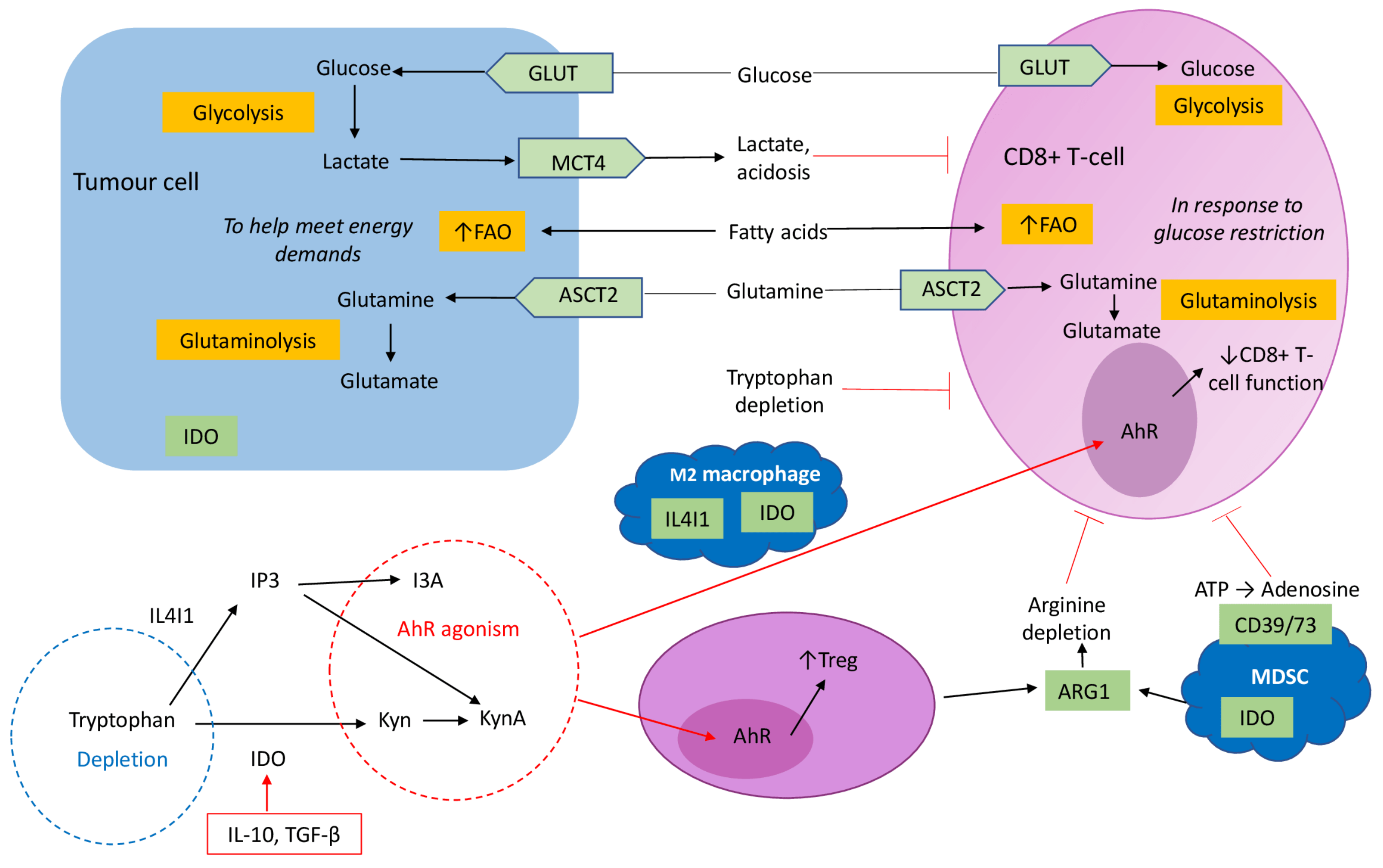
Figure 1. Effect of metabolic changes in the tumour microenvironment (TME) on immune cell differentiation. Increased tumour aerobic glycolysis, supported by upregulation of GLUT-mediated glucose provision provides energy for rapidly dividing cancer cells and leads to lactate accumulation within the TME. Fatty acid oxidation (FAO) is also utilised by tumour cells to supply energy for growth such that CD8+ T-cells starved of glucose increase FAO. Cancer cells also enhance amino acid metabolism to fulfil the energetic demands of rapid growth. Glutamine metabolism provides intermediates for the tricarboxylic acid cycle and maintains intracellular redox balance, making glutamine key to supporting cancer cell proliferation. This depletes glutamine from the TME, limiting its availability to CD8+ T-cells. Increased arginase 1 (ARG1) production by Tregs and MDSCs also limits arginine supply for CD8+ T-cell function. Cancer cell and M2 macrophage indolamine 2,3-dioxygenase (IDO) expression instead causes tryptophan depletion, which arrests CD8+ T-cell proliferation and upregulates regulatory T-cell (Treg) expansion. Tryptophan metabolism by IDO and IL4I1 yield the metabolites kynurenine (kyn), kynurenic acid (kyn A) and the indole I3a (indole-3-carboxaldehyde). Kyn and Kyn A are both ligands of the AhR receptor, which promote Treg differentiation and suppresses CD8+ T-cell function. Tumour transforming growth factor (TGF)-β induces upregulation of CD39/CD73 on MDSCs. These ectoenzymes hydrolyse adenosine triphosphate (ATP) to produce extracellular adenosine within the TME, which inhibits CD8+ T-cell proliferation. Abbreviations: GLUT: glucose transporters; MCT4: monocarboxylate transporter 4; ASCT2: glutamine transporter; IL-2/10: interleukin-2/10; TGF-β: transforming growth factor-β; IP3: indole-3-propionic acid; I3a: indole-3-carboxaldehyde; IL4I1: L-amino acid oxidase interleukin-4-induced-1.
Cancers 13 06231 g001
Figure 2. Hypoxia-driven immune escape within the tumour microenvironment (TME). The hypoxic environment promotes tumour production of interleukin (IL)-10 to recruit myeloid-derived suppressor cells (MDSCs) and regulatory T-cells (Tregs) to the TME. Hypoxia-induced expression of CC-chemokine ligand 28 (CCL28) and CXCL12 recruits Tregs to the TME. Hypoxia inducible factors (HIF)-1α and -2α transcriptionally upregulate vascular endothelial growth factors (VEGFs) to promote proangiogenic signalling in addition to recruiting Tregs to the TME. The HIF pathway also increases glucose transporters (GLUTs) to support substrate provision for tumour glycolysis. Upregulation of PD-L1 on tumour cells is HIF-1α-dependent. Impaired maturation and reduced production of cytokines such as interferon (IFN)-γ in dendritic cells (DCs) occurs in hypoxic environments. Upregulation of inhibitor programmed cell death ligand 1 (PD-L1) on DCs, Tregs, macrophages and MDSCs are all dependent on HIF signalling. TME Hypoxia regulates the expression of inhibitory checkpoint proteins. Cytotoxic T-lymphocyte antigen-4 (CTLA-4), lymphocyte activating 3 (LAG3), T-cell immunoglobulin domain and mucin domain 3 (TIM3) and PD-1 are up-regulated on CD8+ T-cells in a HIF-1α-dependent manner.
Cancers 13 06231 g002
Table 1. Clinical trials in OC using immune checkpoint inhibition in combination with two other therapies. Advanced recurrent (AR), Platinum-resistant (PR), Recurrent or refractory (RR), Ovarian cancer (OC), Primary peritoneal cancer (PPC), Fallopian tube carcinoma (FTC), High-grade serous ovarian carcinoma (HGSOC).
Study Phase and DesignInclusion CriteriaNo. of PatientsTreatmentNCT (ClinicalTrials.gov)
Phase I/II, open-label, sequential assignment
Recruiting
AR-PR OC, triple negative breast, lung, prostate or colorectal carcinoma384Durvalumab + olaparib +/− cediranib (anti-VEGF)NCT02484404
Phase I/II, open-label, single group
Active, not recruiting
RR OC/PPC/FTC with BRCA 1/2 mutation40Olaparib + tremelimumab + durvalumabNCT02953457
Phase II, triple masked, randomised
Active, not recruiting
Recurrent PR OC/PPC/FTC122Atezolizumab + bevacizumab +/− placebo or acetylsalicylic acidNCT02659384
Phase II, open-label, single group assignment
Recruiting
Recurrent PR HGSOC29Atezolizumab + bevacizumab + cobimetinib (mitogen-activated protein kinase inhibitor)NCT03363867
Phase II, open-label, single assignment
Not yet recruiting
Recurrent PR OC/PPC/FTC or endometrial cancer47Lenvatinib + pembrolizumab + paclitaxelNCT04781088
Phase II, open-labelled, randomised
Not yet recruiting
Recurrent OC/PPC/FTC with BRCA wild-type184Maintainence post platinum chemotherapy of olaparib +/− durvalumab +/− UV1 vaccine (hTERT)NCT04742075
Phase III, randomised, masked, parallel assignment
Recruiting
Advanced epithelial OC with BRCA mutation1284First line treatment of carboplatin/paclitaxel + pembrolizumab or placebo
Followed by maintenance of olaparib or placebo
NCT03740165
Phase III, randomized, double blinded, placebo controlled
Active, not recruiting
Stage III/IV, high grade non-mucinous epithelial OC/PPC/FTC1405Carboplatin/paclitaxel + bevacizumab;
+ placebo
Or + dostarlimab (anti-PD-1)
Or + niraparib
NCT03602859
Phase III, randomized, double blinded, placebo controlled
Active, not yet recruiting
Stage III/IV EOC/PPC/FTC who have completed cytoreductive surgery 1000Maintenance post primary platinum-based chemotherapy;
+ nivolumab and rucaparib
Or + nivolumab or placebo
Or + rucaparib or placebo
NCT03522246
Phase III, randomised, double-blinded
Recruiting
Recurrent high-grade serous or endometroid OC/PPC/FTC414Carboplatin/paclitaxel + atezolizumab or placebo
Followed by niraparib maintenance + atezolizumab or placebo
NCT03598270
Phase III, randomised, parallel assignment
Recruiting
Recurrent OC/PPC/FTC664Chemotherapy + bevacizumab + atezolizumab or placeboNCT03353831
Phase III, randomised, parallel assignment
Recruiting
Recurrent, high-grade, PR OC444Doxorubicin +/− atezolizumab +/− bevacizumabNCT02839707
Phase III, randomised, double-blinded, placebo
Recruiting
Advanced (III/IV) high-grade epithelial OC/PPC/FTC1374Platinum-based chemotherapy after primary/interval cytoreductive surgery and bevacizumab, followed by maintenance bevacizumab +/− durvalumab or placebo +/− olaparib or placeboNCT03737643
Table 2. Chimaeric antigen receptor (CAR) T-cell therapy clinical trials in ovarian cancer.
Study Phase and DesignCAR TargetEligibleNo. of PatientsTreatmentNCT
Interventional open-label single group
Recruiting
Anti-mesothelinRR OC with mesothelin positive tumour10
20
Cyclophosphamide
+ Fludarabine
With CAR T-cells
NCT03814447
Interventional open-label single group
Recruiting
Anti-mesothelinRR OC with mesothelin positive tumour20CAR T-cellsNCT03916679
Phase 1 open-label single group
Recruiting
Anti-mesothelinRR OC with mesothelin positive tumour34Cyclophosphamide
+ Fludarabine
With CAR T-cells
NCT04562298
Phase 1 open-label single group
Recruiting
Anti-B7-H3 antigenRR OC21Cyclophosphamide+ Fludarabine
With CAR T-cells
NCT04670068
Phase 1 open-label single group
Recruiting
Anti-MUC16 (gene encoding ca 125)RR OC/PPC/FTC71Biological: PRGN-3005 UltraCAR T-cellsNCT03907527
Phase 1 open-label single group
Not yet recruiting
Anti-ALPPMetastatic ALPP positive OC and EC20CAR T-cellsNCT04627740
Phase 1 open-label single group
Recruiting
Anti-α-FRRR HGSOC/PPC/FTC with α-FR positive tumour18CAR T-cells with or without Cyclophosphamide
+ Fludarabine
NCT03585764
Exploratory open-label single group
Recruiting
Anti-mesothelin T cells secreting PD-1 nanobodiesMesothelin positive advanced solid tumours10CAR T-cellsNCT04503980
Interventional open-label single group
Recruiting
Autogolous Immunogene-modified T-Cells (IgT)Stage III/IV OC in complete remission post primary treatment100CAR T-cellsNCT03184753
Phase I open-label
Recruiting
Anti-MUC1Advanced MUC1+ solid tumours (refractory OC)112Cyclophosphamide
+ Fludarabine
With CAR T-cells
NCT04025216
Relapsed refractory (RR), Ovarian cancer (OC), primary peritoneal cancer (PPC), Fallopian tube carcinoma (FTC), Endometrial cancer (EC), Alkaline phosphatase placental (ALPP), α-folate receptor (α-FR).
Table 3. Clinical trials in ovarian cancer using dendritic cell-based vaccines (DCVs).
Study Phase and DesignEligibleNo. of PatientsControlsNCT
Phase I open-label single-armActive, not yet recruitingIIIc/IV OC no residual disease post primary treatment19Folate receptor alpha loaded DCV onlyNCT02111941
Phase I open-label single-armActive, not yet recruitingHGSOC (=/>IIIb) post primary cytoreductive surgery + chemotherapy17DCV onlyNCT04739527
Phase I/IIa open-label single-arm
Active, not yet recruiting
Stage II-IV OC no residual disease
post primary treatment
18Alpha-type-1 polarised
DCV and intra-peritoneal infusion of CTL
NCT03735589
Phase II
Open-label single-arm
Active, not yet recruiting
First recurrence of platinum-sensitive OC33Autologous maintenance DCV after standard chemotherapyNCT03657966
Phase II open-label
single-arm
Recruiting
AR OC36Autologous DCV only loaded with tumour lysate or for patients who are HLA-A2 with peptides of MUC1 and WT1 therapyNCT00703105
Phase II
Multicentre, randomised, double-blind, placebo-controlled
Recruiting
Stage III/IV OC/PPC no residual disease post primary treatment99Autologous DCV only loaded with tumour antigen versus loaded with peripheral blood mononuclear CellsNCT02033616
Phase II open-label randomised
Active, not yet recruiting
AR OC23Autologous DCV plus GM-CSF NCT00799110
Phase III Multicentre, randomised, double-blind, placebo-controlled
Active, not yet recruiting
AR platinum-sensitive OC678Induction:
DCV verus placebo with carboplatin + gemcitabine/paclitaxel/doxorubicin
+/− bevacizumab
Maintenance:
DCV versus placebo + bevacizumab +/− PARPi
NCT03905902
Advanced recurrent (AR), Ovarian cancer (OC), Primary peritoneal cancer (PPC), Fallopian tube carcinoma (FTC), High-grade ovarian serous carcinoma (HGSOC), Wilms tumour 1 (WT1), Granulocyte–macrophage colony stimulating factor (GM-CSF), Poly (ADP-ribose) polymerase inhibitor (PARPi).
Publisher’s Note: MDPI stays neutral with regard to jurisdictional claims in published maps and institutional affiliations.

