Article

Comparative Analysis of Genetic Alterations, HPV-Status, and PD-L1 Expression in Neuroendocrine Carcinomas of the Cervix

1
Division of Genome Biology, National Cancer Center Research Institute, 5-1-1 Tsukiji, Chuo-ku, Tokyo 104-0045, Japan
2
Department of Obstetrics and Gynecology, The Jikei University School of Medicine, 3-19-18, Nishishinbashi, Minato-ku, Tokyo 105-8471, Japan
3
Medical Oncology, Osaka International Cancer Institute, 3-1-69, Otemae, Chuo-ku, Osaka 541-8567, Japan
4
Department of Obstetrics and Gynecology, Showa University School of Medicine, 1-5-8 Hatanodai, Shinagawa-ku, Tokyo 142-8666, Japan
5
Department of Radiation Oncology, National Cancer Center Hospital, 5-1-1 Tsukiji, Chuo-ku, Tokyo 104-0045, Japan
6
Division of Molecular Modification and Cancer Biology, National Cancer Center Research Institute, 5-1-1 Tsukiji, Chuo-ku, Tokyo 104-0045, Japan
7
Cancer Translational Research Team, RIKEN Center for Advanced Intelligence Project, 1-4-1 Nihonbashi, Chuo-ku, Tokyo 103-0027, Japan
8
Department of Gynecology, National Cancer Center Hospital, 5-1-1 Tsukiji, Chuo-ku, Tokyo 104-0045, Japan
9
Department of Diagnostic Pathology, National Cancer Center Hospital, 5-1-1 Tsukiji, Chuo-ku, Tokyo 104-0045, Japan
*
Authors to whom correspondence should be addressed.
Cancers 2021, 13(6), 1215; https://doi.org/10.3390/cancers13061215
Submission received: 28 January 2021 / Revised: 6 March 2021 / Accepted: 8 March 2021 / Published: 10 March 2021

Abstract

:

Simple Summary

Patients with neuroendocrine carcinoma of the cervix (NECC) have limited treatment options due to its rarity and aggressiveness. In this study, we performed a comparative genetic analysis between 25 NECC and other cervical cancer types (180 squamous cell carcinoma, 53 adenocarcinoma, and 14 adenosquamous carcinoma). Furthermore, the expression of programmed cell death-ligand 1 (PD-L1) was assessed by immunohistochemistry. PIK3CA and TP53 were commonly altered genes in cervical cancer, while SMAD4, RET, EGFR, and APC were NECC-specific altered genes. Of note, 11 NECC cases showed at least one actionable mutation linked to molecular targeted therapies, and 14 cases showed more than one combined positive score for PD-L1 expression. These results may boost the generation of effective treatment strategies for NECC in the future.

Abstract

Neuroendocrine carcinoma of the cervix (NECC) is a rare and highly aggressive tumor with no efficient treatment. We examined genetic features of NECC and identified potential therapeutic targets. A total of 272 patients with cervical cancer (25 NECC, 180 squamous cell carcinoma, 53 adenocarcinoma, and 14 adenosquamous carcinoma) were enrolled. Somatic hotspot mutations in 50 cancer-related genes were detected using the Ion AmpliSeq Cancer Hotspot Panel v2. Human papillomavirus (HPV)-positivity was examined by polymerase chain reaction (PCR)-based testing and in situ hybridization assays. Programmed cell death-ligand 1 (PD-L1) expression was examined using immunohistochemistry. Somatic mutation data for 320 cases of cervical cancer from the Project GENIE database were also analyzed. NECC showed similar (PIK3CA, 32%; TP53, 24%) and distinct (SMAD4, 20%; RET, 16%; EGFR, 12%; APC, 12%) alterations compared with other histological types. The GENIE cohort had similar profiles and RB1 mutations in 27.6% of NECC cases. Eleven (44%) cases had at least one actionable mutation linked to molecular targeted therapies and 14 (56%) cases showed more than one combined positive score for PD-L1 expression. HPV-positivity was observed in all NECC cases with a predominance of HPV-18. We report specific gene mutation profiles for NECC, which can provide a basis for the development of novel therapeutic strategies.

1. Introduction

Neuroendocrine carcinoma of the cervix (NECC) is an uncommon and highly aggressive tumor. Based on Surveillance, Epidemiology, and End Results (SEER) data from the National Cancer Institute, the annual incidence of this cancer is 0.05/100,000 [1]. In Japan, NECC reportedly accounts for less than 2% of all cervical cancers [2]. Approximately 50% of patients with NECC have distant metastases at the time of diagnosis, and the median survival is 18.2 months for all stages [1]. The standard systemic therapy for NECC has not been established in a randomized prospective clinical trial owing to its rare occurrence. Some retrospective studies suggest that chemotherapy complies with that for small-cell lung cancer (SCLC) [3,4] or more common cervical cancer types, such as squamous cell carcinoma (SCC). Platinum-based chemotherapy, combined with etoposide, is generally used as an adjuvant or first-line chemotherapy [3,5], whereas regimens including topotecan, paclitaxel, and bevacizumab are used for some recurrent cases of NECC [4]. Nevertheless, the prognosis of patients with NECC remains dismal; thus, novel and more efficient systemic therapeutic options, including molecular targeted therapy or immunotherapy, are required.
Genomic data of NECC are expected to provide basis for novel and more efficient treatment options for this aggressive tumor. In fact, molecular targeted therapies have contributed to the improvement of patient outcomes in various cancers [6]. However, few studies have focused on molecular targeted therapies for NECC [7,8]. Unfortunately, genomic data available for NECC are still very limited, particularly for Asian patients, although some previous studies reported recurrent genetic alterations involving the phosphoinositide 3-kinase (PI3K)/protein kinase B (AKT)/mechanistic target of rapamycin (mTOR), mitogen-activated protein kinase (MAPK), and P53 pathways in NECC [9,10,11,12]. One of these reports on comparison of genomic profiles between NECC and SCLC demonstrated considerably different molecular characteristics [11], whereas another report revealed genetic similarities between NECC and other major cervical cancer subtypes, including SCC and adenocarcinoma (ADC), using published genomic data [12]. Considering the genomic differences between NECC and SCLC, the present treatment strategy for NECC complying with that for SCLC could be optimized based on the specific genetic features of NECC. Furthermore, deciphering genetic differences between NECC and other subtypes of cervical cancer is expected to help identify specific targets for the treatment of NECC.
The relationship between genotypes of human papillomavirus (HPV) and mutational profiles of NECC remains to be elucidated. High-risk HPV is reportedly associated with most cervical cancers, including SCC, ADC, and adenosquamous carcinoma (ASC) of the cervix [13]. Recently, NECC was reported to be associated with high-risk HPV, primarily HPV16 and HPV18 [14]. Although we previously reported the association between HPV genotypes and histological types and genetic alterations in cervical cancer [15], we could not demonstrate features specific to NECC owing to a very limited number of such cases.
In this study, we aimed to decipher the genetic characteristics of NECC compared with cervical cancer of other histological types using targeted sequencing and analysis of data available in a public database. Furthermore, we explored the links between the identified mutations and targeted therapies. We also elucidated the HPV genotypes in NECC and their association with genetic alterations.

2. Materials and Methods

2.1. Patients

The study protocol was approved by the Institutional Review Board of the National Cancer Center Research Institute (2017–136) and the study was conducted in accordance with the ethical guidelines of the Helsinki Declaration. Written informed consent was obtained from all patients through an opt-out form. Patients who refused to provide consent were excluded from the study.
Two-hundred and eighty-seven patients with pathologically confirmed cervical cancers, including NECC (n = 26), SCC (n = 191), ADC (n = 55), and ASC (n = 15), who received treatment between 2002 and 2018 at the National Cancer Center Hospital, Japan were retrospectively enrolled. Of the 287 cases, sequencing data from 272 patients met the quality control criteria and were included in this study. All the cases were reviewed by at least two gynecological pathologists, and the pathological diagnoses were confirmed according to the World Health Organization (WHO) tumor classification [16].

2.2. DNA Preparation and Next-Generation Sequencing

Genomic DNA was extracted from formalin-fixed paraffin-embedded (FFPE) tumor tissues using the QIAamp DNA FFPE Tissue Kit (Qiagen, Hilden, Germany), according to the manufacturer’s instructions. Library construction was performed using purified genomic DNA (50 ng) obtained from the tumor tissues and the Ion AmpliSeqTM Cancer Hotspot Panel v2 (Thermo Fisher Scientific, Waltham, MA, USA), which targets approximately 2800 Catalog of Somatic Mutations in Cancer (COSMIC) mutational hotspot regions of 50 cancer-related genes. An Ion AmpliSeqTM Custom Panel that was designed for the TP53 gene (coverage: all coding regions) using Ion AmpliSeqTM Designer (https://www.ampliseq.com, accessed on 8 November 2019) was also used. Sequencing was performed using the Ion Proton platform (Thermo Fisher Scientific). For quality control, samples with a mean read depth of coverage over 1000 and a base quality score of 20 (≤1% probability of being incorrect) were selected, which accounted for 90% of the total reads.

2.3. Classification of Oncogenic/Pathogenic Mutations

The sequencing reads were mapped to the University of California, Santa Cruz (UCSC) human reference genome GRCh37, and data analysis was carried out using the Torrent Suite Software v5.0.4 (Thermo Fisher Scientific). Somatic mutations were initially selected using the following criteria: (1) variant allele frequency of somatic mutations was >4% in tumor tissues, (2) single nucleotide polymorphisms were removed if they showed a threshold allele frequency ≥0.01 in either the National Heart, Lung, And Blood Institute (NHLBI) Grand Opportunity Exome Sequencing Project (ESP6500; http://evs.gs.washington.edu/EVS/ (accessed on 1 July 2019)) or the integrative Japanese Genome Variation Database (iJGVD, 20181105; https://ijgvd.megabank.tohoku.ac.jp/ (accessed on 1 July 2019)), and (3) the mutations were registered as “pathogenic/likely pathogenic variants” in the ClinVar or as “oncogenic/likely oncogenic variants” in OncoKB (http://oncokb.org) databases using oncokb-annotator, commit 8910b65 (accessed on 29 June 2019). All the selected variants were then evaluated manually using the Integrative Genomics Viewer (IGV; http://www.broadinstitute.org/igv/ (accessed on 29 May 2019)).

2.4. Detection of Copy Number Alterations Using the Taqman Assay

Copy number alterations were detected by real-time genomic polymerase chain reaction (PCR) using the TaqMan copy number assay and the ABI 7900HT real-time PCR system (Applied Biosystems). Four genes (PIK3CA, ERBB2, PTEN, and STK11) were selected from among the 50 targeted genes in the Ion AmpliSeq™ Cancer Hotspot Panel v2, and the frequency of copy number alterations in these four genes was detected to be >5% in the TCGA dataset of cervical cancer patients within cBioPortal. All TaqMan probes, including PIK3CA (ID Hs02202946_cn), ERBB2 (ID Hs02803918_cn), PTEN (ID Hs05128032_cn), STK11 (ID Hs04013006_cn), and RNase P (cat. no. 4403328), which was used as a reference, were purchased from Thermo Fisher Scientific. Genome data were analyzed using the ABI PRISM 7900HT Sequence Detection Software CopyCaller v2.1 (Thermo Fisher Scientific) for copy number analysis. Copy number amplification was defined as the process resulting in the presence of >8 copies, whereas copy number loss was defined as the process resulting in the presence of <1.2 copy.

2.5. Identification of Human Papillomavirus (HPV) Genotyping by Sanger Sequencing

HPV genotypes were identified as follows: Genomic DNA (10 ng) was amplified using PCR for two distinct HPV genomic regions. The E6/E7 region of HPV was amplified using the primer set pU-1M/pU2R (HPVpU-1M: 5′-TGTCAAAAACCGTTGTGTCC-3′ and HPVpU-2R: 5′-GAGCTGTCGCTTAATTGCTC-3′); the region containing the HPV L1 gene was amplified using the primer set GP5+/GP6+ (GP5+: 5′-TTTGTTACTGTGGTAGATACTAC-3′, GP6+: 5′-GAAAAATAAACTGTAAATCATATTC-3′). PCR reactions were performed using the TaKaRa PCR Human Papillomavirus Typing Set (TaKaRa Bio Inc., Shiga, Japan). PCR products were purified using the NucleoSpin Gel (TaKaRa Bio Inc.) or a PCR Clean-up Kit (TaKaRa Bio Inc.). Sanger sequencing was performed using an ABI 3130xl DNA Sequencer (Applied Biosystems, Foster City, CA, USA), according to the manufacturer’s instructions. Similarity between the obtained sequences and various HPV genotypes in the GenBank database was determined by Basic Local Alignment Search Tool (BLAST) analysis (https://blast.ncbi.nlm.nih.gov/Blast.cgi (accessed on 17 June 2019)).

2.6. Detection of High-Risk HPV Types

To determine the frequency of HPV-positive samples, we performed in situ hybridization (HPV-ISH) using HPV-III High Risk probes (Roche Diagnostics, Mannheim, Germany), according to the manufacturer’s instructions. The HPV-ISH assay is able to detect high-risk HPV genotypes in cervical cancer specimens, including HPV-16, HPV-18, HPV-31, HPV-33, HPV-35, HPV-45, HPV-52, HPV-56, HPV-58, and HPV-66.

2.7. Immunohistochemistry

Immunohistochemistry for retinoblastoma protein, RB1, was performed on 4 μm thick paraffin-embedded tissue sections, which had the same tumor areas provided for genetic analysis, using an RB1-specific mouse monoclonal antibody (clone G3-245, 1:400, BD Pharmingen). Tumors were scored as negative for RB1 protein if more than 95% of cells showed no RB1 staining, with positive nuclear staining in the surrounding endothelial cells serving as an internal control. The expression of programmed cell death-ligand 1 (PD-L1) was examined in tumor cells and infiltrating histiocytes by immunohistochemistry using a rabbit polyclonal antibody (SP142). The expression of PD-L1 was scored by both tumor proportion score (TPS) and combined positive score (CPS), as previously described [17].

2.8. Large-Scale Genomic Datasets

Somatic mutations called from targeted sequencing data in the American Association for Cancer Research (AACR) Project Genomics Evidence Neoplasia information Exchange (GENIE) database (GENIE version 8.0-public) were downloaded as MAF files via the Synapse Platform (http://www.synapse.org/genie (accessed on 2 July 2020)).

2.9. Clinical Association and Actionability Analysis

OncoKB, a precision oncology knowledge base containing information regarding the actionability and therapeutic implications of specific genomic alterations in cancer patients, was used. Somatic mutations were classified into four levels. Gene aberrations with evidence levels of 1–3B were identified as actionable mutations for molecular targeted drugs.

2.10. Statistical Analysis

Statistical analysis was performed using the SPSS (version 27.0, IBM, Armonk, NY, USA) and R (version 3.6.0; R Foundation, Vienna, Austria) software. Categorical variables were compared using the chi-square test or Fisher’s exact test for small sample sizes. Continuous variables were compared using the unpaired Student’s t-test. All p-values were two-tailed, and p-values < 0.05, were considered statistically significant. Overall survival (OS) was defined as the time from the start of treatment to death from any cause. Survival curves were computed using Kaplan–Meier estimates with log-rank tests. Cox regression analysis was applied to test the predictors of OS and to calculate hazard ratios with 95% confidence intervals (95% CI).

3. Results

3.1. Patient Demographics

The clinicopathological characteristics of 272 patients are summarized in Table 1. The median age of patients with NECC was 43 years (range, 28–68 years), which was lower than that of patients with other histological types. The rate of occurrence of advanced International Federation of Gynecology and Obstetrics (FIGO) stages (III/IV) in NECC was 32%, which was higher than that of ADC and ASC (18.9% and 7.1%, respectively). A total of 249 patients (91.5%) were positive for high-risk HPV; these included 25 patients with NECC (100%), 169 with SCC (93.8%), 44 with ADC (83.0%), and 11 with ASC (78.6%).

3.2. Different Genetic Alterations between Neuroendocrine Carcinoma of the Cervix (NECC) and Other Histological Types in Patients with Japanese Cervical Cancer

The profiles of the frequent genetic alterations in the NECC and other histological types are shown in Figure 1. We identified 64 mutations in patients with NECC as being oncogenic/likely oncogenic or pathogenic/likely pathogenic mutations in the OncoKB and ClinVar databases, respectively. In patients with NECC, the mutations included 50 non-synonymous mutations, 13 stop-gain mutations, and one splicing-site mutation (Table S1). For the most frequently mutated genes in patients with NECC, PIK3CA and TP53 variants were detected in 6/25 (24%) of the patients each, followed by SMAD4 variants in 5/25 (20%), PTEN variants in 4/25 (16%), and RET variants in 4/25 (16%) patients. Copy number losses were detected in 4/25 (16%) patients for STK11 and in 3/25 (12%) patients for PTEN. Copy number amplifications were detected in 3/25 (12%) patients for PIK3CA. The PI3K pathway is frequently activated in cervical cancer, and the PI3K pathway-related gene PIK3CA was frequently altered in all histological types (Figure 1 and Figure 2A). The frequency of mutation of TP53 in patients with NECC was significantly higher than that in those with SCC (24.0% vs. 6.1%, p < 0.05, Figure 2A). The frequency of mutations in RET, EGFR, SMAD4, PTPN11, and APC in patients with NECC were higher than those in patients with other histological types. The mTOR signaling-related gene, STK11, previously reported by our group as a poor prognostic factor for cervical cancer [15], was altered significantly more frequently in patients with NECC than in those with SCC (16.0% vs. 4.4%, p < 0.05, Figure 2A). No statistically significant differences were observed between the frequency of MAPK pathway-related genes, KRAS and BRAF, and NRAS mutations in patients with NECC than in those with other histological subtypes.

3.3. Comparison of Frequency of Mutation between NECC and Other Histological Types of Cervical Cancer in the Project GENIE Database

We also examined differences in the mutation frequency between NECC and the other histological subtypes of cervical cancer according to the Project GENIE data (Table S2), and compared the results with those obtained for our cohort. The Project GENIE database contained 11 NECC, 223 SCC, 50 ADC, and 36 ASC cases (Table S3). The median age of patients with NECC was 38 years (range, 29–51 years), which was lower than those of patients with other histological types. The data were obtained from patients mainly from the USA, and only 24 cases (7.5%) were from Asia. Mutations in PIK3CA were most frequently observed and detected in all the histological types (Figure 2B). The frequency of TP53 mutations in patients with NECC was higher than that in patients with other histological types, and was significantly higher than that in patients with SCC (27.2% vs. 6.7%, p < 0.05, Figure 2B). In addition, genetic mutations in RB1 were detected more frequently in patients with NECCs than in those with SCC and ADC (27.2% vs. 5.8%, 0%, p < 0.05; Figure 2B).

3.4. Immunohistichemical Detection of the Expression of RB1 and PD-L1

Because the sequence targeted by us only covered the hotspot regions of RB1 mutations, we could not detect the relatively infrequent mutations of RB1. Therefore, we additionally assessed the expression of RB1 by immunohistochemistry in all the 25 patients with NECC. Loss of RB1 expression was not observed in any of the samples.
For identifying the putative therapeutic targets for NECC, we also evaluated the expression of PD-L1, which is reportedly a predictor of response to immunotherapy, by immunohistochemical staining. Of the 25 patients with NECC, none of the cases showed more than 1% TPS, whereas 56% (14/25) of the cases presented more than 1 CPS (Table S4).

3.5. Actionable Mutations in NECC

Actionable mutations registered as evidence levels of 1–3B in OncoKB were detected in 11 (44%) patients with NECC (Figure 3). Accordingly, 8 (32%) and 4 (16%) patients with NECC may have benefited from mTOR/AKT/PI3K and RET inhibitors, respectively. RET mutation is a therapeutic target not found in other histological types.

3.6. Association between HPV Genotypes and Genetic Alterations in NECC

Approximately 80% of the patients with NECC had detectable HPV-18, and HPV-18-positivity was statistically more frequent in patients with NECC than in those with other histological subtypes (Figure 4). Next, we examined the association between HPV genotypes and genetic alterations frequently detected in NECC; however, there was no significant difference in mutation profiles between NECC with and without HPV-18 (Table S5).

3.7. Prognostic Factors of NECC

Two patients with NECC were excluded from the prognostic analysis because follow-up data were not available. For others, the median survival time was 29.1 months (range, 0.53–140 months). The OS was significantly worse among patients with advanced FIGO stages (median OS of stage I: 129.3 vs. stage II: 39.9 vs. stage III: 49.4 vs. stage IV: 18.5 months; log-rank, p = 0.002; Figure S1A). Patients with KRAS mutations had significantly shorter OS than those without these mutations (log-rank p < 0.001; Figure S1B).
In the univariate Cox regression analysis, the FIGO stage and KRAS mutation were the variables that were significantly associated with shorter OS. The multivariate Cox survival analysis indicated that the FIGO stage and KRAS mutation remained significant prognostic factors for shorter OS (HR: 5.95, 95% CI: 1.58–22.34, p = 0.0083 and HR: 12.92, 95% CI: 1.12–148.89, p = 0.04; Table S6).

4. Discussion

NECC is an extremely aggressive malignancy with high mortality rates, even in patients diagnosed at an early stage [3]. Systemic treatment strategies based on prospective clinical trials are lacking owing to the low incidence of NECC. Novel therapeutics, including molecular-targeted therapy or immunotherapy, have been in demand. In the present study, we analyzed the genetic features of NECC and the clinical value of genetic alterations associated with molecular targeted therapies. We unravel the genetic similarities and differences between NECC and other histological types of cervical cancer. We also show that more than half of the patients with NECC may benefit from molecular targeted therapies and immune checkpoint inhibitors. Furthermore, an important role of high-risk HPV infections in patients with NECC has been demonstrated.
Our targeted sequencing results, comparing NECC with other subtypes of cervical cancers, reveal some similarities. Alterations in PIK3CA were more frequently detected among all histological types, predominantly at two sites (E542K and R545K) in the helical domain of PIK3CA. In Project GENIE, PIK3CA mutations in patients with NECC were less frequent (9.1%), whereas those in patients with other histological types were comparable to our results. Activation of the PI3K pathway through PIK3CA regulates various transformed phenotypes as well as the growth and differentiation of HPV-16 and HPV-18-immortalized cells; thus, it may play a pivotal role in HPV-induced carcinogenesis [18]. Our cohort and the Project GENIE data showed a similar distribution of TP53 mutations, more frequently observed in patients with NECC than in those with SCC and being similar to that in patients with ADC. Moreover, mutations in the MAPK pathway genes, including KRAS, BRAF, and NRAS, were observed at similar frequencies among the histological types. This observation was comparable to that reported previously [12]. Of note, these similarities in genetic alterations between NECC and the other histological types suggest that targeted therapies for genetic alterations, which are widely observed in cervical cancer, can be extended to NECC.
Notably, we identified that several recurrent gene mutations, such as those in EGFR, RET, SMAD4, APC, CTNNB1, and PTPN11, are limited in NECC. These gene mutations are uncommon in cervical cancer [19]. EGFR regulates multiple intracellular target pathways and affects a wide range of biological processes [20]. EGFR mutations are significantly correlated with a highly differentiated grade in patients with cervical cancer [21]. SMAD4, which regulates the canonical transforming growth factor (TGF)-β signaling pathway in porcine granulosa cells, was mutated in 5 (20%) patients with NECC. SMAD4 plays a critical role in tumor progression and induces proinflammatory cytokines, such as interleukin (IL)-5, IL-6, and IL-13, which might induce a tumor-promoting microenvironment [22]. Oncogenic RET mutations were first identified in 4 (16%) patients with NECC in this study. To date, only two cervical SCC cases harboring RET mutations have been reported in Project GENIE. Somatic RET mutations has been reported in about 25–45% cases of sporadic medullary thyroid carcinoma (MTC), a type of carcinoma with neuroendocrine differentiation. Similarly, RET mutations may be associated with the neuroendocrine differentiation in some patients with NECC. In this study, APC, which is a negative regulator of the Wnt/β-catenin pathway, was mutated in 3 (12%) patients with NECC. The Wnt/β-catenin pathway plays a key role in the sequential development (initiation, expansion, and transformation) of tumors into cancer [23].
These genetic differences and similarities between NECC and other types of cervical cancer seem to be consistent with the current understanding of NECC; NECC derives from major histological subtypes, such as SCC or ADC, showing trans-differentiation of the neuroendocrine phenotype [24,25]. We hypothesized that these differences could determine the distinct aggressive behavior of NECC. However, survival analysis did not show any prognostic significance of gene mutations limited to NECC, although KRAS mutation appears to be a poor prognostic factor. Interestingly, Lyons et al. reported that a mitogen-activated protein kinase 1 inhibitor (trametinib) achieved complete radiologic response in a patient with recurrent NECC with KRAS mutation [7].
We also evaluated NECC for actionable mutations with OncoKB evidence levels of 1 to 3B. Actionable mutations were detected in all histological types of cervical cancer, including 11 patients with NECC (44%). PIK3CA mutations are the most frequent actionable mutations in cervical cancers and have been approved by the US Food and Drug Administration (FDA) as a predictive biomarker for the use of the PI3K inhibitor, alpelisib [26]. Various PI3K inhibitors have been developed, and numerous clinical trials have been designed to evaluate various solid malignancies, including neuroendocrine tumors [27]. One study demonstrated that combination therapy of etoposide and cisplatin with the PI3K inhibitor, dactolisib, resulted in enhanced cell cytotoxic responses in a NECC cell line by reducing cell viability and increasing cell apoptosis, which may be a potential new treatment strategy against NECC [28]. Four oncogenic RET mutations, R886Q, S891L, A883T, and C611Y, were first identified in four patients with NECC. Patients with oncogenic RET mutations reportedly benefit from treatment with RET inhibitors, BLU-667 and LOXO-292 [29,30]. Loss-of-function mutations in RB1 were observed in three patients with NECC in Project GENIE. Although loss of RB1 is not registered in OncoKB, two recent studies described that the deregulation of cell cycle transitions upon loss of RB1 can represent a high dependency on aurora kinases, which can be targeted therapeutically [31,32]. Recently, Ramez et al. reported that 73% of patients with NECC had potentially actionable genetic alterations by analyzing 97 cases of NECC using comprehensive genomic profiling (182–315 genes) [11]. Despite extensive research, some genetic alterations, including RET mutations, were first identified in our study. Therefore, the data seem to be insufficient to elucidate the true nature of NECC and, thus, the published data must be integrated and utilized to identify a novel therapeutic target in future studies.
We first provided both TPS and CPS for PD-L1 expression in patients with NECC. Notably, 56% of patients with NECC showed more than one CPS, whereas no cases presented more than 1% TPS. The expression of PD-L1 on tumor and/or immune cells is predictive of the benefit of anti–PD-1 and anti–PD-L1 therapy in several tumor types [33]. Although the expression of PD-L1 in NECC has been consistently reported to be low [3], an exceptional response to nivolumab in recurrent NECC has also been reported [8]. Recently, CPS was used interchangeably with TPS and may be more sensitive than TPS at lower cut-offs in head and neck SCC [34]. Moreover, in one study, it was demonstrated that the expression of PD-L1 on histiocytes in patients with ovarian cancer and melanoma correlated with the efficacy of treatment with either anti-PD-1 alone or in combination with anti-CTLA-4 [35]. The drug, bintrafusp alfa (M7824), was designed to simultaneously bind to two target proteins, PD-L1 and TGF-β, which help prevent the immune system from effectively attacking tumor cells [36]. Bintrafusp alfa shrank tumors in approximately 40% of patients with advanced HPV-associated cancers, including cervical and anal cancers, and showed a manageable safety profile in a phase 1 clinical trial [37]. Overall, the majority of patients with NECC may benefit from precision medicine based on the molecular profiling of genetic alterations and targets of immunotherapy.
Our results show that all patients with NECC were positive for high-risk HPV and 76% of them were positive for HPV-18; however, no significant correlation between HPV genotypes and specific gene mutations was observed. Although HPV-positivity was observed in more than 90% of all cervical cancer patients, NECC was significantly more associated with HPV-18 than other histological types. A large proportion of NECC cases are reportedly caused by high-risk HPV, primarily HPV-16 and HPV-18 [14]. A previous study also reported a predominance of HPV-18 (41% of NECC vs. 10% of other histological types, p < 0.001) [38]. Furthermore, HPV-18-related cervical cancers are characterized by marked lymphatic infiltration and poor prognosis [39]. Although the molecular basis is still unknown, several previous studies have described HPV-16 as a favorable prognostic factor, whereas HPV-18 is a poor prognostic factor in HPV-associated cancers, such as cervical cancer and head and neck cancer [39,40,41]. Importantly, considering the close relationship between NECC and high-risk HPV, prophylactic HPV vaccines would be the most effective measure to prevent death from this highly aggressive cancer. Furthermore, immunotherapy or therapeutic vaccines may be developed as an option for HPV-associated cancer.
Frequent HPV-positivity in NECC also has diagnostic utility for detection of a primary site of metastatic NEC. A significant subset of patients with advanced neuroendocrine carcinomas (NECs) are diagnosed after the detection of distant metastases. However, 11–22% of NECs are reportedly of unknown origin [42]. Considering that HPV-positive NECs originate from HPV-associated cancers including cervical, anal, and oropharyngeal cancer [43], clinicians should consider NECs of these primary sites as differential diagnosis when they encounter HPV-positive NEC of unknown primary origin. In addition, frequent HPV-positivity in NECC would also be useful for distinguishing between primary small cell lung carcinoma, which is HPV-independent [44], and lung metastasis of NECC.
This study has several limitations that need to be addressed. First, the number of patients with NECC included in this study was limited. The survival analysis was not robust due to the small number of cases, and further external validation should be performed in a future study. However, considering its rarity, the mutational analysis of 25 cases of NECC has been a relatively large cohort in Asia. Second, targeted sequencing for mutational analysis used in this study contained only hotspot mutations in 50 cancer-related genes. Nevertheless, we could identify new therapeutic targets for NECC, such as alterations in RET, even for limited genes and their regions. Plans are currently underway to test therapies targeting the candidate identified in this study.

5. Conclusions

We demonstrate genetic similarities and differences between NECC and other histological types of cervical cancers. PIK3CA (32.0%), TP53 (24.0%), SMAD4 (20%), PTEN (20.0%), STK11 (16.0%), and RET (16.0%) were frequently altered in patients with NECC. Nineteen patients (76%) with NECC could potentially benefit from molecular targeted therapies and immune checkpoint inhibitors. Furthermore, high-risk HPV infections, particularly HPV-18, may play a critical role in the carcinogenesis and aggressive behavior of NECC. Therefore, the current HPV vaccines could prevent a large proportion of NECC cases. Collectively, this study reveals specific gene mutation profiles and HPV status in NECC, which can provide a basis for the development of novel therapeutic strategies for this highly aggressive malignancy.

Supplementary Materials

The following are available online at https://www.mdpi.com/2072-6694/13/6/1215/s1, Figure S1: Kaplan–Meier analysis of the overall survival (OS) in patients with neuroendocrine carcinoma of the cervix, Table S1: Candidate somatic mutations of cervical cancers in this study, Table S2: Candidate somatic mutations of cervical cancers in Project GENIE, Table S3: Characteristics of 320 patients with cervical cancers in Project GENIE, Table S4: Combined positive score of PD-L1 expression in NECC, Table S5: Association between HPV genotypes and genetic mutations in NECC, Table S6: Univariate and multivariate analysis for overall survival of patients with NECC.

Author Contributions

D.T., K.S. (Kouya Shiraishi), T.K., and H.Y. designed the study. D.T. wrote the initial draft of the manuscript. K.S. (Kouya Shiraishi), M.M., and H.Y. contributed to the analysis and interpretation of the data and assisted in manuscript preparation. All other authors contributed to the data collection, interpretation, and critical review of the manuscript. All authors approved the final version of the manuscript and agreed to be accountable for all aspects of the study. We ensure that questions related to the accuracy or integrity of any part of the study will be appropriately investigated and resolved. All authors have read and agreed to the published version of the manuscript.

Funding

This work was supported by grants-in-aid from the MITSUI LIFE SOCIAL WELFARE FOUNDATION, PUBLIC FOUNDATION OF THE VACCINATION RESEARCH CENTER, GRANT-IN-AID FOR YOUNG SCIENTISTS (grant numbers 18K15654 and 20K17668), and the NATIONAL CANCER CENTER RESEARCH AND DEVELOPMENT FUND (29-A-2, NCC Biobank, and NCC Core Facility).

Institutional Review Board Statement

The study protocol was approved by the Institutional Review Board of the National Cancer Center Research Institute (2017–136, 30 June 2018) and the study was conducted in accordance with the ethical guidelines of the Helsinki Declaration. Written informed consent was obtained from all patients through an opt-out form. Patients who refused to provide consent were excluded from the study.

Informed Consent Statement

Informed consent was obtained from all subjects involved in the study.

Data Availability Statement

The data presented in this study are available in the present manuscript or in the Supplementary Materials.

Acknowledgments

The results of our study are based in part on data generated by the AACR Project GENIE (https://www.aacr.org/professionals/research/aacr-project-genie/). The authors are grateful to Hitoshi Ichikawa, Sachiyo Mitani, and Tsuyuka Otsuki, the other physicians and staff at the National Cancer Center, and the physicians and staff of other hospitals for their help and support. We would like to thank Editage (https://www.editage.jp (last requested on 3 March 2021)) for English language editing.

Conflicts of Interest

The authors state that there are no conflict of interest to declare.

References

  1. Dasari, A.; Mehta, K.; Byers, L.A.; Sorbye, H.; Yao, J.C. Comparative Study of Lung and Extrapulmonary Poorly Differentiated Neuroendocrine Carcinomas: A SEER Database Analysis of 162,983 Cases. Cancer 2018, 124, 807–815. [Google Scholar] [CrossRef]
  2. Nagase, S.; Ohta, T.; Takahashi, F.; Enomoto, T. 2017 Committee on Gynecologic Oncology of the Japan Society of Obstetrics and Gynecology. Annual Report of the Committee on Gynecologic Oncology, the Japan Society of Obstetrics and Gynecology: Annual Patients Report for 2015 and Annual Treatment Report for 2010. J. Obstet. Gynaecol. Res. 2019, 45, 289–298. [Google Scholar] [CrossRef] [PubMed] [Green Version]
  3. Salvo, G.; Gonzalez Martin, A.; Gonzales, N.R.; Frumovitz, M. Updates and Management Algorithm for Neuroendocrine Tumors of the Uterine Cervix. Int. J. Gynecol. Cancer 2019, 29, 986–995. [Google Scholar] [CrossRef]
  4. Tempfer, C.B.; Tischoff, I.; Dogan, A.; Hilal, Z.; Schultheis, B.; Kern, P.; Rezniczek, G.A. Neuroendocrine Carcinoma of the Cervix: A Systematic Review of the Literature. BMC Cancer 2018, 18, 530. [Google Scholar] [CrossRef] [PubMed] [Green Version]
  5. Ishikawa, M.; Kasamatsu, T.; Tsuda, H.; Fukunaga, M.; Sakamoto, A.; Kaku, T.; Nakanishi, T.; Hasumi, Y.; Iwata, T.; Baba, T.; et al. Prognostic Factors and Optimal Therapy for Stages I-II Neuroendocrine Carcinomas of the Uterine Cervix: A Multi-Center Retrospective Study. Gynecol. Oncol. 2018, 148, 139–146. [Google Scholar] [CrossRef] [PubMed]
  6. Dancey, J.E.; Bedard, P.L.; Onetto, N.; Hudson, T.J. The Genetic Basis for Cancer Treatment Decisions. Cell 2012, 148, 409–420. [Google Scholar] [CrossRef] [Green Version]
  7. Lyons, Y.A.; Frumovitz, M.; Soliman, P.T. Response to MEK Inhibitor in Small Cell Neuroendocrine Carcinoma of the Cervix with a KRAS mutation. Gynecol. Oncol. Rep. 2014, 10, 28–29. [Google Scholar] [CrossRef] [Green Version]
  8. Paraghamian, S.E.; Longoria, T.C.; Eskander, R.N. Metastatic Small Cell Neuroendocrine Carcinoma of the Cervix Treated with the PD-1 Inhibitor, Nivolumab: A Case Report. Gynecol. Oncol. Res. Pract. 2017, 4, 3. [Google Scholar] [CrossRef] [PubMed] [Green Version]
  9. Xing, D.; Zheng, G.; Schoolmeester, J.K.; Li, Z.; Pallavajjala, A.; Haley, L.; Conner, M.G.; Vang, R.; Hung, C.F.; Wu, T.C.; et al. Next-Generation Sequencing Reveals Recurrent Somatic Mutations in Small Cell Neuroendocrine Carcinoma of the Uterine Cervix. Am. J. Surg. Pathol. 2018, 42, 750–760. [Google Scholar] [CrossRef]
  10. Frumovitz, M.; Burzawa, J.K.; Byers, L.A.; Lyons, Y.A.; Ramalingam, P.; Coleman, R.L.; Brown, J. Sequencing of Mutational Hotspots in Cancer-Related Genes in Small Cell Neuroendocrine Cervical Cancer. Gynecol. Oncol. 2016, 141, 588–591. [Google Scholar] [CrossRef] [Green Version]
  11. Eskander, R.N.; Elvin, J.; Gay, L.; Ross, J.S.; Miller, V.A.; Kurzrock, R. Unique Genomic Landscape of High-Grade Neuroendocrine Cervical Carcinoma: Implications for Rethinking Current Treatment Paradigms. JCO Precis. Oncol. 2020, 4. [Google Scholar] [CrossRef]
  12. Hillman, R.T.; Cardnell, R.; Fujimoto, J.; Lee, W.C.; Zhang, J.; Byers, L.A.; Ramalingam, P.; Leitao, M.; Swisher, E.; Futreal, P.A.; et al. Comparative Genomics of High Grade Neuroendocrine Carcinoma of the Cervix. PLoS ONE 2020, 15, e0234505. [Google Scholar] [CrossRef] [PubMed]
  13. Muñoz, N.; Bosch, F.X.; de Sanjosé, S.; Herrero, R.; Castellsagué, X.; Shah, K.V.; Snijders, P.J.; Meijer, C.J. Epidemiologic Classification of Human Papillomavirus Types Associated with Cervical Cancer. N. Engl. J. Med. 2003, 348, 518–527. [Google Scholar] [CrossRef] [PubMed] [Green Version]
  14. Castle, P.E.; Pierz, A.; Stoler, M.H. A systematic Review and Meta-Analysis on the Attribution of Human Papillomavirus (HPV) in Neuroendocrine Cancers of the Cervix. Gynecol. Oncol. 2018, 148, 422–429. [Google Scholar] [CrossRef]
  15. Hirose, S.; Murakami, N.; Takahashi, K.; Kuno, I.; Takayanagi, D.; Asami, Y.; Matsuda, M.; Shimada, Y.; Yamano, S.; Sunami, K.; et al. Genomic Alterations in STK11 Can Predict Clinical Outcomes in Cervical Cancer Patients. Gynecol. Oncol. 2020, 156, 203–210. [Google Scholar] [CrossRef] [PubMed] [Green Version]
  16. Kurman, R.J.; Carcangiu, M.L.; Herrington, C.S.; Young, R.H. WHO Classification of Tumours of Female Reproductive Organs, 4th ed.; International Agency for Research on Cancer: Lyon, France, 2014; 307p. [Google Scholar]
  17. Kulangara, K.; Zhang, N.; Corigliano, E.; Guerrero, L.; Waldroup, S.; Jaiswal, D.; Ms, M.J.; Shah, S.; Hanks, D.; Wang, J.; et al. Clinical Utility of the Combined Positive Score for Programmed Death Ligand-1 Expression and the Approval of Pembrolizumab for Treatment of Gastric Cancer. Arch. Pathol. Lab. Med. 2019, 143, 330–337. [Google Scholar] [CrossRef] [Green Version]
  18. Henken, F.E.; Banerjee, N.S.; Snijders, P.J.; Meijer, C.J.; De-Castro Arce, J.; Rösl, F.; Broker, T.R.; Chow, L.T.; Steenbergen, R.D. PIK3CA-Mediated PI3-Kinase Signalling Is Essential for HPV-Induced Transformation in Vitro. Mol. Cancer 2011, 10, 71. [Google Scholar] [CrossRef] [Green Version]
  19. Cancer Genome Atlas Research Network; Albert Einstein College of Medicine; Analytical Biological Services; Barretos Cancer Hospital; Baylor College of Medicine; Beckman Research Institute of City of Hope; Buck Institute for Research on Aging; Canada’s Michael Smith Genome Sciences Centre; Harvard Medical School; Helen, F.; et al. Integrated Genomic and Molecular Characterization of Cervical Cancer. Nature 2017, 543, 378–384. [Google Scholar] [CrossRef]
  20. Fraguas, S.; Barberán, S.; Cebrià, F. EGFR Signaling Regulates Cell Proliferation, Differentiation and Morphogenesis during Planarian Regeneration and Homeostasis. Dev. Biol. 2011, 354, 87–101. [Google Scholar] [CrossRef]
  21. Wei, H.; Wang, X.W.; Chen, K.M.; Ling, S.R.; Yi, C.J. Analysis of Gene Mutation Associated with Tyrosine Kinase Inhibitor Sensitivity of Epidermal Growth Factor Receptor in Cervical Cancer Patients. Eur. Rev. Med. Pharmacol. Sci. 2018, 22, 6280–6287. [Google Scholar] [CrossRef]
  22. Pickup, M.; Novitskiy, S.; Moses, H.L. The Roles of TGFβ in the Tumour Microenvironment. Nat. Rev. Cancer 2013, 13, 788–799. [Google Scholar] [CrossRef] [PubMed] [Green Version]
  23. Zhan, T.; Rindtorff, N.; Boutros, M. Wnt Signaling in Cancer. Oncogene 2017, 36, 1461–1473. [Google Scholar] [CrossRef]
  24. Ambros, R.A.; Park, J.S.; Shah, K.V.; Kurman, R.J. Evaluation of Histologic, Morphometric, and Immunohistochemical Criteria in the Differential Diagnosis of Small Cell Carcinomas of the Cervix with Particular Reference to Human Papillomavirus Types 16 and 18. Mod. Pathol. 1991, 4, 586–593. [Google Scholar] [PubMed]
  25. Stoler, M.H.; Mills, S.E.; Gersell, D.J.; Walker, A.N. Small-Cell Neuroendocrine Carcinoma of the Cervix. A Human Papillomavirus Type 18-Associated Cancer. Am. J. Surg. Pathol. 1991, 15, 28–32. [Google Scholar] [CrossRef] [PubMed]
  26. André, F.; Ciruelos, E.; Rubovszky, G.; Campone, M.; Loibl, S.; Rugo, H.S.; Iwata, H.; Conte, P.; Mayer, I.A.; Kaufman, B.; et al. Alpelisib for PIK3CA-Mutated, Hormone Receptor-Positive Advanced Breast Cancer. N. Engl. J. Med. 2019, 380, 1929–1940. [Google Scholar] [CrossRef]
  27. Alqahtani, A.; Ayesh, H.S.K.; Halawani, H. PIK3CA Gene Mutations in Solid Malignancies: Association with Clinicopathological Parameters and Prognosis. Cancers 2019, 12, 93. [Google Scholar] [CrossRef] [PubMed] [Green Version]
  28. Lai, Z.Y.; Yeo, H.Y.; Chen, Y.T.; Chang, K.M.; Chen, T.C.; Chuang, Y.J.; Chang, S.J. PI3K Inhibitor Enhances the Cytotoxic Response to Etoposide and Cisplatin in a Newly Established Neuroendocrine Cervical Carcinoma Cell Line. Oncotarget 2017, 8, 45323–45334. [Google Scholar] [CrossRef] [PubMed] [Green Version]
  29. Subbiah, V.; Gainor, J.F.; Rahal, R.; Brubaker, J.D.; Kim, J.L.; Maynard, M.; Hu, W.; Cao, Q.; Sheets, M.P.; Wilson, D.; et al. Precision Targeted Therapy with BLU-667 for RET-Driven Cancers. Cancer Discov. 2018, 8, 836–849. [Google Scholar] [CrossRef] [Green Version]
  30. Drilon, A.E.; Subbiah, V.; Oxnard, G.R.; Bauer, T.M.; Velcheti, V.; Lakhani, N.J.; Besse, B.; Park, K.; Patel, J.D.; Cabanillas, M.E.; et al. A phase 1 Study of LOXO-292, a Potent and Highly Selective RET Inhibitor, in Patients with RET-Altered Cancers. J. Clin. Oncol. 2018, 36 (Suppl. 15), 102. [Google Scholar] [CrossRef]
  31. Oser, M.G.; Fonseca, R.; Chakraborty, A.A.; Brough, R.; Spektor, A.; Jennings, R.B.; Flaifel, A.; Novak, J.S.; Gulati, A.; Buss, E.; et al. Cells Lacking the RB1 Tumor Suppressor Gene Are Hyperdependent on Aurora B Kinase for Survival. Cancer Discov. 2019, 9, 230–247. [Google Scholar] [CrossRef] [Green Version]
  32. Gong, X.; Du, J.; Parsons, S.H.; Merzoug, F.F.; Webster, Y.; Iversen, P.W.; Chio, L.C.; Van Horn, R.D.; Lin, X.; Blosser, W.; et al. Aurora A Kinase Inhibition Is Synthetic Lethal with Loss of the RB1 Tumor Suppressor Gene. Cancer Discov. 2019, 9, 248–263. [Google Scholar] [CrossRef] [PubMed] [Green Version]
  33. Caldwell, C., Jr.; Johnson, C.E.; Balaji, V.N.; Balaji, G.A.; Hammer, R.D.; Kannan, R. Identification and Validation of a PD-L1 Binding Peptide for Determination of PDL1 Expression in Tumors. Sci. Rep. 2017, 7, 13682. [Google Scholar] [CrossRef] [Green Version]
  34. Emancipator, K.; Huang, L.; Aurora-Garg, D.; Bal, T.; Cohen, E.E.W.; Harrington, K.; Soulières, D.; Le Tourneau, C.; Licitra, L.; Burtness, B.; et al. Comparing Programmed Death Ligand 1 Scores for Predicting Pembrolizumab Efficacy in Head and Neck Cancer. Mod. Pathol. 2020. [Google Scholar] [CrossRef]
  35. Lin, H.; Wei, S.; Hurt, E.M.; Green, M.D.; Zhao, L.; Vatan, L.; Szeliga, W.; Herbst, R.; Harms, P.W.; Fecher, L.A.; et al. Host Expression of PD-L1 Determines Efficacy of PD-L1 Pathway Blockade-Mediated Tumor Regression. J. Clin. Investig. 2018, 128, 805–815. [Google Scholar] [CrossRef] [PubMed] [Green Version]
  36. Lan, Y.; Zhang, D.; Xu, C.; Hance, K.W.; Marelli, B.; Qi, J.; Yu, H.; Qin, G.; Sircar, A.; Hernández, V.M.; et al. Enhanced Preclinical Antitumor Activity of M7824, a Bifunctional Fusion Protein Simultaneously Targeting PD-L1 and TGF-β. Sci. Transl. Med. 2018, 10. [Google Scholar] [CrossRef] [Green Version]
  37. Strauss, J.; Heery, C.R.; Schlom, J.; Madan, R.A.; Cao, L.; Kang, Z.; Lamping, E.; Marté, J.L.; Donahue, R.N.; Grenga, I.; et al. Phase I Trial of M7824 (MSB0011359C), a Bifunctional Fusion Protein Targeting PD-L1 and TGFβ, in Advanced Solid Tumors. Clin. Cancer. Res. 2018, 24, 1287–1295. [Google Scholar] [CrossRef] [Green Version]
  38. Alejo, M.; Alemany, L.; Clavero, O.; Quiros, B.; Vighi, S.; Seoud, M.; Cheng-Yang, C.; Garland, S.M.; Juanpere, N.; Lloreta, J.; et al. Contribution of Human Papillomavirus in Neuroendocrine Tumors from a Series of 10,575 Invasive Cervical Cancer Cases. Papillomavirus Res. 2018, 5, 134–142. [Google Scholar] [CrossRef] [PubMed]
  39. Lai, C.H.; Chang, C.J.; Huang, H.J.; Hsueh, S.; Chao, A.; Yang, J.E.; Lin, C.T.; Huang, S.L.; Hong, J.H.; Chou, H.H.; et al. Role of Human Papillomavirus Genotype in Prognosis of Early-Stage Cervical Cancer Undergoing Primary Surgery. J. Clin. Oncol. 2007, 25, 3628–3634. [Google Scholar] [CrossRef]
  40. Onuki, M.; Matsumoto, K.; Tenjimbayashi, Y.; Tasaka, N.; Akiyama, A.; Sakurai, M.; Minaguchi, T.; Oki, A.; Satoh, T.; Yoshikawa, H. Human Papillomavirus Genotype and Prognosis of Cervical Cancer: Favorable Survival of Patients with HPV16-Positive Tumors. Papillomavirus Res. 2018, 6, 41–45. [Google Scholar] [CrossRef]
  41. Bratman, S.V.; Bruce, J.P.; O’Sullivan, B.; Pugh, T.J.; Xu, W.; Yip, K.W.; Liu, F.F. Human Papillomavirus Genotype Association with Survival in Head and Neck Squamous Cell Carcinoma. JAMA Oncol. 2016, 2, 823–826. [Google Scholar] [CrossRef] [Green Version]
  42. Alexandraki, K.I.; Tsoli, M.; Kyriakopoulos, G.; Angelousi, A.; Nikolopoulos, G.; Kolomodi, D.; Kaltsas, G.A. Current Concepts in the Diagnosis and Management of Neuroendocrine Neoplasms of Unknown Primary Origin. Minerva. Endocrinol. 2019, 44, 378–386. [Google Scholar] [CrossRef] [PubMed]
  43. Bishop, J.A.; Westra, W.H. Human Papillomavirus-Related Small Cell Carcinoma of the Oropharynx. Am. J. Surg. Pathol. 2011, 35, 1679–1684. [Google Scholar] [CrossRef] [PubMed]
  44. Carlson, J.W.; Nucci, M.R.; Brodsky, J.; Crum, C.P.; Hirsch, M.S. Biomarker-Assisted Diagnosis of Ovarian, Cervical and Pulmonary Small Cell Carcinomas: The Role of TTF-1, WT-1 and HPV Analysis. Histopathology 2007, 51, 305–312. [Google Scholar] [CrossRef] [PubMed]
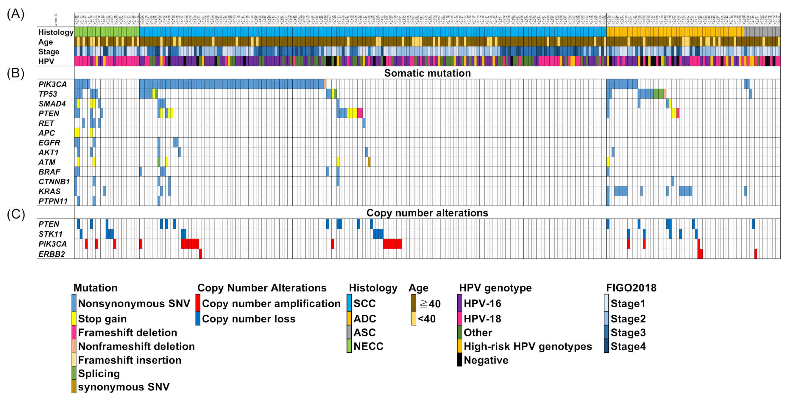
Figure 1. Somatic alterations in cervical cancer and associated clinicopathological features. Two hundred and seventy-two cases were categorized according to their (A) histological types and clinicopathological features, (B) major mutated genes of neuroendocrine carcinoma, and (C) copy number alterations. Mutated genes are color-coded according to their mutation type.
Cancers 13 01215 g001
Figure 2. Association between genetic alterations and histological types in cervical cancer. Percentages of samples mutated in individual tumor types are shown. (A) the present study, (B) Project GENIE v8.0. The p-value was calculated using the Fisher’s exact test, * p < 0.05, ** p < 0.001. NEC: neuroendocrine carcinoma; SCC: squamous cell carcinoma; ADC: adenocarcinoma; ASC: adenosquamous carcinoma.
Cancers 13 01215 g002
Figure 3. Frequency of actionable genetic mutations in cervical cancers. Percentages of samples mutated in individual tumor types are shown. NEC, neuroendocrine carcinoma; SCC, squamous cell carcinoma; ADC, adenocarcinoma; ASC, adenosquamous carcinoma.
Cancers 13 01215 g003
Figure 4. Correlation between histological types and human papillomavirus (HPV) genotypes in 272 cervical cancer specimens. HPV genotypes were compared with neuroendocrine carcinoma (NEC) and other histological types of cervical cancer, including squamous cell carcinoma (SCC), adenocarcinoma (ADC), and adenosquamous carcinoma (ASC). The P-value was calculated using the Fisher’s exact test, * p < 0.05, ** p < 0.01.
Cancers 13 01215 g004
Table 1. Clinicopathological characteristics of 272 patients with cervical cancers.
CharacteristicsNECC (25)SCC (180)ADC (53)ASC (14)
Age (Year)Median (Range)43 (2868)55 (25–89)51 (30–82)47 (37–60)
Stage (FIGO2018), n (%)I10 (40.0)40 (22.2)18 (34.0)7 (50.0)
II7 (28.0)58 (32.2)25 (47.2)6 (42.9)
III4 (16.0)60 (33.3)3 (5.7)1 (7.1)
IV4 (16.0)22 (12.2)7 (13.2)0 (0.0)
HPV positivity, n(%)25 (100)169 (93.8)44 (83.0)11 (78.6)
Treatment, n(%)Surgery only6 (24.0)34 (18.9)23 (43.4)4 (28.6)
RH6 (24.0)33 (13.3)21 (39.6)4 (28.6)
RH+PAN0 (0.0)1 (0.6)1 (1.9)0 (0.0)
TAH+BSO+PLND+OMT0 (0.0)0 (0.0)1 (1.9)0 (0.0)
Surgery+adj-Treatment9 (36.0)57 (31.7)21 (39.6)9 (64.3)
RH7 (24.0)43 (20.6)16 (30.2)6 (42.9)
RH+PAN1 (4.0)10 (5.6)3 (5.7)3 (21.4)
MRHx+BSO+PLND0 (0.0)1 (0.6)1 (1.9)0 (0.0)
TAH+BSO+PLND0 (0.0)2 (0.6)1 (0.0)0 (0.0)
TAH+BSO1 (4.0)0 (0.0)0 (0.0)0 (0.0)
NACT+RH+adjCT1 (4.0)1 (0.6)0 (0.0)0 (0.0)
NACRT+RT0 (0.0)1 (0.6)0 (0.0)0 (0.0)
RT1 (4.0)25 (13.9)3 (5.7)0 (0.0)
CCRT only3 (12.0)62 (34.4)6 (11.3)1 (7.1)
RT following CT1 (4.0)0 (0.0)0 (0.0)0 (0.0)
CT only2 (7.0)0 (0.0)0 (0.0)0 (0.0)
Palliative care only2 (7.0)0 (0.0)0 (0.0)0 (0.0)
Abbreviations: NECC, Neuroendocrine carcinoma of the cervix; SCC, Squamous cell carcinoma; ADC, Adenocarcinoma; ASC, Adenosquamous carcinoma; HPV, human papillomavirus; NACT, Neoadjuvant chemotherapy; adj, Adjuvant; CT, Chemotherapy; RT, Radiotherapy; CCRT, concurrent chemoradiation therapy; NACRT, Neoadjuvant chemoradiation therapy; RH, radical hysterectomy; BSO, bilateral salpingo-oophorectomy; PLND, pelvic lymph mode dissection; PAN, para-aortic lymphadectomy; TAH, total hysterectomy; OMT, omentectomy; MRHx, modified radical hysterectomy.
Publisher’s Note: MDPI stays neutral with regard to jurisdictional claims in published maps and institutional affiliations.