
Single-Cell Deconvolution of Head and Neck Squamous Cell Carcinoma

 , , , , , ,
Abstract
:Simple Summary
Abstract
1. Introduction
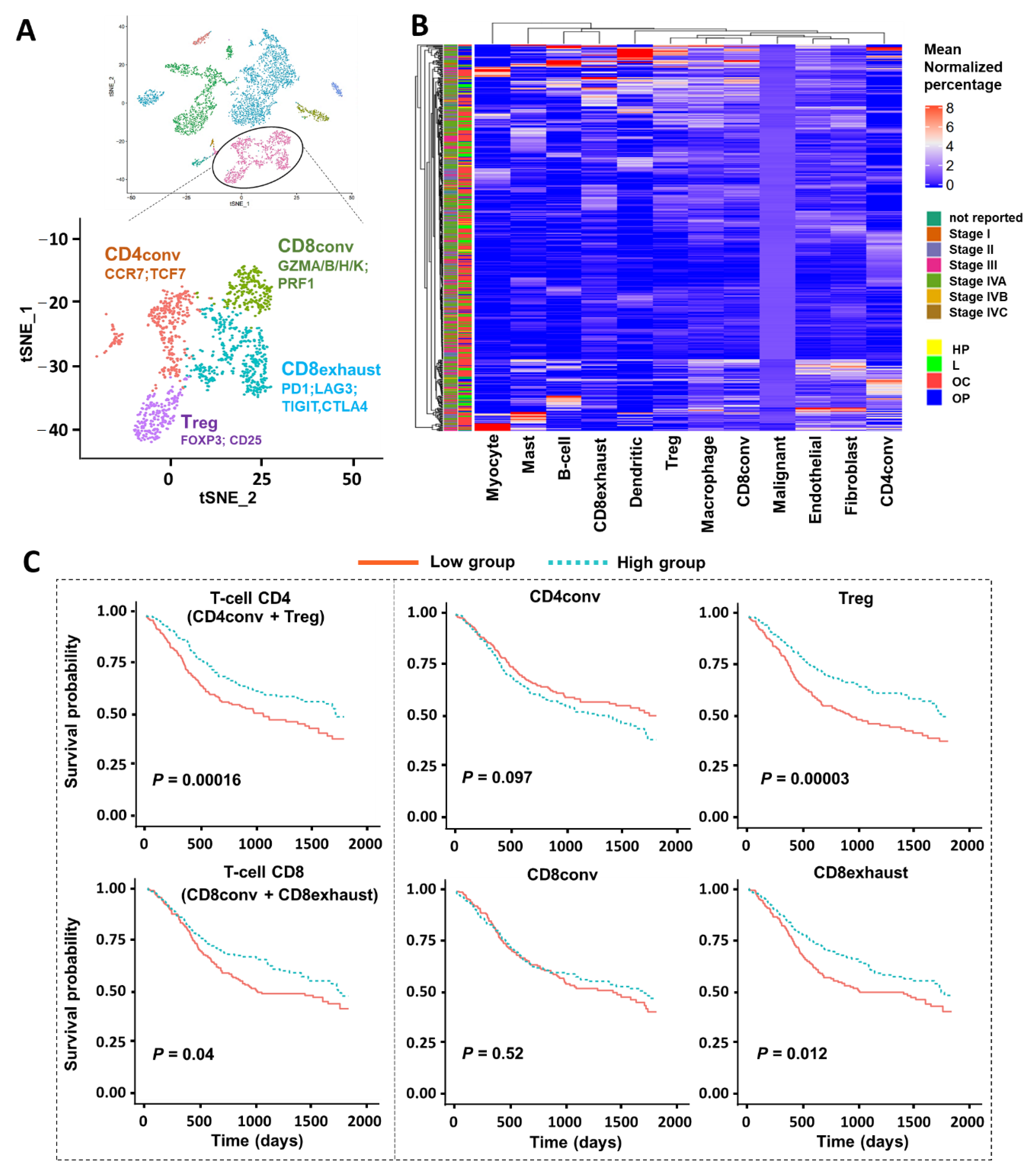
2. Results
2.1. Overview of Deconvolution Approach
2.2. CIBERSORTx Analysis with scRNA-seq Reference for Nine Major Cell Types
2.3. CIBERSORTx Analysis with T-Cell Subtypes/Subpopulations
2.4. MuSiC Deconvolution Based on T-Cell Subtypes/Subpopulations
2.5. CIBERSORTx Analysis of Gene Expression of Regulatory T-Cells
3. Discussion
4. Methods
4.1. Bulk RNA-Seq Data and Clinical Information of HNSCC Tumors from TCGA
4.2. Single-Cell RNA-seq Data of HNSCC Tumors
4.3. CIBERSORTx Deconvolution Analysis
4.4. MuSiC Deconvolution Analysis
4.5. Single-Cell RNA-seq Data of HNSCC Tumors
5. Conclusions
Supplementary Materials
Author Contributions
Funding
Institutional Review Board Statement
Informed Consent Statement
Data Availability Statement
Acknowledgments
Conflicts of Interest
References
- Puram, S.V.; Rocco, J.W. Molecular aspects of head and neck cancer therapy. Hematol. Clin. N. Am. 2015, 29, 971–992. [Google Scholar] [CrossRef] [PubMed] [Green Version]
- Bray, F.; Ferlay, J.; Soerjomataram, I.; Siegel, R.L.; Torre, L.A.; Jemal, A. Global cancer statistics 2018: GLOBOCAN estimates of incidence and mortality worldwide for 36 cancers in 185 countries. CA Cancer J. Clin. 2018, 68, 394–424. [Google Scholar] [CrossRef] [Green Version]
- Chow, L.Q.M. Head and neck cancer. N. Engl. J. Med. 2020, 382, 60–72. [Google Scholar] [CrossRef]
- Alsahafi, E.; Begg, K.; Amelio, I.; Raulf, N.; Lucarelli, P.; Sauter, T.; Tavassoli, M. Clinical update on head and neck cancer: Molecular biology and ongoing challenges. Cell Death Dis. 2019, 10, 1–17. [Google Scholar] [CrossRef] [PubMed] [Green Version]
- Hanahan, D.; Weinberg, R.A. Hallmarks of cancer: The next generation. Cell 2011, 144, 646–674. [Google Scholar] [CrossRef] [PubMed] [Green Version]
- Coussens, L.M.; Zitvogel, L.; Palucka, A.K. Neutralizing tumor-promoting chronic inflammation: A magic bullet? Science 2013, 339, 286–291. [Google Scholar] [CrossRef] [Green Version]
- Hyman, D.M.; Taylor, B.S.; Baselga, J. Implementing genome-driven oncology. Cell 2017, 168, 584–599. [Google Scholar] [CrossRef] [PubMed] [Green Version]
- Puram, S.V.; Tirosh, I.; Parikh, A.S.; Patel, A.P.; Yizhak, K.; Gillespie, S.; Rodman, C.; Luo, C.L.; Mroz, E.A.; Emerick, K.S.; et al. Single-Cell Transcriptomic Analysis of Primary and Metastatic Tumor Ecosystems in Head and Neck Cancer. Cell 2017, 171, 1611–1624. [Google Scholar] [CrossRef] [Green Version]
- Izar, B.; Tirosh, I.; Stover, E.H.; Wakiro, I.; Cuoco, M.S.; Alter, I.; Rodman, C.; Leeson, R.; Su, M.-J.; Shah, P.; et al. A single-cell landscape of high-grade serous ovarian cancer. Nat. Med. 2020, 26, 1271–1279. [Google Scholar] [CrossRef] [PubMed]
- Parikh, A.S.; Puram, S.V.; Faquin, W.C.; Richmon, J.D.; Emerick, K.S.; Deschler, D.G.; Varvares, M.A.; Tirosh, I.; Bernstein, B.E.; Lin, D.T. Immunohistochemical quantification of partial-EMT in oral cavity squamous cell carcinoma primary tumors is associated with nodal metastasis. Oral Oncol. 2019, 99, 104458. [Google Scholar] [CrossRef] [PubMed]
- Mandal, R.; Şenbabaoğlu, Y.; Desrichard, A.; Havel, J.J.; Dalin, M.G.; Riaz, N.; Lee, K.-W.; Ganly, I.; Hakimi, A.A.; Chan, T.A.; et al. The head and neck cancer immune landscape and its immunotherapeutic implications. JCI Insight 2016, 1, e89829. [Google Scholar] [CrossRef] [Green Version]
- The Cancer Genome Atlas Network. Comprehensive genomic characterization of head and neck squamous cell carcinomas. Nature 2015, 517, 576–582. [Google Scholar] [CrossRef] [PubMed] [Green Version]
- Stransky, N.; Egloff, A.M.; Tward, A.D.; Kostic, A.D.; Cibulskis, K.; Sivachenko, A.; Kryukov, G.V.; Lawrence, M.S.; Sougnez, C.; McKenna, A.; et al. The mutational landscape of head and neck squamous cell carcinoma. Science 2011, 333, 1157–1160. [Google Scholar] [CrossRef] [PubMed] [Green Version]
- Qi, Z.; Barrett, T.; Parikh, A.S.; Tirosh, I.; Puram, S.V. Single-cell sequencing and its applications in head and neck cancer. Oral Oncol. 2019, 99, 104441. [Google Scholar] [CrossRef]
- Puram, S.V.; Parikh, A.S.; Tirosh, I. Single cell RNA-seq highlights a role for a partial EMT in head and neck cancer. Mol. Cell. Oncol. 2018, 5, e1448244. [Google Scholar] [CrossRef] [PubMed]
- Peltanova, B.; Raudenska, M.; Masarik, M. Effect of tumor microenvironment on pathogenesis of the head and neck squamous cell carcinoma: A systematic review. Mol. Cancer 2019, 18, 63. [Google Scholar] [CrossRef] [PubMed]
- Mroz, E.A.; Rocco, J.W. MATH, a novel measure of intratumor genetic heterogeneity, is high in poor-outcome classes of head and neck squamous cell carcinoma. Oral Oncol. 2013, 49, 211–215. [Google Scholar] [CrossRef] [Green Version]
- Rocco, J.; Patel, K.; Mroz, E. A combination of three biomarkers for HNSCC prognostication following chemoradiotherapy. Int. J. Radiat. Oncol. Biol. Phys. 2020, 106, 1119–1120. [Google Scholar] [CrossRef]
- Mroz, E.A.; Tward, A.M.; Hammon, R.J.; Ren, Y.; Rocco, J.W. Intra-tumor genetic heterogeneity and mortality in head and neck cancer: Analysis of data from the cancer genome atlas. PLoS Med. 2015, 12, e1001786. [Google Scholar] [CrossRef] [Green Version]
- Koenigs, M.B.; Lefranc-Torres, A.; Bonilla-Velez, J.; Patel, K.B.; Hayes, D.N.; Glomski, K.; Busse, P.M.; Chan, A.W.; Clark, J.R.; Deschler, D.G.; et al. Association of estrogen receptor alpha expression with survival in oropharyngeal cancer following chemoradiation therapy. JNCI J. Natl. Cancer Inst. 2019, 111, 933–942. [Google Scholar] [CrossRef]
- Haber, A.L.; Biton, M.; Rogel, N.; Herbst, R.H.; Shekhar, K.; Smillie, C.; Burgin, G.; DeLorey, T.M.; Howitt, M.R.; Katz, Y.; et al. A single-cell survey of the small intestinal epithelium. Nature 2017, 551, 333–339. [Google Scholar] [CrossRef] [PubMed]
- Slyper, M.; Porter, C.B.M.; Ashenberg, O.; Waldman, J.; Drokhlyansky, E.; Wakiro, I.; Smillie, C.; Smith-Rosario, G.; Wu, J.; Dionne, D.; et al. A single-cell and single-nucleus RNA-Seq toolbox for fresh and frozen human tumors. Nat. Med. 2020, 26, 792–802. [Google Scholar] [CrossRef] [PubMed]
- Hwang, B.; Lee, J.H.; Bang, D. Single-cell RNA sequencing technologies and bioinformatics pipelines. Exp. Mol. Med. 2018, 50, 1–14. [Google Scholar] [CrossRef] [Green Version]
- Kagohara, L.T.; Zamuner, F.; Davis-Marcisak, E.F.; Sharma, G.; Considine, M.; Allen, J.; Yegnasubramanian, S.; Gaykalova, D.A.; Fertig, E.J. Integrated single-cell and bulk gene expression and ATAC-seq reveals heterogeneity and early changes in pathways associated with resistance to cetuximab in HNSCC-sensitive cell lines. Br. J. Cancer 2020, 123, 101–113. [Google Scholar] [CrossRef]
- Yu, X.; Chen, Y.A.; Conejo-Garcia, J.R.; Chung, C.H.; Wang, X. Estimation of immune cell content in tumor using single-cell RNA-seq reference data. BMC Cancer 2019, 19, 715. [Google Scholar] [CrossRef] [PubMed] [Green Version]
- Dissecting the Multicellular Ecosystem of Metastatic Melanoma by Single-Cell RNA-seq|Science. Available online: https://science.sciencemag.org/content/352/6282/189 (accessed on 1 March 2020).
- Li, H.; Van Der Leun, A.M.; Yofe, I.; Lubling, Y.; Gelbard-Solodkin, D.; Van Akkooi, A.C.; Braber, M.V.D.; Rozeman, E.A.; Haanen, J.B.; Blank, C.U.; et al. Dysfunctional CD8 T cells form a proliferative, dynamically regulated compartment within human melanoma. Cell 2019, 176, 775–789. [Google Scholar] [CrossRef]
- Jerby-Arnon, L.; Shah, P.; Cuoco, M.S.; Rodman, C.; Su, M.-J.; Melms, J.C.; Leeson, R.; Kanodia, A.; Mei, S.; Lin, J.-R.; et al. A cancer cell program promotes T Cell exclusion and resistance to checkpoint blockade. Cell 2018, 175, 984–997. [Google Scholar] [CrossRef] [Green Version]
- Andor, N.; Simonds, E.F.; Czerwinski, D.K.; Chen, J.; Grimes, S.M.; Wood-Bouwens, C.; Zheng, G.X.Y.; Kubit, M.A.; Greer, S.; Weiss, W.A.; et al. Single-cell RNA-Seq of follicular lymphoma reveals malignant B-cell types and coexpression of T-cell immune checkpoints. Blood 2019, 133, 1119–1129. [Google Scholar] [CrossRef] [Green Version]
- Karaayvaz, M.; Cristea, S.; Gillespie, S.M.; Patel, A.P.; Mylvaganam, R.; Luo, C.C.; Specht, M.C.; Bernstein, B.E.; Michor, F.; Ellisen, L.W. Unravelling subclonal heterogeneity and aggressive disease states in TNBC through single-cell RNA-seq. Nat. Commun. 2018, 9, 3588. [Google Scholar] [CrossRef] [PubMed] [Green Version]
- Giraddi, R.R.; Chung, C.-Y.; Heinz, R.E.; Balcioglu, O.; Novotny, M.; Trejo, C.L.; Dravis, C.; Hagos, B.M.; Mehrabad, E.M.; Rodewald, L.W.; et al. Single-Cell transcriptomes distinguish stem cell state changes and lineage specification programs in early mammary gland development. Cell Rep. 2018, 24, 1653–1666. [Google Scholar] [CrossRef] [Green Version]
- Newman, A.M.; Steen, C.B.; Liu, C.L.; Gentles, A.J.; Chaudhuri, A.A.; Scherer, F.; Khodadoust, M.S.; Esfahani, M.S.; Luca, B.A.; Steiner, D.; et al. Determining cell type abundance and expression from bulk tissues with digital cytometry. Nat. Biotechnol. 2019, 37, 773–782. [Google Scholar] [CrossRef] [PubMed]
- Bulk Tissue Cell Type Deconvolution with Multi-Subject Single-Cell Expression Reference|Nature Communications. Available online: https://www.nature.com/articles/s41467-018-08023-x (accessed on 1 March 2020).
- Picelli, S.; Faridani, O.R.; Björklund, A.K.; Winberg, G.; Sagasser, S.; Sandberg, R. Full-length RNA-seq from single cells using Smart-seq2. Nat. Protoc. 2014, 9, 171–181. [Google Scholar] [CrossRef] [PubMed]
- Weinstein, J.N.; The Cancer Genome Atlas Research Network; Collisson, E.A.; Mills, G.B.; Shaw, K.R.M.; Ozenberger, B.A.; Ellrott, K.; Shmulevich, I.; Sander, C.; Stuart, J.M. The cancer genome atlas pan-cancer analysis project. Nat. Genet. 2013, 45, 1113–1120. [Google Scholar] [CrossRef]
- Wang, H.-F.; Wang, S.-S.; Tang, Y.-L.; Chen, Y.; Zheng, M.; Liang, X.-H. The double-edged sword—How human papillomaviruses interact with immunity in head and neck cancer. Front. Immunol. 2019, 10, 653. [Google Scholar] [CrossRef] [PubMed]
- Liu, Y.; Yu, W.; Zhu, Y.; Sun, Z.; Huang, X.; Zhou, J.; Yao, R.; Zhang, Q.; Qiu, J.; Yue, L. Myocyte-specific enhancer factor 2D promotes tumorigenesis and progression in tongue squamous cell carcinoma. Int. J. Clin. Exp. Pathol. 2020, 13, 934–943. [Google Scholar] [PubMed]
- Shevyrev, D.; Tereshchenko, V. Treg heterogeneity, function, and homeostasis. Front. Immunol. 2020, 10, 3100. [Google Scholar] [CrossRef] [Green Version]
- Guo, G.; Luc, S.; Marco, E.; Lin, T.-W.; Peng, C.; Kerenyi, M.A.; Beyaz, S.; Kim, W.; Xu, J.; Das, P.P.; et al. Mapping cellular hierarchy by single-cell analysis of the cell surface repertoire. Cell Stem Cell 2013, 13, 492–505. [Google Scholar] [CrossRef] [Green Version]
- Tsoucas, D.; Dong, R.; Chen, H.; Zhu, Q.; Guo, G.; Yuan, G.-C. Accurate estimation of cell-type composition from gene expression data. Nat. Commun. 2019, 10, 2975. [Google Scholar] [CrossRef]
- GTEx consortium Erratum: Genetic effects on gene expression across human tissues. Nat. Cell Biol. 2018, 553, 530. [CrossRef] [Green Version]
- Salama, P.; Phillips, M.; Grieu, F.; Morris, M.; Zeps, N.; Joseph, D.; Platell, C.; Iacopetta, B. Tumor-infiltrating FOXP3+ T regulatory cells show strong prognostic significance in colorectal cancer. J. Clin. Oncol. Off. J. Am. Soc. Clin. Oncol. 2009, 27, 186–192. [Google Scholar] [CrossRef]
- Liyanage, U.K.; Moore, T.T.; Joo, H.-G.; Tanaka, Y.; Herrmann, V.; Doherty, G.; Drebin, J.A.; Strasberg, S.M.; Eberlein, T.J.; Goedegebuure, P.S.; et al. Prevalence of regulatory T cells is increased in peripheral blood and tumor microenvironment of patients with pancreas or breast adenocarcinoma. J. Immunol. 1950, 169, 2756–2761. [Google Scholar] [CrossRef]
- Karagöz, B.; Bilgi, O.; Gumus, M.; Erikci, A.A.; Sayan, O.; Turken, O.; Kandemir, E.G.; Ozturk, A.; Yaylacı, M. CD8+CD28- cells and CD4+CD25+ regulatory T cells in the peripheral blood of advanced stage lung cancer patients. Med. Oncol. 2010, 27, 29–33. [Google Scholar] [CrossRef] [PubMed]
- Bates, G.J.; Fox, S.B.; Han, C.; Leek, R.D.; Garcia, J.F.; Harris, A.L.; Banham, A.H. Quantification of regulatory T cells enables the identification of high-risk breast cancer patients and those at risk of late relapse. J. Clin. Oncol. 2006, 24, 5373–5380. [Google Scholar] [CrossRef] [PubMed]
- Wang, Y.; Liu, T.; Tang, W.; Deng, B.; Chen, Y.; Zhu, J.; Shen, X. Hepatocellular carcinoma cells induce regulatory T cells and lead to poor prognosis via production of transforming growth factor-β1. Cell. Physiol. Biochem. 2016, 38, 306–318. [Google Scholar] [CrossRef] [PubMed]
- Hu, M.; Li, K.; Maskey, N.; Xu, Z.; Peng, C.; Wang, B.; Li, Y.; Yang, G. Decreased intratumoral Foxp3 Tregs and increased dendritic cell density by neoadjuvant chemotherapy associated with favorable prognosis in advanced gastric cancer. Int. J. Clin. Exp. Pathol. 2014, 7, 4685–4694. [Google Scholar] [PubMed]
- Curiel, T.J.; Coukos, G.; Zou, L.; Alvarez, X.; Cheng, P.; Mottram, P.; Evdemon-Hogan, M.; Conejo-Garcia, J.R.; Zhang, L.; Burow, M.; et al. Specific recruitment of regulatory T cells in ovarian carcinoma fosters immune privilege and predicts reduced survival. Nat. Med. 2004, 10, 942–949. [Google Scholar] [CrossRef] [PubMed]
- Ladányi, A.; Somlai, B.; Gilde, K.; Fejös, Z.; Gaudi, I.; Tímár, J. T-cell activation marker expression on tumor-infiltrating lymphocytes as prognostic factor in cutaneous malignant melanoma. Clin. Cancer Res. 2004, 10, 521–530. [Google Scholar] [CrossRef] [PubMed] [Green Version]
- Raghavan, S.; Quiding-Järbrink, M. Regulatory T cells in gastrointestinal tumors. Expert Rev. Gastroenterol. Hepatol. 2011, 5, 489–501. [Google Scholar] [CrossRef]
- Ladoire, S.; Martin, F.; Ghiringhelli, F. Prognostic role of FOXP3+ regulatory T cells infiltrating human carcinomas: The paradox of colorectal cancer. Cancer Immunol. Immunother. 2011, 60, 909–918. [Google Scholar] [CrossRef]
- Ke, X.; Wang, J.; Li, L.; Chen, I.H.; Wang, H.; Yang, X.-F. Roles of CD4+CD25(high) FOXP3+ Tregs in lymphomas and tumors are complex. Front. Biosci. J. Virtual Libr. 2008, 13, 3986–4001. [Google Scholar]
- Näsman, A.; Romanitan, M.; Nordfors, C.; Grün, N.; Johansson, H.; Hammarstedt, L.; Marklund, L.; Munck-Wikland, E.; Dalianis, T.; Ramqvist, T. Tumor infiltrating CD8+ and Foxp3+ lymphocytes correlate to clinical outcome and human papillomavirus (HPV) status in tonsillar cancer. PLoS ONE 2012, 7, e38711. [Google Scholar] [CrossRef]
- Wansom, D.; Light, E.; Thomas, D.; Worden, F.; Prince, M.; Urba, S.; Chepeha, D.; Kumar, B.; Cordell, K.; Eisbruch, A.; et al. Infiltrating lymphocytes and human papillomavirus-16--associated oropharyngeal cancer. Laryngoscope 2012, 122, 121–127. [Google Scholar] [CrossRef] [Green Version]
- Zhang, Y.-L.; Li, J.; Mo, H.-Y.; Qiu, F.; Zheng, L.-M.; Qian, C.-N.; Zeng, Y.-X. Different subsets of tumor infiltrating lymphocytes correlate with NPC progression in different ways. Mol. Cancer 2010, 9, 4. [Google Scholar] [CrossRef] [Green Version]
- Badoual, C.; Hans, S.; Rodriguez, J.; Peyrard, S.; Klein, C.; Agueznay, N.E.H.; Mosseri, V.; Laccourreye, O.; Bruneval, P.; Fridman, W.H.; et al. Prognostic value of tumor-infiltrating CD4+ T-cell subpopulations in head and neck cancers. Clin. Cancer Res. 2006, 12, 465–472. [Google Scholar] [CrossRef] [PubMed] [Green Version]
- Lukesova, E.; Boucek, J.; Rotnaglova, E.; Salakova, M.; Koslabova, E.; Grega, M.; Eckschlager, T.; Rihova, B.; Prochazka, B.; Klozar, J.; et al. High level of Tregs is a positive prognostic marker in patients with HPV-positive oral and oropharyngeal squamous cell carcinomas. BioMed Res. Int. 2014, 2014, 1–11. [Google Scholar] [CrossRef]
- Liang, B.; Tao, Y.; Wang, T. Profiles of immune cell infiltration in head and neck squamous carcinoma. Biosci. Rep. 2020, 40. [Google Scholar] [CrossRef] [Green Version]
- Jin, Y.; Qin, X. Profiles of immune cell infiltration and their clinical significance in head and neck squamous cell carcinoma. Int. Immunopharmacol. 2020, 82, 106364. [Google Scholar] [CrossRef] [PubMed]
- He, Y.; Van Bommel, P.E.; Samplonius, D.F.; Bremer, E.; Helfrich, W. A versatile pretargeting approach for tumour-selective delivery and activation of TNF superfamily members. Sci. Rep. 2017, 7, 13301. [Google Scholar] [CrossRef] [PubMed]
- A Schaer, D.; Hirschhorn-Cymerman, D.; Wolchok, J.D. Targeting tumor-necrosis factor receptor pathways for tumor immunotherapy. J. Immunother. Cancer 2014, 2, 7. [Google Scholar] [CrossRef] [Green Version]
- Hassan, S.B.; Sørensen, J.F.; Olsen, B.N.; Pedersen, A.E. Anti-CD40-mediated cancer immunotherapy: An update of recent and ongoing clinical trials. Immunopharmacol. Immunotoxicol. 2014, 36, 96–104. [Google Scholar] [CrossRef] [PubMed]
- Fanale, M.; Assouline, S.; Kuruvilla, J.; Solal-Céligny, P.; Heo, D.S.; Verhoef, G.; Corradini, P.; Abramson, J.S.; Offner, F.; Engert, A.; et al. Phase IA/II, multicentre, open-label study of the CD40 antagonistic monoclonal antibody lucatumumab in adult patients with advanced non-Hodgkin or Hodgkin lymphoma. Br. J. Haematol. 2014, 164, 258–265. [Google Scholar] [CrossRef]
- Forero-Torres, A.; Infante, J.R.; Waterhouse, D.; Wong, L.; Vickers, S.; Arrowsmith, E.; He, A.R.; Hart, L.; Trent, D.; Wade, J.; et al. Phase 2, multicenter, open-label study of tigatuzumab (CS-1008), a humanized monoclonal antibody targeting death receptor 5, in combination with gemcitabine in chemotherapy-naive patients with unresectable or metastatic pancreatic cancer. Cancer Med. 2013, 2, 925–932. [Google Scholar] [CrossRef] [PubMed]
- Li, B.; Dewey, C.N. RSEM: Accurate transcript quantification from RNA-Seq data with or without a reference genome. BMC Bioinform. 2011, 12, 323. [Google Scholar] [CrossRef] [PubMed] [Green Version]
- Durinck, S.; Moreau, Y.; Kasprzyk, A.; Davis, S.; De Moor, B.; Brazma, A.; Huber, W. BioMart and Bioconductor: A powerful link between biological databases and microarray data analysis. Bioinformatics 2005, 21, 3439–3440. [Google Scholar] [CrossRef] [PubMed] [Green Version]


| Variables | CIBERSORTx | MuSiC | ||||
|---|---|---|---|---|---|---|
| HR | 95% CI | p | HR | 95% CI | p | |
| Cell type prop. | ||||||
| T-cell low (ref) | 1.00 | 1.00 | ||||
| T-cell high | 0.63 | 0.47–0.83 | 0.001 | 0.71 | 0.53–0.93 | 0.014 |
| Stage | ||||||
| Stage I (ref) | 1.00 | 1.00 | ||||
| Stage II | 1.50 | 0.58–3.90 | 0.404 | 1.49 | 0.57–3.87 | 0.415 |
| Stage III | 1.83 | 0.71–4.71 | 0.21 | 1.80 | 0.70–4.63 | 0.224 |
| Stage IVA | 2.53 | 1.03–6.21 | 0.043 | 2.45 | 1.00–6.04 | 0.051 |
| Stage IVB | 5.53 | 1.66–18.39 | 0.005 | 5.56 | 1.67–18.51 | 0.005 |
| Stage IVC | 17.61 | 1.92–161.32 | 0.011 | 18.38 | 2.00–168.47 | 0.01 |
| Not reported | 2.21 | 0.84–5.81 | 0.107 | 2.05 | 0.78–5.38 | 0.145 |
| Race | ||||||
| White (ref) | 1.00 | 1.00 | ||||
| Black | 1.39 | 0.86–2.26 | 0.18 | 1.36 | 0.84–2.21 | 0.212 |
| Hispanic | 1.53 | 0.85–2.75 | 0.159 | 1.46 | 0.81–2.63 | 0.21 |
| Other | 1.11 | 0.56–2.19 | 0.761 | 1.18 | 0.60–2.33 | 0.631 |
| Smoke | ||||||
| Never (ref) | 1.00 | 1.00 | ||||
| Ever | 0.88 | 0.63–1.22 | 0.437 | 0.88 | 0.63–1.22 | 0.446 |
| Age | 1.02 | 1.00–1.03 | 0.007 | 1.02 | 1.01–1.03 | 0.004 |
| Variables | CIBERSORTx | MuSiC | ||||
|---|---|---|---|---|---|---|
| HR | 95% CI | p | HR | 95% CI | p | |
| Cell type prop. | ||||||
| B-cell low (ref) | 1.00 | 1.00 | ||||
| B-cell high | 0.59 | 0.45–0.79 | 3 × 10⁻⁴ | 0.69 | 0.44–1.11 | 0.126 |
| Stage | ||||||
| Stage I (ref) | 1.00 | 1.00 | ||||
| Stage II | 1.57 | 0.60–4.08 | 0.353 | 1.67 | 0.64–4.34 | 0.293 |
| Stage III | 1.93 | 0.75–4.96 | 0.173 | 1.94 | 0.76–4.99 | 0.168 |
| Stage IVA | 2.71 | 1.11–6.65 | 0.029 | 2.72 | 1.11–6.66 | 0.029 |
| Stage IVB | 5.95 | 1.79–19.74 | 0.004 | 6.75 | 2.03–22.44 | 0.002 |
| Stage IVC | 27.77 | 3.03–254.86 | 0.003 | 21.37 | 2.34–194.94 | 0.007 |
| Not reported | 2.34 | 0.89–6.14 | 0.085 | 2.18 | 0.83–5.73 | 0.114 |
| Race | ||||||
| White (ref) | 1.00 | 1.00 | ||||
| Black | 1.57 | 0.97–2.55 | 0.069 | 1.46 | 0.9–2.37 | 0.127 |
| Hispanic | 1.54 | 0.86–2.78 | 0.15 | 1.52 | 0.84–2.74 | 0.163 |
| Other | 1.08 | 0.55–2.12 | 0.834 | 1.09 | 0.56–2.16 | 0.795 |
| Smoke | ||||||
| Never (ref) | 1.00 | 1.00 | ||||
| Ever | 0.85 | 0.61–1.19 | 0.343 | 0.88 | 0.63–1.22 | 0.432 |
| Age | 1.02 | 1.01–1.03 | 0.003 | 1.02 | 1.01–1.03 | 0.006 |
| Variables | CIBERSORTx | MuSiC | ||||
|---|---|---|---|---|---|---|
| HR | 95% CI | p | HR | 95% CI | p | |
| Cell type prop. | ||||||
| Treg low (ref) | 1.00 | 1.00 | ||||
| Treg high | 0.61 | 0.46–0.80 | 4 × 10⁻⁴ | 0.70 | 0.52–0.95 | 0.021 |
| Stage | ||||||
| Stage I (ref) | 1.00 | 1.00 | ||||
| Stage II | 1.75 | 0.67–4.54 | 0.252 | 1.54 | 0.59–4.01 | 0.372 |
| Stage III | 2.05 | 0.79–5.26 | 0.138 | 1.86 | 0.72–4.79 | 0.196 |
| Stage IVA | 2.87 | 1.17–7.05 | 0.022 | 2.59 | 1.06–6.36 | 0.038 |
| Stage IVB | 6.21 | 1.87–20.63 | 0.003 | 5.87 | 1.77–19.46 | 0.004 |
| Stage IVC | 31.03 | 3.36–286.33 | 0.002 | 18.37 | 2.01–168.11 | 0.01 |
| Not reported | 2.51 | 0.95–6.61 | 0.064 | 2.05 | 0.78–5.37 | 0.146 |
| Race | ||||||
| White (ref) | 1.00 | 1.00 | ||||
| Black | 1.46 | 0.90–2.37 | 0.126 | 1.52 | 0.94–2.48 | 0.089 |
| Hispanic | 1.48 | 0.82–2.67 | 0.191 | 1.55 | 0.86–2.79 | 0.148 |
| Other | 1.06 | 0.54–2.10 | 0.862 | 1.09 | 0.56–2.15 | 0.796 |
| Smoke | ||||||
| Never (ref) | 1.00 | 1.00 | ||||
| Ever | 0.87 | 0.62–1.21 | 0.409 | 0.85 | 0.61–1.18 | 0.335 |
| Age | 1.02 | 1.01–1.03 | 0.006 | 1.02 | 1.01–1.03 | 0.005 |
| Variables | Bulk RNAseq | CIBERSORTx—Treg | ||||
|---|---|---|---|---|---|---|
| HR | 95% CI | p | HR | 95% CI | p | |
| Cell type prop. | ||||||
| TNFRSF4 low (ref) | 1.00 | 1.00 | ||||
| TNFRSF4 high | 0.57 | 0.43–0.75 | 8 × 10⁻⁵ | 0.59 | 0.46–0.75 | 2 × 10⁻⁵ |
| Stage | ||||||
| Stage I (ref) | 1.00 | 1.00 | ||||
| Stage II | 2.31 | 0.88–6.02 | 0.088 | 2.03 | 0.88–4.70 | 0.097 |
| Stage III | 1.54 | 0.59–4.00 | 0.376 | 1.60 | 0.72–3.56 | 0.249 |
| Stage IVA | 1.78 | 0.69–4.59 | 0.232 | 1.87 | 0.84–4.17 | 0.128 |
| Stage IVB | 2.65 | 1.08–6.52 | 0.033 | 2.64 | 1.23–5.65 | 0.013 |
| Stage IVC | 4.79 | 1.44–15.92 | 0.011 | 5.28 | 1.76–15.84 | 0.003 |
| Not reported | 16.43 | 1.80–150.11 | 0.013 | 22.36 | 2.59–193.19 | 0.005 |
| Race | ||||||
| White (ref) | 1.00 | 1.00 | ||||
| Black | 1.55 | 0.96–2.49 | 0.073 | 1.55 | 1.00–2.41 | 0.05 |
| Hispanic | 1.55 | 0.88–2.74 | 0.128 | 1.64 | 0.98–2.73 | 0.058 |
| Other | 1.10 | 0.55–2.17 | 0.792 | 1.21 | 0.69–2.13 | 0.51 |
| Smoke | ||||||
| Never (ref) | 1.00 | 1.00 | ||||
| Ever | 0.83 | 0.59–1.15 | 0.258 | 0.74 | 0.55–0.98 | 0.036 |
| Age | 1.02 | 1.00–1.03 | 0.009 | 1.02 | 1.01–1.03 | 0.002 |
Publisher’s Note: MDPI stays neutral with regard to jurisdictional claims in published maps and institutional affiliations. |
© 2021 by the authors. Licensee MDPI, Basel, Switzerland. This article is an open access article distributed under the terms and conditions of the Creative Commons Attribution (CC BY) license (http://creativecommons.org/licenses/by/4.0/).
Share and Cite
Qi, Z.; Liu, Y.; Mints, M.; Mullins, R.; Sample, R.; Law, T.; Barrett, T.; Mazul, A.L.; Jackson, R.S.; Kang, S.Y.; et al. Single-Cell Deconvolution of Head and Neck Squamous Cell Carcinoma. Cancers 2021, 13, 1230. https://doi.org/10.3390/cancers13061230
Qi Z, Liu Y, Mints M, Mullins R, Sample R, Law T, Barrett T, Mazul AL, Jackson RS, Kang SY, et al. Single-Cell Deconvolution of Head and Neck Squamous Cell Carcinoma. Cancers. 2021; 13(6):1230. https://doi.org/10.3390/cancers13061230
Chicago/Turabian StyleQi, Zongtai, Yating Liu, Michael Mints, Riley Mullins, Reilly Sample, Travis Law, Thomas Barrett, Angela L. Mazul, Ryan S. Jackson, Stephen Y. Kang, and et al. 2021. "Single-Cell Deconvolution of Head and Neck Squamous Cell Carcinoma" Cancers 13, no. 6: 1230. https://doi.org/10.3390/cancers13061230