Article

Full-Thickness Tumor Resection of Oral Cancer Involving the Facial Skin—Microsurgical Reconstruction of Extensive Defects after Radical Treatment of Advanced Squamous Cell Carcinoma

1
Department of Oral and Cranio-Maxillofacial Surgery, University of Heidelberg, Im Neuenheimer Feld 400, D-69120 Heidelberg, Germany
2
Department of Oral and Cranio-Maxillofacial Surgery, Saarland University Hospital, Kirrberger Straße, D-66424 Homburg, Germany
*
Author to whom correspondence should be addressed.
Cancers 2021, 13(9), 2122; https://doi.org/10.3390/cancers13092122
Submission received: 15 March 2021 / Revised: 22 April 2021 / Accepted: 27 April 2021 / Published: 28 April 2021
(This article belongs to the Special Issue Surgical Treatment of Head and Neck Squamous Cell Carcinomas (HNSCC))

Abstract

:

Simple Summary

Advanced malignant tumors of the oral cavity are challenging because they impose serious oncological and functional requirements on the treatment specialist. Depending on the localization and the extent of the primary tumor, a full-thickness resection affecting the facial skin may be necessary to achieve a complete tumor resection. The resulting defects need adequate reconstruction in order to restore the aesthetics and functionality of the orofacial system. In this retrospective analysis, the authors aimed to evaluate treatment techniques for these tumors and analyze the clinical outcome of the related procedures. Full-thickness tumor resection with free flap reconstruction due to advanced cancer was performed in 33 patients.

Abstract

Advanced tumors of the head and neck are challenging for the treatment specialist due to the need to synergize oncological and functional requirements. Free flap reconstruction has been established as the standard of care for defects following tumor resection. However, depending on the affected anatomic subsite, advanced tumors may impose specific difficulties regarding reconstruction, especially when full-thickness resection is required. This study aimed to evaluate reconstructive strategies and oncological outcomes in patients with full-thickness resection of the oral cavity. A total of 33 patients with extensive defects due to squamous cell carcinoma of the oral cavity were identified. Indications, reconstructive procedures, and clinical outcome were evaluated. Thirty-two patients (97%) presented locally advanced tumors (T3/T4). Complete tumor resection was achieved in 26 patients (78.8%). The anterolateral thigh flap was the most frequently used flap (47.1%), and the primary flap success rate was 84.8%. The cohort demonstrated a good local control rate and moderate overall and progression-free survival rates. Most patients regained full competence regarding oral alimentation and speech. Full-thickness tumor resections of the head and neck area may be necessary due to advanced tumors in critical anatomic areas. In many cases, radical surgical treatment leads to good oncological results. Free flap reconstruction has been shown to be a suitable option for extensive defects in aesthetically challenging regions.

1. Introduction

Head and neck squamous cell carcinomas (HNSCCs) are among the seven most common malignant tumors global and a significant portion of these tumors arises from the mucosa of the oral cavity [1]. Despite recent advances in the therapy of HNSCC by the introduction of immune checkpoint inhibition for recurrent/metastatic tumors, surgery and/or primary (chemo-)radiotherapy remain the only primary therapeutic options for most patients [2,3,4]. Advanced tumors, in particular, impose a challenge to both the patients suffering from the disease and the treating specialist, due to a trade-off between oncological necessities, and functional and aesthetic requirements, that is inevitable in many cases of head and neck tumor therapy. Although primary chemo-radiation is a relevant option for head and neck cancer patients, factors including tumor localization, prior adjuvant therapy in cases of recurrent tumors, other medical reasons, or the individual decisions of patients and treating specialists, leave surgery as the only curative therapy for advanced tumors in many cases.
The need for a complete resection of oral tumors with safe surgical margins may lead to extensive defects including full-thickness perforations involving the facial or the cervical skin. While small defects after surgical tumor ablation may be restored using local wound closure, split skin grafts, or regional flaps, extensive defects in most cases require the use of free flaps to achieve an adequate result in terms of function and aesthetics [5,6,7,8,9].
Although reconstructive surgery of the head and neck region is a well-established field in many centers, the preparedness and the expertise for extensive resections resulting in full-thickness defects and consequent reconstructions may vary. This may lead to undertreatment of patients, affecting both oncologic and functional outcomes in terms of post-therapeutic quality of life. The purpose of the presented study, therefore, was to present an overview of reconstructive strategies and clinical outcomes in a cohort of patients with full-thickness tumor resections of squamous cell carcinomas of the oral cavity.

2. Results

2.1. Patients

A total of 33 patients were included in the analysis; 12 (36.4%) were female and 21 (63.6%) were male, with a mean age of 67 ± 13.7 years and a range from 25 to 95 years. All patients received surgical therapy in the Department of Oral and Cranio-Maxillofacial Surgery of the Heidelberg University Hospital. Twenty-two (66.7%) patients suffered from primary oral squamous cell carcinoma and 11 (33.3%) were treated because of a local or locoregional tumor recurrence. Table 1 provides detailed information on demographic, clinical, and pathological features of the investigated cohort.

2.2. Therapeutic Procedures

All patients underwent surgical therapy due to primary or recurrent squamous cell carcinoma of the oral cavity. Complete tumor resection (R0) was achieved in 26 patients (78.8%), incomplete tumor resection (R1) was performed in six patients (18.2%). In one patient (3.0%) resection status was unclear (Rx) due to a fragmented preparation. Most cases of incomplete tumor resections (5/6, 83.3%) were related to tumors of the floor of the mouth/mandible.
The whole cohort of 33 patients had an indication for postoperative adjuvant therapy due to advanced tumor stage, the presence of neck node metastases, incomplete tumor resection status, histopathological risk factors (L+, V+, pN+), or a combination of several factors. In fact, only 15 patients (45.5%) received postoperative (chemo-)radiation. The remaining patients did not receive adjuvant treatment due to a history of prior radiotherapy (n = 11/33.3%), reduced medical condition or incomplete wound healing after surgery (n = 3/9.1%), and patients’ refusal (n = 4/12.1%).

2.3. Surgical Technique

All patients received full-thickness tumor resection due to an involvement of the external facial/cervical skin by the tumor mass and the intention to obtain tumor-free resection margins. The most commonly affected area was the cheek (n = 23/69.7%), followed by the submental/submandibular region (n = 10/30.3%), whereas the most commonly affected primary tumor sites were the mandible, including the floor of the mouth (n = 20/60.6%) and the buccal mucosa (n = 8/24.2%) (Table 1).
Free flap reconstruction was performed in all patients. Primary reconstruction was successful in 28 patients (84.8%) and flap loss occurred in five patients (15.2%). In all patients with a failure of the first flap, a second free flap reconstruction was performed successfully using an ALT flap (n = 5). The types of free flaps used for defect restoration included the anterolateral thigh flap (ALT, n = 24/82.7%), the radial forearm flap (RFF, n = 3/9.1%), the scapula flap (n = 3/9.1%), the fibula flap (n = 2/6.1%), and the latissimus dorsi flap (n = 1/3%). Soft-tissue flaps were prepared for the restoration of full-thickness defects by partial de-epithelization to create two separated skin paddles (Figure 1).
Table 2 and Table 3 provide an overview of types of free flaps used for reconstruction after full-thickness resection and corresponding success-rates and Table 4 provides a set of considerations regarding the ALT flap as technique of choice for full-thickness defects of the head and neck area.

2.4. Perioperative Management

The mean hospitalization period for all patients was 23.1 ± 10.9 days with a range from 9 to 53 days. Two patients (6.1%) died during their hospitalization period due to postoperative complications. A total of 24 patients (72.7%) were temporarily dependent on a tracheostomy for a mean duration of 23.4 ± 32.4 days. Twelve patients (36.4%) were temporarily or constantly dependent on a percutaneous endoscopic gastrostomy (PEG).

2.5. Outcome

2.5.1. Oncological Outcome

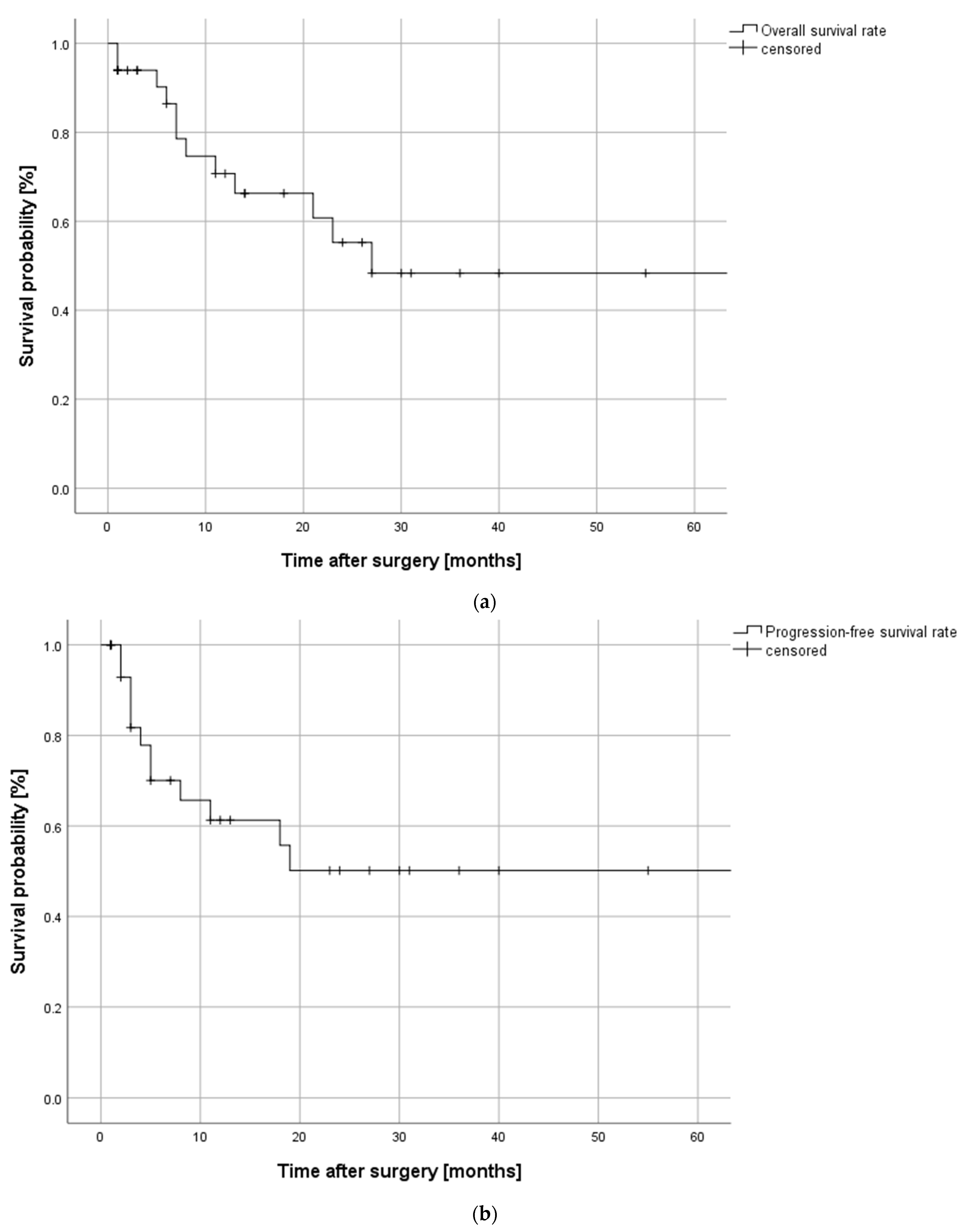
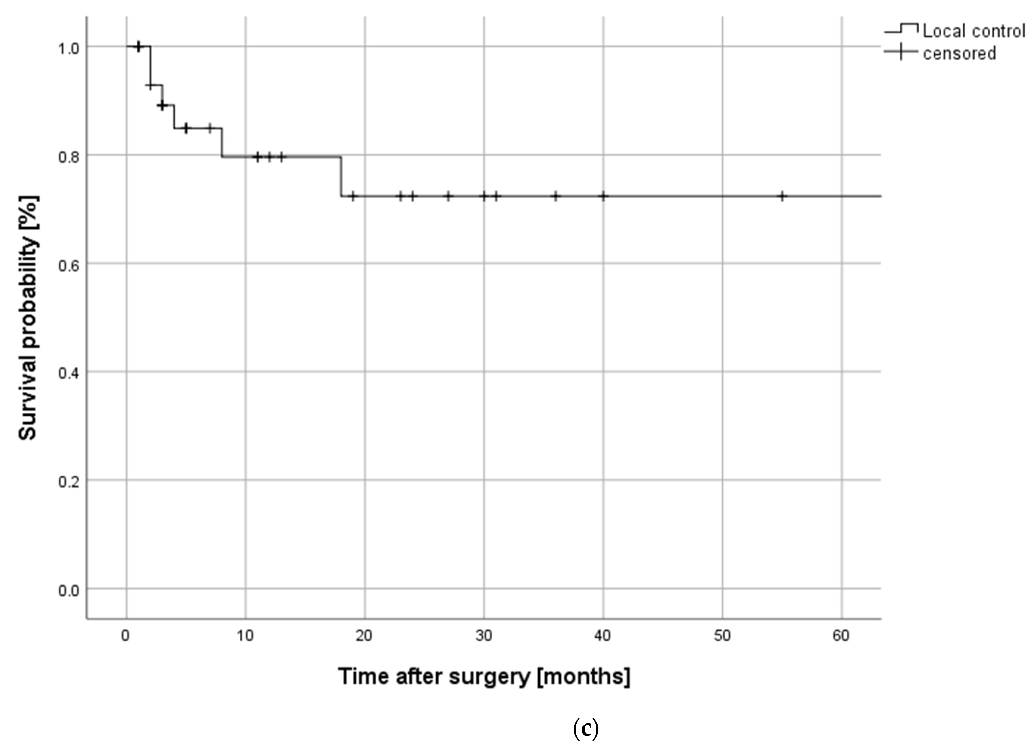
The mean follow-up period was 21 ± 23 months with a range from 1 to 91 months. Overall survival, progression-free survival and the local control rate for the investigated cohort are depicted in Figure 2. Twelve patients (36.4%) experienced disease progression and 12 patients (36.4%) died during follow-up. The patients with disease progression developed a local recurrence in six cases (50%), regional recurrence in two cases (16.7%), and distant metastases in four cases (33.3%), all of which were located in the lung.

2.5.2. Functional Outcome

In all patients, adequate reconstruction was achieved using a free flap. Although 12 patients (36.4%) were temporarily dependent on a percutaneous gastrostomy, the remaining patients (n = 21/63.6%) were able to maintain sufficient oral nutrition postoperatively. Figure 2 exemplifies the preoperative situation, surgical procedures, and postoperative outcome of a patient with recurrent squamous cell carcinoma of the maxilla. Figure 3 and Figure 4 exemplify the preoperative situation, and postoperative outcome of two patients with advanced squamous cell carcinoma of the oral cavity.

3. Discussion

Despite advances in screening and diagnosis of head and neck cancer, a significant proportion of patients presents with locally advanced disease [10,11]. As surgical therapy remains the primary modality for the treatment of locally advanced oral squamous cell carcinomas (OSCCs), the extent of tumor resection has to be carefully balanced against possible functional and aesthetic impairments affecting the patient’s quality of life. In patients in which oral cavity tumors exhibit involvement of the external skin, in particular, extensive resection might be challenging and usually requires the use of free flaps for reconstruction. As residual disease is accompanied by worse overall and progression-free survival, a critical goal of surgery must be the achievement of clear oncologic margins [12]. Nonetheless, radical surgical intervention may lead to functional and aesthetic impairments.
Hence, the present manuscript exclusively focuses on patients with locally advanced OSCCs involving the external skin, independent of their medical history regarding the sequence of the disease. This heterogeneous cohort integrating patients with primary and recurrent disease was chosen because it offers the possibility to exemplify and discuss aspects of synergism between surgical oncology and reconstructive surgery.
The main objective of oncological surgery is to create tumor-free margins, which in this cohort was successfully achieved in 78.8% of the cases. Although this rate generally might be critically discussed, it has to be considered under the aspect of the dominance of advanced tumors in this cohort (97% T3/T4 tumors), including 36.4% recurrent tumors and a history of prior multi-modality-treatment in 33.3% of the patients. Therapeutic considerations for patients with primary disease are generally different from those with recurrent disease, especially regarding potential alternatives. However, the presented cohort offers certain peculiarities that should be respected concerning treatment planning. Although primary radiotherapy is a valuable option for radiation-naive patients suffering from head and neck cancer that offers good local and regional control rates, there are advantages of a primary surgical approach that should be considered [13]. These include the possibility to further improve the outcome if complete tumor resection is achieved, to guide adjuvant therapy by possibly detecting occult lymph node metastases, and to restore areas where extensive tumor growth may lead to defect healing if primary radiotherapy is applied. As other therapeutic approaches including the newly introduced immune-checkpoint inhibition are, at present, restricted to palliative settings and therapeutic response is limited to a fraction of patients, the affected patients need profound guidance regarding the implications of the proposed therapies and their rejection [4,14].
Several retrospective and prospective studies evaluated the concept of salvage surgery for patients suffering from recurrent head and neck cancer, mostly advocating the use of free flaps for the reconstruction of large tissue defects [14,15,16,17,18]. Although the term “salvage surgery” mostly applies to tumor resections with curative intent in patients with recurrent tumors and a history of previous (radio-) therapies, and consequently lacking alternatives to the surgical approach, the focus of the present study lies on extensive resections of head and neck tumors irrespective of the sequence of prior therapy to exemplify the possibilities and limitations of tumor surgery of the head and neck in general. Consequently, the presented cohort is heterogeneous regarding the medical history of the patients and limited in size due to the focus on cases with a need to perform full-thickness tumor resections. Although most of the mentioned publications focus on salvage surgery approaches, the general considerations mostly also apply to patients with advanced primary tumors in sensitive localizations. In every case, critical balancing of the patient’s individual situation and wishes is urgently warranted, including prognosis, the patient’s physical and mental capacity, and the chance for therapeutic success.
The oncological results in our analysis demonstrate a high local control rate and, although the overall and progression-free survival rate is typical for a cohort of patients suffering from advanced head and neck cancer, only six cases (18.2%) of local recurrence occurred, and the remaining six cases of disease recurrence were attributable to regional or distant metastases. These results, although drawn from a small cohort, are comparable to those reported in the available literature and confirm the validity of our data [19,20]. Furthermore, in many of the presented cases, surgery was the only available curative therapeutic option due to a history of radiotherapy in the cases of recurrent tumors. In other cases, a definitive radio(-chemo)-therapy was abandoned because of the tumor’s vicinity or its manifest infiltration of sensitive structures with a high risk of postoperative osteonecrosis (e.g., the mandible) [21]. Additionally, extensive tumors carry a risk of a defect healing after radiotherapy, especially if they cause a connection between the oral cavity and the facial skin. In those cases, a primary surgical approach to restore the anatomic integrity of the oral cavity and the facial skin appears sensible for obvious aesthetic and functional reasons, because there is plethora of publications on the higher complication rate of reconstructive surgical approaches in patients with a history of radiotherapy [22,23,24].
The ALT was the most frequently used flap in our cohort. It is a versatile option for the extensive defects due to its volume, good pedicle length, and comparably low donor site morbidity [25,26,27,28]. It is especially suitable for full-thickness defects, because partial de-epithelization of the flap allows for the creation of separate skin paddles that may be tunneled through the defect and may be used for the restoration of the intra- and extraoral parts of the defect even if only one perforator is present. Other flaps used in our cohort were the radial forearm flap, the scapula flap, the fibula flap, and the latissimus dorsi flap. The overall flap success rate in our cohort was 84.8%, which is inferior to that reported by other authors [18,24]. Flap success rates were different between patients with primary (flap success: 90.5%) and recurrent disease (75%), which may be explained by the oft-reported tendency towards higher complication rates in patients with a history of prior reconstructive approaches and radiotherapy [24,29,30]. Additionally, the general need for bigger flaps for full-thickness defects may carry an increased risk of flap failure.
Several authors have reported on techniques for the restoration of facial full-thickness defects. They proposed free flaps including the ALT, the fibula flap, and the radial forearm flap, alone or in combination with rotational flaps for the extraoral parts of the defect [31,32,33,34]. As most of the reported studies focused on small numbers of patients, to the best of our knowledge this article describes one of the largest cohorts with full-thickness defects.
Although there was an accumulation of tumors in distinct subsites of the oral cavity (mandible/floor of the mouth, buccal mucosa, maxilla) eventually leading to full-thickness tumor resection, each patient needs individual treatment planning and, thus, patients with similar expected defects may require different reconstructive approaches to achieve an optimal or at least satisfying outcome. Several factors are of critical relevance for the choice of the reconstructive technique, including medical history, the sequence of reconstruction, and the patient’s individual physical condition.
This study has several limitations. The retrospective nature of the analysis is problematic regarding the case selection and the significance of deduced statements. As stated previously, the focus on a special subset of tumor patients that was mainly defined by the necessity for full-thickness resection led to a small cohort. The inclusion of primary and recurrent tumors aggravates the interpretation of the survival data and does not allow for valid conclusions. Nevertheless, the authors think that the presented study may be regarded as a legitimate suggestion for a possible treatment regime for a subset of head and neck cancer patients that is relevant and challenging regarding oncological and reconstructive considerations.

4. Materials and Methods

The presented retrospective study was conducted with respect to the principles of the Declaration of Helsinki and was approved by the local ethics committee of the University of Heidelberg (Ethic vote: S-183/2015). Written informed consent was given by all patients. All patients receiving full-thickness tumor resection of a squamous cell carcinoma of the oral cavity (primary and recurrent tumors) in the Department of Oral and Cranio-Maxillofacial Surgery of the Heidelberg University Hospital between 2010 and 2020 were included in the analysis. Secondly, patients who received full-thickness tumor resections were identified for further investigation. Demographic, clinical, and pathological data were extracted from the electronic patient records and transferred to an anonymized data processing sheet.
Statistical analysis was performed using IBM SPSS Statistics® 25 (IBM, Armonk, New York, NY, USA). Categorial data were described using descriptive analysis.
Data regarding overall and progression-free survival data and follow-up recalls were collected from the electronic patient document and transferred in SPSS. Survival was assessed using the Kaplan–Meier method as the time between initial therapy and the date of death or last follow-up (censored) for overall survival, and between initial therapy and the date of local, regional or distant disease progression or last follow-up (censored) for progression-free survival and local control rate, accordingly. Data cutoff refers to 31st December 2020.

5. Conclusions

Surgical tumor therapy with curative intent is based on the achievement of tumor-free margins wherever possible. If clear margins seem achievable preoperatively, the surgical procedures including reconstructive strategies should integrate oncological and functional considerations to obtain an optimal outcome in terms of survival and postoperative quality of life. Extensive tumor resections in the head and neck area including full-thickness resections are feasible, and free flap reconstruction offers a versatile tool to restore function and aesthetics while providing good local disease control rates.

Author Contributions

Conceptualization, J.M. (Julius Moratin) and C.F.; methodology, J.M. (Julius Moratin), K.M. and D.H.; software, J.M. (Julius Moratin), S.Z. and K.M.; validation, J.M. (Jan Mrosek), O.R., C.F., J.H., S.Z., M.E., K.F. and D.H.; formal analysis, J.M. (Julius Moratin), K.M., J.M. (Jan Mrosek) and O.R.; investigation, J.M. (Julius Moratin), K.M., J.M. (Jan Mrosek), S.Z.; resources, D.H., K.F., C.F.; data curation, J.M. (Julius Moratin), D.H., J.M. (Jan Mrosek), K.M., S.Z.; writing—original draft preparation, J.M. (Julius Moratin), K.M., J.M. (Jan Mrosek); writing—review and editing, O.R., S.Z., M.E., J.H., D.H., and J.H.; visualization, J.M. (Julius Moratin) and S.Z.; supervision, C.F.; project administration, C.F., K.F. and J.H. All authors have read and agreed to the published version of the manuscript.

Funding

This research received no external funding.

Institutional Review Board Statement

The study was conducted according to the guidelines of the Declaration of Helsinki, and approved by the Institutional Ethics Committee of the University of Heidelberg (S-183/2015, 27.04.2015).

Informed Consent Statement

Informed consent was obtained from all subjects involved in the study.

Data Availability Statement

The data presented in this study are available on request from the corresponding author.

Acknowledgments

This study was supported by the Medical Faculty of the University of Heidelberg.

Conflicts of Interest

The authors declare no conflict of interest.

References

  1. Ferlay, J.; Soerjomataram, I.; Dikshit, R.; Eser, S.; Mathers, C.; Rebelo, M.; Parkin, D.M.; Forman, D.; Bray, F. Cancer incidence and mortality worldwide: Sources, methods and major patterns in GLOBOCAN 2012. Int. J. Cancer 2015, 136, E359–E386. [Google Scholar] [CrossRef]
  2. Ferris, R.L.; Blumenschein, G., Jr.; Fayette, J.; Guigay, J.; Colevas, A.D.; Licitra, L.; Harrington, K.J.; Kasper, S.; Vokes, E.E.; Even, C.; et al. Nivolumab vs investigator’s choice in recurrent or metastatic squamous cell carcinoma of the head and neck: 2-year long-term survival update of CheckMate 141 with analyses by tumor PD-L1 expression. Oral Oncol. 2018, 81, 45–51. [Google Scholar] [CrossRef]
  3. Harrington, K.J.; Ferris, R.L.; Blumenschein, G., Jr.; Colevas, A.D.; Fayette, J.; Licitra, L.; Kasper, S.; Even, C.; Vokes, E.E.; Worden, F.; et al. Nivolumab versus standard, single-agent therapy of investigator’s choice in recurrent or metastatic squamous cell carcinoma of the head and neck (CheckMate 141): Health-related quality-of-life results from a randomised, phase 3 trial. Lancet Oncol. 2017, 18, 1104–1115. [Google Scholar] [CrossRef]
  4. Burtness, B.; Harrington, K.J.; Greil, R.; Soulieres, D.; Tahara, M.; de Castro, G., Jr.; Psyrri, A.; Baste, N.; Neupane, P.; Bratland, A.; et al. Pembrolizumab alone or with chemotherapy versus cetuximab with chemotherapy for recurrent or metastatic squamous cell carcinoma of the head and neck (KEYNOTE-048): A randomised, open-label, phase 3 study. Lancet 2019, 394, 1915–1928. [Google Scholar] [CrossRef]
  5. Urken, M.L.; Weinberg, H.; Buchbinder, D.; Moscoso, J.F.; Lawson, W.; Catalano, P.J.; Biller, H.F. Microvascular free flaps in head and neck reconstruction. Report of 200 cases and review of complications. Arch. Otolaryngol. Head Neck Surg. 1994, 120, 633–640. [Google Scholar] [CrossRef]
  6. McConnel, F.M.; Pauloski, B.R.; Logemann, J.A.; Rademaker, A.W.; Colangelo, L.; Shedd, D.; Carroll, W.; Lewin, J.; Johnson, J. Functional results of primary closure vs flaps in oropharyngeal reconstruction: A prospective study of speech and swallowing. Arch. Otolaryngol. Head Neck Surg. 1998, 124, 625–630. [Google Scholar] [CrossRef] [PubMed] [Green Version]
  7. Di Taranto, G.; Chen, S.H.; Elia, R.; Sitpahul, N.; Chan, J.C.Y.; Losco, L.; Cigna, E.; Ribuffo, D.; Chen, H.C. Outcomes following head neck free flap reconstruction requiring interposition vein graft or vascular bridge flap. Head Neck 2019, 41, 2914–2920. [Google Scholar] [CrossRef]
  8. Podrecca, S.; Salvatori, P.; Squadrelli Saraceno, M.; Fallahdar, D.; Calabrese, L.; Cantu, G.; Molinari, R. Review of 346 patients with free-flap reconstruction following head and neck surgery for neoplasm. J. Plast. Reconstr. Aesthet. Surg. 2006, 59, 122–129. [Google Scholar] [CrossRef] [PubMed]
  9. Hurvitz, K.A.; Kobayashi, M.; Evans, G.R.D. Current options in head and neck reconstruction. Plast. Reconstr. Surg. 2006, 118, 122e–133e. [Google Scholar] [CrossRef] [PubMed]
  10. Gatta, G.; Botta, L.; Sanchez, M.J.; Anderson, L.A.; Pierannunzio, D.; Licitra, L.; Group, E.W. Prognoses and improvement for head and neck cancers diagnosed in Europe in early 2000s: The EUROCARE-5 population-based study. Eur. J. Cancer 2015, 51, 2130–2143. [Google Scholar] [CrossRef] [PubMed]
  11. Madhura, M.G.; Rao, R.S.; Patil, S.; Fageeh, H.N.; Alhazmi, A.; Awan, K.H. Advanced diagnostic aids for oral cancer. Dis. Mon. 2020, 66, 101034. [Google Scholar] [CrossRef]
  12. Hinni, M.L.; Ferlito, A.; Brandwein-Gensler, M.S.; Takes, R.P.; Silver, C.E.; Westra, W.H.; Seethala, R.R.; Rodrigo, J.P.; Corry, J.; Bradford, C.R.; et al. Surgical margins in head and neck cancer: A contemporary review. Head Neck 2013, 35, 1362–1370. [Google Scholar] [CrossRef]
  13. Hosni, A.; Chiu, K.; Huang, S.H.; Xu, W.; Huang, J.; Bayley, A.; Bratman, S.V.; Cho, J.; Giuliani, M.; Kim, J.; et al. Non-operative management for oral cavity carcinoma: Definitive radiation therapy as a potential alternative treatment approach. Radiother. Oncol. 2020, 154, 70–75. [Google Scholar] [CrossRef] [PubMed]
  14. Patil, V.M.; Noronha, V.; Thiagarajan, S.; Joshi, A.; Chandrasekharan, A.; Talreja, V.; Agarwal, J.; Ghosh-Laskar, S.; Budrukkar, A.; Juvekar, S.; et al. Salvage surgery in head and neck cancer: Does it improve outcomes? Eur. J. Surg. Oncol. 2020, 46, 1052–1058. [Google Scholar] [CrossRef] [PubMed]
  15. Horn, D.; Bodem, J.; Freudlsperger, C.; Zittel, S.; Weichert, W.; Hoffmann, J.; Freier, K. Outcome of heavily pretreated recurrent oral squamous cell carcinoma after salvage resection: A monocentric retrospective analysis. J. Cranio-Maxillofac. Surg. 2016, 44, 1061–1066. [Google Scholar] [CrossRef] [PubMed]
  16. Horn, D.; Zittel, S.; Moratin, J.; Metzger, K.; Ristow, O.; Krisam, J.; Bodem, J.; Engel, M.; Freudlsperger, C.; Hoffmann, J.; et al. Prospective feasibility analysis of salvage surgery in recurrent oral cancer in terms of quality of life. Oral Oncol. 2020, 102, 104580. [Google Scholar] [CrossRef] [PubMed]
  17. van Weert, S.; Leemans, C.R. Salvage surgery in head and neck cancer. Oral Dis. 2021, 27, 117–124. [Google Scholar] [CrossRef] [PubMed]
  18. Baek, C.H.; Park, W.; Choi, N.; Gu, S.; Sohn, I.; Chung, M.K. Free flap outcome of salvage surgery compared to primary surgery for head and neck defects: A propensity score analysis. Oral Oncol. 2016, 62, 85–89. [Google Scholar] [CrossRef] [PubMed]
  19. Carvalho, A.L.; Nishimoto, I.N.; Califano, J.A.; Kowalski, L.P. Trends in incidence and prognosis for head and neck cancer in the United States: A site-specific analysis of the SEER database. Int. J. Cancer 2005, 114, 806–816. [Google Scholar] [CrossRef]
  20. Elbers, J.B.W.; Veldhuis, L.I.; Bhairosing, P.A.; Smeele, L.E.; Jozwiak, K.; van den Brekel, M.W.M.; Verheij, M.; Al-Mamgani, A.; Zuur, C.L. Salvage surgery for advanced stage head and neck squamous cell carcinoma following radiotherapy or chemoradiation. Eur. Arch. Oto-Rhino-Laryngol. 2019, 276, 647–655. [Google Scholar] [CrossRef]
  21. Aarup-Kristensen, S.; Hansen, C.R.; Forner, L.; Brink, C.; Eriksen, J.G.; Johansen, J. Osteoradionecrosis of the mandible after radiotherapy for head and neck cancer: Risk factors and dose-volume correlations. Acta Oncol. 2019, 58, 1373–1377. [Google Scholar] [CrossRef]
  22. Mijiti, A.; Kuerbantayi, N.; Zhang, Z.Q.; Su, M.Y.; Zhang, X.H.; Huojia, M. Influence of preoperative radiotherapy on head and neck free-flap reconstruction: Systematic review and meta-analysis. Head Neck 2020, 42, 2165–2180. [Google Scholar] [CrossRef]
  23. Halle, M.; Eriksson, B.O.; Docherty Skogh, A.C.; Sommar, P.; Hammarstedt, L.; Gahm, C. Improved Head and Neck Free Flap Outcome-Effects of a Treatment Protocol Adjustment from Pre- to Postoperative Radiotherapy. Plast. Reconstr. Surg. Glob. Open 2017, 5, e1253. [Google Scholar] [CrossRef]
  24. Benatar, M.J.; Dassonville, O.; Chamorey, E.; Poissonnet, G.; Ettaiche, M.; Pierre, C.S.; Benezery, K.; Hechema, R.; Demard, F.; Santini, J.; et al. Impact of preoperative radiotherapy on head and neck free flap reconstruction: A report on 429 cases. J. Plast. Reconstr. Aesthet. Surg. 2013, 66, 478–482. [Google Scholar] [CrossRef] [PubMed]
  25. Kesting, M.R.; Holzle, F.; Wales, C.; Steinstraesser, L.; Wagenpfeil, S.; Mucke, T.; Rohleder, N.H.; Wolff, K.D.; Hasler, R.J. Microsurgical reconstruction of the oral cavity with free flaps from the anterolateral thigh and the radial forearm: A comparison of perioperative data from 161 cases. Ann. Surg. Oncol. 2011, 18, 1988–1994. [Google Scholar] [CrossRef] [PubMed]
  26. Agostini, T.; Lazzeri, D.; Agostini, V.; Shokrollahi, K. Anterolateral thigh flap as the ideal flap to full-thickness cheek reconstruction. J. Craniofac. Surg. 2010, 21, 1897–1898. [Google Scholar] [CrossRef] [PubMed]
  27. Agostini, T.; Lazzeri, D.; Spinelli, G. Anterolateral thigh flap: Systematic literature review of specific donor-site complications and their management. J. Cranio-Maxillofac. Surg. 2013, 41, 15–21. [Google Scholar] [CrossRef] [PubMed]
  28. Horn, D.; Jonas, R.; Engel, M.; Freier, K.; Hoffmann, J.; Freudlsperger, C. A comparison of free anterolateral thigh and latissimus dorsi flaps in soft tissue reconstruction of extensive defects in the head and neck region. J. Cranio-Maxillofac. Surg. 2014, 42, 1551–1556. [Google Scholar] [CrossRef] [PubMed]
  29. Mulholland, S.; Boyd, J.B.; McCabe, S.; Gullane, P.; Rotstein, L.; Brown, D.; Yoo, J. Recipient vessels in head and neck microsurgery: Radiation effect and vessel access. Plast. Reconstr. Surg. 1993, 92, 628–632. [Google Scholar] [CrossRef]
  30. Demirkan, F.; Wei, F.C.; Chen, H.C.; Chen, I.H.; Hau, S.P.; Liau, C.T. Microsurgical reconstruction in recurrent oral cancer: Use of a second free flap in the same patient. Plast. Reconstr. Surg. 1999, 103, 829–838. [Google Scholar] [CrossRef]
  31. Jeng, S.F.; Kuo, Y.R.; Wei, F.C.; Su, C.Y.; Chien, C.Y. Reconstruction of concomitant lip and cheek through-and-through defects with combined free flap and an advancement flap from the remaining lip. Plast. Reconstr. Surg. 2004, 113, 491–498. [Google Scholar] [CrossRef] [PubMed]
  32. Sasaki, K.; Sasaki, M.; Oshima, J.; Aihara, Y.; Nishijima, A.; Sekido, M. Free-flap reconstruction for full-thickness oral defects involving the oral commissure combined with oral modiolus reconstruction using a fascial sling. Microsurgery 2020, 40, 553–560. [Google Scholar] [CrossRef] [PubMed]
  33. Savant, D.N.; Patel, S.G.; Deshmukh, S.P.; Gujarati, R.; Bhathena, H.M.; Kavarana, N.M. Folded free radial forearm flap for reconstruction of full-thickness defects of the cheek. Head Neck 1995, 17, 293–296. [Google Scholar] [CrossRef]
  34. Katou, F.; Shirai, N.; Kamakura, S.; Ohki, H.; Motegi, K. Full-thickness reconstruction of cheek defect involving oral commissure with forearm tendinocutaneous flap. Br. J. Oral Maxillofac. Surg. 1996, 34, 26–27. [Google Scholar] [CrossRef]
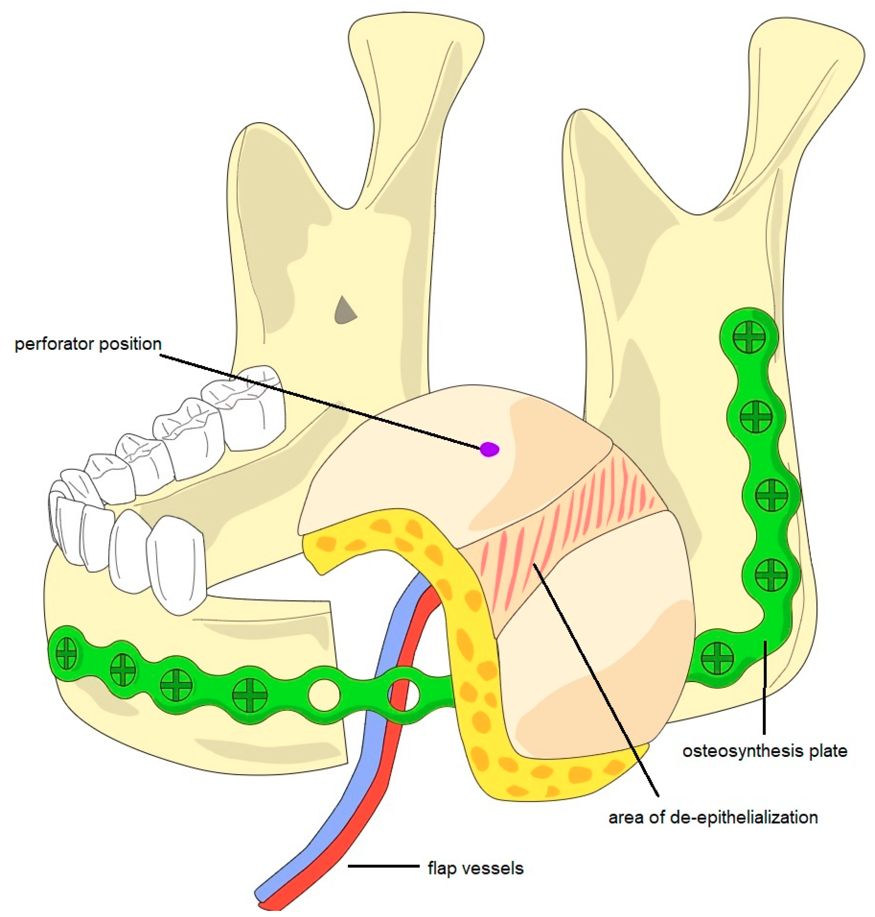
Figure 1. Schematic diagram of a partially de-epithelialized ALT flap and an osteosynthesis plate for the reconstruction of a full-thickness defect after tumor resection with segmental resection of the left-sided mandible. After segmental resection and the individualization of an osteosynthesis plate, the ALT flap was harvested. The prepared flap then underwent partial de-epithelialization in order to create two skin paddles for restoration of the oral and the facial defect.
Cancers 13 02122 g001
Figure 2. Kaplan–Meier curves depicting (a) overall survival, (b) progression-free survival, and (c) local control rate of the investigated cohort with full-thickness resections following advanced oral squamous cell carcinoma during a follow-up period of up to 60 months. Overall and progression-free survival rates exhibit a decrease to about 50% after 20–30 months, the local control rate shows a decrease to about 75% after 20 months.
Cancers 13 02122 g002aCancers 13 02122 g002b
Figure 3. Male patient with recurrent squamous cell carcinoma of the left-sided maxilla with infiltration of the maxillary sinus and facial skin. (a) CT scan depicting the infiltration of the paranasal sinus and the facial skin. (b) Full-frontal image of the preoperative situation with tumor progression of the left cheek. (c) Full-frontal image of the postoperative situation after full-thickness tumor resection and reconstruction with ALT flap.
Cancers 13 02122 g003
Figure 4. Male patient with primary squamous cell carcinoma of the right-sided mandible with infiltration of the facial skin. (a) Tumor infiltration of the right cheek. (b) CT scan depicting the tumor of the right-sided mandible with infiltration of the facial skin. (c) Full-frontal image of the postoperative situation after full-thickness tumor resection and reconstruction with ALT flap.
Cancers 13 02122 g004
Table 1. Descriptive data regarding demographic and clinical features of the investigated cohort.
ParameterNumber of Cases (%)
Gender
Female12 (36.4)
Male21 (63.6)
Age
Mean: 67 ± 13.7 years
Range: 25–95 years
Risk Factors
Tobacco17 (51.5)
Alcohol10 (30.3)
Primary or Recurrent Tumor
Primary tumor21 (63.6)
Recurrent tumor (local recurrence)12 (36.4)
Tumor Localization
Mandible + Floor of the mouth20 (60.6)
Buccal mucosa8 (24.2)
Maxilla5 (15.2)
T Stage
T1-
T21 (3.0)
T36 (18.2)
T426 (78.8)
N Stage
021 (63.6)
11 (3.0)
2a-
2b1 (3.0)
2c2 (6.1)
38 (24.2)
M Stage
033 (100)
1-
UICC Stage
1-
2-
34 (12.1)
429 (87.9)
Histopathological Tumor Features
Lymphatic invasion15 (45.5)
Vascular invasion4 (12.1)
Perineural invasion12 (36.4)
Table 2. Overview of reconstructive strategies and success rates in relation to affected anatomical subsite of the oral cavity.
Affected Subsite of the Oral CavityFree Flap (no.)Success Rate
Mandible + Floor of the Mouth
ALT (n = 15)12/15 (80%)
RFF (n = 1)1/1 (100%)
Scapula (n = 1)1/1 (100%)
Fibula (n = 2)2/2 (100%)
Buccal Mucosa
ALT (n = 7)7/7 (100%)
RFF (n = 1)1/1 (100%)
Maxilla
ALT (n = 2)2/2 (100%)
RFF (n = 1)1/1 (100%)
Scapula (n = 2)0/2 (0%)
Table 3. Overview on free flaps used for the restoration of full-thickness defects with revision and success rates.
Type of Free FlapRevision RateSuccess Rate
ALT4/24 (16.7%)21/24 (87.5%)
RFF0/33/3 (100%)
Scapula3/3 (100%)1/3 (33.3%)
Fibula0/22/2 (100%)
Latissimus dorsi0/11/1 (100%)
Table 4. Considerations regarding the ALT flap as reconstructive technique of choice for full-thickness defects of the head and neck area.
Advantages of ALT Flaps
  • Good pedicle length
  • Especially suitable for extensive defects due to its bulkiness
  • Partial de-epithelization allows for the restoration of several neighboring defects, even if only one perforator is found
  • Low donor-site morbidity and high rate of primary closure of donor-sites
  • Low surgery times due to the possibility of a two-team approach
Disadvantages of ALT Flaps
  • Risk of aesthetic and functional impairment if a bulky flap is used for narrow defects
  • Risk of compromising the perforator if ALT is used for the coverage of metal plates
  • Need for secondary osseous reconstruction in cases of mandibular resections
Publisher’s Note: MDPI stays neutral with regard to jurisdictional claims in published maps and institutional affiliations.