Review

The Tubulin Code and Tubulin-Modifying Enzymes in Autophagy and Cancer

by
Daniela Trisciuoglio
* and
Francesca Degrassi
*
IBPM Institute of Molecular Biology and Pathology, CNR Consiglio Nazionale delle Ricerche, c/o Department of Biology and Biotechnology “Charles Darwin”, Sapienza University of Rome, 00185 Rome, Italy
*
Authors to whom correspondence should be addressed.
Cancers 2022, 14(1), 6; https://doi.org/10.3390/cancers14010006
Submission received: 11 November 2021 / Revised: 15 December 2021 / Accepted: 16 December 2021 / Published: 21 December 2021
(This article belongs to the Special Issue Microtubule Dynamics and Cancer)

Abstract

:

Simple Summary

Microtubules are tubulin polymers that constitute the structure of eukaryotic cells. They control different cell functions that are often deregulated in cancer, such as cell shape, cell motility and the intracellular movement of organelles. Here, we focus on the crucial role of tubulin modifications in determining different cancer characteristics, including metastatic cell migration and therapy resistance. We also discuss the influence of microtubule modifications on the autophagic process—the cellular degradation pathway that influences cancer growth. We discuss findings showing that inducing microtubule modifications can be used as a means to kill cancer cells by inhibiting autophagy.

Abstract

Microtubules are key components of the cytoskeleton of eukaryotic cells. Microtubule dynamic instability together with the “tubulin code” generated by the choice of different α- and β- tubulin isoforms and tubulin post-translational modifications have essential roles in the control of a variety of cellular processes, such as cell shape, cell motility, and intracellular trafficking, that are deregulated in cancer. In this review, we will discuss available evidence that highlights the crucial role of the tubulin code in determining different cancer phenotypes, including metastatic cell migration, drug resistance, and tumor vascularization, and the influence of modulating tubulin-modifying enzymes on cancer cell survival and aggressiveness. We will also discuss the role of post-translationally modified microtubules in autophagy—the lysosomal-mediated cellular degradation pathway—that exerts a dual role in many cancer types, either promoting or suppressing cancer growth. We will give particular emphasis to the role of tubulin post-translational modifications and their regulating enzymes in controlling the different stages of the autophagic process in cancer cells, and consider how the experimental modulation of tubulin-modifying enzymes influences the autophagic process in cancer cells and impacts on cancer cell survival and thereby represents a new and fruitful avenue in cancer therapy.

1. Introduction

1.1. Microtubule Dynamic Instability

Microtubules (MTs) are hollow cylinders of approximately 25 nm formed by the polymerization of α-/β-tubulin dimers. MT polymers are intrinsically polar with a faster addition and release of tubulin dimers at the plus end and a slower dynamics at the more stable minus end.
In interphase cells, MTs are key components of the filamentous cytoskeletal network, acting in concert with actin filaments and intermediate filaments. In most cell types, MTs regulate the spatial organization of the cell, contributing to cell shape and cell motility, and control intracellular spatial functions, such as organelle positioning and intracellular transport. MTs have a fundamental role in intracellular trafficking, allowing the movement of signaling molecules or organelles, such as the endoplasmic reticulum, the Golgi complex, mitochondria, and autophagic vesicles. In this regard, MTs function as preassembled tracks on which kinesins (plus-end-directed) or cytoplasmic dynein (minus-end-directed) motor proteins transport cargoes by exerting pulling and pushing forces on dynamic MTs or by sliding interdigitating MTs (for a review see [1]).
In dividing cells, MTs control the process of mitosis at different stages of its progression. At mitotic entry, the MT network extensively reorganizes to form the mitotic spindle, a fusiform structure of two radial arrays of short, dynamic MTs that govern chromosome segregation. At cytokinesis, cytoplasmic division intervenes at the residual MT structure called midbody. At the heart of these different functions lies the “dynamic instability” of MTs, the filament property for which individual MTs undergo continuous cycles of rapid depolymerization (catastrophe) and polymerization (rescue) at their plus end [2]. Dynamic instability continuously remodels the MT network, allowing individual MTs to explore the intracellular space, a property that is fundamental during mitosis when MTs grow and shrink rapidly until they bind a chromosome at kinetochore [2]. This binding restrains MT dynamics, allowing a robust attachment of chromosomes to the mitotic spindle, a prerequisite for correct chromosome segregation. Indeed, control of MT dynamics at mitosis has been shown to be fundamental in faithful chromosome segregation, so that altered MT dynamics can lead to chromosome segregation errors and chromosome instability, a key feature of cancer cells. A plethora of studies in the last 20 years have delineated the connection between cancer, chromosome instability, and control of MT dynamics, and these factors are discussed in several excellent reviews to which the reader can refer [3,4,5]. Importantly, enhanced tubulin dynamics at mitosis is also the molecular target for numerous so-called anti-tubulin agents that have been shown to interact with several sites on α- or β-tubulin and have been successfully used as chemotherapeutic drugs to cause mitotic arrest and cell death of cancer cells [6,7,8,9].

1.2. The α-/β-Tubulin Code

The tubulin superfamily in humans comprises five families: alpha-(α), beta-(β), gamma-(γ), delta-(δ), and epsilon-(ε) tubulin. Nine α-tubulin (TubA), ten β-tubulin (TubB), two γ-tubulin (TubG), one δ-tubulin (TubD), and one ε-tubulin (TubE) isoforms have been identified. Tubulin isoforms derive from different genes that are located on different chromosomes, and their aberrant expression or mutation is associated with several human pathologies [10,11]. TubA and TubB isoforms are the main components of MTs, and they share a high degree of homology at the N-terminal and intermediate domain, while they differ significantly in the length of the C-terminal tail, the region that harbors interaction sites for MT-associated proteins (MAPs) and MT molecular motors [12,13,14,15].
TubG is the main component of multiprotein complexes that are found at MT organ-izing centers, such as centrosomes in most animal cells. TubG-containing complexes nucleate MTs both at MT organizing centers and at other intracellular sites, such as the Golgi apparatus and preexisting MTs [16,17,18]. TubD and TubE are also centrosomal proteins, but they display specific localization patterns: TubD is associated with centrioles, whereas TubE localizes to the pericentriolar material with a cell cycle-specific localization pattern [19].
In the last years, the idea that different TubA and TubB gene products, together with a variety of tubulin post-translational modifications (PTMs), generate a code, named the “tubulin code”, has attracted a great deal of interest [20]. Hence, individual MTs, harboring a specific tubulin code, have been shown to have different properties or architectures between cell types or even within a single cell due to their peculiar ability to interact with MAPs or MT molecular motors [21,22].
TubA and TubB isoforms are for the most part ubiquitously expressed but some of them have cell- or tissue-specific expression. For example, TubB1 is expressed only in platelets and megakaryocytes [15], whereas TubB3 is physiologically expressed in cells of neuronal origins [12,13,14] and TubB4 in the axonemes of cilia and flagella [23]. The tubulin isotype composition of MTs has a clear impact on MT dynamics. In 1994, Panda and coauthors showed that MTs composed of TubA and TubB3 isotypes exhibit unique polymerization dynamics, being more dynamic than MTs formed of TubA and TubB2 or TubB4 isotypes [24]. A more recent study, comparing the dynamics of neuronal MTs and MTs obtained from human embryonic kidney cells, has shown that the latter MTs are more stable and grow faster than neuronal MTs [25]. Overall, these studies confirmed the role of tubulin isoform composition on MT dynamics.
Tubulin PTMs are a key player in the tubulin code, and they have been shown to exert a clear impact on different MT functions. In the last few years, the discovery of tubulin-modifying enzymes has allowed researchers to decipher the role of tubulin PTMs in MT dynamics (Table 1) and to unveil the mechanisms by which specific PTMs regulate MT function in different cell contexts (for a comprehensive review see [26]). PTMs relevant to cancer occur abundantly and reversibly on tubulin and are mostly localized at the C-terminal tail of the protein; some PTMs, including acetylation, detyrosination, and polyglutamylation, are specifically associated with polymerized tubulin, while others, like tyrosination, occur only on soluble tubulin; polyamination intervenes both on soluble and polymerized tubulin.
Tubulin acetylation refers to the transfer of the acetyl group from acetyl-coenzyme A (CoA) at the Lysine-40 residue (K40) of TubA, which is exposed in the lumen of MTs. In normal cells, acetylated TubA is associated with long-lived stable MTs within centrioles, primary cilia, and flagella [27]. To date, the relationship between tubulin acetylation and MT stability is not fully understood. The predominant idea was that only stable MTs may be acetylated, based on experiments showing that drug-induced MT stabilization determines an accumulation of acetylated MTs [28] or on studies linking decreased tubulin acetylation with reduced MT stability [29]. More recent studies have proved that acetylation reduces the rigidity of MTs, making them more resistant to mechanical breakage and disassembly [30,31]. The alpha-tubulin N-acetyltransferase 1 (ATAT1 or MEC17) has been identified as being responsible for K40 acetylation on TubA in mammalian cells [32], whereas the reverse reaction is catalyzed by the HDAC6 and/or sirtuin type 2 (SIRT2) deacetylases [29,33]. However, other acetyltransferases, such as GCN5, can also acetylate α-tubulin in different cell contexts [34]. More recently, another reaction occurring at Lysine-252 (K252) of TubB, favoring MT depolymerization, has also been described [35]. The acetyltransferase SAN catalyzes K252 acetylation of TubB, whereas the enzyme catalyzing the reverse reaction has not been defined yet [35]. Functionally, tubulin acetylation has been linked to the recruitment of specific motor proteins to MTs and to intracellular trafficking [36,37]. Consequently, tubulin acetylation plays an important role in several cellular activities, including cell polarity, cell migration, vesicle transport, and cell development. Indeed, abnormal tubulin acetylation levels have been linked to a number of different diseases, including ciliopathies, neurodegenerative disorders, and cancer (for a review see [38]). Detyrosination/tyrosination of TubA also occurs in the C-terminal tail and the cycling of this modification on tubulin is closely connected to MT dynamics: newly polymerized MTs are mostly tyrosinated, while long-lived MTs are typically detyrosinated, so that detyrosination has long been considered a marker of MT longevity [39]. The tyrosination state of MTs can also modify the binding affinity for motor proteins: some kinesins bind preferentially detyrosinated MTs, while cytoplasmic dynein interacts more efficiently with tyrosinated MTs [40]. Consequently, specific cargo transport has been found to use different MT subsets, with important implications in cell division, neuronal, and cardiac physiology [41,42,43]. The tubulin terminal tyrosine is removed by cytosolic carboxypeptidases (CCPs) to expose a glutamate, whereas the reverse reaction is catalyzed by tubulin–tyrosine ligase (TTL). Two redundant CCPs, vasohibin-1 (VASH-1) and vasohibin-2 (VASH-2), have been found to detyrosinate MTs in a complex with a chaperone-like small vasohibin-binding peptide (SVBP) [44,45]. The detyrosination/tyrosination cycle is completed by TTL, which binds only tubulin dimers and adds a tyrosine residue to detyrosinated tubulin [46].
Finally, glutamate side chains of different lengths can be added to the C-terminal tail of both TubA and TubB on glutamate residues. This reaction, named mono- or polyglutamylation, adds one or multiple glutamic acids to a γ-carboxyl group of a glutamate residue of tubulin and is catalyzed by several TTL-like (TTLL) mono- or polyglutamylases, a large family of proteins harboring a TTL homology domain. The reverse reaction is carried out by a deglutamylase from the CCP family. Tubulin polyglutamylation, preferentially occurring in cilia MTs, regulates multiple interactions between MTs and their associated proteins, such as MAPs and molecular motors. For instance, the activity of several molecular motors, including kinesin-1 and kinesin-2, can be regulated by tubulin isotypes and various degrees of MT polyglutamylation [37].
Overall, the combination of tubulin PTMs and isotypes may generate a subpopulation of MTs with specific dynamic properties that influence different cellular effectors, such as MAPs or motor proteins. However, work is still needed to determine the impact of less well characterized tubulin PTMs (e.g., methylation, polyamination) on MT structure in diverse cell types.

2. The Tubulin Code and Its Associated Enzymes in Cancer

In cancer cells, alterations in MT dynamics, often associated with cancer-specific tubulin isotypes and tubulin PTMs, have been shown to be involved in metastatic cell migration, drug resistance, and tumor vascularization [5,38]. Accordingly, anti-tubulin agents have been proved to have anti-angiogenic and vascular-disrupting properties, as well as effects on cellular migration and intracellular trafficking [47]. However, the relevance of these effects to the anti-tumor activity of anti-tubulin drugs has been overlocked in the past due to the efficacy of their mitotic action.
In several cancer cell contexts, emerging studies highlight a role for tubulin isotypes in influencing MT behavior and function in the metastatic ability and chemotherapy resistance of cancer cells (Table 2). Indeed, a differential TubA1 isotype expression has been found in various types of cancer and correlates with poor outcome/prognosis as well as with resistance to therapy [48,49,50,51,52]. TubA1B expression was found to be upregulated in hepatocellular carcinoma (HCC) tumor tissues and correlated with poor overall survival and resistance to paclitaxel in HCC patients [48]. Recently, it was demonstrated that glioma tissues have higher TubA1C expression than normal brain tissues and that high TubA1C levels are an indicator of worse prognoses in glioma patients, thereby suggesting that TubA1C may be a therapeutic biomarker for gliomas [49,52].
The frequency and impact of TubB isoforms and mutations in cancer is still not fully understood. TubB1 is the most common isotype in human lung cancer and breast cancer cell lines [53]. TubB1 expression is correlated with taxane resistance in breast cancer [54].
Class 3 of TubB is the prominent isoform linked to neoplastic disease and has been identified as a biomarker for resistance to MT-targeting chemotherapeutics in breast and other types of solid cancer [55]. TubB3 overexpression has been linked to aggressive tumor features, genetic instability, and poor prognosis in urinary bladder cancer and clear cell renal cell carcinoma [56,57]. Aberrant expression of TubB3, TubB2, TubB4A, TubB4B, and TubB5 isotypes has been detected in several tumor types and is also associated with resistance to tubulin-binding agents, such as taxanes and vinca alkaloids, in different cancer cell types [54,58,59,60,61,62,63,64,65,66]; this may be due to the specific properties of tubulin isoforms which could alter the sensitivity of MTs to this class of drugs.
Among tubulin PTMs, tubulin acetylation was first described 20 years ago and has recently attracted growing interest in the field of cancer research. Many studies have suggested alterations in tubulin acetylation as potential prognostic biomarkers in different cancers, including head and neck, breast, and pancreatic cancer [67,68]. Boggs and collaborators have linked acetylated tubulin levels to the metastatic process in breast cancer, suggesting a relationship between high levels of acetylation and metastatic behavior of basal-like breast cancers [68]. More recently, acetylated tubulin levels have been associated with paclitaxel sensitivity in lung cancer. Indeed, tubulin acetylation enhances the resistance to paclitaxel-induced cell death. Mechanistically, tubulin acetylation stabilizes the level of the anti-apoptotic protein Mcl-1 by protecting it from degradation [69]. In line with these studies, aberrant expression of tubulin deacetylase HDAC6 has been reported in several cancer cell lines and tumor models [70,71]. In addition, upregulation of HDAC6 increases cell motility in breast cancer cells, thus contributing to cancer metastasis [72], whereas in glioblastoma cells genetic silencing of HDAC6 decreases cellular malignancy and reverses the mesenchymal phenotype [73,74].
Table 2. Significance of aberrant expression of tubulin isotypes in cancer.
Tubulin IsotypeAlterationCancer TypeEffectReferences
TubA1AHigh levelsGastricMacrophage infiltration in tumor microenvironment[50]
TubA1BHigh levelHepatocellular carcinomaPoor overall survival and resistance to paclitaxel[48]
TubA1CHigh levelGliomaPoor prognosis[52]
High levelLungImmune cell infiltration[51]
TubB1High levelBreastTaxane resistance[54]
TubB2DepletionLungEnhanced sensitivity to Vinca alkaloids[58]
Low levelOvarian and breastResistance to taxanes; correlated with advanced stages[59,60]
High levelLungBiomarker for tumor differentiation and aggressiveness[61]
Tub3High levelOvarianCorrelated with advanced stages[59]
High levelClear cell renal cell carcinomaPoor prognosis[56]
High levelProstatePoor overall survival[65]
High levelUrinary bladder cancerPoor prognosis[57]
High levelThyroid carcinomaInvasive potential and poor prognosis[62]
TubB4AHigh levelLungResistance to paclitaxel[63]
TubB4BDepletionLung cancer cellsEnhanced sensitivity to Vinca alkaloids[58]
Tub5High levelLungBiomarker for tumor differentiation and aggressiveness[61]
High levelLungTreatment response to paclitaxel[64]
On the other hand, the role of tubulin deacetylase SIRT2 in cancer is controversial, as SIRT2 exerts either tumor-suppressive or oncogenic properties. Overexpression of SIRT2 promotes cell stemness in renal cell carcinoma and endometrial cancer [75,76]. By contrast, overexpression of SIRT2 in lung cancer cell lines induces cell cycle arrest and apoptosis induction [77]. Conversely, the role of ATAT1 in cancer is greatly understudied, as compared with the role of this acetyltransferase in development and non-cancer diseases [27]. Some recent studies have reported that acetylation of tubulin by ATAT1 can affect various pathways, such as cell motility and mitosis, which in turn impinge on cancer cell proliferation, adhesion, invasion, and metastasis [78,79,80]. Notably, despite recent evidence which points to ATAT-1 as a significant player in several types of cancer, little is known about the underlying mechanisms and the relevance of ATAT1 acetyltransferase activity on cancer.
Tubulin detyrosination/tyrosination as well as the associated enzymes have a vital role in several physiological conditions and are associated with malignant transformation and cancer aggressiveness. Tubulin detyrosination seems to represent a selective advantage for cancer cells. Low TTL levels correlate with poor prognoses in several forms of cancer [81,82,83]. For example, tubulin detyrosination is frequent in breast cancer and has been linked to tumor aggressiveness [83]. Concordantly, TTL downregulation induces epithelial–mesenchymal transition and increases in vitro tumor invasion and the in vivo metastatic potential of breast cancer cells [81]. The VASH family, which includes vasohibin-1 (VASH1) and vasohibin-2 (VASH2), is a novel family of angiogenesis regulators [84] that act as tubulin-specific CCPs [40,45]. According to their role in angiogenesis, high expression of VASH1 and VASH2 was associated with poor clinical outcomes in gastric, ovarian, and esophageal squamous carcinoma patients [85,86,87,88]. Despite this, the relationship between the tubulin CCP activity of VASH1/2 and cancer progression or drug sensitivity is still a matter of debate. In ovarian cancer cells, the ablation of VASH2 reduced CCP activity and increased cyclin B1 expression results in increased paclitaxel sensitivity in ovarian cancer cells [89].
In a cancer cell context, altered polyglutamylation is linked to tumorigenesis and resistance to drug targeting MTs. A recent study showed that tubulin tyrosine ligase-like 4 (TTLL4) overexpression in breast cancer cells is associated with increased polyglutamylation of TubB, alteration of exosome homeostasis, and brain metastasis [90]. In pancreatic cancer cells, genetic downregulation of TTLL4 attenuates cell proliferation [91]. A new TTLL isoform named TTLL12 has recently been identified [92], and it has been suggested it acts as a potential molecular marker for predicting the invasion and progression of ovarian cancer [93]. On the other hand, the opposite enzyme, AGBL2, promotes tumorigenesis and cancer progression in breast, ovarian, renal, and hepatocellular carcinoma [94,95,96]. This suggests that AGBL2 may serve as a prognostic molecular marker and/or a potential target for therapy. In conclusion, an emerging role of tubulin-modifying enzymes in cancer-associated properties has been identified in different cancer models. Results are summarized in Table 3.

3. The Tubulin Code and Its Associated Enzymes in Autophagy

MT dynamic instability and the associated tubulin code have essential roles also in autophagy, a fundamental cellular process. Autophagy is a lysosomal-mediated cellular degradation pathway that exerts a dual role in many cancer types, either supporting cancer growth or acting as a tumor suppressor mechanism (for a review, see [97]). Thus, modulation of autophagy is a promising therapeutic strategy to fight cancer. In this part of the review, we will discuss the impact of tubulin isoforms and tubulin PTMs along with global MT dynamics in the different stages of the autophagic process and the ways in which they modulate the autophagic process in cancer cells. Finally, we will discuss recent findings that have implicated tubulin PTMs and their regulating enzymes in controlling the autophagic process in normal and cancer cells and how tubulin PTMs might be implicated in cancer cell properties or response to therapy.

3.1. The Autophagic Machinery: Mechanisms and Regulation

Autophagy is an evolutionarily conserved cellular process in which cellular debris, damaged proteins/organelles, and pathogens are degraded and/or recycled to maintain physiological cell homeostasis. Accordingly, autophagy dysfunction is involved in many diseases, including bacterial or viral infections, neurogenerative diseases, and cancer [97,98]. Beside baseline autophagy, several stimuli can trigger the process, including nutrient deprivation (non-selective macroautophagy) or the intracellular presence of specific degradation targets (selective macroautophagy). In both cases, autophagy originates by the nucleation of a double membrane around the material to be degraded, also referred as phagophore. Membrane expansion and shaping intervenes successively and reflects the form of the engulfed material in selective macroautophagy [99,100].
Membrane nucleation is stimulated by the activation of the unc-51-like autophagy activating kinase 1 (ULK1) complex consisting of ULK1, the non-catalytic focal adhesion kinase family interacting protein of 200 KD (FIP200), autophagy-related protein 13 (ATG13) and the ATG101 subunit. The activity of the ULK complex is under multi-layered regulation, so that the ULK1 complex is inhibited by the mammalian target of rapamycin complex 1 (mTORC1) through mTORC1-dependent inactivating phosphorylation of ULK1. Conversely, mTORC1 inactivation by different stimuli promotes ULK1 complex activation and autophagy [101]. In response to amino acid and ATP depletion, a further autophagy-inducing pathway involves the AMP-activated protein kinase (AMPK) that activates the ULK1 complex both by direct phosphorylation of ULK1 and by inhibitory phosphorylation of mTOR. Once activated, the ULK1 complex localizes at sites of condensed cargo via its interaction with the adaptor protein p62, promoting membrane nucleation [102]. Indeed, ULK1-dependent phosphorylation of Beclin 1, a subunit of the class III phosphatidylinositol 3-kinase (PI3K) complex leads to a local increment in phosphatidylinositol 3 phosphate at membrane sites known as pre-autophagosomal structures or omegasomes. Then, membrane expansion is sustained by different membrane sources, especially the endoplasmic reticulum, through the action of the lipid transporter ATG2A [103,104]. The ubiquitin-like ATG5–ATG12 conjugation and the autophagy-related 16-like 1 (ATG16L1) complex are then recruited to phagophores where phosphatidylethanolamine (PtdETn) is conjugated to cytosolic LC3 (known as LC3-I) to produce membrane-associated LC3-II, which acts as a universal adaptor for several protein cargoes and is commonly used as a marker for autophagy activation [105]. Finally, phagophore growth around its cargo leads to membrane convergence and closure of the two membrane ends by the endosomal sorting complexes required for transport (ESCRT) machinery [106]. The different steps in autophagosome biogenesis are outlined in Figure 1. Mature autophagosomes are then transported toward the centrosome, a cell region where lysosomes accumulate [107]. There, autophagosomes and lysosomes fuse to form autolysosomes, where the sequestered materials are degraded by the lysosomal lytic activities. Finally, the degradation products, including sugars, amino acids and nucleic acid precursors, are reused in cellular metabolism [108]. In conclusion, autophagy is a dynamic and complex process that includes autophagosome formation, maturation, fusion with lysosomes, and subsequent degradation of cargoes and autophagosomes themselves into the cytosol. The term “autophagic flux” refers to this whole process and is operationally used to assess whether the process is functional, with autophagosomes forming and dismantling, or blocked in the late stages (autophagic flux blockage), with accumulation of unfused autophagosomes or unfunctional autolysosomes.

3.2. The Role of Microtubules in Autophagosome Formation and Fusion with Lysosomes

MT dynamics and MT-based motors have been implicated for a long time in both autophagosome formation and trafficking by studies using drugs interfering with MT assembly and dynamics [109].
Starvation-induced autophagosome formation has been shown to require dynamic MTs, since the overall inhibition of their dynamics with the destabilizing drug nocodazole or the stabilizing agent taxol at nanomolar concentrations impairs LC3-II accumulation [110]. Several independent pieces of evidence reinforce the role of MT dynamics in the early stage of autophagosome formation. As suggested by its name, both the nonlipidated and the PtdETn-conjugated forms of LC3 are associated with MTs, either directly or by their interaction with the microtubule-associated protein 1S (MAP1S), a protein that confers cisplatin sensitivity in non-small cell lung cancer cells through autophagy activation [111,112]. Furthermore, several early markers of autophagosome formation (ULK1, Beclin 1, WIPI1, ATG5, ATG12) associated with labile, dynamic MTs suggest that pre-autophagosomal structures originate at dynamic MTs [110,113].
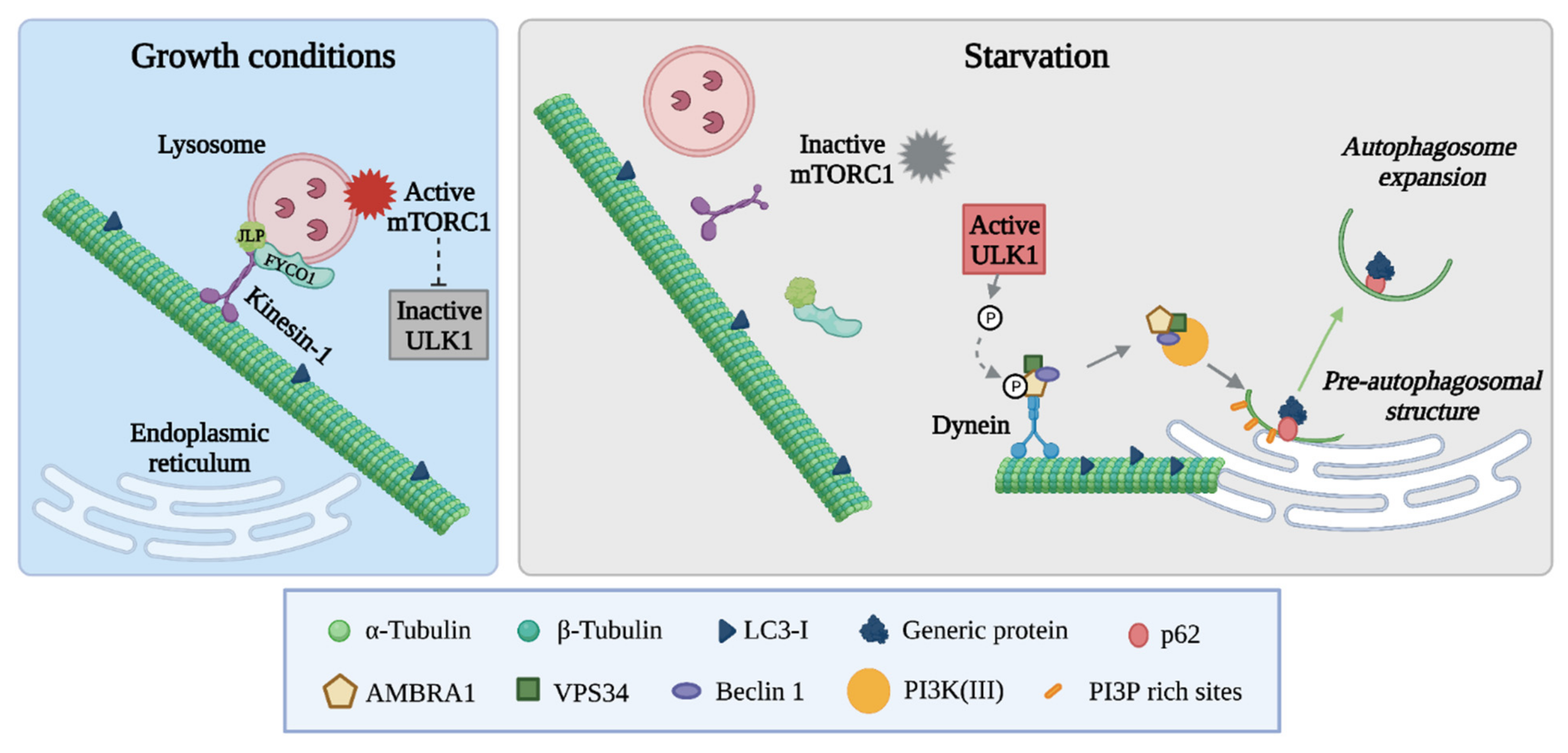
In addition, MTs regulate two important apical complexes of the autophagic pathway, namely, mTORC1 and PI3K complexes, through MT-associated motor proteins (Figure 2). MTORC1 activity is controlled by lysosome localization, so that in the presence of nutrients the plus end directed kinesin KIF2A and KIF1Bb maintain lysosomes at the cell periphery, keeping lysosome-associated mTORC1 active to suppresses autophagy [114]. Genetic manipulation of the kinesin adaptors FYVE and coiled-coil domain autophagy adaptor 1 (FYCO1) and c-Jun NH2-terminal kinase-associated leucine zipper protein (JLP) has also connected kinesin I activity with lysosomal positioning by showing that kinesin I functional inhibition promotes lysosome translocation to the juxtanuclear area and autophagy activation [114,115]. Starvation-dependent increase in intracellular pH has been implicated in lysosomal centripetal movement through the release of the molecular motors from MTs. In these conditions, dissociation of lysosomes from membrane-associated mTORC1 activators promotes autophagy [114]. These findings, together with recent work on other lysosome-associated nutrient-responsive growth mediators [116,117], have identified MT-dependent lysosome positioning as a dynamic regulator of cell homeostasis [118].
MT-driven intracellular positioning of autophagy mediators is also responsible for the activation of the PI3K complex. Under basal conditions, the autophagy and Beclin 1 regulator 1 (AMBRA1) is sequestered to MTs in a complex with Beclin 1 and phosphatidylinositol 3-kinase (VPS34) through its interaction with the dynein light chain 1 (DLC1), a subunit of the dynein motor complex. Upon autophagy stimulation, ULK1-mediated phosphorylation releases AMBRA1 from dynein interaction, allowing the translocation of Beclin 1/VPS34/AMBRA1 to the omegasome, where the PI3K complex promotes membrane formation [117].
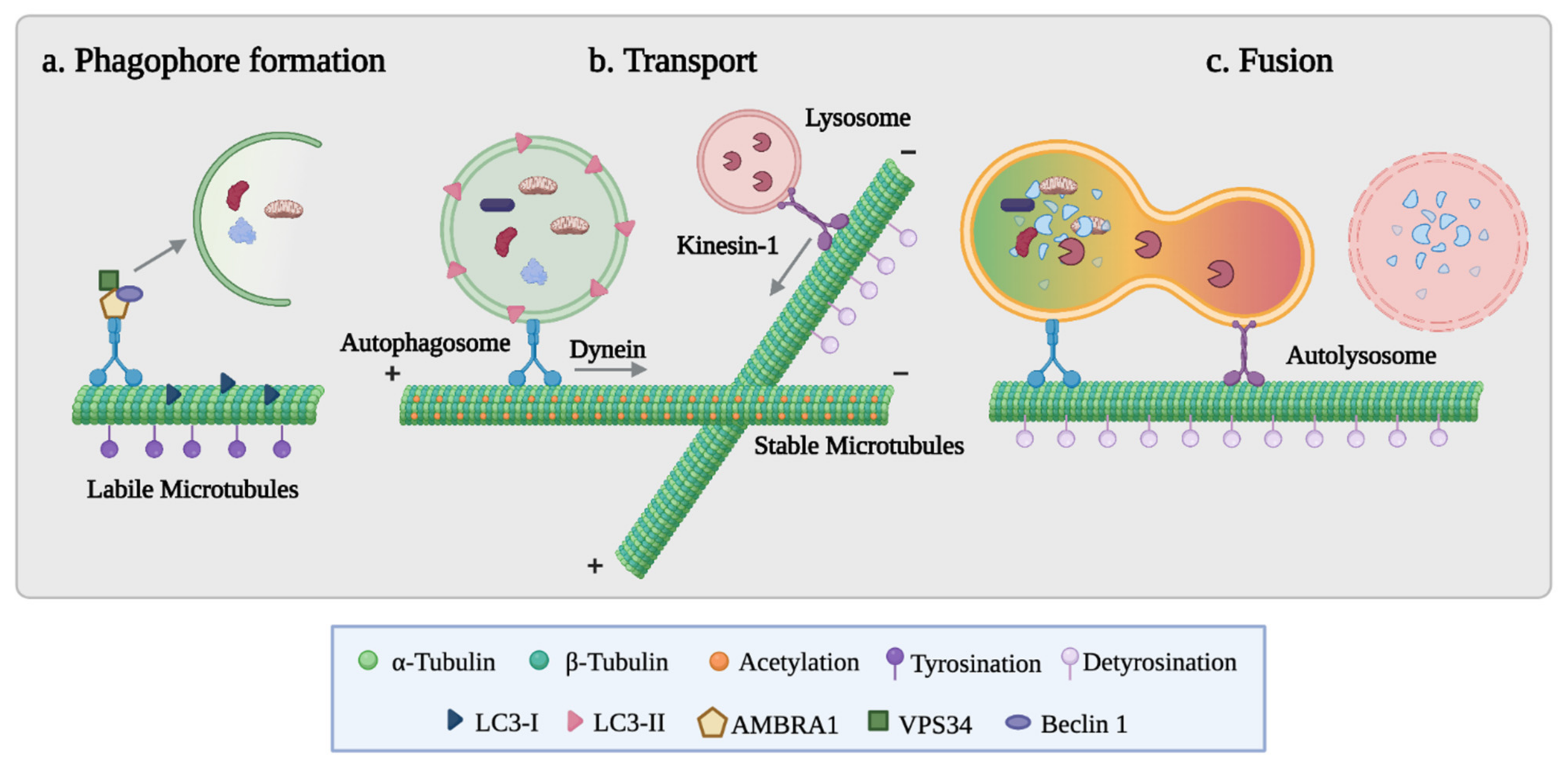
Live cell tracking of individual autophagosomes has shown that autophagosomes form randomly within the cell, then move bidirectionally along stable MTs until they concentrate around the centrosome in the perinuclear region. The centripetal movement of autophagosomes is mediated by the minus-end motor dynein, as shown by using a chemical dynein ATPase inhibitor or by disrupting the dynein complex through p50 dynamitin overexpression [119,120,121]. Autophagosomal centripetal movement is critical for an efficient autophagosome lysosome fusion, since these organelles localize at the perinuclear region. Conversely, plus-end-directed autophagosome movement involves kinesin 1, possibly through the FYCO1 kinesin 1 adaptor that is recruited to autophagosomes by LC3 and Rab7 [110,122,123]. Although the dependence of autophagosomal trafficking on molecular motors has been known from many years, it is still to be elucidated how and why bidirectional movement switches to a net centripetal movement of autophagosomes after autophagy induction and whether the switch in association from labile to stable MTs of nascent vs. mature autophagosomes could contribute to this phenomenon. Furthermore, the role of tubulin PTMs in driving autophagosome formation and trafficking is under debate.

3.3. The Tubulin Code in the Autophagic Process

Despite the acquired knowledge on the role of MT dynamics in autophagy, the role of different tubulin isoforms and PTMs in the autophagic process is still not fully clarified. An overview of the current information connecting differently post-translationally modified MTs to the different stages of the autophagic process is presented in Figure 3.
In 2016, a mass spectrometry study reported TubB3 as a binding partner of the autophagic player LC3 [124]. However, the functional significance of this interaction is still unknown and the role of this isoform in autophagy is still undefined. Recently, PCB118, a 2,3′,4,4′,5-pentachlorobiphenyl, has been reported to induce thyroid autophagy by promoting the binding to TubB3 of death-associated protein kinase 2 (DAPK2), a serine/threonine kinase implicated in autophagy and apoptosis, and thereby triggering the DAPK2/PKD/VPS34 pathway [125]. Apart from these studies, little is known of the impact of specific tubulin isoforms on autophagy.
In the last decade, several groups have focused their attention on the role of tubulin PTMs and their regulating enzymes in controlling the autophagic process (Table 4). Tubulin acetylation is the most studied tubulin PTM in connection with autophagy control. Different studies have used genetic or pharmacological approaches to modulate the enzymes that catalyze the acetylation reaction and have demonstrated the relevance of this PTM in the autophagic process in different experimental models. Upon starvation, tubulin acetylation is involved in regulating the formation of pre-autophagosomal structures and in influencing MT-based autophagosome movements [110]. Indeed, tubulin acetylation determines the spatial localization of autophagosomes and the complete resolving of the autophagic flux [126].
Tubulin acetylation is also essential for the formation of autolysosomes [113,127], and several studies have highlighted the relevance of HDAC6 in autophagosome–lysosome fusion. In cervical carcinoma cells, HDAC6 inhibition promotes tubulin acetylation and impairs serum starvation-induced autophagy by increasing LC3 acetylation and decreasing autophagic flux [128]. Notably, HDAC6 depletion impairs the fusion of autophagosomes and lysosomes by perturbing actin networks [129]. Concordantly, a highly selective HDAC6 inhibitor induces the accumulation of autophagic vacuoles and abrogates the autophagic flux by inhibiting autophagosome–lysosome fusion in glioblastoma cells [130]. In line with these data, HDAC6 has been shown to control autophagic flux in several cancer contexts, including differentiated cancer cells [131,132,133,134] and in cancer stem-like cells [135]. Interestingly, the latter study pointed out that HDAC6 inhibition differentially regulates autophagy in differentiated cancer cells as compared with cancer stem-like cells and identified autophagy as a target for developing anticancer stem cell therapies. The tubulin deacetylase SIRT2 has also been associated with autophagy regulation in several cancer cell lines, although its impact on cancer autophagy is less well understood, in comparison with HDAC6. In colon cancer, SIRT2 downregulation enhanced basal autophagy and mitotic post-slippage death [136]. In human neuroblastoma cells, autophagic flux was inhibited upon SIRT2 overexpression, as evidenced by increased accumulation of LC3-II and p62 proteins. In the same experimental model, SIRT2 overexpression interfered with the accumulation of autophagosomes following proteasome inhibition, leading to neuroblastoma cell death [137]. Notably, a direct link between SIRT2 function in autophagy and modulation of MT acetylation has been already identified in neurodegenerative models, such as Alzheimer’s and Parkinson’s diseases [138].
Less evident is the role of tubulin acetyltransferases in the autophagic process. Dowregulation of ATAT1 acetyltransferase, using a siRNA approach in H1299 lung cancer cells stably expressing the EGFP-LC3 fusion protein, markedly increased EGFP-LC3 puncta compared with control cells, suggesting that ATAT1 may modulate the autophagic process [139]. In another, more recent study, downregulation of ATAT1 promoted tubulin deacetylation and abrogated autophagic flux in response to glucose starvation [126]. Overall, these data support the role of tubulin acetylation as well as the role of deacetylases and acetyltransferases in the control of autophagic flux. Despite this, further work is required to demonstrate the connection between acetylated tubulin-controlled modulation of autophagy and cancer cell features.
Table 4. Functional roles of tubulin-modifying enzymes in autophagy progression and cell death.
EnzymesCancer CellsExperimental ApproachImpact on Cancer AutophagyReferences
Lysine Acetyltransferase
ATAT1LungDownregulationAutophagosome accumulation[139]
Lysine Deacetylase
HDAC6Glioblastoma, cancer stem-likeDownregulationInduction of cancer stem cell differentiation by promoting autophagy[135]
head and neckPharmacological inhibitionAutophagy inhibition[130]
SIRT2NeuroblastomaOverexpressionInhibition of autophagic flux[137]
ColonDownregulationAutophagy and mitotic post-slippage death induction[136]
Leukemic linesPharmacological inhibitionApoptosis and autophagic cell death[140]
Tubulin–tyrosine ligase
TTLBreast, pancreaticPharmacological inhibitionApoptosis and autophagic cell death[141,142]
Tubulin-specific carboxypeptidase
AGBL2Hepatocellular carcinomaOverexpressionEnhanced autophagy by upregulating immunity-related GTPase M[95]
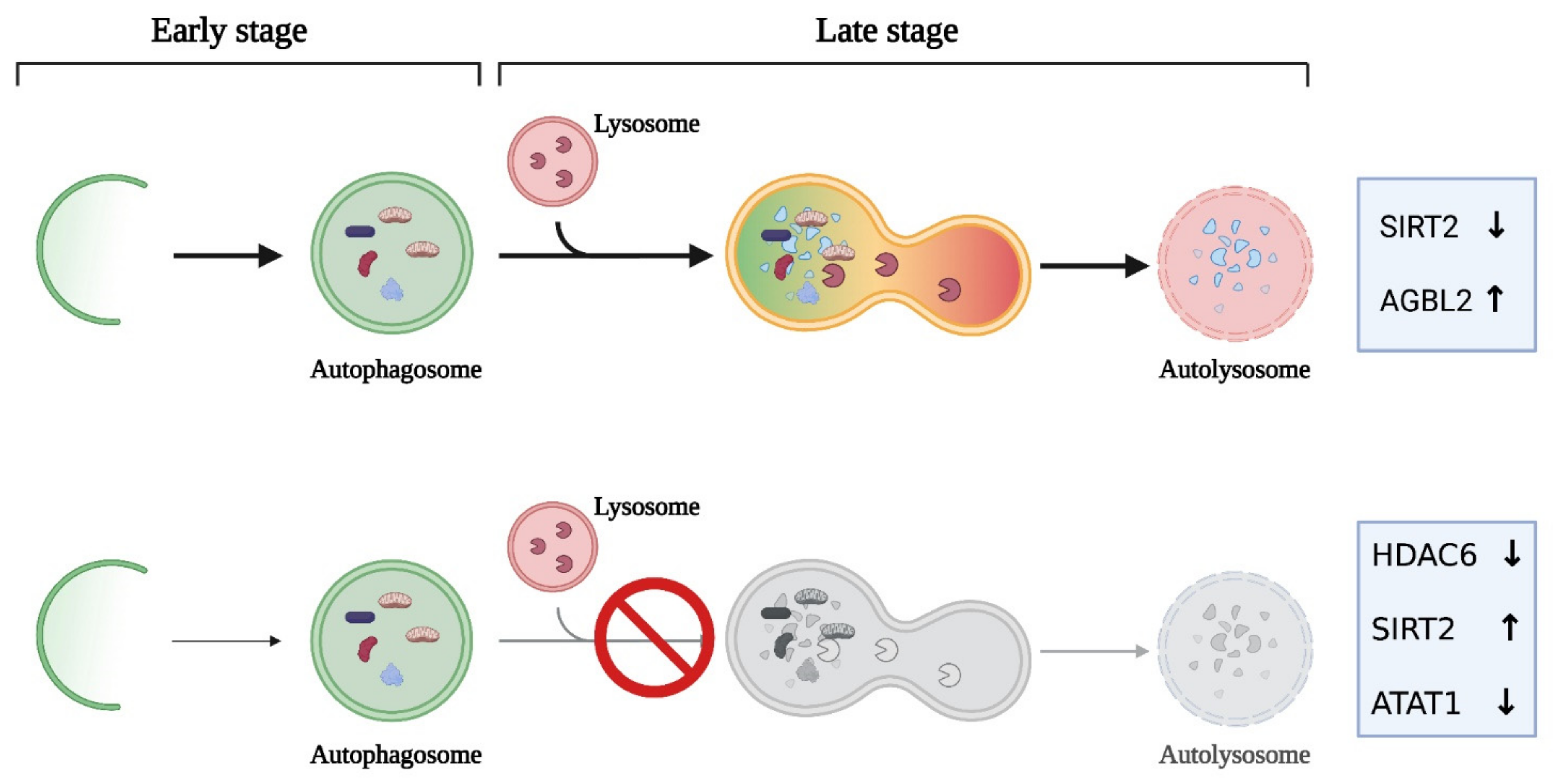
In 2019, Mohan and collaborators showed that tubulin detyrosination/tyrosination has a key role in mediating efficient lysosome–autophagosome encounters during the autophagic process. By using super-resolution microscopy, the authors showed that lysosomes were specifically enriched on detyrosinated MTs and that depletion of detyrosinated MTs reduced the number of autolysosomes, thus highlighting a new role of detyrosinated MTs in key steps of autophagy, namely, autophagosome trafficking and fusion [143]. Concordantly, AGBL2 overexpression increases tubulin detyrosination and enhances autophagy by upregulating immunity-related GTPase M, protecting HCC cells from apoptosis [95]. Collectively, all these studies highlight the role of tubulin PTMs in the regulation of the autophagic process and identify tubulin-modifying enzymes as key modulators of the dynamics of the autophagic flux (Figure 4).

4. Autophagy–Microtubule Crosstalk as a Possible Target for Cancer Growth Control

MT-interacting drugs, specifically interacting with either TubA or TubB [7,8,9], are widely used in the therapy of several cancer types. However, limitations on their use due to their neurotoxic side effects and to acquired resistance have stimulated the search for agents with greater effectiveness. In this field, much attention has been given to the capacity of new anti-TubA/TubB agents to affect the autophagic process, their capacity to stimulate or inhibit the autophagic flux and to promote autophagic or apoptosis-related cancer cell death.
Early work showed that the stabilizing drug paclitaxel inhibits autophagosome movement in interphase cells, thereby preventing autophagosome maturation and lysosome fusion in breast cancer cells [144]. The contribution of this block in the autophagic process to paclitaxel-induced cytotoxicity was investigated by the use of the early-stage autophagy inhibitor 3MA. The decreased cell killing effect, observed when autophagosome formation was inhibited by 3MA, demonstrated that blockage of autophagosome traffic and the accumulation of autophagosomes promotes paclitaxel-induced cancer cell death [144]. In line with these findings, several new tubulin binding agents have been shown to activate apoptosis as a result of autophagic flux inhibition in cancer cells [145,146,147,148]. It could be envisaged that induction of autophagosome formation in response to prolonged mitotic arrest and/or mitotic slippage, together with the reduction of autophagosome turnover by flux blockage, could result in the accumulation of autophagosomes, the intracellular persistence of toxic substances and/or damaged organelles, the production of oxygen reactive species, and cytotoxicity [149]. On the other hand, several reports have highlighted a role for autophagy induction in promoting cancer cell survival after exposure to tubulin-binding agents [150,151,152], confirming the dual role of autophagy in cancer [97]. Identifying whether the response to a specific MT-binding agent involves autophagy induction or implies both autophagy induction and flux blockage could be instrumental in determining whether the autophagic response would cooperate or compete with apoptosis in promoting cancer cell death.
Concerning tubulin PTMs, the direct effects of modulating tubulin PTMs on autophagy induction and cancer growth have not yet been identified. On the other hand, several papers on cancer therapeutics suggest that autophagy can act to promote cell death of tumor cells in response to exposure to HDAC6 inhibitors [130,135,153,154]. Moreover, the novel small molecule SIRT2-specific inhibitor NCO-90/141 has been reported to inhibit cell growth of leukemic cell lines by simultaneously causing apoptosis and autophagic cell death [140]. It should be recognized that the potential mechanisms by which HDAC6 or SIRT2 inhibitors can modulate the autophagic flux and induce autophagic cell death may be only partly dependent on the modulation of tubulin acetylation. It is also possible that deacetylase inhibition can inactivate HSP90 and other chaperones, increasing the levels of denatured proteins and enhancing ER stress signaling [155], thereby influencing autophagy. In line with studies revealing a role for tubulin detyrosination/tyrosination in autophagy, several reports have demonstrated the ability of parthenolide, an inhibitor of MT detyrosination, to induce autophagy and cell death in multiple cancer types, including breast and pancreatic cancer [141,142]. However, in none of these studies the effect of pathenolide on autophagy has been directly linked to its effect on tubulin tyrosination.
Future studies are needed to directly link tubulin PTMs to autophagy regulation. This research will unveil new avenues for modulating autophagy and will offer new perspectives for using tubulin-binding agents and related compounds to target autophagy in cancer.

5. Conclusions

MT dynamics have been implicated in a variety of cellular processes that are profoundly deregulated in cancer. Despite this, the contribution of altered MT dynamics to the cancer phenotypes has been often under-investigated. The recent flowering of reports on the role of tubulin PTMs on MT behavior, both on isolated MTs and in cellular contexts, has provided an opportunity to investigate the contribution of tubulin PTMs and tubulin-modifying enzymes in cancer and autophagy. Since autophagy has variable effects on tumor cells, depending on the cellular context and cancer stage, the role of the tubulin code in providing better survival capacity or promoting cancer cell death is an emerging field in cancer studies. As depicted in Figure 4, experimental modulation of the activity of tubulin-modifying enzymes has clearly been shown to impact on the autophagic process. However, both autophagy stimulation, associated with increased survival, and blockage of the autophagic flux, promoting cancer cell death, have been recorded. These findings indicate the need to carefully investigate the molecular determinants that differentiate the two opposing outcomes produced by modulating autophagy. Critical factors, such as cancer progression, cancer metabolic requirements, and autophagy–apoptosis crosstalk, should be carefully examined. Identifying the occurrence of an autophagic block by simple tools, such as late-stage inhibitor co-treatment or GFP-RFP-LC3 expression [105], will enable the determination of whether a genetic or pharmacological intervention on tubulin-modifying enzymes will promote cancer cell death by the cell toxicity associated with autophagosome accumulation [149] and could be usefully applied in the identification of therapeutically promising molecules.
In conclusion, although the molecular pathways underlying the action of tubulin-modifying enzymes on autophagy need further clarification, the targeted induction of autophagic cell death by inhibitors may represent a new fruitful avenue in cancer therapy.

Author Contributions

D.T. and F.D. wrote, revised, and edited the manuscript. All authors have read and agreed to the published version of the manuscript.

Funding

The research leading to these results has received funding from AIRC under IG 2020—ID 24942 project, PI Daniela Trisciuoglio—and POR FESR Lazio 2014-2020, “Progetti di Gruppi di Ricerca 2020” grant number 108016-0300-0327 (to FD).

Acknowledgments

The authors gratefully acknowledge the contribution of Francesca Romana Pellegrini in preparing the figures.

Conflicts of Interest

The authors declare no conflict of interest.

References

  1. Etienne-Manneville, S. From Signaling Pathways to Microtubule Dynamics: The Key Players. Curr. Opin. Cell Biol. 2010, 22, 104–111. [Google Scholar] [CrossRef]
  2. Mitchison, T.; Kirschner, M. Dynamic Instability of Microtubule Growth. Nature 1984, 312, 237–242. [Google Scholar] [CrossRef]
  3. Bakhoum, S.F.; Compton, D.A. Chromosomal Instability and Cancer: A Complex Relationship with Therapeutic Potential. J. Clin. Investig. 2012, 122, 1138–1143. [Google Scholar] [CrossRef] [Green Version]
  4. Cirillo, L.; Gotta, M.; Meraldi, P. The elephant in the room: The role of microtubules in cancer. In Cell Division Machinery and Disease. Advances in Experimental Medicine and Biology; Springer: Cham, Switzerland, 2017; Volume 1002, pp. 93–124. [Google Scholar] [CrossRef]
  5. Lopes, D.; Maiato, H. The Tubulin Code in Mitosis and Cancer. Cells 2020, 9, 2356. [Google Scholar] [CrossRef]
  6. Dumontet, C.; Jordan, M.A. Microtubule-Binding Agents: A Dynamic Field of Cancer Therapeutics. Nat. Rev. Drug Discov. 2010, 9, 790–803. [Google Scholar] [CrossRef] [Green Version]
  7. Steinmetz, M.O.; Prota, A.E. Microtubule-Targeting Agents: Strategies To Hijack the Cytoskeleton. Trends Cell Biol. 2018, 28, 776–792. [Google Scholar] [CrossRef] [PubMed]
  8. Zhang, D.; Kanakkanthara, A. Beyond the Paclitaxel and Vinca Alkaloids: Next Generation of Plant-Derived Microtubule-Targeting Agents with Potential Anticancer Activity. Cancers 2020, 12, 1721. [Google Scholar] [CrossRef] [PubMed]
  9. Matthew, S.; Chen, Q.Y.; Ratnayake, R.; Fermaintt, C.S.; Lucena-Agell, D.; Bonato, F.; Prota, A.E.; Lim, S.T.; Wang, X.; Díaz, J.F.; et al. Gatorbulin-1, a Distinct Cyclodepsipeptide Chemotype, Targets a Seventh Tubulin Pharmacological Site. Proc. Natl. Acad. Sci. USA 2021, 118, e2021847118. [Google Scholar] [CrossRef] [PubMed]
  10. Hussey, S.P.; Fritz-Laylin, L.K. “The Missing Link”: The Tubulin Mutation Database Connects Over 1500 Missense Mutations With Phenotypes Across Eukaryotes. Cytoskeleton 2019, 76, 175–176. [Google Scholar] [CrossRef]
  11. Breuss, M.W.; Leca, I.; Gstrein, T.; Hansen, A.H.; Keays, D.A. Tubulins and Brain Development—The Origins of Functional Specification. Mol. Cell. Neurosci. 2017, 84, 58–67. [Google Scholar] [CrossRef]
  12. Lewis, S.A.; Lee, M.G.; Cowan, N.J. Five Mouse Tubulin Isotypes and Their Regulated Expression during Development. J. Cell Biol. 1985, 101, 852–861. [Google Scholar] [CrossRef] [PubMed] [Green Version]
  13. Joshi, H.C.; Cleveland, D.W. Differential Utilization of Beta-Tubulin Isotypes in Differentiating Neurites. J. Cell Biol. 1989, 109, 663–673. [Google Scholar] [CrossRef]
  14. Denoulet, P.; Eddé, B.; Gros, F. Differential Expression of Several Neurospecific β-Tubulin MRNAs in the Mouse Brain during Development. Gene 1986, 50, 289–297. [Google Scholar] [CrossRef]
  15. Wang, D.; Villasante, A.; Lewis, S.; Cowan, N. The Mammalian Beta-Tubulin Repertoire: Hematopoietic Expression of a Novel, Heterologous Beta-Tubulin Isotype. J. Cell Biol. 1986, 103, 1903–1910. [Google Scholar] [CrossRef] [Green Version]
  16. Stearns, T.; Evans, L.; Kirschner, M. γ-Tubulin Is a Highly Conserved Component of the Centrosome. Cell 1991, 65, 825–836. [Google Scholar] [CrossRef]
  17. Kollman, J.M.; Merdes, A.; Mourey, L.; Agard, D.A. Microtubule Nucleation by γ-Tubulin Complexes. Nat. Rev. Mol. Cell Biol. 2011, 12, 709–721. [Google Scholar] [CrossRef]
  18. Prosser, S.L.; Pelletier, L. Mitotic Spindle Assembly in Animal Cells: A Fine Balancing Act. Nat. Rev. Mol. Cell Biol. 2017, 18, 187–201. [Google Scholar] [CrossRef]
  19. Chang, P.; Stearns, T. δ-Tubulin and ε-Tubulin: Two New Human Centrosomal Tubulins Reveal New Aspects of Centrosome Structure and Function. Nat. Cell Biol. 1999, 2, 30–35. [Google Scholar] [CrossRef]
  20. Verhey, K.J.; Gaertig, J. The Tubulin Code. Cell Cycle 2007, 6, 2152–2160. [Google Scholar] [CrossRef]
  21. Bodakuntla, S.; Jijumon, A.; Villablanca, C.; Gonzalez-Billault, C.; Janke, C. Microtubule-Associated Proteins: Structuring the Cytoskeleton. Trends Cell Biol. 2019, 29, 804–819. [Google Scholar] [CrossRef]
  22. Roll-Mecak, A. The Tubulin Code in Microtubule Dynamics and Information Encoding. Dev. Cell 2020, 54, 7–20. [Google Scholar] [CrossRef] [PubMed]
  23. Renthal, R.; Schneider, B.G.; Miller, M.M.; Ludueña, R.F. Beta IV Is the Major Beta-Tubulin Isotype in Bovine Cilia. Cell Motil. Cytoskelet. 1993, 25, 19–29. [Google Scholar] [CrossRef]
  24. Panda, D.; Miller, H.P.; Banerjee, A.; Ludueña, R.F.; Wilson, L. Microtubule Dynamics in Vitro Are Regulated by the Tubulin Isotype Composition. Proc. Natl. Acad. Sci. USA 1994, 91, 11358–11362. [Google Scholar] [CrossRef] [PubMed] [Green Version]
  25. Vemu, A.; Atherton, J.; Spector, J.O.; Moores, C.A.; Roll-Mecak, A. Tubulin Isoform Composition Tunes Microtubule Dynamics. Mol. Biol. Cell 2017, 28, 3564–3572. [Google Scholar] [CrossRef] [Green Version]
  26. Janke, C.; Magiera, M.M. The Tubulin Code and Its Role in Controlling Microtubule Properties and Functions. Nat. Rev. Mol. Cell Biol. 2020, 21, 307–326. [Google Scholar] [CrossRef]
  27. Li, L.; Yang, X.-J. Tubulin Acetylation: Responsible Enzymes, Biological Functions and Human Diseases. Cell. Mol. Life Sci. 2015, 72, 4237–4255. [Google Scholar] [CrossRef]
  28. Palazzo, A.; Ackerman, B.; Gundersen, G.G. Tubulin Acetylation and Cell Motility. Nature 2003, 421, 230. [Google Scholar] [CrossRef] [PubMed]
  29. Hubbert, C.; Guardiola, A.; Shao, R.; Kawaguchi, Y.; Ito, A.; Nixon, A.; Yoshida, A.; Wang, X.F.; Yao, T.P. HDAC6 Is a Microtubule-Associated Deacetylase. Nature 2002, 417, 455–458. [Google Scholar] [CrossRef]
  30. Portran, D.; Schaedel, L.; Xu, Z.; Théry, M.; Nachury, M.V. Tubulin Acetylation Protects Long-Lived Microtubules against Mechanical Ageing. Nat. Cell Biol. 2017, 19, 391–398. [Google Scholar] [CrossRef] [Green Version]
  31. Eshun-Wilson, L.; Zhang, R.; Portran, D.; Nachury, M.V.; Toso, D.B.; Löhr, T.; Vendruscolo, M.; Bonomi, M.; Fraser, J.S.; Nogales, E. Effects of α-Tubulin Acetylation on Microtubule Structure and Stability. Proc. Natl. Acad. Sci. USA 2019, 116, 10366–10371. [Google Scholar] [CrossRef] [Green Version]
  32. Akella, J.S.; Wloga, D.; Kim, J.; Starostina, N.G.; Lyons-Abbott, S.; Morrissette, N.S.; Dougan, S.T.; Kipreos, E.T.; Gaertig, J. MEC-17 Is an α-Tubulin Acetyltransferase. Nature 2010, 467, 218–222. [Google Scholar] [CrossRef] [Green Version]
  33. North, B.J.; Marshall, B.L.; Borra, M.T.; Denu, J.M.; Verdin, E. The Human Sir2 Ortholog, SIRT2, Is an NAD+-Dependent Tubulin Deacetylase. Mol. Cell 2003, 11, 437–444. [Google Scholar] [CrossRef]
  34. Ouyang, C.; Mu, J.; Lu, Q.; Li, J.; Zhu, H.; Wang, Q.; Zou, M.-H.; Xie, Z. Autophagic Degradation of KAT2A/GCN5 Promotes Directional Migration of Vascular Smooth Muscle Cells by Reducing TUBA/α-Tubulin Acetylation. Autophagy 2019, 16, 1753–1770. [Google Scholar] [CrossRef] [PubMed]
  35. Chu, C.-W.; Hou, F.; Zhang, J.; Phu, L.; Loktev, A.V.; Kirkpatrick, D.S.; Jackson, P.K.; Zhao, Y.; Zou, H. A Novel Acetylation of β-Tubulin by San Modulates Microtubule Polymerization via down-Regulating Tubulin Incorporation. Mol. Biol. Cell 2011, 22, 448. [Google Scholar] [CrossRef]
  36. Reed, N.A.; Cai, D.; Blasius, T.L.; Jih, G.T.; Meyhofer, E.; Gaertig, J.; Verhey, K.J. Microtubule Acetylation Promotes Kinesin-1 Binding and Transport. Curr. Biol. 2006, 16, 2166–2172. [Google Scholar] [CrossRef] [Green Version]
  37. Sirajuddin, M.; Rice, L.M.; Vale, R.D. Regulation of Microtubule Motors by Tubulin Isotypes and Posttranslational Modifications. Nat. Cell Biol. 2014, 16, 335. [Google Scholar] [CrossRef] [Green Version]
  38. Nekooki-Machida, Y.; Hagiwara, H. Role of Tubulin Acetylation in Cellular Functions and Diseases. Med. Mol. Morphol. 2020, 53, 191–197. [Google Scholar] [CrossRef]
  39. Barra, H.S.; Arce, C.A.; Argaraña, C.E. Posttranslational Tyrosination/Detyrosination of Tubulin. Mol. Neurobiol. 1988, 2, 133–153. [Google Scholar] [CrossRef]
  40. Nieuwenhuis, J.; Brummelkamp, T.R. The Tubulin Detyrosination Cycle: Function and Enzymes. Trends Cell Biol. 2019, 29, 80–92. [Google Scholar] [CrossRef]
  41. Robison, P.; Caporizzo, M.A.; Ahmadzadeh, H.; Bogush, A.I.; Chen, C.Y.; Margulies, K.B.; Shenoy, V.B.; Prosser, B.L. Detyrosinated Microtubules Buckle and Bear Load in Contracting Cardiomyocytes. Science 2016, 352, aaf0659. [Google Scholar] [CrossRef] [Green Version]
  42. Erck, C.; Peris, L.; Andrieux, A.; Meissirel, C.; Gruber, A.D.; Vernet, M.; Schweitzer, A.; Saoudi, Y.; Pointu, H.; Bosc, C.; et al. A Vital Role of Tubulin-Tyrosine-Ligase for Neuronal Organization. Proc. Natl. Acad. Sci. USA 2005, 102, 7853–7858. [Google Scholar] [CrossRef] [PubMed] [Green Version]
  43. Barisic, M.; Sousa, R.S.E.; Tripathy, S.K.; Magiera, M.M.; Zaytsev, A.V.; Pereira, A.L.; Janke, C.; Grishchuk, E.L.; Maiato, H. Microtubule Detyrosination Guides Chromosomes during Mitosis. Science 2015, 348, 799. [Google Scholar] [CrossRef] [Green Version]
  44. Nieuwenhuis, J.; Adamopoulos, A.; Bleijerveld, O.B.; Mazouzi, A.; Stickel, E.; Celie, P.; Altelaar, M.; Knipscheer, P.; Perrakis, A.; Blomen, V.A.; et al. Vasohibins Encode Tubulin Detyrosinating Activity. Science 2017, 358, 1453–1456. [Google Scholar] [CrossRef] [Green Version]
  45. Aillaud, C.; Bosc, C.; Peris, L.; Bosson, A.; Heemeryck, P.; van Dijk, J.; Friec, J.L.; Boulan, B.; Vossier, F.; Sanman, L.E.; et al. Vasohibins/SVBP Are Tubulin Carboxypeptidases (TCPs) That Regulate Neuron Differentiation. Science 2017, 358, 1448–1453. [Google Scholar] [CrossRef] [Green Version]
  46. Ersfeld, K.; Wehland, J.; Plessmann, U.; Dodemont, H.; Gerke, V.; Weber, K. Characterization of the Tubulin-Tyrosine Ligase. J. Cell Biol. 1993, 120, 725. [Google Scholar] [CrossRef] [Green Version]
  47. Field, J.J.; Kanakkanthara, A.; Miller, J.H. Microtubule-Targeting Agents Are Clinically Successful Due to Both Mitotic and Interphase Impairment of Microtubule Function. Bioorg. Med. Chem. 2014, 22, 5050–5059. [Google Scholar] [CrossRef] [PubMed]
  48. Lu, C.; Zhang, J.; He, S.; Wan, C.; Shan, A.; Wang, Y.; Yu, L.; Liu, G.; Chen, K.; Shi, J.; et al. Increased α-Tubulin1b Expression Indicates Poor Prognosis and Resistance to Chemotherapy in Hepatocellular Carcinoma. Dig. Dis. Sci. 2013, 58, 2713–2720. [Google Scholar] [CrossRef]
  49. Gui, S.; Chen, P.; Liu, Y.; Chen, Q.; Cheng, T.; Lv, S.; Zhou, T.; Song, Z.; Xiao, J.; He, W.; et al. TUBA1C Expression Promotes Proliferation by Regulating the Cell Cycle and Indicates Poor Prognosis in Glioma. Biochem. Biophys. Res. Commun. 2021, 577, 130–138. [Google Scholar] [CrossRef] [PubMed]
  50. Wang, D.; Jiao, Z.; Ji, Y.; Zhang, S. Elevated TUBA1A Might Indicate the Clinical Outcomes of Patients with Gastric Cancer, Being Associated with the Infiltration of Macrophages in the Tumor Immune Microenvironment. J. Gastrointest. Liver Dis. JGLD 2020, 29, 509–522. [Google Scholar] [CrossRef]
  51. Bian, T.; Zheng, M.; Jiang, D.; Liu, J.; Sun, H.; Li, X.; Liu, L.; Zhang, J.; Liu, Y. Prognostic Biomarker TUBA1C Is Correlated to Immune Cell Infiltration in the Tumor Microenvironment of Lung Adenocarcinoma. Cancer Cell Int. 2021, 21, 144. [Google Scholar] [CrossRef] [PubMed]
  52. Zhu, H.; Hu, X.; Gu, L.; Jian, Z.; Li, L.; Hu, S.; Qiu, S.; Xiong, X. TUBA1C Is a Prognostic Marker in Low-Grade Glioma and Correlates with Immune Cell Infiltration in the Tumor Microenvironment. Front. Genet. 2021, 12, 759953. [Google Scholar] [CrossRef]
  53. Nicoletti, M.I.; Valoti, G.; Giannakakou, P.; Zhan, Z.; Kim, J.-H.; Lucchini, V.; Landoni, F.; Mayo, J.G.; Giavazzi, R.; Fojo, T. Expression Of Tubulin Isotypes in Human Ovarian Carcinoma Xenografts and in a Sub-Panel of Human Cancer Cell Lines from the NCI-Anticancer Drug Screen: Correlation with Sensitivity to Microtubule Active Agents. Clin. Cancer Res. 2001, 7, 2912–2922. [Google Scholar]
  54. Nami, B.; Wang, Z. Genetics and Expression Profile of the Tubulin Gene Superfamily in Breast Cancer Subtypes and Its Relation to Taxane Resistance. Cancers 2018, 10, 274. [Google Scholar] [CrossRef] [Green Version]
  55. Kanakkanthara, A.; Miller, J.H. ΒIII-Tubulin Overexpression in Cancer: Causes, Consequences, and Potential Therapies. Biochim. Biophys. Acta (BBA) Rev. Cancer 2021, 1876, 188607. [Google Scholar] [CrossRef] [PubMed]
  56. Quaas, A.; Rahvar, A.-H.; Burdelski, C.; Koop, C.; Eichelberg, C.; Rink, M.; Dahlem, R.; Schlomm, T.; Tsourlakis, M.C.; Simon, R.; et al. ΒIII-Tubulin Overexpression Is Linked to Aggressive Tumor Features and Shortened Survival in Clear Cell Renal Cell Carcinoma. World J. Urol. 2014, 33, 1561–1569. [Google Scholar] [CrossRef] [PubMed]
  57. Hinsch, A.; Chaker, A.; Burdelski, C.; Koop, C.; Tsourlakis, M.C.; Steurer, S.; Rink, M.; Eichenauer, T.S.; Wilczak, W.; Wittmer, C.; et al. ΒIII-Tubulin Overexpression Is Linked to Aggressive Tumor Features and Genetic Instability in Urinary Bladder Cancer. Hum. Pathol. 2017, 61, 210–220. [Google Scholar] [CrossRef]
  58. Gan, P.P.; Kavallaris, M. Tubulin-Targeted Drug Action: Functional Significance of Class II and Class IVb β-Tubulin in Vinca Alkaloid Sensitivity. Cancer Res. 2008, 68, 9817–9824. [Google Scholar] [CrossRef] [Green Version]
  59. Ohishi, Y.; Oda, Y.; Basaki, Y.; Kobayashi, H.; Wake, N.; Kuwano, M.; Tsuneyoshi, M. Expression of Beta-Tubulin Isotypes in Human Primary Ovarian Carcinoma. Gynecol. Oncol. 2007, 105, 586–592. [Google Scholar] [CrossRef]
  60. Bernard-Marty, C.; Treilleux, I.; Dumontet, C.; Cardoso, F.; Fellous, A.; Gancberg, D.; Bissery, M.C.; Paesmans, M.; Larsimont, D.; Piccart, M.J.; et al. Microtubule-Associated Parameters as Predictive Markers of Docetaxel Activity in Advanced Breast Cancer Patients: Results of a Pilot Study. Clin. Breast Cancer 2002, 3, 341–345. [Google Scholar] [CrossRef]
  61. Cucchiarelli, V.; Hiser, L.; Smith, H.; Frankfurter, A.; Spano, A.; Correia, J.J.; Lobert, S. β-Tubulin Isotype Classes II and V Expression Patterns in Nonsmall Cell Lung Carcinomas. Cell Motil. Cytoskelet. 2008, 65, 675–685. [Google Scholar] [CrossRef] [PubMed]
  62. Na, H.Y.; Park, M.; Kim, Y.A.; Won, J.K.; Park, Y.J.; Shin, S.A.; Lee, S.; Oh, S.; Kim, J.E. Expression of Class III Beta-Tubulin Is Associated with Invasive Potential and Poor Prognosis in Thyroid Carcinoma. J. Clin. Med. 2020, 9, 3830. [Google Scholar] [CrossRef] [PubMed]
  63. Atjanasuppat, K.; Lirdprapamongkol, K.; Jantaree, P.; Svasti, J. Non-Adherent Culture Induces Paclitaxel Resistance in H460 Lung Cancer Cells via ERK-Mediated up-Regulation of ΒIVa-Tubulin. Biochem. Biophys. Res. Commun. 2015, 466, 493–498. [Google Scholar] [CrossRef]
  64. Christoph, D.C.; Kasper, S.; Gauler, T.C.; Loesch, C.; Engelhard, M.; Theegarten, D.; Poettgen, C.; Hepp, R.; Peglow, A.; Loewendick, H.; et al. ΒV-Tubulin Expression Is Associated with Outcome Following Taxane-Based Chemotherapy in Non-Small Cell Lung Cancer. Br. J. Cancer 2012, 107, 823–830. [Google Scholar] [CrossRef] [Green Version]
  65. Sekino, Y.; Han, X.; Babasaki, T.; Miyamoto, S.; Kobatake, K.; Kitano, H.; Ikeda, K.; Goto, K.; Inoue, S.; Hayashi, T.; et al. TUBB3 Is Associated with PTEN, Neuroendocrine Differentiation, and Castration Resistance in Prostate Cancer. Urol. Oncol. Semin. Orig. Investig. 2021, 39, 368.e1–368.e9. [Google Scholar] [CrossRef] [PubMed]
  66. Sekino, Y.; Han, X.; Babasaki, T.; Miyamoto, S.; Kitano, H.; Kobayashi, G.; Goto, K.; Inoue, S.; Hayashi, T.; Teishima, J.; et al. TUBB3 Is Associated with High-Grade Histology, Poor Prognosis, P53 Expression, and Cancer Stem Cell Markers in Clear Cell Renal Cell Carcinoma. Oncology 2020, 98, 689–698. [Google Scholar] [CrossRef]
  67. Bailey, J.M.; Alsina, J.; Rasheed, Z.A.; McAllister, F.M.; Fu, Y.Y.; Plentz, R.; Zhang, H.; Pasricha, P.J.; Bardeesy, N.; Matsui, W.; et al. DCLK1 Marks a Morphologically Distinct Subpopulation of Cells With Stem Cell Properties in Preinvasive Pancreatic Cancer. Gastroenterology 2014, 146, 245–256. [Google Scholar] [CrossRef] [Green Version]
  68. Boggs, A.E.; Vitolo, M.I.; Whipple, R.A.; Charpentier, M.S.; Goloubeva, O.G.; Ioffe, O.B.; Tuttle, K.C.; Slovic, J.; Lu, Y.; Mills, G.B.; et al. α-Tubulin Acetylation Elevated in Metastatic and Basal-like Breast Cancer Cells Promotes Microtentacle Formation, Adhesion, and Invasive Migration. Cancer Res. 2015, 75, 203–215. [Google Scholar] [CrossRef] [Green Version]
  69. Wattanathamsan, O.; Thararattanobon, R.; Rodsiri, R.; Chanvorachote, P.; Vinayanuwattikun, C.; Pongrakhananon, V. Tubulin Acetylation Enhances Lung Cancer Resistance to Paclitaxel-Induced Cell Death through Mcl-1 Stabilization. Cell Death Discov. 2021, 7, 67. [Google Scholar] [CrossRef] [PubMed]
  70. Levidou, G.; Gajdzis, P.; Cassoux, N.; Donizy, P.; Masaoutis, C.; Gajdzis, M.; Gardrat, S.; Pergaris, A.; Danas, E.; Klijanienko, J.; et al. Histone Deacetylase (HDAC)-1, -2, -4, and -6 in Uveal Melanomas: Associations with Clinicopathological Parameters and Patients’ Survival. Cancers 2021, 13, 4763. [Google Scholar] [CrossRef] [PubMed]
  71. Zhang, Z.; Zhang, X.; Huang, A. Aggresome–Autophagy Associated Gene HDAC6 Is a Potential Biomarker in Pan-Cancer, Especially in Colon Adenocarcinoma. Front. Oncol. 2021, 11, 2625. [Google Scholar] [CrossRef] [PubMed]
  72. Saji, S.; Kawakami, M.; Hayashi, S.; Yoshida, N.; Hirose, M.; Horiguchi, S.; Itoh, A.; Funata, N.; Schreiber, S.L.; Yoshida, M.; et al. Significance of HDAC6 Regulation via Estrogen Signaling for Cell Motility and Prognosis in Estrogen Receptor-Positive Breast Cancer. Oncogene 2005, 24, 4531–4539. [Google Scholar] [CrossRef] [Green Version]
  73. Shi, P.; Hoang-Minh, L.B.; Tian, J.; Cheng, A.; Basrai, R.; Kalaria, N.; Lebowitz, J.J.; Khoshbouei, H.; Deleyrolle, L.P.; Sarkisian, M.R. HDAC6 Signaling at Primary Cilia Promotes Proliferation and Restricts Differentiation of Glioma Cells. Cancers 2021, 13, 1644. [Google Scholar] [CrossRef] [PubMed]
  74. Urdiciain, A.; Erausquin, E.; Zelaya, M.V.; Zazpe, I.; Lanciego, J.L.; Meléndez, B.; Rey, J.A.; Idoate, M.A.; Galdo, N.A.R.-D.; Castresana, J.S. Silencing of Histone Deacetylase 6 Decreases Cellular Malignancy and Contributes to Primary Cilium Restoration, Epithelial-to-Mesenchymal Transition Reversion, and Autophagy Inhibition in Glioblastoma Cell Lines. Biology 2021, 10, 467. [Google Scholar] [CrossRef] [PubMed]
  75. Wei, R.; He, D.; Zhang, X. Role of SIRT2 in Regulation of Stemness of Cancer Stem-Like Cells in Renal Cell Carcinoma. Cell. Physiol. Biochem. 2018, 49, 2348–2357. [Google Scholar] [CrossRef]
  76. Zhao, N.; Guo, Y.; Liu, P.; Chen, Y.; Wang, Y. Sirtuin 2 Promotes Cell Stemness and MEK/ERK Signaling Pathway While Reduces Chemosensitivity in Endometrial Cancer. Arch. Gynecol. Obstet. 2021, 1, 1–9. [Google Scholar] [CrossRef]
  77. Li, Z.; Xie, Q.R.; Chen, Z.; Lu, S.; Xia, W. Regulation of SIRT2 Levels for Human Non-Small Cell Lung Cancer Therapy. Lung Cancer 2013, 82, 9–15. [Google Scholar] [CrossRef] [PubMed]
  78. Oh, S.; You, E.; Ko, P.; Jeong, J.; Keum, S.; Rhee, S. Genetic Disruption of Tubulin Acetyltransferase, ATAT1, Inhibits Proliferation and Invasion of Colon Cancer Cells through Decreases in Wnt1/β-Catenin Signaling. Biochem. Biophys. Res. Commun. 2017, 482, 8–14. [Google Scholar] [CrossRef] [PubMed]
  79. Lee, C.-C.; Cheng, Y.-C.; Chang, C.-Y.; Lin, C.-M.; Chang, J.-Y. Alpha-Tubulin Acetyltransferase/MEC-17 Regulates Cancer Cell Migration and Invasion through Epithelial–Mesenchymal Transition Suppression and Cell Polarity Disruption. Sci. Rep. 2018, 8, 17477. [Google Scholar] [CrossRef] [Green Version]
  80. Chien, J.-Y.; Tsen, S.-D.; Chien, C.-C.; Liu, H.-W.; Tung, C.-Y.; Lin, C.-H. ATAT1 Downregulation Induces Mitotic Catastrophe in HeLa and A549 Cells. Cell Death Discov. 2016, 2, 16006. [Google Scholar] [CrossRef]
  81. Whipple, R.A.; Matrone, M.A.; Cho, E.H.; Balzer, E.M.; Vitolo, M.I.; Yoon, J.R.; Ioffe, O.B.; Tuttle, K.C.; Yang, J.; Martin, S.S. Epithelial-to-Mesenchymal Transition Promotes Tubulin Detyrosination and Microtentacles That Enhance Endothelial Engagement. Cancer Res. 2010, 70, 8127–8137. [Google Scholar] [CrossRef] [Green Version]
  82. Kato, C.; Miyazaki, K.; Nakagawa, A.; Ohira, M.; Nakamura, Y.; Ozaki, T.; Imai, T.; Nakagawara, A. Low Expression of Human Tubulin Tyrosine Ligase and Suppressed Tubulin Tyrosination/Detyrosination Cycle Are Associated with Impaired Neuronal Differentiation in Neuroblastomas with Poor Prognosis. Int. J. Cancer 2004, 112, 365–375. [Google Scholar] [CrossRef] [PubMed]
  83. Mialhe, A.; Lafanechèere, L.; Treilleux, I.; Peloux, N.; Dumontet, C.; Brémond, A.; Panh, M.-H.; Payan, R.; Wehland, J.; Margolis, R.-L.; et al. Tubulin Detyrosination Is a Frequent Occurrence in Breast Cancers of Poor Prognosis. Cancer Res. 2001, 61, 5024–5027. [Google Scholar] [PubMed]
  84. Sato, Y. The Vasohibin Family: A Novel Family for Angiogenesis Regulation. J. Biochem. 2013, 153, 5. [Google Scholar] [CrossRef] [Green Version]
  85. Hara, H.; Ozawa, S.; Ninomiya, Y.; Yamamoto, M.; Ogimi, M.; Nabeshima, K.; Nakamura, K.; Kajiwara, H.; Nakamura, N.; Sato, Y. Prognostic Significance of Vasohibin-1 and Vasohibin-2 Immunohistochemical Expression in Gastric Cancer. Surg. Today 2020, 50, 1530–1543. [Google Scholar] [CrossRef] [PubMed]
  86. Yamamoto, M.; Ozawa, S.; Ninomiya, Y.; Koyanagi, K.; Oguma, J.; Kazuno, A.; Hara, H.; Yatabe, K.; Kajiwara, H.; Nakamura, N.; et al. Plasma Vasohibin-1 and Vasohibin-2 Are Useful Biomarkers in Patients with Esophageal Squamous Cell Carcinoma. Esophagus Off. J. Jpn. Esophageal Soc. 2020, 17, 289–297. [Google Scholar] [CrossRef]
  87. Ninomiya, Y.; Ozawa, S.; Oguma, J.; Kazuno, A.; Nitta, M.; Kajiwara, H.; Sato, Y. Expression of Vasohibin-1 and −2 Predicts Poor Prognosis among Patients with Squamous Cell Carcinoma of the Esophagus. Oncol. Lett. 2018, 16, 5265. [Google Scholar] [CrossRef]
  88. Sano, R.; Kanomata, N.; Suzuki, S.; Shimoya, K.; Sato, Y.; Moriya, T.; Shiota, M. Vasohibin-1 Is a Poor Prognostic Factor of Ovarian Carcinoma. Tohoku J. Exp. Med. 2017, 243, 107–114. [Google Scholar] [CrossRef] [PubMed] [Green Version]
  89. Koyanagi, T.; Saga, Y.; Takahashi, Y.; Tamura, K.; Yoshiba, T.; Takahashi, S.; Taneichi, A.; Takei, Y.; Urabe, M.; Mizukami, H.; et al. Knockout of Vasohibin-2 Reduces Tubulin Carboxypeptidase Activity and Increases Paclitaxel Sensitivity in Ovarian Cancer. Cancer Med. 2021, 10, 2732–2739. [Google Scholar] [CrossRef] [PubMed]
  90. Arnold, J.; Schattschneider, J.; Blechner, C.; Krisp, C.; Schlüter, H.; Schweizer, M.; Nalaskowski, M.; Oliveira-Ferrer, L.; Windhorst, S. Tubulin Tyrosine Ligase Like 4 (TTLL4) Overexpression in Breast Cancer Cells Is Associated with Brain Metastasis and Alters Exosome Biogenesis. J. Exp. Clin. Cancer Res. 2020, 39, 1–15. [Google Scholar] [CrossRef]
  91. Kashiwaya, K.; Nakagawa, H.; Hosokawa, M.; Mochizuki, Y.; Ueda, K.; Piao, L.; Chung, S.; Hamamoto, R.; Eguchi, H.; Ohigashi, H.; et al. Involvement of the Tubulin Tyrosine Ligase-Like Family Member 4 Polyglutamylase in PELP1 Polyglutamylation and Chromatin Remodeling in Pancreatic Cancer Cells. Cancer Res. 2010, 70, 4024–4033. [Google Scholar] [CrossRef] [PubMed] [Green Version]
  92. Wen, R.; Xiao, Y.; Zhang, Y.; Yang, M.; Lin, Y.; Tang, J. Identification of a Novel Transcript Isoform of the TTLL12 Gene in Human Cancers. Oncol. Rep. 2016, 36, 3172–3180. [Google Scholar] [CrossRef] [PubMed] [Green Version]
  93. Yang, S.; Liang, Y.; Qian, H.; Li, Q. TTLL12 Expression in Ovarian Cancer Correlates with a Poor Outcome. Int. J. Clin. Exp. Pathol. 2020, 13, 239. [Google Scholar]
  94. He, W.; Wang, L. High Expression of AGBL2 Is a Novel Prognostic Factor of Adverse Outcome in Patients with Ovarian Carcinoma. Oncol. Lett. 2019, 18, 4900–4906. [Google Scholar] [CrossRef] [PubMed] [Green Version]
  95. Wang, L.L.; Jin, X.H.; Cai, M.Y.; Li, H.G.; Chen, J.W.; Wang, F.W.; Wang, C.Y.; Hu, W.W.; Liu, F.; Xie, D. AGBL2 Promotes Cancer Cell Growth through IRGM-Regulated Autophagy and Enhanced Aurora A Activity in Hepatocellular Carcinoma. Cancer Lett. 2018, 414, 71–80. [Google Scholar] [CrossRef] [PubMed]
  96. Peterfi, L.; Banyai, D.; Yusenko, M.V.; Bjercke, T.; Kovacs, G. Expression of RARRES1 and AGBL2 and Progression of Conventional Renal Cell Carcinoma. Br. J. Cancer 2020, 122, 1818. [Google Scholar] [CrossRef]
  97. Yang, Y.; Klionsky, D.J. Autophagy and Disease: Unanswered Questions. Cell Death Differ. 2020, 27, 858. [Google Scholar] [CrossRef] [PubMed]
  98. Levine, B.; Kroemer, G. Biological Functions of Autophagy Genes: A Disease Perspective. Cell 2019, 176, 11. [Google Scholar] [CrossRef] [Green Version]
  99. Lamb, C.A.; Yoshimori, T.; Tooze, S.A. The Autophagosome: Origins Unknown, Biogenesis Complex. Nat. Rev. Mol. Cell Biol. 2013, 14, 759–774. [Google Scholar] [CrossRef]
  100. Zaffagnini, G.; Martens, S. Mechanisms of Selective Autophagy. J. Mol. Biol. 2016, 428, 1714. [Google Scholar] [CrossRef] [Green Version]
  101. Dossou, A.S.; Basu, A. The Emerging Roles of MTORC1 in Macromanaging Autophagy. Cancers 2019, 11, 1422. [Google Scholar] [CrossRef] [Green Version]
  102. Turco, E.; Witt, M.; Abert, C.; Bock-Bierbaum, T.; Su, M.-Y.; Trapannone, R.; Sztacho, M.; Danieli, A.; Shi, X.; Zaffagnini, G.; et al. FIP200 Claw Domain Binding to P62 Promotes Autophagosome Formation at Ubiquitin Condensates. Mol. Cell 2019, 74, 330. [Google Scholar] [CrossRef] [PubMed] [Green Version]
  103. Zhao, Y.G.; Zhang, H. Formation and Maturation of Autophagosomes in Higher Eukaryotes: A Social Network. Curr. Opin. Cell Biol. 2018, 53, 29–36. [Google Scholar] [CrossRef] [PubMed]
  104. Maeda, S.; Otomo, C.; Otomo, T. The Autophagic Membrane Tether ATG2A Transfers Lipids between Membranes. eLife 2019, 8, e45777. [Google Scholar] [CrossRef]
  105. Klionsky, D.J.; Abdelmohsen, K.; Abe, A.; Abedin, M.J.; Abeliovich, H.; Arozena, A.A.; Adachi, H.; Adams, C.M.; Adams, P.D.; Adeli, K.; et al. Guidelines for the Use and Interpretation of Assays for Monitoring Autophagy (3rd Edition). Autophagy 2016, 12, 1. [Google Scholar] [CrossRef] [PubMed] [Green Version]
  106. Takahashi, Y.; He, H.; Tang, Z.; Hattori, T.; Liu, Y.; Young, M.M.; Serfass, J.M.; Chen, L.; Gebru, M.; Chen, C.; et al. An Autophagy Assay Reveals the ESCRT-III Component CHMP2A as a Regulator of Phagophore Closure. Nat. Commun. 2018, 9, 2855. [Google Scholar] [CrossRef] [Green Version]
  107. Oyarzún, J.E.; Lagos, J.; Vázquez, M.C.; Valls, C.; de la Fuente, C.; Yuseff, M.I.; Alvarez, A.R.; Zanlungo, S. Lysosome Motility and Distribution: Relevance in Health and Disease. Biochim. Biophys. Acta (BBA) Mol. Basis Dis. 2019, 1865, 1076–1087. [Google Scholar] [CrossRef]
  108. Kaur, J.; Debnath, J. Autophagy at the Crossroads of Catabolism and Anabolism. Nat. Rev. Mol. Cell Biol. 2015, 16, 461–472. [Google Scholar] [CrossRef] [Green Version]
  109. Mackeh, R.; Perdiz, D.; Lorin, S.; Codogno, P.; Poüs, C. Autophagy and Microtubules—New Story, Old Players. J. Cell Sci. 2013, 126, 1071–1080. [Google Scholar] [CrossRef] [Green Version]
  110. Geeraert, C.; Ratier, A.; Pfisterer, S.G.; Perdiz, D.; Cantaloube, I.; Rouault, A.; Pattingre, S.; Proikas-Cezanne, T.; Codogno, P.; Poüs, C. Starvation-Induced Hyperacetylation of Tubulin Is Required for the Stimulation of Autophagy by Nutrient Deprivation. J. Biol. Chem. 2010, 285, 24184–24194. [Google Scholar] [CrossRef] [Green Version]
  111. Xie, R.; Nguyen, S.; McKeehan, K.; Wang, F.; McKeehan, W.L.; Liu, L. Microtubule-Associated Protein 1S (MAP1S) Bridges Autophagic Components with Microtubules and Mitochondria to Affect Autophagosomal Biogenesis and Degradation. J. Biol. Chem. 2011, 286, 10367. [Google Scholar] [CrossRef] [PubMed] [Green Version]
  112. Wang, J.; Zhang, X.; Yang, F.; Yang, Y.; Wang, T.; Liu, W.; Zhou, H.; Zhao, W. RASSF1A Enhances Chemosensitivity of NSCLC Cells Through Activating Autophagy by Regulating MAP1S to Inactivate Keap1-Nrf2 Pathway. Drug Des. Dev. Ther. 2021, 15, 21. [Google Scholar] [CrossRef]
  113. Köchl, R.; Hu, X.W.; Chan, E.Y.W.; Tooze, S.A. Microtubules Facilitate Autophagosome Formation and Fusion of Autophagosomes with Endosomes. Traffic 2006, 7, 129–145. [Google Scholar] [CrossRef] [PubMed]
  114. Hong, Z.; Pedersen, N.M.; Wang, L.; Torgersen, M.L.; Stenmark, H.; Raiborg, C. PtdIns3P Controls MTORC1 Signaling through Lysosomal Positioning. J. Cell Biol. 2017, 216, 4217. [Google Scholar] [CrossRef] [Green Version]
  115. Suzuki, R.; Gunarta, I.K.; Boldbaatar, J.; Erdenebaatar, P.; Odongoo, R.; Yoshioka, K. Functional Role of C-Jun NH2-Terminal Kinase-Associated Leucine Zipper Protein (JLP) in Lysosome Localization and Autophagy. Drug Discov. Ther. 2020, 14, 35–41. [Google Scholar] [CrossRef] [PubMed] [Green Version]
  116. Korolchuk, V.I.; Saiki, S.; Lichtenberg, M.; Siddiqi, F.H.; Roberts, E.A.; Imarisio, S.; Jahreiss, L.; Sarkar, S.; Futter, M.; Menzies, F.M.; et al. Lysosomal Positioning Coordinates Cellular Nutrient Responses. Nat. Cell Biol. 2011, 13, 453–460. [Google Scholar] [CrossRef]
  117. Jia, R.; Bonifacino, J.S. Lysosome Positioning Influences MTORC2 and AKT Signaling. Mol. Cell 2019, 75, 26. [Google Scholar] [CrossRef]
  118. Ballabio, A.; Bonifacino, J.S. Lysosomes as Dynamic Regulators of Cell and Organismal Homeostasis. Nat. Rev. Mol. Cell Biol. 2019, 21, 101–118. [Google Scholar] [CrossRef] [PubMed]
  119. Ravikumar, B.; Acevedo-Arozena, A.; Imarisio, S.; Berger, Z.; Vacher, C.; O’Kane, C.J.; Brown, S.D.M.; Rubinsztein, D.C. Dynein Mutations Impair Autophagic Clearance of Aggregate-Prone Proteins. Nat. Genet. 2005, 37, 771–776. [Google Scholar] [CrossRef] [PubMed]
  120. Kimura, S.; Noda, T.; Yoshimori, T. Dynein-Dependent Movement of Autophagosomes Mediates Efficient Encounters with Lysosomes. Cell Struct. Funct. 2008, 33, 109–122. [Google Scholar] [CrossRef] [Green Version]
  121. Jahreiss, L.; Menzies, F.M.; Rubinsztein, D.C. The Itinerary of Autophagosomes: From Peripheral Formation to Kiss-and-Run Fusion with Lysosomes. Traffic 2008, 9, 574–587. [Google Scholar] [CrossRef] [PubMed] [Green Version]
  122. Cardoso, C.M.P.; Groth-Pedersen, L.; Høyer-Hansen, M.; Kirkegaard, T.; Corcelle, E.; Andersen, J.S.; Jäättelä, M.; Nylandsted, J. Depletion of Kinesin 5B Affects Lysosomal Distribution and Stability and Induces Peri-Nuclear Accumulation of Autophagosomes in Cancer Cells. PLoS ONE 2009, 4, e4424. [Google Scholar] [CrossRef] [Green Version]
  123. Pankiv, S.; Alemu, E.A.; Brech, A.; Bruun, J.-A.; Lamark, T.; Øvervatn, A.; Bjørkøy, G.; Johansen, T. FYCO1 Is a Rab7 Effector That Binds to LC3 and PI3P to Mediate Microtubule plus End–Directed Vesicle Transport. J. Cell Biol. 2010, 188, 253–269. [Google Scholar] [CrossRef] [PubMed] [Green Version]
  124. Kraft, L.J.; Manral, P.; Dowler, J.; Kenworthy, A.K. Nuclear LC3 Associates with Slowly Diffusing Complexes That Survey the Nucleolus. Traffic 2016, 17, 369. [Google Scholar] [CrossRef] [PubMed] [Green Version]
  125. Zhou, Q.; Wang, L.; Chen, H.; Xu, B.; Xu, W.; Sheng, Y.; Duan, Y. 2,3′,4,4′,5-Pentachlorobiphenyl Induced Autophagy of the Thyrocytes via DAPK2/PKD/VPS34 Pathway. Arch. Toxicol. 2019, 93, 1639–1648. [Google Scholar] [CrossRef] [PubMed]
  126. Nowosad, A.; Creff, J.; Jeannot, P.; Culerrier, R.; Codogno, P.; Manenti, S.; Nguyen, L.; Besson, A. P27 Controls Autophagic Vesicle Trafficking in Glucose-Deprived Cells via the Regulation of ATAT1-Mediated Microtubule Acetylation. Cell Death Dis. 2021, 12, 481. [Google Scholar] [CrossRef]
  127. Xie, R.; Nguyen, S.; McKeehan, W.L.; Liu, L. Acetylated Microtubules Are Required for Fusion of Autophagosomes with Lysosomes. BMC Cell Biol. 2010, 11, 89. [Google Scholar] [CrossRef] [Green Version]
  128. Liu, K.P.; Zhou, D.; Ouyang, D.Y.; Xu, L.H.; Wang, Y.; Wang, L.X.; Pan, H.; He, X.H. LC3B-II Deacetylation by Histone Deacetylase 6 Is Involved in Serum-Starvation-Induced Autophagic Degradation. Biochem. Biophys. Res. Commun. 2013, 441, 970–975. [Google Scholar] [CrossRef]
  129. Lee, J.-Y.; Koga, H.; Kawaguchi, Y.; Tang, W.; Wong, E.; Gao, Y.-S.; Pandey, U.B.; Kaushik, S.; Tresse, E.; Lu, J.; et al. HDAC6 Controls Autophagosome Maturation Essential for Ubiquitin-Selective Quality-Control Autophagy. EMBO J. 2010, 29, 969. [Google Scholar] [CrossRef] [Green Version]
  130. Liu, J.R.; Yu, C.W.; Hung, P.Y.; Hsin, L.W.; Chern, J.W. High-Selective HDAC6 Inhibitor Promotes HDAC6 Degradation Following Autophagy Modulation and Enhanced Antitumor Immunity in Glioblastoma. Biochem. Pharmacol. 2019, 163, 458–471. [Google Scholar] [CrossRef] [PubMed]
  131. Yang, Y.; Sun, Y.; Wang, H.; Li, H.; Zhang, M.; Zhou, L.; Meng, X.; Wu, Y.; Liu, P.; Liu, X.; et al. MicroRNA-221 Induces Autophagy through Suppressing HDAC6 Expression and Promoting Apoptosis in Pancreatic Cancer. Oncol. Lett. 2018, 16, 7295. [Google Scholar] [CrossRef] [Green Version]
  132. Yang, F.; Wang, F.; Liu, Y.; Wang, S.; Li, X.; Huang, Y.; Xia, Y.; Cao, C. Sulforaphane Induces Autophagy by Inhibition of HDAC6-Mediated PTEN Activation in Triple Negative Breast Cancer Cells. Life Sci. 2018, 213, 149–157. [Google Scholar] [CrossRef]
  133. Kaliszczak, M.; van Hechanova, E.; Li, Y.; Alsadah, H.; Parzych, K.; Auner, H.W.; Aboagye, E.O. The HDAC6 Inhibitor C1A Modulates Autophagy Substrates in Diverse Cancer Cells and Induces Cell Death. Br. J. Cancer 2018, 119, 1278. [Google Scholar] [CrossRef]
  134. Jung, K.H.; Noh, J.H.; Kim, J.K.; Eun, J.W.; Bae, H.J.; Chang, Y.G.; Kim, M.G.; Park, W.S.; Lee, J.Y.; Lee, S.-Y.; et al. Histone Deacetylase 6 Functions as a Tumor Suppressor by Activating C-Jun NH2-Terminal Kinase-Mediated Beclin 1-Dependent Autophagic Cell Death in Liver Cancer. Hepatology 2012, 56, 644–657. [Google Scholar] [CrossRef] [PubMed]
  135. Sharif, T.; Martell, E.; Dai, C.; Ghassemi-Rad, M.S.; Hanes, M.R.; Murphy, P.J.; Margam, N.N.; Parmar, H.B.; Giacomantonio, C.A.; Duncan, R.; et al. HDAC6 Differentially Regulates Autophagy in Stem-like versus Differentiated Cancer Cells. Autophagy 2018, 15, 686–706. [Google Scholar] [CrossRef]
  136. Inoue, T.; Nakayama, Y.; Li, Y.; Matsumori, H.; Takahashi, H.; Kojima, H.; Wanibuchi, H.; Katoh, M.; Oshimura, M. SIRT2 Knockdown Increases Basal Autophagy and Prevents Postslippage Death by Abnormally Prolonging the Mitotic Arrest That Is Induced by Microtubule Inhibitors. FEBS J. 2014, 281, 2623–2637. [Google Scholar] [CrossRef] [PubMed]
  137. Gal, J.; Bang, Y.; Choi, H.J. SIRT2 Interferes with Autophagy-Mediated Degradation of Protein Aggregates in Neuronal Cells under Proteasome Inhibition. Neurochem. Int. 2012, 61, 992–1000. [Google Scholar] [CrossRef]
  138. Esteves, A.R.; Palma, A.M.; Gomes, R.; Santos, D.; Silva, D.F.; Cardoso, S.M. Acetylation as a Major Determinant to Microtubule-Dependent Autophagy: Relevance to Alzheimer’s and Parkinson Disease Pathology. Biochim. Biophys. Acta (BBA) Mol. Basis Dis. 2019, 1865, 2008–2023. [Google Scholar] [CrossRef]
  139. Ragazzoni, Y.; Desideri, M.; Gabellini, C.; de Luca, T.; Carradori, S.; Secci, D.; Nescatelli, R.; Candiloro, A.; Condello, M.; Meschini, S.; et al. The Thiazole Derivative CPTH6 Impairs Autophagy. Cell Death Dis. 2013, 4, e524. [Google Scholar] [CrossRef] [PubMed]
  140. Kozako, T.; Mellini, P.; Ohsugi, T.; Aikawa, A.; Uchida, Y.; Honda, S.; Suzuki, T. Novel Small Molecule SIRT2 Inhibitors Induce Cell Death in Leukemic Cell Lines. BMC Cancer 2018, 18, 791. [Google Scholar] [CrossRef] [PubMed]
  141. D’Anneo, A.; Carlisi, D.; Lauricella, M.; Puleio, R.; Martinez, R.; di Bella, S.; di Marco, P.; Emanuele, S.; di Fiore, R.; Guercio, A.; et al. Parthenolide Generates Reactive Oxygen Species and Autophagy in MDA-MB231 Cells. A Soluble Parthenolide Analogue Inhibits Tumour Growth and Metastasis in a Xenograft Model of Breast Cancer. Cell Death Dis. 2013, 4, e891. [Google Scholar] [CrossRef] [Green Version]
  142. Liu, W.; Wang, X.; Sun, J.; Yang, Y.; Li, W.; Song, J. Parthenolide Suppresses Pancreatic Cell Growth by Autophagy-Mediated Apoptosis. OncoTargets Ther. 2017, 10, 453–461. [Google Scholar] [CrossRef] [Green Version]
  143. Mohan, N.; Sorokina, E.M.; Verdeny, I.V.; Alvarez, A.S.; Lakadamyali, M. Detyrosinated Microtubules Spatially Constrain Lysosomes Facilitating Lysosome–Autophagosome Fusion. J. Cell Biol. 2019, 218, 632. [Google Scholar] [CrossRef] [Green Version]
  144. Veldhoen, R.A.; Banman, S.L.; Hemmerling, D.R.; Odsen, R.; Simmen, T.; Simmonds, A.J.; Underhill, D.A.; Goping, I.S. The Chemotherapeutic Agent Paclitaxel Inhibits Autophagy through Two Distinct Mechanisms That Regulate Apoptosis. Oncogene 2012, 32, 736–746. [Google Scholar] [CrossRef] [PubMed] [Green Version]
  145. Yagdi Efe, E.; Mazumder, A.; Lee, J.Y.; Gaigneaux, A.; Radogna, F.; Nasim, M.J.; Christov, C.; Jacob, C.; Kim, K.W.; Dicato, M.; et al. Tubulin-Binding Anticancer Polysulfides Induce Cell Death via Mitotic Arrest and Autophagic Interference in Colorectal Cancer. Cancer Lett. 2017, 410, 139–157. [Google Scholar] [CrossRef] [PubMed]
  146. Hasanain, M.; Sahai, R.; Pandey, P.; Maheshwari, M.; Choyal, K.; Gandhi, D.; Singh, A.; Singh, K.; Mitra, K.; Datta, D.; et al. Microtubule Disrupting Agent-mediated Inhibition of Cancer Cell Growth Is Associated with Blockade of Autophagic Flux and Simultaneous Induction of Apoptosis. Cell Prolif. 2020, 53, e12749. [Google Scholar] [CrossRef] [PubMed] [Green Version]
  147. Mercier, A.E.; Prudent, R.; Pepper, M.S.; De Koning, L.; Nolte, E.; Peronne, L.; Nel, M.; Lafanechère, L.; Joubert, A.M. Characterization of Signalling Pathways That Link Apoptosis and Autophagy to Cell Death Induced by Estrone Analogues Which Reversibly Depolymerize Microtubules. Molecules 2021, 26, 706. [Google Scholar] [CrossRef]
  148. das Mukherjee, D.; Kumar, N.M.; Tantak, M.P.; Datta, S.; Ghosh Dastidar, D.; Kumar, D.; Chakrabarti, G. NMK-BH2, a Novel Microtubule-Depolymerising Bis (Indolyl)-Hydrazide-Hydrazone, Induces Apoptotic and Autophagic Cell Death in Cervical Cancer Cells by Binding to Tubulin at Colchicine—Site. Biochim. Biophys. Acta (BBA) Mol. Cell Res. 2020, 1867, 118762. [Google Scholar] [CrossRef]
  149. Button, R.W.; Roberts, S.L.; Willis, T.L.; Oliver Hanemann, C.; Luo, S. Accumulation of Autophagosomes Confers Cytotoxicity. J. Biol. Chem. 2017, 292, 13599–13614. [Google Scholar] [CrossRef] [Green Version]
  150. Wei, R.J.; Lin, S.S.; Wu, W.R.; Chen, L.R.; Li, C.F.; Chen, H.D.; Chou, C.T.; Chen, Y.C.; Liang, S.S.; Chien, S.T.; et al. A Microtubule Inhibitor, ABT-751, Induces Autophagy and Delays Apoptosis in Huh-7 Cells. Toxicol. Appl. Pharmacol. 2016, 311, 88–98. [Google Scholar] [CrossRef]
  151. Hasanpourghadi, M.; Majid, N.A.; Mustafa, M.R. Activation of Autophagy by Stress-Activated Signals as a Cellular Self-Defense Mechanism against the Cytotoxic Effects of MBIC in Human Breast Cancer Cells in Vitro. Biochem. Pharmacol. 2018, 152, 174–186. [Google Scholar] [CrossRef] [PubMed]
  152. Cao, Y.; Wang, J.; Tian, H.; Fu, G.H. Mitochondrial ROS Accumulation Inhibiting JAK2/STAT3 Pathway Is a Critical Modulator of CYT997-Induced Autophagy and Apoptosis in Gastric Cancer. J. Exp. Clin. Cancer Res. 2020, 39, 119. [Google Scholar] [CrossRef] [PubMed]
  153. Dent, P.; Booth, L.; Poklepovic, A.; Hancock, J.F. Signaling Alterations Caused by Drugs and Autophagy. Cell. Signal. 2019, 64, 109416. [Google Scholar] [CrossRef] [PubMed]
  154. Hattori, K.; Takano, N.; Kazama, H.; Moriya, S.; Miyake, K.; Hiramoto, M.; Tsukahara, K.; Miyazawa, K. Induction of Synergistic Non-Apoptotic Cell Death by Simultaneously Targeting Proteasomes with Bortezomib and Histone Deacetylase 6 with Ricolinostat in Head and Neck Tumor Cells. Oncol. Lett. 2021, 22, 680. [Google Scholar] [CrossRef]
  155. Liu, P.; Xiao, J.; Wang, Y.; Song, X.; Huang, L.; Ren, Z.; Kitazato, K.; Wang, Y. Posttranslational Modification and beyond: Interplay between Histone Deacetylase 6 and Heat-Shock Protein 90. Mol. Med. 2021, 27, 1–12. [Google Scholar] [CrossRef] [PubMed]
Figure 1. Overview of autophagosome formation. The main complexes involved in initiation/nucleation of the autophagosome membrane and in growth and closure of the autophagosome are reported. Image created with Biorender.
Cancers 14 00006 g001
Figure 2. MTs regulate the initiation of autophagy. In normal growth conditions (left panel), lysosomes are maintained at the cell periphery through their FYCO1- and JLP-mediated interaction with the plus-end-directed kinesin 1 and MTs. In these conditions, the lysosome-associated mTORC1 complex is active and inactivates the ULK1 complex. Upon starvation (right panel), lysosomes detach from kinesin 1 and the mTORC1 complex becomes inactive. This stimulates ULK1-dependent phosphorylation of AMBRA1, releasing AMBRA1 from dynein interaction and allowing the translocation of the PI3K complex, comprising Beclin 1/VPS34/AMBRA1, to the endoplasmic reticulum, where it promotes membrane formation at PI3 rich sites. Image created with Biorender.
Cancers 14 00006 g002
Figure 3. MTs harboring different tubulin post-translational modifications are involved in various stages of autophagy. (a) Phagophore formation occurs on labile, dynamic MTs that are characterized by tubulin tyrosination. Upon autophagy stimulation, ULK1-dependent phosphorylation of AMBRA1, which is sequestered on labile MTs in growth conditions, initiates autophagosome formation. (b) Closed autophagosomes are transported along stable, acetylated MTs prevalently toward the MT minus end, while lysosomes are enriched on detyrosinated MTs through a kinesin 1-dependent mechanism. (c) Autophagosome–lysosome fusion intervenes on detyrosinated MTs. Image created with BioRender.com.
Cancers 14 00006 g003
Figure 4. Modulation of tubulin-modifying enzymes impacts differently on autophagic flux. Downregulation of SIRT2 or AGBL2 upregulation promotes functional autophagy. On the contrary, differential expression of the tubulin acetylase ATAT1 or of HDAC6 and SIRT2 deacetylases induces autophagosome accumulation by blocking late autophagic stages. Image created with BioRender.com.
Cancers 14 00006 g004
Table 1. Tubulin post-translational modifications (PTMs) and MT dynamics and properties.
Tubulin PTMModification SitesEnzymeImpact on MTs
AcetylationTubA Lys40ATAT1Resistance to mechanical bending
DeacetylationTubA Lys40SIRT2, HDAC6Sensitivity to mechanical bending
AcetylationTubB Lys252SAN acetyltransferaseMT depolymerization
TyrosinationC-terminal Tyr
residue
TTLBinding of specific MAPs (e.g., MCAK121, CLIP170, dynein/dynactin/BICD2 complex)
DetyrosinationC-terminal Tyr
residue
VASH1/2Associated with MT longevity and binding of specific MAPs
(e.g., CENPE, kinesin-2)
Glutamylation/PolyglutamylationC-terminal Glu
residues
Monoglutamylases (TTLL4, -5, 7);
Poliglutamylases (TTL-1, -6, -11, -13)
Fine-tuning of MT–MAP interactions
Deglutamylation/PolydeglutamylationC-terminal Glu
residues
CCP -1, -2, -3, -4,
-5, -6
Fine-tuning of MT–MAP interactions
Table 3. Impact of tubulin-modifying enzymes on different cancer properties.
EnzymeCancer TypeExperimental ApproachImpact on Cancer Cell PropertiesReferences
Lysine AcetylTransferase
ATAT1LungOverexpressionAttenuated cell migration, invasion, and metastasis[79]
LungOverexpressionDrug resistance[44]
LungDownregulationMitotic catastrophe[80]
BreastDownregulationAttenuated tumor growth[68]
ColonDownregulationAttenuated cell invasion[78]
Lysine Deacetylase
HDAC6GlioblastomaDownregulationProliferation, clonogenicity and cell migration[73,74]
BreastOverexpressionCell migration[72]
SIRT2LungOverexpressionCell cycle arrest and apoptosis induction[77]
Endometrial, renal cell carcinomaOverexpressionProliferation and stemness[75,76]
Tubulin-specific carboxypeptidase
VASH2OvarianDownregulationDrug sensitivity[89]
AGBL2Breast, ovarian, prostate and hepatocellular carcinomaOverexpressionTumorigenesis and cancer progression[94,95,96]
Tubulin monoglutamylase
TTLL4BreastOverexpressionIncreased metastasis[90]
PancreaticDownregulationIncreased cell proliferation[91]
Publisher’s Note: MDPI stays neutral with regard to jurisdictional claims in published maps and institutional affiliations.