Review

The Role of m6A Modification and m6A Regulators in Esophageal Cancer

1
Department of Computed Tomography, The Fourth Hospital of Hebei Medical University, Shijiazhuang 050011, China
2
Department of Surgery, Shijiazhuang Ping’an Hospital, Shijiazhuang 050021, China
3
Hebei Provincial Cancer Institute, The Fourth Hospital of Hebei Medical University, Shijiazhuang 050011, China
*
Author to whom correspondence should be addressed.
Cancers 2022, 14(20), 5139; https://doi.org/10.3390/cancers14205139
Submission received: 21 September 2022 / Revised: 15 October 2022 / Accepted: 18 October 2022 / Published: 20 October 2022

Abstract

:

Simple Summary

N6-methyladenosine (m6A) modification and m6A regulators play important roles in the occurrence and development of various cancers. Esophageal cancer (ESCA), one of the most common gastrointestinal tumors, seriously affects people’s health. In this review article, we summarized the role and possible mechanism of m6A modification and m6A regulators in the occurrence, progression, remedy and prognosis of ESCA, which might be useful for determining the pathogenesis of ESCA and providing direction for the future research of ESCA.

Abstract

N6-methyladenosine (m6A) modification, the most prevalent RNA modification, is involved in all aspects of RNA metabolism, including RNA processing, nuclear export, stability, translation and degradation. Therefore, m6A modification can participate in various physiological functions, such as tissue development, heat shock response, DNA damage response, circadian clock control and even in carcinogenesis through regulating the expression or structure of the gene. The deposition, removal and recognition of m6A are carried out by methyltransferases, demethylases and m6A RNA binding proteins, respectively. Aberrant m6A modification and the dysregulation of m6A regulators play critical roles in the occurrence and development of various cancers. The pathogenesis of esophageal cancer (ESCA) remains unclear and the five-year survival rate of advanced ESCA patients is still dismal. Here, we systematically reviewed the recent studies of m6A modification and m6A regulators in ESCA and comprehensively analyzed the role and possible mechanism of m6A modification and m6A regulators in the occurrence, progression, remedy and prognosis of ESCA. Defining the effect of m6A modification and m6A regulators in ESCA might be helpful for determining the pathogenesis of ESCA and providing some ideas for an early diagnosis, individualized treatment and improved prognosis of ESCA patients.

1. Introduction

N6-methyladenosine (m6A) refers to the methylation at the N6 position of adenosine, which is considered to be the most prevalent RNA modification. Approximately one to two m6A residues are found in every 1000 nucleotides [1,2], and are mainly located in the RRACH sequence (R = A or G, H = A, C, or U) [3,4]. However, not all RRACH motifs are methylated, which suggests that methylation at the N6 position of adenosine is specific and selective [5,6]. N6-methyladenosine occurs in mRNA, rRNA, long non-coding RNA (lncRNA), microRNA (miRNA), circular RNA (circRNA), etc., and is involved in all aspects of RNA metabolism, including RNA processing, nuclear export, stability, translation and degradation. Therefore, m6A modification can participate in various physiological functions, such as tissue development, heat shock response, DNA damage response, circadian clock control and even in carcinogenesis through regulating the expression or structure of the gene [5,6,7,8,9,10,11,12,13,14,15,16,17,18,19].
Esophageal cancer (ESCA) is one of the most common malignant tumors, ranking tenth all over the world in 2020 [20]. Esophageal squamous cell carcinoma (ESCC) and esophageal adenocarcinoma (EAC) are the main pathological subtypes of ESCA. Genetic factors and environmental factors interplay to cause the occurrence of ESCA. Smoking, drinking, obesity, etc., are associated with a higher risk of ESCA. Genome-wide association studies and candidate gene association studies both found some susceptible genes of ESCA [21,22,23,24,25]. Furthermore, epigenetic modifications, such as DNA methylation, RNA methylation, histone modifications, et al., also play a critical role in the occurrence and development of ESCA [26,27]. The m6A modification is the most frequent RNA modification, and was firstly identified in 1974 [28]. It was considered as a static process until the fat mass and obesity-associated (FTO) gene was firstly found to be a demethylase in 2010 [29]. With the discovery of FTO and the application of m6A detection technology, m6A modification became a hotspot in the research field of cancer, including ESCA. Recent studies indicated that aberrant m6A modification and the dysregulation of m6A regulators, the proteins that participate in the m6A-modification-related process, played critical roles in the initiation and progression of various cancers. At present, the pathogenesis of ESCA remains unclear. Owing to a poor early diagnosis rate, ESCA patients are commonly diagnosed at advanced stages. Although great progress has been made in the treatment of esophageal cancer, the five-year survival rate of advanced ESCA patients is still dismal because of uncontrolled tumor growth and relapse. Therefore, we systematically reviewed the recent studies of m6A modification and m6A regulators in ESCA and comprehensively analyzed the role and possible mechanism of m6A modification and m6A regulators in the occurrence, progression, remedy and prognosis of ESCA. Defining the effect of m6A modification and m6A regulators in ESCA might be helpful for determining the pathogenesis of ESCA and providing some ideas for an early diagnosis, individualized treatment and improved prognosis of ESCA patients.

2. m6A Modification and m6A Regulators

N6-methyladenosine modification is a dynamic and reversible process. In this process, methyltransferases are responsible for the addition of methyl, while removals of m6A are performed by demethylases. The fate of m6A-modified RNA depends on the protein that recognizes and binds to it. All of the methyltransferases, demethylases and m6A-modified RNA bound proteins that take part in the m6A-modification-associated process belong to m6A regulators.

2.1. Methyltransferases

Methyltransferases are also called “writers”. They catalyze the installation of m6A in the form of a complex, the m6A methyltransferase complex (MTC), which consists of methyltransferase-like 3 (METTL3), methyltransferase-like 14 (METTL14), Wilms = tumor-1-associated protein (WTAP), RNA binding protein 15 (RBM15), RNA binding protein 15B (RBM15B), vir like m6Amethyltransferase associated (VIRMA) (also known as KIAA1429), zinc finger CCCH-type containing13 (ZC3H13) and HAKAI. METTL3, METTL14 and WTAP constitute the catalytic core of MTC. METTL3 is the main subunit of MTC, with catalytic activity, and can bind S-adenosylmethionine (SAM) and transfer methyl to the N6 position of adenosine [30]. In addition, cytoplasmic METTL3 can bind to the m6A-modified 3′-untranslated region (UTR) of target mRNA, recruit eukaryotic translation initiation factor 3 subunit H (EIF3H) and enhance the translation efficiency [31,32]. METTL14 forms a stable heterodimer with METTL3 at a 1:1 ratio, stabilizes METTL3 and recognizes and binds to target RNA through its C-terminal RGG repeats [30,33]. With the help of WTAP, the METTL3-METTL14 heterodimer can be located in the nuclear speckle [34]. In addition to the catalytic core of MTC, other components are associated with WTAP and are helpful for the recruitment and localization of the METTL3-METTL14 heterodimer [35,36,37,38,39]. In a WTAP-dependent manner, RBM15/RBM15B correlates with METTL3 and recruits MTC to U-rich regions immediately adjacent to RRACH motifs to catalyze the deposition of m6A [37,40,41]. VIRMA is also a WTAP-associated factor and recruits MTC to 3′-UTR and near the stop codon for m6A catalysis [39]. ZC3H13 connects RBM15/RBM15B with WTAP through its C-terminal structure domain [36,38]. The knockdown of ZC3H13 leads to translocation from the nucleus to cytoplasm for a large proportion of WTAP, VIRMA, HAKAI, METTL3 and METTL14, indicating the important role of ZC3H13 for the nuclear localization of MTC [38]. HAKAI is an E3 ubiquitin-protein ligase, while its role in m6A catalysis is unclear. It is worth noting that m6A deposition depends on transcription. METTL3 and METTL14 form a heterodimer in the cytoplasm and then the heterodimer enters the nucleus with the help of a nuclear localization signal in METTL3. METTL4 can recognize and bind to histone H3 trimethylation at Lys36 (H3k36me3), promote the binding of MTC with RNA polymerase II and transfer MTC to actively transcribing RNAs to install m6A cotranscriptionally [42].
In addition to MTC, there are other methyltransferases, such as methyltransferase-like protein 16 (METTL16), cap-specific adenosine methyltransferase (CAPAM), methyltransferase-like protein 5 (METTL5)/tRNA methyltransferase activator subunit 11-2 (TRMT112) complex and zinc finger CCHC-type containing 4 (ZCCHC4). METTL16 catalyzes m6A deposition in the A43 of U6 small nuclear RNA (snRNA), and is involved in the splicing of RNA [43,44]. The m6A 43 is considered to affect the interaction between snRNA and pre-mRNA and thus regulate the splicing of pre-mRNA [44]. Methionine adenosyl transferase 2A (MAT2A) encodes SAM synthetase. The 3′-UTR hairpins of MAT2A mRNA are substrates of METTL16. The m6A modification of 3′-UTR hairpins influences the splicing of MAT2A pre-mRNA and maintains SAM homeostasis [43,45]. If 2′-O-methyladenosine (Am) is the first transcribed nucleotide of eukaryotic capped mRNAs, CAPAM can recognize it and deposit m6A on it to form a m7GPPPm6Am motif [46,47]. The METTL5/TRMT112 complex and ZCCHC4 are responsible for the methylation of the A1832 of 18S and A4220 of 28S rRNA, respectively [48,49,50].

2.2. Demethylases

Demethylases are termed as “erasers”. The m6A demethylation occurs on nascent transcripts. FTO is the first identified demethylase, and alkB homolog 5 (ALKBH5) is the second one. Ferrous iron and α-ketoglutarate are cofactors of FTO and ALKBH5 [51]. Both FTO and ALKBH5 can remove m6A modification on single RNA and DNA [8,52]. In addition, FTO also can act as demethylase for N6,-2′-O-dimethyladenosine (m6Am) near the N7-methylguanosine (m7G) cap [53].

2.3. m6A RNA Binding Proteins

The fate of m6A-modified RNA depends on the protein that binds to it. This kind of RNA binding protein is referred to as a “reader”. Readers include YT521-B homology (YTH) family proteins, insulin like growth factor 2 mRNA binding proteins (IGF2BPs), heterogeneous nuclear ribonucleoproteins (HNRNPs) and eukaryotic translation initiation factor 3 (EIF3).

3. m6A Modification and Its Effect on Various RNAs in ESCA

A proper m6A level, mainly relying on the appropriate expression and function of m6A regulators, is necessary for sustaining normal bioprocesses. The disruption of the dynamic balance between the installation and removal of m6A modification will lead to the development of diseases, including cancer. An aberrant m6A level associated with the dysregulation of m6A regulators has been reported in a variety of cancers, such as gastric cancer, hepatocellular carcinoma, bladder cancer, etc. [54,55,56]. Nucleotide sequence changes could also result in the gain or loss of m6A sites and contribute to carcinogenesis [57,58]. For example, a base transition from the G to A of rs5746136 in SOD2 led to an increased m6A modification level of SOD2 and an increased binding of HNRNPC with SOD2 through an “m6A switch” mechanism followed by the upregulation of SOD2. The overexpression of SOD2 inhibited the proliferation, migration and invasion of bladder cancer cells, which suggested that SOD2 acted as a tumor suppressor gene for bladder cancer. Thus, the A allele of rs5746136 in SOD2 was associated with a reduced risk of bladder cancer [57]. m6A could deposit on various type of RNA, such as coding RNA and non-coding RNA, participate in all steps of RNA metabolism and post-transcriptionally regulate the expression of the gene (Table 1) [59,60,61,62,63,64,65,66,67,68,69,70,71,72,73,74,75,76,77,78,79,80,81,82,83].

3.1. m6A Modification in ESCA

Most studies revealed that the m6A level was elevated in ESCC tissues and cell lines [73,84,85,86]. However, Cui et al. [83] found lower m6A levels in ESCC cell lines and tissues compared to their counterparts. Some studies indicated that the m6A level could act as a diagnostic and prognostic marker. In gastric cancer, the m6A level might be used to distinguish patients from healthy individuals and predict the prognosis of patients treated with immunotherapy [87,88]. In lung cancer, a higher m6A level in circulating tumor cells than in whole blood cells might be associated with tumor metastasis [89]. Inconsistent results of the m6A level in ESCC suggested that further large sample studies are needed to draw a reliable conclusion.

3.2. The Effect of m6A Modification on mRNA in ESCA

The fate of m6A-modified RNA relies on the m6A reader that binds to it. YTH family proteins, consisting of YTH m6A-binding protein 1 (YTHDF1), YTHDF2, YTHDF3, YTH domain-containing 1 (YTHDC1) and YTHDC2, contain a specific YTH domain, through which, they can recognize and bind to target RNA in an m6A-dependent way [90,91]. YTHDF1 promotes translation initiation and protein synthesis [10]. On the contrary, YTHDF2, the first discovered m6A reader, enhances the degradation of m6A-modified mRNA, either by delivering them to the mRNA decay site or recruiting the CCR4-NOT deadenylase complex to initiate mRNA degradation [9,92]. YTHDF3 interacts with YTHDF1 or YTHDF2, playing opposite roles by promoting mRNA translation or enhancing mRNA degradation [93,94]. YTHDC1 regulates the splicing of the exon and promotes the translocation of m6A-modified mRNA from the nucleus to the cytoplasm [95,96,97]. YTHDC2 elevates the translation efficiency of m6A-modified mRNA; accordingly, the abundance of target mRNA is reduced [98,99]. IGF2BPs, including IGF2BP1, IGF2BP2 and IGF2BP3, enhance mRNA stability [100]. HNRNPs contain HNRNPA2B1, HNRNPC and HNRNPG. HNRNPC and HNRNPG regulate the alternative splicing of mRNA in an m6A-dependent way [13,101]. EIF3 can act as reader of m6A in the 5′-UTR of mRNA [17]. EIF3 participates in almost all steps of translation initiation, which is a rate-limiting process. EIF3 promotes the formation of the 43S pre-initiation complex (PIC), bridges 43S PIC and mRNA bound to the EIF4F complex and takes part in the AUG start codon scanning process [102,103,104,105].
It is generally acknowledged that YTHDF1 plays a facilitating role in translation initiation and protein synthesis [10]. That was true in the study from Zhao et al. [73], which demonstrated that YTHDF1 upregulated the protein level of ERBB2 through recognizing m6A-modified ERBB2 mRNA. Conversely, the knockdown of YTHDF1 enhanced the protein level of HSD17B11, which suggested that YTHDF1 decreased the translation efficiency of m6A-modified HSD17B11 mRNA [74]. YTHDF2 promoted the degradation of APC mRNA and decreased APC expression via binding to m6A-modified APC mRNA [69]. IGF2BP2 enhanced the stability of m6A-modified TK1 and KIF18A mRNA and upregulated their expression [66]. In addition, m6A readers could regulate the stability and expression of their downstream mRNAs through interacting with lncRNA [63,64,65,68,70]. For example, LBX2-AS1 and HNRNPC synergized to increase the stability of ZEB1 and ZEB2 mRNA, upregulated their expression and consequently promoted the migration and epithelial mesenchymal transition (EMT) of ESCC cells [68].

3.3. The Effect of m6A Modification on Non-Coding RNAs in ESCA

Although without a coding ability, non-coding RNAs (ncRNAs), such as miRNA, lncRNA and circRNA, serve a critical role in regulating the expression of the gene.
The miRNAs bind to the 3′-UTR of the target mRNA, and then silence or inhibit the expression of corresponding genes. miRNA biogenesis includes the following steps: firstly, primary miRNA (pri-miRNA) is transcribed from DNA; secondly, pri-miRNA is cleaved into precursor miRNA (pre-miRNA), which requires a microprocessor complex composed of drosha ribonuclease III (DROSHA) and DiGeorge syndrome critical region 8 (DGCR8); thirdly, pre-miRNA is cleaved into mature miRNA. In ESCC, m6A writers and erasers deposit and remove m6A on pri-miRNA, respectively [75,77,78,79]. For instance, METTL3 elevates the m6A level of pri-miR-200-5p, whereas ALKBH5 reduces the m6A level of pri-miR-194-2 [75,78]. A previous study showed that HNRNPA2B1 played a crucial role in the maturation of miRNA through recognizing m6A on pri-miRNA and interacting with DROSHA and DDGCR8 [106]. In ESCC, HNRNPA2B1 promoted the proliferation of ESCC cells through binding to a m6A-modified miR-17-92 cluster and upregulating the expression of a miR-17-92 cluster [79].
m6A modification could also be found on lncRNAs, which might be involved in the regulation of gene expression via influencing the interaction of lncRNAs with RNA binding proteins through an “m6A switch” mechanism or impacting the interaction between lncRNAs and miRNAs [13,107]. In ESCC, the overexpression of FTO significantly reduced the enrichment of m6A at site 2 of the LINC00022 transcript and led to a decrease in the degradation of LINC00022 by YTHDF2 [83]. The study of Wu et al. [81] showed that lncRNA LINC00278 encoded a micropeptide named Yin Yang 1 (YY1)-binding micropeptide (YY1BM), and the binding of YTHDF1 to m6A-modified LINC00278 led to an increased translation of YY1BM. LncRNA metastasis-associated lung adenocarcinoma transcript 1 (MALAT1) is located at nuclear speckles (NSs). The MALAT1-m6A-enriched sequence and the binding of YTHDC1 to the m6A of MALAT1 were necessary for maintaining the composition of NSs and migratory capability of ESCC cells [82]. In addition, as mentioned above, lncRNA could interact with m6A readers to regulate the expression of target mRNAs [64,65,68,70].
circRNAs perform different biological functions based on their diverse distribution in cells. Nuclear circRNAs might affect transcription and splicing [108,109]. Cytoplasmic circRNAs might not only absorb miRNAs and alleviate their depression of the target mRNA [110,111] but also might interact with RNA binding proteins and enhance their functional impacts [112,113]. It is worth mentioning that the exon-derived circRNAs might have a protein-encoding ability [114,115]. m6A regulators regulated the expression, distribution and function of circRNAs through installing, removing and recognizing m6A on circRNAs in all sorts of cancers [112,113,116,117,118,119]. In gastric cancer and cervical cancer, METTL14 and ALKBH5 acted as a transmethylase of circORC5 and demethyltransferase of circCCDC134, respectively [116,117]. YTHDC1 facilitated m6A-modified circMET and circNSUN2 exportation from the nucleus to cytoplasm in NONO-TFE3 fusion renal cell carcinoma and colorectal carcinoma, respectively [112,113]. IGF2BP2 interacted with circNSUN2 to increase the stability of HMGA2 mRNA and promote the metastasis of colorectal carcinoma [113]. IGF2BP1 promoted the translation of m6A-modified circMAP3K4 into circMAP3K4-455aa via recognizing m6A modification on circMAP3K4 in hepatocellular carcinoma [118]. Interestingly, m6A modification on circALG1 enhanced its ability as competitive endogenous RNA (ceRNA) of miR-342-5p by increasing its binding to miR-342-5p in colorectal cancer [119]. It is a pity that no study has reported how m6A modification regulates the expression and function of circRNAs during the development and progression of ESCA until now, which provides a direction for future research in ESCA.

4. The Role of m6A Regulators in Development, Progression, Prognosis and Treatment of ESCA

ESCC and EAC are the main pathological subtypes of ESCA. To date, only two studies have been conducted to investigate the expression of m6A regulators and their association with clinicopathological characteristics and prognosis in EAC. In some of the studies, although the histological type of tissue samples employed was not indicated clearly, the cells used for cytology experiments were ESCC cells. Therefore, these studies were described alongside studies about ESCC.

4.1. m6A Regulators and EAC

Plum et al. [120] examined the expression of IGF2BP3 in 371 EAC samples, including 109 early invasive EAC (pT1a/pT1b) and 262 locally advanced EAC (>pT2), with a higher IGF2BP3 expression in locally advanced EAC. The pT1a and pT1b were used to describe a cancerous lesion restricted to the mucosa and submucosa, respectively. The pT1b was further divided into sm1, sm2 and sm3. The IGF2BP3 expression had an elevated trend with an increase in the invasive depth from pT1a to pT1b (sm3). A high IGF2BP3 expression predicted a shorter survival for early invasive EAC patients. The researchers thought that IGF2BP3 might be useful for therapeutic decisions in early invasive EAC. In addition, Burdelski et al. [121] reported an association of a high IGF2BP3 expression with adverse features such as a high grade and metastatic phenotype in EAC.

4.2. m6A Regulators and ESCA (Not including EAC)

4.2.1. The Expression of m6A Regulators and their Association with Clinicopathological Characteristic in ESCA

The expression, clinical significance and biological function of m6A regulators in ESCA are shown in Supplementary Table S1 [59,60,61,66,69,72,73,74,75,76,79,80,83,84,85,86,122,123,124,125,126,127,128,129,130,131,132,133,134,135,136,137,138,139].
  • Writers
Methyltransferases, including METTL3, METTL14, WTAP, RBM15 and KIAA1429, were upregulated in ESCA. In addition, METTL3 expression was associated with sex, tumor size, depth of invasion, differentiation extent, lymph node metastasis, distant metastasis and TNM stage [59,72,75,85,123,125,126,127,128]. WTAP expression was associated with TNM stage and lymph node metastasis [129].
  • Erasers
The expression of ALKBH5 was downregulated in ESCA tissues [80,86,122,130,131], which was more frequently observed in advanced ESCC such as T3–T4, N1–N3, clinical stage III–IV and histological grade III tumors [122,130,131]. FTO was reported to be increased in ESCA tissues and cell lines in most studies [73,74,83,132]. Female patients and advanced stage patients had a significantly higher FTO expression than male patients and early stage patients, respectively [83]. On the contrary, Zhang et al. [122] found a lower FTO expression in ESCA tissues.
  • Readers
A higher expression of YTHDF1, IGF2BP1, IGF2BP2, HNRNPA2B1, HNRNPC, EIF3B, EIF3E and EIF3H was found in ESCA [66,79,84,127,130,134,135,136,137,138,139]. Moreover, there was a positive correlation of HNRNPA2B1 expression with tumor diameter, lymphatic metastasis, distant metastasis and advanced lymph node stage [79,84]. A significant discrepancy of EIF3B expression was shown in different invasion depths, with or without lymph node metastasis and different TNM stages [137]. Takata et al. [140] tested the expression of IGF2BP3 in 191 ESCC specimens using an IHC assay and found 113 tumors that were IGF2BP3-positive. The expression of IGF2BP3 was related to the depth of invasion, lymph node metastasis and pathological stage. The expression of YTHDF2 and YTHDC2 was downregulated in ESCA [83,133].
The expression of m6A regulators was dysregulated in ESCA and was associated with a clinicopathological characteristic of ESCA, which implied an important role of m6A regulators in the development and progression of ESCA. To date, all studies about METTL3 in ESCA showed an upregulation of METTL3. Therefore, METTL3 expression had the potential as a diagnostic marker for ESCA, with a sensitivity of 75% and specificity of 72.06% [85]. In addition, a high standard uptake value (SUVmax) of 18F-FDG PET/CT could be used as a non-invasive predictive marker for a high METTL3 expression of ESCA patients [125]. The expression of ALKBH5 was downregulated in ESCA. The difference in sample size and detection methods might contribute to inconsistent results about FTO. The amount of studies on other m6A regulators is insufficient to draw a preliminary conclusion. The alteration of the nucleotide sequence and epigenetic modification could both impact the expression of m6A regulators. For instance, rs2416282 regulated YTHDC2 expression [133]. KAT2A and SIRT2 activated and inhibited the transcription of METTL3 through increasing and decreasing the H3K27 acetylation level in the METTL3 promoter region, respectively [72]. m6A regulators not only regulated the expression of various type of RNAs but also received regulation by non-coding RNAs (Table 2) [66,80,122,129,135,136]. LncRNA EMS sponged miR-758-3p, weakened its repression of WTAP and led to an increased expression of WTAP [129]. Furthermore, external stimulus could change the expression of m6A regulators. Specifically, cigarette smoking decreased the methylation of the ALKBH5 promoter, enhanced ALKBH5 expression and affected the progression of male ESCC [81].

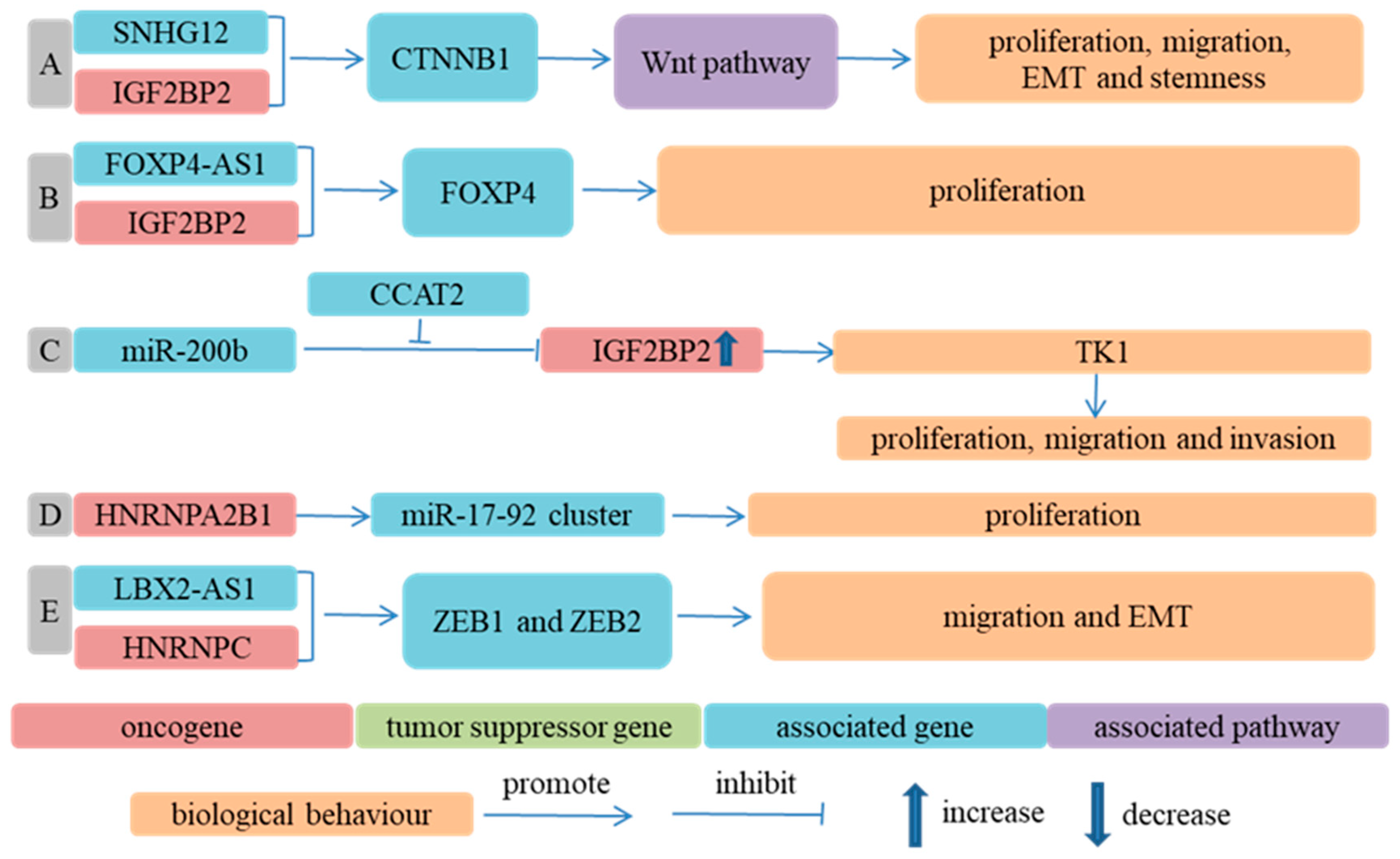
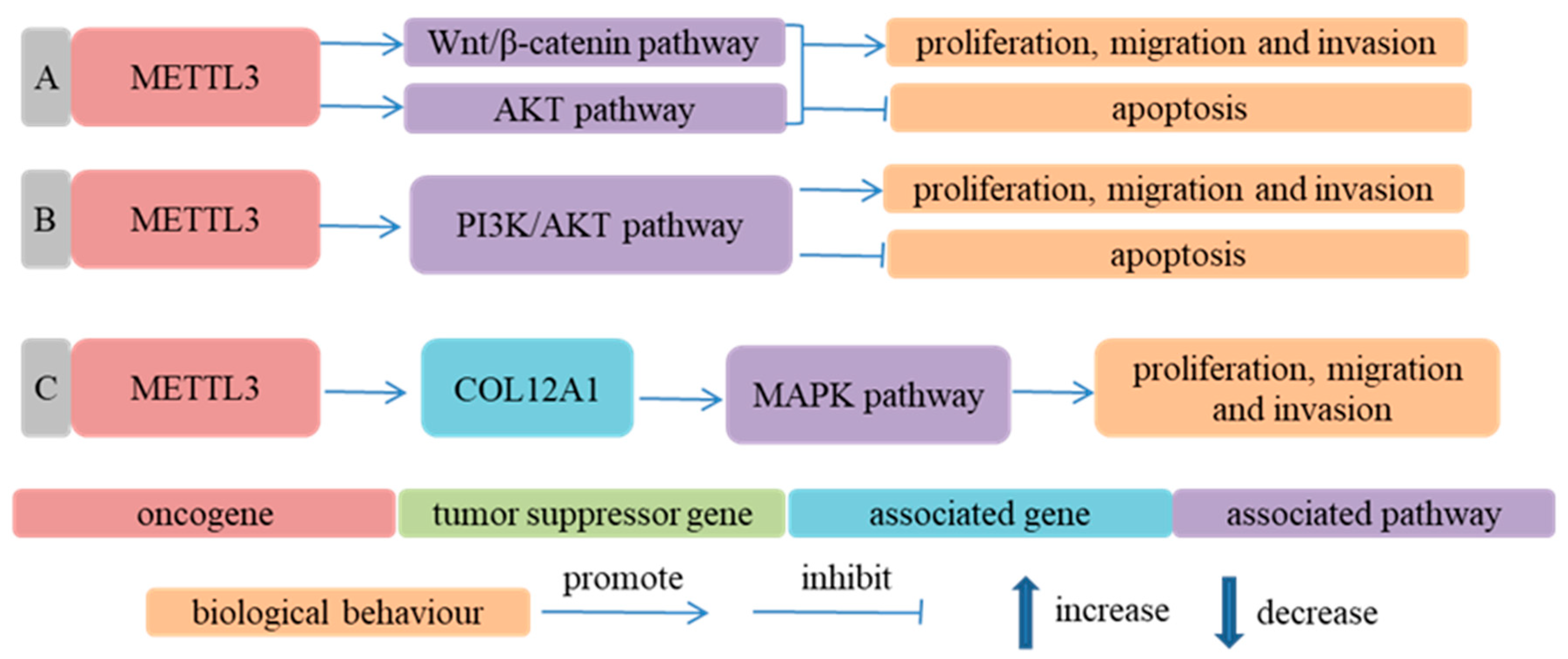
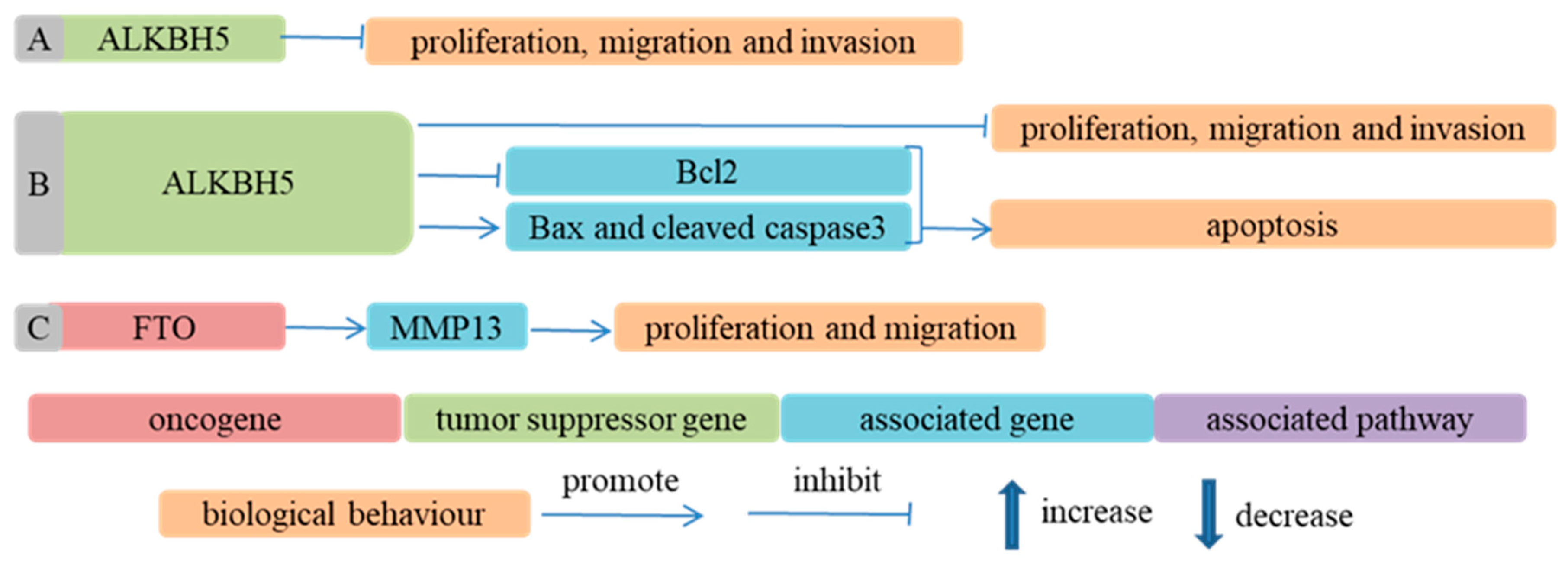
4.2.2. The Effect and Mechanism of m6A Regulators in Progression of ESCA

The m6A regulators affected the biological behavior of ESCC cells through the same or a different gene/signaling pathway, which is shown in Figure 1, Figure 2, Figure 3, Figure 4, Figure 5, Figure 6 and Figure 7 [59,60,61,62,63,64,65,66,68,69,70,71,72,73,74,75,76,78,79,80,81,82,83,84,85,86,124,126,127,131,132,133,134,135,136,137,138,139,141].

m6A Regulators Regulated Biological Behavior of ESCC Cells in an m6A-Dependent Way

  • Writers
METTL3 could promote the proliferation, migration and invasion of ESCC cells and inhibited ESCC cells’ apoptosis in an m6A-dependent way [59,60,61,75,76,85,122]. As methyltransferase, METTL3 affected the expression of coding RNA and the biogenesis of microRNA through changing the m6A level. In METTL3 knockdown cells, m6A peaks in the UTRs of glutaminase 2 (GLS2) and expression of GLS2 were decreased, which suggested that METTL3 could upregulate GLS2 expression through increasing the m6A level of GLS2. GLS2 knockdown alleviated the migration and invasion ability of ESCC cells increased by METTL3 overexpression. That is, METTL3 could regulate GLS2 expression in an m6A-dependent way and GLS2 mediated the effect of METTL3 on the malignant phenotype of ESCC cells [59]. Similarly, METTL3 played a crucial role in ESCC initiation and progression through regulating the expression of NOTCH1 in an m6A-dependent manner and activating the Notch signaling pathway [60]. The downregulation of METTL3 decreased the m6A level of pri-miR-200-5p, inhibited the binding of DGCR8 with pri-miR-200-5p and reduced the expression of miR-200-5p. The upregulation of miR-200-5p could target and inhibit nuclear factor IC (NFIC), which had been reported to play a suppressive role in the proliferation, metastasis and EMT process of ESCC cells [142]. Therefore, METTL3 influenced the malignancy of ESCC cells through regulating the expression of miR-200-5p and NFIC [75]. Similarly, the overexpression of METTL3 elevated the expression of miR-320b, which inhibited the expression of programmed cell death 4 (PDCD4), and then activated the AKT signaling pathway and led to lymphangiogenesis and the lymphatic metastasis of ESCC [76] (Figure 1).
Figure 1. Methyltransferases participate in ESCA progression in an m6A-dependent way. (A) Ref. [59] METTL3 upregulated GLS2 expression through increasing m6A modification level of GLS2 and promoted migration and invasion of ESCC cells; (B) Ref. [60] METTL3 upregulated NOTCH1 expression through increasing m6A modification level of NOTCH1, activated Notch signaling pathway and thus promoted proliferation, migration and invasion of ESCC cells; (C) Ref. [85] Depletion of METTL3 significantly reduced m6A modification levels of ESCC cells. METTL3 promoted proliferation of ESCC cells by increasing CDC25C, CDK1, cyclin A2 and cyclin B1 amounts and reducing p21 amounts; (D) Ref. [75] METTL3 upregulated miR-200-5p expression through increasing m6A modification level of pri-miR-200-5p, inhibited expression of NFIC and promoted malignant biological behavior of ESCC cells; (E) Ref. [76] METTL3 elevated miR-320b expression through increasing m6A modification level of pri-miR-320b, inhibited expression of PDCD4 and then activated AKT signaling pathway, leading to lymphangiogenesis and lymphatic metastasis of ESCC cells; (F) Ref. [61] ATXN2 bound to TNFR1 methylated by METTL3 (upregulated in ESCC samples) and augmented translation of TNFR1. TNFR1 promoted malignant behavior of ESCC cells, such as proliferation, migration and invasion, by activating MAPK and NF-κB signaling pathways.
Cancers 14 05139 g001
  • Erasers
Whether ALKBH5 promoted or inhibited the progression of ESCA was uncertain. ALKBH5 inhibited the biogenesis of miR-194-2 through decreasing its m6A modification, upregulated the expression of RAI1, activated the Hippo signaling pathway and suppressed the growth and motility of ESCC cells in vitro and in vivo [78]. Conversely, Nagaki et al. [62] reported that the depletion of ALKBH5 suppressed the proliferation and migration of ESCC cells. Mechanistically, ALKBH5 knockdown increased the m6A modification and stability of CDKN1A mRNA and upregulated the expression of p21, a cell cycle inhibitor. Furthermore, the knockdown of ALKBH5 suppressed tumor growth in nude mice. Collectively, ALKBH5 downregulated the expression of p21 in an m6A-dependent way and facilitated cell cycle progression and the proliferation of ESCC cells in vitro and in vivo. LncRNAs may influence mRNA expression through m6A RNA modification [143,144]. Qin et al. [63] reported that cancer susceptibility candidate 15 (CASC15) interacted with FTO, reduced the m6A level of single-minded 2 (SIM2) mRNA, decreased SIM2 expression and facilitated ESCC progression (Figure 2).
Figure 2. Demethylases participate in ESCA progression in an m6A-dependent way. (A) Ref. [62] ALKBH5 downregulated expression of p21 by decreasing m6A modification level of CDKN1A and promoted proliferation and migration of ESCC cells; (B) Ref. [78] ALKBH5 inhibited biogenesis of miR-194-2 through decreasing its m6A modification level, upregulated expression of RAI1, activated Hippo signaling pathway and suppressed malignant biological behavior of ESCC cells; (C) Ref. [63] FTO, as demethyltransferase of SIM2, interacted with CASC15 to decrease SIM2 expression and facilitate progression of ESCC.
Cancers 14 05139 g002
  • Readers
IGF2BP2 could increase the stability of mRNA individually [66] or in combination with lncRNA [64,65]. LncRNA colon-cancer-associated transcript 2 (CCAT2) absorbed miR-200b, weakened the inhibition of miR-200b on IGF2BP2 and upregulated the expression of IGF2BP2. The overexpression of IGF2BP2 enhanced the expression of TK1 by recognizing the m6A of TK1 mRNA, which promoted the oncogenesis of ESCC cells in nude mice and facilitated the migration and invasion of ESCC cells [66]. The interaction of IGF2BP2 with lncRNA small nucleolar RNA host gene 12 (SNHG12) and the interaction of IGF2BP2 with lncRNA forkhead box P4 antisense RNA 1 (FOXP4-AS1) could enhance the mRNA stability of CTNNB1 and forkhead box P4 (FOXP4), respectively, and partly contributed to the proliferation, migration, EMT and stemness of ESCC cells [64,65].
HNRNPA2B1 could increase the expression of the miR-17-92 cluster in an m6A-dependent manner, promote the proliferation of ESCC cells and affect the prognosis of ESCA patients negatively [79]. Similarly, HNRNPA2B1 might also facilitate ESCC progression by accelerating fatty acid synthesis via increasing the expression of ACLY and ACC1 [84]. LBX2-AS1 played a critical role in promoting the migration and EMT of ESCC cells, and this role was mediated by HNRNPC through increasing ZEB1 and ZEB2 mRNA stability [68] (Figure 3).
Figure 3. m6A RNA binding proteins participate in ESCA progression in an m6A-dependent way. (A) Ref. [64] IGF2BP2 interacted with SNHG12 to enhance mRNA stability of CTNNB1, elevated expression of β-catenin encoded by CTNNB1, activated Wnt signaling pathway and thus facilitated proliferation, migration, EMT and stemness of ESCC cells; (B) Ref. [65] IGF2BP2 interacted with FOXP4-AS1, enhanced FOXP4 mRNA stability, upregulated FOXP4 expression and promoted proliferation of ESCC cells; (C) Ref. [66] CCAT2 alleviated inhibition of IGF2BP2 by miR-200b and elevated IGF2BP2 expression, which upregulated expression of TK1 and facilitated proliferation, migration and invasion of ESCC cells; (D) Ref. [79] HNRNPA2B1 bound to m6A modification sites of miR-17-92 cluster, enhanced expression of miR-17-92 cluster and facilitated proliferation of ESCC cells; (E) Ref. [68] Interaction of HNRNPC with LBX2-AS1 enhanced ZEB1 and ZEB2 mRNA stability, upregulated expression of ZEB1 and ZEB2 and then promoted migration and EMT of ESCC cells.
Cancers 14 05139 g003

m6A Regulators Regulated Biological Behavior of ESCC Cells in an m6A-Independent Way

  • Writers
METTL3 was also involved in the progression of ESCC in an m6A-independent way [124,126,127]. For example, METTL3 might influence the biological behavior of ESCC cells by regulating Wnt/β-catenin and AKT signaling pathways [124]. In addition, METTL3 facilitated the malignant phenotype of ESCC cells by upregulating the expression of COL12A1 and activating the MAPK signaling pathway [127] (Figure 4).
Figure 4. Methyltransferases participate in ESCA progression in an m6A-independent way. (A) Ref. [124] METTL3 promoted proliferation, migration and invasion of ESCC cells and suppressed apoptosis of ESCC cells by activating Wnt/β-catenin and AKT signaling pathways; (B) Ref. [126] METTL3 facilitated proliferation, migration and invasion of ESCC cells and repressed apoptosis of ESCC cells by activating PI3K/AKT signaling pathway; (C) Ref. [127] METTL3 increased COL12A1 expression, activated MAPK signaling pathway and promoted proliferation, migration and invasion of ESCC cells.
Cancers 14 05139 g004
  • Erasers
The overexpression of ALKBH5 induced the apoptosis of ESCC cells by changing the expression of Bax, cleaved caspase 3 and Bcl2 in an m6A-independent way [86]. FTO promoted the proliferation and migration of ESCC cells by increasing the expression of MMP13 [132] (Figure 5).
Figure 5. Demethylases participate in ESCA progression in an m6A-independent way. (A) Ref. [131] ALKBH5 inhibited proliferation, migration and invasion of ESCC cells; (B) Ref. [86] ALKBH5 not only inhibited apoptosis of ESCC cells by regulating expression of Bcl2, Bax and cleaved caspase3 but also promoted proliferation, migration and invasion of ESCC cells; (C) Ref. [132] FTO facilitated proliferation and migration of ESCC cells through upregulating MMP13 expression.
Cancers 14 05139 g005
  • Readers
The downregulation of YTHDC2 changed the expression of some genes, which were enriched in p53, NF-kappa B and the JAK-STAT signaling pathway. Therefore, the downregulation of YTHDC2 might enhance the growth of ESCC cells through regulating certain pathological signaling pathways [133].
miR-98-5p and miR-454-3p could suppress the biological phenotype of ESCC cells by targeting IGF2BP1 [134,135].
The downregulation of miR-186 alleviated its inhibition of HNRNPC, upregulated the expression of HNRNPC and facilitated the malignant behavior of ESCC cells [136].
EIF3B cooperated with TEX9 to promote the malignant phenotype of ESCC cells via activating the AKT signaling pathway [141]. EIF3B also promoted ESCC cells’ proliferation and invasion and inhibited ESCC cells’ apoptosis through activating the β-catenin signaling pathway [137]. The knockdown of EIF3E repressed the proliferation and migration of ESCC cells [138]. EIF3H could stabilize Snail through deubiquitination and could promote the aggressiveness of ESCC cells [139] (Figure 6).
Figure 6. m6A RNA binding proteins participate in ESCA progression in an m6A-independent way. (A) Ref. [133] YTHDC2 inhibited proliferation of ESCC cells through changing expression of some genes enriched in p53 signaling pathway, NF-kappa B signaling pathway and JAK-STAT signaling pathway; (B) Ref. [134] IGF2BP1 and UHRF2 interacted to promote proliferation and invasion of ESCC cells and inhibit apoptosis of ESCC cells, which mediated regulation of miR-98-5p on biological phenotype of ESCC cells; (C) Ref. [135] miR-454-3p suppressed ERK and AKT signaling pathway by targeting IGF2BP1, then inhibited proliferation and invasion of ESCC cells; (D) Ref. [84] HNRNPA2B1 facilitated ESCC progression by accelerating fatty acid synthesis via increasing expression of ACLY and ACC1; (E) Ref. [136] miR-186 inhibited malignant behavior of ESCC cells by downregulating expression of HNRNPC; (F) Ref. [139] EIF3H promoted the proliferation, migration and invasion of ESCC cells through increasing stability of Snail protein; (G) Ref. [141] EIF3B cooperated with TEX9 to promote malignant phenotype of ESCC cells via activating AKT signaling pathway; (H) Ref. [137] EIF3B promoted proliferation and invasion of ESCC cells and inhibited apoptosis of ESCC cells through activating β-catenin signaling pathway; (I) Ref. [138] Knockdown of EIF3E repressed proliferation and migration of ESCC cells; in other words, EIF3E could facilitate malignant behavior of ESCC cells.
Cancers 14 05139 g006
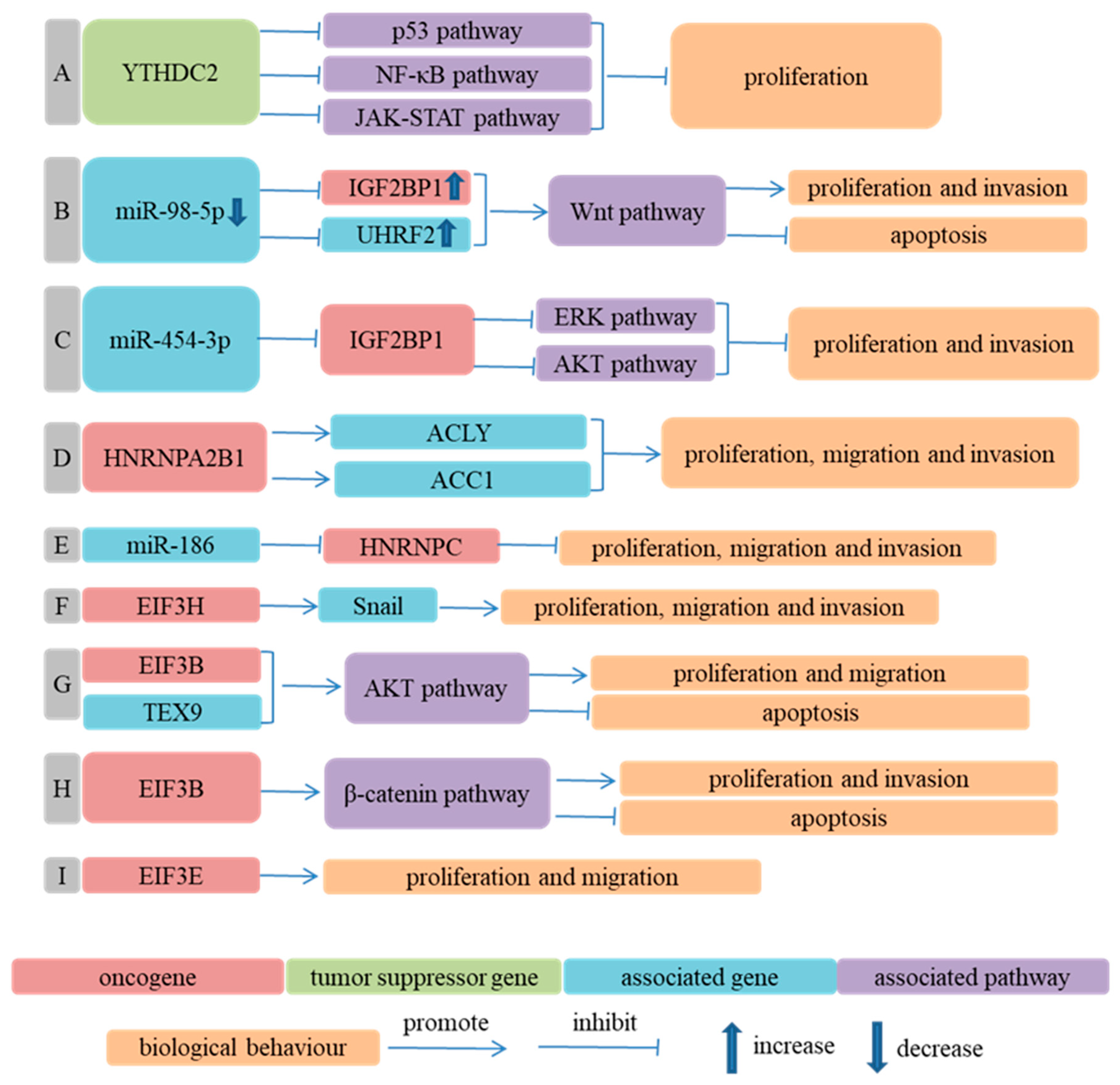
  • Combined Effects of m6A Regulators in Progression of ESCA
METTL3 overexpression enhanced the m6A level of adenomatous polyposis coli (APC) mRNA in an METTL14-dependent way, elevated the binding of YTHDF2 with APC mRNA and promoted the degradation of APC mRNA. Ultimately, the downregulation of APC enhanced aerobic glycolysis, ESCC cells growth and tumor formation in mice through activating the Wnt/β-catenin signaling pathway [69].
RBM15 interacted with METTL3 in a WTAP-dependent way to deposit m6A onto lncRNA metastasis-associated lung adenocarcinoma transcript 1 (MALAT1), located at nuclear speckles (NSs). YTHDC1 bound to the m6A of MALAT1, which maintained the composition of NSs and increased the migration of ESCC cells [82].
The combined regulation of EGR1 by METTL3 and YTHDF3 enhanced the expression of EGR1, activated the Snail signaling pathway and promoted the invasion and metastasis of ESCC cells [72].
Cigarette smoking and sexual hormones might be associated with different incidence rates of ESCC between men and women [145,146,147,148]. Wu et al. [81] reported whether and how a micropeptide affected the progression of male ESCC. The micropeptide was named Yin Yang 1 (YY1)-binding micropeptide (YY1BM), which was encoded by Y-linked lncRNA LINC00278. Specifically, cigarette smoking decreased the methylation of an ALKBH5 promoter, enhanced ALKBH5 expression, downregulated the m6A modification of LINC00278 and binding of YTHDF1 with LINC00278 and led to a translation suppression of YY1BM. The downregulation of YY1BM increased the survival of ESCC cells through a series of cascade reactions. Furthermore, Wu et al. [81] also reported that METTL3, METTL14 and WTAP acted as “writers” for the m6A modification of LINC00278 to participate in the development of male ESCC.
miR-193a-3p could target ALKBH5 and inhibit the expression of ALKBH5. The knockdown of ALKBH5 increased the m6A level of pri-miR-193a-3p and promoted the maturation of pri-miR-193a-3p. Thus, miR-193a-3p and ALKBH5 formed a positive feedback loop, which facilitated the proliferation and metastasis of ESCC cells in vitro and in vivo. The researchers also revealed that METTL3, as a methyltransferase of pri-miR-193a-3p, played an opposite role in the maturation of miR-193a-3p compared to ALKBH5 [80].
The overexpression of FTO promoted the proliferation and cell cycle progression of ESCC cells, which was realized by accelerating the expression of LINC00022 in a YTHDF2-dependent pattern and decay of p21 [83].
FTO could promote the proliferation, migration and invasion of ESCC cells by regulating the m6A level and expression of ERBB2 and HSD17B11. Interestingly, YTHDF1, as a reader of ERBB2 and HSD17B11, increased the stability of ERBB2 mRNA but decreased the translation efficiency of HSD17B11 mRNA [73,74].
YTHDF1 could recognize, bind and increase the stability of m6A-modified HK2 mRNA and upregulate the expression of HK2, which mediated the role of HCP5 in promoting the progression of ESCC. In this process, METTL3 acted as a methyltransferase of HK2 mRNA [71].
Linc01305 interacted with IGF2BP2 and IGF2BP3 to increase the stability of HTR3A mRNA and expression of HTR3A, and consequently promoted the proliferation and migration of ESCC cells [70] (Figure 7).
Figure 7. Combined effects of m6A regulators on ESCA progression. (A) Ref. [81] Cigarette smoking enhanced ALKBH5 expression through decreasing methylation of ALKBH5 promoter, downregulated m6A modification of LINC00278 and binding of YTHDF1 with LINC00278 and consequently led to translation suppression of Yin Yang 1 (YY1)-binding micropeptide (YY1BM), a micropeptide encoded by LINC00278. Downregulation of YY1BM promoted the interaction of YY1 with androgen receptor (AR), activated AR signaling pathway and increased eEF2K expression. Under nutrient deprivation, the elevated phosphorylation of eEF2 inhibited activity of eEF2 and translation elongation and increased survival of ESCC cells; (B) Ref. [80] miR-193a-3p could target ALKBH5 and inhibit expression of ALKBH5. Interestingly, knockdown of ALKBH5 promoted the maturation of miR-193a-3p. Thus, miR-193a-3p and ALKBH5 formed a positive feedback loop, which facilitated proliferation and metastasis of ESCC cells in vitro and in vivo; (C) Ref. [69] Overexpression of METTL3 enhanced m6A level of adenomatous polyposis coli (APC) mRNA in an METTL14-dependent way, elevated binding of YTHDF2 with APC mRNA and promoted degradation of APC mRNA. Decreased APC expression facilitated proliferation of ESCC cells through activating Wnt/β-catenin signaling pathway; (D) Ref. [70] Linc01305 interacted with IGF2BP2 and IGF2BP3 to increase stability of HTR3A mRNA and expression of HTR3A, then promoted proliferation and migration of ESCC cells; (E) Ref. [82] RBM15 interacted with METTL3 in a WTAP-dependent way to deposit m6A onto MALAT1, specifically located at nuclear speckles (NSs), and YTHDC1 could bind to m6A of MALAT1, which was important for NSs homeostasis and metastasis of ESCC cells; (F) Ref. [83] Overexpression of FTO significantly reduced the enrichment of m6A at site 2 of LINC00022 transcript and led to a decrease in degradation of LINC00022 by YTHDF2. FTO promoted proliferation of ESCC cells by upregulating expression of LINC00022 in a YTHDF2-dependent pattern; (G) Ref. [71] YTHDF1 increased stability of m6A-modified HK2 mRNA and upregulated expression of HK2, which mediated the role of HCP5 in promoting progression of ESCC. In this process, METTL3 acted as methyltransferase of HK2 mRNA; (H) Ref. [73] FTO promoted proliferation, migration and invasion of ESCC cells by regulating m6A level of ERBB2, in which, YTHDF1, as reader of ERBB2, increased stability of ERBB2 mRNA; (I) Ref. [74] Combined role of FTO and YTHDF1 resulted in upregulation of HSD17B11 and progression of ESCC, in which, reduced m6A level of ERBB2 by FTO led to inhibition of YTHDF1-induced decreased translation efficiency of HSD17B11; (J) Ref. [72] Increased m6A level of EGR1 mRNA by METTL3 and increased binding of m6A-modified EGR1 mRNA with YTDF3 led to upregulation of EGR1, activated Snail signaling pathway and promoted invasion and metastasis of ESCC cells.
Cancers 14 05139 g007
Various signaling pathways mediated the regulation of m6A regulators on ESCC, consisting of AKT, NF-κB, MAPK, Wnt/β-catenin, etc. Interestingly, the same gene could target different genes or signaling pathways to influence the malignancy of ESCC. For instance, the target genes and signaling pathways of METTL3 included GLS2, NOTCH1, TNFR1, miR-200-5p, miR-320b, miR-99a-5p, Notch pathway, AKT pathway, MAPK pathway, NF-κB pathway and Wnt/β-catenin pathway [59,60,61,75,76,124,126]. Different genes could also regulate the same gene, such as p21, a cell cycle inhibitor, which participated in the regulation process of METTL3, ALKBH5, FTO and YTHDF2 in ESCC [62,83,85]. Different m6A-modified genes could play their regulatory role in ESCC through the same pathway, such as the AKT pathway [76,77,124,126,135,141], NF-κB pathway [61,133] and Wnt/β-catenin pathway [64,69,124,134,137]. Therefore, a combined analysis of m6A regulators might be beneficial for better understanding their effect and mechanism in ESCA and providing an experimental basis for the drug research of ESCA.

4.2.3. The Role of m6A Regulators in Treatment of ESCA

The survival time of ESCA patients has improved dramatically with the improvement in the surgical level and development of drugs. However, the five-year survival rate of ESCA patients remains dismal due to treatment failure resulting from therapeutic resistance. Thus, it is urgent to identify the mechanism responsible for therapeutic resistance and to find new drugs alone or in combination with other therapeutic agents used to improve the prognosis of ESCA patients. Considering the crucial role of m6A regulators in the occurrence and progression of ESCA, m6A regulators might be promising therapeutic targets. A previous study demonstrated that the depletion of METTL3 enhanced the sensitivity of pancreatic cancer cells to cisplatin and irradiation [149]. Compared to wild-type mice, YTHDF1-/- mice acquired a better therapeutic efficacy of PD-L1 checkpoint blockade, implying the potential of YTHDF1 as a target of antitumor immunotherapy [150]. Similarly, m6A regulators played a non-negligible role in the therapeutic resistance of ESCA [67,77,122,129,151].
Platinum-based agents have been widely applied to treat esophageal cancer patients. Platinum treatment resulted in the upregulation of SNHG3. SNHG3 could sponge miR-186-5p, alleviated the inhibition of METTL3 by miR-186-5p, increased the expression of MTLL3 and promoted the growth of ESCC cells in vitro and in vivo. Therefore, targeting SNHG3/miR-186-5p/METTL3 might enhance the platinum efficacy [122]. Hypoxia could induce the expression of some genes associated with resistance to anticancer drugs, then decreased the sensitivity of cells to chemotherapy. Hypoxia induced the expression of lncRNA E2F1 messenger RNA stabilizing factor (EMS), which could sponge miR-758-3p and weaken its repression of WTAP, increased the expression of WTAP and led to resistance of ESCC cells to cisplatin. Therefore, EMS, miR-758-3p and WTAP might be considered as a therapeutic target to enhance cisplatin efficiency [129].
m6A regulators were also associated with radioresistance. Cancer stem-like cells (CSCs) have been reported to be associated with the radioresistance of ESCC cells [152]. METTL14 was downregulated in CSCs, which resulted in a decreased expression of miR-99a-5p in an m6A-dependent manner. The downregulation of miR-99a-5p led to an increase in the expression of TRIB2. TIRB2 bridged METTL14 to COP1 and one E3 ligase, and resulted in the ubiquitination and degradation of METTL14. Thus, METTL14, miR-99a-5p and TRIB2 formed a positive feedback circuit. TRIB2 could promote CSCs persistence and the radioresistance of ESCC cells through activating the Akt/mTOR/S6K1 signaling pathway [77]. IGF2BP3 enhanced KIF18A mRNA stability, upregulated KIF18A expression and facilitated ESCC cells’ proliferation, migration, invasion and radioresistance [67]. The knockdown of IGF2BP3 increased the susceptibility of radioresistant TE-5 and TE-9 cells to radiotherapy [153]. These results suggested that METTL14/miR-99a-5p/TRIB2 axis and IGF2BP3 might be potential targets for improving the sensitivity of ESCC cells to radiotherapy.
m6A regulators could be used as a treatment target of herbal medicine for esophageal cancer. HNRNPA2B1 codes for two isoforms, HNRNPA2 and HNRNPB1. The crude extract of a South African medicinal plant, Cotyledon orbiculata, reduced colon cancer cells and ESCC cells proliferation and induced their apoptosis through regulating the alternative splicing of HNRNPA2B1 (decreasing the expression of HNRNPB1). A further investigation on HCT116 cells showed that the silencing of HNRNPB1 led to a switch in the splicing of BCL2L1 from a Bcl-xL anti-apoptotic isoform to a Bcl-xS pro-apoptotic isoform and promoted the apoptosis of HCT116 cells. Thus, HNRNPA2B1 might be a potential treatment target of herbal medicine for ESCA. [151].
Elvitegravir, originally developed to treat human immunodeficiency virus (HIV) infection, could repress the invasion and metastasis of ESCC cells by enhancing the proteasomal degradation of METTL3 mediated by STUB1 [72]. If the therapeutic effect of elvitegravir is proven clinically, there will be an additional option for the treatment of ESCA.

4.2.4. The Association between Expression of m6A Regulators and Prognosis of ESCA Patients

The association between the expression of m6A regulators and prognosis of ESCA patients was shown in Supplementary Table S2. A higher expression or positive expression of METTL3, WTAP, HNRNPA2B1, HNRNPC and EIF3B was associated with a poor prognosis of ESCA patients [59,60,61,69,72,79,123,126,127,129,130,137]. On the contrary, a lower METTL14 expression predicted a poor overall survival (OS) of ESCA patients [77]. There was no relation between the expression of EIF3E and prognosis of ESCA patients [138]. Xu et al. [130] revealed that a low ALKBH5 expression was associated with a poor survival of ESCA patients. Similarly, ESCA patients with a positive ALKBH5 expression showed a longer OS and disease-free survival (DFS) and less recurrence/distant metastasis after surgery than those with a negative ALKBH5 expression [78]. However, a study on 177 patients showed that a higher ALKBH5 expression was related to a lower OS [62]. There were two studies that reported the association of FTO expression with the prognosis of ESCA patients. One showed the relation of a poor OS with a high FTO expression [74]; the other suggested no relation between them [62]. The difference in subjects, sample size and detection methods might partly explain the discrepancy. For patients treated with surgery alone, a high IGF2BP3 expression was associated with a poor OS, disease-specific survival (DSS) and DFS [154]. Takata et al. [140] reported that IGF2BP3-positive patients had a poorer OS and recurrence-free survival (RFS) than IGF2BP3-negative patients.
Present studies on the association between METTL3 expression and the prognosis of ESCA patients reached a consistent conclusion, which was that a “higher METTL3 expression predicted a poor prognosis of ESCA patients”. Results about ALKBH5 and FTO were opposite or inconsistent. The number of studies about other m6A regulators is limited. Therefore, further studies are needed to comprehensively analyze the relation between the expression of m6A regulators and the survival of ESCA patients due to the complexity of the m6A regulatory network. Based on information from the public database, some researchers constructed a prognostic signature according to the expression of m6A regulators to predict the survival of ESCA patients [79,84,130,136,155,156]. For example, Xu et al. [130] analyzed the RNA sequencing transcriptome data of 13 m6A regulators from the TCGA ESCA database, constructed a prognostic signature, which consisted of HNRNPC and ALKBH5, and established a formula to calculate the risk score. According to the risk score, the ESCA patients were divided into a low risk group and high risk group. Compared with the low risk group patients, the patients in the high risk group had a significantly poorer survival. Therefore, the prognostic signature could be used as an independent prognostic marker for ESCA patients.

5. Conclusions and Perspectives

After transcription, different chemical modifications can occur on cellular RNA, such as N6-methyladenosine (m6A), N6,2′-O-dimethyladenosine (m6Am), N1-methyla- denosine (m1A) and 5-methylcytosine (m5C) [157]. Among them, m6A is the most abundant RNA modification, and is dynamic and reversible. m6A is catalyzed by methyltransferase, then removed by demethylase or recognized by m6A RNA binding protein. During the development and progression of ESCA, m6A on various types of RNA was involved in all steps of RNA metabolism, post-transcriptionally regulated the expression of the gene and affected the biological behavior of ESCC cells. m6A regulators not only regulated the expression of various type of RNAs but also received regulation by genetic variation, epigenetic modification and non-coding RNAs. During the development of ESCA, METTL3 acted as an oncogene. The mRNA expression level of METTL3 might be used for the diagnosis of ESCA with a sensitivity of 75% and specificity of 72.06%. Furthermore, a high SUVmax of 18F-FDG PET/CT could be used as a non-invasive predictive marker for a high METTL3 expression of ESCA patients. m6A regulators participated in the progression of ESCA in an m6A-dependent or m6A-indepent manner. The same m6A regulator could target different genes or signaling pathways to influence the malignancy of ESCA. Similarly, different m6A-modified genes could play their regulatory role in ESCA through the same gene or signaling pathway. The m6A regulators played a key role in the occurrence and progression of ESCA, which suggested that they might serve as potential therapeutic targets for ESCA. At present, chemotherapy and radiotherapy remain the primary treatment for ESCA. Platinum resistance and radioresistance often result in a poor prognosis of ESCA patients. Targeting the SNHG3/miR-186-5p/METTL3 axis and EMS/miR-758-3p/WTAP axis might help to increase the sensitivity of ESCC cells to platinum; the METTL14/miR-99a-5p/TRIB2 axis and IGF2BP3 might be useful targets for decreasing the radioresistance of ESCC cells. In addition, an extract of Cotyledon orbiculata, a South African medicinal plant, exerted an anti-proliferative effect in ESCC cells through possibly regulating the alternative splicing of hnRNPA2B1 [151]. Moreover, elvitegravir could enhance the proteasomal degradation of METTL3 mediated by STUB1 to repress the invasion and metastasis of ESCC cells [72]. A prognostic signature based on m6A regulators is helpful for predicting the prognosis of ESCA patients accurately.
Of note, YTHDF1 has been widely reported to promote protein synthesis. However, YTHDF1 decreased the translation efficiency of HSD17B11 mRNA in the progression of ESCC. In addition, whether ALKBH5 and FTO play roles as an oncogene or cancer suppressor gene is inconclusive, which might be associated with a difference in the sample size, detection method and cell lines employed. In future, a combined analysis of m6A regulators in a multi-center study with a large sample size and identical detection method might be beneficial for better understanding their effect and mechanism in ESCA and for drawing a consistent and reliable conclusion.
Inflammation is a physiological response of organisms to various injuries, such as an infection of microorganisms, autoimmunity and physical damage. The disequilibrium of pro-inflammatory factors and anti-inflammatory factors will result in a persistent inflammatory state, so-called chronic inflammation, in which, a large number of cytokines arise and facilitate the initiation, promotion, invasion and metastasis of a tumor [158]. Previous studies revealed a close relation between inflammatory esophageal disease and ESCA [159,160]. In particular, in 58 ESCA patients and 10,614 non-ESCA subjects who received endoscopic examination, there was a significantly higher proportion of esophagitis in ESCA patients (89.7%) than in non-ESCA subjects (14.3%) [161]. The association of m6A regulators with pro-inflammatory genes in tumor development and progression has been widely reported. YTHDF2 deletion aggravated the inflammation, angiogenesis and metastatic progression of hepatocellular carcinoma [55]. In another study on intrahepatic cholangiocarcinoma, FTO changed the expression of inflammatory genes via upregulating NR5A2 [162]. In ESCA, there is no similar report. Identifying the target of m6A regulators during the process from chronic esophagitis to ESCA would help to determine the pathogenesis of ESCA and provide experimental evidence for the development of a new drug.
To meet the needs of uncontrolled proliferation, one hallmark of cancer, cancer cells must acquire sufficient nutrients from the surrounding tumor microenvironment through regulating various metabolism-associated enzymes, signaling pathways and transcription factors [163]. This ability of cancer cells is termed metabolic reprogramming, which includes the metabolic recombination of glucose, lipid and glutamine; for instance, whether under aerobic conditions or under anoxic conditions cancer cells depend on glycolysis to obtain energy preferentially, which is also known as the “Warburg effect” [164,165]. Glucose transporter 1 (GLUT1) and hexokinase 2 (HK2) are rate-limiting enzymes of the “Warburg effect”, responsible for glucose transport and transformation, respectively. Based on multiple public databases, some studies showed a relation between the expression of these two enzymes and m6A regulators, which hinted that m6A-modified genes participated in glucose metabolic reprogramming [66,125,166], which was validated in some cytological experiments [69,71]. Pyruvate kinase M2 isozyme (PKM2), as a downstream gene of APC regulated by METTl3, METTL14 and YTHDF2, enhanced aerobic glycolysis and, in turn, promoted the proliferation of ESCC cells [69]. The combined effect of METTL3 and YTHDF1, as a writer of HK2 mRNA and reader of m6A-modified HK2 mRNA, respectively, caused an upregulation of HK2 and promoted the glycolysis of ESCC cells [71]. m6A regulators were also involved in lipid metabolic reprogramming [74,84]. The knockdown of HNRNPA2B1 inhibited the malignant biological behavior of ESCC cells by downregulating the expression of de novo fatty acid synthetic enzymes and suppressing lipid accumulation in ESCC cells [84]. FTO facilitated the growth and invasion of ESCC cells by enhancing the formation of lipid droplets via the regulation of HSD17B11 expression in ESCC cells [74]. So far, whether m6A regulators affect glutamine metabolic reprogramming in ESCA has not been disclosed. Targeting metabolism-associated m6A regulators might be new strategies for the treatment of ESCA. As shown in one study, some drugs targeting glutaminase isoenzymes had an anticancer effect [167]. Determining the role of m6A regulators in ESCA metabolic reprogramming will be beneficial for the development of a new drug.
Immune escape is one of the basic characteristics of tumor cells. The application of immune checkpoint inhibitors (ICIs) that activate antitumor immune cells has shown a significant therapeutic effect on various malignant tumors, including ESCA [168]. However, only a few patients benefit from ICIs treatment because of resistance, thus limiting its application. The tumor-immune microenvironment (TIME), consisting of PD-L1 expression on tumors, tumor-infiltrating lymphocytes, tumor-associated macrophages, etc., might impact the patients’ response to ICIs. m6A modification is involved in remodeling TIME through regulating the expression of PD-L1 on tumors and regulating the differentiation and activation of tumor-associated immune cells [169,170,171,172,173]. The continual expression of PD-L1 in tumor cells facilitates tumor immune escape. IGF2BP1 recognized PD-L1 mRNA modified by METTL3 and enhanced the expression of PD-L1 in bladder cancer cells, which decreased the attack of CD8+ T cells [170]. The deletion of METTL3 disturbed T cells differentiation and macrophage activation via regulating different downstream target genes [172,173]. In addition, a change in the expression of m6A regulators also might influence the distribution and function of immune cells in TIME [174,175,176]. In hepatocellular carcinoma, ALKBH5 promoted macrophage recruitment through upregulating MAP3K8 expression in an m6A-dependent manner [174]. In ESCA, some studies revealed that the expression of m6A regulators was associated with PD-L1 expression and immune cells infiltration, suggesting the role of m6A regulators as an immune therapy target individually or in combination with ICIs [128,155,156,177]. However, how m6A regulators affect the TIME of ESCA has not been reported, which provides a direction for future research in ESCA.
In summary, determining the role and mechanism of m6A modification and m6A regulators in the occurrence, progression, remedy and prognosis of ESCA is necessary for an early diagnosis, individualized treatment and improved prognosis of ESCA patients. Identifying the target of m6A regulators during the process from chronic esophagitis to ESCA, the role of m6A regulators in metabolic reprogramming and the impact of m6A regulators on TIME in ESCA might be a future research direction.

Supplementary Materials

The following supporting information can be downloaded at: https://www.mdpi.com/article/10.3390/cancers14205139/s1, Table S1: Expression, clinical significance and biological function of m6A regulators in ESCA; Table S2: The association between expression of m6A regulators and prognosis of ESCA patients.

Author Contributions

Literature collection, reading and analysis, Y.L., C.N., N.W., X.H. (Xi Huang), S.C. (Shiru Cao), S.C. (Saijin Cui), T.C., X.H. (Xiangran Huo) and R.Z.; table making, C.N., N.W. and X.H. (Xi Huang); figure drawing, S.C. (Shiru Cao), S.C. (Saijin Cui), T.C. and X.H. (Xiangran Huo); writing—original draft preparation, Y.L.; writing—review and editing, Y.L. and R.Z.; supervision, R.Z.; funding acquisition, R.Z. All authors have read and agreed to the published version of the manuscript.

Funding

This research was funded by Hebei Province Medical Science Research Key Project [No. 20180533].

Conflicts of Interest

The authors declare no conflict of interest.

References

  1. Krug, R.M.; Morgan, M.A.; Shatkin, A.J. Influenza viral mRNA contains internal N6-methyladenosine and 5′-terminal 7-methylguanosine in cap structures. J. Virol. 1976, 20, 45–53. [Google Scholar] [CrossRef] [PubMed] [Green Version]
  2. Beemon, K.; Keith, J. Localization of N6-methyladenosine in the Rous sarcoma virus genome. J. Mol. Biol. 1977, 113, 165–179. [Google Scholar] [CrossRef]
  3. Bokar, J.A.; Shambaugh, M.E.; Polayes, D.; Matera, A.G.; Rottman, F.M. Purification and cDNA cloning of the AdoMet-binding subunit of the human mRNA (N6-adenosine)-methyltransferase. RNA 1997, 3, 1233–1247. [Google Scholar] [PubMed]
  4. Wei, C.M.; Moss, B. Nucleotide sequences at the N6-methyladenosine sites of HeLa cell messenger ribonucleic acid. Biochemistry 1977, 16, 1672–1676. [Google Scholar] [CrossRef]
  5. Dominissini, D.; Moshitch-Moshkovitz, S.; Schwartz, S.; Salmon-Divon, M.; Ungar, L.; Osenberg, S.; Cesarkas, K.; Jacob-Hirsch, J.; Amariglio, N.; Kupiec, M.; et al. Topology of the human and mouse m6A RNA methylomes revealed by m6A-seq. Nature 2012, 485, 201–206. [Google Scholar] [CrossRef]
  6. Meyer, K.D.; Saletore, Y.; Zumbo, P.; Elemento, O.; Mason, C.E.; Jaffrey, S.R. Comprehensive analysis of mRNA methylation reveals enrichment in 3′ UTRs and near stop codons. Cell 2012, 149, 1635–1646. [Google Scholar] [CrossRef] [Green Version]
  7. Wang, Y.; Li, Y.; Toth, J.I.; Petroski, M.D.; Zhang, Z.; Zhao, J.C. N6-methyladenosine modification destabilizes developmental regulators in embryonic stem cells. Nat. Cell Biol. 2014, 16, 191–198. [Google Scholar] [CrossRef] [Green Version]
  8. Zheng, G.; Dahl, J.A.; Niu, Y.; Fedorcsak, P.; Huang, C.M.; Li, C.J.; Vågbø, C.B.; Shi, Y.; Wang, W.L.; Song, S.H.; et al. ALKBH5 is a mammalian RNA demethylase that impacts RNA metabolism and mouse fertility. Mol. Cell 2013, 49, 18–29. [Google Scholar] [CrossRef] [Green Version]
  9. Wang, X.; Lu, Z.; Gomez, A.; Hon, G.C.; Yue, Y.; Han, D.; Fu, Y.; Parisien, M.; Dai, Q.; Jia, G.; et al. N6-methyladenosine-dependent regulation of messenger RNA stability. Nature 2014, 505, 117–120. [Google Scholar] [CrossRef] [Green Version]
  10. Wang, X.; Zhao, B.S.; Roundtree, I.A.; Lu, Z.; Han, D.; Ma, H.; Weng, X.; Chen, K.; Shi, H.; He, C. N(6)-methyladenosine Modulates Messenger RNA Translation Efficiency. Cell 2015, 161, 1388–1399. [Google Scholar] [CrossRef]
  11. Zhao, X.; Yang, Y.; Sun, B.F.; Shi, Y.; Yang, X.; Xiao, W.; Hao, Y.J.; Ping, X.L.; Chen, Y.S.; Wang, W.J.; et al. FTO-dependent demethylation of N6-methyladenosine regulates mRNA splicing and is required for adipogenesis. Cell Res. 2014, 24, 1403–1419. [Google Scholar] [CrossRef]
  12. Chen, T.; Hao, Y.J.; Zhang, Y.; Li, M.M.; Wang, M.; Han, W.; Wu, Y.; Lv, Y.; Hao, J.; Wang, L.; et al. m(6)A RNA methylation is regulated by microRNAs and promotes reprogramming to pluripotency. Cell Stem Cell 2015, 16, 289–301. [Google Scholar] [CrossRef] [PubMed] [Green Version]
  13. Liu, N.; Dai, Q.; Zheng, G.; He, C.; Parisien, M.; Pan, T. N(6)-methyladenosine-dependent RNA structural switches regulate RNA-protein interactions. Nature 2015, 518, 560–564. [Google Scholar] [CrossRef] [Green Version]
  14. Alarcón, C.R.; Lee, H.; Goodarzi, H.; Halberg, N.; Tavazoie, S.F. N6-methyladenosine marks primary microRNAs for processing. Nature 2015, 519, 482–485. [Google Scholar] [CrossRef] [PubMed] [Green Version]
  15. Geula, S.; Moshitch-Moshkovitz, S.; Dominissini, D.; Mansour, A.A.; Kol, N.; Salmon-Divon, M.; Hershkovitz, V.; Peer, E.; Mor, N.; Manor, Y.S.; et al. Stem cells. m6A mRNA methylation facilitates resolution of naïve pluripotency toward differentiation. Science 2015, 347, 1002–1006. [Google Scholar] [CrossRef]
  16. Zhou, J.; Wan, J.; Gao, X.; Zhang, X.; Jaffrey, S.R.; Qian, S.B. Dynamic m(6)A mRNA methylation directs translational control of heat shock response. Nature 2015, 526, 591–594. [Google Scholar] [CrossRef] [Green Version]
  17. Meyer, K.D.; Patil, D.P.; Zhou, J.; Zinoviev, A.; Skabkin, M.A.; Elemento, O.; Pestova, T.V.; Qian, S.B.; Jaffrey, S.R. 5′ UTR m(6)A Promotes Cap-Independent Translation. Cell 2015, 163, 999–1010. [Google Scholar] [CrossRef] [PubMed] [Green Version]
  18. Xiang, Y.; Laurent, B.; Hsu, C.H.; Nachtergaele, S.; Lu, Z.; Sheng, W.; Xu, C.; Chen, H.; Ouyang, J.; Wang, S.; et al. RNA m(6)A methylation regulates the ultraviolet-induced DNA damage response. Nature 2017, 543, 573–576. [Google Scholar] [CrossRef] [Green Version]
  19. Zhao, B.S.; Wang, X.; Beadell, A.V.; Lu, Z.; Shi, H.; Kuuspalu, A.; Ho, R.K.; He, C. m(6)A-dependent maternal mRNA clearance facilitates zebrafish maternal-to-zygotic transition. Nature 2017, 542, 475–478. [Google Scholar] [CrossRef] [Green Version]
  20. Sung, H.; Ferlay, J.; Siegel, R.L.; Laversanne, M.; Soerjomataram, I.; Jemal, A.; Bray, F. Global Cancer Statistics 2020: GLOBOCAN Estimates of Incidence and Mortality Worldwide for 36 Cancers in 185 Countries. CA Cancer J. Clin. 2021, 71, 209–249. [Google Scholar] [CrossRef]
  21. Wang, L.D.; Zhou, F.Y.; Li, X.M.; Sun, L.D.; Song, X.; Jin, Y.; Li, J.M.; Kong, G.Q.; Qi, H.; Cui, J.; et al. Genome-wide association study of esophageal squamous cell carcinoma in Chinese subjects identifies susceptibility loci at PLCE1 and C20orf54. Nat. Genet. 2010, 42, 759–763. [Google Scholar] [CrossRef]
  22. Wu, C.; Hu, Z.; He, Z.; Jia, W.; Wang, F.; Zhou, Y.; Liu, Z.; Zhan, Q.; Liu, Y.; Yu, D.; et al. Genome-wide association study identifies three new susceptibility loci for esophageal squamous-cell carcinoma in Chinese populations. Nat. Genet. 2011, 43, 679–684. [Google Scholar] [CrossRef] [PubMed]
  23. Wu, C.; Kraft, P.; Zhai, K.; Chang, J.; Wang, Z.; Li, Y.; Hu, Z.; He, Z.; Jia, W.; Abnet, C.C.; et al. Genome-wide association analyses of esophageal squamous cell carcinoma in Chinese identify multiple susceptibility loci and gene-environment interactions. Nat. Genet. 2012, 44, 1090–1097. [Google Scholar] [CrossRef]
  24. Zhou, R.M.; Li, Y.; Wang, N.; Huang, X.; Cao, S.R.; Shan, B.E. Association of programmed death-1 polymorphisms with the risk and prognosis of esophageal squamous cell carcinoma. Cancer Genet. 2016, 209, 365–375. [Google Scholar] [CrossRef] [PubMed]
  25. Zhou, R.; Li, Y.; Wang, N.; Niu, C.; Huang, X.; Cao, S.; Huo, X. PARP1 rs1136410 C/C genotype associated with an increased risk of esophageal cancer in smokers. Mol. Biol. Rep. 2021, 48, 1485–1491. [Google Scholar] [CrossRef] [PubMed]
  26. Grady, W.M.; Yu, M.; Markowitz, S.D. Epigenetic Alterations in the Gastrointestinal Tract: Current and Emerging Use for Biomarkers of Cancer. Gastroenterology 2021, 160, 690–709. [Google Scholar] [CrossRef]
  27. Cao, W.; Lee, H.; Wu, W.; Zaman, A.; McCorkle, S.; Yan, M.; Chen, J.; Xing, Q.; Sinnott-Armstrong, N.; Xu, H.; et al. Multi-faceted epigenetic dysregulation of gene expression promotes esophageal squamous cell carcinoma. Nat. Commun. 2020, 11, 3675. [Google Scholar] [CrossRef]
  28. Desrosiers, R.; Friderici, K.; Rottman, F. Identification of methylated nucleosides in messenger RNA from Novikoff hepatoma cells. Proc. Natl. Acad. Sci. USA 1974, 71, 3971–3975. [Google Scholar] [CrossRef] [Green Version]
  29. Jia, G.; Fu, Y.; Zhao, X.; Dai, Q.; Zheng, G.; Yang, Y.; Yi, C.; Lindahl, T.; Pan, T.; Yang, Y.G.; et al. N6-methyladenosine in nuclear RNA is a major substrate of the obesity-associated FTO. Nat. Chem. Biol. 2011, 7, 885–887. [Google Scholar] [CrossRef]
  30. Wang, P.; Doxtader, K.A.; Nam, Y. Structural Basis for Cooperative Function of Mettl3 and Mettl14 Methyltransferases. Mol. Cell 2016, 63, 306–317. [Google Scholar] [CrossRef]
  31. Choe, J.; Lin, S.; Zhang, W.; Liu, Q.; Wang, L.; Ramirez-Moya, J.; Du, P.; Kim, W.; Tang, S.; Sliz, P.; et al. mRNA circularization by METTL3-eIF3h enhances translation and promotes oncogenesis. Nature 2018, 561, 556–560. [Google Scholar] [CrossRef] [PubMed] [Green Version]
  32. Lin, S.; Choe, J.; Du, P.; Triboulet, R.; Gregory, R.I. The m(6)A Methyltransferase METTL3 Promotes Translation in Human Cancer Cells. Mol. Cell 2016, 62, 335–345. [Google Scholar] [CrossRef] [PubMed] [Green Version]
  33. Schöller, E.; Weichmann, F.; Treiber, T.; Ringle, S.; Treiber, N.; Flatley, A.; Feederle, R.; Bruckmann, A.; Meister, G. Interactions, localization, and phosphorylation of the m(6)A generating METTL3-METTL14-WTAP complex. RNA 2018, 24, 499–512. [Google Scholar] [CrossRef] [Green Version]
  34. Ping, X.L.; Sun, B.F.; Wang, L.; Xiao, W.; Yang, X.; Wang, W.J.; Adhikari, S.; Shi, Y.; Lv, Y.; Chen, Y.S.; et al. Mammalian WTAP is a regulatory subunit of the RNA N6-methyladenosine methyltransferase. Cell Res. 2014, 24, 177–189. [Google Scholar] [CrossRef] [PubMed] [Green Version]
  35. Guo, J.; Tang, H.W.; Li, J.; Perrimon, N.; Yan, D. Xio is a component of the Drosophila sex determination pathway and RNA N(6)-methyladenosine methyltransferase complex. Proc. Natl. Acad. Sci. USA 2018, 115, 3674–3679. [Google Scholar] [CrossRef] [Green Version]
  36. Knuckles, P.; Lence, T.; Haussmann, I.U.; Jacob, D.; Kreim, N.; Carl, S.H.; Masiello, I.; Hares, T.; Villaseñor, R.; Hess, D.; et al. Zc3h13/Flacc is required for adenosine methylation by bridging the mRNA-binding factor Rbm15/Spenito to the m(6)A machinery component Wtap/Fl(2)d. Genes Dev. 2018, 32, 415–429. [Google Scholar] [CrossRef] [Green Version]
  37. Patil, D.P.; Chen, C.K.; Pickering, B.F.; Chow, A.; Jackson, C.; Guttman, M.; Jaffrey, S.R. m(6)A RNA methylation promotes XIST-mediated transcriptional repression. Nature 2016, 537, 369–373. [Google Scholar] [CrossRef] [Green Version]
  38. Wen, J.; Lv, R.; Ma, H.; Shen, H.; He, C.; Wang, J.; Jiao, F.; Liu, H.; Yang, P.; Tan, L.; et al. Zc3h13 Regulates Nuclear RNA m(6)A Methylation and Mouse Embryonic Stem Cell Self-Renewal. Mol. Cell 2018, 69, 1028–1038.e1026. [Google Scholar] [CrossRef] [Green Version]
  39. Yue, Y.; Liu, J.; Cui, X.; Cao, J.; Luo, G.; Zhang, Z.; Cheng, T.; Gao, M.; Shu, X.; Ma, H.; et al. VIRMA mediates preferential m(6)A mRNA methylation in 3′UTR and near stop codon and associates with alternative polyadenylation. Cell Discov. 2018, 4, 10. [Google Scholar] [CrossRef] [Green Version]
  40. Meyer, K.D.; Jaffrey, S.R. Rethinking m(6)A Readers, Writers, and Erasers. Annu. Rev. Cell Dev. Biol. 2017, 33, 319–342. [Google Scholar] [CrossRef]
  41. Kan, L.; Grozhik, A.V.; Vedanayagam, J.; Patil, D.P.; Pang, N.; Lim, K.S.; Huang, Y.C.; Joseph, B.; Lin, C.J.; Despic, V.; et al. The m(6)A pathway facilitates sex determination in Drosophila. Nat. Commun. 2017, 8, 15737. [Google Scholar] [CrossRef] [PubMed] [Green Version]
  42. Huang, H.; Weng, H.; Zhou, K.; Wu, T.; Zhao, B.S.; Sun, M.; Chen, Z.; Deng, X.; Xiao, G.; Auer, F.; et al. Histone H3 trimethylation at lysine 36 guides m(6)A RNA modification co-transcriptionally. Nature 2019, 567, 414–419. [Google Scholar] [CrossRef] [PubMed]
  43. Pendleton, K.E.; Chen, B.; Liu, K.; Hunter, O.V.; Xie, Y.; Tu, B.P.; Conrad, N.K. The U6 snRNA m(6)A Methyltransferase METTL16 Regulates SAM Synthetase Intron Retention. Cell 2017, 169, 824–835.e814. [Google Scholar] [CrossRef] [PubMed] [Green Version]
  44. Warda, A.S.; Kretschmer, J.; Hackert, P.; Lenz, C.; Urlaub, H.; Höbartner, C.; Sloan, K.E.; Bohnsack, M.T. Human METTL16 is a N(6)-methyladenosine (m(6)A) methyltransferase that targets pre-mRNAs and various non-coding RNAs. EMBO Rep. 2017, 18, 2004–2014. [Google Scholar] [CrossRef]
  45. Shima, H.; Matsumoto, M.; Ishigami, Y.; Ebina, M.; Muto, A.; Sato, Y.; Kumagai, S.; Ochiai, K.; Suzuki, T.; Igarashi, K. S-Adenosylmethionine Synthesis Is Regulated by Selective N(6)-Adenosine Methylation and mRNA Degradation Involving METTL16 and YTHDC1. Cell Rep. 2017, 21, 3354–3363. [Google Scholar] [CrossRef] [Green Version]
  46. Akichika, S.; Hirano, S.; Shichino, Y.; Suzuki, T.; Nishimasu, H.; Ishitani, R.; Sugita, A.; Hirose, Y.; Iwasaki, S.; Nureki, O.; et al. Cap-specific terminal N (6)-methylation of RNA by an RNA polymerase II-associated methyltransferase. Science 2019, 363, eaav0080. [Google Scholar] [CrossRef] [PubMed]
  47. Sun, H.; Zhang, M.; Li, K.; Bai, D.; Yi, C. Cap-specific, terminal N(6)-methylation by a mammalian m(6)Am methyltransferase. Cell Res. 2019, 29, 80–82. [Google Scholar] [CrossRef] [Green Version]
  48. van Tran, N.; Ernst, F.G.M.; Hawley, B.R.; Zorbas, C.; Ulryck, N.; Hackert, P.; Bohnsack, K.E.; Bohnsack, M.T.; Jaffrey, S.R.; Graille, M.; et al. The human 18S rRNA m6A methyltransferase METTL5 is stabilized by TRMT112. Nucleic Acids Res. 2019, 47, 7719–7733. [Google Scholar] [CrossRef] [Green Version]
  49. Pinto, R.; Vågbø, C.B.; Jakobsson, M.E.; Kim, Y.; Baltissen, M.P.; O’Donohue, M.F.; Guzmán, U.H.; Małecki, J.M.; Wu, J.; Kirpekar, F.; et al. The human methyltransferase ZCCHC4 catalyses N6-methyladenosine modification of 28S ribosomal RNA. Nucleic Acids Res. 2020, 48, 830–846. [Google Scholar] [CrossRef]
  50. Ma, H.; Wang, X.; Cai, J.; Dai, Q.; Natchiar, S.K.; Lv, R.; Chen, K.; Lu, Z.; Chen, H.; Shi, Y.G.; et al. N(6-)Methyladenosine methyltransferase ZCCHC4 mediates ribosomal RNA methylation. Nat. Chem. Biol. 2019, 15, 88–94. [Google Scholar] [CrossRef]
  51. Fedeles, B.I.; Singh, V.; Delaney, J.C.; Li, D.; Essigmann, J.M. The AlkB Family of Fe(II)/α-Ketoglutarate-dependent Dioxygenases: Repairing Nucleic Acid Alkylation Damage and Beyond. J. Biol. Chem. 2015, 290, 20734–20742. [Google Scholar] [CrossRef] [PubMed] [Green Version]
  52. Jia, G.; Yang, C.G.; Yang, S.; Jian, X.; Yi, C.; Zhou, Z.; He, C. Oxidative demethylation of 3-methylthymine and 3-methyluracil in single-stranded DNA and RNA by mouse and human FTO. FEBS Lett. 2008, 582, 3313–3319. [Google Scholar] [CrossRef] [Green Version]
  53. Mauer, J.; Luo, X.; Blanjoie, A.; Jiao, X.; Grozhik, A.V.; Patil, D.P.; Linder, B.; Pickering, B.F.; Vasseur, J.J.; Chen, Q.; et al. Reversible methylation of m(6)A(m) in the 5′ cap controls mRNA stability. Nature 2017, 541, 371–375. [Google Scholar] [CrossRef] [PubMed] [Green Version]
  54. Wang, Q.; Chen, C.; Ding, Q.; Zhao, Y.; Wang, Z.; Chen, J.; Jiang, Z.; Zhang, Y.; Xu, G.; Zhang, J.; et al. METTL3-mediated m(6)A modification of HDGF mRNA promotes gastric cancer progression and has prognostic significance. Gut 2020, 69, 1193–1205. [Google Scholar] [CrossRef] [PubMed]
  55. Hou, J.; Zhang, H.; Liu, J.; Zhao, Z.; Wang, J.; Lu, Z.; Hu, B.; Zhou, J.; Zhao, Z.; Feng, M.; et al. YTHDF2 reduction fuels inflammation and vascular abnormalization in hepatocellular carcinoma. Mol. Cancer 2019, 18, 163. [Google Scholar] [CrossRef] [PubMed] [Green Version]
  56. Gu, C.; Wang, Z.; Zhou, N.; Li, G.; Kou, Y.; Luo, Y.; Wang, Y.; Yang, J.; Tian, F. Mettl14 inhibits bladder TIC self-renewal and bladder tumorigenesis through N(6)-methyladenosine of Notch1. Mol. Cancer 2019, 18, 168. [Google Scholar] [CrossRef] [Green Version]
  57. Liu, H.; Gu, J.; Jin, Y.; Yuan, Q.; Ma, G.; Du, M.; Ge, Y.; Qin, C.; Lv, Q.; Fu, G.; et al. Genetic variants in N6-methyladenosine are associated with bladder cancer risk in the Chinese population. Arch. Toxicol. 2021, 95, 299–309. [Google Scholar] [CrossRef]
  58. Tian, J.; Ying, P.; Ke, J.; Zhu, Y.; Yang, Y.; Gong, Y.; Zou, D.; Peng, X.; Yang, N.; Wang, X.; et al. ANKLE1 N(6) -Methyladenosine-related variant is associated with colorectal cancer risk by maintaining the genomic stability. Int. J. Cancer 2020, 146, 3281–3293. [Google Scholar] [CrossRef]
  59. Chen, X.; Huang, L.; Yang, T.; Xu, J.; Zhang, C.; Deng, Z.; Yang, X.; Liu, N.; Chen, S.; Lin, S. METTL3 Promotes Esophageal Squamous Cell Carcinoma Metastasis through Enhancing GLS2 Expression. Front. Oncol. 2021, 11, 667451. [Google Scholar] [CrossRef]
  60. Han, H.; Yang, C.; Zhang, S.; Cheng, M.; Guo, S.; Zhu, Y.; Ma, J.; Liang, Y.; Wang, L.; Zheng, S.; et al. METTL3-mediated m(6)A mRNA modification promotes esophageal cancer initiation and progression via Notch signaling pathway. Mol. Ther.-Nucleic Acids 2021, 26, 333–346. [Google Scholar] [CrossRef]
  61. Li, R.; Zeng, L.; Zhao, H.; Deng, J.; Pan, L.; Zhang, S.; Wu, G.; Ye, Y.; Zhang, J.; Su, J.; et al. ATXN2-mediated translation of TNFR1 promotes esophageal squamous cell carcinoma via m(6)A-dependent manner. Mol. Ther. J. Am. Soc. Gene Ther. 2022, 30, 1089–1103. [Google Scholar] [CrossRef] [PubMed]
  62. Nagaki, Y.; Motoyama, S.; Yamaguchi, T.; Hoshizaki, M.; Sato, Y.; Sato, T.; Koizumi, Y.; Wakita, A.; Kawakita, Y.; Imai, K.; et al. m(6) A demethylase ALKBH5 promotes proliferation of esophageal squamous cell carcinoma associated with poor prognosis. Genes Cells Devoted Mol. Cell. Mech. 2020, 25, 547–561. [Google Scholar] [CrossRef] [PubMed]
  63. Qin, B.; Dong, M.; Wang, Z.; Wan, J.; Xie, Y.; Jiao, Y.; Yan, D. Long non-coding RNA CASC15 facilitates esophageal squamous cell carcinoma tumorigenesis via decreasing SIM2 stability via FTO-mediated demethylation. Oncol. Rep. 2021, 45, 1059–1071. [Google Scholar] [CrossRef] [PubMed]
  64. Wu, D.; He, X.; Wang, W.; Hu, X.; Wang, K.; Wang, M. Long noncoding RNA SNHG12 induces proliferation, migration, epithelial-mesenchymal transition, and stemness of esophageal squamous cell carcinoma cells via post-transcriptional regulation of BMI1 and CTNNB1. Mol. Oncol. 2020, 14, 2332–2351. [Google Scholar] [CrossRef] [Green Version]
  65. Li, Y.; Li, T.; Yang, Y.; Kang, W.; Dong, S.; Cheng, S. YY1-induced upregulation of FOXP4-AS1 and FOXP4 promote the proliferation of esophageal squamous cell carcinoma cells. Cell Biol. Int. 2020, 44, 1447–1457. [Google Scholar] [CrossRef]
  66. Wu, X.; Fan, Y.; Liu, Y.; Shen, B.; Lu, H.; Ma, H. Long Non-Coding RNA CCAT2 Promotes the Development of Esophageal Squamous Cell Carcinoma by Inhibiting miR-200b to Upregulate the IGF2BP2/TK1 Axis. Front. Oncol. 2021, 11, 680642. [Google Scholar] [CrossRef]
  67. Qian, L.X.; Cao, X.; Du, M.Y.; Ma, C.X.; Zhu, H.M.; Peng, Y.; Hu, X.Y.; He, X.; Yin, L. KIF18A knockdown reduces proliferation, migration, invasion and enhances radiosensitivity of esophageal cancer. Biochem. Biophys. Res. Commun. 2021, 557, 192–198. [Google Scholar] [CrossRef]
  68. Zhang, Y.; Chen, W.; Pan, T.; Wang, H.; Zhang, Y.; Li, C. LBX2-AS1 is activated by ZEB1 and promotes the development of esophageal squamous cell carcinoma by interacting with HNRNPC to enhance the stability of ZEB1 and ZEB2 mRNAs. Biochem. Biophys. Res. Commun. 2019, 511, 566–572. [Google Scholar] [CrossRef]
  69. Wang, W.; Shao, F.; Yang, X.; Wang, J.; Zhu, R.; Yang, Y.; Zhao, G.; Guo, D.; Sun, Y.; Wang, J.; et al. METTL3 promotes tumour development by decreasing APC expression mediated by APC mRNA N(6)-methyladenosine-dependent YTHDF binding. Nat. Commun. 2021, 12, 3803. [Google Scholar] [CrossRef]
  70. Huang, G.W.; Chen, Q.Q.; Ma, C.C.; Xie, L.H.; Gu, J. linc01305 promotes metastasis and proliferation of esophageal squamous cell carcinoma through interacting with IGF2BP2 and IGF2BP3 to stabilize HTR3A mRNA. Int. J. Biochem. Cell Biol. 2021, 136, 106015. [Google Scholar] [CrossRef]
  71. Wang, Y.; Yu, Z.; Shi, W.; Shen, J.; Guan, Y.; Ni, F. HLA complex P5 upregulation is correlated with poor prognosis and tumor progression in esophageal squamous cell carcinoma. Bioengineered 2022, 13, 9301–9311. [Google Scholar] [CrossRef]
  72. Liao, L.; He, Y.; Li, S.J.; Zhang, G.G.; Yu, W.; Yang, J.; Huang, Z.J.; Zheng, C.C.; He, Q.Y.; Li, Y.; et al. Anti-HIV drug elvitegravir suppresses cancer metastasis via increased proteasomal degradation of m6A methyltransferase METTL3. Cancer Res. 2022, 82, 2444–2457. [Google Scholar] [CrossRef] [PubMed]
  73. Zhao, F.; Ge, F.; Xie, M.; Li, Z.; Zang, C.; Kong, L.; Pu, Y.; Zheng, X.; Tan, Y. FTO mediated ERBB2 demethylation promotes tumor progression in esophageal squamous cell carcinoma cells. Clin. Exp. Metastasis 2022, 39, 623–639. [Google Scholar] [CrossRef] [PubMed]
  74. Duan, X.; Yang, L.; Wang, L.; Liu, Q.; Zhang, K.; Liu, S.; Liu, C.; Gao, Q.; Li, L.; Qin, G.; et al. m6A demethylase FTO promotes tumor progression via regulation of lipid metabolism in esophageal cancer. Cell Biosci. 2022, 12, 60. [Google Scholar] [CrossRef] [PubMed]
  75. Liang, X.; Zhang, Z.; Wang, L.; Zhang, S.; Ren, L.; Li, S.; Xu, J.; Lv, S. Mechanism of methyltransferase like 3 in epithelial-mesenchymal transition process, invasion, and metastasis in esophageal cancer. Bioengineered 2021, 12, 10023–10036. [Google Scholar] [CrossRef] [PubMed]
  76. Liu, T.; Li, P.; Li, J.; Qi, Q.; Sun, Z.; Shi, S.; Xie, Y.; Liu, S.; Wang, Y.; Du, L.; et al. Exosomal and intracellular miR-320b promotes lymphatic metastasis in esophageal squamous cell carcinoma. Mol. Ther. Oncolytics 2021, 23, 163–180. [Google Scholar] [CrossRef]
  77. Liu, Z.; Wu, K.; Gu, S.; Wang, W.; Xie, S.; Lu, T.; Li, L.; Dong, C.; Wang, X.; Zhou, Y. A methyltransferase-like 14/miR-99a-5p/tribble 2 positive feedback circuit promotes cancer stem cell persistence and radioresistance via histone deacetylase 2-mediated epigenetic modulation in esophageal squamous cell carcinoma. Clin. Transl. Med. 2021, 11, e545. [Google Scholar] [CrossRef]
  78. Chen, P.; Li, S.; Zhang, K.; Zhao, R.; Cui, J.; Zhou, W.; Liu, Y.; Zhang, L.; Cheng, Y. N(6)-methyladenosine demethylase ALKBH5 suppresses malignancy of esophageal cancer by regulating microRNA biogenesis and RAI1 expression. Oncogene 2021, 40, 5600–5612. [Google Scholar] [CrossRef]
  79. Li, K.; Chen, J.; Lou, X.; Li, Y.; Qian, B.; Xu, D.; Wu, Y.; Ma, S.; Zhang, D.; Cui, W. HNRNPA2B1 Affects the Prognosis of Esophageal Cancer by Regulating the miR-17-92 Cluster. Front. Cell Dev. Biol. 2021, 9, 658642. [Google Scholar] [CrossRef]
  80. Xue, J.; Xiao, P.; Yu, X.; Zhang, X. A positive feedback loop between AlkB homolog 5 and miR-193a-3p promotes growth and metastasis in esophageal squamous cell carcinoma. Hum. Cell 2021, 34, 502–514. [Google Scholar] [CrossRef]
  81. Wu, S.; Zhang, L.; Deng, J.; Guo, B.; Li, F.; Wang, Y.; Wu, R.; Zhang, S.; Lu, J.; Zhou, Y. A Novel Micropeptide Encoded by Y-Linked LINC00278 Links Cigarette Smoking and AR Signaling in Male Esophageal Squamous Cell Carcinoma. Cancer Res. 2020, 80, 2790–2803. [Google Scholar] [CrossRef] [PubMed] [Green Version]
  82. Wang, X.; Liu, C.; Zhang, S.; Yan, H.; Zhang, L.; Jiang, A.; Liu, Y.; Feng, Y.; Li, D.; Guo, Y.; et al. N(6)-methyladenosine modification of MALAT1 promotes metastasis via reshaping nuclear speckles. Dev. Cell 2021, 56, 702–715.e708. [Google Scholar] [CrossRef] [PubMed]
  83. Cui, Y.; Zhang, C.; Ma, S.; Li, Z.; Wang, W.; Li, Y.; Ma, Y.; Fang, J.; Wang, Y.; Cao, W.; et al. RNA m6A demethylase FTO-mediated epigenetic up-regulation of LINC00022 promotes tumorigenesis in esophageal squamous cell carcinoma. J. Exp. Clin. Cancer Res. CR 2021, 40, 294. [Google Scholar] [CrossRef] [PubMed]
  84. Guo, H.; Wang, B.; Xu, K.; Nie, L.; Fu, Y.; Wang, Z.; Wang, Q.; Wang, S.; Zou, X. m(6)A Reader HNRNPA2B1 Promotes Esophageal Cancer Progression via Up-Regulation of ACLY and ACC1. Front. Oncol. 2020, 10, 553045. [Google Scholar] [CrossRef] [PubMed]
  85. Zou, J.; Zhong, X.; Zhou, X.; Xie, Q.; Zhao, Z.; Guo, X.; Duan, Y. The M6A methyltransferase METTL3 regulates proliferation in esophageal squamous cell carcinoma. Biochem. Biophys. Res. Commun. 2021, 580, 48–55. [Google Scholar] [CrossRef]
  86. Li, J.; Liu, H.; Dong, S.; Zhang, Y.; Li, X.; Wang, J. ALKBH5 Is Lowly Expressed in Esophageal Squamous Cell Carcinoma and Inhibits the Malignant Proliferation and Invasion of Tumor Cells. Comput. Math. Methods Med. 2021, 2021, 1001446. [Google Scholar] [CrossRef]
  87. Zhang, B.; Wu, Q.; Li, B.; Wang, D.; Wang, L.; Zhou, Y.L. m(6)A regulator-mediated methylation modification patterns and tumor microenvironment infiltration characterization in gastric cancer. Mol. Cancer 2020, 19, 53. [Google Scholar] [CrossRef] [Green Version]
  88. Ge, L.; Zhang, N.; Chen, Z.; Song, J.; Wu, Y.; Li, Z.; Chen, F.; Wu, J.; Li, D.; Li, J.; et al. Level of N6-Methyladenosine in Peripheral Blood RNA: A Novel Predictive Biomarker for Gastric Cancer. Clin. Chem. 2020, 66, 342–351. [Google Scholar] [CrossRef]
  89. Huang, W.; Qi, C.B.; Lv, S.W.; Xie, M.; Feng, Y.Q.; Huang, W.H.; Yuan, B.F. Determination of DNA and RNA Methylation in Circulating Tumor Cells by Mass Spectrometry. Anal. Chem. 2016, 88, 1378–1384. [Google Scholar] [CrossRef]
  90. Xu, C.; Wang, X.; Liu, K.; Roundtree, I.A.; Tempel, W.; Li, Y.; Lu, Z.; He, C.; Min, J. Structural basis for selective binding of m6A RNA by the YTHDC1 YTH domain. Nat. Chem. Biol. 2014, 10, 927–929. [Google Scholar] [CrossRef]
  91. Zhu, T.; Roundtree, I.A.; Wang, P.; Wang, X.; Wang, L.; Sun, C.; Tian, Y.; Li, J.; He, C.; Xu, Y. Crystal structure of the YTH domain of YTHDF2 reveals mechanism for recognition of N6-methyladenosine. Cell Res. 2014, 24, 1493–1496. [Google Scholar] [CrossRef] [PubMed] [Green Version]
  92. Du, H.; Zhao, Y.; He, J.; Zhang, Y.; Xi, H.; Liu, M.; Ma, J.; Wu, L. YTHDF2 destabilizes m(6)A-containing RNA through direct recruitment of the CCR4-NOT deadenylase complex. Nat. Commun. 2016, 7, 12626. [Google Scholar] [CrossRef] [PubMed]
  93. Li, A.; Chen, Y.S.; Ping, X.L.; Yang, X.; Xiao, W.; Yang, Y.; Sun, H.Y.; Zhu, Q.; Baidya, P.; Wang, X.; et al. Cytoplasmic m(6)A reader YTHDF3 promotes mRNA translation. Cell Res. 2017, 27, 444–447. [Google Scholar] [CrossRef] [PubMed] [Green Version]
  94. Shi, H.; Wang, X.; Lu, Z.; Zhao, B.S.; Ma, H.; Hsu, P.J.; Liu, C.; He, C. YTHDF3 facilitates translation and decay of N(6)-methyladenosine-modified RNA. Cell Res. 2017, 27, 315–328. [Google Scholar] [CrossRef] [Green Version]
  95. Xiao, W.; Adhikari, S.; Dahal, U.; Chen, Y.S.; Hao, Y.J.; Sun, B.F.; Sun, H.Y.; Li, A.; Ping, X.L.; Lai, W.Y.; et al. Nuclear m(6)A Reader YTHDC1 Regulates mRNA Splicing. Mol. Cell 2016, 61, 507–519. [Google Scholar] [CrossRef] [Green Version]
  96. Roundtree, I.A.; Luo, G.Z.; Zhang, Z.; Wang, X.; Zhou, T.; Cui, Y.; Sha, J.; Huang, X.; Guerrero, L.; Xie, P.; et al. YTHDC1 mediates nuclear export of N(6)-methyladenosine methylated mRNAs. eLife 2017, 6, e31311. [Google Scholar] [CrossRef]
  97. Lesbirel, S.; Viphakone, N.; Parker, M.; Parker, J.; Heath, C.; Sudbery, I.; Wilson, S.A. The m(6)A-methylase complex recruits TREX and regulates mRNA export. Sci. Rep. 2018, 8, 13827. [Google Scholar] [CrossRef] [Green Version]
  98. Wojtas, M.N.; Pandey, R.R.; Mendel, M.; Homolka, D.; Sachidanandam, R.; Pillai, R.S. Regulation of m(6)A Transcripts by the 3′→5′ RNA Helicase YTHDC2 Is Essential for a Successful Meiotic Program in the Mammalian Germline. Mol. Cell 2017, 68, 374–387.e312. [Google Scholar] [CrossRef] [Green Version]
  99. Hsu, P.J.; Zhu, Y.; Ma, H.; Guo, Y.; Shi, X.; Liu, Y.; Qi, M.; Lu, Z.; Shi, H.; Wang, J.; et al. Ythdc2 is an N(6)-methyladenosine binding protein that regulates mammalian spermatogenesis. Cell Res. 2017, 27, 1115–1127. [Google Scholar] [CrossRef] [Green Version]
  100. Huang, H.; Weng, H.; Sun, W.; Qin, X.; Shi, H.; Wu, H.; Zhao, B.S.; Mesquita, A.; Liu, C.; Yuan, C.L.; et al. Recognition of RNA N(6)-methyladenosine by IGF2BP proteins enhances mRNA stability and translation. Nat. Cell Biol. 2018, 20, 285–295. [Google Scholar] [CrossRef]
  101. Liu, N.; Zhou, K.I.; Parisien, M.; Dai, Q.; Diatchenko, L.; Pan, T. N6-methyladenosine alters RNA structure to regulate binding of a low-complexity protein. Nucleic Acids Res. 2017, 45, 6051–6063. [Google Scholar] [CrossRef] [PubMed] [Green Version]
  102. Pestova, T.V.; Kolupaeva, V.G.; Lomakin, I.B.; Pilipenko, E.V.; Shatsky, I.N.; Agol, V.I.; Hellen, C.U. Molecular mechanisms of translation initiation in eukaryotes. Proc. Natl. Acad. Sci. USA 2001, 98, 7029–7036. [Google Scholar] [CrossRef] [PubMed] [Green Version]
  103. Jackson, R.J.; Hellen, C.U.; Pestova, T.V. The mechanism of eukaryotic translation initiation and principles of its regulation. Nat. Rev. Mol. Cell Biol. 2010, 11, 113–127. [Google Scholar] [CrossRef] [PubMed] [Green Version]
  104. Sokabe, M.; Fraser, C.S. Human eukaryotic initiation factor 2 (eIF2)-GTP-Met-tRNAi ternary complex and eIF3 stabilize the 43 S preinitiation complex. J. Biol. Chem. 2014, 289, 31827–31836. [Google Scholar] [CrossRef] [Green Version]
  105. Pestova, T.V.; Kolupaeva, V.G. The roles of individual eukaryotic translation initiation factors in ribosomal scanning and initiation codon selection. Genes Dev. 2002, 16, 2906–2922. [Google Scholar] [CrossRef] [Green Version]
  106. Alarcón, C.R.; Goodarzi, H.; Lee, H.; Liu, X.; Tavazoie, S.; Tavazoie, S.F. HNRNPA2B1 Is a Mediator of m(6)A-Dependent Nuclear RNA Processing Events. Cell 2015, 162, 1299–1308. [Google Scholar] [CrossRef] [Green Version]
  107. Yang, D.; Qiao, J.; Wang, G.; Lan, Y.; Li, G.; Guo, X.; Xi, J.; Ye, D.; Zhu, S.; Chen, W.; et al. N6-Methyladenosine modification of lincRNA 1281 is critically required for mESC differentiation potential. Nucleic Acids Res. 2018, 46, 3906–3920. [Google Scholar] [CrossRef] [Green Version]
  108. Yang, L.; Han, B.; Zhang, Z.; Wang, S.; Bai, Y.; Zhang, Y.; Tang, Y.; Du, L.; Xu, L.; Wu, F.; et al. Extracellular Vesicle-Mediated Delivery of Circular RNA SCMH1 Promotes Functional Recovery in Rodent and Nonhuman Primate Ischemic Stroke Models. Circulation 2020, 142, 556–574. [Google Scholar] [CrossRef]
  109. Conn, V.M.; Hugouvieux, V.; Nayak, A.; Conos, S.A.; Capovilla, G.; Cildir, G.; Jourdain, A.; Tergaonkar, V.; Schmid, M.; Zubieta, C.; et al. A circRNA from SEPALLATA3 regulates splicing of its cognate mRNA through R-loop formation. Nat. Plants 2017, 3, 17053. [Google Scholar] [CrossRef]
  110. Thomson, D.W.; Dinger, M.E. Endogenous microRNA sponges: Evidence and controversy. Nat. Rev. Genet. 2016, 17, 272–283. [Google Scholar] [CrossRef]
  111. Hansen, T.B.; Jensen, T.I.; Clausen, B.H.; Bramsen, J.B.; Finsen, B.; Damgaard, C.K.; Kjems, J. Natural RNA circles function as efficient microRNA sponges. Nature 2013, 495, 384–388. [Google Scholar] [CrossRef] [PubMed]
  112. Yang, L.; Chen, Y.; Liu, N.; Lu, Y.; Ma, W.; Yang, Z.; Gan, W.; Li, D. CircMET promotes tumor proliferation by enhancing CDKN2A mRNA decay and upregulating SMAD3. Mol. Cancer 2022, 21, 23. [Google Scholar] [CrossRef] [PubMed]
  113. Chen, R.X.; Chen, X.; Xia, L.P.; Zhang, J.X.; Pan, Z.Z.; Ma, X.D.; Han, K.; Chen, J.W.; Judde, J.G.; Deas, O.; et al. N(6)-methyladenosine modification of circNSUN2 facilitates cytoplasmic export and stabilizes HMGA2 to promote colorectal liver metastasis. Nat. Commun. 2019, 10, 4695. [Google Scholar] [CrossRef] [PubMed] [Green Version]
  114. Zhou, C.; Molinie, B.; Daneshvar, K.; Pondick, J.V.; Wang, J.; Van Wittenberghe, N.; Xing, Y.; Giallourakis, C.C.; Mullen, A.C. Genome-Wide Maps of m6A circRNAs Identify Widespread and Cell-Type-Specific Methylation Patterns that Are Distinct from mRNAs. Cell Rep. 2017, 20, 2262–2276. [Google Scholar] [CrossRef] [PubMed] [Green Version]
  115. Yang, Y.; Fan, X.; Mao, M.; Song, X.; Wu, P.; Zhang, Y.; Jin, Y.; Yang, Y.; Chen, L.L.; Wang, Y.; et al. Extensive translation of circular RNAs driven by N(6)-methyladenosine. Cell Res. 2017, 27, 626–641. [Google Scholar] [CrossRef] [Green Version]
  116. Fan, H.N.; Chen, Z.Y.; Chen, X.Y.; Chen, M.; Yi, Y.C.; Zhu, J.S.; Zhang, J. METTL14-mediated m(6)A modification of circORC5 suppresses gastric cancer progression by regulating miR-30c-2-3p/AKT1S1 axis. Mol. Cancer 2022, 21, 51. [Google Scholar] [CrossRef]
  117. Liang, L.; Zhu, Y.; Li, J.; Zeng, J.; Wu, L. ALKBH5-mediated m6A modification of circCCDC134 facilitates cervical cancer metastasis by enhancing HIF1A transcription. J. Exp. Clin. Cancer Res. 2022, 41, 261. [Google Scholar] [CrossRef]
  118. Duan, J.L.; Chen, W.; Xie, J.J.; Zhang, M.L.; Nie, R.C.; Liang, H.; Mei, J.; Han, K.; Xiang, Z.C.; Wang, F.W.; et al. A novel peptide encoded by N6-methyladenosine modified circMAP3K4 prevents apoptosis in hepatocellular carcinoma. Mol. Cancer 2022, 21, 93. [Google Scholar] [CrossRef]
  119. Lin, C.; Ma, M.; Zhang, Y.; Li, L.; Long, F.; Xie, C.; Xiao, H.; Liu, T.; Tian, B.; Yang, K.; et al. The N(6)-methyladenosine modification of circALG1 promotes the metastasis of colorectal cancer mediated by the miR-342-5p/PGF signalling pathway. Mol. Cancer 2022, 21, 80. [Google Scholar] [CrossRef]
  120. Plum, P.S.; Ulase, D.; Bollschweiler, E.; Chon, S.H.; Berlth, F.; Zander, T.; Alakus, H.; Hölscher, A.H.; Bruns, C.J.; Schallenberg, S.; et al. Upregulation of insulin-like growth factor II mRNA-binding protein 3 (IMP3) has negative prognostic impact on early invasive (pT1) adenocarcinoma of the esophagus. J. Cancer Res. Clin. Oncol. 2018, 144, 1731–1739. [Google Scholar] [CrossRef]
  121. Burdelski, C.; Jakani-Karimi, N.; Jacobsen, F.; Möller-Koop, C.; Minner, S.; Simon, R.; Sauter, G.; Steurer, S.; Clauditz, T.S.; Wilczak, W. IMP3 overexpression occurs in various important cancer types and is linked to aggressive tumor features: A tissue microarray study on 8,877 human cancers and normal tissues. Oncol. Rep. 2018, 39, 3–12. [Google Scholar] [CrossRef] [PubMed] [Green Version]
  122. Zhang, M.; Bai, M.; Wang, L.; Lu, N.; Wang, J.; Yan, R.; Cui, M.; Yan, H.; Zhang, L. Targeting SNHG3/miR-186-5p reverses the increased m6A level caused by platinum treatment through regulating METTL3 in esophageal cancer. Cancer Cell Int. 2021, 21, 114. [Google Scholar] [CrossRef] [PubMed]
  123. Xia, T.L.; Yan, S.M.; Yuan, L.; Zeng, M.S. Upregulation of METTL3 Expression Predicts Poor Prognosis in Patients with Esophageal Squamous Cell Carcinoma. Cancer Manag. Res. 2020, 12, 5729–5737. [Google Scholar] [CrossRef] [PubMed]
  124. Hou, H.; Zhao, H.; Yu, X.; Cong, P.; Zhou, Y.; Jiang, Y.; Cheng, Y. METTL3 promotes the proliferation and invasion of esophageal cancer cells partly through AKT signaling pathway. Pathol. Res. Pract. 2020, 216, 153087. [Google Scholar] [CrossRef] [PubMed]
  125. Liu, X.S.; Yuan, L.L.; Gao, Y.; Zhou, L.M.; Yang, J.W.; Pei, Z.J. Overexpression of METTL3 associated with the metabolic status on (18)F-FDG PET/CT in patients with Esophageal Carcinoma. J. Cancer 2020, 11, 4851–4860. [Google Scholar] [CrossRef]
  126. Hu, W.; Liu, W.; Liang, H.; Zhang, C.; Zou, M.; Zou, B. Silencing of methyltransferase-like 3 inhibits oesophageal squamous cell carcinoma. Exp. Ther. Med. 2020, 20, 138. [Google Scholar] [CrossRef] [PubMed]
  127. Li, J.; Li, Z.; Xu, Y.; Huang, C.; Shan, B. METTL3 Facilitates Tumor Progression by COL12A1/MAPK Signaling Pathway in Esophageal Squamous Cell Carcinoma. J. Cancer 2022, 13, 1972–1984. [Google Scholar] [CrossRef]
  128. Zhou, Y.; Guo, S.; Li, Y.; Chen, F.; Wu, Y.; Xiao, Y.; An, J. METTL3 Is Associated with the Malignancy of Esophageal Squamous Cell Carcinoma and Serves as a Potential Immunotherapy Biomarker. Front. Oncol. 2022, 12, 824190. [Google Scholar] [CrossRef] [PubMed]
  129. Zhu, Z.J.; Pang, Y.; Jin, G.; Zhang, H.Y.; Wang, W.H.; Liu, J.W.; Tuo, G.X.; Wu, P.; Yang, Y.; Wang, Z.Q.; et al. Hypoxia induces chemoresistance of esophageal cancer cells to cisplatin through regulating the lncRNA-EMS/miR-758-3p/WTAP axis. Aging 2021, 13, 17155–17176. [Google Scholar] [CrossRef] [PubMed]
  130. Xu, L.C.; Pan, J.X.; Pan, H.D. Construction and Validation of an m6A RNA Methylation Regulators-Based Prognostic Signature for Esophageal Cancer. Cancer Manag. Res. 2020, 12, 5385–5394. [Google Scholar] [CrossRef] [PubMed]
  131. Xiao, D.; Fang, T.X.; Lei, Y.; Xiao, S.J.; Xia, J.W.; Lin, T.Y.; Li, Y.L.; Zhai, J.X.; Li, X.Y.; Huang, S.H.; et al. m(6)A demethylase ALKBH5 suppression contributes to esophageal squamous cell carcinoma progression. Aging 2021, 13, 21497–21512. [Google Scholar] [CrossRef]
  132. Liu, S.; Huang, M.; Chen, Z.; Chen, J.; Chao, Q.; Yin, X.; Quan, M. FTO promotes cell proliferation and migration in esophageal squamous cell carcinoma through up-regulation of MMP13. Exp. Cell Res. 2020, 389, 111894. [Google Scholar] [CrossRef] [PubMed]
  133. Yang, N.; Ying, P.; Tian, J.; Wang, X.; Mei, S.; Zou, D.; Peng, X.; Gong, Y.; Yang, Y.; Zhu, Y.; et al. Genetic variants in m6A modification genes are associated with esophageal squamous-cell carcinoma in the Chinese population. Carcinogenesis 2020, 41, 761–768. [Google Scholar] [CrossRef] [PubMed]
  134. Fang, X.Y.; Sun, J.J.; Chen, S.Y.; Wu, K.J.; Yu, Y.; Zhang, C.; Duan, C.Z. IGF2BP1/UHRF2 Axis Mediated by miR-98-5p to Promote the Proliferation of and Inhibit the Apoptosis of Esophageal Squamous Cell Carcinoma. Ann. Clin. Lab. Sci. 2021, 51, 329–338. [Google Scholar]
  135. Yan, A.; Wang, C.; Zheng, L.; Zhou, J.; Zhang, Y. MicroRNA-454-3p inhibits cell proliferation and invasion in esophageal cancer by targeting insulin-like growth factor 2 mRNA-binding protein 1. Oncol. Lett. 2020, 20, 359. [Google Scholar] [CrossRef] [PubMed]
  136. Li, L.; Xie, R.; Wei, Q. Network analysis of miRNA targeting m6A-related genes in patients with esophageal cancer. PeerJ 2021, 9, e11893. [Google Scholar] [CrossRef] [PubMed]
  137. Xu, F.; Xu, C.Z.; Gu, J.; Liu, X.; Liu, R.; Huang, E.; Yuan, Y.; Zhao, G.; Jiang, J.; Xu, C.; et al. Eukaryotic translation initiation factor 3B accelerates the progress63ion of esophageal squamous cell carcinoma by activating β-catenin signaling pathway. Oncotarget 2016, 7, 43401–43411. [Google Scholar] [CrossRef] [PubMed] [Green Version]
  138. Xu, F.; Gu, J.; Wang, L.; Liu, R.; Yuan, Y.; Wang, H.; Jiang, J.; Mao, W.; Lu, C.; Ge, D. Up-regulation Of EIF3e Is Associated with The Progression of Esophageal Squamous Cell Carcinoma and Poor Prognosis in Patients. J. Cancer 2018, 9, 1135–1144. [Google Scholar] [CrossRef] [Green Version]
  139. Guo, X.; Zhu, R.; Luo, A.; Zhou, H.; Ding, F.; Yang, H.; Liu, Z. EIF3H promotes aggressiveness of esophageal squamous cell carcinoma by modulating Snail stability. J. Exp. Clin. Cancer Res. 2020, 39, 175. [Google Scholar] [CrossRef]
  140. Takata, A.; Takiguchi, S.; Okada, K.; Takahashi, T.; Kurokawa, Y.; Yamasaki, M.; Miyata, H.; Nakajima, K.; Mori, M.; Doki, Y. Expression of insulin-like growth factor-II mRNA-binding protein-3 as a marker for predicting clinical outcome in patients with esophageal squamous cell carcinoma. Oncol. Lett. 2014, 8, 2027–2031. [Google Scholar] [CrossRef] [PubMed] [Green Version]
  141. Xu, F.; Zhang, S.; Liu, Z.; Gu, J.; Li, Y.; Wang, L.; Mao, W.; Zhu, Q.; Shou, H.; Ge, D.; et al. TEX9 and eIF3b functionally synergize to promote the progression of esophageal squamous cell carcinoma. BMC Cancer 2019, 19, 875. [Google Scholar] [CrossRef]
  142. Wang, H.; Shi, X.; Wu, S. miR-550a-3/NFIC plays a driving role in esophageal squamous cell cancer cells proliferation and metastasis partly through EMT process. Mol. Cell. Biochem. 2020, 472, 115–123. [Google Scholar] [CrossRef] [PubMed]
  143. Yoneda, R.; Ueda, N.; Uranishi, K.; Hirasaki, M.; Kurokawa, R. Long noncoding RNA pncRNA-D reduces cyclin D1 gene expression and arrests cell cycle through RNA m(6)A modification. J. Biol. Chem. 2020, 295, 5626–5639. [Google Scholar] [CrossRef] [PubMed] [Green Version]
  144. Sun, T.; Wu, Z.; Wang, X.; Wang, Y.; Hu, X.; Qin, W.; Lu, S.; Xu, D.; Wu, Y.; Chen, Q.; et al. LNC942 promoting METTL14-mediated m(6)A methylation in breast cancer cell proliferation and progression. Oncogene 2020, 39, 5358–5372. [Google Scholar] [CrossRef] [PubMed]
  145. Li, Q.; Hsia, J.; Yang, G. Prevalence of smoking in China in 2010. N. Engl. J. Med. 2011, 364, 2469–2470. [Google Scholar] [CrossRef] [PubMed]
  146. Chen, W.; Zheng, R.; Baade, P.D.; Zhang, S.; Zeng, H.; Bray, F.; Jemal, A.; Yu, X.Q.; He, J. Cancer statistics in China, 2015. CA Cancer J. Clin. 2016, 66, 115–132. [Google Scholar] [CrossRef] [PubMed] [Green Version]
  147. Sukocheva, O.A.; Li, B.; Due, S.L.; Hussey, D.J.; Watson, D.I. Androgens and esophageal cancer: What do we know? World J. Gastroenterol. 2015, 21, 6146–6156. [Google Scholar] [CrossRef] [PubMed]
  148. Yamashita, Y.; Hirai, T.; Mukaida, H.; Kawano, K.; Toge, T.; Niimoto, M.; Hattori, T. Detection of androgen receptors in human esophageal cancer. Jpn. J. Surg. 1989, 19, 195–202. [Google Scholar] [CrossRef]
  149. Taketo, K.; Konno, M.; Asai, A.; Koseki, J.; Toratani, M.; Satoh, T.; Doki, Y.; Mori, M.; Ishii, H.; Ogawa, K. The epitranscriptome m6A writer METTL3 promotes chemo- and radioresistance in pancreatic cancer cells. Int. J. Oncol. 2018, 52, 621–629. [Google Scholar] [CrossRef] [PubMed] [Green Version]
  150. Han, D.; Liu, J.; Chen, C.; Dong, L.; Liu, Y.; Chang, R.; Huang, X.; Liu, Y.; Wang, J.; Dougherty, U.; et al. Anti-tumour immunity controlled through mRNA m(6)A methylation and YTHDF1 in dendritic cells. Nature 2019, 566, 270–274. [Google Scholar] [CrossRef] [PubMed]
  151. Makhafola, T.J.; Mbele, M.; Yacqub-Usman, K.; Hendren, A.; Haigh, D.B.; Blackley, Z.; Meyer, M.; Mongan, N.P.; Bates, D.O.; Dlamini, Z. Apoptosis in Cancer Cells Is Induced by Alternative Splicing of hnRNPA2/B1 through Splicing of Bcl-x, a Mechanism that Can Be Stimulated by an Extract of the South African Medicinal Plant, Cotyledon orbiculata. Front. Oncol. 2020, 10, 547392. [Google Scholar] [CrossRef] [PubMed]
  152. Chen, G.Z.; Zhu, H.C.; Dai, W.S.; Zeng, X.N.; Luo, J.H.; Sun, X.C. The mechanisms of radioresistance in esophageal squamous cell carcinoma and current strategies in radiosensitivity. J. Thorac. Dis. 2017, 9, 849–859. [Google Scholar] [CrossRef] [Green Version]
  153. Yoshino, K.; Motoyama, S.; Koyota, S.; Shibuya, K.; Sato, Y.; Sasaki, T.; Wakita, A.; Saito, H.; Minamiya, Y.; Sugiyama, T.; et al. Identification of insulin-like growth factor 2 mRNA-binding protein 3 as a radioresistance factor in squamous esophageal cancer cells. Dis. Esophagus Off. J. Int. Soc. Dis. Esophagus 2014, 27, 479–484. [Google Scholar] [CrossRef]
  154. Wakita, A.; Motoyama, S.; Sato, Y.; Nagaki, Y.; Fujita, H.; Terata, K.; Imai, K.; Maeda, E.; Minamiya, Y. IGF2BP3 Expression Correlates with Poor Prognosis in Esophageal Squamous Cell Carcinoma. J. Surg. Res. 2021, 259, 137–144. [Google Scholar] [CrossRef] [PubMed]
  155. Guo, W.; Tan, F.; Huai, Q.; Wang, Z.; Shao, F.; Zhang, G.; Yang, Z.; Li, R.; Xue, Q.; Gao, S.; et al. Comprehensive Analysis of PD-L1 Expression, Immune Infiltrates, and m6A RNA Methylation Regulators in Esophageal Squamous Cell Carcinoma. Front. Immunol. 2021, 12, 669750. [Google Scholar] [CrossRef] [PubMed]
  156. Pu, Y.; Lu, X.; Yang, X.; Yang, Y.; Wang, D.; Li, M.; Guan, W.; Xu, M. Estimating the prognosis of esophageal squamous cell carcinoma based on The Cancer Genome Atlas (TCGA) of m6A methylation-associated genes. J. Gastrointest. Oncol. 2022, 13, 1–12. [Google Scholar] [CrossRef] [PubMed]
  157. Boccaletto, P.; Machnicka, M.A.; Purta, E.; Piatkowski, P.; Baginski, B.; Wirecki, T.K.; de Crécy-Lagard, V.; Ross, R.; Limbach, P.A.; Kotter, A.; et al. MODOMICS: A database of RNA modification pathways. 2017 update. Nucleic Acids Res. 2018, 46, D303–D307. [Google Scholar] [CrossRef]
  158. Luo, J.; Xu, T.; Sun, K. N6-Methyladenosine RNA Modification in Inflammation: Roles, Mechanisms, and Applications. Front. Cell Dev. Biol. 2021, 9, 670711. [Google Scholar] [CrossRef] [PubMed]
  159. Muñoz, N.; Crespi, M.; Grassi, A.; Qing, W.G.; Qiong, S.; Cai, L.Z. Precursor lesions of oesophageal cancer in high-risk populations in Iran and China. Lancet 1982, 1, 876–879. [Google Scholar] [CrossRef]
  160. Mandard, A.M.; Hainaut, P.; Hollstein, M. Genetic steps in the development of squamous cell carcinoma of the esophagus. Mutat. Res. 2000, 462, 335–342. [Google Scholar] [CrossRef]
  161. Wang, L.D.; Bi, X.; Song, X.; Pohl, N.M.; Cheng, Y.; Zhou, Y.; Shears, S.; Ansong, E.; Xing, M.; Wang, S.; et al. A sequence variant in the phospholipase C epsilon C2 domain is associated with esophageal carcinoma and esophagitis. Mol. Carcinog. 2013, 52 (Suppl. S1), E80–E86. [Google Scholar] [CrossRef]
  162. Rong, Z.X.; Li, Z.; He, J.J.; Liu, L.Y.; Ren, X.X.; Gao, J.; Mu, Y.; Guan, Y.D.; Duan, Y.M.; Zhang, X.P.; et al. Downregulation of Fat Mass and Obesity Associated (FTO) Promotes the Progression of Intrahepatic Cholangiocarcinoma. Front. Oncol. 2019, 9, 369. [Google Scholar] [CrossRef] [Green Version]
  163. An, Y.; Duan, H. The role of m6A RNA methylation in cancer metabolism. Mol. Cancer 2022, 21, 14. [Google Scholar] [CrossRef]
  164. Koppenol, W.H.; Bounds, P.L.; Dang, C.V. Otto Warburg’s contributions to current concepts of cancer metabolism. Nat. Rev. Cancer 2011, 11, 325–337. [Google Scholar] [CrossRef] [PubMed]
  165. Hay, N. Reprogramming glucose metabolism in cancer: Can it be exploited for cancer therapy? Nat. Rev. Cancer 2016, 16, 635–649. [Google Scholar] [CrossRef] [Green Version]
  166. Liu, X.S.; Gao, Y.; Wu, L.B.; Wan, H.B.; Yan, P.; Jin, Y.; Guo, S.B.; Wang, Y.L.; Chen, X.Q.; Zhou, L.M.; et al. Comprehensive Analysis of GLUT1 Immune Infiltrates and ceRNA Network in Human Esophageal Carcinoma. Front. Oncol. 2021, 11, 665388. [Google Scholar] [CrossRef] [PubMed]
  167. Matés, J.M.; Campos-Sandoval, J.A.; de Los Santos-Jiménez, J.; Segura, J.A.; Alonso, F.J.; Márquez, J. Metabolic Reprogramming of Cancer by Chemicals that Target Glutaminase Isoenzymes. Curr. Med. Chem. 2020, 27, 5317–5339. [Google Scholar] [CrossRef]
  168. Baba, Y.; Nomoto, D.; Okadome, K.; Ishimoto, T.; Iwatsuki, M.; Miyamoto, Y.; Yoshida, N.; Baba, H. Tumor immune microenvironment and immune checkpoint inhibitors in esophageal squamous cell carcinoma. Cancer Sci. 2020, 111, 3132–3141. [Google Scholar] [CrossRef] [PubMed]
  169. Wan, W.; Ao, X.; Chen, Q.; Yu, Y.; Ao, L.; Xing, W.; Guo, W.; Wu, X.; Pu, C.; Hu, X.; et al. METTL3/IGF2BP3 axis inhibits tumor immune surveillance by upregulating N(6)-methyladenosine modification of PD-L1 mRNA in breast cancer. Mol. Cancer 2022, 21, 60. [Google Scholar] [CrossRef]
  170. Ni, Z.; Sun, P.; Zheng, J.; Wu, M.; Yang, C.; Cheng, M.; Yin, M.; Cui, C.; Wang, G.; Yuan, L.; et al. JNK Signaling Promotes Bladder Cancer Immune Escape by Regulating METTL3-Mediated m6A Modification of PD-L1 mRNA. Cancer Res. 2022, 82, 1789–1802. [Google Scholar] [CrossRef]
  171. Peng, L.; Pan, B.; Zhang, X.; Wang, Z.; Qiu, J.; Wang, X.; Tang, N. Lipopolysaccharide facilitates immune escape of hepatocellular carcinoma cells via m6A modification of lncRNA MIR155HG to upregulate PD-L1 expression. Cell Biol. Toxicol. 2022. [Google Scholar] [CrossRef] [PubMed]
  172. Li, H.B.; Tong, J.; Zhu, S.; Batista, P.J.; Duffy, E.E.; Zhao, J.; Bailis, W.; Cao, G.; Kroehling, L.; Chen, Y.; et al. m(6)A mRNA methylation controls T cell homeostasis by targeting the IL-7/STAT5/SOCS pathways. Nature 2017, 548, 338–342. [Google Scholar] [CrossRef] [Green Version]
  173. Tong, J.; Wang, X.; Liu, Y.; Ren, X.; Wang, A.; Chen, Z.; Yao, J.; Mao, K.; Liu, T.; Meng, F.L.; et al. Pooled CRISPR screening identifies m(6)A as a positive regulator of macrophage activation. Sci. Adv. 2021, 7, eabd4742. [Google Scholar] [CrossRef] [PubMed]
  174. You, Y.; Wen, D.; Zeng, L.; Lu, J.; Xiao, X.; Chen, Y.; Song, H.; Liu, Z. ALKBH5/MAP3K8 axis regulates PD-L1+ macrophage infiltration and promotes hepatocellular carcinoma progression. Int. J. Biol. Sci. 2022, 18, 5001–5018. [Google Scholar] [CrossRef] [PubMed]
  175. Bai, X.; Wong, C.C.; Pan, Y.; Chen, H.; Liu, W.; Zhai, J.; Kang, W.; Shi, Y.; Yamamoto, M.; Tsukamoto, T.; et al. Loss of YTHDF1 in gastric tumors restores sensitivity to antitumor immunity by recruiting mature dendritic cells. J. Immunother. Cancer 2022, 10, e003663. [Google Scholar] [CrossRef]
  176. Liu, Y.; Liang, G.; Xu, H.; Dong, W.; Dong, Z.; Qiu, Z.; Zhang, Z.; Li, F.; Huang, Y.; Li, Y.; et al. Tumors exploit FTO-mediated regulation of glycolytic metabolism to evade immune surveillance. Cell Metab. 2021, 33, 1221–1233.e1211. [Google Scholar] [CrossRef] [PubMed]
  177. Zhao, H.; Xu, Y.; Xie, Y.; Zhang, L.; Gao, M.; Li, S.; Wang, F. m6A Regulators Is Differently Immune Response of Esophageal Cancer. Front. Cell Dev. Biol. 2021, 9, 650023. [Google Scholar] [CrossRef] [PubMed]
Table 1. The m6A methylation modified various RNAs in ESCA.
m6A RegulatorTargetType of Target RNAFunctionMolecular MechanismReference
METTL3GLS2mRNAWriterIncrease m6A level of GLS2 mRNA, upregulate GLS2 expression[59]
METTL3NOTCH1mRNAWriterIncrease m6A level of NOTCH1 mRNA, upregulate NOTCH1 expression[60]
METTL3TNFR1mRNAWriterIncrease m6A level of TNFR1 mRNA, upregulate TNFR1 expression[61]
ALKBH5CDKN1AmRNAEraserDecrease m6A level of CDKN1A mRNA, decrease stability of CDKN1A mRNA, downregulate p21 expression[62]
FTOSIM2mRNAEraserDecrease m6A level of SIM2 mRNA, decrease stability of SIM2 mRNA, downregulate SIM2 expression[63]
IGF2BP2CTNNB1mRNAReaderIncrease stability of CTNNB1 mRNA, upregulate CTNNB1 expression[64]
IGF2BP2FOXP4mRNAReaderIncrease stability of FOXP4 mRNA, upregulate FOXP4 expression[65]
IGF2BP2TK1mRNAReaderRecognize m6A of TK1 mRNA, upregulate TK1 expression[66]
IGF2BP2KIF18AmRNAReaderIncrease stability of KIF18A mRNA, upregulate expression of KIF18A[67]
HNRNPCZEB1 and ZEB2mRNAReaderIncrease stability of ZEB1 and ZEB2 mRNA, upregulate expression of ZEB1 and ZEB2[68]
METTL3, METTL14, and YTHDF2APCmRNAMETTL3 and METTL14 as writer; YTHDF2 as readerMETTL3 increases m6A level of APC mRNA in an METTL14-dependent way; YTHDF2 promotes degradation of APC mRNA[69]
IGF2BP2 and IGF2BP3HTR3AmRNAReaderIncrease stability of HTR3A mRNA, upregulate HTR3A expression[70]
METTL3 and YTHDF1HK2mRNAMETTL3 as writer; YTHDF1 as readerIncrease stability of HK2 mRNA, upregulate HK2 expression[71]
METTL3 and YTHDF3EGR1mRNAMETTL3 as writer; YTHDF3 as readerIncrease stability of EGR1 mRNA, upregulate EGR1 expression[72]
FTO and YTHDF1ERBB2mRNAFTO as eraser; YTHDF1 as readerIncrease stability of ERBB2 mRNA, upregulate ERBB2 expression[73]
FTO and YTHDF1HSD17B11mRNAFTO as eraser; YTHDF1 as readerDecrease the translation efficiency of HSD17B11 mRNA, downregulate HSD17B11 expression[74]
METTL3pri-miR-200-5pmiRNAWriterIncrease m6A level of pri-miR-200-5p, upregulate miR-200-5p expression[75]
METTL3pri-miR-320bmiRNAWriterIncrease m6A level of pri-miR-320b, upregulate miR-320b expression[76]
METTL14pri-miR-99amiRNAWriterIncrease m6A level of pri-miR-99a, upregulate miR-99a-5p expression[77]
ALKBH5pri-miR-194-2miRNAEraserDecrease m6A level of pri-miR-194-2, downregulate miR-194-2 expression[78]
HNRNPA2B1miR-17-92 clustermiRNAReaderBind to m6A of miR-17-92 cluster, upregulate expression of miR-17-92 cluster[79]
METTL3 and ALKBH5pri-miR-193a-3pmiRNAMETTL3 as writer; ALKBH5 as eraserMETTL3 increases m6A level of pri-miR-193a-3p, upregulates miR-193a-3p expression; ALKBH5 decreases m6A level of pri-miR-193a-3p, downregulates miR-193a-3p expression [80]
METTL3, METTL14, WTAP, ALKBH5, and YTHDF1LINC00278lncRNAMETTL3, METTL14, and WTAP as writers; ALKBH5 as eraser; YTHDF1 as readerMETTL3, METTL14, and WTAP increase m6A level of LINC00278; ALKBH5 decreases m6A level of LINC00278; YTHDF1 promotes translation of LINC00278[81]
METTL3, RBM15, WTAP, and YTHDC2MALAT1lncRNAMETTL3, RBM15, and WTAP as writer; YTHDC2 as readerRBM15 interacts with METTL3 in a WTAP-dependent way to deposit m6A onto MALAT1; YTHDC1 binds to m6A of MALAT1 and maintains composition of nuclear speckle[82]
FTO and YTHDF2LINC00022lncRNAFTO as eraser; YTHDF2 as readerFTO reduces the enrichment of m6A at site 2 of LINC00022 transcript; YTHDF2 promotes degradation of LINC00022[83]
Table 2. The m6A regulators regulated by non-coding RNAs in ESCA.
m6A RegulatorUpstream GeneType of Upstream GeneMolecular MechanismReference
METTL3SNHG3 and miR-186-5plncRNA and miRNASNHG3 sponges miR-186-5p and alleviates inhibition of METTL3 by miR-186-5p[122]
WTAPEMS and miR-758-3plncRNA and miRNAEMS sponges miR-758-3p and alleviates inhibition of METTL3 by miR-758-3p[129]
IGF2BP2CCAT2 and miR-200blncRNA and miRNACCAT2 sponges miR-200b and alleviates inhibition of IGF2BP2 by miR-200b[66]
IGF2BP1miR-454-3pmiRNAmiR-454-3p inhibits expression of IGF2BP1[135]
HNRNPCmiR-186miRNAmiR-186 inhibits expression of HNRNPC[136]
ALKBH5miR-193a-3pmiRNAmiR-193a-3p inhibits expression of ALKBH5[80]
Publisher’s Note: MDPI stays neutral with regard to jurisdictional claims in published maps and institutional affiliations.

