Article

Early Referral for Breast-Cancer-Related Lymphedema: Do We Follow the Evidence? A Two-Year Prospective Multicenter Cohort Study

by
Ad A. Hendrickx
1,2,3,*,†,
Saskia W. Küthe
2,4,
Cees P. van der Schans
2,3,5,
Wim P. Krijnen
2,
Chantal M. Mouës-Vink
4 and
Robert J. Damstra
1,†
1
Center of Expertise for Lymphovascular Medicine, Nij Smellinghe Hospital, Compagnonsplein 1, 9202 NN Drachten, The Netherlands
2
Research Group Healthy Ageing Allied Health Care and Nursing, Hanze University of Applied Sciences, 9747 AS Groningen, The Netherlands
3
Department of Health Psychology, University Medical Center Groningen, University of Groningen, 9712 CP Groningen, The Netherlands
4
Department of Plastic, Reconstructive and Hand Surgery, Medical Center Leeuwarden, 8934 AD Leeuwarden, The Netherlands
5
Department of Rehabilitation Medicine, University Medical Center Groningen, University of Groningen, 9712 CP Groningen, The Netherlands
*
Author to whom correspondence should be addressed.
Member European reference Network ERN (VASCERN PPL).
Cancers 2022, 14(23), 6016; https://doi.org/10.3390/cancers14236016
Submission received: 26 October 2022 / Revised: 28 November 2022 / Accepted: 1 December 2022 / Published: 6 December 2022
(This article belongs to the Special Issue New Insights in Lymphedema after Cancer to Enhance Clinical Practice)

Abstract

:

Simple Summary

The early detection of breast-cancer-related lymphedema and referral for therapy can reduce lymphedema-related problems. We aimed to determine whether the early detection of lymphedema and referral for treatment is adequate. Women treated for breast cancer were followed up within a standard protocol for two years with several measurements, including arm volumes. A 5% or greater Relative Volume Change was used to diagnose for lymphedema and as an indication for therapy referral. Among the patients with early signs of lymphedema, 83% of them were not referred for therapy. This remained consistent over a 2-year follow-up period. Additionally, we noticed a significant improvement of the mean Relative Volume Change at 24 months within this group. We concluded that waiting with a therapy referral and carefully monitoring if symptoms change may represent an appropriate choice when lymphedema is detected within the first year post-surgery.

Abstract

The early detection of breast-cancer-related lymphedema and referral for therapy has the potential to reduce lymphedema-related morbidity. Although research shows the benefits, a gap is observed between evidence and daily practice. We aimed to determine whether the early detection of lymphedema and referral for treatment is adequate following the current guidelines. Women with primary breast cancer treated with breast-conserving therapy or ablative treatment were included. Demographic-, general health-, tumor-, and treatment-related data were recorded. Bilateral arm volume measurements were performed preoperatively and 3, 6, 12, and 24 months post-surgery. A 5% or greater Relative Volume Change was considered the cutoff point for lymphedema and as an indication for therapy referral. After 24 months post-surgery, the main outcomes show that among the patients with early signs of lymphedema, based on a Relative Volume Change ≥5%, a nonreferral for therapy was noted in 83%. Additionally, we observed a significant improvement of the mean Relative Volume Change at 24 months within this group, which might implicate that nonreferral was an adequate choice and that watchful waiting is appropriate when lymphedema is detected within the first year post-surgery.

1. Introduction

Lymphedema is one of the most common and well-known complications of breast cancer treatment. It is estimated that over one in five women will develop lymphedema (LE), potentially resulting in substantial physical, functional, and psychosocial burdens, along with financial burdens for patients and the health care system [1,2,3,4]. The early detection of breast-cancer-related lymphedema (BCRL) and referral for therapy has the potential to reduce lymphedema-related morbidity [5,6,7,8].
Breast-cancer-related lymphedema is a chronic condition that involves swelling of the arm and represents an imbalance between the rate of interstitial fluid generation (lymphatic load) and the lymphatic transport capacity. Breast cancer in general and specific components of its treatment can trigger this imbalance [9,10]. Well established risk-factors for the development of breast-cancer-related lymphedema include axillar lymphnode dissection (ALND), regional lymphnode radiation (RLNR), and high Body Mass Index (BMI) at the time of diagnosis [11]. Two phases in the development of LE are recognized: (I) a subclinical phase that involves fluid accumulation in the interstitium without clinical symptoms and (II) a phase of persistent LE with chronic inflammation that causes depositions of fatty tissue and fibrosis. Both stages reflect a compromised lymphatic system. The subclinical phase is generally reversible with conservative therapy, using treatment modalities such as patient education, self-management, and compression therapy. Conservative measures in the second phase will improve swelling in some patients, but the deposition of fatty tissue and fibrosis is generally irreversible. Therefore, the early detection and treatment of LE is important to prevent irreversible skin changes and will save intensive and expensive treatments [5,12,13,14,15].
Prospective surveillance programs aim to detect LE in an early phase. These programs should include objective measurement tools, standardized protocols, a pre-operative baseline assessment, and regular postoperative measurements to detect changes in the circumference of the upper extremities and weight changes [11,16,17]. Because most patients seem to present LE within the first two years after breast cancer surgery, more frequent surveillance during this time (e.g., once every 3–6 months) seems reasonable [1]. A cutoff point of 5% RVC for close monitoring or intervention was described by Specht et al. and is used in the Dutch guideline for lymphedema as well [13,18,19,20].
Although research shows the benefits of the early detection, referral, and treatment of BCRL, there is a gap between the evidence and daily practice [5]. Poor awareness of LE results in treatment delay and patients who lack the knowledge and skills to deal adequately with BCRL [21,22,23,24,25]. Alvarado et al. reported a difference between health care professionals regarding the risks they attribute to breast-cancer-related late effects such as LE and the indication for interventions such as patient education, more intensive screening, preventive measurements, and referral for therapy. Although relatively small, these differences suggest that late effect-related care among breast cancer patients varies and suggests that a substantial amount of patients do not receive appropriate treatment [26].
In this prospective cohort study, we aimed to determine whether the early detection and referral for lymphedema treatment is adequate in daily practice following the current guidelines.

2. Methods

2.1. Study Design and Setting

This study used data from the Lymphedema Study Friesland (LOF), a prospective multicenter observational cohort study. The study was performed at the Medical Centre Leeuwarden and Nij Smellinghe Hospital in Drachten, The Netherlands. The study was conducted in accordance with the Declaration of Helsinki and approved by the local Medical Ethical Committees in Leeuwarden (number RTPO 974a) and in Drachten (number NH/JF/ID 1225). Patient enrollment started in December 2016 and closed in December 2019. Measurements were performed in a standardized manner by nurses specialized in oncology. A Case Report Form was developed to collect and store all data.

2.2. Participants

Patients with a confirmed diagnosis of primary breast cancer scheduled for curative treatment were included after oral and written informed consent was provided to the department. Women treated with breast-conserving therapy or ablative treatment, with sentinel node dissection or axillar lymphnode dissection and with or without reconstructive surgery were included.
Patients with a history of prior treatment for breast cancer, preexisting lymphedema, preventive ablative treatment, and patients without axillar intervention (DCIS level 1 and partly DCIS level 2) were excluded. Patients who underwent bilateral breast cancer surgery were excluded from the analysis. Informed consent was obtained from all subjects involved in the study.

2.3. Study Outcomes

Demographic- and general health-related data were recorded prior to surgery, i.e., age, smoking, American Society of Anesthesiologists classification (ASA), body mass index (BMI), side of operation, and hand dominance. Tumor- and treatment-related data were recorded as soon as they were available during the diagnostic and treatment processes. Tumor-related data included tumor stage, the number of lymph nodes removed, and the number of positive lymph nodes. Treatment-related data included information on breast conserving therapy or mastectomy, sentinel node procedure or axillary lymph node dissection, neoadjuvant and adjuvant chemotherapy, hormonal therapy, and radiotherapy (including location)

2.4. Arm Volume Measurement

Bilateral arm volume measurements were performed preoperatively and 3, 6, 12, and 24 months post-surgery. Arm volume measurements were obtained from both arms using the method of “Inverse Water Volumetry”, which is the gold standard for arm volume measurements [27,28]. To evaluate the arm volume changes over time, the formula described by Ancukiewicz was used [29]. This relative volume change (RVC) equation takes into account preoperative asymmetry between the arms and incorporates contralateral arm volume to account for changes in arm size caused by factors unrelated to lymphedema, such as weight gain. The formula is RVC = (A2U1)/(U2A1) − 1, where A1 and A2 are arm volumes on the side of the treated breast at two different time points, and U1 and U2 are volumes on the contralateral side. Patients who underwent bilateral breast cancer surgery were excluded from the analysis given that the RVC formula cannot be used for patients at risk for bilateral lymphedema. Based on the percentage values obtained from the RVC equation, the total group of patients was divided into two categories. The first category had an RVC < 5%, and the second category had an RVC ≥ 5%.

2.5. Early Therapy Referral

A 5% or greater volume increase (relative volume change) of the arm at the side of the surgery was considered the cutoff point for the diagnosis of LE and was considered an indication for therapy referral [18,20]. Therapy referral exclusively based on LE of the arm was recorded at 3, 6, 12, and 24 months in the Case Report Form and was categorized as follows: (0) no referral for therapy and (1) referral for therapy. At each of these time points, the incidence was scored for newly diagnosed patients with LE.

2.6. Self-Reported Signs and Symptoms

The medical records of the newly diagnosed LE patients were assessed for the presence of any self-reported signs and symptoms of LE (i.e., swelling, heaviness, tightness, stiffness, pain, sensory disturbance, and functional changes of the arm at the side of surgery) at the time point when the RVC increased by 5% or greater. The two first authors checked the files independently, argued the results, and resolved the differences by consensus.

2.7. Statistical Analyses

SPSS (IBM SPSS Statistics for Apple, Version 28.0.1.0, IBM Corp, 2021, Amsterdam, Netherlands) was used to generate the frequency tables, the proportion estimates, and crosstabulation, and to perform the paired-sample t tests. The chi-squared test and confidence intervals for the proportions of the RVC categories were calculated with the programming language R version 4.2.1 for statistical computing. Frequency tables were used to describe the demographic-, general health-, tumor-, and treatment-related characteristics of the study population. The proportions of patients with an RVC < 5% and with an RVC ≥ 5% were calculated, and these values were reported along with confidence intervals, mean RVCs, and standard deviations. Crosstabs were used to compute the number of patients referred and not referred for therapy in relation to the diagnosis of LE based on the RVC categories. A chi-squared test was used to test the hypothesis that there was no change in time since surgery for patients “not referred for therapy”, given a RVC of 5% or more was performed. Paired-sample t tests were conducted to compare the mean RVC of the newly diagnosed LE patients at 3, 6, and 12 months, with the mean RVC at 24 months. A p value of 0.05 or less was considered statistically significant.

3. Results

In total, 548 patients were included in the LOF study. After the exclusion of bilateral surgery patients and dropouts, data from 472 patients were included in the analyses. Figure 1 presents a flow diagram of the study.

3.1. Patient Characteristics

Table 1 shows the most relevant data regarding the demographic-, general health-, tumor-, and treatment-related characteristics of the study population.

3.2. Relative Volume Change Data

Table 2 shows the proportions of all patients based on RVC category, including non-LE with an RVC < 5% and LE with an RVC ≥ 5%, at 3, 6, 12, and 24 months.

3.3. Therapy Referral

Table 3 shows the number and percentages of patients referred and not referred for therapy in relation to the cutoff point of 5% or greater RVC. The number of newly diagnosed LE patients was 43 at 3 months, 19 at 6 months, 14 at 12 months, and 12 at 24 months.
The most interesting findings in Table 3 reflect the inappropriate choices made for specific groups: (I) the group with a diagnosis of LE (based on the RVC ≥ 5%) without a referral for therapy (this group is of upmost importance and directly related to our research question) and (II) the group without a diagnosis of LE (based on the RVC < 5%), but still referred for therapy. In the group with LE at 3 months, 35 patients (81.4%) were inappropriately not referred. Eight patients (18.6%) were properly diagnosed and referred for therapy. At 6, 12, and 24 months, the percentages of patients inappropriately not referred were 89.5%, 78.6%, and 83.3%, respectively. A chi-squared test on the frequencies of patients inappropriately not receiving a referral for therapy over the four time points since surgery shows no significant difference in the proportions (X-squared = 0.87, df = 3, p value = 0.84). The mean rate of nonreferral since surgery was 83% (95% CI, 0.73, 0.90).

3.4. Self-Reported Signs and Symptoms

Table 4 shows the number and percentages of newly diagnosed LE patients with self-reported signs and symptoms at different time points.

3.5. Assessment of the Mean RVC in Time within the LE Group

Table 5 shows the mean RVC data of newly diagnosed LE patients with and without a referral for therapy at 3, 6, and 12 months compared with the mean RVC at 24 months.
A significant mean decrease in RVC is observed in the group of newly diagnosed LE patients without a referral for therapy at all time points compared to 24 months. The group of patients who did receive a referral showed no significant change in RVC at all time intervals based on paired t tests.

4. Discussion

4.1. Key Findings

Our data show that 9.5% of the patients developed LE 3 months post-surgery, increasing to 10.5% at 2 years, using the cut-off point of 5% or greater RVC for the diagnosis. A minority of the newly diagnosed patients with LE also reported signs and symptoms in the first year post-surgery: 25.6% at 3 months, 26.3 % at 6 months, and 28.6% at 12 months, respectively. This percentage increased to 58.3% at 2 years. Among the patients with LE, an inappropriate nonreferral for therapy was noted in 83%. This value remained consistent over a 2-year follow-up period. At the same time, we observed a significant improvement of the mean RVC at 24 months in this group of LE patients without a referral for therapy, indicating that spontaneous improvement may occur.

4.2. Consideration of Possible Mechanisms and Explanations

The current guidelines for lymphedema recommend referring patients for therapy in case of an RVC of ≥5%. If referral would strictly depend on the RVC value, without including any other (clinical) aspects or patient preferences, our results would show that these guidelines are not followed in the majority of the cases, since only 18.6, 10.5, 21.4, and 16.7 % of the patients with an RVC ≥ 5% were referred for therapy at 3, 6, 12, and 24 months post-surgery, respectively. An important question related to guideline adherence is whether deviations are intentional and supported by valid reasons or not [30]. In our case, the specialized oncological nurses may have considered, in dialogue with the patient, that “watchful waiting” is appropriate. In these cases, the first step is to provide additional information about LE to the patient and to schedule more frequent check-ups for clinical signs of LE and extra volume measurements. This stepped care model is supported by our study as: (I) there is an improvement in the mean RVC at 24 months compared with 3, 6, and 12 months, demonstrating that spontaneous resolution is possible; and (II) only a small percentage of the patients reported clinical signs and symptoms at 3, 6, and 12 months. However, at two years post-surgery, 58.3% of the newly diagnosed patients also reported signs and symptoms related to LE, which implicates that therapy could have been beneficial. Nevertheless, only 28.6% of them were referred. Additionally, if LE occurs two years after surgery, the chances for a spontaneous improvement are expected to be smaller, considering the time interval since surgery and the progressive nature of the disease [31,32]. In conclusion, the detection of LE in the first year after surgery seems adequate and the substantial number of patients with spontaneous recovery of the RVC supports the decision not to refer in some cases, and, therefore, supports the deviation from the guidelines. Although the group of patients inappropriately referred for therapy without a positive diagnosis of LE is small in size, they are a burden for health care costs. We will not further discuss this group, as it is beyond the scope of our research question.

4.3. Relevant Findings from Other Published Studies

The observation in our group of newly diagnosed LE patients at 3, 6, and 12 months showing a significant improvement of the mean RVC at 24 months without a referral for therapy is consistent with research outcomes by Kilbreath et al. [33]. Using bioimpedance spectroscopy to detect LE, they found that swelling in the first year following surgery is likely to be transient. Other studies showed that LE might resolve with or without treatment in some patients, at least during the first months after surgery [2,34]. Additionally, research outcomes by Li Zou showed that LE can be accurately diagnosed only 1 month post-surgery, and Keeley demonstrated a relative volume increase of 5% or more at 1 month post-surgery to be a strong predictor for LE at 36 months [12,35].
Specht et al. assessed the optimal threshold for intervention in a group of 1173 patients treated for breast cancer and concluded that patients with a relative arm volume increase of 3% to <5% occurring later than 3 months after surgery did not have a statistically significant increase in the risk of progression to 10%. These researchers suggest the use of a 5 to <10% threshold for close monitoring or intervention in combination with risk stratification based on the presence of additional risk factors for the deterioration of LE [19].
Conservative treatment of more advanced stages of LE consists of Complex Decongestive Therapy (CDT), with elements such as manual lymph drainage, compression bandages, skincare, and exercise. In relation to the current debate about the relevance and contribution of the different treatment modalities within the CDT, a recent review of de Site et al. underlines the importance of compression therapy as the cornerstone of the treatment and advocates the use of a multidisciplinary integrated approach emphasizing exercise therapy, self-management, and lifestyle [36].
Screening for subclinical lymphoedema to allow early intervention is internationally recommended, although the evidence is inconclusive and limited due to small sample sizes, the lack of a control group, and limited follow-up data [12]. Stout et al. were the first to demonstrate the benefits of early detection and used compression garments for a period of 4 weeks to prevent progression [37]. Similar results were found by Whitworth et al. [38,39]. Koelmeyer et al. demonstrated a low rate of progression from subclinical LE to LE, with an RVC ≥ 10% based on early recognition and interventions, including education, self-monitoring, and compression garments [5]. Finally, a systematic review by Jeffs et al. on the clinical effectiveness of decongestive treatments in early BCRL found weak evidence (grade B) for the impact of decongestive lymphedema treatment. In this study, early LE was defined as BRCL symptoms with less than 12 months of duration. These researchers could not identify the optimal treatment components to reduce excess arm volume or determine the optimal duration of the treatment [40].

4.4. Potential Implications of Our Results for Daily Practice

The period of swelling can be considered as a first signal of a compromised balance between the lymphatic load and the lymphatic transport capacity, and as an indication for the risk for recurrence. However, this period is also considered as transient. From this perspective, our results suggest that “watchful waiting” and starting with additional information and extra monitoring (in our situation, performed by the oncological nurses) might represent a good treatment option for the patients newly diagnosed in the first year post-surgery, with a cut-off point for LE of RVC ≥ 5%. The limited evidence regarding the treatment after early detection supports this choice for watchful waiting. Choosing for watchful waiting could also reduce health care costs. Given that we did not collect data regarding the additional interventions performed by the nurses, we cannot draw conclusions about the impact of these interventions. It is reasonable to expect that these interventions were performed adequately and played a positive role. In addition, the nurses play a central role during the first years post-surgery by guiding these patients on a regular basis during oncological follow-up. As qualified “case managers”, they are aware of the undesirable side effects of cancer treatment and are equipped to inform and instruct patients about LE. Another relevant aspect to note is that patients can easily approach them when they have new questions, notice an increase in volume, or experience other symptoms. More generally, we believe that the growing awareness in cancer-survivorship care regarding exercise, as well as for the risks of obesity, will have played a positive role in the maintenance of the balance between the lymphatic load and the lymphatic transport capacity.
Based on our results, questions could be raised about the current guidelines. The cutoff point of a 5% or greater increase in RVC could be too low given the number of patients doing well without a referral for therapy. If these patients had been referred for therapy, over-treatment could have occurred. However, considering the importance of early detection, the discussion is probably better focused on the question “after early detection, what is next?”. In addition to watchful waiting with the health care professional in the lead in the first period after the detection of LE, patients should be encouraged to improve their self-management. Regarding the risks of recurrence of LE, patients should be educated about LE in general and more specifically about the risks for progression, and taught how to monitor themselves and when to seek professional support.

4.5. Strength, Limitations, and Recommendations

Our study has several strengths. First, the observational design created an optimal reflection on daily practice. Second, the follow up of two years allowed us to evaluate arm volume changes in probably the most relevant period in the development of BCRL. Third, the data collection protocol assured reliable and accurate results. Our study also has some limitations. The first limitation is the lack of insight in the arguments and considerations used by the oncological nurses in their decision-making process for therapy referral. A second limitation concerns the lack of data regarding the additional interventions performed by the oncological nurses. A third limitation concerns the use of data concerning the self-reported signs and symptoms derived from the medical records, as this is associated with risks of incomplete files and interpretation errors. Further comprehensive research on LE detection and referral for therapy will help us to better understand and optimize the decision-making process. Research regarding the amount and content of the interventions performed by the nursing staff and by the patients themselves in the early phases of LE will help us to better understand the best treatment methods to employ when early symptoms of LE are detected. An on-site observational or participative study design could be a good choice for these purposes.

4.6. Conclusions

Within the growing body of evidence for the early detection of BCRL, our study highlights the need to focus on the step after the detection of early signs of LE. The results show that RVC outcomes should be combined with other clinical features, risk factors, and patient preferences in order to find the most optimal and individually tailored intervention for the patient.
Our data support the use of watchful waiting as an appropriate choice when LE is detected within the first year post-surgery, using a threshold of RVC ≥ 5%.

Author Contributions

Conceptualization, A.A.H., S.W.K., C.M.M.-V. and R.J.D.; methodology, A.A.H., S.W.K., C.P.v.d.S., W.P.K., C.M.M.-V. and R.J.D.; validation, A.A.H. and S.W.K.; formal analysis, A.A.H., S.W.K., C.P.v.d.S. and W.P.K.; investigation, A.A.H., S.W.K., C.M.M.-V. and R.J.D.; resources, A.A.H., S.W.K. and C.M-V.; data curation, A.A.H., S.W.K. and C.M.M.-V.; writing—original draft preparation, A.A.H.; writing—review and editing, A.A.H., S.W.K., C.P.v.d.S., W.P.K., C.M.M.-V. and R.J.D.; supervision, C.P.v.d.S., C.M.M.-V. and R.J.D.; project administration, A.A.H. and S.W.K.; funding acquisition, S.W.K. and C.M.M.-V. All authors have read and agreed to the published version of the manuscript.

Funding

This research was financially supported by the Scientific Fund of the Medical Center Leeuwarden.

Institutional Review Board Statement

The study was conducted in accordance with the Declaration of Helsinki and approved by the local Medical Ethical Committees in Leeuwarden (number RTPO 974a) and in Drachten (number NH/JF/ID 1225).

Informed Consent Statement

Informed consent was obtained from all subjects involved in the study.

Data Availability Statement

The data are available from the corresponding author upon reasonable request.

Acknowledgments

The authors thank all partners, including Medical Center Leeuwarden, Nij Smellinghe Hospital, and Hanze University of Applied Sciences. In particular, the authors thank the nurses of the breast cancer surgery outpatient departments of both hospitals for their efforts and dedication to data collection.

Conflicts of Interest

The authors declare no conflict of interest.

References

  1. DiSipio, T.; Rye, S.; Newman, B.; Hayes, S. Incidence of unilateral arm lymphoedema after breast cancer: A systematic review and meta-analysis. Lancet Oncol. 2013, 14, 500–515. [Google Scholar] [CrossRef] [PubMed]
  2. Hayes, S.C.; Janda, M.; Cornish, B.; Battistutta, D.; Newman, B. Lymphedema After Breast Cancer: Incidence, Risk Factors, and Effect on Upper Body Function. J. Clin. Oncol. 2008, 26, 3536–3542. [Google Scholar] [CrossRef]
  3. Fu, M.R.; Kang, Y. Psychosocial Impact of Living with Cancer-Related Lymphedema. Semin. Oncol. Nurs. 2013, 29, 50–60. [Google Scholar] [CrossRef] [PubMed]
  4. De Vrieze, T.; Nevelsteen, I.; Thomis, S.; De Groef, A.; Tjalma, W.A.A.; Gebruers, N.; Devooglt, N. What are the economic burden and costs associated with the treatment of breast cancer-related lymphoedema? A systematic review. Support. Care Cancer 2020, 28, 439–449. [Google Scholar] [CrossRef] [PubMed]
  5. Koelmeyer, L.; Gaitatzis, K.; Ridner, S.H.; Boyages, J.; Nelms, J.; Hughes, T.M.; Elder, E.; French, J.; Ngui, N.; Hsu, J.; et al. Implementing a prospective surveillance and early intervention model of care for breast cancer–related lymphedema into clinical practice: Application of the RE-AIM framework. Support. Care Cancer 2020, 29, 1081–1089. [Google Scholar] [CrossRef] [PubMed]
  6. Koelmeyer, L.A.; Borotkanics, R.J.; Alcorso, J.; Prah, P.; Winch, C.J.; Nakhel, K.; Dean, C.M.; Boyages, J. Early surveillance is associated with less incidence and severity of breast cancer–related lymphedema compared with a traditional referral model of care. Cancer 2018, 125, 854–862. [Google Scholar] [CrossRef]
  7. Gençay Can, A.; Ekşioǧlu, E.; Çakçl, F.A. Early Detection and Treatment of Subclinical Lymphedema in Patients with Breast Cancer. Lymphat. Res. Biol. 2019, 17, 368–373. [Google Scholar] [CrossRef]
  8. Kilgore, L.J.; Korentager, S.S.; Hangge, A.N.; Amin, A.; Balanoff, C.R.; Larson, K.E.; Mitchell, M.P.; Chen, J.G.; Burgen, E.; Khan, Q.J.; et al. Reducing Breast Cancer-Related Lymphedema (BCRL) Through Prospective Surveillance Monitoring Using Bioimpedance Spectroscopy (BIS) and Patient Directed Self-Interventions. Ann. Surg. Oncol. 2018, 25, 2948–2952. [Google Scholar] [CrossRef]
  9. McLaughlin, S.A.; Staley, A.C.; Vicini, F.; Thiruchelvam, P.; Hutchison, N.A.; Mendez, J.; MacNeill, F.; Rockson, S.G.; DeSnyder, S.M.; Klimberg, S.; et al. Considerations for Clinicians in the Diagnosis, Prevention, and Treatment of Breast Cancer-Related Lymphedema: Recommendations from a Multidisciplinary Expert ASBrS Panel: Part 1: Definitions, Assessments, Education, and Future Directions. Ann. Surg. Oncol. 2017, 24, 2818–2826. [Google Scholar] [CrossRef]
  10. Rockson, S.G. Update on the Biology and Treatment of Lymphedema. Curr. Treat. Options Cardiovasc. Med. 2012, 14, 184–192. [Google Scholar] [CrossRef]
  11. Gillespie, T.C.; Sayegh, H.E.; Brunelle, C.L.; Daniell, K.M.; Taghian, A.G. Breast cancer-related lymphedema: Risk factors, precautionary measures, and treatments. Gland Surg. 2018, 7, 379–403. [Google Scholar] [CrossRef] [PubMed]
  12. Keeley, V. The Early Detection of Breast Cancer Treatment-Related Lymphedema of the Arm. Lymphat. Res. Biol. 2021, 19, 51–55. [Google Scholar] [CrossRef] [PubMed]
  13. Bucci, L.K.; Brunelle, C.L.; Bernstein, M.C.; Shui, A.M.; Gillespie, T.C.; Roberts, S.A.; Naoum, G.E.; Taghian, A.G. Subclinical Lymphedema After Treatment for Breast Cancer: Risk of Progression and Considerations for Early Intervention. Ann. Surg. Oncol. 2021, 28, 8624–8633. [Google Scholar] [CrossRef] [PubMed]
  14. Shah, C.; Arthur, D.W.; Wazer, D.; Khan, A.; Ridner, S.; Vicini, F. The impact of early detection and intervention of breast cancer-related lymphedema: A systematic review. Cancer Med. 2016, 5, 1154–1162. [Google Scholar] [CrossRef]
  15. Stout, N.L.; Pfalzer, L.A.; Springer, B.; Levy, E.; McGarvey, C.L.; Danoff, J.V.; Gerber, L.; Soballe, P.W. Breast Cancer–Related Lymphedema: Comparing Direct Costs of a Prospective Surveillance Model and a Traditional Model of Care. Phys. Ther. 2012, 92, 152–163. [Google Scholar] [CrossRef]
  16. Stout Gergich, N.L.; Pfalzer, L.A.; McGarvey, C.; Springer, B.; Gerber, L.H.; Soballe, P. Preoperative assessment enables the early 426 diagnosis and successful treatment of lymphedema. Cancer 2008, 112, 2809–2819. [Google Scholar] [CrossRef] [PubMed] [Green Version]
  17. Heller, D.R.; Killelea, B.K.; Sanft, T. Prevention Is Key: Importance of Early Recognition and Referral in Combating Breast Cancer–Related Lymphedema. J. Oncol. Pract. 2019, 15, 263–264. [Google Scholar] [CrossRef] [PubMed]
  18. Gebruers, N.; Verbelen, H.; De Vrieze, T.; Vos, L.; Devoogdt, N.; Fias, L.; Tjalma, W. Current and future perspectives on the evaluation, prevention and conservative management of breast cancer related lymphoedema: A best practice guideline. Eur. J. Obstet. Gynecol. Reprod. Biol. 2017, 216, 245–253. [Google Scholar] [CrossRef] [PubMed]
  19. Specht, M.C.; Miller, C.L.; Russell, T.A.; Horick, N.; Skolny, M.N.; O’Toole, J.A.; Jammallo, L.S.; Niemierko, A.; Sadek, B.T.; Shenouda, M.N.; et al. Defining a threshold for intervention in breast cancer-related lymphedema: What level of arm volume increase predicts progression? Breast Cancer Res. Treat. 2013, 140, 485–494. [Google Scholar] [CrossRef] [Green Version]
  20. Damstra, R.J.; Halk, A.-B.; Halk, B.; Berg, J.V.D.; Born, Y.; Butter, E.; van Dorst, E.; van Everdingen, J.; Feenstra, C.; Gielink, P.; et al. The Dutch lymphedema guidelines based on the International Classification of Functioning, Disability, and Health and the chronic care model. J. Vasc. Surgery: Venous Lymphat. Disord. 2017, 5, 756–765. [Google Scholar] [CrossRef]
  21. Fu, M.R.; Chen, C.M.; Haber, J.; Guth, A.A.; Axelrod, D. The Effect of Providing Information about Lymphedema on the Cognitive and Symptom Outcomes of Breast Cancer Survivors. Ann. Surg. Oncol. 2010, 17, 1847–1853. [Google Scholar] [CrossRef] [PubMed]
  22. Borman, P.; Yaman, A.; Yasrebi, S.; Özdemir, O. The Importance of Awareness and Education in Patients with Breast Cancer-Related Lymphedema. J. Cancer Educ. 2016, 32, 629–633. [Google Scholar] [CrossRef]
  23. DeSnyder, S.M.; Yi, M.; Boccardo, F.; Feldman, S.; Klimberg, V.S.; Smith, M.; Thiruchelvam, P.T.R.; McLaughlin, S. American Society of Breast Surgeons’ Practice Patterns for Patients at Risk and Affected by Breast Cancer-Related Lymphedema. Ann. Surg. Oncol. 2021, 28, 5742–5751. [Google Scholar] [CrossRef] [PubMed]
  24. Reichart, K. yLmphedema: Improving Screening and Treatment among At-Risk Breast Cancer Survivors. Clin. J. Oncol. Nurs. 2017, 21, 21–25. [Google Scholar] [CrossRef] [PubMed]
  25. Tam, E.K.; Shen, L.; Munneke, J.R.; Ackerson, L.M.; Partee, P.N.; Somkin, C.P.; André, M.; Kutner, S.E.; Thiadens, S.R.J.; Kwan, M.L. Clinician awareness and knowledge of breast cancer-related lymphedema in a large, integrated health care delivery setting. Breast Cancer Res. Treat. 2012, 131, 1029–1038. [Google Scholar] [CrossRef] [PubMed]
  26. Alvarado, I.; Wisotzky, E.; Cheville, A.L. Differences in Perceived Risk at Which Clinician and Patient Stakeholders Initiate Activities to Prevent Late Effects Among Breast Cancer Survivors. Arch. Rehabil. Res. Clin. Transl. 2019, 1, 100006. [Google Scholar] [CrossRef] [PubMed]
  27. Damstra, R.J.; Glazenburg, E.J.; Hop, W.C.J. Validation of the inverse water volumetry method: A new gold standard for arm volume measurements. Breast Cancer Res. Treat. 2006, 99, 267–273. [Google Scholar] [CrossRef]
  28. Hidding, J.T.; Viehoff, P.B.; Beurskens, C.H.G.; van Laarhoven, H.W.M.; Nijhuis-van der Sanden, M.W.G.; van der Wees, P.J. Measurement Properties of Instruments for Measuring of Lymphedema: Systematic Review. Phys. Ther. 2016, 96, 1965–1981. [Google Scholar] [CrossRef] [PubMed]
  29. Ancukiewicz, M.; Russell, T.A.; Otoole, J.; Specht, M.; Singer, M.; Kelada, A.; Murphy, C.D.; Pogachar, J.; Gioioso, V.; Patel, M.; et al. Standardized Method for Quantification of Developing Lymphedema in Patients Treated for Breast Cancer. Int. J. Radiat. Oncol. 2011, 79, 1436–1443. [Google Scholar] [CrossRef] [PubMed] [Green Version]
  30. Arts, D.L.; Voncken, A.G.; Medlock, S.; Abu-Hanna, A.; van Weert, H.C. Reasons for intentional guideline non-adherence: A systematic review. Int. J. Med. Inform. 2016, 89, 55–62. [Google Scholar] [CrossRef] [PubMed]
  31. Johansson, K.; Branje, E. Arm lymphoedema in a cohort of breast cancer survivors 10 years after diagnosis. Acta Oncol. 2010, 49, 166–173. [Google Scholar] [CrossRef]
  32. Michelotti, A.; Invernizzi, M.; Lopez, G.; Lorenzini, D.; Nesa, F.; de Sire, A.; Fusco, N. Tackling the diversity of breast cancer related lymphedema: Perspectives on diagnosis, risk assessment, and clinical management. Breast 2019, 44, 15–23. [Google Scholar] [CrossRef] [PubMed]
  33. Kilbreath, S.L.; Lee, M.-J.; Refshauge, K.M.; Beith, J.M.; Ward, L.C.; Simpson, J.M.; Black, D. Transient swelling versus lymphoedema in the first year following surgery for breast cancer. Support. Care Cancer 2013, 21, 2207–2215. [Google Scholar] [CrossRef]
  34. Norman, S.A.; Localio, A.R.; Potashnik, S.L.; Torpey, H.A.S.; Kallan, M.J.; Weber, A.L.; Miller, L.T.; DeMichele, A.; Solin, L.J. Lymphedema in Breast Cancer Survivors: Incidence, Degree, Time Course, Treatment, and Symptoms. J. Clin. Oncol. 2009, 27, 390–397. [Google Scholar] [CrossRef]
  35. Zou, L.; Liu, F.; Shen, P.; Hu, Y.; Liu, X.; Xu, Y.; Pen, Q.; Wang, B.; Zhu, Y.; Tian, Y. The incidence and risk factors of related lymphedema for breast cancer survivors post-operation: A 2-year follow-up prospective cohort study. Breast Cancer 2018, 25, 309–314. [Google Scholar] [CrossRef]
  36. de Sire, A.; Losco, L.; Lippi, L.; Spadoni, D.; Kaciulyte, J.; Sert, G.; Ciamarra, P.; Marcasciano, M.; Cuomo, R.; Bolletta, A.; et al. Surgical Treatment and Rehabilitation Strategies for Upper and Lower Extremity Lymphedema: A Comprehensive Review. Medicina 2022, 58, 954. [Google Scholar] [CrossRef] [PubMed]
  37. Stout, N.L.; Binkley, J.M.; Schmitz, K.; Andrews, K.; Hayes, S.; Campbell, K.L.; Pt, M.L.M.; Soballe, P.W.; Berger, A.M.; Cheville, A.L.; et al. A prospective surveillance model for rehabilitation for women with breast cancer. Cancer 2012, 118, 2191–2200. [Google Scholar] [CrossRef]
  38. Whitworth, P.W.; Cooper, A. Reducing chronic breast cancer-related lymphedema utilizing a program of prospective surveillance with bioimpedance spectroscopy. Breast J. 2017, 24, 62–65. [Google Scholar] [CrossRef] [PubMed] [Green Version]
  39. Whitworth, P.W.; Shah, C.; Vicini, F.; Cooper, A. Preventing Breast Cancer-Related Lymphedema in High-Risk Patients: The Impact of a Structured Surveillance Protocol Using Bioimpedance Spectroscopy. Front. Oncol. 2018, 8, 197. [Google Scholar] [CrossRef] [Green Version]
  40. Jeffs, E.; Ream, E.; Taylor, C.; Bick, D. Clinical effectiveness of decongestive treatments on excess arm volume and patient-centered outcomes in women with early breast cancer-related arm lymphedema: A systematic review. JBI Database Syst. Rev. Implement Rep. 2018, 16, 453–506. [Google Scholar] [CrossRef]
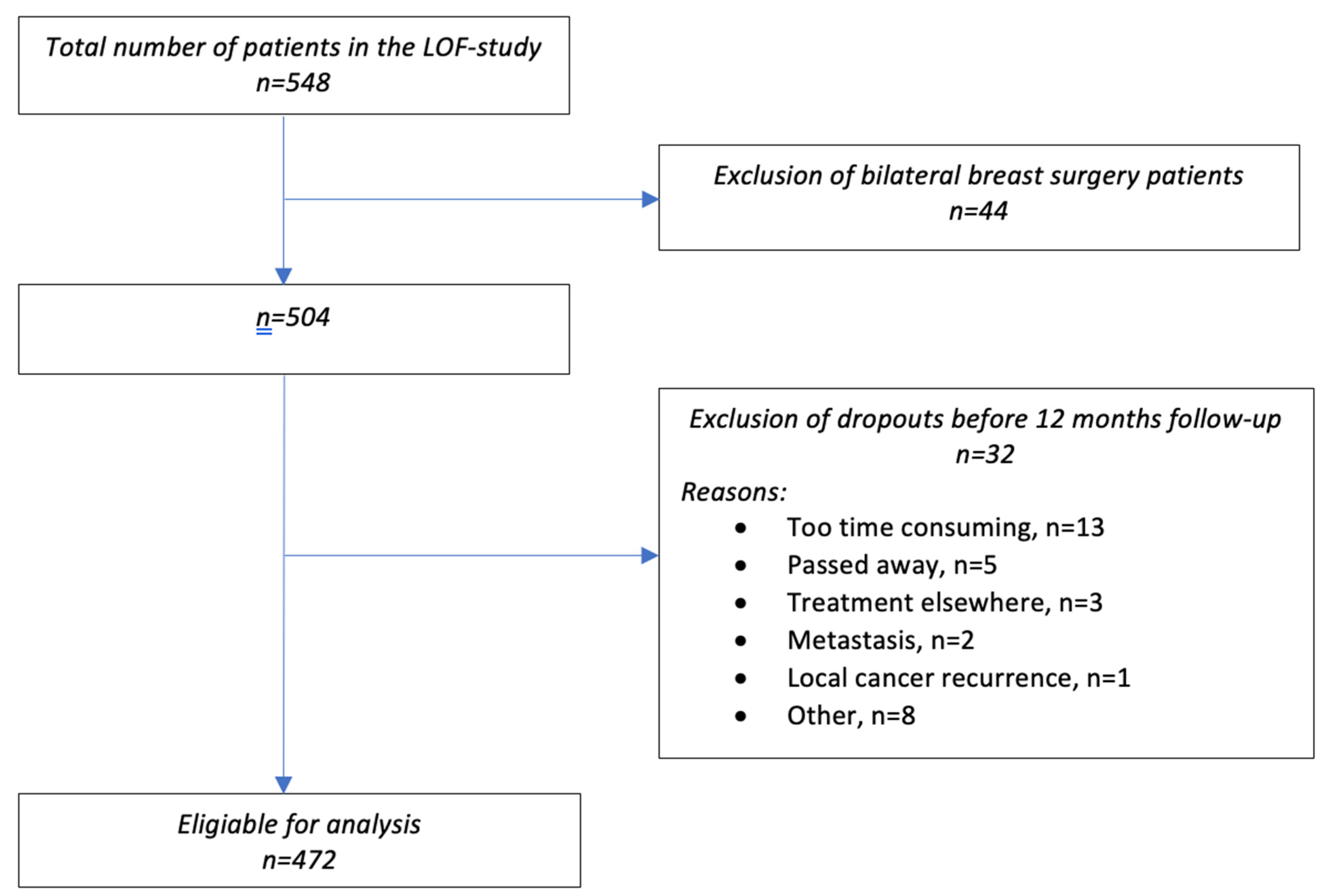
Figure 1. Flow diagram of eligible patients and those ultimately included in the analysis.
Cancers 14 06016 g001
Table 1. Patient characteristics.
CharacteristicsMean ± SD Number of PatientsFrequency %
Demographic & general health related
Age (years)59.2 ± 10.4
Smoking
-
Current smoker
6012.7
-
Never
20844.2
-
Ex-smoker
20343.1
ASA classification
-
ASA 1
8518.1
-
ASA 2
29462.6
-
ASA 3
8818.7
-
ASA 4
30.6
BMI preoperative 27.3 ± 5.1
-
Cat 1, ≤25
16234.5
-
Cat 2, >25–≤30
19842.1
-
Cat 3, >30
11023.4
Dominant side surgery 22649.9
Tumor related
Tumor stage
-
0
30.9
-
1
7221.5
-
2
16749.9
-
3
9327.8
Number of removed lymphnodes
-
<5
42890.7
-
≥5
449.3
Total number of positive nodes ≥1 14129.9
Treatment related
Breast conserving therapy (BCT) 36176.5
Mastectomy 12025.4
Sentinel node procedure 45095.3
Axillar lymph node dissection 347.2
Neo-adjuvant chemotherapy 11123.5
Adjuvant chemotherapy 10722.7
Hormonal therapy 28760.8
Radiation 40084.7
-
Including the axilla
13233.0
Valid percentages are used to correct for missing data.
Table 2. RVC development over time of all patients based on RVC category.
Follow-UpRVC < 5%RVC ≥ 5%
Proportion95% CIProportion95% CI
3 months (n = 455)90.587.4, 93.09.56.9, 12.6
6 months (n = 449)91.388.2, 93.78.76.3, 11.8
12 months (n = 448)91.388.2, 93.78.76.3, 11.8
24 months (n = 437)89.586.1, 92.110.57.9, 13.9
RVC = Relative Volume Change. Valid percentages are used to correct for missing data.
Table 3. Numbers and percentages of newly diagnosed patients referred and not referred for therapy, in relation to the diagnosis LE.
Therapy Referral RVC < 5%RVC ≥ 5%
n (%)n (%)
3 months
No 411 (99.8)35 (81.4)
Yes 1 (0.2)8 (18.6)
6 months
No 371 (97.9)17 (89.5)
Yes 8 (2.1)2 (10.5)
12 months
No 340 (97.1)11 (78.6)
Yes 10 (2.9)3 (21.4)
24 months
No 314 (98.4)10 (83.3)
Yes 5 (1.6)2 (16.7)
RVC = Relative Volume Change.
Table 4. Percentages of newly diagnosed LE patients with self-reported signs and symptoms.
Follow-Up% (95% CI)
3 months (n = 43)25.6 (14.0, 41.5)
Not referred45.5 (18.1, 75.4)
Referred54.5 (24.6, 81.9)
6 months (n = 19)26.3 (10.1, 51.4)
Not referred60.0 (17.0, 92.7)
Referred40.0 (7.3, 83.0)
12 months (n = 14)28.6 (9.6, 58.0)
Not referred25.0 (1.3, 78.1)
Referred75.0 (21.9, 98.7)
24 months (n = 12)58.3 (28.6, 83.5)
Not referred71.4 (30.3, 94.9)
Referred28.6 (5.1, 69.7)
Table 5. Group level data of the mean RVC of the LE patients, over three time points (the results are obtained by paired t testing).
Follow Up.Mean (SD) RVC %T24, Mean (SD) RVC % Mean Difference (%, SD)p Value
No treatment referral
T3 (n = 33)8.7 (4.6)5.3 4.0)−3.4 (5.8)0.002 *
T6 (n = 16)9.9 (11.6)3.6 (3.2)−6.3 (11.6)0.045 *
T12 (n = 11)7.8 (2.6)2.4 (4.3)−5.4 (5.7)0.011 *
Treatment referral
T3 (n = 7)10.8 (5.9)15.9 (16.5)5.1 (17.2)0.467
T6 (n = 2)8.2 (1.2)9.2 (10.3)1.0 (9.1)0.899
T12 (n = 3)9.4 (2.2)6.6 (3.4)−2.8 (2.2)0.163
RVC= Relative Volume Change. Mean RVC’s at 3, 6 and 12 months post-op are compared with the mean RVC at 24 months. * Significant at p < 0.05.
Publisher’s Note: MDPI stays neutral with regard to jurisdictional claims in published maps and institutional affiliations.