Article

Outcome of Children and Adolescents with Recurrent Classical Hodgkin Lymphoma: The Italian Experience

by
Alberto Garaventa
1,
Stefano Parodi
2,
Giulia Guerrini
3,
Piero Farruggia
4,
Alessandra Sala
5,
Marta Pillon
6,
Salvatore Buffardi
7,
Francesca Rossi
8,
Maurizio Bianchi
9,
Marco Zecca
10,
Luciana Vinti
11,
Elena Facchini
12,
Tommaso Casini
13,
Sayla Bernasconi
14,
Loredana Amoroso
1,
Salvatore D’Amico
15,
Massimo Provenzi
16,
Raffaela De Santis
17,
Antonella Sau
18,
Paola Muggeo
19,
Rosa Maria Mura
20,
Riccardo Haupt
2,
Maurizio Mascarin
21 and
Roberta Burnelli
22,*
add Show full author list remove Hide full author list
1
Paediatric Oncology Unit, IRCCS Istituto Giannina Gaslini, L.go G. Gaslini 5, 16147 Genova, Italy
2
Epidemiology and Biostatistics Unit, IRCCS Istituto Giannina Gaslini, L.go G. Gaslini 5, 16147 Genova, Italy
3
UOC Pediatria e Neonatologia, Grosseto USL-Toscana Sud-Est, Via Senese, 58100 Grosseto, Italy
4
Paediatric Haematology and Oncology Unit, A.R.N.A.S. Civic Hospital, Piazza Leotta Nicola 4, 90127 Palermo, Italy
5
Department of Paediatrics, Ospedale San Gerardo, University of Milano-Bicocca, Fondazione MBBM, Via Cadore, 20900 Monza, Italy
6
Dipartimento di Oncoematologia Pediatrica, Università di Padova, Via Gattamelata 5687, 35128 Padova, Italy
7
Paediatric Haemato-Oncology Department, Santobono-Pausilipon Children’s Hospital, Via Mario Fiore 6, 80129 Naples, Italy
8
Dipartimento di Pediatria II Ateneo di Napoli, Servizio di Oncologia Pediatrica, Via Luigi De Crecchio 2, 80138 Naples, Italy
9
Pediatric Onco-Hematology and Stem Cell Transplant Division, City of Health and Science, Regina Margherita Children’s Hospital, Piazza Polonia 94, 10126 Turin, Italy
10
Oncoematologia Pediatrica, Fondazione IRCCS Policlinico San Matteo, Viale Camillo Golgi 19, 27100 Pavia, Italy
11
Department of Hematology/Oncology and Stem Cell Transplantation, Bambino Gesù Children’s Hospital, Piazza di Sant’Onofrio 4, 00165 Rome, Italy
12
Pediatric Oncology and Hematology Unit “LallaSeràgnoli”, Department of Pediatrics, University of Bologna, Sant’Orsola Malpighi Hospital, Via Giuseppe Massarenti 9, 40138 Bologna, Italy
13
Division of Pediatric Oncology/Hematology, Meyer University Children’s Hospital, Via Gaetano Pieraccini 24, 50139 Florence, Italy
14
Pediatric Hematology Oncology, Bone Marrow Transplant, S. Chiara University Hospital of Pisa, Via Bonanno Pisano 93, 56126 Pisa, Italy
15
Paediatric Haemato-Oncology Unit, Department of Clinical and Experimental Medicine, University of Catania, Piazza Università 2, 95124 Catania, Italy
16
Department of Pediatrics, Civic Hospital, Piazza OMS 1, 24127 Bergamo, Italy
17
IRCCS Casa Sollievo della Sofferenza, San Giovanni Rotondo, Viale Cappuccini, 47156 Foggia, Italy
18
Pediatric Hematology-Oncology Unit, Ospedale Civico, Via Fonte Romana 8, 65124 Pescara, Italy
19
Department of Biomedicine of Developmental Age, University of Bari, Piazza Umberto I 1, 70121 Bari, Italy
20
Department of Paediatric Oncohaematology, Microcitemico Hospital, Via Edward Jenner 18, 09121 Cagliari, Italy
21
AYA Oncology and Pediatric Radiotherapy Unit, CRO-Centro di Riferimento Oncologico di Aviano, IRCCS Aviano, Via Franco Gallini 2, 33081 Aviano, Italy
22
Pediatric Hemato-Oncology Unit, Azienda Ospedaliero Universitaria Sant’Anna di Ferrara, Cona, Via Aldo Moro, 44124 Ferrara, Italy
*
Author to whom correspondence should be addressed.
Cancers 2022, 14(6), 1471; https://doi.org/10.3390/cancers14061471
Submission received: 11 February 2022 / Revised: 4 March 2022 / Accepted: 11 March 2022 / Published: 13 March 2022
(This article belongs to the Special Issue Childhood and Adolescent Cancer)

Abstract

:

Simple Summary

Survival of classical Hodgkin’s lymphoma (cHL) in Western countries is excellent. However, about 10% of patients with stage I–II disease and 15–30% of those with advanced stages require salvage therapy for resistant or relapsing disease. Many studies have investigated prognostic factors in adult patients, but data on children and adolescents are scarce. We analyzed a cohort of 272 patients aged <18 years with recurrent cHL, enrolled in two Italian subsequent protocols between 1996 and 2016. Overall and event-free survival after 10 years since the first recurrence were 65.3% and 53.3%, respectively. Major prognostic risk factors were progressive disease, advanced stage, ≥5 involved sites, and extra-nodal involvement at the recurrence. Patients with progressive disease, advanced stage, or ≥5 involved sites had a very poor survival and might benefit from more innovative approaches since the first progression. Patients who relapsed later with localized cHL might be considered for a conservative approach.

Abstract

The objective of this study was to identify prognostic factors for children and adolescents with relapsed or progressive classical Hodgkin’s lymphoma (cHL) to design salvage therapy tailored to them. We analyzed a homogeneous pediatric population, diagnosed with progressive/relapsed cHL previously enrolled in two subsequent protocols of the Italian Association of Pediatric Hematology and Oncology in the period 1996–2016. There were 272 eligible patients, 17.5% of treated patients with cHL. Overall survival (OS) and event-free survival (EFS) after a 10-year follow-up were 65.3% and 53.3%, respectively. Patients with progressive disease (PD), advanced stage at recurrence, and ≥5 involved sites showed a significantly worse OS. PD, advanced stage, and extra-nodal involvement at recurrence were significantly associated with a poorer EFS. Multivariable analysis identified three categories for OS based on the type of recurrence and number of localizations: PD and ≥5 sites: OS 34%; PD and <5 sites: OS 56.5%; relapses: OS 73.6%. Four categories were obtained for EFS based on the type of recurrence and stage: PD and stage 3–4: EFS 25.5%; PD and stage 1–2: EFS 43%; relapse and stage 3–4: EFS 55.4%; relapse and stage 1–2: EFS 72.1%. Patients with PD, in advanced stage, or with ≥5 involved sites had a very poor survival and they should be considered refractory to first- and second-line standard chemotherapy. Probably, they should be considered for more innovative approaches since the first progression. Conversely, patients who relapsed later with localized disease had a better prognosis, and they could be considered for a conservative approach.

1. Introduction

Classical Hodgkin’s lymphoma (cHL) is a common hematological malignancy in children, adolescents, and young adults. In pediatric patients, it is considered one of the most curable neoplastic diseases. The 5-year survival rate of pediatric cHL patients treated with the modern protocols available in Western countries now exceeds 90% [1]. Based on past experience, these protocols are designed to provide at least similar survival rates as in the past, but with lower treatment burden, to minimize the risk of early- and long-term toxicities. Still, 10% of stage I–II patients and 15–30% of those with advanced stages experience resistant or relapsing disease and therefore require salvage therapy [2]. The chance of rescuing these patients is around 50–60%, with different salvage protocols including standard-dose chemotherapy, high-dose chemotherapy with autologous stem cell rescue (ASCR), haploidentical transplant, related or unrelated transplant, and, quite recently, immunotherapy [3,4,5].
Several studies on adult cHL patients investigated the prognostic significance of several risk factors at relapse (i.e., timing, stage, bulky disease, B symptoms) and led to the development of a prognostic index and treatment guidelines [6]. Conversely, few studies focused on children and adolescents with progressive/relapsed cHL, and their unique conclusion was that the interval between diagnosis and recurrence is the only significant factor affecting survival [2,7].
The objective of this study was to identify any other prognostic factor for children with relapsing or progressive cHL, which could contribute to the design of salvage therapy. With this purpose, we analyzed a large homogeneous population of children and adolescents with progressive/relapsed cHL after being enrolled in two subsequent front-line protocols adopted by the Italian Association of Pediatric Hematology and Oncology (AIEOP) in the period 1996–2016 [8].

2. Patients and Methods

Eligible patients were diagnosed with cHL, aged <18 years, treated with MH96 protocol (February 1996–May 2004) or LH2004 (June 2004–December 2016) [8], and who experienced progressive disease (PD) or relapse during or after front-line therapy. Inclusion criteria, procedures, staging, and treatment outlines were recently reported [8] and are summarized in Table 1. After completion of treatment, both protocols required minimum 6-month monitoring for the subsequent 5 years.
Tumor regrowth was defined based on its timing during treatment or after the first elective end of treatment (EOT), namely (i) progressive or refractory disease (PD) when tumor regrowth occurs either during treatment or within 3 months after EOT, (ii) early relapse when it occurs between 3 and 12 months after EOT, and (iii) late relapse when it occurs >12 months after EOT [2,7].
For each patient, data were collected on tumor characteristics at diagnosis: gender, age, tumor histology [9], stage, presence of symptoms A or B, bulky disease, number of involved sites, involvement of extra-nodal sites, protocol, and treatment group including radiotherapy. Collected data about tumor regrowth were as follows: timing of regrowth, age, staging at recurrence, number of involved sites, extra-nodal site involvement, recurrence at the same primary site, and recurrence within the radiotherapy field. Since fluorodeoxyglucose positron emission tomography (FDG-PET) scan was not a standard investigation during the study period, the FDG-PET response was not considered as a prognostic factor.
No AIEOP approved salvage protocol was available, and the most common schemes used as second-line therapies [10,11,12] are reported in Table 2. They were followed by radiotherapy and/or high-dose chemotherapy (HDCT) with ASCR. The individual choice was made according to institutional strategies and, mostly, to previous treatment and characteristics of relapse; further modifications might have been influenced by the initial response to salvage therapy.
Follow-up data after recurrence included date and type of any new event—further progression/relapse, subsequent malignant neoplasm (SMN), date, and status at the last follow-up.
Second-line strategies were approved by ethics committee or institutional review board of each participating institution. Written informed consent was obtained from parents or legal guardians of all patients. The data lock point was September 2019 and data on previous relapses were updated in April 2021.

Statistical Methods

All data were collected in a central database. Overall survival (OS) after the first recurrence was calculated from the date of PD or relapse to the date of either death due to any cause or last contact. Event-free survival (EFS) was calculated from the date of progression/relapse to the date of either first subsequent event (further progression, second relapse, SMN, or death, whichever occurred first) or date of the last follow-up. The Kaplan–Meier method was used to estimate EFS and OS, while differences between groups were assessed using the log-rank test. Univariable and multivariable survival analysis was carried out by the Cox regression model [13].
All analyses were performed by STATA 13.1 (Stata Corporation, College Station, TX, USA). p-values < 0.05 were considered as statistically significant.

3. Results

Patient selection for data analysis is illustrated in Figure 1. In total, 82 (16.4%) of the 499 cHL patients treated with MH96 protocol and 190 (17.7%) of the 1074 treated with LH2004 protocol experienced either PD or recurrence, for a total of 272 eligible cHL patients.
Patient characteristics at diagnosis are resumed in Table 3. Patients treated with LH2004 protocol were 69.8%. There was a higher number of males and patients <15 years with nodular sclerosis, B symptoms, bulky disease, and treatment group 3. Patient characteristics were quite similar in the two protocols, except for a significant higher number of adolescents 37.9% vs. 20.7%), nodular sclerosis (93.2% vs. 80.5%), and multiple involved sites (≥8 sites: 37.4% vs. 25.6%) in the LH2004 group (Supplemental Table S1). Furthermore, 31% of the LH2004 patients did not receive radiotherapy due to PD vs. 12.2% of the MH96 patients.
Patient characteristics at disease recurrence are reported in Table 4. Forty-three percent had PD. Time from off-therapy to recurrence was reported for non-refractory patients: 51% relapsed between 3 months and 1 year from stop therapy. About half of the patients were ≥15 years. For 34 subjects (12.5%), no information was available about disease stage at relapse. Advanced stages (3 and 4) were observed in about half of patients. The large majority (64.7%) had no extra-nodal involvement and had recurrence in the site of diagnosis (83.1%) and in an irradiated site (83.3%).
A smaller proportion of relapsed patients was observed in the LH2004 group (49.5% vs. 74.4%, Supplemental Table S2), which was characterized by a greater number of patients ≥15 years (60.5% vs. 40.2%), multiple involved sites (≥5: 35.7% vs. 10.8%), and recurrence at the same site of diagnosis (87.5% vs. 73.0%).
Overall, 170 patients received HDCT with ASCR as consolidation of a complete response after second-line therapy. Only 18 patients (9 in complete response) received allogeneic transplant.
Since the date of the first cHL recurrence, 158 further events were observed in 121 patients: 64 further cHL relapses, 5 SNMs, and 89 deaths (Supplemental Figure S1). In details, relapses occurred between 1 and 118 months (median 11 months); SMNs were: one lung adenocarcinoma at 53 months, one soft tissue sarcoma at 85 months after secondary acute myeloblastic leukemia, one osteosarcoma of the chest wall at 64 months, one colon adenocarcinoma associated with a clear-cell renal carcinoma at 182 months, and a papillary and follicular thyroid carcinoma at 125 months in a patient who had experienced a second cHL relapse at 20 months. The 89 deaths happened between 12 days and 128 months (median 19 months): 33 after HL recurrence, 3 after SMN, and 53 because of a persistent HL.
Figure 2 shows the Kaplan–Meier survival curves for the OS and EFS after disease recurrence in the 272 cHL patients under study. After a 10-year follow-up, OS was 65.3% (95% CI: 59–71) and EFS 53.3% (95% CI: 47–59).
Table 5 shows OS after recurrence by patient characteristics at diagnosis. A significantly better result was reported for patients treated with LH2004 protocol. No difference was observed for gender, age, histology, stage, bulky disease, number, and type of involved sites, as well as radiotherapy administration. B symptoms were associated with a poorer survival, but statistical significance was not reached. A not significant better survival was observed in patients without bulky disease and in those in the first treatment group.
Table 6 shows OS after recurrence by patient characteristics at the time of the event. OS was significantly poorer for patients with PD, advanced stage, a large number of involved sites and extra-nodal site involvement (in this latter not statistically significant). No difference was observed between early vs. late relapse.
Supplemental Table S3 shows EFS after recurrence by patient characteristics at diagnosis. Only the presence of B symptoms was associated with poorer survival (47.7% vs. 61.6%), without reaching the statistical significance.
Table 7 shows EFS after recurrence by patient characteristics at disease recurrence. Progressive disease, advanced stage, and extra-nodal site involvement were significantly associated with a poorer survival. A not statistically significant lower EFS was observed for patients with at least five involved sites and recurrence at previously irradiated sites.
Multivariable analysis (Table 8) confirmed a higher OS for patients treated with LH2004 protocol (HR = 0.43) and a lower OS for those with progressive disease (HR = 0.33) and with a high number of involved sites at recurrence (HR = 2.5 for ≥5 involved sites)
EFS was significantly related to type of recurrence (HR = 0.34 for relapse vs. progression), stage at relapse (HR = 1.7 for advanced vs. not-advanced stages), and recurrence at the same irradiated site (Table 9). The highest survival of patients treated with LH2004 protocol was confirmed (HR = 0.56).
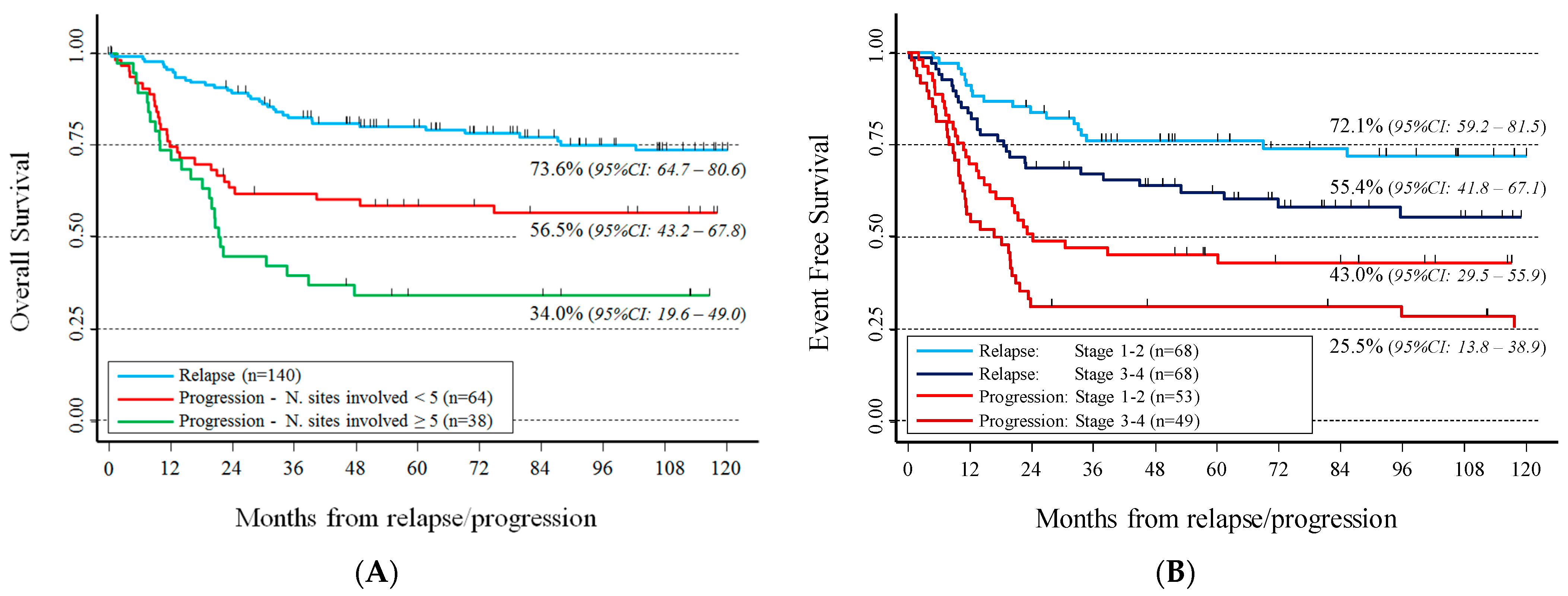
Combining the potential prognostic factors, evaluated by multivariable analysis, three categories were identified for OS (Figure 3A): progressive patients with ≥5 involved sites and the worst survival (34%, 95% CI: 19.6–49), progressive patients with <5 involved sites with intermediate survival (56.5%, 95% CI: 43.2–67.8), and relapsed patients with the best survival (73.6%, 95% CI: 64.7–80.6). The small number of deaths in this latter group prevented to further separate patients by the number of involved sites.
With regard to EFS, type of recurrence combined with stage identified four categories (Figure 3B). The worst EFS was observed for patients with progression in advanced stage (25.5%, 95% CI: 13.8–38.9) and the best for those with relapse in non-advanced stage (72.1%, 95% CI: 59.2–81.5). Analysis restricted to patients with recurrence after radiotherapy (n = 174) showed a similar trend (data not shown) and an extremely poor survival in patients in advanced stage, with PD relapsed in an irradiated site (EFS = 9.5%, 95% CI: 1.6–26.1).

4. Discussion

Despite improvements in the overall prognosis for children with cHL, a proportion of patients are not cured by their primary treatment. In adults, clinical risk factors for patients with recurrent disease have been well described and they include refractory disease, time to relapse, advanced stage, bulky disease, extra-nodal disease, and age [1,6]. In addition, an interim FDG-PET positivity was found associated with an increased risk of failure [1]. Preliminary data also suggested that cell-free DNA may be an important biomarker of response and outcome [6].
Limited data are available in the pediatric population [7,14,15] and pediatric series include few cases collected over a long period of time. The first investigation published in 1992 reported 35 cases failing first-line therapy with 45% OS at 10 years; the authors identified a poor risk group of patients not achieving a complete response to first-line therapy or relapsing within 1 year since diagnosis [15]. Another study analyzed 176 patients in the period 1986–2003 (51 after progression during or shortly after first-line therapy) [7] and, in agreement with our findings, reported that disease refractory to first-line therapy was the strongest prognostic factor. Patients with progression had 41% disease-free survival after 10 years compared with 86% in patients with relapse, although none of them received ASCR in second remission. Friedmann et al. described 64 relapsed patients in the period 1990–2006 focusing on methods of detection and timing of relapse and concluded that frequent imaging does not appear to impact on survival as most relapses are identified through history and physical examination [14]. Unfortunately, this interesting point could not be addressed in our study.
The overall relapse rate of 17.5% observed in our cohort is similar to that reported in other pediatric studies. OS for our patients was 64%, with a median survival time of 88.6 months, and 65.3% of them survived ≥10 years after recurrence. Survival was better for patients recurring after LH2004 protocol, probably due to an improvement of second- and third-line rescue therapy.
The duration of first remission was a highly significant prognostic factor. Children who relapsed before or within 3 months from completion of treatment had significantly poorer EFS and OS when compared to those who relapsed later. However, we did not observe a difference between early and late relapse. It is difficult to explain the difference in PD between the two protocols: in the period of enrolment of LH2004, it seems to occur a more aggressive disease, as suggested by the higher number of involved sites. The more frequent assessment of response and a larger use of FDG-PET at the EOT in LH2004 protocol could also have played a role.
The patients with PD, in stage 3–4 or with ≥5 involved sites had a very poor survival. These patients should be considered refractory to first-line therapy and also to second-line standard chemotherapy. Probably, they should be treated with more innovative approaches since first progression. Conversely, patients who relapsed later on with a localized disease had a better prognosis and they could be considered for a conservative approach. However, in such patients we were unable to identify a more favorable subset only on the basis of timing of relapse. Even if consistent with evidence from other studies, our results of stratified analysis should be considered cautiously due to the adopted post hoc approach, and they need to be validated in a large independent prospective cohort.
The strength of our study is the huge number of patients treated at diagnosis with only two homogenous protocols, and the consequent large population of relapsed patients. To our knowledge, our study is the largest one in a pediatric population. Limitations include the multicenter retrospective design and the analysis of only static variables, like stage and number of involved sites. Early response to salvage therapy was not available, due to the lack of detailed information on salvage therapies, even though they were likely to be quite homogeneous. However, when designing second-line treatment, early response to salvage therapy should be considered and poor responder patients should be moved to a higher risk group and treated with novel therapies [2,16].
In conclusion, current salvage therapeutic approaches are quite effective, with a 65.3% 10-year OS in cHL patients after progression or relapse. However, identification of several prognostic groups should be advisable to address different therapeutic approaches. Our results, in agreement with previous studies, strongly indicate that refractory disease at the first-line treatment represents the most relevant prognostic factor. Advanced stage at recurrence, the number of involved sites and recurrence at the same previously irradiated district could also significantly affect patient survival.
Alongside the clinical characteristics, liquid biopsy-based biomarkers, such as cell-free DNA can represent new tools to assess a prognostic stratification at diagnosis. Moreover, it can monitor the quality of response detecting the minimal residual disease, or the recurrence of the disease. These new biomarkers should be considered among the future predictive features, considering the improving technology and the emerging results, even though the requirement of validation in prospective analyses in childhood cHL [17,18,19].
Innovative therapy like Brentuximab alone [20] or combined with Nivolumab [2] or with chemotherapy [21] has been studied in refractory or relapsed patients achieving very high overall and complete response rates, and it should be considered pre and/or post HDCT/ASCR in the poor prognosis patients or in patients resistant to second-line chemotherapies. The use of these drugs in the treatment of low- and standard-risk groups could be considered to decrease long-term toxicity in selected patients and in cooperative trials as their long-term side effects are not well known yet.

5. Conclusions

The identification of prognostic groups of children and adolescents with refractory or relapsing cHL is needed in order to address different therapeutic approaches. In our study, patients with PD, in advanced stage or with ≥5 involved sites had a very poor survival. They should be considered refractory to first- and second-line standard chemotherapy and could be considered for more innovative approaches since the first progression. On the contrary, patients who relapsed later with localized disease had a very good outcome, and they could be addressed to a more conservative therapeutic approach.

Supplementary Materials

The following supporting information can be downloaded at: https://www.mdpi.com/article/10.3390/cancers14061471/s1, Figure S1: Patients’ outcome after the first disease recurrence; Table S1: Characteristics at diagnosis of 272 recurring patients with classical Hodgkin lymphoma by first-line treatment protocol; Table S2: Characteristics at the disease recurrence of 272 patients with classical Hodgkin lymphoma by first-line treatment protocol. Table S3: Ten-year Event Free Survival (EFS) of 272 patients with classical Hodgkin lymphoma after recurrence by characteristics at diagnosis.

Author Contributions

Conceptualization, A.G. and R.B.; validation, A.G., S.P. and R.H.; formal analysis, S.P.; resources, P.F., A.S. (Alessandra Sala), M.P. (Marta Pillon), S.B. (Salvatore Buffardi), F.R., M.B., M.Z., L.V., E.F., T.C., S.B. (Sayla Bernasconi), L.A., S.D., M.P. (Massimo Provenzi), R.D.S., A.S. (Antonella Sau), P.M., R.M.M., M.M. and R.B.; data curation, G.G. and R.B.; writing—original draft preparation, A.G., and R.B.; writing—review and editing, A.G., S.P., G.G., P.F., A.S. (Alessandra Sala), M.P. (Marta Pillon), S.B. (Salvatore Buffardi), F.R., M.B., M.Z., L.V., E.F., T.C., S.B. (Sayla Bernasconi), L.A., S.D., M.P. (Massimo Provenzi), R.D.S., A.S., P.M., R.M.M., R.H., M.M. and R.B.; visualization, A.G., S.P., G.G., P.F., A.S. (Antonella Sau) and R.B.; supervision, R.H.; funding acquisition, A.G. All authors have read and agreed to the published version of the manuscript.

Funding

This work was supported by the Italian Ministry of Health through “Cinque per mille” and “RicercaCorrente”.

Institutional Review Board Statement

The study was conducted in accordance with the Declaration of Helsinki and approved by the following: Comitato Etico—Centro di Riferimento Oncologico di Aviano (prot. n°206/D, 15/02/2005), Comitato Etico—A. O. U. C. Policlinico di Bari (prot. n°2960, 31/05/2006), Comitato Etico—AO di Bologna, Policlinico S. Orsola-Malpighi (prot. n°1103/2004, 23/04/2004), Comitato Etico Indipendente—Azienda U.S.L. N.8 Cagliari (prot. n°146/CE/04, 22/09/2004), Comitato Etico—AOU Policlinico di Catania (prot. n°68, 29/07/2005), Comitato Etico Sperimentazione dei Farmaci—AO Meyer-Firenze (prot. n°60, 25/07/2005), Comitato di etica per la ricerca scientifica biomedica, per la buona pratica clinica e per la sperimentazione dei farmaci–Istituto G.Gaslini, Genova (15/07/2004), Comitato Etico—AO “San Gerardo” Monza (15/07/2005), IRB of SSN-AO di rilievo Nazionale “Santobono-Pausillipon” (prot. n°422, 16/06/2005), Comitato Etico della Facoltà di Medicina e Chirurgia—II Università degli Studi di Napoli (prot. n°624, 16/12/2004), Comitato Etico per la Sperimentazione Clinica della Provincia di Padova (prot. n°908P, 10/04/2006), Comitato Etico dell’Azienda di Rilievo Nazionale e di Alta Specializzazione Ospedale Civico e Benfratelli, G.Di Cristina e M.Ascoli—Palermo (prot. n°56/1, 04/05/2004), Comitato Etico per la Sperimentazione clinica dei farmaci—AUSL di Pescara (Prot. n°901/CE, 02/08/2005), Sezione del Comitato Etico IRCCS Istituto Tumori “Giovanni Paolo II “di Bari presso la Fondazione Casa Sollievo della Sofferenza di San Giovanni Rotondo (FG) (15/10/2005), Commissione Regionale per le Sperimentazioni Cliniche dell’Assessorato alla Sanità della Regione Piemonte- Ospedale Infantile Regina Margherita, Torino (prot. n°15184/28.3, 11/10/2004).

Informed Consent Statement

Informed consent was obtained from all subjects involved in the study.

Data Availability Statement

The Database is in CINECA, which is a Consortium composed by 102 Italian universities and public institutions. Since its origins in 1969, Cineca offers support to scientific research, public and industrial, through supercomputing and the use of the most innovative computing systems based on state-of-the-art architectures and technologies. The database is not public, as it is affirmed in the informed consent signed by patients or parents.

Acknowledgments

The authors express their gratitude and appreciation to AIEOP investigators, treating physicians, clinical research and care teams, and, most importantly, to patients and families facing Hodgkin’s lymphoma. We are indebted with Anna Capurro for editorial assistance.

Conflicts of Interest

The authors declare no conflict of interest.

References

  1. Voss, S.D.; Chen, L.; Constine, L.S.; Chauvenet, A.; Fitzgerald, T.J.; Kaste, S.C.; Slovis, T.; Schwartz, C.L. Surveillance Computed Tomography Imaging and Detecting of Relapse in Intermediate-and Advanced-Stage Pediatric Hodgkin’s Lymphoma: A Report from the Children’s Oncology Group. J. Clin. Oncol. 2012, 30, 2635–2640. [Google Scholar] [CrossRef] [PubMed] [Green Version]
  2. Daw, S.; Hasenclever, D.; Mascarin, M.; Fernández-Teijeiro, A.; Balwierz, W.; Beishuizen, A.; Burnelli, R.; Cepelova, M.; Claviez, A.; Dieckmann, K.; et al. Risk and Response Adapted Treatment Guidelines for Managing First Relapsed and Refractory Classical Hodgkin Lymphoma in Children and Young People. Recommendations from the EuroNet Pediatric Hodgkin Lymphoma Group. HemaSphere 2020, 4, e329. [Google Scholar] [CrossRef] [PubMed]
  3. La Casce, A.S. Treating Hodgkin lymphoma in the new millennium: Relapsed and refractory disease. Hematol. Oncol. 2019, 37 (Suppl. 1), 87–91. [Google Scholar] [CrossRef] [PubMed] [Green Version]
  4. Warlick, E.D.; DeFor, T.E.; Bejanyan, N.; Holtan, S.; MacMillan, M.; Blazar, B.R.; Dusenbery, K.; Arora, M.; Bachanova, V.; Cooley, S.; et al. Reduced-Intensity Conditioning Followed by Related and Unrelated Allografts for Hematologic Malignancies: Expanded Analysis and Long-Term Follow-Up. Biol. Blood Marrow Transpl. 2019, 25, 56–62. [Google Scholar] [CrossRef] [PubMed] [Green Version]
  5. Moskowitz, C.H.; Nademanee, A.; Masszi, T.; Agura, E.; Holowiecki, J.; Abidi, M.H.; Chen, A.I.; Stiff, P.; Gianni, A.M.; Carella, A.; et al. Brentuximab vedotin as consolidation therapy after autologous stem-cell transplantation in patients with Hodgkin’s lymphoma at risk of relapse or progression (AETHERA): A randomised, double-blind, placebo-controlled, phase 3 trial. Lancet 2015, 385, 1853–1862. [Google Scholar] [CrossRef]
  6. Bröckelmann, P.J.; Von Tresckow, B. Risk stratification and prognostic biomarkers in relapsed Hodgkin lymphoma. Br. J. Haematol. 2020, 190, 813–814. [Google Scholar] [CrossRef] [PubMed]
  7. Schellong, G.; Dörffel, W.; Claviez, A.; Körholz, D.; Mann, G.; Scheel-Walter, H.G.; Bökkerink, J.P.; Riepenhausen, M.; Lüders, H.; Pötter, R.; et al. Salvage therapy of progressive and recurrent Hodgkin’s disease: Results from a multicenter study of the pediatric DAL/GPOH-HD study group. J. Clin. Oncol. 2005, 23, 6181–6189. [Google Scholar] [CrossRef] [PubMed]
  8. Burnelli, R.; Fiumana, G.; Rondelli, R.; Pillon, M.; Sala, A.; Garaventa, A.; D’Amore, E.S.G.; Sabattini, E.; Buffardi, S.; Bianchi, M.; et al. Comparison of Hodgkin’s Lymphoma in Children and Adolescents. A Twenty Year Experience with MH’96 and LH2004 AIEOP (Italian Association of Pediatric Hematology and Oncology) Protocols. Cancers 2020, 12, 1620. [Google Scholar] [CrossRef] [PubMed]
  9. Swerdlow, S.H.; Campo, E.; Pileri, S.A.; Harris, N.L.; Stein, H.; Siebert, R.; Advani, R.; Ghielmini, M.; Salles, G.A.; Zelenetz, A.D.; et al. The 2016 revision of the World Health Organization classification of lymphoid neoplasms. Blood 2016, 127, 2375–2390. [Google Scholar] [CrossRef] [PubMed] [Green Version]
  10. Salar, A.; Martino, R.; Perea, G.; Ribera, J.M.; López-Guillermo, A.; Guardia, R.; Escoda, L.; Altés, A.; Sierra, J.; Montserrat, E. High-dose infusional ifosfamide, etoposide plus methylprednisolone followed by dexamethasone, high-dose ara-C and cisplatinum and autologous stem cell transplantation for refractory or relapsed aggressive non-Hodgkin’s lymphoma. Haematologica 2002, 87, 1028–1035. [Google Scholar] [PubMed]
  11. Cairo, M.S.; Krailo, M.D.; Morse, M.; Hutchinson, R.J.; Harris, R.E.; Kjeldsberg, C.R.; Kadin, M.E.; Radel, E.; Steinherz, L.J.; Morris, E.; et al. Long-term follow-up of short intensive multiagent chemotherapy without high-dose methotrexate (‘Orange’) in children with advanced non-lymphoblastic non-Hodgkin’s lymphoma: A children’s cancer group report. Leukemia 2002, 16, 594–600. [Google Scholar] [CrossRef] [PubMed] [Green Version]
  12. Argiris, A.; Seropian, S.; Cooper, D.L. High-dose BEAM chemotherapy with autologous peripheral blood progenitor-cell transplantation for unselected patients with primary refractory or relapsed Hodgkin’s disease. Ann. Oncol. 2000, 11, 665–672. [Google Scholar] [CrossRef] [PubMed]
  13. Hosmer, D.W.; Lemeshow, S. Applied Survival Analysis—Regression Modelling of Time to Event Data; John Wiley & Sons, Inc.: New York, NY, USA, 1999. [Google Scholar]
  14. Friedmann, A.M.; Wolfson, J.A.; Hudson, M.M.; Weinstein, H.J.; Link, M.P.; Billett, A.; Larsen, E.C.; Yock, T.; Donaldson, S.S.; Marcus, K.; et al. Relapse after treatment of pediatric Hodgkin lymphoma: Outcome and role of surveillance after end of therapy. Pediatric Blood Cancer 2013, 60, 1458–1463. [Google Scholar] [CrossRef] [PubMed] [Green Version]
  15. James, N.D.; Kingston, J.E.; Plowman, P.N.; Meller, S.; Pinkerton, R.; Barrett, A.; Sandland, R.; McElwain, T.J.; Malpas, J.S. Outcome of children with resistant and relapsed Hodgkin’s disease. Br. J. Cancer 1992, 66, 1155–1158. [Google Scholar] [CrossRef] [PubMed] [Green Version]
  16. Metzger, M.L.; Hudson, M.M.; Krasin, M.J.; Wu, J.; Kaste, S.C.; Kun, L.E.; Sandlund, J.T.; Howard, S.C. Initial response to salvage therapy determines prognosis in relapsed pediatric Hodgkin lymphoma patients. Cancer 2010, 116, 4376–4384. [Google Scholar] [CrossRef] [PubMed] [Green Version]
  17. Camus, V.; Jardin, F. Cell-Free DNA for the Management of Classical Hodgkin Lymphoma. Pharmaceuticals 2021, 14, 207. [Google Scholar] [CrossRef] [PubMed]
  18. Primerano, S.; Burnelli, R.; Carraro, E.; Pillon, M.; Elia, C.; Farruggia, P.; Sala, A.; Vinti, L.; Buffardi, S.; Basso, G.; et al. Kinetics of Circulating Plasma Cell-Free DNA in Paediatric Classical Hodgkin Lymphoma. J. Cancer 2016, 7, 364–366. [Google Scholar] [CrossRef] [PubMed] [Green Version]
  19. Buedts, L.; Wlodarska, I.; Finalet-Ferreiro, J.; Gheysens, O.; Dehaspe, L.; Tousseyn, T.; Fornecker, L.-M.; Lazarovici, J.; Casasnovas, R.-O.; Gac, A.-C.; et al. The landscape of copy number variations in classical Hodgkin lymphoma: A joint KU Leuven and LYSA study on cell-free DNA. Blood Adv. 2021, 5, 1991–2002. [Google Scholar] [CrossRef] [PubMed]
  20. Locatelli, F.; Mauz-Koerholz, C.; Neville, K.; Llort, A.; Beishuizen, A.; Daw, S.; Pillon, M.; Aladjidi, N.; Klingebiel, T.; Landman-Parker, J.; et al. Brentuximab vedotin for paediatric relapsed or refractory Hodgkin’s lymphoma and anaplastic large-cell lymphoma: A multicentre, open-label, phase 1/2 study. Lancet Haematol. 2018, 5, e450–e461. [Google Scholar] [CrossRef]
  21. Cole, P.D.; McCarten, K.M.; Pei, Q.; Spira, M.; Metzger, M.L.; Drachtman, R.A.; Horton, T.M.; Bush, R.; Blaney, S.M.; Weigel, B.J.; et al. Brentuximab-vedotin with gemcitabine for paediatric and young adult patients with relapsed or refractory Hodgkin’s lymphoma (AHOD1221): A Children’s Oncology Group, multicentre single-arm, phase 1–2 trial. Lancet Oncol. 2018, 19, 1229–1238. [Google Scholar] [CrossRef]
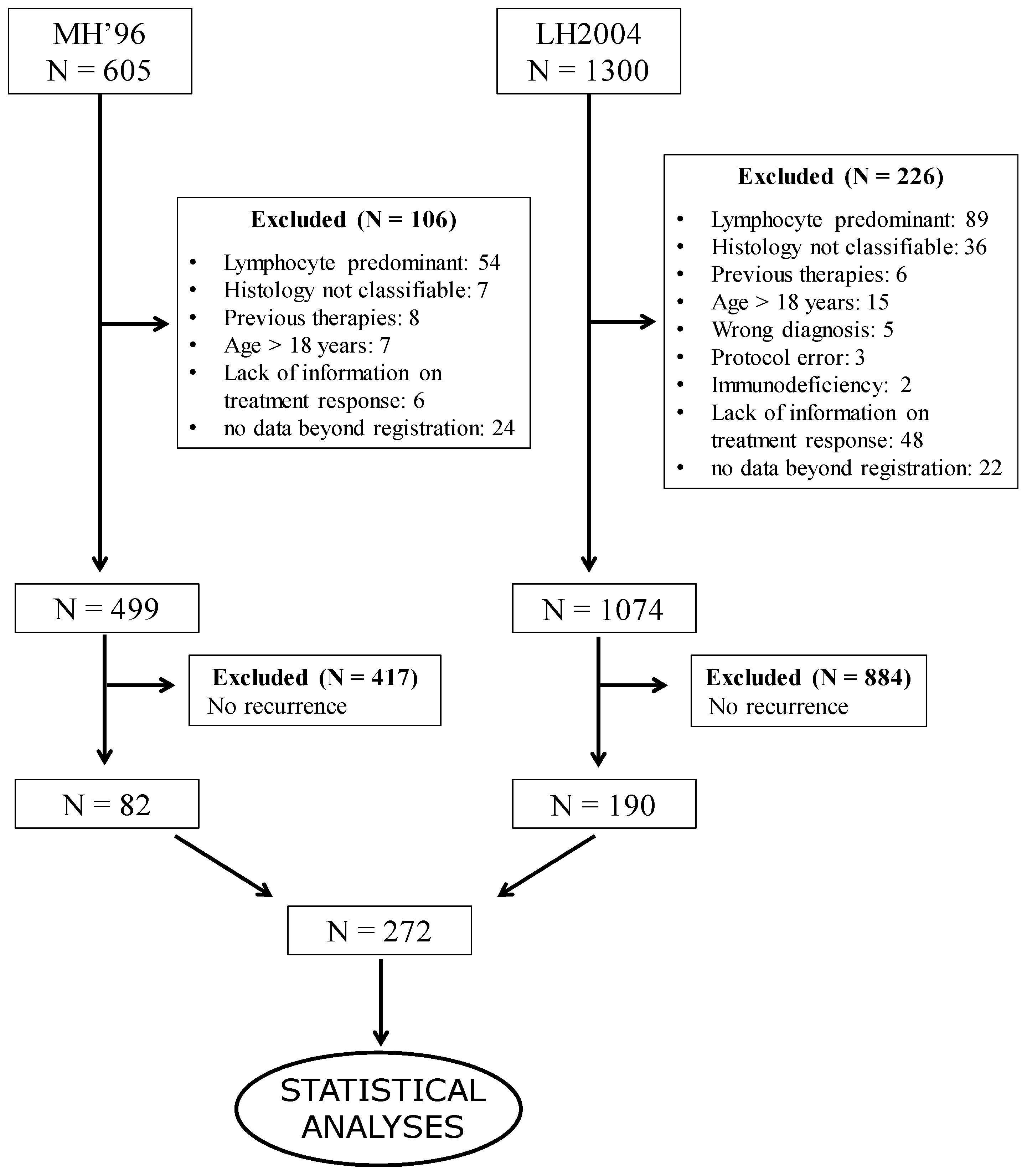
Figure 1. Patient selection for statistical analyses.
Cancers 14 01471 g001
Figure 2. Overall and event-free survival after disease recurrence in 272 cHL patients.
Cancers 14 01471 g002
Figure 3. Survival after relapse or progression of 272 classical Hodgkin’s lymphoma patients by characteristics at relapse identified by multivariable Cox regression model. Panel (A): overall survival. Panel (B): event-free survival.
Cancers 14 01471 g003
Table 1. Summary of MH96 and LH2004 AIEOP protocols.
Treatment GroupsMH’96LH2004
1
IA, IIA supradiaphragmatic no bulky, no pulmonary hilum, <4 lymphatic sites, or IA, IIA infradiaphragmatic <4 lymphatic sites
3× ABVD+:
-
CR no initial mediastinal involvement: stop
-
others: RT
-
CR o PR ≥ 75%: 20 Gy IF
-
PR <75%: 36 Gy IF
3× ABVD+:
-
CR: stop
-
others: RT 25.2 Gy IF
2
patients not included in groups 1 or 3
4× COPP/ABV+RT:
-
CR or PR ≥ 75%: 20 Gy IF
-
PR < 75%: 36 Gy IF
4× COPP/ABV+:
-
CR: RT 14.4 Gy IF
-
PR: 2× IEP + RT
-
CR: 14.4 Gy IF
-
PR: 25.2 Gy IF
3
IIIB, IV; M/T ≥ 0.33 all stages
6× COPP/ABV+RT:
-
CR or PR ≥ 75%: 20 Gy IF
-
PR <75%: 36 Gy IF
4× COPP/ABV+:
-
CR: 2× COPP/ABV + RT 14.4 Gy IF
-
PR: 2× IEP + RT
-
CR: 14.4 Gy IF
-
PR: 25.2 Gy IF
If RP ≤ 50% after 2° cycle: GR1: IEP/OPPA/COPP/IEP + RT; GR2 and 3: IEP/OPPA/IEP/OPPA/IEP + RT
ABVD
Doxorubicin: 25 mg/m2 IV days 1
Bleomycin: 10 mg/m2 IV days 1 and 15
Vinblastine: 6 mg/m2 IV days 1 and 15
DTIC: 375 mg/m2 IV days 1 and 15
COPP/ABV
Cyclophosphamide: 600 mg/m2 IV day 1
Vincristine: 1.4 mg/m2 IV day 1
Prednisone: 40 mg/m2 orally days 1–14
Procarbazine: 100 mg/m2 orally days 1–7
Doxorubicin: 35 mg/m2 IV day 8
Bleomycin: 10 mg/m2 IV day 8
Vinblastine: 6 mg/m2 IV day 8
IEP
Ifosfamide: 2000 mg/m2 IV days 1–5
Etoposide: 120 mg/m2 IV days 1–5
Prednisone: 100 mg/m2 orally days 1–5
OPPA
Vincristine: 1.5 mg/m2 IV days 1 and 8 and 15
Procarbazine: 100 mg/m2 orally days 1–14
Prednisone: 60 mg/m2 IV days 1–14
Doxorubicin: 40 mg/m2 IV days 1 and 15
COPP
Cyclophosphamide: 500 mg/m2 IV days 1 and 8
Vincristine: 1.5 mg/m2 IV days 1 and 8
Procarbazine: 100 mg/m2 orally days 1–15
Prednisone: 40 mg/m2 orally days 1–15
A: symptoms A (absent), B: symptoms B (present). CR: complete response. IF: involved fields. M/T: mediastinal/thoracic ratio. PR: partial response. RT: radiotherapy.
Table 2. Therapeutic regimens utilized as a second line treatment for cHL.
AcronymsDrugs
IEPHigh-dose IfosfamideEtoposideMetil-Prednisolone--
DHAPDexamethasoneHigh dose Cytosine-ArabinosideCisplatin--
DECALDexamethasoneEtoposideCisplatinCytosine-ArabinosideL-asparaginase
BEAMBCNUEtoposideCytosine-ArabinosideMelphalan-
Table 3. Characteristics at diagnosis of 272 recurring patients with classical Hodgkin’s lymphoma.
Patient Characteristicsn%
Protocol
MH 968230.2
LH 200419069.8
Gender
Male15858.1
Female11441.9
Age (years)
<531.1
5–1418066.2
≥158932.7
Histology
Lymphocyte depleted51.8
Mixed cellularity248.8
Nodular sclerosis24389.3
Stage
172.6
212546.0
36222.8
47828.7
Symptoms
A11140.8
B16159.2
Bulky
No9735.7
Yes17564.3
Number of involved sites
1–36423.5
4–711642.7
≥89233.8
Extra-nodal site involvement
No17865.4
Yes9434.6
Treatment group
1124.4
23512.9
322582.7
Radiotherapy
No according to protocol62.2
No for disease progression6825.0
Yes19571.7
Missing31.1
Extra-nodal sites: parenchymal sites, including liver, lung, bone, and bone marrow. %: percentages calculated on valid data only.
Table 4. Characteristics at disease recurrence of 272 patients with classical Hodgkin’s lymphoma.
Patient Characteristics n%
Type of recurrence
Progression11743.0
Relapse15557.0
Early relapse (3–12 months from OT)7951.0
Late relapse (≥12 months from OT)7649.0
Age (years)
<531.1
5–1412144.5
≥514854.4
Stage
13012.6
29138.2
33615.1
48134.0
Missing3412.5
Number of involved sites *
17229.8
2–410242.5
≥56828.1
Missing3011.0
Extra-nodal site involvement **
No15664.7
Yes8535.3
Missing3111.4
Recurrence at the same site
No4116.9
Yes20183.1
Missing3011.0
Recurrence after Radiotherapy
Recurrence in non-irradiated sites3016.7
Recurrence in the irradiated site15083.3
Missing157.7
* The number of involved sites was grouped according to the tertiles of its distribution. ** Extra-nodal sites: parenchymal sites including liver, lung, bone, and bone marrow. % percentages calculated on valid data only.
Table 5. Ten-year overall survival of 272 patients with classical Hodgkin’s lymphoma after recurrence by characteristics at diagnosis.
Patient CharacteristicsN/DOS95% CIp
Whole cohort272/8965.359.0–70.9
Protocol 0.042
MH9682/3656.545.0–66.5
LH2004190/5369.561.7–76.0
Gender 0.687
Male158/5362.353.2–70.2
Female114/3666.956.7–75.3
Age (years) 0.729
0–14183/6264.556.8–71.2
≥1589/2766.653.9–76.5
Histology 0.356
Lymphocyte depleted5/250.05.8–84.5
Mixed cellularity24/1057.435.2–74.4
Nodular sclerosis243/7766.459.7–72.3
Stage 0.885
1–2132/4465.556.2–73.2
3–4140/4565.356.2–73.0
Symptoms 0.052
A111/2972.061.8–80.0
B161/6060.852.5–68.2
Bulky disease 0.141
No97/2671.260.0–79.8
Yes175/6362.354.4–69.2
Number of involved sites * 0.667 *
1–364/2265.652.2–76.1
4–7116/3964.154.1–72.5
≥892/2866.454.8–75.7
Extra-nodal site involvement ** 0.220
No178/6263.755.8–70.5
Yes94/2768.657.4–77.5
Treatment Group 0.220
112/383.348.2–95.6
235/776.956.9–88.5
3225/7962.655.5–68.8
Radiotherapy (post-chemotherapy) 0.197
No according to protocol6/0100
No for disease progression68/2562.349.5–72.8
Yes195/6365.257.5–71.9
* The number of involved sites was grouped according to the tertiles of its distribution. ** Extra-nodal sites: parenchymal sites including liver, lung, bone, and bone marrow. % percentages calculated on valid data only. Bold: significant p value.
Table 6. Ten-year overall survival of 272 patients with classical Hodgkin’s lymphoma after recurrence by characteristics at relapse/progression.
Patient CharacteristicsN/DOS95% CIp
Type of recurrence <0.001
Progression117/5452.142.5–61.0
Relapse155/3575.366.9–81.8
Early relapse79/1873.760.9–82.90.991
Late relapse76/1776.564.3–85.0
Age (years) 0.252
0–14124/4661.552.0–69.6
≥15148/4368.759.7–76.1
Stage 0.029
1–2121/3669.059.6–76.7
3–4117/4857.247.1–66.1
Number of involved sites * 0.023 *
172/2268.456.0–78.0
2–4102/3265.855.0–74.7
≥568/3252.539.3–64.2
Extra-nodal site involvement ** 0.087
No156/5166.958.7–73.8
Yes85/3554.942.7–65.5
Recurrence at the same site 0.412
No41/1366.449.0–79.0
Yes201/7362.354.9–68.9
Recurrence after Radiotherapy 0.110
Recurrence in a non-irradiated site30/775.755.5–87.6
Recurrence in the same irradiated site150/5461.852.8–69.5
* The number of involved sites was grouped according to the tertiles of its distribution. ** Extra-nodal sites: parenchymal sites including liver, lung, bone, and bone marrow. % percentages calculated on valid data only. Bold: significant p value.
Table 7. Ten-year event-free survival of 272 patients with classical Hodgkin’s lymphoma after recurrence by characteristics at relapse/progression.
Patient CharacteristicsN/EEFS95% CIp
Type of recurrence <0.001
Progression117/6938.028.6–47.4
Relapse155/5264.656.0–71.9
Early relapse79/3059.747.3–70.00.244
Late relapse76/2269.757.2–79.3
Age (years) 0.571
0–14124/5951.642.2–60.3
≥15148/6254.345.1–62.7
Stage 0.011
1–2121/4859.349.8–67.6
3–4117/6442.332.4–51.8
Number of involved sites * 0.079 *
172/3156.444.0–67.0
2–4102/4552.441.2–62.4
≥568/3843.631.6–55.0
Extra-nodal site involvement ** 0.019
No156/6756.548.1–64.0
Yes85/4740.028.4–51.3
Recurrence at the same site 0.331
No41/1856.839.8–70.7
Yes201/9649.842.3–56.9
Recurrence after radiotherapy 0.067
Recurrence in a non-irradiated site30/1069.449.4–82.8
Recurrence in the same irradiated site150/7050.041.0–58.3
* The number of involved sites was grouped according to the tertiles of its distribution. ** Extra-nodal sites: parenchymal sites including liver, lung, bone, and bone marrow. % percentages calculated on valid data only. Bold: significant p value.
Table 8. Univariable and multivariable Cox regression model to assess the overall survival after recurrence by characteristics at diagnosis and at relapse/progression of 272 patients with classical Hodgkin’s lymphoma.
Univariable AnalysisMultivariable Analysis
Patient CharacteristicsN/DHR95% CIpHR95% CIp
Characteristics at diagnosis
Protocol 0.042 <0.001
MH9682/361 (ref)- 1 (ref)-
LH2004190/530.650.42–0.99 0.430.27–0.70
Symptoms at diagnosis 0.054 0.184
A111/291 (ref)- 1 (ref)-
B161/601.50.99–2.4 1.40.86–2.2
Characteristics at recurrence
Type of recurrence <0.001 <0.001
Progression117/541 (ref)- 1 (ref)-
Relapse155/350.380.25–0.58 0.330.21–0.52
Stage 0.029 0.147
1–2121/361 (ref)- 1 (ref)-
3–4117/481.61.0–2.5 1.40.88–2.3
Extra-nodal site involvement * 0.093 0.409
No156/511 (ref)- 1 (ref)-
Yes85/351.50.95–2.2 1.20.76–2.0
Number of involved sites ** 0.054 0.009
172/221 (ref)- 1 (ref)-
2–4102/321.1 0.66–1.9 1.50.85–2.5
≥568/321.91.1–3.2 2.51.4–4.4
Continuous variable242/861.11.04–1.20.0031.11.1–1.20.001
N/D = Number of patients/deaths. HR = hazard ratio. * Extra-nodal sites: parenchymal sites including liver, lung, bone, and bone marrow. % percentages calculated on valid data only. ** The number of involved sites was grouped according to the tertiles of its distribution. Bold: significant p value.
Table 9. Univariable and multivariable Cox regression model to assess the event-free survival after recurrence by characteristics at diagnosis and at relapse/progression of 272 patients with classical Hodgkin’s lymphoma.
Univariable AnalysisMultivariable Analysis
Patient CharacteristicsN/EHR95% CIpHR95% CIp
Characteristics at diagnosis
Protocol 0.099 0.006
MH9682/451 (ref)- 1 (ref)-
LH2004190/760.730.50–1.1 0.560.38–0.84
Symptoms 0.067 0.200
A111/421 (ref)- 1 (ref)-
B161/791.40.98–2.1 1.30.87–1.9
Characteristics at recurrence
Type <0.001 <0.001
Progression117/691 (ref)- 1 (ref)-
Relapse155/520.430.30–0.61 0.340.23–0.51
Stage 0.012 0.005
1–2121/481 (ref)- 1 (ref)-
3–4117/641.61.1–2.4 1.71.2–2.5
Number of involved sites * 0.072 0.492
Continuous variable242/1141.061.0–1.12 1.020.96–1.10
Extra-nodal site involvement ** 0.022 0.783
No156/671 (ref)- 1 (ref)-
Yes85/471.61.1–2.3 1.10.62–1.9
Recurrence after radiotherapy *** 0.052 0.031
Non-irradiated site30/101 (ref.)- 1 (ref.)-
Same irradiated site150/701.80.95–3.6 2.01.0–4.1
N/E: Number of patients/events; HR = hazard ratio. Extra-nodal sites: parenchymal sites, including liver, lung, bone, and bone marrow. * The number of involved sites was grouped according to the tertiles of its distribution ** Extra-nodal sites: parenchymal sites including liver, lung, bone, and bone marrow. % percentages calculated on valid data only. *** Analysis performed in the subcohort of patients receiving radiotherapy after first-line chemotherapy (n = 174). Bold: significant p value.
Publisher’s Note: MDPI stays neutral with regard to jurisdictional claims in published maps and institutional affiliations.