Article

GISTs with NTRK Gene Fusions: A Clinicopathological, Immunophenotypic, and Molecular Study

1
Department of Pathology, Tianjin Medical University Cancer Institute and Hospital, National Clinical Research Center for Cancer, Key Laboratory of Cancer Prevention and Therapy, Tianjin’s Clinical Research Center for Cancer, Tianjin 300202, China
2
Shanghai OrigiMed Co., Ltd., Shanghai 201112, China
*
Author to whom correspondence should be addressed.
These authors contributed equally to this work.
Cancers 2023, 15(1), 105; https://doi.org/10.3390/cancers15010105
Submission received: 1 November 2022 / Revised: 16 December 2022 / Accepted: 19 December 2022 / Published: 23 December 2022
(This article belongs to the Section Cancer Pathophysiology)

Abstract

:

Simple Summary

Wild-type GISTs are generally not sensitive to tyrosine kinase inhibitors. Tropomyosin receptor kinase inhibitors have been approved to be effective in multiple cancers with neurotrophic tyrosine receptor kinase (NTRK) fusions. Although NTRK fusions are rare in wild-type GISTs, the unambiguous diagnosis can bring clinical benefits to the patients. The immunohistochemistry staining of Pan-TRK, next-generation sequencing or fluorescence in situ hybridization have been used to screen NTRK fusions in a few cases of wild-type GIST, and each technique has its advantages and drawbacks. This study aimed to identify NTRK fusions in wild-type GISTs with the above three methods and explore the clinicopathological and genetic features of GISTs with NTRK functions based on our patients and the literature. The findings from this study provide new evidence to establish a clinical protocol for screening GISTs with NTRK fusions and an overall view of the clinicopathological characteristics of GISTs with NTRK fusions.

Abstract

The most common mutations in gastrointestinal stromal tumors (GISTs) are KIT or PDGFRA mutations. Recently, neurotrophic tyrosine receptor kinase (NTRK) fusions have been reported in WT GISTs, which increased interest in introducing tropomyosin receptor kinase (TRK) inhibitors as treatments for GISTs with NTRK fusions. Hence, we aimed to screen NTRK fusions in WT GISTs; we used fluorescence in situ hybridization (FISH), next-generation sequencing (NGS), and immunohistochemistry (IHC) to screen NTRK fusions in 46 WT GISTs and evaluate each method. We further reviewed NTRK fusion-positive GISTs from the literature and performed clinical and pathological analyses; two GISTs with an ETV6-NTRK3 fusion (5%) were identified, while only one (50%) was positive for Pan-TRK expression. On the other hand, among the six GISTs with Pan-TRK-positive expression, only one (17%) harbored NTRK fusion. The literature review revealed the strong consistency between FISH and NGS and the limited value of Pan-TRK IHC in screening NTRK fusions in GISTs. In addition, the clinical and pathological analysis showed that GISTs with NTRK rearrangement occurred less frequently in the stomach, were more frequently larger in size, and the epithelioid type presented with a higher risk of recurrence. The NTRK3 fusion has been more common than the NTRK1 fusion in GISTs to date; our study identified two ETV6-NTRK3 fusions in 46 WT GISTs. Compared with FISH and IHC, NGS is preferred for screening WT GISTs, including NTRK rearrangements. However, since GISTs with NTRK fusions are rare, further studies including more samples and mechanistic investigations should be conducted in the future.

1. Introduction

Gastrointestinal stromal tumors (GISTs) are the most common mesenchymal tumors of the gastrointestinal tract, with an annual incidence of 15 cases per million [1,2]. The majority of GISTs occur in the stomach (60%), followed by the small intestine (35%). GISTs are considered to originate from interstitial Cajal cells [3], with KIT or PDGFRA mutations detected in approximately 85% of cases [4,5]. GISTs without KIT or PDGFRA mutations are considered wild-type (WT) GISTs that are generally not sensitive to tyrosine kinase inhibitors (TKIs). Approximately 20–40% of WT GISTs are succinate dehydrogenase (SDH)-deficient GISTs, which are predominant in children and young patients, particularly in females, and are exclusively found in the stomach [6,7]. Among non-SDH-deficient WT GISTs, approximately 15% of cases carry mutations in BRAF or RAS [8,9,10]. The GISTs without mutations in KIT, PDGFRA, SDH, or BRAF/RAS/NF1 (RAS-Pathway) were termed ‘quadruple WT’ GISTs [11] whose diverse molecular alterations and tumor biological behaviors remain unclear. Recently, several molecular alterations, including the PIK3CA mutation, neurotrophic tyrosine receptor kinase (NTRK) fusions (ETV6-NTRK3 and LMNA-NTRK1), FGFR1 gene fusions (FGFR1-HOOK3 and FGFR1-TACC), BRAF gene fusions (BRAF-AGAP3 and BRAF-MKRN1) and ALK gene fusion (CDC42BPB-ALK), have been reported in ‘quadruple WT’ GIST [12,13,14,15,16,17,18].
The NTRK family consists of NTRK1, NTRK2, and NTRK3, which encode tropomyosin receptor kinase (TRK) A, B, and C, respectively. Oncogenic TRK activation is mainly caused by the fusion of NTRK genes and is involved in the pathogenesis of many tumors, including infantile fibrosarcoma [19], mesoblastic nephroma [20], acute myeloid and chronic eosinophilic leukemia [21,22], secretory breast carcinoma [23], mammary analog secretory carcinoma of the salivary gland [24], radiation-induced thyroid carcinoma [25], myofibroblast tumors [26] and WT GISTs [12,13,14,15,27]. TRK inhibitors, such as larotrectinib and entrectinib, have been approved to treat multiple cancers with NTRK fusions [28,29,30,31]. Although NTRK fusions were detected in WT GISTs at a low frequency, patients with GIST presenting NTRK fusions had a good chance of responding to treatment with TRK inhibitors, particularly patients with recurrent tumors that are resistant to TKIs. Accordingly, WT GISTs with NTRK gene fusions must be identified. Fluorescence in situ hybridization (FISH) was identified as the gold standard for assessing gene fusions [32] and recommended by the European Society for Medical Oncology (ESMO) Translational Research and Precision Medicine Working Group when the 5′ partners of NTRK fusions were expected at high frequency [33]. However, NTRK fusions are rare in GISTs and involve several fusions that must be tested using NTRK1, NTRK2, and NTRK3 probes [34]. With the application of next-generation sequencing (NGS), especially RNA sequencing, some NTRK fusions were identified in GISTs without the validation of FISH assessment [14,15]. In addition, immunohistochemical (IHC) staining of Pan-TRK was used to indicate NTRK fusions in some types of tumors; however, inconsistencies between Pan-TRK IHC staining and FISH results for NTRK rearrangements have been reported [13,35]. Therefore, the efficiency of the aforementioned methods in screening NTRK fusions in GISTs must be evaluated. In this study, we performed IHC staining for Pan-TRK in 39 non-SDH-deficient WT GISTs and NGS in 36 GISTs without KIT/PDGFRA/SDH/BRAF/RAS mutations and validated the results by performing a FISH assessment. Based on our results and the cases from the literature, we further explored the clinicopathological and genetic features of GISTs with NTRK rearrangements.

2. Materials and Methods

2.1. Patient Selection

We reviewed the medical records of all patients with GIST who underwent resection and genetic testing in our center between October 2014 and December 2019. The inclusion criteria were as follows: (a) primary GISTs, (b) endoscopic or surgical resection, (c) a diagnosis of GIST confirmed by pathologists, and (d) WT GISTs diagnosed based on Sanger sequencing of KIT and PDGFRA mutations. The exclusion criteria were as follows: (a) patients who were suspected to have GISTs by clinicians but not confirmed by pathologists, (b) patients who received neoadjuvant therapy before surgery, and (c) patients with recurrent or metastatic GISTs. According to these criteria, 46 patients with WT GISTs were included in this study.

2.2. Immunohistochemistry

IHC was performed on formalin-fixed, paraffin-embedded (FFPE) tissue sections with a thickness of 4 µm using an automated staining instrument (BENCHMARKXT, Roche, Tucson, AZ, USA). Primary antibodies against CD117 (YR145, maxim-BIO, Fuzhou, China), DOG-1 (OTI1C6, ZSGB-BIO, Beijing, China), CD34 (EP88, ZSGB-BIO, Beijing, China), SDHB (MAB-0736, maxim-BIO, Fuzhou China), BRAF V600E (H03529, Roche Diagnostics, Tucson, AZ, USA), and Pan-TRK (EPR17341, Roche Diagnostics, Monza, Italy) were used. The testing and assessment were performed according to the manufacturer’s instructions for every biomarker. For Pan-TRK, nuclear, cytoplasmic, or membranous staining in more than 5% of tumor cells was considered positive [36]. All IHC-stained sections were interpreted by two experienced pathologists.

2.3. Sanger Sequencing

DNA was extracted from FFPE sections using the QIAamp DNA FFPE Tissue Kit (Qiagen, Valencia, CA, USA). The degree of DNA degradation and RNA contamination were evaluated by the agarose gel electrophoresis. The purity and concentrations of DNA were accessed by measuring the A260/A280 ratio using NanoDrop2000 (Thermo Scientific, Waltham, MA, USA). DNA was quantified using Qubit2.0 (Thermo Scientific, Waltham, MA, USA) and then amplified with the following primers (Table S1). PCRs were performed using a HotStarTaq Plus Master Mix Kit (Qiagen, Valencia, CA, USA), and PCR products were purified using an Axyprep PCR Cleanup Kit (Axygen, Union City, CA, USA), labeled with a BigDye Terminator v3.1 Cycle Sequencing Kit (Thermo Scientific, Waltham, MA, USA) and sequenced with a 3500 Genetic Analyzer (Thermo Scientific, Waltham, MA, USA). The raw data were analyzed with Sequencing Analysis Software v5.4.

2.4. Next-Generation Target Sequencing (DNA and RNA)

DNA was extracted using QIAamp DNA FFPE Tissue Kit (Qiagen, CA, USA) and quantified using Qubit dsDNA HS Assay Kit (Life Technologies, Carlsbad, CA, USA). The gDNA library was captured using a customized 671 gene individually-synthesized 5′-biotinylated DNA 120 bp oligonucleotides probe panel with xGen Hybridization and quantified using Qubit dsDNA HS Assay Kit. RNA was extracted using a miRNeasy FFPE Kit (Qiagen, Hilden, Germany) and quantified using Qubit RNA HS Assay Kit (Thermo Fisher, Waltham, MA, USA). The cDNA library was captured using a customized 632 gene individually-synthesized 5′-biotinylated DNA 120 bp oligonucleotides probe panel with xGen Hybridization and Wash Kit (IDT, Coralville, IA, USA). The captured libraries were sequenced on Illumina NovaSeq 6000 with 2 × 150 bp paired-end reads, following the manufacturer instructions (Illumina, San Diego, CA, USA). A comprehensive genomic analysis of the sample was performed with a DNA + RNA cancer-related gene panel (YuanSu S, 671 DNA and 632 RNA gene panel, OrigiMed, Shanghai, China). DNA-seq reads were mapped to the hg19 reference sequence with BWA (version 0.7.12). PCR duplicates were removed by Pi-card (version 2.5.0), and recalibrated by the BaseRecalibrator tool from GATK (version 3.1.1). The in-house-developed algorithm was used for DNA fusion detection [37]. RNA-seq reads were using STAR (version 2.5.3) algorithm for mapping and STAR-fusion (version 0.8) for fusion detection [38]. Gene fusions were identified when total number of supportive reads spanning the fusion junction ≥5 [39].

2.5. Fluorescence In Situ Hybridization

FISH tests were performed on FFPE tissue sections with a thickness of 4 µm. Procedures, including denaturation at 73 °C for 5 min and hybridization at 37 °C for 16 h, were automated on a ThermoBrite hybridizer (Abbott, Chicago, IL, USA). DAPI (517,529, Abbott, Chicago, IL, USA) was used for counterstaining. Hybridization signals were analyzed under an Olympus BX53 (Olympus, Tokyo, Japan) fluorescence microscope. The probe signals were counted in at least 200 tumor cell nuclei per slide at 1000× magnification. The ETV6-NTRK3 fusion probes (F.01258-01, LBP, Guangzhou, China) consisting of a spectrum green-labeled NTRK3 (15q25) probe and a spectrum red-labeled ETV6 probe (12q13.2) were used to identify the fusion of ETV6 and NTRK3. The ETV6-NTRK3 fusion was indicated by fused red and green signals in more than 10% of tumor cell nuclei [40]. NTRK1 (220,101, HealthCare Biotechnology, Wuhan, China), NTRK2 (220,501, HealthCare Biotechnology, Wuhan, China) and NTRK3 (220,401, HealthCare Biotechnology, Wuhan, China) break-apart probes were used to detect the rearrangement of NTRK1, NTRK2 and NTRK3, respectively. The NTRK rearrangements were interpreted by the presence of separated green and orange signals in more than 15% of tumor cell nuclei [27,41].

3. Results

3.1. Preliminary Testing of WT GISTs

The 46 WT GISTs were divided into SDH-deficient (n = 7, Figure S1A) and non-SDH-deficient (n = 39, Figure S1B) groups based on the expression of SDHB. We performed IHC staining for BRAF V600E and Pan-TRK and Sanger sequencing for BRAF, KRAS, NRAS, and HRAS in the 39 non-SDH-deficient WT GISTs. One patient was positive for BRAF V600E (Figure S2A), followed by confirmation with Sanger sequencing for BRAF (c.1799 T > A, p.Val600Glu, Figure S2B). In addition, a KRAS exon 3 mutation (c.193A > G, p.Ser65Gly) and NRAS exon 2 mutation (c.35G > A, p.Gly12Asp) were detected in each patient (Figure S3A,B). Finally, we obtained 36 cases of GISTs without KIT/PDGFRA/SDH/BRAF/RAS mutations after IHC staining and Sanger sequencing, in which NGS was performed. No mutations of KIT, PDGFRA, SDHA, SDHB, SDHC, SDHD, BRAF, KRAS, NRAS or HRAS were detected by NGS in these 36 cases, which were consistent with the above results of Sanger sequencing and IHC. The Flow diagram of the preliminary testing of WT GISTs was presented in Figure 1.

3.2. NTRK Fusions Identified with NGS

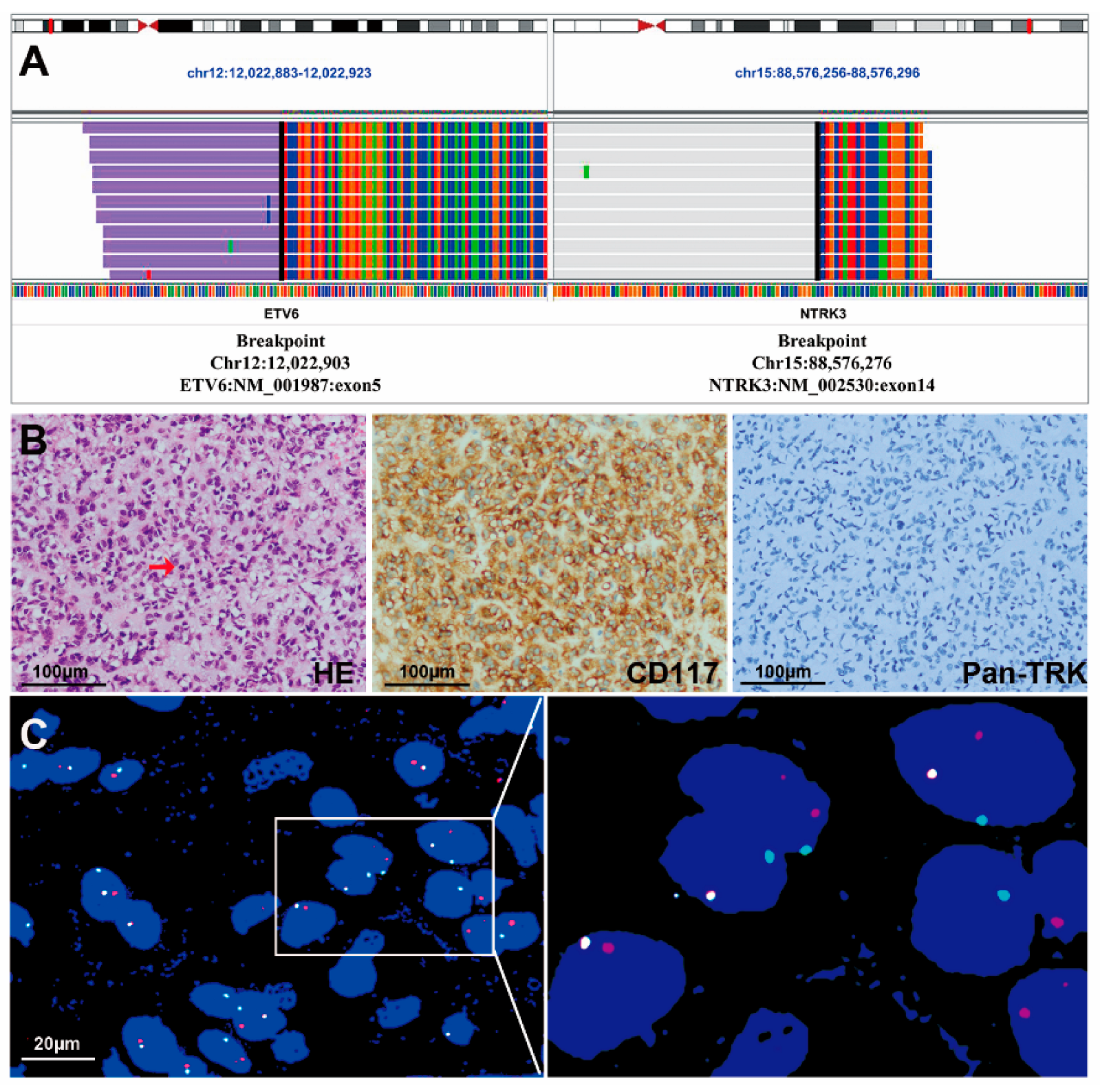
NTRK fusions were detected with NGS in two samples from the 36 GISTs without KIT/PDGFRA/SDH/BRAF/RAS mutations. ETV6-NTRK3 fusion was identified by DNA NGS (74 reads), showing that exon 1 to exon 5 of ETV6 were fused with exon 15 to exon 19 of NTRK3 in case #1 (Figure 2A). No gene rearrangement was detected using RNA-NGS in this case. The second case (case #2) was indicated to contain an ETV6-NTRK3 fusion using RNA NGS (215 reads), showing that exon 1 to exon 5 of ETV6 was fused with exon 14 to exon 19 of NTRK3 (Figure 3A), but the gene fusion was not detected using DNA NGS.

3.3. IHC Staining of Pan-TRK

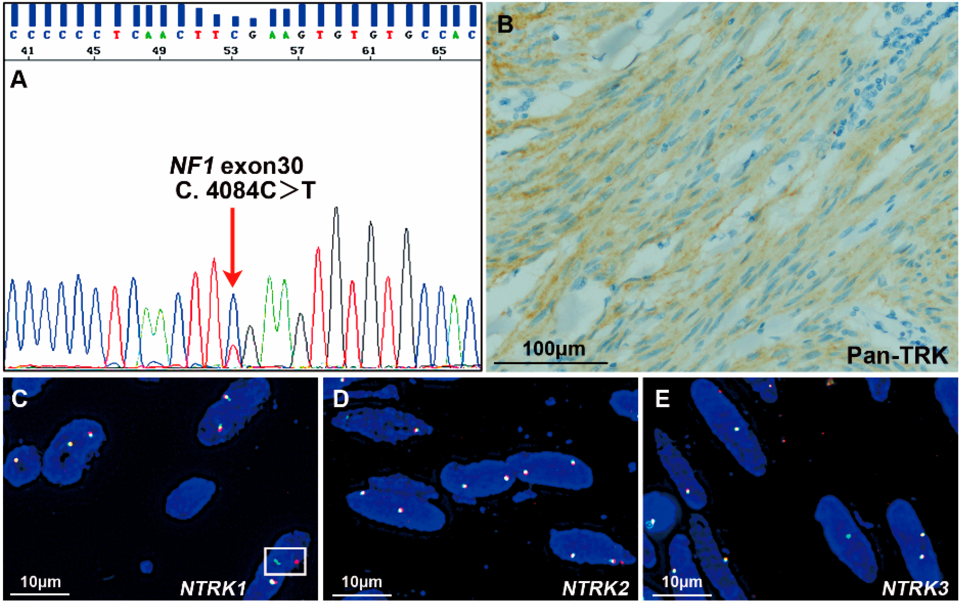
Among the 39 non-SDH-deficient WT GISTs, Pan-TRK was negative in 33 tissues and positive in 6 samples (Table 1), including strong cytoplasmic expression in 3 tumors (cases #1, #4 and #5) and weak-moderate cytoplasmic expression in 3 tumors (cases #3, #6 and #7). Among the 3 tumors with strong Pan-TRK expression, an ETV6-NTRK3 fusion was identified by NGS in only one case, which was a spindle-type GIST (case #1, Figure 2B). On the other hand, case #2 with an ETV6-NTRK3 fusion identified by NGS was negative for Pan-TRK (Figure 3B). In addition, the 3 tumors with weak-moderate cytoplasmic Pan-TRK expression were all negative for NTRK rearrangement according to NGS. Among them, the NF1 mutation was indicated by NGS and confirmed using Sanger sequencing (Figure 4A) in one tumor (case #3) showing weak-moderate expression of Pan-TRK (Figure 4B), and this patient was diagnosed with NF1 syndrome based on the clinical characteristics.

3.4. FISH Assessments

For the two patients (cases #1 and #2) with ETV6-NTRK3 fusions that were identified using NGS described above, we performed a FISH analysis using ETV6-NTRK3 fusion probes. FISH assays confirmed the fusion of ETV6 and NTRK3 as a result of the (12p13.2;15q25) chromosome translocation in both cases (Figure 2C and Figure 3C).
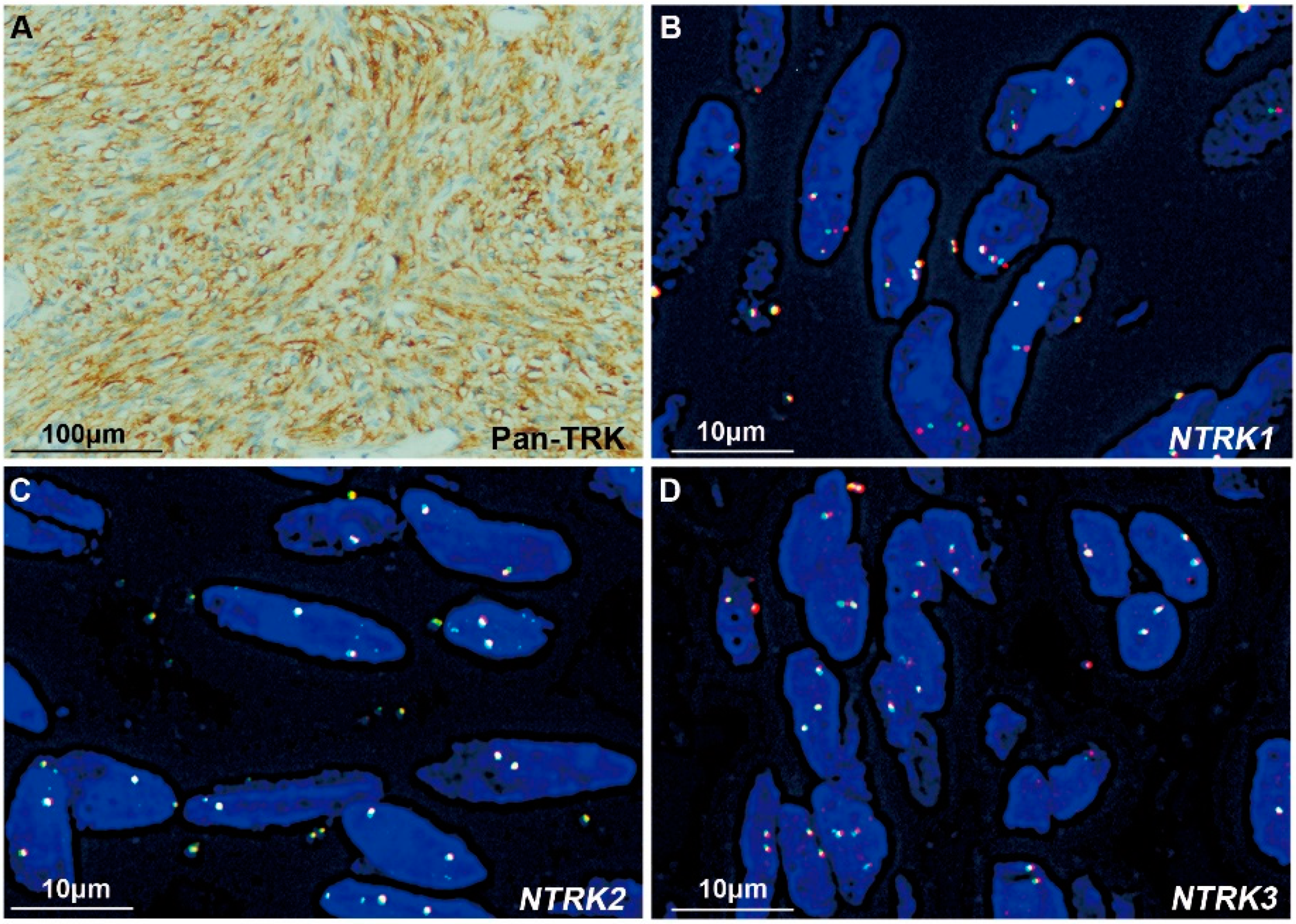
We subsequently performed FISH assessments with NTRK1, NTRK2, and NTRK3 break-apart probes in the 6 tumors with Pan-TRK positive expression. In case #1 with ETV6-NTRK3 fusion and strong Pan-TRK expression, NTRK3 was shown to be fragmented using the NTRK3 break-apart probe, consistent with the results obtained using the ETV6-NTRK3 fusion probe. Among the other two tumors with strong Pan-TRK expression, the NTRK1, NTRK2 or NTRK3 rearrangement was negative in one tumor (case #4, Figure 5), and no rearrangement was evaluated because of the poor DNA quality in the other sample (case #5). Among the 3 tumors with weak-moderate expression of Pan-TRK, case #3 was diagnosed as NF1-related GIST, as described above, in which NTRK1 break-apart was captured in only a few tumor cells (<15%, Figure 4C) and could not be diagnosed as an NTRK1 rearrangement. Moreover, this tumor was negative for NTRK2 and NTRK3 rearrangements (Figure 4D,E). Polyploidy was detected in most tumor cells using the NTRK1 break-apart probe in case #6, but no rearrangement of NTRK1, NTRK2, or NTRK3 was detected using the three break-apart probes (Figure 6). Another tumor (case #7) with weak-moderate expression of Pan-TRK was negative for NTRK1, NTRK2 and NTRK3 rearrangements (Figure S4). The FISH results were also shown in Table 1.

3.5. Comparisons among the Results of NGS, FISH, and IHC

Finally, we identified two GISTs (cases #1 and #2) with NTRK rearrangements among 39 non-SDH-deficient WT GISTs (5%). As shown in Table 1, the NGS and FISH results were absolutely consistent in screening GISTs with NTRK rearrangements. However, only one GIST (case #1) with a NTRK rearrangement (50%) was positive for Pan-TRK with IHC. On the other hand, among the six GISTs with Pan-TRK-positive expression, only one (case #1, 17%) contained an NTRK fusion, as determined using NGS or FISH.

3.6. The Clinicopathological Features of the Two GISTs with the ETV6-NTRK3 Fusion

In this study, case #1 with the ETV6-NTRK3 fusion was a 52-year-old female patient who occasionally found a pelvic mass. The pelvic ultrasound scan showed an irregular mass of 13.3 × 13.8 × 6.6 cm in front of the anterior uterine wall with an unclear boundary. As the tumor had a rich blood supply and adhered extensively to the adjacent organs and vessels, cytoreductive surgery was performed to remove the mesenteric mass in November 2018 in our center. The gross appearance of the mass was a solid tumor with a largest diameter of 10.0 cm, gray-yellow-tan color, and tough texture. Microscopically, the tumor was composed of spindle or short-spindle cells (Figure 2B). Tumor cells surrounded thin- and thick-walled blood vessels in some areas, and hyalinization and myxoid matrix background were observed in some regions. The mitoses of tumor cells were obvious, with a mitotic rate of 8/5 mm2 and a Ki-67 index of 30%. Except for ETV6-NTRK3 fusion, none of clinically significant mutations or fusions were found by NGS (Table S2). In addition, case #1 was indicated as microsatellite stable (MSS), and tumor mutation burden (TMB) was 4.4. The patient did not receive any adjuvant therapy based on the request of the patient and her family. The patient died 11 months after surgery.
Case #2 with ETV6-NTRK3 fusion was a 56-year-old male patient who presented with intermittent epigastric pain in December 2017. An upper abdomen computed tomography scan revealed a mass of 16.2 × 9.5 × 8.0 cm in the lower part of the gastric body. The intraoperative exploration revealed a large mass with a size of 16.0 × 10.0 × 8.0 cm originating from the greater curvature of the gastric body and invading the mesocolon transversum, spleen, and pancreas. The surgeon performed a complete margin negative (R0) resection. Microscopically, hyalinized, and myxoid matrix backgrounds were observed in some tumor areas. Most of the tumor cells were epithelioid (Figure 3B). The mitotic index was 3/5 mm2 and the Ki-67 index was 8%. Although Pan-TRK staining was negative (Figure 3B), NGS and FISH detected an ETV6-NTRK3 fusion (Figure 3A,C). Similar to case #1, except for ETV6-NTRK3 fusion, no clinically significant mutations or fusions were found by NGS in case #2 (Table S2). In addition, case #2 was indicated as MSS, and TMB was 5.5. The patient received adjuvant treatment with 400 mg/qd imatinib and his disease did not progress during the follow-up period (58 months).

3.7. The Clinicopathological and Genetic Features of 12 GISTs with NTRK Fusions

We searched the related literature in PubMed to determine the clinicopathological and molecular characteristics of GISTs with NTRK fusions. We only found 10 GISTs with NTRK fusions from five papers thus far [12,13,14,15,27]. We summarized the clinicopathological parameters, immunohistochemical profile, and genetic features of the previously reported 10 GISTs (cases #8–#17) together with our two patients with NTRK fusions (cases #1 and #2) in Table 2. Among the 12 patients, the male: female ratio was 2:1. Except for one patient who was 20 years old, the other patients were all over 40 years old (50 ± 12 years old). The tumors were frequently found in the intestine (75%), including the rectum (4/12), duodenum (1/12), jejunum (1/12), colon (1/12), small bowel (1/12), and mesentery (1/12), followed by the stomach (16.7%) and unknown sites (8.3%). Except for the unknown tumor size in 3 patients, the maximum diameter of tumors was >10 cm in 3 patients (33.3%), 5.1–10.0 cm in 2 patients (22.2%), 2.1–5.0 cm in 3 patients (33.3%) and less than 2.0 cm in 1 patient (11.1%). Among the 4 patients with the information of localized or metastatic tumors, 3 patients (cases #1, #2, and #11) had localized tumors, and case #9 found metastasis at diagnosis. Probably due to metastasis at diagnosis, case #9 did not undergo surgery but received five lines of TKIs therapy. When the tumor progressed after five lines of TKIs, the patient was treated with a TRK inhibitor (larotrectinib) for 4 months and had an ongoing partial response (44%) according to the Response Evaluation Criteria in Solid Tumor (RECIST) version 1.114. Case #9 had been alive for 159 months at the end of the follow-up. Similar to case #9, case #10 did not undergo surgery but received four lines of TKI therapy. Case #10 had been alive for 12 months at the end of the follow-up. Unfortunately, case #12 died soon after the biopsy for diagnosis, so he did not undergo surgery or drug treatment. Except for the above 3 patients without surgery, 9 patients (cases #1, #2, #8, #11, #13~#17) underwent surgery. Among the 9 patients, 3 patients received adjuvant therapy with imatinib. Of the three patients with imatinib adjuvant therapy, 2 (cases #2 and #16) had no tumor progression at the end of follow-up, and another patient (case #15) died of tumor progression. Among the 6 patients who underwent surgery but not adjuvant therapy, 2 patients (cases #1 and #13) died of tumor progression, and 4 patients (cases #8, #11, #14, and #17) did not experience tumor progression and were alive at the end of follow-up. The treatment and follow-up of the 12 patients are also listed in Table 2.
Histologically, the epithelioid type was the most frequent (3/5, 60%), followed by the spindle type (1/5, 20%) and mixed spindle-epithelioid type (1/5, 20%), based on the information reported. Among the nine cases with known mitotic counts, the mitosis rate was >10/5 mm2 in four patients (44.4%), 5–10/5 mm2 in two patients (22.2%), and less than 5/5 mm2 in three patients (33.3%). According to the modified NIH risk classification [42], eight patients were classified as high risk (8/9, 88.9%), one patient was classified as extremely low risk (1/9, 11.1%) and the other three patients were unable to be classified without the information on tumor size and mitosis number. CD117 was positive in 9 patients (9/10, 90%), and DOG-1 was positive in all of the patients with known IHC results (10/10, 100%). All of the 12 GISTs with NTRK fusions were WT GIST without KIT/PDGFRA mutation. SDHB was positive in all of the patients with known results (5/5, 100%). IHC for Pan-TRK was performed in only 8 patients. Among them, Pan-TRK was positive in two patients (25%). NGS was performed in 7 patients, in which ETV6-NTRK3 fusion was detected in 6 patients and LMNA-NTRK1 fusion was identified in one patient. Among them, fusions were detected in 4 patients using DNA NGS and 3 patients using RNA NGS. FISH was performed in 9 patients. Among them, ETV6-NTRK3 fusion probes were used in 2 patients (this study), both NTRK3 break-apart probe and ETV6 break-apart probe were used in one patient, ETV6 break-apart probe was used in one patient, and NTRK1 or NTRK3 break-apart probe was used in 5 patients. Both NGS and FISH were conducted in four cases (cases #1, #2, #3 and #17) with consistent results. Finally, NTRK3 fusion and NTRK1 fusion were identified in 8 (66.7%) and 4 patients (33.3%), respectively.

4. Discussion

The NTRK genes encode TRK proteins that exert oncogenic effects on tumors, and the activation of most TRK proteins is caused by NTRK fusions [43]. NTRK fusions occur in a variety of cancers with different incidences [19,20,21,22,23,24,25,26,44]. TRK inhibitors, such as larotrectinib and entrectinib, showed encouraging antitumor efficacies in tumors with NTRK rearrangements and have been approved as treatments for multiple cancers harboring NTRK fusions [29,30,31,34,45,46,47]. Although the prevalence of NTRK fusions in GIST is extremely low and only a few patients were enrolled in the clinical trials, the antitumor efficacies of TRK inhibitors were observed [30,31]. In clinical trials (LOXO-TRK-14001, SCOUT, and NAVIGATE), 55 patients with tumors carrying NTRK rearrangements, including three GISTs, were enrolled to evaluate the efficacy of larotrectinib. All three patients with GIST experienced tumor shrinkage >30%, and one had a pathological complete response with sufficient tumor shrinkentrage >90% [30]. In trials (ALKA-372–001, STARTRK-1 and STARTRK-2) including 54 patients with different tumors carrying NTRK fusions (including one with WT GIST), entrectinib was reported to be beneficial in general, although the detailed information for the patient with GIST was not revealed [31]. In addition, one patient with GIST carrying an ETV6-NTRK3 fusion received larotrectinib after five lines of TKIs (case #9 in Table 2) and achieved a partial response (44%) after 4 months of treatment [14]. Furthermore, larotrectinib has been recommended by the Belgian multidisciplinary expert panel as a first-line treatment for WT GIST with NTRK fusions [48]. These evidences suggest that NTRK fusions define a unique subgroup of GIST and TRK inhibitors have the potential to benefit GIST with NTRK fusions. Hence, it is clinically significant to screen NTRK fusions in WT GIST.
In this study, we identified ETV6-NTRK3 fusion in two patients among 46 with WT GISTs using several methods. Our NGS and FISH results were absolutely consistent in screening NTRK rearrangements in GISTs. FISH has been recommended as the gold standard for detecting gene rearrangements, including break-apart probes and fusion probes. Break-apart probes are more broad-spectrum than fusion probes but do not supply information about partner genes involved in the fusions. In tumors with a high frequency of NTRK rearrangements, such as infantile fibrosarcoma, congenital mesoblastic nephroma, and secretory breast carcinoma, NTRK1, NTRK2, and NTRK3 break-apart probes have been routinely used for the triple detection of FISH. In this study, we used NTRK1, NTRK2, and NTRK3 break-apart probes, which were also used in other studies [27], to detect NTRK fusions in patients with Pan-TRK positive expression, but NGS did not show NTRK fusions. Considering the high cost of triple detection with three pairs of break-apart probes, this approach does not seem superior to screening NTRK fusions using FISH in GISTs, which are tumors with a low incidence of NTRK fusions. In this study, based on the detailed fusion information identified using NGS, we used ETV6-NTRK3 fusion probes to verify the known fusions. As ETV6 is the most frequent partner in NTRK3 fusions, Castillon et al. sequentially used an ETV6 break-apart probe and NTRK3 break-apart probe to indicate the ETV6-NTRK3 fusion in one WT GIST [13]. FISH appeared more suitable for verification than for screening gene fusions in tumors with a low incidence of the fusions.
Compared to FISH with specific probes, NGS may provide information on broad-spectrum molecular alterations in many genes, including mutations, amplification/deletion, gene fusions, and SNPs. Therefore, NGS has been recommended in WT GISTs and patients with GIST exhibiting acquired resistance [49,50,51,52]. Genomic profiling can be performed through DNA-based NGS in which DNA is stable, but it is impossible to design probes covering all introns since exons account for only a small proportion of the total gene; therefore, missed inspection is unavoidable for DNA-based NGS. This limitation is a probable cause of the lack of DNA-based NGS data for case #2 in this study. Compared with DNA-based NGS, RNA-based NGS completely covers all exons, which may decrease the probability of missed detection and can supply direct evidence of fusion proteins based on mRNA levels. However, RNA is unstable and easily degrades in FFPE samples, which decreases the efficiency of RNA-based NGS. We detected the ETV6-NTRK3 fusion using DNA-based NGS but did not detect the fusion using RNA-based NGS in Patient One in the present study, which may result from the poor quality of RNA. Therefore, DNA + RNA NGS may reduce the false negative rate. In previous studies, some GISTs with the ETV6-NTRK3 fusion were identified using RNA sequencing [12,27] and others were identified by performing genomic profiling of DNA [14]. NGS requires strict quality control and was not available in some primary hospitals. The doctors in those hospitals are suggested to refer the patients with non-SDH-deficient WT GIST to expert centers, so that the patients could gain appropriate treatment based on the comprehensive landscape of targeting alterations from NGS.
Compared with FISH and NGS, IHC is a cheap and fast testing method and is available in many medical centers. IHC staining for Pan-TRK was used to indicate NTRK fusions in several cancers [35,36,53,54,55]. However, both the sensitivity (50%) and specificity (16.7%) of Pan-TRK staining were low in our study. Moreover, in the 8 GISTs with NTRK rearrangement for which Pan-TRK results were available across several studies, only 2 tumors (25%) were positive for Pan-TRK. Therefore, the validity of Pan-TRK IHC staining in screening NTRK rearrangements was dubious in GISTs. In this study, polyploidy of NTRK1 was detected in one tumor, and NTRK1 break-apart signals were captured in a few tumor cells in another sample, which might be one explanation for the weak-to-intermediate expression of Pan-TRK. However, we could not explain the possible mechanism of the other two cases with strong or weak-to-intermediate expression of Pan-TRK but without any NTRK rearrangement, amplification, or mutation based on the current results of FISH and NGS. We also have no insights into the drug response to TRK inhibitors in patients with GIST presenting Pan-TRK-positive expression but not NTRK rearrangements. The relationship between Pan-TRK expression and NTRK alterations should be further studied, and more clinical trials are needed. However, considering the practicability, IHC staining for Pan-TRK is a cheap way to screen patients in a targeted population of non-SDH-deficient WT GIST if NGS is not available.
In this study, we also explored the clinicopathological and genetic features of GISTs with NTRK fusions based on our patients and the literature. Compared with the classic GISTs frequently observed in the stomach, more GISTs with NTRK fusions occurred in the intestine, especially in the rectum. GISTs with NTRK fusions tended to present as the epithelioid type, while approximately 70% of classic GISTs were the spindle type [56]. In addition, according to the current data, most GISTs with NTRK rearrangements had a high risk of recurrence. In addition, NTRK3 fusions were more frequent than NTRK1 fusions in GISTs based on the present data.
However, there were some limitations in our study. In the first place, we couldn’t provide further treatment information about the TRK inhibitors in our patients with ETV6-NTRK fusion, since one patient died before she had a chance to use TRK inhibitors and the other have no evidence to use TRK inhibitors according to the current status of no tumor recurrence. Secondly, although we described the patient’s survival status in detail in this study, we could not perform a survival analysis based on such small sample size. Furthermore, we used a pooled cohort to analyze the screening efficiency of IHC, NGS and FISH as well as the clinicopathological characteristics of the GISTs with NTRK fusions, but the information from different studies is inhomogeneous. The conclusions should be validated in multicenter studies with large sample size.

5. Conclusions

It is clinically significant to screen NTRK fusions in WT GIST. Among the techniques used for screening NTRK fusion, NGS and FISH presented strong consistency, while IHC staining for Pan-TRK had limited sensitivity and specificity. However, IHC is a cheap way to screen patients in a targeted population of non-SDH-deficient WT GIST if NGS is not available. On the other hand, NGS can allow a comprehensive landscape of targeting alterations and is recommended to be used in WT GISTs and the GISTs exhibiting acquired resistance. It is suggested to refer the patients with WT GIST to expert centers at the earliest practicable stage in their disease course for precise diagnosis and treatment. Based on the study in small sample size, GISTs with NTRK rearrangement less frequently occurred in the stomach, were more frequently large in size and presented with epithelioid type, and had a higher risk of recurrence. NTRK3 fusion was more frequent than NTRK1 fusion in GISTs thus far. However, since the GISTs with NTRK fusions are rare, further studies including more cases and mechanism investigations should be conducted in future.

Supplementary Materials

The following supporting information can be downloaded at: https://www.mdpi.com/article/10.3390/cancers15010105/s1, Table S1:The primer sequences for exon amplification of target genes.; Table S2:The genetic alterations of the two GISTs with ETV6-NTRK3 fusion through NGS; Figure S1: The expression of SDHB protein in wild-type GISTs; Figure S2: Pathological images and genetic testing results of a BRAF-mutant GIST; Figure S3: Pathological images and genetic testing results of two RAS-mutant GISTs; Figure S4: The tumor (case #7) with weak-moderate expression of Pan-TRK but without NTRK1, NTRK2 or NTRK3 rearrangement.

Author Contributions

Y.S. designed the study and authorized the manuscript; Z.C. and J.L. performed the experiments and wrote the manuscript; L.S. and B.S. performed Sanger sequencing; and Y.G. performed IHC staining. Z.X. and Y.K. performed NGS and analyzed the data. All authors have read and agreed to the published version of the manuscript.

Funding

This research was funded by the National Natural Science Foundation of China (81871990), the Construction Project of Cancer Precision Diagnosis and Drug Treatment Technology from TUCIH (ZLJZZDYYWZL13), Tianjin Key Medical Discipline (Pathology) Construction Project (TJYXZDXK-012A), and Project of Tumor Translational Medicine Seed Fund of the Tianjin Medical University Cancer Institute and Hospital (1902).

Institutional Review Board Statement

This study was approved by the Institutional Review Board of Tianjin Medical University Cancer Institute and Hospital (approval no. bc2022133) in compliance with the Helsinki Declaration. Written informed consent was obtained from individual or guardian participants.

Informed Consent Statement

Informed consent was obtained from all subjects involved in the study.

Data Availability Statement

The datasets used and analyzed during the current study are available from the corresponding author on reasonable request.

Conflicts of Interest

The authors declare no conflict of interest.

References

  1. Søreide, K.; Sandvik, O.M.; Søreide, J.A.; Giljaca, V.; Jureckova, A.; Bulusu, V.R. Global epidemiology of gastrointestinal stromal tumours (GIST): A systematic review of population-based cohort studies. Cancer Epidemiol. 2016, 40, 39–46. [Google Scholar] [CrossRef] [Green Version]
  2. Verschoor, A.J.; Bovée, J.; Overbeek, L.; PALGA group; Hogendoorn, P.; Gelderblom, H. The incidence, mutational status, risk classification and referral pattern of gastro-intestinal stromal tumours in the Netherlands: A nationwide pathology registry (PALGA) study. Virchows Arch. 2018, 472, 221–229. [Google Scholar] [CrossRef] [PubMed] [Green Version]
  3. Sanders, K.M.; Ward, S.M.; Koh, S.D. Interstitial cells: Regulators of smooth muscle function. Physiol. Rev. 2014, 94, 859–907. [Google Scholar] [CrossRef] [PubMed]
  4. Parab, T.M.; DeRogatis, M.J.; Boaz, A.M.; Grasso, S.A.; Issack, P.S.; Duarte, D.A.; Urayeneza, O.; Vahdat, S.; Qiao, J.H.; Hinika, G.S. Gastrointestinal stromal tumors: A comprehensive review. J. Gastrointest. Oncol. 2019, 10, 144–154. [Google Scholar] [CrossRef] [PubMed]
  5. Corless, C.L.; Barnett, C.M.; Heinrich, M.C. Gastrointestinal stromal tumours: Origin and molecular oncology. Nat. Rev. Cancer. 2011, 11, 865–878. [Google Scholar] [CrossRef] [PubMed]
  6. Settas, N.; Faucz, F.R.; Stratakis, C.A. Succinate dehydrogenase (SDH) deficiency, Carney triad and the epigenome. Mol. Cell. Endocrinol. 2018, 469, 107–111. [Google Scholar] [CrossRef]
  7. Ibrahim, A.; Chopra, S. Succinate Dehydrogenase-Deficient Gastrointestinal Stromal Tumors. Arch. Pathol. Lab. Med. 2020, 144, 655–660. [Google Scholar] [CrossRef] [PubMed] [Green Version]
  8. Hostein, I.; Faur, N.; Primois, C.; Boury, F.; Denard, J.; Emile, J.F.; Bringuier, P.P.; Scoazec, J.Y.; Coindre, J.M. BRAF mutation status in gastrointestinal stromal tumors. Am. J. Clin. Pathol. 2010, 133, 141–148. [Google Scholar] [CrossRef] [Green Version]
  9. Huss, S.; Pasternack, H.; Ihle, M.A.; Merkelbach-Bruse, S.; Heitkötter, B.; Hartmann, W.; Trautmann, M.; Gevensleben, H.; Büttner, R.; Schildhaus, H.U.; et al. Clinicopathological and molecular features of a large cohort of gastrointestinal stromal tumors (GISTs) and review of the literature: BRAF mutations in KIT/PDGFRA wild-type GISTs are rare events. Hum. Pathol. 2017, 62, 206–214. [Google Scholar] [CrossRef]
  10. Miranda, C.; Nucifora, M.; Molinari, F.; Conca, E.; Anania, M.C.; Bordoni, A.; Saletti, P.; Mazzucchelli, L.; Pilotti, S.; Pierotti, M.; et al. KRAS and BRAF mutations predict primary resistance to imatinib in gastrointestinal stromal tumors. Clin. Cancer Res. 2012, 18, 1769–1776. [Google Scholar] [CrossRef]
  11. Pantaleo, M.A.; Nannini, M.; Corless, C.L.; Heinrich, M.C. Quadruple wild-type (WT) GIST: Defining the subset of GIST that lacks abnormalities of KIT, PDGFRA, SDH, or RAS signaling pathways. Cancer Med. 2015, 4, 101–103. [Google Scholar] [CrossRef] [PubMed]
  12. Brenca, M.; Rossi, S.; Polano, M.; Gasparotto, D.; Zanatta, L.; Racanelli, D.; Valori, L.; Lamon, S.; Dei Tos, A.P.; Maestro, R. Transcriptome sequencing identifies ETV6-NTRK3 as a gene fusion involved in GIST. J. Pathol. 2016, 238, 543–549. [Google Scholar] [CrossRef] [PubMed]
  13. Castillon, M.; Kammerer-Jacquet, S.F.; Cariou, M.; Costa, S.; Conq, G.; Samaison, L.; Douet-Guilbert, N.; Marcorelles, P.; Doucet, L.; Uguen, A. Fluorescent In Situ Hybridization Must be Preferred to pan-TRK Immunohistochemistry to Diagnose NTRK3-rearranged Gastrointestinal Stromal Tumors (GIST). Appl. Immunohistochem. Mol. Morphol. 2021, 29, 626–634. [Google Scholar] [CrossRef] [PubMed]
  14. Shi, E.; Chmielecki, J.; Tang, C.M.; Wang, K.; Heinrich, M.C.; Kang, G.; Corless, C.L.; Hong, D.; Fero, K.E.; Murphy, J.D.; et al. FGFR1 and NTRK3 actionable alterations in “Wild-Type” gastrointestinal stromal tumors. J. Transl. Med. 2016, 14, 339. [Google Scholar] [CrossRef] [Green Version]
  15. D’Alpino Peixoto, R.; Medeiros, B.A.; Cronemberger, E.H. Resected High-Risk Rectal GIST Harboring NTRK1 Fusion: A Case Report and Review of the Literature. J. Gastrointest. Cancer 2021, 52, 316–319. [Google Scholar] [CrossRef]
  16. Nannini, M.; Urbini, M.; Astolfi, A.; Biasco, G.; Pantaleo, M.A. The progressive fragmentation of the KIT/PDGFRA wild-type (WT) gastrointestinal stromal tumors (GIST). J. Transl. Med. 2017, 15, 113. [Google Scholar] [CrossRef]
  17. Pantaleo, M.A.; Urbini, M.; Indio, V.; Ravegnini, G.; Nannini, M.; De Luca, M.; Tarantino, G.; Angelini, S.; Gronchi, A.; Vincenzi, B.; et al. Genome-Wide Analysis Identifies MEN1 and MAX Mutations and a Neuroendocrine-Like Molecular Heterogeneity in Quadruple WT GIST. Mol. Cancer Res. 2017, 15, 553–562. [Google Scholar] [CrossRef] [Green Version]
  18. Huang, W.; Yuan, W.; Ren, L.; Xu, C.; Luo, R.; Zhou, Y.; Lu, W.; Hao, Q.; Xu, M.; Hou, Y. A novel fusion between CDC42BPB and ALK in a patient with quadruple wild-type gastrointestinal stromal tumor. Mol. Genet. Genom. Med. 2022, 10, e1881. [Google Scholar] [CrossRef]
  19. Knezevich, S.R.; McFadden, D.E.; Tao, W.; Lim, J.F.; Sorensen, P.H. A novel ETV6-NTRK3 gene fusion in congenital fibrosarcoma. Nat. Genet. 1998, 18, 184–187. [Google Scholar] [CrossRef]
  20. Rubin, B.P.; Chen, C.J.; Morgan, T.W.; Xiao, S.; Grier, H.E.; Kozakewich, H.P.; Perez-Atayde, A.R.; Fletcher, J.A. Congenital mesoblastic nephroma t(12;15) is associated with ETV6-NTRK3 gene fusion: Cytogenetic and molecular relationship to congenital (infantile) fibrosarcoma. Am. J. Pathol. 1998, 153, 1451–1458. [Google Scholar] [CrossRef]
  21. Eguchi, M.; Eguchi-Ishimae, M.; Tojo, A.; Morishita, K.; Suzuki, K.; Sato, Y.; Kudoh, S.; Tanaka, K.; Setoyama, M.; Nagamura, F.; et al. Fusion of ETV6 to neurotrophin-3 receptor TRKC in acute myeloid leukemia with t(12;15)(p13;q25). Blood 1999, 93, 1355–1363. [Google Scholar] [CrossRef] [PubMed]
  22. Forghieri, F.; Morselli, M.; Potenza, L.; Maccaferri, M.; Pedrazzi, L.; Paolini, A.; Bonacorsi, G.; Artusi, T.; Giacobbi, F.; Corradini, G.; et al. Chronic eosinophilic leukaemia with ETV6-NTRK3 fusion transcript in an elderly patient affected with pancreatic carcinoma. Eur. J. Haematol. 2011, 86, 352–355. [Google Scholar] [CrossRef] [PubMed]
  23. Tognon, C.; Knezevich, S.R.; Huntsman, D.; Roskelley, C.D.; Melnyk, N.; Mathers, J.A.; Becker, L.; Carneiro, F.; MacPherson, N.; Horsman, D.; et al. Expression of the ETV6-NTRK3 gene fusion as a primary event in human secretory breast carcinoma. Cancer Cell 2002, 2, 367–376. [Google Scholar] [CrossRef] [PubMed] [Green Version]
  24. Skálová, A.; Vanecek, T.; Sima, R.; Laco, J.; Weinreb, I.; Perez-Ordonez, B.; Starek, I.; Geierova, M.; Simpson, R.H.; Passador-Santos, F.; et al. Mammary analogue secretory carcinoma of salivary glands, containing the ETV6-NTRK3 fusion gene: A hitherto undescribed salivary gland tumor entity. Am. J. Surg. Pathol. 2010, 34, 599–608. [Google Scholar] [CrossRef]
  25. Leeman-Neill, R.J.; Kelly, L.M.; Liu, P.; Brenner, A.V.; Little, M.P.; Bogdanova, T.I.; Evdokimova, V.N.; Hatch, M.; Zurnadzy, L.Y.; Nikiforova, M.N.; et al. ETV6-NTRK3 is a common chromosomal rearrangement in radiation-associated thyroid cancer. Cancer 2014, 120, 799–807. [Google Scholar] [CrossRef] [Green Version]
  26. Alassiri, A.H.; Ali, R.H.; Shen, Y.; Lum, A.; Strahlendorf, C.; Deyell, R.; Rassekh, R.; Sorensen, P.H.; Laskin, J.; Marra, M.; et al. ETV6-NTRK3 Is Expressed in a Subset of ALK-Negative Inflammatory Myofibroblastic Tumors. Am. J. Surg. Pathol. 2016, 40, 1051–1061. [Google Scholar] [CrossRef]
  27. Lee, J.H.; Shin, S.J.; Choe, E.A.; Kim, J.; Hyung, W.J.; Kim, H.S.; Jung, M.; Beom, S.H.; Kim, T.I.; Ahn, J.B.; et al. Tropomyosin-Related Kinase Fusions in Gastrointestinal Stromal Tumors. Cancers 2022, 14, 2659. [Google Scholar] [CrossRef]
  28. Cocco, E.; Scaltriti, M.; Drilon, A. NTRK fusion-positive cancers and TRK inhibitor therapy. Nat. Rev. Clin. Oncol. 2018, 15, 731–747. [Google Scholar] [CrossRef]
  29. Drilon, A.; Laetsch, T.W.; Kummar, S.; DuBois, S.G.; Lassen, U.N.; Demetri, G.D.; Nathenson, M.; Doebele, R.C.; Farago, A.F.; Pappo, A.S.; et al. Efficacy of Larotrectinib in TRK Fusion-Positive Cancers in Adults and Children. N. Engl. J. Med. 2018, 378, 731–739. [Google Scholar] [CrossRef]
  30. Laetsch, T.W.; DuBois, S.G.; Mascarenhas, L.; Turpin, B.; Federman, N.; Albert, C.M.; Nagasubramanian, R.; Davis, J.L.; Rudzinski, E.; Feraco, A.M.; et al. Larotrectinib for paediatric solid tumours harbouring NTRK gene fusions: Phase 1 results from a multicentre, open-label, phase 1/2 study. Lancet Oncol. 2018, 19, 705–714. [Google Scholar] [CrossRef]
  31. Doebele, R.C.; Drilon, A.; Paz-Ares, L.; Siena, S.; Shaw, A.T.; Farago, A.F.; Blakely, C.M.; Seto, T.; Cho, B.C.; Tosi, D.; et al. Entrectinib in patients with advanced or metastatic NTRK fusion-positive solid tumours: Integrated analysis of three phase 1-2 trials. Lancet Oncol. 2020, 21, 271–282. [Google Scholar] [CrossRef] [PubMed]
  32. Solomon, J.P.; Hechtman, J.F. Detection of NTRK Fusions: Merits and Limitations of Current Diagnostic Platforms. Cancer Res. 2019, 79, 3163–3168. [Google Scholar] [CrossRef] [PubMed]
  33. Marchiò, C.; Scaltriti, M.; Ladanyi, M.; Iafrate, A.J.; Bibeau, F.; Dietel, M.; Hechtman, J.F.; Troiani, T.; López-Rios, F.; Douillard, J.Y.; et al. ESMO recommendations on the standard methods to detect NTRK fusions in daily practice and clinical research. Ann. Oncol. 2019, 30, 1417–1427. [Google Scholar] [CrossRef] [PubMed] [Green Version]
  34. Hechtman, J.F. NTRK insights: Best practices for pathologists. Mod. Pathol. 2022, 35, 298–305. [Google Scholar] [CrossRef] [PubMed]
  35. Bell, D.; Ferrarotto, R.; Liang, L.; Goepfert, R.P.; Li, J.; Ning, J.; Broaddus, R.; Weber, R.S.; El-Naggar, A.K. Pan-Trk immunohistochemistry reliably identifies ETV6-NTRK3 fusion in secretory carcinoma of the salivary gland. Virchows Arch. 2020, 476, 295–305. [Google Scholar] [CrossRef]
  36. Hechtman, J.F.; Benayed, R.; Hyman, D.M.; Drilon, A.; Zehir, A.; Frosina, D.; Arcila, M.E.; Dogan, S.; Klimstra, D.S.; Ladanyi, M.; et al. Pan-Trk Immunohistochemistry Is an Efficient and Reliable Screen for the Detection of NTRK Fusions. Am. J. Surg. Pathol. 2017, 41, 1547–1551. [Google Scholar] [CrossRef]
  37. Cao, J.; Chen, L.; Li, H.; Chen, H.; Yao, J.; Mu, S.; Liu, W.; Zhang, P.; Cheng, Y.; Liu, B.; et al. An Accurate and Comprehensive Clinical Sequencing Assay for Cancer Targeted and Immunotherapies. Oncologist 2019, 24, e1294–e1302. [Google Scholar] [CrossRef] [Green Version]
  38. Haas, B.J.; Dobin, A.; Li, B.; Stransky, N.; Pochet, N.; Regev, A. Accuracy assessment of fusion transcript detection via read-mapping and de novo fusion transcript assembly-based methods. Genome Biol. 2019, 20, 213. [Google Scholar] [CrossRef] [Green Version]
  39. Moes-Sosnowska, J.; Skupinska, M.; Lechowicz, U.; Szczepulska-Wojcik, E.; Skronska, P.; Rozy, A.; Stepniewska, A.; Langfort, R.; Rudzinski, P.; Orlowski, T.; et al. FGFR1-4 RNA-Based Gene Alteration and Expression Analysis in Squamous Non-Small Cell Lung Cancer. Int. J. Mol. Sci. 2022, 23, 10506. [Google Scholar] [CrossRef]
  40. Yang, Y.; Wang, Z.; Pan, G.; Li, S.; Wu, Y.; Liu, L. Pure secretory carcinoma in situ: A case report and literature review. Diagn. Pathol. 2019, 14, 95. [Google Scholar] [CrossRef]
  41. Chen, L.; Yang, F.; Feng, T.; Wu, S.; Li, K.; Pang, J.; Shi, X.; Liang, Z. PD-L1, Mismatch Repair Protein, and NTRK Immunohistochemical Expression in Cervical Small Cell Neuroendocrine Carcinoma. Front. Oncol. 2021, 11, 752453. [Google Scholar] [CrossRef] [PubMed]
  42. Joensuu, H. Risk stratification of patients diagnosed with gastrointestinal stromal tumor. Hum. Pathol. 2008, 39, 1411–1419. [Google Scholar] [CrossRef] [PubMed]
  43. Vaishnavi, A.; Le, A.T.; Doebele, R.C. TRKing down an old oncogene in a new era of targeted therapy. Cancer Discov. 2015, 5, 25–34. [Google Scholar] [CrossRef] [PubMed] [Green Version]
  44. Klug, L.R.; Khosroyani, H.M.; Kent, J.D.; Heinrich, M.C. New treatment strategies for advanced-stage gastrointestinal stromal tumours. Nat. Rev. Clin. Oncol. 2022, 19, 328–341. [Google Scholar] [CrossRef] [PubMed]
  45. Al-Salama, Z.T.; Keam, S.J. Entrectinib: First Global Approval. Drugs 2019, 79, 1477–1483. [Google Scholar] [CrossRef]
  46. Scott, L.J. Larotrectinib: First Global Approval. Drugs 2019, 79, 201–206. [Google Scholar] [CrossRef]
  47. Drilon, A.; Siena, S.; Ou, S.I.; Patel, M.; Ahn, M.J.; Lee, J.; Bauer, T.M.; Farago, A.F.; Wheler, J.J.; Liu, S.V.; et al. Safety and Antitumor Activity of the Multitargeted Pan-TRK, ROS1, and ALK Inhibitor Entrectinib: Combined Results from Two Phase I Trials (ALKA-372-001 and STARTRK-1). Cancer Discov. 2017, 7, 400–409. [Google Scholar] [CrossRef] [Green Version]
  48. Awada, A.; Berghmans, T.; Clement, P.M.; Cuppens, K.; De Wilde, B.; Machiels, J.P.; Pauwels, P.; Peeters, M.; Rottey, S.; Van Cutsem, E. Belgian expert consensus for tumor-agnostic treatment of NTRK gene fusion-driven solid tumors with larotrectinib. Crit. Rev. Oncol. Hematol. 2022, 169, 103564. [Google Scholar] [CrossRef]
  49. NCCN Clinical Practice Guidelines in Oncology. Gastrointestinal Stromal Tumors (GISTs), Version 1.2021. 2021. Available online: https://www.nccn.org/professionals/physician_gls/default.aspx#gist (accessed on 23 January 2021).
  50. Qian, H.; Yan, N.; Hu, X.; Jiang, J.; Cao, Z.; Shen, D. Molecular Portrait of GISTs Associated with Clinicopathological Features: A Retrospective Study with Molecular Analysis by a Custom 9-Gene Targeted Next-Generation Sequencing Panel. Front. Genet. 2022, 13, 864499. [Google Scholar] [CrossRef]
  51. Mavroeidis, L.; Metaxa-Mariatou, V.; Papoudou-Bai, A.; Lampraki, A.M.; Kostadima, L.; Tsinokou, I.; Zarkavelis, G.; Papadaki, A.; Petrakis, D.; Gκoura, S.; et al. Comprehensive molecular screening by next generation sequencing reveals a distinctive mutational profile of KIT/PDGFRA genes and novel genomic alterations: Results from a 20-year cohort of patients with GIST from north-western Greece. ESMO Open 2018, 3, e000335. [Google Scholar] [CrossRef]
  52. Unk, M.; Bombač, A.; Jezeršek Novaković, B.; Stegel, V.; Šetrajčič Dragoš, V.; Blatnik, O.; Klančar, G.; Novaković, S. Correlation of treatment outcome in sanger/RT-qPCR KIT/PDGFRA wild-type metastatic gastrointestinal stromal tumors with next-generation sequencing results: A single-center report. Oncol. Rep. 2022, 48, 167. [Google Scholar] [CrossRef] [PubMed]
  53. Suurmeijer, A.J.; Dickson, B.C.; Swanson, D.; Zhang, L.; Sung, Y.S.; Huang, H.Y.; Fletcher, C.D.; Antonescu, C.R. The histologic spectrum of soft tissue spindle cell tumors with NTRK3 gene rearrangements. Genes Chromosomes Cancer 2019, 58, 739–746. [Google Scholar] [CrossRef] [PubMed]
  54. Atiq, M.A.; Davis, J.L.; Hornick, J.L.; Dickson, B.C.; Fletcher, C.D.M.; Fletcher, J.A.; Folpe, A.L.; Mariño-Enríquez, A. Mesenchymal tumors of the gastrointestinal tract with NTRK rearrangements: A clinicopathological, immunophenotypic, and molecular study of eight cases, emphasizing their distinction from gastrointestinal stromal tumor (GIST). Mod. Pathol. 2021, 34, 95–103. [Google Scholar] [CrossRef]
  55. Rudzinski, E.R.; Lockwood, C.M.; Stohr, B.A.; Vargas, S.O.; Sheridan, R.; Black, J.O.; Rajaram, V.; Laetsch, T.W.; Davis, J.L. Pan-Trk Immunohistochemistry Identifies NTRK Rearrangements in Pediatric Mesenchymal Tumors. Am. J. Surg. Pathol. 2018, 42, 927–935. [Google Scholar] [CrossRef] [PubMed]
  56. Li, J.; Ye, Y.; Wang, J.; Zhang, B.; Qin, S.; Shi, Y.; He, Y.; Liang, X.; Liu, X.; Zhou, Y.; et al. Chinese consensus guidelines for diagnosis and management of gastrointestinal stromal tumor. Chin. J. Cancer Res. 2017, 29, 281–293. [Google Scholar] [CrossRef]
Figure 1. Flow diagram of preliminary testing of WT GISTs.
Cancers 15 00105 g001
Figure 2. IGV, HE staining, immunohistochemical staining and FISH images of a mesenteric GIST with an ETV6-NTRK3 fusion (case #1). (A) IGV images of NGS showed the ETV6-NTRK3 fusion. (B) The tumor was composed of spindle-shaped or short spindle-shaped cells, and mitosis was observed (red arrow) (HE staining, 400×). Immunohistochemical staining showed strong positive expression of CD117 (400×) and Pan-TRK (400×). Each scale bar is 100 μm. (C) The FISH assay showed a pair of fused red and green signals using ETV6-NTRK3 fusion probes in almost all tumor cells. Scale bar is 20 μm.
Cancers 15 00105 g002
Figure 3. IGV, HE staining, immunohistochemical staining and FISH images of a gastric GIST with an ETV6-NTRK3 fusion (case #2). (A) IGV images of NGS indicated an ETV6-NTRK3 fusion. (B) HE staining showed that the GIST was composed of epithelioid cells, and the red arrow indicates tumor cell mitosis (HE staining, 400×). The tumor cells were positive for CD117 (400×) and negative for Pan-TRK (400×). Each scale bar is 100 μm. (C) The FISH assay showed a pair of fused red and green signals using ETV6-NTRK3 fusion probes in almost all tumor cells. Scale bar is 20 μm.
Cancers 15 00105 g003
Figure 4. A NF1-related GIST with weak-moderate expression of Pan-TRK (case #3). (A) Sanger sequencing indicated a heterozygous point mutation in exon 30 of the NF1 gene (c.4084C > T, p.Arg1362*). (B) IHC staining showed weak-moderate cytoplasmic expression of Pan-TRK (400×). Scale bar is 100 μm. (CE) FISH images obtained using NTRK1, NTRK2, and NTRK3 break-apart probes. NTRK1 break-apart signals (in the white rectangle) were captured in only a few tumor cells (<15%), which could not be diagnosed as an NTRK1 rearrangement. Neither NTRK2 nor NTRK3 rearrangements were detected in this tumor. Each scale bar is 10 μm.
Cancers 15 00105 g004
Figure 5. The tumor (case #4) with strong Pan-TRK expression but without NTRK1, NTRK2 or NTRK3 rearrangement. (A) IHC staining showed strong cytoplasmic expression of Pan-TRK (400×). Scale bar is 100 μm. (BD) FISH images obtained using NTRK1, NTRK2, and NTRK3 break-apart probes. The tumor was negative for NTRK1, NTRK2 and NTRK3 rearrangements. Each scale bar is 10 μm.
Cancers 15 00105 g005
Figure 6. The tumor (case #6) with moderate expression of Pan-TRK and polyploidy involving NTRK1. (A) IHC staining showed moderate cytoplasmic expression of Pan-TRK (400×). Scale bar is 100 μm. (BD) FISH images obtained using NTRK1, NTRK2, and NTRK3 break-apart probes. Polyploidy, but not rearrangement, was observed in some tumor cells using the NTRK1 break-apart probe. The tumor was negative for NTRK2 and NTRK3 rearrangements. Each scale bar is 10 μm.
Cancers 15 00105 g006
Table 1. FISH assessments in the cases with positive expression of Pan-TRK or NTRK fusion from NGS.
CaseIHC for Pan-TRKNGSFISH
IntensityPositive
Proportion
NTRK FusionsETV6-NTRK3NTRK3NTRK2NTRK1
#1Strong50%ETV6-NTRK3PositivePositiveNegativeNegative
#2Negative0ETV6-NTRK3PositiveNPNegativeNegative
#3Weak-moderate70%No aNPNegativeNegativeNegative
#4Strong80%NoNPNegativeNegativeNegative
#5Strong50%NoNPNA bNA bNA b
#6Weak-moderate30%NoNPNegativeNegativeNegative
#7Weak-moderate30%NoNPNegativeNegativeNegative
Abbreviations: NA: not available; a: the NF1 mutation was indicated by NGS and confirmed using Sanger sequencing; b: no rearrangement was evaluated because of the poor DNA quality in the other sample, NP: not performed.
Table 2. Gastrointestinal Stromal Tumors with NTRK Gene Fusions Reported in the Literature.
CaseAge
(Years),
Sex
Location/
Localized or Metastatic
Size
(cm)
MorphologyMI
(/5 mm²)
ImmunohistochemistryKIT/
PDGFRA
Mutation
NTRK FusionSurgeryDrug
Treatment
Progression/PFS
(Months)
Status/OS
(Months)
Reference
CD117DOG-1SDHBPTRKTypeNGSFISH
#152, FMesentery/
Localized
10S8PPPPNoETV6-NTRK3DNA-basedETV6-NTRK3YesNoYes/11Dead/11this study
#256, MStomach/
Localized
16E3PPPNNoETV6-NTRK3RNA-basedETV6-NTRK3YesImatinibNo/58Alive/58this study
#844, MRectum/NA5E34PPPNANoETV6-NTRK3RNA-basedETV6YesNoNo/44Alive/44[12]
#955, MSmall bowel/MetastaticNANANANANAPNANoETV6-NTRK3DNA-basedNANoImatinibYes/NA aAlive/159[14]
Sunitinib
Sorafenib
Nilotinib
Regorafenib Larotrectinib
#1054, MColon/NANANANANANAPNANoETV6-NTRK3DNA-basedNANoImatinib Sunitinib
Sorafenib
Linsitinib
Yes/NA bAlive/12[14]
#1120, MRectum/
Localized
7S,E10NPNANANoLMNA-NTRK1DNA-basedNAYesNoNo/7Alive/7[15]
#1259, MNA/NANAENAPPNANNoETV6-NTRK3NoNTRK3 and ETV6, respectivelyNoNoNo/0Dead/0[13]
#1344, FRectum/NA2.8NA17PNANAWeakly positive in only one of the following five patientsNoNTRK1NoNTRK1YesNoYes/108Dead/132[27]
#1445, MDuodenum/NA1.7NA1PNANANoNTRK3NoNTRK3YesNoNo/72Alive/72[27]
 
 
#1565, FStomach/NA17NA70PNANANoNTRK1NoNTRK1YesImatinibYes/26Dead/96[27]
#1661, FJejunum/NA3.9NA12PNANANoNTRK1NoNTRK1YesImatinibNo/48Alive/48[27]
#1743, MRectum/NA11NA0PNANANoETV6-NTRK3RNA-basedNTRK3YesNoNo/36Alive/36[27]
Abbreviations: M: male; F: female; S: spindled; MI: mitotic index; E: epithelioid; P: positive; N: negative; NA: not available; PFS: progression-free survival; OS: overall survival; a The patient experienced tumor progression during treatment with five lines of TKI therapy (155 months) and then achieved an ongoing partial response (44%) after larotrectinib therapy for 4 months. b The patient experienced tumor progression during treatment with four lines of TKI therapy (12 months).
Disclaimer/Publisher’s Note: The statements, opinions and data contained in all publications are solely those of the individual author(s) and contributor(s) and not of MDPI and/or the editor(s). MDPI and/or the editor(s) disclaim responsibility for any injury to people or property resulting from any ideas, methods, instructions or products referred to in the content.