Article

TIM-3 Qualifies as a Potential Immunotherapeutic Target in Specific Subsets of Patients with High-Risk Soft Tissue Sarcomas (HR-STS)

by
Luc M. Berclaz
1,*,
Annelore Altendorf-Hofmann
2,
Lars H. Lindner
1,
Anton Burkhard-Meier
1,
Dorit Di Gioia
1,
Hans Roland Dürr
3,
Alexander Klein
3,
Markus Albertsmeier
4,
Nina-Sophie Schmidt-Hegemann
5,
Frederick Klauschen
6 and
Thomas Knösel
6,*
1
Department of Internal Medicine III, University Hospital, Ludwig-Maximilians-University (LMU) Munich, 81377 Munich, Germany
2
Department of General, Visceral and Vascular Surgery, Friedrich-Schiller University Jena, 07747 Jena, Germany
3
Orthopaedic Oncology, Department of Orthopaedics and Trauma Surgery, University Hospital, Ludwig-Maximilians-University (LMU) Munich, 81377 Munich, Germany
4
Department of General, Visceral and Transplantation Surgery, University Hospital, Ludwig-Maximilians-University (LMU) Munich, 81377 Munich, Germany
5
Department of Radiation Oncology, University Hospital, Ludwig-Maximilians-University (LMU) Munich, Marchioninistr. 15, 81377 Munich, Germany
6
Institute of Pathology, Ludwig-Maximilians-University (LMU) Munich, 81377 Munich, Germany
*
Authors to whom correspondence should be addressed.
Cancers 2023, 15(10), 2735; https://doi.org/10.3390/cancers15102735
Submission received: 13 April 2023 / Revised: 8 May 2023 / Accepted: 10 May 2023 / Published: 12 May 2023
(This article belongs to the Section Cancer Biomarkers)

Abstract

:

Simple Summary

T cell immunoglobulin and mucin domain-containing protein 3 (TIM-3) acts as an immune checkpoint on exhausted T cells and has been associated with dismal outcomes in various solid tumors. TIM3 is currently being evaluated as an immunotherapeutic target in multiple clinical trials. Our study shows the significant tumor cell expression of TIM-3 in specific subsets of patients with high risk soft tissue sarcomas (HR-STS). We demonstrate an interaction between TIM-3, tumor infiltrating lymphocyte (TIL) counts and PD-1/PD-L1 expression in patients with HR-STS. TIM-3 could qualify as a potential immunotherapeutic target in HR-STS.

Abstract

(1) Background: The expression of T cell immunoglobulin and mucin domain-containing protein 3 (TIM-3), an immune checkpoint receptor on T cells, has been associated with dismal outcomes and advanced tumor stages in various solid tumors. The blockade of TIM-3 is currently under examination in several clinical trials. This study examines TIM-3 expression in high-risk soft tissue sarcomas (HR-STS). (2) Methods: Tumor cell expression of TIM-3 on protein level was analyzed in pre-treatment biopsies of patients with HR-STS. TIM-3 expression was correlated with clinicopathological parameters including tumor-infiltrating lymphocyte (TIL) counts, programmed cell death 1 (PD-1) and programmed cell death ligand 1 (PDL-1) expression in patients with HR-STS. Survival dependent on the expression of TIM-3 was analyzed. (3) Results: TIM-3 expression was observed in 101 (56%) out of 179 pre-treatment biopsies of patients with HR-STS. TIM-3 expression was significantly more often observed in undifferentiated pleomorphic sarcomas (UPS) compared to other histological subtypes (p < 0.001), high TIL counts (p < 0.001), and high PD-1 (p < 0.001) and PD-L1 expression (p < 0.001). TIM-3 expression did not have a prognostic impact on survival in patients with HR-STS. (4) Conclusions: This is the first study to demonstrate a significant tumor cell expression of TIM-3 in specific subsets of patients with HR-STS. TIM-3 qualifies as a potential immunotherapeutic target in HR-STS.

1. Introduction

High-risk soft tissue sarcomas (HR-STS) are rare tumors with multiple distinct histopathological subtypes, the most common being liposarcoma, leiomyosarcoma and undifferentiated pleomorphic sarcomas (UPS). They account for approximately 1% of adult malignancies [1,2]. Despite optimal local treatment, almost half of patients will die within 5 years of their diagnosis [3,4]. In patients with advanced and metastatic soft tissue sarcomas, median survival rates range around 12–18 months [5,6,7,8]. Standard treatment for locally advanced and metastatic HR-STS is systemic chemotherapy with anthracycline-based regimens [9,10,11]. Different second- or third-line regimens, including trabectedin, or targeted therapies such as pazopanib have been approved in recent years, with only limited effects on PFS and OS [12,13]. Considering the lack of efficient therapy lines and poor survival rates, there is a great need for new systemic treatment strategies.
While checkpoint inhibitors (CPI) revolutionized the treatment of multiple cancers with high somatic mutation rates such as melanoma and lung cancer, they have demonstrated only limited effects in sarcomas and are currently not part of international treatment guidelines [14,15,16,17,18]. T cell immunoglobulin and mucin domain-containing protein 3 (TIM-3), an emerging immune checkpoint receptor, is a member of the TIM family and was originally identified as a receptor expressed on interferon-γ-producing CD4+ and CD8+ T cells [19]. The working mechanisms of TIM-3 are not fully understood. In lymphocytes, TIM-3 is recruited to the immunological synapse on T cell activation [20]. Depending on the interplay with its interacting ligands such as CEACAM1 or lectin galectin 9, TIM-3 is differently phosphorylated and either permissive or inhibitory to T cell activation [21,22]. TIM-3 is expressed in different tumor cells, including lung cancer and melanoma [23,24]. It is co-stimulated and co-regulated with other checkpoint receptors, and the co-expression of TIM-3 with PD-1 is associated with a specific subset of particularly dysfunctional or “exhausted” T cells [22]. TIM-3+/PD-1+ cells appear to express significantly lower amounts of effector cytokines such as IFN-γ, TNF and IL-2 [25]. Both checkpoint receptors are co-regulated by immunosuppressive cytokines such as IL-27, which finally results in a diminished immune response in cancer and chronic viral infections [25,26,27]. The expression of TIM-3 was associated with a poor overall survival and advanced tumor stages in several solid malignancies, including colorectal and non-small cell lung cancer [28]. In soft tissue and bone sarcomas, TIM-3 expression in tumor-infiltrating lymphocytes (TIL) did not significantly correlate with PFS or OS in previous studies [29,30].
TIM-3 inhibition has shown promising results in pre-clinical models and is currently being evaluated as a novel immunotherapeutic approach in several clinical trials [31,32,33,34]. Clinical trials often combine TIM-3 inhibitors with checkpoint inhibitors targeting PD-1, as pre-clinical models demonstrated a synergistic effect and a better restoration of T cell responses in CPI “co-blockades” [25,35,36]. Ongoing clinical trials include NCT03446040 combining an anti-TIM-3 antibody with Nivolumab, and NCT03744468 combining anti-TIM-3 antibodies with Tislelizumab. In the present study, we analyzed the tumor cell expression of TIM-3 in a large and well-characterized cohort of HR-STS patients with long-term follow-up. We correlated our findings with clinical tumor characteristics, tumor-infiltrating lymphocyte (TIL) counts, PD-1 and PD-L1 expression status, and survival data. Our study demonstrates a significant expression of TIM-3 in specific subsets of patients with HR-STS.

2. Materials and Methods

2.1. Patient Selection

An exploratory retrospective cohort study design was chosen to address the research question. Eligible patients had pathologically confirmed high-risk soft tissue sarcoma (Tumor diameter 5 cm or larger, French Fédération Nationale des Centres de Lutte Contre le Cancer (FNCLCC) grade 2 or 3, deep to the fascia). Clinical, pathological, and outcomes data were extracted from our clinical sarcoma database. Most patients were to receive a multimodal treatment including neoadjuvant doxorubicin-based chemotherapy and regional hyperthermia (RHT), surgery, adjuvant chemotherapy + RHT and radiotherapy in select cases. Treatment continued unless disease progression or unacceptable toxic effects occurred. Follow-up was performed until December 2022.

2.2. Histopathology and Tissue Microarray Construction

Tumor samples originated from tumor biopsies that were taken before the initiation of neoadjuvant treatment at the Ludwig Maximilians University hospitals, Munich. In addition to the original pathology reports, microscopic findings (tumor type according to current WHO classifications and degree of differentiation) were reassessed. For tissue microarray (TMA) assembly, representative tumor areas were marked on H&E-stained slides of formalin-fixed, paraffin-embedded tumor samples from all patients according to standard procedures, and two 0.6 mm punch biopsies were taken from each sample [37]. Normal tonsillar tissue samples were used as controls on the TMA. In the end, seven tissue microarrays containing 179 pre-treatment tumor samples from 179 patients with high-grade soft tissue sarcomas (HR-STS) were constructed.

2.3. TIM-3 Immunohistochemistry

For the immunohistochemical detection of TIM-3, commercially available and validated monoclonal antibodies were used (TIM-3 D5D5R, Cell Signaling Tech., Danvers, MA, USA). Antigen retrieval was carried out by heat treatment with Target Retrieval Solution Citrate (Agilent Technologies, Santa Clara, CA, USA). Staining was performed on a Ventana Benchmark XT Autostainer (Ventana Medical Systems, Tucson, AZ, USA) with a DAB+ Kit (Agilent Technologies, Santa Clara, CA, USA). All slides were counterstained with hematoxylin (Vector Laboratories, Burlingame, CA, USA). An ImmPRESS Anti-Rabbit IgG Polymer Kit was used for detection (Vector Laboratories, Burlingame, CA, USA). To exclude unspecific staining, system controls were included. Tonsillar tissue served as a positive control for immunohistochemistry. The immunostaining of cells was evaluated and scored semi-quantitatively (0 = negative; 1 = ≥5% positive and weakly stained, 2 = ≥25% positive and moderately stained, 3 = ≥50% positive and strongly stained). All immunohistochemical and pathologic evaluations were carried out independently and blinded with an experienced sarcoma pathologist (T.K.). In the case of discrepancy, the slides were reevaluated under a multiheaded microscope and a consensus was reached.

2.4. TILs, PD-1 and PD-L1

Tumor-infiltrating lymphocytes (TILs), PD-1 and PD-L1 were previously investigated in our HR-STS cohort [38,39]. TILs were counted per high-power field (HPF) (400× magnification, field of view 0.237 mm2) in H&E-stained TMA slides. As previously described, slides were pre-treated with heat and Target Retrieval solution (S1699, Agilent, Santa Clara, CA, USA) before incubation with the monoclonal primary anti-PD-1 mouse antibody (315M; 1:80; Cell Marque, Rocklin, CA, USA) for 60 min at room temperature. The Vectastain Elite ABC HRP Kit (Vector Laboratories, Burlingame, CA, USA) and the chromogen DAB+ (Agilent) were used for detection, and Hematoxylin (Vector Laboratories) for counterstaining. For PD-L1 staining, slides were pre-treated with heat and the Epitope Retrieval Solution pH8 Novocastra (Leica Biosystems, Wetzlar, Germany) before incubation with the monoclonal primary anti-PD-L1 rabbit antibody (E1L3N; 1:50; Cell Signaling Technology, Danvers, MA, USA) for 60 min at room temperature. We used the SignalStain Boost IHC Detection Reagent (Cell Signalling Technology) and the chromogen DAB+ (Agilent) for detection according to previous studies [39].

2.5. Statistical Analysis

Categorical variables were tested for independence using the Chi square test. Binary variables were compared using Fisher’s Exact Test, and continuous variables were compared using t-tests. Logistic regression was used for univariate and multivariate analyses. The forward stepwise procedure was set to a threshold of 0.05. Data analysis was performed using SAS 9.4 (SAS Inst Inc., Cary, NC, USA). All p-values were based on a two-tailed hypothesis test, with values less than 0.05 considered statistically significant.

3. Results

3.1. Patient Cohort

In total, 179 patients treated between 1997 and 2019 were included in this study. The median age was 54 years (range, 18–79 years), and 87 (48.6%) patients were female. The most common histological subtypes were undifferentiated pleomorphic sarcomas (UPS) (33%), leiomyosarcomas (17%), and liposarcomas (22%). The clinicopathologic characteristics of the study cohort are summarized in Table 1.

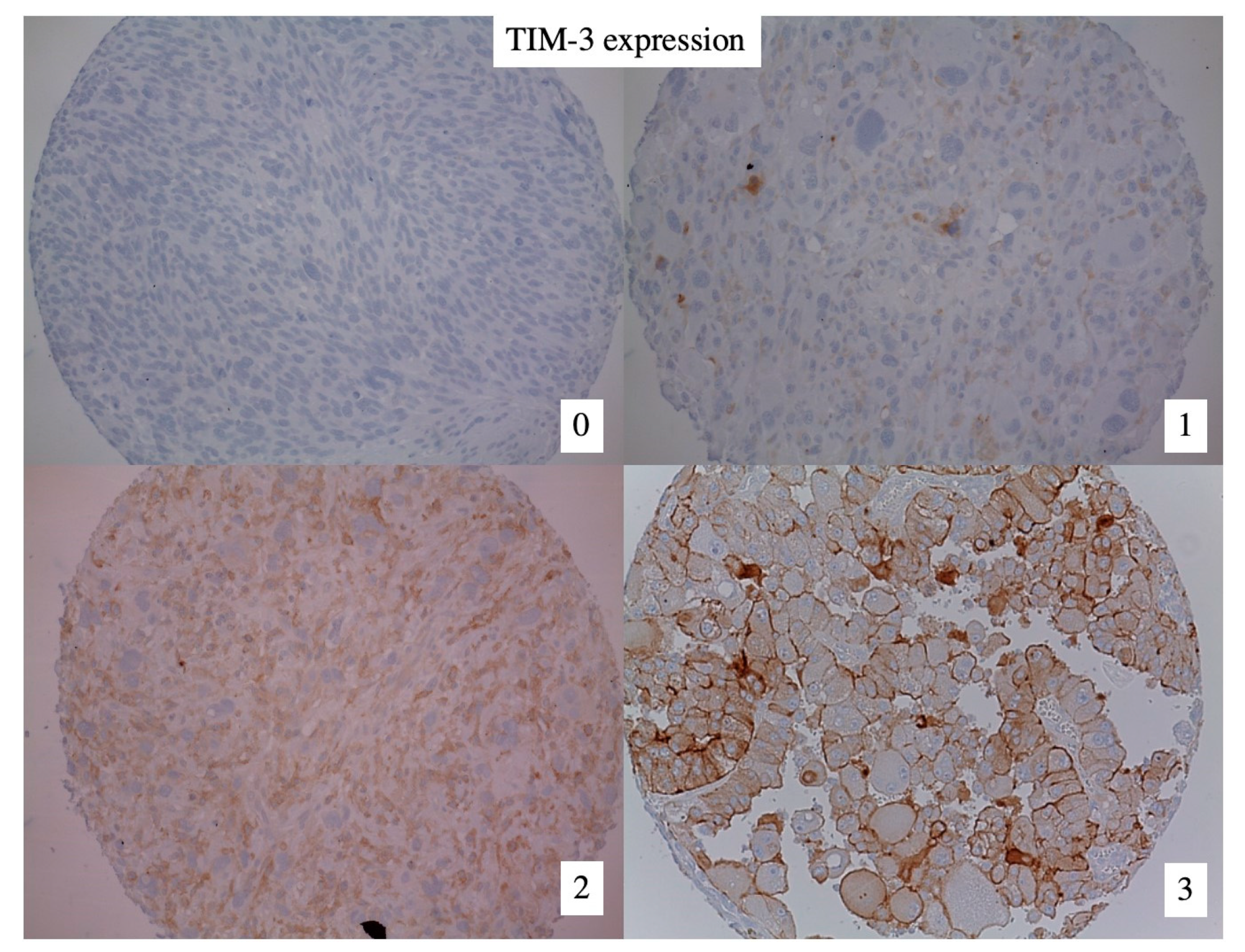
3.2. TIM-3 Expression in High-Risk Soft Tissue Sarcomas (HR-STS)

TIM-3 expression was observed in 101 (56%) out of 179 pre-treatment biopsies of patients with HR-STS. Examples of immunohistochemistry staining for TIM-3 are shown in Figure 1. TIM-3 was more often positive in male than female patients (64% vs. 48%, p = 0.036) and associated with older age (67% vs. 47%, p = 0.010). TIM-3 expression was more common in undifferentiated pleomorphic sarcomas (UPS) compared to other histological subtypes (75% vs. 47%, p < 0.001). There was no significant association between TIM-3 expression and FNCLCC grade (p = 0.229). A large proportion of patients received neoadjuvant anthracycline-based chemotherapy (80%), and nearly all patients underwent R0/R1 resection (n = 152, 89%) (Table 2).

3.3. TIM-3 Expression Is Associated with TILs, PD-1 and PD-L1 Expression Status

TIM-3 expression was associated with high tumor-infiltrating lymphocyte (TIL) counts (77% vs. 43%, p < 0.001), high positive PD-1 (60% vs. 30%, p < 0.001) and positive PD-L1 expression (91% vs. 47%, p < 0.001). We performed a logistic regression analysis of TIM-3 expression using an inclusion approach. Sex, age, increased TIL counts, PD-L1 expression and UPS histological subtype remained statistically significant predictors of TIM-3 expression (Table 3).

3.4. TIM-3 Expression and Survival

The median follow-up duration was 119 months (95% CI 109–128 months). In total, 71 patients (40%) died within 5 years after diagnosis. Statistically significant risk factors for an unfavorable outcome in univariate survival analysis were positive surgical margins (p < 0.001), grade (p = 0.015), presence of distant metastases (p < 0.001) and chemotherapy (p = 0.010) (Table 4). Expression of TIM-3 was not associated with statistically significant changes in overall survival (p = 0.339) (Figure 2).
Observed 5-year overall survival (OS) was not significantly influenced by TIM-3 expression in different histological subtypes (UPS (p = 0.207), leiomyosarcoma (p = 0.660), liposarcoma (p = 0.767), and other histological subtypes (p = 0.681)). All tested immune markers including TIM-3, PD-1, PD-L1 and tumor-infiltrating lymphocytes (TIL) did not have a statistically significant impact on 5-year OS in univariate analysis. In a multivariate Cox proportional hazards model, grade (p = 0.014), surgical margins (p < 0.001), and presence of distant metastases (p = 0.003) remained statistically significant independent predictors of 5-year OS. In conclusion, TIM-3 did not have a statistically significant prognostic impact on overall survival.

4. Discussion

To our knowledge, this is the first study to analyze the tumor cell expression of TIM-3, a novel immune checkpoint receptor and potential biomarker, in a well-characterized cohort of patients with HR-STS. TIM-3 expression was observed in 56% of patients. Our analysis indicates that patients with undifferentiated pleomorphic sarcomas (UPS), male gender, age ≥ 55 years and high expression of other immune markers (high TIL counts, positive PD-1 and PD-L1 expression) are more likely to demonstrate strong TIM-3 expression. These results remain significant in a logistic regression model, and indicate that specific subgroups of patients with HR-STS are more likely to express TIM-3.
We demonstrate the strong tumor cell expression of TIM-3 in undifferentiated pleomorphic sarcomas compared to other histological subtypes (75% vs. 47%, p < 0.001). UPS belong to non-translocation associated sarcomas and are associated with abundant immune infiltrates due to a higher mutational burden, higher neoantigen counts, and greater intratumoral heterogeneity compared to other entities [40]. Dancsok et al. described higher levels of PD-1, PD-L1 and TIM-3 expression on tumor-infiltrating lymphocytes in non-translocation-associated sarcomas including UPS [29]. In a study by Klaver et al., UPS had the highest fraction of PD-1+/LAG3+/TIM-3+/CD8+ T cell infiltrates, which was comparable to known “immune-dense” tumors such as malignant melanoma [41]. These findings correlate with clinical studies on immune checkpoint inhibitors in sarcomas, where UPS generally were among the best responders [14,17,42]. The strong expression of TIM-3 in UPS tumor cells supports the notion of an immunogenic signature in both tumor cells and immune infiltrates in this entity.
Our results suggest differences in TIM-3 expression according to age and sex. Reitsema et al. have provided evidence that both age and sex modulate the expression of immune checkpoints by human T cells [43]. Interestingly, their results described a decline in PD-1 expression with age and female sex, while our results demonstrate a stronger expression of TIM-3 in male patients ≥ 55 years of age. Age-related differences in immune checkpoint expression have shown direct effects on the treatment efficacy in other tumors, including head and neck cancer or malignant melanoma [44,45]. In consequence, age- and sex-associated differences in TIM-3 expression should be considered as relevant clinical parameters in ongoing clinical trials.
In addition to TIM-3, 60% of patients demonstrated a significant co-expression of PD-1. The expression of PD-L1 in combination with TIM-3 was observed in 91% of patients. In pre-clinical models, the co-expression of TIM-3 and PD-1 was observed in strongly dysfunctional T cells [25,27,46]. In addition, Koyama et al. demonstrated that TIM-3 can be upregulated as a result of PD-1-directed therapy [35]. With regard to these results, studies in murine models of melanoma, colorectal cancer and AML have analyzed checkpoint co-blockades, and demonstrated greater T cell responses following TIM-3 and PD-1 co-blockades compared to PD-1 inhibition alone [36,47,48]. In metastatic sarcomas, D’Angelo et al. have demonstrated increased response rates in co-blockades with anti-PD-1 and anti-CTLA4 antibodies, while anti-CTLA4 antibodies did not prove effective [17]. Our results provide an additional rationale for checkpoint co-blockades in high-risk soft tissue sarcomas, and support current clinical trials on combinations of anti-TIM-3 and anti-PD-1 antibodies in solid tumors.
We were not able to demonstrate differences in overall survival (OS) in TIM-3+ vs. TIM-3- patients with high-risk soft tissue sarcomas (p = 0.339). These results are in line with previous studies on TIM-3 in soft tissue and bone sarcomas: Ligon et al. analyzed tumor-infiltrating lymphocytes in osteosarcoma pulmonary metastases and compared them with primary bone tumors. While PD-L1 and LAG3 significantly predicted progression-free survival (PFS), there was no correlation between TIM-3 status and survival [30]. In addition, Dancsok et al. were not able to correlate TIM-3 expression on tumor-infiltrating lymphocytes of soft tissue and bone sarcomas with OS or PFS [29]. In contrast, a meta-analysis conducted by Zhang et al. reported significantly shorter OS rates and advanced tumor stages in patients with positive TIM-3 expression in various solid tumors including colon cancer, gastric cancer, renal cell carcinoma and non-small cell lung cancer (NSCLC) [28]. Furthermore, Wang et al. associated TIM-3 expression with a shorter OS in esophageal squamous cell carcinoma [49]. It is currently not clear why there seems to be no significant association between survival and TIM-3 expression in high-risk soft tissue sarcomas. Possible reasons could be the large number of histological subtypes and typically small sample size in rare tumors.
Our results demonstrate TIM-3 expression in tumor cells of patients with high-risk soft tissue sarcomas. These findings indicate that tumors with low tumor-infiltrating lymphocyte (TIL) counts can still express TIM-3 and perhaps benefit from future TIM-3 targeting therapies. Currently, there are only limited data on TIM-3 expression in tumor cells: Wiener et al. demonstrated the expression of TIM-3 in melanoma cells, and Zhuang et al. were able to detect TIM-3 in non-small cell lung cancer (NSCLC) [23,24]. In their study, TIM-3 stained positive on tumor cells in 86.7% of patients with primary NSCLC, and was associated with higher T classification and shorter OS. Interestingly, TIM-3 only stained positive in tumor cells and tumor-infiltrating lymphocytes, but not in normal (control) lung tissue, which adds to the current notion of TIM-3 playing an active role in carcinogenesis.
In addition to the typical limitations of a retrospective study design and immunohistochemical analyses, not all patients underwent the same treatment, which could have an impact on our survival analyses. In conclusion, new systemic therapy options are needed for high-risk soft tissue sarcomas. Immunotherapeutic approaches have become a cornerstone of modern oncology, with many drugs becoming approved for a variety of tumors. This study might help us to better select the patients with HR-STS who might express higher levels of TIM-3, and therefore be candidates for potential new clinical trials.

5. Conclusions

To date, checkpoint inhibitors have shown only limited efficacy in patients with high-risk soft tissue sarcomas (HR-STS). Selective TIM-3 blockade has demonstrated promising results in pre-clinical trials, and acts as a potential immunotherapeutic target in combination with established checkpoint inhibitors in ongoing clinical trials. This is the first study to demonstrate a significant tumor cell expression of TIM-3 in specific subsets of patients with HR-STS. We were able to correlate TIM-3 expression with high levels of tumor-infiltrating lymphocytes and PD-1/PD-L1 expression. Our results promote the identification of potential candidates for immunotherapy in HR-STS to expand therapeutic options and move on from the current “one-size-fits-all” paradigm in the therapy of advanced HR-STS.

Author Contributions

Conceptualization L.M.B., T.K. and L.H.L.; data curation L.M.B., A.A.-H. and T.K.; formal analysis L.M.B. and A.A.-H.; investigation L.M.B., A.A.-H. and T.K.; methodology L.M.B., A.A.-H. and T.K.; project administration T.K.; resources T.K.; software A.A.-H.; supervision L.H.L. and T.K.; validation T.K.; visualization A.A.-H.; writing—original draft L.M.B.; writing—review and editing A.A.-H., L.H.L., A.B.-M., D.D.G., H.R.D., A.K., M.A., N.-S.S.-H., F.K. and T.K. All authors have read and agreed to the published version of the manuscript.

Funding

This research received no external funding.

Institutional Review Board Statement

The Internal Review Board and the Ethical Review Committee at the Ludwig Maximilians University (LMU) Hospital, Munich, Germany, approved the protocol of this research project (Protocol Nr. 23-0113). All data were irreversibly anonymized.

Informed Consent Statement

Patient consent was waived for this analysis due to its retrospective design and irreversible anonymization of all data.

Data Availability Statement

The data presented in this study are available on specific request from the corresponding author. The data are not publicly available for reasons of data protection and data economy.

Conflicts of Interest

There are no conflicts of interest to disclose.

References

  1. Brennan, M.F.; Antonescu, C.R.; Moraco, N.; Singer, S. Lessons learned from the study of 10,000 patients with soft tissue sarcoma. Ann. Surg. 2014, 260, 416. [Google Scholar] [CrossRef] [PubMed] [Green Version]
  2. Siegel, R.L.; Miller, K.D.; Jemal, A. Cancer statistics, 2020. CA Cancer J. Clin. 2020, 70, 7–30. [Google Scholar] [CrossRef] [PubMed]
  3. Callegaro, D.; Miceli, R.; Bonvalot, S.; Ferguson, P.; Strauss, D.C.; Levy, A.; Griffin, A.; Hayes, A.J.; Stacchiotti, S.; Le Pechoux, C.; et al. Development and external validation of two nomograms to predict overall survival and occurrence of distant metastases in adults after surgical resection of localised soft-tissue sarcomas of the extremities: A retrospective analysis. Lancet Oncol. 2016, 17, 671–680. [Google Scholar] [CrossRef] [PubMed]
  4. Kane, J.M.; Magliocco, A.; Zhang, Q.; Wang, D.; Klimowicz, A.; Harris, J.; Simko, J.; Delaney, T.; Kraybill, W.; Kirsch, D.G. Correlation of High-Risk Soft Tissue Sarcoma Biomarker Expression Patterns with Outcome following Neoadjuvant Chemoradiation. Sarcoma 2018, 2018, 8310950. [Google Scholar] [CrossRef]
  5. In, G.K.; Hu, J.S.; Tseng, W.W. Treatment of advanced, metastatic soft tissue sarcoma: Latest evidence and clinical considerations. Ther. Adv. Med. Oncol. 2017, 9, 533–550. [Google Scholar] [CrossRef] [Green Version]
  6. Nielsen, O.; Judson, I.; van Hoesel, Q.; le Cesne, A.; Keizer, H.; Blay, J.; van Oosterom, A.; Radford, J.; Svancárová, L.; Krzemienlecki, K.; et al. Effect of high-dose ifosfamide in advanced soft tissue sarcomas. A multicentre phase II study of the EORTC Soft Tissue and Bone Sarcoma Group. Eur. J. Cancer 2000, 36, 61–67. [Google Scholar] [CrossRef]
  7. Maki, R.G.; Wathen, J.K.; Patel, S.R.; Priebat, D.A.; Okuno, S.H.; Samuels, B.; Fanucchi, M.; Harmon, D.C.; Schuetze, S.M.; Reinke, D.; et al. Randomized Phase II Study of Gemcitabine and Docetaxel Compared with Gemcitabine Alone in Patients with Metastatic Soft Tissue Sarcomas: Results of Sarcoma Alliance for Research Through Collaboration Study 002. J. Clin. Oncol. 2007, 25, 2755–2763. [Google Scholar] [CrossRef]
  8. Leahy, M.; del Muro, X.G.; Reichardt, P.; Judson, I.; Staddon, A.; Verweij, J.; Baffoe-Bonnie, A.; Jönsson, L.; Musayev, A.; Justo, N.; et al. Chemotherapy treatment patterns and clinical outcomes in patients with metastatic soft tissue sarcoma. The SArcoma treatment and Burden of Illness in North America and Europe (SABINE) study. Ann. Oncol. 2012, 23, 2763–2770. [Google Scholar] [CrossRef]
  9. Von Mehren, M.; Randall, R.L.; Benjamin, R.S.; Boles, S.; Bui, M.M.; Ganjoo, K.N.; George, S.; Gonzalez, R.J.; Heslin, M.J.; Kane, J.M.; et al. Soft Tissue Sarcoma, Version 2.2018, NCCN Clinical Practice Guidelines in Oncology. J. Natl. Compr. Cancer Netw. 2018, 16, 536–563. [Google Scholar] [CrossRef]
  10. Gronchi, A.; Miah, A.B.; Dei Tos, A.; Abecassis, N.; Bajpai, J.; Bauer, S.; Biagini, R.; Bielack, S.; Blay, J.Y.; Bolle, S.; et al. Soft tissue and visceral sarcomas: ESMO–EURACAN–GENTURIS Clinical Practice Guidelines for diagnosis, treatment and follow-up☆. Ann. Oncol. 2021, 32, 1348–1365. [Google Scholar] [CrossRef]
  11. Judson, I.; Verweij, J.; Gelderblom, H.; Hartmann, J.T.; Schöffski, P.; Blay, J.-Y.; Kerst, J.M.; Sufliarsky, J.; Whelan, J.; Hohenberger, P.; et al. Doxorubicin alone versus intensified doxorubicin plus ifosfamide for first-line treatment of advanced or metastatic soft-tissue sarcoma: A randomised controlled phase 3 trial. Lancet Oncol. 2014, 15, 415–423. [Google Scholar] [CrossRef]
  12. van der Graaf, W.T.; Blay, J.-Y.; Chawla, S.P.; Kim, D.-W.; Bui-Nguyen, B.; Casali, P.G.; Schöffski, P.; Aglietta, M.; Staddon, A.P.; Beppu, Y.; et al. Pazopanib for metastatic soft-tissue sarcoma (PALETTE): A randomised, double-blind, placebo-controlled phase 3 trial. Lancet 2012, 379, 1879–1886. [Google Scholar] [CrossRef]
  13. Demetri, G.D.; von Mehren, M.; Jones, R.L.; Hensley, M.L.; Schuetze, S.M.; Staddon, A.; Milhem, M.; Elias, A.; Ganjoo, K.; Tawbi, H.; et al. Efficacy and Safety of Trabectedin or Dacarbazine for Metastatic Liposarcoma or Leiomyosarcoma After Failure of Conventional Chemotherapy: Results of a Phase III Randomized Multicenter Clinical Trial. J. Clin. Oncol. 2016, 34, 786–793. [Google Scholar] [CrossRef] [Green Version]
  14. Tawbi, H.A.; Burgess, M.; Bolejack, V.; Van Tine, B.A.; Schuetze, S.M.; Hu, J.; D’Angelo, S.; Attia, S.; Riedel, R.F.; Priebat, D.A.; et al. Pembrolizumab in advanced soft-tissue sarcoma and bone sarcoma (SARC028): A multicentre, two-cohort, single-arm, open-label, phase 2 trial. Lancet Oncol. 2017, 18, 1493–1501. [Google Scholar] [CrossRef]
  15. Ben-Ami, E.; Barysauskas, C.M.; Solomon, S.; Tahlil, K.; Malley, R.; Hohos, M.; Polson, K.; Loucks, M.; Severgnini, M.; Patel, T.; et al. Immunotherapy with Single Agent Nivolumab for Advanced Leiomyosarcoma of the Uterus: Results of a Phase 2 Study. Cancer 2017, 123, 3285–3290. [Google Scholar] [CrossRef] [Green Version]
  16. Paoluzzi, L.; Cacavio, A.; Ghesani, M.; Karambelkar, A.; Rapkiewicz, A.; Weber, J.; Rosen, G. Response to anti-PD1 therapy with nivolumab in metastatic sarcomas. Clin. Sarcoma Res. 2016, 6, 24. [Google Scholar] [CrossRef] [Green Version]
  17. D’Angelo, S.P.; Mahoney, M.R.; Van Tine, B.A.; Atkins, J.; Milhem, M.M.; Jahagirdar, B.N.; Antonescu, C.R.; Horvath, E.; Tap, W.D.; Schwartz, G.K.; et al. Nivolumab with or without ipilimumab treatment for metastatic sarcoma (Alliance A091401): Two open-label, non-comparative, randomised, phase 2 trials. Lancet Oncol. 2018, 19, 416–426. [Google Scholar] [CrossRef]
  18. Toulmonde, M.; Penel, N.; Adam, J.; Chevreau, C.; Blay, J.-Y.; Le Cesne, A.; Bompas, E.; Piperno-Neumann, S.; Cousin, S.; Grellety, T.; et al. Use of PD-1 Targeting, Macrophage Infiltration, and IDO Pathway Activation in Sarcomas. JAMA Oncol. 2018, 4, 93–97. [Google Scholar] [CrossRef]
  19. Wolf, Y.; Anderson, A.C.; Kuchroo, V.K. TIM3 comes of age as an inhibitory receptor. Nat. Rev. Immunol. 2019, 20, 173–185. [Google Scholar] [CrossRef]
  20. Clayton, K.L.; Haaland, M.S.; Douglas-Vail, M.B.; Mujib, S.; Chew, G.M.; Ndhlovu, L.C.; Ostrowski, M.A. T Cell Ig and Mucin Domain–Containing Protein 3 Is Recruited to the Immune Synapse, Disrupts Stable Synapse Formation, and Associates with Receptor Phosphatases. J. Immunol. 2014, 192, 782–791. [Google Scholar] [CrossRef] [Green Version]
  21. van de Weyer, P.S.; Muehlfeit, M.; Klose, C.; Bonventre, J.V.; Walz, G.; Kuehn, E.W. A highly conserved tyrosine of Tim-3 is phosphorylated upon stimulation by its ligand galectin-9. Biochem. Biophys. Res. Commun. 2006, 351, 571–576. [Google Scholar] [CrossRef] [PubMed]
  22. Huang, Y.-H.; Zhu, C.; Kondo, Y.; Anderson, A.C.; Gandhi, A.; Russell, A.F.; Dougan, S.K.; Petersen, B.-S.; Melum, E.; Pertel, T.; et al. CEACAM1 regulates TIM-3-mediated tolerance and exhaustion. Nature 2015, 517, 386–390. [Google Scholar] [CrossRef] [PubMed] [Green Version]
  23. Wiener, Z.; Kohalmi, B.; Pocza, P.; Jeager, J.; Tolgyesi, G.; Toth, S.; Gorbe, E.; Papp, Z.; Falus, A. TIM-3 Is Expressed in Melanoma Cells and Is Upregulated in TGF-Beta Stimulated Mast Cells. J. Investig. Dermatol. 2007, 127, 906–914. [Google Scholar] [CrossRef] [PubMed] [Green Version]
  24. Zhuang, X.; Zhang, X.; Xia, X.; Zhang, C.; Liang, X.; Gao, L.; Zhang, X.; Ma, C. Ectopic Expression of TIM-3 in Lung Cancers. Am. J. Clin. Pathol. 2012, 137, 978–985. [Google Scholar] [CrossRef] [PubMed] [Green Version]
  25. Sakuishi, K.; Apetoh, L.; Sullivan, J.M.; Blazar, B.R.; Kuchroo, V.K.; Anderson, A.C. Targeting Tim-3 and PD-1 pathways to reverse T cell exhaustion and restore anti-tumor immunity. J. Exp. Med. 2010, 207, 2187–2194. [Google Scholar] [CrossRef]
  26. Zhu, C.; Sakuishi, K.; Xiao, S.; Sun, Z.; Zaghouani, S.; Gu, G.; Wang, C.; Tan, D.J.; Wu, C.; Rangachari, M.; et al. An IL-27/NFIL3 signalling axis drives Tim-3 and IL-10 expression and T-cell dysfunction. Nat. Commun. 2015, 6, 6072. [Google Scholar] [CrossRef] [Green Version]
  27. Fourcade, J.; Sun, Z.; Pagliano, O.; Chauvin, J.-M.; Sander, C.; Janjic, B.; Tarhini, A.A.; Tawbi, H.A.; Kirkwood, J.M.; Moschos, S.; et al. PD-1 and Tim-3 Regulate the Expansion of Tumor Antigen–Specific CD8+ T Cells Induced by Melanoma Vaccines. Cancer Res. 2014, 74, 1045–1055. [Google Scholar] [CrossRef] [Green Version]
  28. Zhang, Y.; Cai, P.; Liang, T.; Wang, L.; Hu, L. TIM-3 is a potential prognostic marker for patients with solid tumors: A systematic review and meta-analysis. Oncotarget 2017, 8, 31705–31713. [Google Scholar] [CrossRef] [Green Version]
  29. Dancsok, A.R.; Setsu, N.; Gao, D.; Blay, J.-Y.; Thomas, D.; Maki, R.G.; Nielsen, T.O.; Demicco, E.G. Expression of lymphocyte immunoregulatory biomarkers in bone and soft-tissue sarcomas. Mod. Pathol. 2019, 32, 1772–1785. [Google Scholar] [CrossRef]
  30. Ligon, J.A.; Choi, W.; Cojocaru, G.; Fu, W.; Hsiue, E.H.-C.; Oke, T.F.; Siegel, N.; Fong, M.H.; Ladle, B.; Pratilas, C.A.; et al. Pathways of immune exclusion in metastatic osteosarcoma are associated with inferior patient outcomes. J. Immunother. Cancer 2021, 9, e001772. [Google Scholar] [CrossRef]
  31. Harding, J.J.; Patnaik, A.; Moreno, V.; Stein, M.; Jankowska, A.M.; de Mendizabal, N.V.; Liu, Z.T.; Koneru, M.; Calvo, E. A phase Ia/Ib study of an anti-TIM-3 antibody (LY3321367) monotherapy or in combination with an anti-PD-L1 antibody (LY3300054): Interim safety, efficacy, and pharmacokinetic findings in advanced cancers. J. Clin. Oncol. 2019, 37, 12. [Google Scholar] [CrossRef]
  32. Ahn, M. MS28.02 Combination IO + IO. J. Thorac. Oncol. 2018, 13, S299–S300. [Google Scholar] [CrossRef] [Green Version]
  33. Murtaza, A.; Laken, H.; Correia, J.D.S.; McNeeley, P.; Altobell, L.; Zhang, J.; Vancutsem, P.; Wilcoxen, K.; Jenkins, D. Discovery of TSR-022, a novel, potent anti-human TIM-3 therapeutic antibody. Eur. J. Cancer 2016, 69, S102. [Google Scholar] [CrossRef]
  34. Burugu, S.; Dancsok, A.R.; Nielsen, T.O. Emerging targets in cancer immunotherapy. Semin. Cancer Biol. 2017, 52, 39–52. [Google Scholar] [CrossRef]
  35. Koyama, S.; Akbay, E.A.; Li, Y.Y.; Herter-Sprie, G.S.; Buczkowski, K.A.; Richards, W.G.; Gandhi, L.; Redig, A.J.; Rodig, S.J.; Asahina, H.; et al. Adaptive resistance to therapeutic PD-1 blockade is associated with upregulation of alternative immune checkpoints. Nat. Commun. 2016, 7, 10501. [Google Scholar] [CrossRef] [Green Version]
  36. Ngiow, S.F.; von Scheidt, B.; Akiba, H.; Yagita, H.; Teng, M.W.L.; Smyth, M.J. Anti-TIM3 Antibody Promotes T Cell IFN-γ–Mediated Antitumor Immunity and Suppresses Established Tumors. Cancer Res. 2011, 71, 3540–3551. [Google Scholar] [CrossRef] [Green Version]
  37. Knösel, T.; Emde, A.; Schlüns, K.; Chen, Y.; Jürchott, K.; Krause, M.; Dietel, M.; Petersen, I. Immunoprofiles of 11 Biomarkers Using Tissue Microarrays Identify Prognostic Subgroups in Colorectal Cancer. Neoplasia 2005, 7, 741–747. [Google Scholar] [CrossRef] [Green Version]
  38. Orth, M.F.; Buecklein, V.L.; Kampmann, E.; Subklewe, M.; Noessner, E.; Cidre-Aranaz, F.; Romero-Pérez, L.; Wehweck, F.S.; Lindner, L.; Issels, R.; et al. A comparative view on the expression patterns of PD-L1 and PD-1 in soft tissue sarcomas. Cancer Immunol. Immunother. 2020, 69, 1353–1362. [Google Scholar] [CrossRef]
  39. Albertsmeier, M.; Altendorf-Hofmann, A.; Lindner, L.H.; Issels, R.D.; Kampmann, E.; Dürr, H.-R.; Angele, M.K.; Klauschen, F.; Werner, J.; Jungbluth, A.A.; et al. VISTA in Soft Tissue Sarcomas: A Perspective for Immunotherapy? Cancers 2022, 14, 1006. [Google Scholar] [CrossRef]
  40. Thorsson, V.; Gibbs, D.L.; Brown, S.D.; Wolf, D.; Bortone, D.S.; Ou Yang, T.-H.; Porta-Pardo, E.; Gao, G.F.; Plaisier, C.L.; Eddy, J.A.; et al. The Immune Landscape of Cancer. Immunity 2018, 48, 812–830.e14. [Google Scholar] [CrossRef] [Green Version]
  41. Klaver, Y.; Rijnders, M.; Oostvogels, A.; Wijers, R.; Smid, M.; Grünhagen, D.; Verhoef, K.; Sleijfer, S.; Lamers, C.; Debets, R. Differential quantities of immune checkpoint-expressing CD8 T cells in soft tissue sarcoma subtypes. J. Immunother. Cancer 2020, 8, e000271. [Google Scholar] [CrossRef] [PubMed]
  42. Toulmonde, M.; Lucchesi, C.; Verbeke, S.; Crombe, A.; Adam, J.; Geneste, D.; Chaire, V.; Laroche-Clary, A.; Perret, R.; Bertucci, F.; et al. High throughput profiling of undifferentiated pleomorphic sarcomas identifies two main subgroups with distinct immune profile, clinical outcome and sensitivity to targeted therapies. Ebiomedicine 2020, 62, 103131. [Google Scholar] [CrossRef] [PubMed]
  43. Reitsema, R.D.; Cadena, R.H.; Nijhof, S.H.; Abdulahad, W.H.; Huitema, M.G.; Paap, D.; Brouwer, E.; Boots, A.M.H.; Heeringa, P. Effect of age and sex on immune checkpoint expression and kinetics in human T cells. Immun. Ageing 2020, 17, 32. [Google Scholar] [CrossRef] [PubMed]
  44. Kugel, C.H., III; Douglass, S.M.; Webster, M.R.; Kaur, A.; Liu, Q.; Yin, X.; Weiss, S.A.; Darvishian, F.; Al-Rohil, R.N.; Ndoye, A.; et al. Age Correlates with Response to Anti-PD1, Reflecting Age-Related Differences in Intratumoral Effector and Regulatory T-Cell Populations. Clin. Cancer Res. 2018, 24, 5347–5356. [Google Scholar] [CrossRef] [Green Version]
  45. Daste, A.; Domblides, C.; Gross-Goupil, M.; Chakiba, C.; Quivy, A.; Cochin, V.; de Mones, E.; Larmonier, N.; Soubeyran, P.; Ravaud, A. Immune checkpoint inhibitors and elderly people: A review. Eur. J. Cancer 2017, 82, 155–166. [Google Scholar] [CrossRef]
  46. Jin, H.-T.; Anderson, A.C.; Tan, W.G.; West, E.E.; Ha, S.-J.; Araki, K.; Freeman, G.J.; Kuchroo, V.K.; Ahmed, R. Cooperation of Tim-3 and PD-1 in CD8 T-cell exhaustion during chronic viral infection. Proc. Natl. Acad. Sci. USA 2010, 107, 14733–14738. [Google Scholar] [CrossRef] [Green Version]
  47. Liu, J.; Zhang, S.; Hu, Y.; Yang, Z.; Li, J.; Liu, X.; Deng, L.; Wang, Y.; Zhang, X.; Jiang, T.; et al. Targeting PD-1 and Tim-3 Pathways to Reverse CD8 T-Cell Exhaustion and Enhance Ex Vivo T-Cell Responses to Autologous Dendritic/Tumor Vaccines. J. Immunother. 2016, 39, 171–180. [Google Scholar] [CrossRef]
  48. Zhou, Q.; Munger, M.E.; Veenstra, R.G.; Weigel, B.J.; Hirashima, M.; Munn, D.H.; Murphy, W.J.; Azuma, M.; Anderson, A.C.; Kuchroo, V.K.; et al. Coexpression of Tim-3 and PD-1 identifies a CD8+ T-cell exhaustion phenotype in mice with disseminated acute myelogenous leukemia. Blood 2011, 117, 4501–4510. [Google Scholar] [CrossRef] [Green Version]
  49. Wang, P.; Chen, Y.; Long, Q.; Li, Q.; Tian, J.; Liu, T.; Wu, Y.; Ding, Z. Increased coexpression of PD-L1 and TIM3/TIGIT is associated with poor overall survival of patients with esophageal squamous cell carcinoma. J. Immunother. Cancer 2021, 9, e002836. [Google Scholar] [CrossRef]
Figure 1. Stained tissue microarray (TMA) cores. Representative micrographs of cores on a TMA stained for TIM-3. Numbers represent semiquantitative scoring of immunostaining: 0, negative. 1–3, positive. Magnification, 20×.
Cancers 15 02735 g001
Figure 2. Overall survival according to TIM-3 expression.
Cancers 15 02735 g002
Table 1. Patient characteristics.
FactorStratan%
Total 179100
SexMale9251
Female8749
Histological subtypeUPS5933
Liposarcoma4022
Leiomyosarcoma3117
Synovial sarcoma1810
MPNST127
Angiosarcoma53
Malignant SFT21
Dediff. chondrosarcoma32
Myxofibrosarcoma53
Other42
LocationExtremities7140
Non-Extremities10860
Size of primary tumor (cm)50–79 mm4626
80–120 mm6235
>120 mm7140
Presence of metastasesNo16793
Yes127
FNCLCC GradeIntermediate (G2)8950
High (G3)9050
TIM-3 expression (Grades 0–3)07844
15631
23721
384
Follow-up status 5 years after initial diagnosisAlive10860
Dead7140
Local recurrence within 5 years after R0/R1 resectionNo local recurrence9160
Local recurrence6140
Distant recurrence within 5 years after R0/R1 resectionNo distant recurrence10368
Distant recurrence4932
UPS: Undifferentiated Pleomorphic sarcoma. SFT: Solitary fibrous tumor. MPNST: Malignant peripheral nerve sheath tumor. Other: 1 rhabdomyosarcoma, 1 alveolar soft part sarcoma, 1 carcinosarcoma, 1 extraosseous osteosarcoma.
Table 2. Correlation between TIM3 expression and clinicopathological parameters.
FactorStrataTotalTIM-3 > 0p-Value
nn%
All Patients 17910156--
SexMale9259640.036
Female874248
Age at initial diagnosis (years)<559243470.010
≥55875867
Histological subtypeUPS594475<0.001
Liposarcoma311135
Leiomyosarcoma402665
Other492041
Tumor LocationExtremities7147660.045
Non-extremities1085450
FNCLCC GradeIntermediate (G2)8946520.229
High (G3)905561
Surgical marginsR06948700.011
R1834149
R214429
No resection13862
ChemotherapyYes13480600.164
No452147
RadiotherapyYes3016530.535
No1064845
Missing43
Regional Hyperthermia (RHT)Yes13986620.007
No401538
TIL counts (cells/50HPF)0–51084643<0.001
≥6705477
Missing1
PD-1 expression0611830<0.001
≥0774660
Missing41
PD-L1 expression01396647<0.001
≥0343191
Table 3. Multiple logistic regression model of relevant clinicopathological parameters.
FactorStrataSignificanceHazard Ratio95.0% CI
SexMale vs. Female0.0262.289(1.106–4.737)
Age<55 vs. ≥550.0271.030(1.003–1.056)
TIL counts0–5 vs. ≥60.0023.499(1.565–7.823)
PD-L1 expression0 vs. >00.0019.173(2.420–34.772)
HistologyUPS vs. other subtypes0.0382.316(1.046–5.128)
Table 4. Univariate and multivariate analysis of overall survival.
UnivariateMultivariate
FactorStrataSig.Hazard RatioSig.Hazard Ratio
SexMale vs. Female0.3660.806 (0.505–1.287)
Age1 year step0.6781.003 (0.987–1.020)
GradeG2 vs. G30.0151.812 (1.122–2.926)0.0141.889 (1.139–3.133)
Surgical marginsR0/1 vs. R2<0.0017.310 (4.339–12.318)<0.0016.866 (3.815–12.357)
Distant metastasesM0 vs. M1<0.0014.187 (2.119–8.273)0.0033.059 (1.476–6.341)
PD-L1 expression0 vs. >00.1801.455 (0.840–2.520)0.5421.227 (0.636–2.364)
TIL counts0–5 vs. ≥60.8301.055 (0.649–1.713)0.2471.406 (0.790–2.502)
TIM3 expression0 vs. >00.3420.798 (0.501–1.271)0.2461.403 (0.792–2.483)
HistologyUPS vs. other0.2590.759 (0.470–1.226)
Tumor locationExtremities vs. non-Extremities0.2851.302 (0.802–2.112)
ChemotherapyYes vs. no0.0101.912 (1.168–3.129)0.4981.212 (0.695–2.114)
RadiotherapyYes vs. no0.2411.440 (0.783–2.647)
Regional hyperthermiaYes vs. no0.7491.091 (0.639–1.865)
PD1 expression0 vs > 00.1061.521 (0.914–2.530)
TIM-3 x PDL1Both 0 vs. both >00.6901.133 (0.613–2.095)
TIL x TIM-3TIL ≥ 6 and TIM-3 > 0 vs.
TIL < 6 and TIM-3 = 0
0.5990.848 (0.459–1.566)
TIM-3 x PD1Both 0 vs. both >00.3231.372 (0.733–2.569)
Disclaimer/Publisher’s Note: The statements, opinions and data contained in all publications are solely those of the individual author(s) and contributor(s) and not of MDPI and/or the editor(s). MDPI and/or the editor(s) disclaim responsibility for any injury to people or property resulting from any ideas, methods, instructions or products referred to in the content.

