Review

Genetics of ABCB1 in Cancer

by
Katie T. Skinner
1,2,
Antara M. Palkar
1,2 and
Andrew L. Hong
1,2,3,*
1
Department of Pediatrics, Emory University School of Medicine, Atlanta, GA 30322, USA
2
Aflac Cancer and Blood Disorders Center, Children’s Healthcare of Atlanta, Atlanta, GA 30322, USA
3
Winship Cancer Institute, Emory University School of Medicine, Atlanta, GA 30322, USA
*
Author to whom correspondence should be addressed.
Cancers 2023, 15(17), 4236; https://doi.org/10.3390/cancers15174236
Submission received: 14 July 2023 / Revised: 10 August 2023 / Accepted: 22 August 2023 / Published: 24 August 2023

Abstract

:

Simple Summary

Overexpression of ABCB1 has been identified in a wide range of multidrug-resistant cancers. ABCB1 can become upregulated in many ways, and understanding these mechanisms of upregulation could provide novel insights into cancer multidrug resistance. In this review, we summarize genetic and epigenetic mechanisms of ABCB1 upregulation in cancer and highlight areas that may be relevant for future research.

Abstract

ABCB1, also known as MDR1, is a gene that encodes P-glycoprotein (P-gp), a membrane-associated ATP-dependent transporter. P-gp is widely expressed in many healthy tissues—in the gastrointestinal tract, liver, kidney, and at the blood–brain barrier. P-gp works to pump xenobiotics such as toxins and drugs out of cells. P-gp is also commonly upregulated across multiple cancer types such as ovarian, breast, and lung. Overexpression of ABCB1 has been linked to the development of chemotherapy resistance across these cancers. In vitro work across a wide range of drug-sensitive and -resistant cancer cell lines has shown that upon treatment with chemotherapeutic agents such as doxorubicin, cisplatin, and paclitaxel, ABCB1 is upregulated. This upregulation is caused in part by a variety of genetic and epigenetic mechanisms. This includes single-nucleotide variants that lead to enhanced P-gp ATPase activity without increasing ABCB1 RNA and protein levels. In this review, we summarize current knowledge of genetic and epigenetic mechanisms leading to ABCB1 upregulation and P-gp-enhanced ATPase activity in the setting of chemotherapy resistance across a variety of cancers.

1. Introduction

The ATP-binding cassette (ABC) family of transporters uses energy in the form of ATP to transport substrates against a concentration gradient. One well-studied member of this family is ABCB1, which encodes P-glycoprotein (P-gp). The role of P-gp in healthy tissue is well established, where it has been found to be expressed on organs and tissues that have roles in the detoxification of xenobiotics and a wide variety of drugs, including anti-cancer drugs [1]. Many chemotherapeutic and targeted agents, such as daunorubicin, docetaxel, doxorubicin, etoposide, imatinib, mitoxantrone, paclitaxel, sunitinib, teniposide, topotecan, vinblastine, and vincristine [2,3] have been established as P-gp substrates, and overexpression of this membrane-bound efflux pump prevents accumulation of these drugs within cells, mediating the development of resistance. Inappropriate upregulation of P-gp in cancer cells leads to the development of therapy-resistant cancers.
In this review, we summarize current knowledge on P-gp structure, function, localization, and substrate specificity. We also focus on the genetics of ABCB1, reviewing publicly available data to understand the prevalence of ABCB1 alterations across different cancers, alongside common mechanisms by which ABCB1 has been found to be upregulated in multidrug-resistant cancers.

2. Overview of ABC Family of Transporters
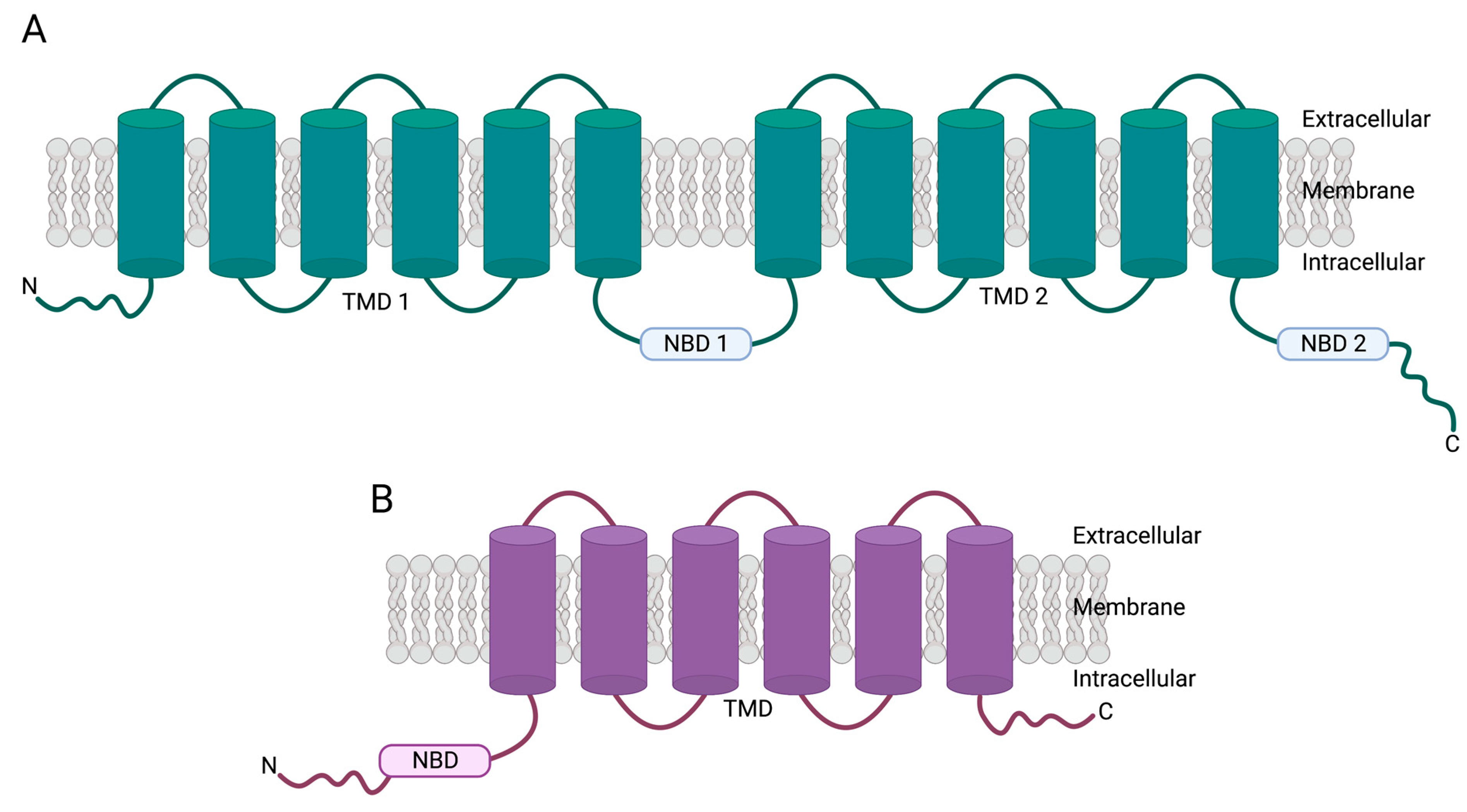
ABCB1 is a member of the ATP-binding cassette (ABC) family of transporter proteins [4]. P-gp, encoded by ABCB1, was first identified in 1976 by Juliano and Ling [5]. In this study, researchers previously isolated Chinese hamster ovarian tissue cells that demonstrated resistance to colchicine, an anti-inflammatory drug. They also observed that these cells were resistant to other non-related compounds such as vinblastine and puromycin [5]. Surface labeling studies revealed a 170 kDa component on the cell surface that was not present on non-resistant wild-type cells [5]. Upon metabolic labeling studies, the component was identified as a glycoprotein [5]. As these mutant cells displayed a decreased permeability, this component was henceforth named “permeability glycoprotein” or P-glycoprotein (P-gp); however, later studies indicated that P-gp functions as an efflux pump, rather than altering permeability status in cells [6,7,8]. In 1985, the ABCB1 gene was cloned, and, using this cDNA as a probe, researchers were able to show that ABCB1 DNA was amplified in the vinblastine-resistant leukemia cell line CEM/VLB compared to its parental counterpart, CCRF-CEM [9].
More broadly, this family of transporters consists of seven sub-families: ABCA to ABCG [4,10]. Within these seven sub-families, there are, to date, forty-eight transporters [4,11]. An additional gene in the ABC family, ABCC13, is expected to produce a nonfunctional protein [12]. One way ABC transporters can be grouped is by structure, whereby they exist as either full or half transporters [13]. Generally, full transporters consist of two transmembrane domains (TMDs) and two nucleotide-binding domains (NBDs) [13] (Figure 1A). On the other hand, half transporters consist of one TMD and one NBD, meaning to function correctly, half transporters are required to form homodimers or heterodimers [14,15,16,17] (Figure 1B).

3. ABCB1 Genetics

The ABCB1 gene is located on chromosome 7q21.12 [NCBI, Gene ID: 5243, GRCh38.p13; Ensembl, Gene ID: ENSG00000085563, GRCh38.p13] and has been found to have two promoters—a proximal “downstream” promoter, and a distal “upstream” promoter [18]. This was first characterized by Chen et al., with the proximal promoter being located in the first exon, and the distal promoter in the second exon [18]. Transcripts derived from the upstream promoter will have 29 exons, and those derived from the downstream promoter will have 28 exons [18] (Figure 2). The literature suggests that the 28 exon ABCB1 is most commonly produced by normally functioning cells, whereas drug-resistant cancer cells might more frequently produce the slightly longer 29 exon form [19,20,21,22]. This is discussed in more depth in Section 5.7. Between NCBI and Ensembl, the 28 exon, 1280 amino acid protein (Table 1, NM_001348946.2 and ENST00000622132.5) is the only isoform to be shared between the two databases.
Besides the one shared database entry (Table 1, NM_001348946.2 and ENST00000622132.5), there are discrepancies regarding mRNA and protein isoforms listed between the NCBI and Ensembl databases. For example, NCBI lists two isoforms containing 30 and 32 exons (Table 1, NM_001348944.2 and NM_001348945.2, respectively) that are not found in Ensembl. These contain additional exons upstream of the ABCB1 promoters identified by Chen et al. These two sequences (Table 1, NM_001348944.2 and NM_001348945.2, respectively) were derived from bacterial artificial chromosomes, and, other than their listing in NCBI, there is no additional evidence in the literature supporting the existence of either variant; furthermore, Pappas et al. identified three hypothetical exons at the 3′ end of the ABCB1 gene. They used RT-PCR to determine whether these hypothetical exons existed, and found no evidence that they did [23]. Taking these data into consideration, additional research is needed to support the NM_001348944.2 and NM_001348945.2 isoforms listed on NCBI. Similarly, in Ensembl, there is a truncated ABCB1 mRNA/protein listed that is not found in NCBI (Table 1, ENST00000416177.1). There is also no evidence of this isoform in the literature, and thus more research should be conducted to verify its validity.

4. ABCB1 in Normal Tissues

4.1. Tissue Localization, Subcellular Localization, and Substrates

Given the role of P-gp as an efflux pump, it is therefore unsurprising that ABCB1 is expressed on tissues with detoxification and/or secretory roles (Table 2). Early studies utilizing slot blot analysis of normal human tissues found that ABCB1 was expressed on the adrenal gland, kidneys, colon, rectum, lungs, and liver [24]; other tissues, such as the brain, prostate, skin, heart, esophagus, and stomach were all found to have lower ABCB1 expression [24]. Characterization of P-gp tissue localization at the protein level via immunohistochemistry confirmed these findings [25]. Of all the cell types where P-gp was found to be expressed, expression typically occurred on the apical surface, facing towards the excretory compartments of each of these organs [25], which is consistent with P-gp function as an efflux pump. P-gp has a long list of substrates that are still being established (summarized in Table 2). At organs such as the kidneys, liver, and colon, P-gp will pump xenobiotics, such as pesticides, and drugs such as antibiotics, calcium channel blockers, and protease inhibitors into compartments such as the proximal tubule [26,27], bile ducts [26,28], and intestinal lumen [29,30] to be removed. This function promotes the elimination of these substances from the blood and prevents them from circulating in the body, potentially causing damage. P-gp has also been found to transport hormones and steroids (Table 2), which is consistent with its high expression on the adrenal gland [24,25]. It is thought that P-gp contributes to the regulation of hormone secretion, thus modulating hormone signaling [31,32,33].
Although earlier studies did not find ABCB1 expression within the central nervous system (regions such as the cerebral cortex, cerebellum, and spinal cord), it was later discovered that P-gp was indeed expressed on endothelial cells located at the blood–brain barrier [34]. Here, it plays an important role in protecting the brain against xenobiotics and drugs [58], such as those outlined in Table 2. Along with protecting the brain from these compounds, P-gp also has roles in the development of neurological disorders. For example, amyloid-β, the protein thought to be causative of Alzheimer’s disease, has been found to be a substrate of P-gp in humans [56], and reduced ABCB1 expression in mice led to an accumulation of amyloid-β in the brains of these animals [59,60,61].
Lastly, research also indicates that ABCB1 expression is prevalent in the placenta and has important roles in protecting the fetus from compounds that may impact development, such as HIV protease inhibitors [62,63], chemotherapeutic agents [63], stress hormones, and steroids [55,62]. For example, human placental tissue obtained between 6 and 10 weeks gestation had significantly higher ABCB1 mRNA expression compared to tissue obtained between 38 and 41 weeks gestation [35]. This is consistent with a protective role for P-gp, as the fetus needs to be protected from harmful compounds during the early stages of development when it is most vulnerable.
In terms of subcellular localization, P-gp is most commonly found on the cell surface, more specifically within the plasma membrane [36,37,64]. This is consistent with its function as an efflux pump. Moreover, a few initial studies suggest that P-gp could localize intracellularly. One study found that in blood–brain endothelial cells, P-gp was expressed on the nuclear envelope in both human and rat brains [64]. Here, the authors proposed that P-gp functions to protect the nucleus and prevent DNA-drug interactions that might cause damage [64]; similarly, it has been proposed that P-gp could be expressed in the mitochondria of three different doxorubicin-resistant cancer cells lines: K562 (leukemia), PLC/PRF/5 (hepatocellular carcinoma), and MCF7 (breast). In K562 cells, researchers found that P-gp may transport substrates into the mitochondria, and they proposed that this organelle acted as a drug sequestration compartment that protected the nucleus and DNA from these toxins [65]. In PLC/PRF/5 and MCF7, however, it was found that P-gp transported substrates from the mitochondria into the cytoplasm, which is more consistent with the protective role as previously described in this review [57,66], although these studies have not been further investigated in recent years and as such, additional research is needed.

4.2. P-gp Mechanism of Action

It has been long established that P-gp is able to utilize ATP to transport substrates against a concentration gradient [67,68,69,70]. The exact mechanism by which P-gp transports substrates, however, is still under debate. Possible mechanisms for P-gp pumping include (1) direct extraction of substrates from the cytoplasm, (2) functioning as a flippase to remove substrates from the inner leaflet of the plasma membrane, or (3) extracting substrates from the outer leaflet to prevent them from reaching the cytoplasm.
Direct extraction proposes that P-gp functions similarly to ion-transporting ATPases, hydrolyzing ATP to maintain ion gradients across cell membranes, and extracting substrates directly from the cytoplasm [71]. In the flippase model, P-gp moves its substrates from the inner to the outer leaflet of the cell membrane [71,72,73]. In this mechanism, the substrates need to be specifically localized within each leaflet of the membrane. Once in the outer leaflet, substrates could passively diffuse into the extracellular aqueous phase or spontaneously move back to the inner leaflet. This flippase activity also requires ATP hydrolysis and is inhibited by certain substances that also inhibit drug transport by P-gp, suggesting that drugs and membrane lipids likely follow the same route through the transporter [73]. The final model suggests that P-gp extracts substrates from the outer leaflet to prevent them from reaching the cytoplasm. Experiments using fluorescent indicators and photolabeling techniques have indicated that P-gp interacts with drug molecules within the membrane, leading to the concept of P-gp as eliminating potentially harmful lipophilic compounds from the membrane [74,75]. Subsequent studies have indicated that the rate of drug efflux by P-gp is linked to the membrane concentration of the drug and inversely related to its concentration in the aqueous phase [76,77]. Experiments using a deletion mutant of P-gp have demonstrated that the transporter’s transmembrane domains alone are sufficient to bind drug substrates [78].
There is still debate as to whether (1) ATP binding and subsequent NBD dimerization or (2) ATP hydrolysis is the force driving the P-gp conformation change [67,68,69,70]. Studies addressing this question have often faced criticism due to the use of physiologically irrelevant conditions. More recent studies have tried to address these issues by studying P-gp function in near-physiological conditions [79] (i.e., in a lipid bilayer and at 37 °C) as well as in living cells [80]. While a consensus remains to be reached, advances in FRET technology, such as FRET-based biosensors, will likely enable us to underpin this mechanism sooner rather than later.

5. ABCB1 in Cancers

5.1. P-gp Substrates: Chemotherapies and Targeted Therapies

As outlined in Section 2, it has been known for a long time that P-gp overexpressing cells are resistant to chemotherapeutic agents such as vinblastine [5,9]. Early studies that sought to establish substrates of P-gp used a photoaffinity labeling technique. Here, photoactive radiolabeled analogs of colchicine, a known P-gp substrate, were co-incubated with chemotherapeutic agents, and the efficiency of photolabeling was assessed [81], with the idea being a compound was a P-gp substrate if it competitively reduced photolabeling. The results of such an experiment indicated that agents such as vincristine, vinblastine, doxorubicin, and actinomycin D were all P-gp substrates, whereas methotrexate was not [81]. In parallel, researchers examined a large number of leukemia and lymphoma cell lines that had never been exposed to chemotherapeutic agents in culture, and tested for P-gp expression via the monoclonal antibody MRK16 [82]. Utilizing the cell lines K562 (doxorubicin-sensitive) and K562/ADM (doxorubicin-resistant) as controls, they identified three cell lines (KYO-1, HEL, and CMK) to have P-gp expression [82]. These three cell lines, as well as K562/ADM, were all found to be resistant to vincristine, vindesine, vinblastine, doxorubicin, daunorubicin, mitoxantrone, etoposide, and actinomycin-D [82]. Other cell lines with no P-gp displayed resistance to some compounds but not others, indicating P-gp-independent mechanisms of resistance [82] (potentially due to the expression of other ABC transporters, such as MRP1 and BCRP, which have an affinity for some of the same substrates as P-gp, however, this requires further investigation [83]). Nowadays, many cell lines and primary tumors have been tested for both P-gp expression and resistance to these agents, whereby a positive correlation has been established (i.e., high P-gp expression is associated with more chemotherapy resistance) across many different cancer types such as breast and ovarian cancers [84], multiple myeloma [85], osteosarcoma [86], and lung cancer [87] to name a few.
As for targeted therapies, Lee et al. conducted a high-throughput screening with the goal of identifying cytotoxic substrates of P-gp. They used the HeLa-derived cell line KB-3-1, and the colchicine-resistant subline KB-8-5-11 [88]. From this screening of 10,804 compounds, they identified 90 as potential substrates [88]. To confirm these hits, they performed IC50 experiments using both ovarian cell lines, as well as P-gp overexpressing HEK 293T cells, in the presence and absence of the P-gp inhibitor tariquidar. From these experiments, they confirmed multiple targeted therapies as substrates including but not limited to inhibitors of the PI3K/AKT pathway (gedatolisib and GSK-690693); cell cycle checkpoint inhibitors (AT7159 and ispinesib); and a Janus kinase 2/3 inhibitor (AT9283) [88]. Using this screening method, substrates of other ABC transporters can be identified. This information will help us to understand ABC transporter redundancy and/or compensation better, as well as cancer therapy resistance.

5.2. Prevalence of ABCB1 Dysregulation in Cancer

To explore the prevalence of ABCB1 dysregulation in cancer, we utilized the NCI’s publicly available GDC Data Portal [89]. Across 38 different projects, ABCB1 mutations were found in 508 out of a total of 13,106 cases (prevalence of approximately 3.9%). Of these 508 patients, 472 had age data, whereby approximately 97% were over the age of 18, illustrating the lack of knowledge we currently have regarding ABCB1 dysregulation in pediatric cancers. ABCB1 mutations occurred most frequently in lung, skin, and uterine malignancies (Figure 3A). Of all 508 cases, 550 total mutations were identified. Of these mutations, almost 70% were missense (Figure 3B), with the most common missense mutation being ABCB1 R467W (frequency of 1.8% in a cohort of patients with an ABCB1 mutation). Interestingly, 2 of the 459 adult cases were derived from recurrent tumors, each with a different ABCB1 missense mutation: ABCB1 A252E (lung) and ABCB1 A599T (brain). Although it is possible that the presence of these mutations is not significant, the functional consequences of these SNPs are unknown, and further study would be required to determine whether they play a role in the tumorigenesis and recurrence of these cancers.
As for gene level copy number gains, across 33 projects, 3345 out of 10,785 total cases had copy number gains of the ABCB1 gene (frequency of 31%). Malignancies that saw the highest frequency of gains originated in the testis, brain, kidneys, adrenal gland, and intestine (Figure 3A). Of these cases, 34 were recurrent tumors, with 30 harboring an ABCB1 copy number gain, and 4 harboring an ABCB1 copy number loss. Of these 34, 30 received treatment, and of these 30, 28 had copy number gains of ABCB1, with 2 cases harboring ABCB1 copy number losses. Currently, it is unclear if increases in ABCB1 gene expression mirror this increase in gene level copy number. As a result, it remains unknown what the functional significance of these copy number changes is. Additional investigation is required to ascertain the significance of ABCB1 gene level copy number increases and how this might relate to cancer therapy resistance.
As aforementioned, much of this data occurs in adult patients, with little to no data on pediatric cases; additionally, the role of ABCB1 in pediatric cancer has also not yet been characterized, so future research should focus on understanding the influence of ABCB1 mutations or copy number gains in this cohort.

5.3. Structural Variants Leading to ABCB1 Upregulation: Chromosome 7 Amplifications

Aneuploidies, or alterations in chromosomal copy number, are commonly found in cancer. Typically, regions containing oncogenes are found to be amplified, and regions containing tumor suppressor genes are lost. As it became more established that ABCB1 was linked to multidrug resistance, researchers sought to understand how the gene was becoming overexpressed. A study by Wang et al. generated 11 multidrug-resistant sublines from a total of 6 ovarian cancer cell lines. These lines were resistant to either paclitaxel or docetaxel [90]. Using cDNA microarrays, they found that a cluster of genes located at chromosome 7q21.11-13 was overexpressed in nine of the resistant lines [90]. In six of these nine lines, they found evidence of copy number amplifications of this region and attributed this to the increased expression of this gene cluster, which included ABCB1 [90]. Similarly, Yabuki et al. generated a paclitaxel-resistant subline of the non-small cell lung carcinoma cell line NCI-H460 and observed both overexpression of ABCB1 and regional amplifications of chromosome 7p21.12, 11- to 13-fold higher than seen in the parental, non-resistant cell line. Analogous findings have also been found in the docetaxel-resistant breast cancer cell line MCF7 [21], and the doxorubicin-resistant leukemic cell line, K562 [91]. This research shows that ABCB1 overexpression can be due to chromosome 7 amplifications across multiple different cancer types that are resistant to different chemotherapeutic agents.

5.4. Structural Variants Leading to ABCB1 Upregulation: Gene Fusions

As well as chromosome amplifications, transcriptional fusions of the N-terminus of ABCB1 to a truncated C-terminus of another gene, resulting in ABCB1 upregulation have been identified. In 2015, Patch et al. conducted a whole-genome sequencing analysis of 92 chemoresistant ovarian cancer patient samples, with additional primary tumor tissue that was chemo-sensitive. From this analysis, they found a transcriptional fusion between ABCB1 and the solute carrier, SLC25A40 in two patients [92]. This fusion placed ABCB1 under the SLC25A40 promoter, causing upregulation in ABCB1 and downregulation in SLC25A40, due to deletion of the rest of the gene [92]. Researchers confirmed that this fusion was not present in the primary, chemo-sensitive, tumors, and stated that these patients had failed to respond to treatments containing P-gp substrates [92]. The same group then sought to characterize this fusion in an even larger ovarian cohort comprising patients with recurrent disease, as well as a smaller group of breast cancer patients [93]. Analysis of this cohort found additional fusions of ABCB1 with other genes such as PRRC2C, ARPC1B, and CNOT4 in ovarian tissue, and NRF1 and TPX2 in breast tissue, alongside the original SLC25A40-ABCB1 fusion identified in the prior study [93]. Many of these novel fusions were found to co-occur with the original SLC25A40-ABCB1 fusion [93]. The majority of these fusions were characterized by a fusion of a non-coding exon of the partner gene, fused to exon 2 onwards of ABCB1, leaving most of the full gene to be transcribed and translated into a functioning protein [93] (Figure 4A). The presence of ABCB1 gene fusions correlated with the number of lines of chemotherapy a patient had received, with the more therapy given to a patient, the more likely it was that a fusion was observed [93]. These two studies provide evidence that, at least in ovarian and breast cancer, ABCB1 gene fusions that retain exon 2 onwards lead to the upregulation of P-gp and are responsible for acquired chemotherapy resistance. This has not yet been established for other cancer types and more research should be conducted to determine whether fusions occur in other malignancies and as a result of different chemotherapy regimens.

5.5. SNPs Leading to Changes in ABCB1 Expression

As well as structural variants, single-nucleotide polymorphisms (SNPs) can impact the expression of ABCB1. Many of the studies investigating ABCB1 SNPs are simply correlative, where they establish whether the presence of an SNP leads to better or worse overall survival [94,95,96,97,98]. From studies such as these, it is difficult to discern whether the presence of the SNP is related to changes in ABCB1 expression or efflux activity, with the possibility that the SNP is completely unrelated to either of those outcomes. Despite this, prior studies have assessed how variants may affect P-gp expression. For example, a study published by Mansoori et al. found that SNPs rs28381943 and rs2032586 (Figure 4B) led to an increase in ABCB1 mRNA stability in gastric cancer, and therefore increased P-gp expression [99]. Interestingly, both SNPs are intronic, and thought to disrupt splicing, resulting in the retention of intron 19 and 11, respectively [99] (Figure 4B). Moreover, the synonymous SNP ABCB1 C3435T (Ile1145Ile, isoform 2) (Figure 4B) has been studied extensively and there is currently conflicting evidence as to whether it increases or decreases ABCB1 mRNA expression. Studies looking at this SNP in the context of intestinal [100], liver [101], and kidney [102] P-gp found expression of ABCB1 to be lower in individuals harboring the TT genotype compared to the CC genotype. It has been predicted that the C>T substitution changes the secondary structure of the mRNA [101], therefore leading to lower mRNA stability and thus lower levels. Conversely, another study looking at the same SNP (ABCB1 C3435T) also in the context of intestinal P-gp found that the TT genotype conferred higher P-gp expression compared to the CC genotype [103]. The differences between these findings could stem from the fact that intestinal P-gp was studied in two different populations, with Hoffmeyer et al.’s study being conducted in a Caucasian population [100] and Nakamura et al.’s study being conducted in a Japanese population [103]. This indicates that this association between ABCB1 C3435T and P-gp expression is more complex than a simple one-to-one correlation. It is also important to note that C3435T is a synonymous SNP, encoding isoleucine regardless of a C or T at position 3435. It is still not fully understood how exactly this single nucleotide change is able to impact the stability of the ABCB1 mRNA, and more research needs to focus on understanding this in normal tissues before we can move on to trying to understand its role in cancer chemotherapy resistance.

5.6. SNPs Leading to Changes in ABCB1 ATPase Activity

ABCB1 SNPs have also been associated with changes in P-gp efflux activity. While there is much debate on whether ABCB1 C3435T influences P-gp expression levels, there is a more unified consensus regarding its impact on P-gp efflux activity. A study utilizing leukocytes from 31 healthy donors representing the three genotypes of ABCB1 C3435T (CC, CT, TT) found that rhodamine 123 efflux was highest in individuals with the CC genotype, followed by the heterozygote CT, and lastly, the TT genotype, which had the lowest levels of rhodamine 123 efflux [104]. Another well-studied synonymous polymorphism is ABCB1 C1236T (Gly412Gly, isoform 2) (Figure 4B). A study was conducted that aimed to characterize the role of ABCB1 SNPs on the rate of docetaxel clearance in patients with a variety of solid tumors [2]. Pharmacokinetically, researchers found that patients harboring the homozygous genotype TT experienced a decreased rate of docetaxel clearance [2] (i.e., lower kidney and liver efflux activity in patients with TT compared to those with the CC or CT genotype). Another study conducted in the Caco-2 human adenocarcinoma cell line aimed to determine the impact of the ABCB1 C1236T polymorphism on the P-gp efflux activity of a number of chemotherapeutic agents [105]. By comparing Caco-2 cell lines stably expressing the wild-type of variant P-gp, they found that cells expressing the wild-type CC genotype transported doxorubicin at a higher rate than variant-expressing cells [105]. Conversely, variant-expressing TT genotype cells transported methotrexate and etoposide more efficiently [105]. Lastly, actinomycin D was found to accumulate at similar levels regardless of whether cells overexpressed the wild-type or variant P-gp [105].
Despite extensive research efforts being focused on these two synonymous variants, there are a few non-synonymous variants that have also been shown to impact P-gp efflux activity. A study found that NIH-3T3 mouse fibroblast cells expressing the mutant ABCB1 G2677T (TT genotype; Ala893Ser) (Figure 4B) had higher efflux activity of digoxin [106], a drug used to treat heart arrhythmias and a known P-gp substrate. They also conducted a population study among a European American and African American cohort and found the presence of all three SNPs in 62% and 13% of their cohorts, respectively [106]. In subjects with all three of these SNPs, using the levels of P-gp substrate fexofenadine (an antihistamine) as a proxy for transporter activity, this group saw that P-gp efflux activity was higher than those without these SNPs [106]. It is unclear as to whether this haplotype correlates to increased efflux of all P-gp substrates but it is clear that these SNPs are involved in the regulation of P-gp efflux activity, and it is worth investigating their role in cancer cell chemotherapy resistance in the future.
Lastly, a less-well-studied non-synonymous SNP, ABCB1 G1199A (Ser400Asn, isoform 2) (Figure 4B), has also been found to impact P-gp efflux activity. Results from the rhodamine 123 uptake experiments indicate that LLC-PK1 kidney epithelial cells harboring the AA genotype (Asn-400) have higher efflux activity than wild-type cells (GG, Ser-400) [107]. Additional EC50 experiments found that both variant and wild-type cell lines were equally as resistant to doxorubicin but the variant (AA, Asn-400) cells were more resistant to both vinblastine and vincristine [107]. This has important clinical implications for patients in determining their response to treatment.
In the cases of the SNPs C1236T [105], G2677T [106], and G1199A [107], researchers found that ABCB1 mRNA and P-gp protein expression levels were consistent between the mutant/variant SNP as compared to the wild-type. These results may indicate that these SNPs can enhance/increase P-gp ATPase activity without a significant increase in mRNA or protein expression. As there are few studies in this area, repeating these experiments using multiple different cell lines and in vivo is necessary before such conclusions can be drawn. Furthermore, in all of these cases, the researchers did not investigate the underlying mechanism by which these SNPs were able to enhance P-gp activity; therefore, more research should be performed on this subject in the future.

5.7. Epigenetic Mechanisms of ABCB1 Upregulation

Epigenetic changes can also lead to the upregulation of ABCB1. Here, the ABCB1 gene can be modified in a way that does not change its underlying sequence or the number of copies of said sequence. One example is promoter methylation (Figure 5A). As outlined in Section 3, it has been established that ABCB1 has two promoters—an upstream, distal promoter and a downstream, proximal promoter—which are separated by around 110 kb [21]. Transcripts generated from these two promoters will only differ by a few hundred bases, and varying promoter usage is likely the difference between variant 3 and variant 4 mRNAs (as outlined in Table 1), both encoding isoform 2 of P-gp (1280 amino acid isoform). Most cells will utilize the downstream promoter; however, it has been found that multidrug-resistant cells will use the upstream promoter, with a small percentage of ABCB1 transcripts being derived from this promoter in these cells [21]. Researchers sought to understand why these multi-drug-resistant cells were using this upstream promoter, using the breast cancer cell line MCF7 and its docetaxel-resistant subline [21]. Through the bisulfite sequence, it was found that the downstream promoter of ABCB1 was hypermethylated in the resistant cells compared to the docetaxel-sensitive cells, thus, forcing the cells to utilize the upstream promoter [21] (Figure 5A). This hypermethylation of the downstream promoter was correlated with ABCB1 RNA expression, and when resistant cells were treated with 5-aza-2′-deoxycytidine, a DNA hypomethylating agent to promote demethylation of the downstream promoter, ABCB1 mRNA expression was found to decrease [21]. This downstream promoter hypermethylation has also been observed in the cisplatin-resistant lung adenocarcinoma cell line, A549 [108], and five different taxane-resistant esophageal cancer cell lines [109]. In these studies, the ABCB1 downstream promoter was hypermethylated also, and demethylation both decreased ABCB1 RNA expression in the lung [108] and esophageal lines [109], and re-sensitized cells to cisplatin in the case of the lung cancer line, A549 [108]. These three works demonstrate that this mechanism of ABCB1 upregulation may be common among different cancer types and across different chemotherapeutic agents.
In addition, the SWI/SNF chromatin remodeling complex has also been implicated in ABCB1 regulation. A forward genetic screen conducted in the near-haploid chronic myeloid leukemia cell line, Hap1, found that upregulation of ABCB1 and loss of SWI/SNF complex members SMARCB1 and SMARCA4 were responsible for doxorubicin resistance [110]. Upon deletion of SMARCB1 in the Hap1 cells, ABCB1 RNA was upregulated by 5.8-fold, whereas in SMARCA4 deleted cells, ABCB1 RNA was upregulated 1.7-fold [110] (Figure 5B). Corresponding protein studies found that in SMARCB1-deleted cells, P-gp was overexpressed but in SMARCA4-deleted cells, P-gp was actually downregulated [110]. To untangle how these two members from the same complex could have opposing roles in the regulation of ABCB1, researchers engineered a double depleted line (SMARCB1- SMARCA4-) where they saw a downregulation of ABCB1 RNA as well as protein [110]. To investigate further, they deleted SMARCB1 in the SMARCA4-deficient lung cancer cell line, A549, and saw no change in ABCB1 RNA levels [110]. Re-expression of SMARCA4 in the A549 SMARCB1-depleted cell line led to an increase in ABCB1 RNA levels [110] (Figure 5B). This led to the conclusion that ABCB1 upregulation is dependent on the presence of functional SMARCA4 and non-functional SMARCB1 [110]. The exact mechanism by which SWI/SNF regulates ABCB1 expression has yet to be elucidated, and it is important to note that while there is a correlation, there is no evidence that the SWI/SNF complex directly regulates ABCB1—the mechanism of regulation could be indirect; therefore, more research needs to be conducted to determine how this regulation occurs.

5.8. Transcriptional Regulation of ABCB1

Alongside genetic and epigenetic regulation of ABCB1, studies have sought to understand how the gene is transcriptionally regulated. For example, a study by Choi et al. found that in the ovarian cancer cell lines A2780 and SKOV3, overexpression of the transcription factor FOXP1 led to an increase in ABCB1 RNA (Figure 5C), and conferred paclitaxel and cisplatin resistance to those cells [111]. On the other hand, gene silencing of FOXP1 via shRNA led to a decrease in ABCB1 RNA and re-sensitized cells to both chemotherapeutic agents [111]. As the study did not establish whether FOXP1 directly transcribes ABCB1, it is difficult to conclude that FOXP1 is a direct regulator of ABCB1. Regardless, more studies are needed to establish whether this relationship is direct and if it is applicable to other cancers and chemotherapeutic agents. FOXP1 is not the only transcription factor that has been found to regulate ABCB1 transcriptionally. An early study by Chin et al., utilizing the chloramphenicol acetyltransferase (CAT) reporter gene under the control of the ABCB1 promoter, found that when mouse fibroblast NIH 3T3 cells expressed a mutant loss-of-function (LOF) p53 (Arg175His) they had 7-fold higher CAT activity than cells with wild-type p53 [112]. Co-transfection with wild-type p53 in these cells found that there was a 50% decrease in CAT activity, indicating that wild-type p53 might directly or indirectly act as a transcriptional repressor of ABCB1 [112] (Figure 5C). These results were also validated in the human adrenocortical carcinoma cell line, SW13, which at baseline, had high ABCB1-CAT fusion RNA expression [112]. The introduction of wild-type p53 into these cells reduced ABCB1-CAT expression by 60-fold, whereas the introduction of the mutant LOF p53 had no impact on expression [112]. While these in vitro findings suggest ABCB1 is regulated by p53, in vivo studies have not been able to replicate these results. Utilizing 34 colorectal tumors, researchers characterized P-gp expression as well as the presence of p53 mutations [113]. Here, they found no significant correlation between p53 mutational presence or protein expression of p53 with P-gp expression, stating that p53-negative (no protein observed) tumors had higher P-gp expression levels compared to p53-positive tumors, although this difference was not significant [113]. From this work, they concluded that p53 does not regulate ABCB1 in vivo; however, if p53 is a transcriptional repressor of ABCB1, and p53-negative tumors had higher P-gp expression, a case could be made that loss of p53 means loss of ABCB1 repression and, thus, overexpression of the gene. Regardless, more research should be performed to understand the relationship between p53 and ABCB1 both in vitro and in vivo.
There have also been many signaling pathways that have also been implicated in the regulation of ABCB1. For instance, the Wnt/β-catenin signaling pathway (Figure 5C). The ABCB1 basal promoter has several sites for β-catenin binding, prompting researchers to investigate the role of this pathway in ABCB1 regulation [114]. Using ChIP and vincristine-resistant K562 leukemia cells, it was established that β-catenin was bound to the ABCB1 promoter at a much higher frequency than in the K562 vincristine-sensitive cells [114]. Using lithium chloride, a Wnt agonist, researchers saw a decrease in phosphorylated GSK3-β as well as increased nuclear translocation of β-catenin, confirming the activation of the pathway [114]. Upon pathway activation, there was an increase in ABCB1 RNA compared to a vehicle-treated group in both sensitive and resistant cells, indicating that the Wnt/β-catenin pathway can activate the gene [114]. A similar study was also conducted in neuroblastoma cells resistant to doxorubicin [115]. Here, they observed overexpression of the Wnt receptor, Frizzled RNA (FZD1), and further analysis saw that this overexpression was correlated to sustained activation of the pathway [115]. ABCB1 RNA was found to be upregulated in this model, and dual suppression of FZD1 via shRNA and P-gp inhibition using verapamil saw the re-sensitization of these cells to doxorubicin [115]. Another study in support of this mechanism was conducted in the cholangiocarcinoma cell line, QBC939 [116]. Researchers developed a fluorouracil-resistant subline termed QBC939/5-FU and found that they overexpressed both P-gp and β-catenin compared to their parental counterparts [116]. Upon silencing of β-catenin in these resistant cells, cells became re-sensitized to fluorouracil, which was accompanied by a decrease in P-gp expression. All of these studies together indicate that the Wnt/β-catenin pathway regulates ABCB1 expression across multiple cancer types and chemotherapies. As these works were conducted in vitro, it would be interesting to see how this translates in vivo, potentially in animal studies, and eventually in patients.
Alongside Wnt, there is evidence that ABCB1 is also regulated by MAPK pathways (Figure 5C). A study conducted in two acute lymphoblastic leukemia cell lines, CCRF-HSB-2 (T-ALL) and YAMN90 (B-ALL), found that ABCB1 mRNA increased upon activation of the MAPK/ERK pathway, indicating a potential interaction there [117]; however, it was not established if this regulation is direct or indirect. Similarly, Katayama et al. found that by inhibiting MEK, a component of the MAPK/ERK pathway, P-gp expression in two human colorectal cancer cells, HCT-15 and SW620, was reduced between 5- and 20-fold compared to untreated cells [118]. Once again, it was not established whether this regulation is direct or indirect. As well as the MAPK/ERK, regulation of ABCB1 via the MAPK/c-Jun pathways has been identified. Using the vincristine-resistant colorectal cancer subline HCT8/V and their parental counterpart, HCT8, researchers found that COX-2 overexpression led to activation of JNK (part of the MAPK/c-Jun pathway), and was correlated with an overexpression of P-gp [119]. By using a JNK inhibitor in combination with a COX-2 inhibitor, ABCB1 promoter activity decreased, which was accompanied by a drop in ABCB1 RNA and P-gp [119]. These findings suggest that the MAPK pathways are also able to regulate ABCB1 expression. Once again, these studies were conducted in vitro, so in vivo studies would need to be performed to determine how applicable this mechanism of regulation is to patients.
Lastly, it has been found that hormone-driven signaling may also regulate ABCB1 transcription. For example, estrogen signaling. A study conducted by Chen et al. sought to understand the underlying mechanisms of doxorubicin resistance in estrogen receptor alpha (ER-α)-positive breast cancer cells. They found the gene WBP2 to be overexpressed in the resistant cells compared to both doxorubicin-sensitive cells and ER-α-negative cells [120]. It has been established that WBP2 directly binds and regulates ER-α, leading to the proliferation and promotion of breast cancer [120]. Suppression of this gene re-sensitized the resistant cells to doxorubicin, and upon overexpression of WBP2 in sensitive cells, induced the expression of ABCB1 [120]. This was specific to the ER-α-positive cells and was not seen in the ER-α-negative cell line [120]. To further elucidate this relationship, ChIP was performed on ER-α-positive, WBP2-overexpressing MCF7 cells, where they found the estrogen response element of the ABCB1 promoter as being co-immunoprecipitated with ER-α [120]. This supports the notion that ER-α is able to positively regulate ABCB1 in the presence of WBP2 (Figure 5C). Other studies have shown that upon treatment of estrogen, multidrug-resistant ER-α-positive breast cancer cells downregulate P-gp but not ABCB1 RNA [121,122], suggesting a post-transcriptional mechanism of regulation. In an ovarian cancer model, treatment with estrogen also downregulated P-gp protein without amplification of ABCB1; however, they found that treatment with progesterone did increase ABCB1 RNA, and a combination treatment of estrogen and progesterone led to lower ABCB1 RNA and P-gp levels [123]. While conflicting results have arisen regarding estrogen- and ER-α-mediated regulation of ABCB1, it appears that there is a connection worth studying, and outcomes may have important implications in hormone-driven resistant cancers such as breast and ovarian.

6. Conclusions

Although ABCB1 is one of the most well-studied members of the ABC family of transporters, much remains to be uncovered. As the list of P-gp substrates continues to grow, it remains unclear what exact features dictate whether a compound has the ability to be transported by the efflux pump. Moreover, we need to push forward in our understanding of P-gp isoforms. Different variants of the protein may have different substrates, and, therefore, different roles in normal bodily functions and disease. Lastly, we have a solid foundation for genetic, epigenetic, and transcriptional mechanisms of ABCB1/P-gp upregulation in cancer, having identified many of the key players and processes involved; however, many of the proposed pathways have not established whether regulation is direct or indirect, and moving forward we need to characterize exactly how ABCB1/P-gp is becoming overexpressed. A deeper understanding will enable us to develop novel therapies to overcome resistance when it occurs or even combination therapies/regimens that circumvent the acquisition of resistance altogether.

Author Contributions

Conceptualization, K.T.S. and A.L.H.; visualization, K.T.S. and A.L.H.; writing—original draft preparation, K.T.S.; writing—review and editing, K.T.S., A.M.P. and A.L.H.; funding acquisition, project administration, supervision, A.L.H. All authors have read and agreed to the published version of the manuscript.

Funding

This research was funded by the American Cancer Society: MRSG-18-202-01-TBG; Rally Foundation: 22IC37; Ian’s Friends Foundation: no grant #; and Department of Defense: W81XWH-19-1-0281.

Conflicts of Interest

The authors declare no conflict of interest.

References

  1. Mao, Q.; Lai, Y.; Wang, J. Drug Transporters in Xenobiotic Disposition and Pharmacokinetic Prediction. Drug Metab. Dispos. 2018, 46, 561–566. [Google Scholar] [CrossRef] [PubMed]
  2. Bosch, T.M.; Huitema, A.D.R.; Doodeman, V.D.; Jansen, R.; Witteveen, E.; Smit, W.M.; Jansen, R.L.; van Herpen, C.M.; Soesan, M.; Beijnen, J.H.; et al. Pharmacogenetic Screening of CYP3A and ABCB1 in Relation to Population Pharmacokinetics of Docetaxel. Clin. Cancer Res. 2006, 12, 5786–5793. [Google Scholar] [CrossRef] [PubMed]
  3. Wolking, S.; Schaeffeler, E.; Lerche, H.; Schwab, M.; Nies, A.T. Impact of Genetic Polymorphisms of ABCB1 (MDR1, P-Glycoprotein) on Drug Disposition and Potential Clinical Implications: Update of the Literature. Clin. Pharmacokinet. 2015, 54, 709–735. [Google Scholar] [CrossRef] [PubMed]
  4. Liu, X. ABC Family Transporters. In Drug Transporters in Drug Disposition, Effects and Toxicity; Liu, X., Pan, G., Eds.; Advances in Experimental Medicine and Biology; Springer: Singapore, 2019; pp. 13–100. ISBN 9789811376474. [Google Scholar]
  5. Juliano, R.L.; Ling, V. A surface glycoprotein modulating drug permeability in Chinese hamster ovary cell mutants. Biochim. Biophys. Acta Biomembr. 1976, 455, 152–162. [Google Scholar] [CrossRef]
  6. Shapiro, A.B.; Ling, V. Reconstitution of Drug Transport by Purified P-glycoprotein. J. Biol. Chem. 1995, 270, 16167–16175. [Google Scholar] [CrossRef] [PubMed]
  7. Sharom, F.J.; Yu, X.; Doige, C.A. Functional reconstitution of drug transport and ATPase activity in proteoliposomes containing partially purified P-glycoprotein. J. Biol. Chem. 1993, 268, 24197–24202. [Google Scholar] [CrossRef]
  8. Ambudkar, S.V.; Lelong, I.H.; Zhang, J.; Cardarelli, C. [36] Purification and reconstitution of human P-glycoprotein. Methods Enzymol. 1998, 292, 492–504. [Google Scholar] [CrossRef]
  9. Riordan, J.R.; Deuchars, K.; Kartner, N.; Alon, N.; Trent, J.; Ling, V. Amplification of P-glycoprotein genes in multidrug-resistant mammalian cell lines. Nature 1985, 316, 817–819. [Google Scholar] [CrossRef]
  10. Dean, M.; Allikmets, R. Complete Characterization of the Human ABC Gene Family. J. Bioenerg. Biomembr. 2001, 33, 475–479. [Google Scholar] [CrossRef]
  11. Dean, M.; Rzhetsky, A.; Allikmets, R. The Human ATP-Binding Cassette (ABC) Transporter Superfamily. Genome Res. 2001, 11, 1156–1166. [Google Scholar] [CrossRef]
  12. Yabuuchi, H.; Takayanagi, S.-I.; Yoshinaga, K.; Taniguchi, N.; Aburatani, H.; Ishikawa, T. ABCC13, an unusual truncated ABC transporter, is highly expressed in fetal human liver. Biochem. Biophys. Res. Commun. 2002, 299, 410–417. [Google Scholar] [CrossRef] [PubMed]
  13. Xiong, J.; Feng, J.; Yuan, D.; Zhou, J.; Miao, W. Tracing the structural evolution of eukaryotic ATP binding cassette transporter superfamily. Sci. Rep. 2015, 5, 16724. [Google Scholar] [CrossRef]
  14. Bhatia, A.; Schäfer, H.-J.; Hrycyna, C.A. Oligomerization of the Human ABC Transporter ABCG2: Evaluation of the Native Protein and Chimeric Dimers. Biochemistry 2005, 44, 10893–10904. [Google Scholar] [CrossRef] [PubMed]
  15. Litman, T.; Jensen, U.; Hansen, A.; Covitz, K.-M.; Zhan, Z.; Fetsch, P.; Abati, A.; Hansen, P.R.; Horn, T.; Skovsgaard, T.; et al. Use of peptide antibodies to probe for the mitoxantrone resistance-associated protein MXR/BCRP/ABCP/ABCG2. Biochim. Biophys. Acta Biomembr. 2002, 1565, 6–16. [Google Scholar] [CrossRef] [PubMed]
  16. van Roermund, C.W.T.; Visser, W.F.; Ijlst, L.; van Cruchten, A.; Boek, M.; Kulik, W.; Waterham, H.R.; Wanders, R.J.A. The human peroxisomal ABC half transporter ALDP functions as a homodimer and accepts acyl–CoA esters. FASEB J. 2008, 22, 4201–4208. [Google Scholar] [CrossRef] [PubMed]
  17. Tordai, H.; Suhajda, E.; Sillitoe, I.; Nair, S.; Varadi, M.; Hegedus, T. Comprehensive Collection and Prediction of ABC Transmembrane Protein Structures in the AI Era of Structural Biology. Int. J. Mol. Sci. 2022, 23, 8877. [Google Scholar] [CrossRef]
  18. Chen, C.J.; Clark, D.; Ueda, K.; Pastan, I.; Gottesman, M.M.; Roninson, I.B. Genomic organization of the human multidrug resistance (MDR1) gene and origin of P-glycoproteins. J. Biol. Chem. 1990, 265, 506–514. [Google Scholar] [CrossRef]
  19. Raguz, S.; Randle, R.A.; Sharpe, E.R.; Foekens, J.A.; Sieuwerts, A.M.; Meijer-van Gelder, M.E.; Melo, J.V.; Higgins, C.F.; Yagüe, E. Production of P-glycoprotein from the MDR1 upstream promoter is insufficient to affect the response to first-line chemotherapy in advanced breast cancer. Int. J. Cancer 2008, 122, 1058–1067. [Google Scholar] [CrossRef]
  20. Raguz, S.; De Bella, M.T.; Tripuraneni, G.; Slade, M.J.; Higgins, C.F.; Coombes, R.C.; Yagüe, E. Activation of the MDR1 Upstream Promoter in Breast Carcinoma as a Surrogate for Metastatic Invasion. Clin. Cancer Res. 2004, 10, 2776–2783. [Google Scholar] [CrossRef]
  21. Reed, K.; Hembruff, S.L.; Laberge, M.L.; Villeneuve, D.J.; Côté, G.B.; Parissenti, A.M. Hypermethylation of the ABCB1 downstream gene promoter accompanies ABCB1 gene amplification and increased expression in docetaxel-resistant MCF-7 breast tumor cells. Epigenetics 2008, 3, 270–280. [Google Scholar] [CrossRef]
  22. Henrique, R.; Oliveira, A.I.; Costa, V.L.; Baptista, T.; Martins, A.T.; Morais, A.; Oliveira, J.; Jerónimo, C. Epigenetic regulation of MDR1 gene through post-translational histone modifications in prostate cancer. BMC Genom. 2013, 14, 898. [Google Scholar] [CrossRef] [PubMed]
  23. Pappas, J.J.; Petropoulos, S.; Suderman, M.; Iqbal, M.; Moisiadis, V.; Turecki, G.; Matthews, S.G.; Szyf, M. The Multidrug Resistance 1 Gene Abcb1 in Brain and Placenta: Comparative Analysis in Human and Guinea Pig. PLoS ONE 2014, 9, e111135. [Google Scholar] [CrossRef] [PubMed]
  24. Fojo, A.T.; Ueda, K.; Slamon, D.J.; Poplack, D.G.; Gottesman, M.M.; Pastan, I. Expression of a multidrug-resistance gene in human tumors and tissues. Proc. Natl. Acad. Sci. USA 1987, 84, 265–269. [Google Scholar] [CrossRef] [PubMed]
  25. Thiebaut, F.; Tsuruo, T.; Hamada, H.; Gottesman, M.M.; Pastan, I.; Willingham, M.C. Cellular localization of the multidrug-resistance gene product P-glycoprotein in normal human tissues. Proc. Natl. Acad. Sci. USA 1987, 84, 7735–7738. [Google Scholar] [CrossRef]
  26. Role of P-Glycoprotein in Drug Disposition. Available online: https://oce-ovid-com.proxy.library.emory.edu/article/00007691-200002000-00029/HTML (accessed on 3 July 2023).
  27. Peeters, K.; Wilmer, M.J.; Schoeber, J.P.; Reijnders, D.; van den Heuvel, L.P.; Masereeuw, R.; Levtchenko, E. Role of P-Glycoprotein Expression and Function in Cystinotic Renal Proximal Tubular Cells. Pharmaceutics 2011, 3, 782–792. [Google Scholar] [CrossRef]
  28. Watanabe, T.; Suzuki, H.; Sawada, Y.; Naito, M.; Tsuruo, T.; Inaba, M.; Hanano, M.; Sugiyama, Y. Induction of hepatic P-glycoprotein enhances biliary excretion of vincristine in rats. J. Hepatol. 1995, 23, 440–448. [Google Scholar] [CrossRef]
  29. Leu, B.-L.; Huang, J.-D. Inhibition of intestinal P-glycoprotein and effects on etoposide absorption. Cancer Chemother. Pharmacol. 1995, 35, 432–436. [Google Scholar] [CrossRef]
  30. Mai, Y.; Dou, L.; Yao, Z.; Madla, C.M.; Gavins, F.K.H.; Taherali, F.; Yin, H.; Orlu, M.; Murdan, S.; Basit, A.W. Quantification of P-Glycoprotein in the Gastrointestinal Tract of Humans and Rodents: Methodology, Gut Region, Sex, and Species Matter. Mol. Pharm. 2021, 18, 1895–1904. [Google Scholar] [CrossRef]
  31. Cufer, T.; Pfeifer, M.; Vrhovec, I.; Frangez, R.; Kosec, M.; Mrhar, A.; Grabnar, I.; Golouh, R.; Vogric, S.; Sikic, B.I. Decreased cortisol secretion by adrenal glands perfused with the P-glycoprotein inhibitor valspodar and mitotane or doxorubicin. Anti-Cancer Drugs 2000, 11, 303–309. [Google Scholar] [CrossRef]
  32. Cufer, T.; Vrhovec, I.; Pfeifer, M.; Skrk, J.; Borstnar, S.; Sikic, B.I. Effect of the multidrug resistance modulator valspodar on serum cortisol levels in rabbits. Cancer Chemother. Pharmacol. 1998, 41, 517–521. [Google Scholar] [CrossRef]
  33. Bello-Reuss, E.; Ernest, S.; Holland, O.B.; Hellmich, M.R. Role of multidrug resistance P-glycoprotein in the secretion of aldosterone by human adrenal NCI-H295 cells. Am. J. Physiol. Physiol. 2000, 278, C1256–C1265. [Google Scholar] [CrossRef] [PubMed]
  34. Cordon-Cardo, C.; O’Brien, J.P.; Casals, D.; Rittman-Grauer, L.; Biedler, J.L.; Melamed, M.R.; Bertino, J.R. Multidrug-resistance gene (P-glycoprotein) is expressed by endothelial cells at blood-brain barrier sites. Proc. Natl. Acad. Sci. USA 1989, 86, 695–698. [Google Scholar] [CrossRef] [PubMed]
  35. Sun, M.; Kingdom, J.; Baczyk, D.; Lye, S.J.; Matthews, S.G.; Gibb, W. Expression of the Multidrug Resistance P-Glycoprotein, (ABCB1 glycoprotein) in the Human Placenta Decreases with Advancing Gestation. Placenta 2006, 27, 602–609. [Google Scholar] [CrossRef] [PubMed]
  36. Kartner, N.; Riordan, J.R.; Ling, V. Cell surface P-glycoprotein associated with multidrug resistance in mammalian cell lines. Science 1983, 221, 1285–1288. [Google Scholar] [CrossRef] [PubMed]
  37. Mlejnek, P.; Kosztyu, P.; Dolezel, P.; Kimura, Y.; Cizkova, K.; Ruzickova, E. Estimation of ABCB1 concentration in plasma membrane. J. Cell. Biochem. 2019, 120, 18406–18414. [Google Scholar] [CrossRef] [PubMed]
  38. Dutheil, F.; Beaune, P.; Tzourio, C.; Loriot, M.-A.; Elbaz, A. Interaction Between ABCB1 and Professional Exposure to Organochlorine Insecticides in Parkinson Disease. Arch. Neurol. 2010, 67, 739–745. [Google Scholar] [CrossRef]
  39. Narayan, S.; Sinsheimer, J.S.; Paul, K.C.; Liew, Z.; Cockburn, M.; Bronstein, J.M.; Ritz, B. Genetic variability in ABCB1, occupational pesticide exposure, and Parkinson’s disease. Environ. Res. 2015, 143, 98–106. [Google Scholar] [CrossRef]
  40. Theile, D.; Staffen, B.; Weiss, J. ATP-binding cassette transporters as pitfalls in selection of transgenic cells. Anal. Biochem. 2010, 399, 246–250. [Google Scholar] [CrossRef]
  41. Kino, K.; Taguchi, Y.; Yamada, K.; Komano, T.; Ueda, K. Aureobasidin A, an antifungal cyclic depsipeptide antibiotic, is a substrate for both human MDR1 and MDR2/P-glycoproteins. FEBS Lett. 1996, 399, 29–32. [Google Scholar] [CrossRef]
  42. Sugie, M.; Asakura, E.; Zhao, Y.L.; Torita, S.; Nadai, M.; Baba, K.; Kitaichi, K.; Takagi, K.; Takagi, K.; Hasegawa, T. Possible Involvement of the Drug Transporters P Glycoprotein and Multidrug Resistance-Associated Protein Mrp2 in Disposition of Azithromycin. Antimicrob. Agents Chemother. 2004, 48, 809–814. [Google Scholar] [CrossRef]
  43. Babić, Ž.; Kučišec-Tepeš, N.; Troskot, R.; Dorosulić, Z.; Svoboda-Beusan, I. The importance of P-glycoprotein multidrug transporter activity measurement in patients with Helicobacter pylori infection. Coll. Antropol. 2009, 33, 1145–1150. [Google Scholar] [PubMed]
  44. Mordi, I.R.; Chan, B.K.; Yanez, N.D.; Palmer, C.N.A.; Lang, C.C.; Chalmers, J.D. Genetic and pharmacological relationship between P-glycoprotein and increased cardiovascular risk associated with clarithromycin prescription: An epidemiological and genomic population-based cohort study in Scotland, UK. PLoS Med. 2020, 17, e1003372. [Google Scholar] [CrossRef] [PubMed]
  45. Römermann, K.; Wanek, T.; Bankstahl, M.; Bankstahl, J.P.; Fedrowitz, M.; Müller, M.; Löscher, W.; Kuntner, C.; Langer, O. (R)-[11C]verapamil is selectively transported by murine and human P-glycoprotein at the blood–brain barrier, and not by MRP1 and BCRP. Nucl. Med. Biol. 2013, 40, 873–878. [Google Scholar] [CrossRef]
  46. Ledwitch, K.V.; Gibbs, M.E.; Barnes, R.W.; Roberts, A.G. Cooperativity between verapamil and ATP bound to the efflux transporter P-glycoprotein. Biochem. Pharmacol. 2016, 118, 96–108. [Google Scholar] [CrossRef] [PubMed]
  47. Lee, C.G.L.; Gottesman, M.M.; Cardarelli, C.O.; Ramachandra, M.; Jeang, K.-T.; Ambudkar, S.V.; Pastan, I.; Dey, S. HIV-1 Protease Inhibitors Are Substrates for the MDR1 Multidrug Transporter. Biochemistry 1998, 37, 3594–3601. [Google Scholar] [CrossRef] [PubMed]
  48. Kim, R.B.; Fromm, M.F.; Wandel, C.; Leake, B.; Wood, A.J.; Roden, D.M.; Wilkinson, G.R. The drug transporter P-glycoprotein limits oral absorption and brain entry of HIV-1 protease inhibitors. J. Clin. Investig. 1998, 101, 289–294. [Google Scholar] [CrossRef] [PubMed]
  49. Weng, H.J.; Tsai, T.F. ABCB1 in dermatology: Roles in skin diseases and their treatment. J. Mol. Med. 2021, 99, 1527–1538. [Google Scholar] [CrossRef]
  50. Ueda, K.; Okamura, N.; Hirai, M.; Tanigawara, Y.; Saeki, T.; Kioka, N.; Komano, T.; Hori, R. Human P-glycoprotein transports cortisol, aldosterone, and dexamethasone, but not progesterone. J. Biol. Chem. 1992, 267, 24248–24252. [Google Scholar] [CrossRef]
  51. Ge, C.; Xu, D.; Yu, P.; Fang, M.; Guo, J.; Xu, D.; Qiao, Y.; Chen, S.; Zhang, Y.; Wang, H. P-gp expression inhibition mediates placental glucocorticoid barrier opening and fetal weight loss. BMC Med. 2021, 19, 311. [Google Scholar] [CrossRef]
  52. Kyle, C.J.; Nixon, M.; Homer, N.Z.M.; Morgan, R.A.; Andrew, R.; Stimson, R.H.; Walker, B.R. ABCC1 modulates negative feedback control of the hypothalamic-pituitary-adrenal axis in vivo in humans. Metabolism 2022, 128, 155118. [Google Scholar] [CrossRef]
  53. Parker, R.B.; Yates, C.R.; Laizure, S.C.; Weber, K.T. P-Glycoprotein Modulates Aldosterone Plasma Disposition and Tissue Uptake. J. Cardiovasc. Pharmacol. 2006, 47, 55–59. [Google Scholar] [CrossRef]
  54. Marques, P.; Courand, P.-Y.; Gouin-Thibault, I.; Zhygalina, V.; Bergerot, D.; Salem, J.-E.; Funck-Brentano, C.; Loriot, M.-A.; Azizi, M.; Blanchard, A. P-glycoprotein influences urinary excretion of aldosterone in healthy individuals. J. Hypertens. 2019, 37, 2225–2231. [Google Scholar] [CrossRef] [PubMed]
  55. Mark, P.J.; Waddell, B.J. P-Glycoprotein Restricts Access of Cortisol and Dexamethasone to the Glucocorticoid Receptor in Placental BeWo Cells. Endocrinology 2006, 147, 5147–5152. [Google Scholar] [CrossRef] [PubMed]
  56. McCormick, J.W.; Ammerman, L.; Chen, G.; Vogel, P.D.; Wise, J.G. Transport of Alzheimer’s associated amyloid-β catalyzed by P-glycoprotein. PLoS ONE 2021, 16, e0250371. [Google Scholar] [CrossRef] [PubMed]
  57. Solazzo, M.; Fantappiè, O.; Lasagna, N.; Sassoli, C.; Nosi, D.; Mazzanti, R. P-gp localization in mitochondria and its functional characterization in multiple drug-resistant cell lines. Exp. Cell Res. 2006, 312, 4070–4078. [Google Scholar] [CrossRef]
  58. Aryal, M.; Fischer, K.; Gentile, C.; Gitto, S.; Zhang, Y.-Z.; McDannold, N. Effects on P-Glycoprotein Expression after Blood-Brain Barrier Disruption Using Focused Ultrasound and Microbubbles. PLoS ONE 2017, 12, e0166061. [Google Scholar] [CrossRef]
  59. Ding, Y.; Zhong, Y.; Baldeshwiler, A.; Abner, E.L.; Bauer, B.; Hartz, A.M.S. Protecting P-glycoprotein at the blood–brain barrier from degradation in an Alzheimer’s disease mouse model. Fluids Barriers CNS 2021, 18, 10. [Google Scholar] [CrossRef]
  60. Brückmann, S.; Brenn, A.; Grube, M.; Niedrig, K.; Holtfreter, S.; Halbach, O.V.B.U.; Groschup, M.; Keller, M.; Vogelgesang, S. Lack of P-glycoprotein Results in Impairment of Removal of Beta-Amyloid and Increased Intraparenchymal Cerebral Amyloid Angiopathy after Active Immunization in a Transgenic Mouse Model of Alzheimer’s Disease. Curr. Alzheimer Res. 2017, 14, 656–667. [Google Scholar] [CrossRef]
  61. Cirrito, J.R.; Deane, R.; Fagan, A.M.; Spinner, M.L.; Parsadanian, M.; Finn, M.B.; Jiang, H.; Prior, J.L.; Sagare, A.; Bales, K.R.; et al. P-glycoprotein deficiency at the blood-brain barrier increases amyloid- deposition in an Alzheimer disease mouse model. J. Clin. Investig. 2005, 115, 3285–3290. [Google Scholar] [CrossRef]
  62. Anoshchenko, O.; Storelli, F.; Unadkat, J.D. Successful Prediction of Human Fetal Exposure to P-Glycoprotein Substrate Drugs Using the Proteomics-Informed Relative Expression Factor Approach and PBPK Modeling and Simulation. Drug Metab. Dispos. 2021, 49, 919–928. [Google Scholar] [CrossRef]
  63. Rubinchik-Stern, M.; Eyal, S. Drug Interactions at the Human Placenta: What Is the Evidence? Front. Pharm. 2012, 3, 126. [Google Scholar] [CrossRef] [PubMed]
  64. Bendayan, R.; Ronaldson, P.T.; Gingras, D.; Bendayan, M. In Situ Localization of P-glycoprotein (ABCB1) in Human and Rat Brain. J. Histochem. Cytochem. 2006, 54, 1159–1167. [Google Scholar] [CrossRef] [PubMed]
  65. Mitochondrial Localization and Activity of P-Glycoprotein in Doxorubicin-Resistant K562 Cells–ScienceDirect. Available online: https://www.sciencedirect.com/science/article/pii/S0006295206000360?via%3Dihub#fig7 (accessed on 24 June 2023).
  66. Shen, Y.; Chu, Y.; Yang, Y.; Wang, Z. Mitochondrial localization of P-glycoprotein in the human breast cancer cell line MCF-7/ADM and its functional characterization. Oncol. Rep. 2012, 27, 1535–1540. [Google Scholar] [CrossRef]
  67. Ambudkar, S.V.; Kim, I.-W.; Sauna, Z.E. The power of the pump: Mechanisms of action of P-glycoprotein (ABCB1). Eur. J. Pharm. Sci. 2006, 27, 392–400. [Google Scholar] [CrossRef] [PubMed]
  68. Seelig, A. P-Glycoprotein: One Mechanism, Many Tasks and the Consequences for Pharmacotherapy of Cancers. Front. Oncol. 2020, 10, 576559. [Google Scholar] [CrossRef] [PubMed]
  69. Kodan, A.; Futamata, R.; Kimura, Y.; Kioka, N.; Nakatsu, T.; Kato, H.; Ueda, K. ABCB1/MDR1/P-gp employs an ATP-dependent twist-and-squeeze mechanism to export hydrophobic drugs. FEBS Lett. 2021, 595, 707–716. [Google Scholar] [CrossRef] [PubMed]
  70. Pote, M.S.; Gacche, R.N. ATP-binding cassette efflux transporters and MDR in cancer. Drug Discov. Today 2023, 28, 103537. [Google Scholar] [CrossRef]
  71. Eytan, G.D.; Kuchel, P.W. Mechanism of Action of P-Glycoprotein in Relation to Passive Membrane Permeation. In International Review of Cytology; Jeon, K.W., Ed.; Academic Press: Cambridge, MA, USA, 1999; Volume 190, pp. 175–250. [Google Scholar]
  72. Sharom, F.J. Complex Interplay between the P-Glycoprotein Multidrug Efflux Pump and the Membrane: Its Role in Modulating Protein Function. Front. Oncol. 2014, 4, 41. [Google Scholar] [CrossRef]
  73. Eckford, P.D.W.; Sharom, F.J. The reconstituted P-glycoprotein multidrug transporter is a flippase for glucosylceramide and other simple glycosphingolipids. Biochem. J. 2005, 389, 517–526. [Google Scholar] [CrossRef]
  74. Homolya, L.; Holló, Z.; Germann, U.; Pastan, I.; Gottesman, M.M.; Sarkadi, B. Fluorescent cellular indicators are extruded by the multidrug resistance protein. J. Biol. Chem. 1993, 268, 21493–21496. [Google Scholar] [CrossRef]
  75. Raviv, Y.; Pollard, H.B.; Bruggemann, E.P.; Pastan, I.; Gottesman, M.M. Photosensitized labeling of a functional multidrug transporter in living drug-resistant tumor cells. J. Biol. Chem. 1990, 265, 3975–3980. [Google Scholar] [CrossRef] [PubMed]
  76. Higgins, C.F.; Gottesman, M.M. Is the multidrug transporter a flippase? Trends Biochem. Sci. 1992, 17, 18–21. [Google Scholar] [CrossRef] [PubMed]
  77. Shapiro, A.B.; Corder, A.B.; Ling, V. P-Glycoprotein-Mediated Hoechst 33342 Transport Out of the Lipid Bilayer. JBIC J. Biol. Inorg. Chem. 1997, 250, 115–121. [Google Scholar] [CrossRef] [PubMed]
  78. Loo, T.W.; Clarke, D.M. The Transmembrane Domains of the Human Multidrug Resistance P-glycoprotein Are Sufficient to Mediate Drug Binding and Trafficking to the Cell Surface. J. Biol. Chem. 1999, 274, 24759–24765. [Google Scholar] [CrossRef]
  79. Zoghbi, M.E.; Mok, L.; Swartz, D.J.; Singh, A.; Fendley, G.A.; Urbatsch, I.L.; Altenberg, G.A. Substrate-induced conformational changes in the nucleotide-binding domains of lipid bilayer–associated P-glycoprotein during ATP hydrolysis. J. Biol. Chem. 2017, 292, 20412–20424. [Google Scholar] [CrossRef]
  80. Futamata, R.; Ogasawara, F.; Ichikawa, T.; Kodan, A.; Kimura, Y.; Kioka, N.; Ueda, K. In vivo FRET analyses reveal a role of ATP hydrolysis–associated conformational changes in human P-glycoprotein. J. Biol. Chem. 2020, 295, 5002–5011. [Google Scholar] [CrossRef]
  81. Safa, A.R.; Mehta, N.D.; Agresti, M. Photoaffinity labeling of P-glycoprotein in multidrug resistant cells with photoactive analogs of colchicine. Biochem. Biophys. Res. Commun. 1989, 162, 1402–1408. [Google Scholar] [CrossRef]
  82. Ishida, Y.; Ohtsu, T.; Hamada, H.; Sugimoto, Y.; Tobinai, K.; Minato, K.; Tsuruo, T.; Shimoyama, M. Multidrug Resistance in Cultured Human Leukemia and Lymphoma Cell Lines Detected by a Monoclonal Antibody, MRK16. Jpn. J. Cancer Res. 1989, 80, 1006–1013. [Google Scholar] [CrossRef]
  83. Dantzig, A.H.; Shepard, R.L.; Cao, J.; Law, K.L.; Ehlhardt, W.J.; Baughman, T.M.; Bumol, T.F.; Starling, J.J. Reversal of P-glycoprotein-mediated multidrug resistance by a potent cyclopropyldibenzosuberane modulator, LY335979. Cancer Res. 1996, 56, 4171–4179. [Google Scholar]
  84. Veneroni, S.; Zaffaroni, N.; Daidone, M.G.; Benini, E.; Villa, R.; Silvestrini, R. Expression of P-glycoprotein and in vitro or in vivo resistance to doxorubicin and cisplatin in breast and ovarian cancers. Eur. J. Cancer 1994, 30, 1002–1007. [Google Scholar] [CrossRef]
  85. Abraham, J.; Salama, N.N.; Azab, A.K. The role of P-glycoprotein in drug resistance in multiple myeloma. Leuk. Lymphoma 2015, 56, 26–33. [Google Scholar] [CrossRef]
  86. Park, Y.B.; Kim, H.S.; Oh, J.H.; Lee, S.H. The co-expression of p53 protein and P-glycoprotein is correlated to a poor prognosis in osteosarcoma. Int. Orthop. 2001, 24, 307–310. [Google Scholar] [CrossRef] [PubMed]
  87. Abe, Y.; Ohnishi, Y.; Yoshimura, M.; Ota, E.; Ozeki, Y.; Oshika, Y.; Tokunaga, T.; Yamazaki, H.; Ueyema, Y.; Ogata, T.; et al. P-glycoprotein-mediated acquired multidrug resistance of human lung cancer cells in vivo. Br. J. Cancer 1996, 74, 1929–1934. [Google Scholar] [CrossRef] [PubMed]
  88. Lee, T.D.; Lee, O.W.; Brimacombe, K.R.; Chen, L.; Guha, R.; Lusvarghi, S.; Tebase, B.G.; Klumpp-Thomas, C.; Robey, R.W.; Ambudkar, S.V.; et al. A High-Throughput Screen of a Library of Therapeutics Identifies Cytotoxic Substrates of P-glycoprotein. Mol. Pharmacol. 2019, 96, 629–640. [Google Scholar] [CrossRef] [PubMed]
  89. Grossman, R.L.; Heath, A.P.; Ferretti, V.; Varmus, H.E.; Lowy, D.R.; Kibbe, W.A.; Staudt, L.M. Toward a Shared Vision for Cancer Genomic Data. N. Engl. J. Med. 2016, 375, 1109–1112. [Google Scholar] [CrossRef]
  90. Wang, Y.C.; Juric, D.; Francisco, B.; Yu, R.X.; Duran, G.E.; Chen, K.G.; Chen, X.; Sikic, B.I. Regional activation of chromosomal arm 7q with and without gene amplification in taxane-selected human ovarian cancer cell lines. Genes Chromosom. Cancer 2006, 45, 365–374. [Google Scholar] [CrossRef]
  91. Ibrahim, S.M.; Karim, S.; Abusamra, H.; Pushparaj, P.N.; Khan, J.A.; Abuzenadah, A.M.; Gari, M.A.; Bakhashab, S.; Ahmed, F.; Al-Qahtan, M.H. Genomic amplification of chromosome 7 in the Doxorubicin resistant K562 cell line. Bioinformation 2018, 14, 587–593. [Google Scholar] [CrossRef]
  92. Patch, A.-M.; Christie, E.L.; Etemadmoghadam, D.; Garsed, D.W.; George, J.; Fereday, S.; Nones, K.; Cowin, P.; Alsop, K.; Bailey, P.J.; et al. Whole–genome characterization of chemoresistant ovarian cancer. Nature 2015, 521, 489–494. [Google Scholar] [CrossRef]
  93. Christie, E.L.; Pattnaik, S.; Beach, J.; Copeland, A.; Rashoo, N.; Fereday, S.; Hendley, J.; Alsop, K.; Brady, S.L.; Lamb, G.; et al. Multiple ABCB1 transcriptional fusions in drug resistant high-grade serous ovarian and breast cancer. Nat. Commun. 2019, 10, 1295. [Google Scholar] [CrossRef]
  94. Priyadarshini, R.; Raj, G.M.; Kayal, S.; Ramesh, A.; Shewade, D.G. Influence of ABCB1 C3435T and C1236T gene polymorphisms on tumour response to docetaxel-based neo-adjuvant chemotherapy in locally advanced breast cancer patients of South India. J. Clin. Pharm. Ther. 2019, 44, 188–196. [Google Scholar] [CrossRef]
  95. Kim, H.J.; Keam, B.; Im, S.; Ham, H.S.; Oh, D.; Kim, J.; Han, W.S.; Kim, T.; Park, I.A.; Bang, Y.J. Use of MDR1/ABCB1 single nucleotide polymorphism (SNP) as a prognostic factor for breast cancer patients receiving docetaxel + doxorubicin neoadjuvant chemotherapy. J. Clin. Oncol. 2008, 26, 569. [Google Scholar] [CrossRef]
  96. Gréen, H.; Söderkvist, P.; Rosenberg, P.; Horvath, G.; Peterson, C. ABCB1 G1199A Polymorphism and Ovarian Cancer Response to Paclitaxel. J. Pharm. Sci. 2008, 97, 2045–2048. [Google Scholar] [CrossRef] [PubMed]
  97. Zawadzka, I.; Jeleń, A.; Pietrzak, J.; Żebrowska-Nawrocka, M.; Michalska, K.; Szmajda-Krygier, D.; Mirowski, M.; Łochowski, M.; Kozak, J.; Balcerczak, E. The impact of ABCB1 gene polymorphism and its expression on non-small-cell lung cancer development, progression and therapy–preliminary report. Sci. Rep. 2020, 10, 6188. [Google Scholar] [CrossRef] [PubMed]
  98. Xiaohui, S.; Aiguo, L.; Xiaolin, G.; Ying, L.; Hongxing, Z.; Yilei, Z. Effect of ABCB1 Polymorphism on the Clinical Outcome of Osteosarcoma Patients after Receiving Chemotherapy. Pak. J. Med. Sci. 2014, 30, 886–890. [Google Scholar]
  99. Mansoori, M.; Golalipour, M.; Alizadeh, S.; Jahangirerad, A.; Khandozi, S.R.; Fakharai, H.; Shahbazi, M. Genetic Variation in the ABCB1 Gene May Lead to mRNA Level Chabge: Application to Gastric Cancer Cases. Asian Pac. J. Cancer Prev. 2016, 16, 8467–8471. [Google Scholar] [CrossRef]
  100. Hoffmeyer, S.; Burk, O.; von Richter, O.; Arnold, H.P.; Brockmöller, J.; Johne, A.; Cascorbi, I.; Gerloff, T.; Roots, I.; Eichelbaum, M.; et al. Functional Polymorphisms of the Human Multidrug-Resistance Gene: Multiple Sequence Variations and Correlation of One Allele with P-Glycoprotein Expression and Activity In Vivo. Proc. Natl. Acad. Sci. USA 2000, 97, 3473–3478. [Google Scholar] [CrossRef] [PubMed]
  101. Wang, D.; Johnson, A.D.; Papp, A.C.; Kroetz, D.L.; Sadée, W. Multidrug resistance polypeptide 1 (MDR1, ABCB1) variant 3435C>T affects mRNA stability. Pharmacogenet. Genom. 2005, 15, 693–704. [Google Scholar] [CrossRef]
  102. Siegsmund, M.; Brinkmann, U.; Scha[Combining Acute Accent]ffeler, E.; Weirich, G.; Schwab, M.; Eichelbaum, M.; Fritz, P.; Burk, O.; Decker, J.; Alken, P.; et al. Association of the P-Glycoprotein Transporter MDR1 C3435T Polymorphism with the Susceptibility to Renal Epithelial Tumors. J. Am. Soc. Nephrol. 2002, 13, 1847–1854. [Google Scholar] [CrossRef]
  103. Nakamura, T.; Sakaeda, T.; Horinouchi, M.; Tamura, T.; Aoyama, N.; Shirakawa, T.; Matsuo, M.; Kasuga, M.; Okumura, K. Effect of the mutation (C3435T) at exon 26 of the MDR1 gene on expression level of MDR1 messenger ribonucleic acid in duodenal enterocytes of healthy Japanese subjects. Clin. Pharmacol. Ther. 2002, 71, 297–303. [Google Scholar] [CrossRef]
  104. Hitzl, M.; Drescher, S.; van der Kuip, H.; Schäffeler, E.; Fischer, J.; Schwab, M.; Eichelbaum, M.; Fromm, M.F. The C3435T mutation in the human MDR1 gene is associated with altered efflux of the P-glycoprotein substrate rhodamine 123 from CD56+ natural killer cells. Pharmacogenetics 2001, 11, 293–298. [Google Scholar] [CrossRef]
  105. Jiang, B.; Yan, L.-J.; Wu, Q. ABCB1(C1236T) Polymorphism Affects P-Glycoprotein-Mediated Transport of Methotrexate, Doxorubicin, Actinomycin D, and Etoposide. DNA Cell Biol. 2019, 38, 485–490. [Google Scholar] [CrossRef] [PubMed]
  106. Kim, R.; Leake, B.F.; Choo, E.F.; Dresser, G.K.; Kubba, S.V.; Schwarz, U.I.; Taylor, A.; Xie, H.-G.; McKinsey, J.; Zhou, S.; et al. Identification of Functionally Variant MDR1 Alleles among European Americans and African Americans. Clin. Pharmacol. Ther. 2001, 70, 189–199. [Google Scholar] [CrossRef] [PubMed]
  107. Woodahl, E.L.; Yang, Z.; Bui, T.; Shen, D.D.; Ho, R.J.Y. Multidrug Resistance Gene G1199A Polymorphism Alters Efflux Transport Activity of P-Glycoprotein. Experiment 2004, 310, 1199–1207. [Google Scholar] [CrossRef] [PubMed]
  108. Li, A.; Song, J.; Lai, Q.; Liu, B.; Wang, H.; Xu, Y.; Feng, X.; Sun, X.; Du, Z. Hypermethylation of ATP-binding cassette B1 (ABCB1) multidrug resistance 1 (MDR1) is associated with cisplatin resistance in the A549 lung adenocarcinoma cell line. Int. J. Exp. Pathol. 2016, 97, 412–421. [Google Scholar] [CrossRef]
  109. Sumarpo, A.; Ito, K.; Saiki, Y.; Ishizawa, K.; Wang, R.; Chen, N.; Sunamura, M.; Horii, A. Genetic and epigenetic aberrations of ABCB1 synergistically boost the acquisition of taxane resistance in esophageal squamous cancer cells. Biochem. Biophys. Res. Commun. 2020, 526, 586–591. [Google Scholar] [CrossRef] [PubMed]
  110. Dubey, R.; Lebensohn, A.M.; Bahrami-Nejad, Z.; Marceau, C.; Champion, M.; Gevaert, O.; Sikic, B.I.; Carette, J.E.; Rohatgi, R. Chromatin-Remodeling Complex SWI/SNF Controls Multidrug Resistance by Transcriptionally Regulating the Drug Efflux Pump ABCB1. Cancer Res 2016, 76, 5810–5821. [Google Scholar] [CrossRef]
  111. Choi, E.J.; Seo, E.J.; Kim, D.K.; Lee, S.I.; Kwon, Y.W.; Jang, I.H.; Kim, K.-H.; Suh, D.-S.; Kim, J.H. FOXP1 functions as an oncogene in promoting cancer stem cell-like characteristics in ovarian cancer cells. Oncotarget 2015, 7, 3506–3519. [Google Scholar] [CrossRef]
  112. Chin, K.-V.; Ueda, K.; Pastan, I.; Gottesman, M.M. Modulation of Activity of the Promoter of the Human MDR1 Gene by Ras and p53. Science 1992, 255, 459–462. [Google Scholar] [CrossRef]
  113. De Angelis, P.; Stokke, T.; Smedshammer, L.; Lothe, R.A.; Lehne, G.; Chen, Y.; Clausen, O.P. P-glycoprotein is not expressed in a majority of colorectal carcinomas and is not regulated by mutant p53 in vivo. Br. J. Cancer 1995, 72, 307–311. [Google Scholar] [CrossRef]
  114. Corrêa, S.; Binato, R.; Du Rocher, B.; Castelo-Branco, M.T.; Pizzatti, L.; Abdelhay, E. Wnt/β-catenin pathway regulates ABCB1 transcription in chronic myeloid leukemia. BMC Cancer 2012, 12, 303. [Google Scholar] [CrossRef]
  115. Flahaut, M.; Muhlethaler, A.; Niggli, F.; Meier, R.; Coulon, A.; Bosman, F.; Joseph, J.-M.; Gross, N. The Wnt/Beta-Catenin Signalling Pathway Cooperates with MDR1 Gene-Encoded P-Glycoprotein in Multi-Drug Resistant Neuroblastoma Cells. Cancer Res. 2008, 68, 2455. [Google Scholar]
  116. Shen, D.-Y.; Zhang, W.; Zeng, X.; Liu, C.-Q. Inhibition of Wnt/β-catenin signaling downregulates P-glycoprotein and reverses multi-drug resistance of cholangiocarcinoma. Cancer Sci. 2013, 104, 1303–1308. [Google Scholar] [CrossRef] [PubMed]
  117. Tomiyasu, H.; Watanabe, M.; Sugita, K.; Goto-Koshino, Y.; Fujino, Y.; Ohno, K.; Sugano, S.; Tsujimoto, H. Regulations of ABCB1 and ABCG2 expression through MAPK pathways in acute lymphoblastic leukemia cell lines. Anticancer Res. 2013, 33, 5317–5323. [Google Scholar] [PubMed]
  118. Katayama, K.; Yoshioka, S.; Tsukahara, S.; Mitsuhashi, J.; Sugimoto, Y. Inhibition of the mitogen-activated protein kinase pathway results in the down-regulation of P-glycoprotein. Mol. Cancer Ther. 2007, 6, 2092–2102. [Google Scholar] [CrossRef] [PubMed]
  119. Sui, H.; Zhou, S.; Wang, Y.; Liu, X.; Zhou, L.; Yin, P.; Fan, Z.; Li, Q. COX-2 contributes to P-glycoprotein-mediated multidrug resistance via phosphorylation of c-Jun at Ser63/73 in colorectal cancer. Carcinog. 2011, 32, 667–675. [Google Scholar] [CrossRef]
  120. Chen, S.; Wang, H.; Li, Z.; You, J.; Wu, Q.-W.; Zhao, C.; Tzeng, C.-M.; Zhang, Z.-M. Interaction of WBP2 with ERα increases doxorubicin resistance of breast cancer cells by modulating MDR1 transcription. Br. J. Cancer 2018, 119, 182–192. [Google Scholar] [CrossRef]
  121. Mutoh, K.; Tsukahara, S.; Mitsuhashi, J.; Katayama, K.; Sugimoto, Y. Estrogen-mediated post transcriptional down-regulation of P-glycoprotein in MDR1-transduced human breast cancer cells. Cancer Sci. 2006, 97, 1198–1204. [Google Scholar] [CrossRef]
  122. Imai, Y.; Tsukahara, S.; Ishikawa, E.; Tsuruo, T.; Sugimoto, Y. Estrone and 17β-Estradiol Reverse Breast Cancer Resistance Protein-mediated Multidrug Resistance. Jpn. J. Cancer Res. 2002, 93, 231–235. [Google Scholar] [CrossRef]
  123. Brayboy, L.M.; Knapik, L.O.; Long, S.; Westrick, M.; Wessel, G.M. Ovarian hormones modulate multidrug resistance transporters in the ovary. Contracept. Reprod. Med. 2018, 3, 26. [Google Scholar] [CrossRef]
Figure 1. Example protein structures: (A) full transporter, P-glycoprotein (P-gp/ABCB1); and (B) half transporter breast-cancer-resistance protein (BCRP/ABCG2). NBD: nucleotide-binding domain; TMD: transmembrane domain. Created with BioRender.com (accessed on 7 August 2023).
Cancers 15 04236 g001
Figure 2. Location and structure of the ABCB1 gene. Gene structure of ABCB1 as in the NCBI entry NM_001348946.2 and Ensembl entry ENST00000622132.5. This is the only gene variant that is common to both databases. Orange depicts exons that code for transmembrane domains (TMDs) and green depicts exons that code for nucleotide-binding domains (NBDs). Created with BioRender.com (accessed on 13 July 2023).
Cancers 15 04236 g002
Figure 3. Prevalence of ABCB1 mutations and copy number changes derived from the NCI’s publicly available GDC Data Portal: (A) Top 5 projects with highest prevalence of ABCB1 mutations (single-nucleotide polymorphisms; pink points on graph), top 5 projects with highest prevalence of ABCB1 copy number gain (blue points on graph), and top 5 projects with highest prevalence of both ABCB1 mutations and copy number gains (green points on graph); (B) Proportions of missense, synonymous, and nonsense ABCB1 mutations in cohort—table depicts top 10 missense mutations in cohort. Genomic data are based on genome assembly GRCh38p13.
Cancers 15 04236 g003
Figure 4. Genetics aberrations in ABCB1: (A) Partners of ABCB1 fusions. Commonly, the promoter of a fusion partner will fuse with exon 2 onwards of ABCB1, promoting ABCB1 overexpression; (B) Distribution of ABCB1 single-nucleotide polymorphisms (SNPs) discussed in this review. Created with BioRender.com (accessed on 13 July 2023).
Cancers 15 04236 g004
Figure 5. Epigenetic and transcriptional regulation of ABCB1: (A) Schematic of ABCB1 gene with proximal and distal promoters. Promoter hypermethylation prevents transcription; (B) Regulation of ABCB1 by chromatin remodeling complex SWI/SNF. SMARCB1-deficient SWI/SNF increases ABCB1 transcription; (C) Transcription factors and pathways that activate or inhibit ABCB1 transcription. Created with BioRender.com (accessed on 4 August 2023).
Cancers 15 04236 g005
Table 1. Differences in transcript and protein data between NCBI and Ensembl databases. mRNA length is listed in base pairs and protein length in amino acids.
NCBI Accession NumberEnsembl Accession NumberNumber of ExonsmRNA Length (bp)Protein Length (AA)
NM_001348945.2 3255861350
NM_001348944.2 3053871280
NM_000927.5
NM_001348946.2
 
ENST00000622132.5
29
28
5534
5205
1280
1280
ENST00000265724.82947201280
ENST00000543898.52945241216
ENST00000416177.1646148
Table 2. Tissue localization, subcellular localization, and non-cancer drug substrates of human ABCB1/P-gp.
Tissue LocalizationSubcellular LocalizationSubstrates
Adrenal gland [24,25]
Kidneys [24,25]
Colon [24,25]
Rectum [24,25]
Lungs [24,25]
Liver [24,25]
Blood–brain endothelial cells [34]
Placenta [35]
On the cell surface/within the plasma membrane of:
  • Healthy tissue [25,27,28]
  • Drug-resistant cells [36,37]
Pesticides [38,39]
Antibiotics:
  • Puromycin [40]
  • G418 [40]
  • Aureobasidin A [41]
  • Azithromycin [42,43]
  • Amoxicillin [43,44]
  • Clarithromycin [43,44];
Calcium channel blockers [45,46]
Protease inhibitors [47,48]
Antihistamines [49]
Hormones and steroids
Cortisol [50,51,52]
Aldosterone [50,53,54]
Dexamethasone [50,55]
Amyloid-β [56]
Rhodamine 123 [57]
Disclaimer/Publisher’s Note: The statements, opinions and data contained in all publications are solely those of the individual author(s) and contributor(s) and not of MDPI and/or the editor(s). MDPI and/or the editor(s) disclaim responsibility for any injury to people or property resulting from any ideas, methods, instructions or products referred to in the content.

