Review

Exploring the Molecular Landscape of Myelofibrosis, with a Focus on Ras and Mitogen-Activated Protein (MAP) Kinase Signaling

1
Division of Hematology/Oncology, Department of Medicine, University of Michigan, Ann Arbor, MI 48109, USA
2
Department of Cell and Developmental Biology, University of Michigan, Ann Arbor, MI 48109, USA
*
Authors to whom correspondence should be addressed.
Cancers 2023, 15(18), 4654; https://doi.org/10.3390/cancers15184654
Submission received: 10 August 2023 / Revised: 12 September 2023 / Accepted: 16 September 2023 / Published: 21 September 2023
(This article belongs to the Special Issue Precision Medicine Approaches to Tackle RAS/RAF-Mutant Cancer)

Abstract

:

Simple Summary

Myelofibrosis is a disease arising from bone marrow driven by mutations in stem cells, which results in progressive failure of marrow production in red and white blood cells as well as platelets. Patients can also develop leukemia (a cancer of white blood cells) over time. While the only cure is a bone marrow transplant, many patients are not candidates and require treatment for symptomatic disease. The most common therapies target a protein known as “Jak” but resistance to these agents is common, making the exploration of alternative mechanisms for how such resistance manifests essential. Beyond Jak, another emerging yet under-explored target in myelofibrosis is a group of proteins known as “Ras/MAP Kinase”. The aim of this review is to present research into Ras/MAP Kinase signaling and its role in myelofibrosis, which we hope will expand how providers view and target this disease, ultimately improving the lives of patients.

Abstract

Myelofibrosis (MF) is a clonal myeloproliferative neoplasm (MPN) characterized clinically by cytopenias, fatigue, and splenomegaly stemming from extramedullary hematopoiesis. MF commonly arises from mutations in JAK2, MPL, and CALR, which manifests as hyperactive Jak/Stat signaling. Triple-negative MF is diagnosed in the absence of JAK2, MPL, and CALR but when clinical, morphologic criteria are met and other mutation(s) is/are present, including ASXL1, EZH2, and SRSF2. While the clinical and classic molecular features of MF are well-established, emerging evidence indicates that additional mutations, specifically within the Ras/MAP Kinase signaling pathway, are present and may play important role in disease pathogenesis and treatment response. KRAS and NRAS mutations alone are reportedly present in up to 15 and 14% of patients with MF (respectively), and other mutations predicted to activate Ras signaling, such as CBL, NF1, BRAF, and PTPN11, collectively exist in as much as 21% of patients. Investigations into the prevalence of RAS and related pathway mutations in MF and the mechanisms by which they contribute to its pathogenesis are critical in better understanding this condition and ultimately in the identification of novel therapeutic targets.

1. Introduction

Myelofibrosis (MF) is a myeloproliferative neoplasm (MPN) arising from a clonal proliferation of hematopoietic stem cells. The clinical manifestations stemming from this proliferation in the bone marrow compartment include constitutional symptoms (fevers, chills, and weight loss), fatigue, and splenomegaly. Long-term sequelae may include leukemic transformation and/or severe anemia and thrombocytopenia in the setting of cytopenic disease [1]. MPNs including polycythemia vera (PV), essential thrombocythemia (ET), and MF are characterized by overactive Jak/Stat pathway signaling, usually resulting from mutations in JAK2, MPL, or CALR.
Management in myelofibrosis varies by clinical risk; lower-risk patients may be closely observed while high risk disease should be approached with consideration for allogeneic stem cell transplantation. Patients with bothersome splenomegaly and/or constitutional symptoms are generally treated with JAK pathway inhibitors, which include ruxolitinib, fedratinib, or for cytopenic disease, pacritinib [2,3]. However, the modest disease modification and survival changes in those treated with JAK inhibitors has been disappointing [4,5,6]. Lack of response or eventual resistance to JAK inhibitors is common, and mechanisms of resistance are incompletely understood.
Understanding the genetic and molecular landscape of MF will inform new and effective therapeutics. Emerging evidence indicates that MF has a diverse mutational profile and many mutated genes such as ASXL1, EZH2, IDH1/2, SRSF2, and U2AF1 are associated with high-risk disease features. Their presence, in fact, confers valuable prognostic information that may be utilized in risk stratification models (e.g., Mutation-Enhanced International Prognostic Score System for Transplantation-Age Patients with Primary Myelofibrosis, MIPSSv2) [7]. Recent data suggest that mutations of the Ras signaling pathway are observed in MF and may be associated with high-risk features and poor outcomes [8]. This review will summarize studies delineating the genetic and molecular features of MF with a focus on mutations that activate Ras signaling and their potential implications in the prognosis and treatment of MF.

2. Driver Mutations in Myelofibrosis

The primary driver mutations in myelofibrosis are well-established and mainly include JAK2, CALR, and MPL mutations. Numerous studies have supported their presence as being essential in the diagnostic consideration of overt myelofibrosis, including the most recent International Consensus Classification (ICC) Guidelines [9]. Specifically, one of the three major criteria for overt MF diagnosis requires the presence of either a CALR, JAK2, or MPL mutation, an alternative marker of clonality (including EZH2, ASXL1, and TET2) or absence of reactive reticulin fibrosis in the bone marrow.

2.1. JAK2

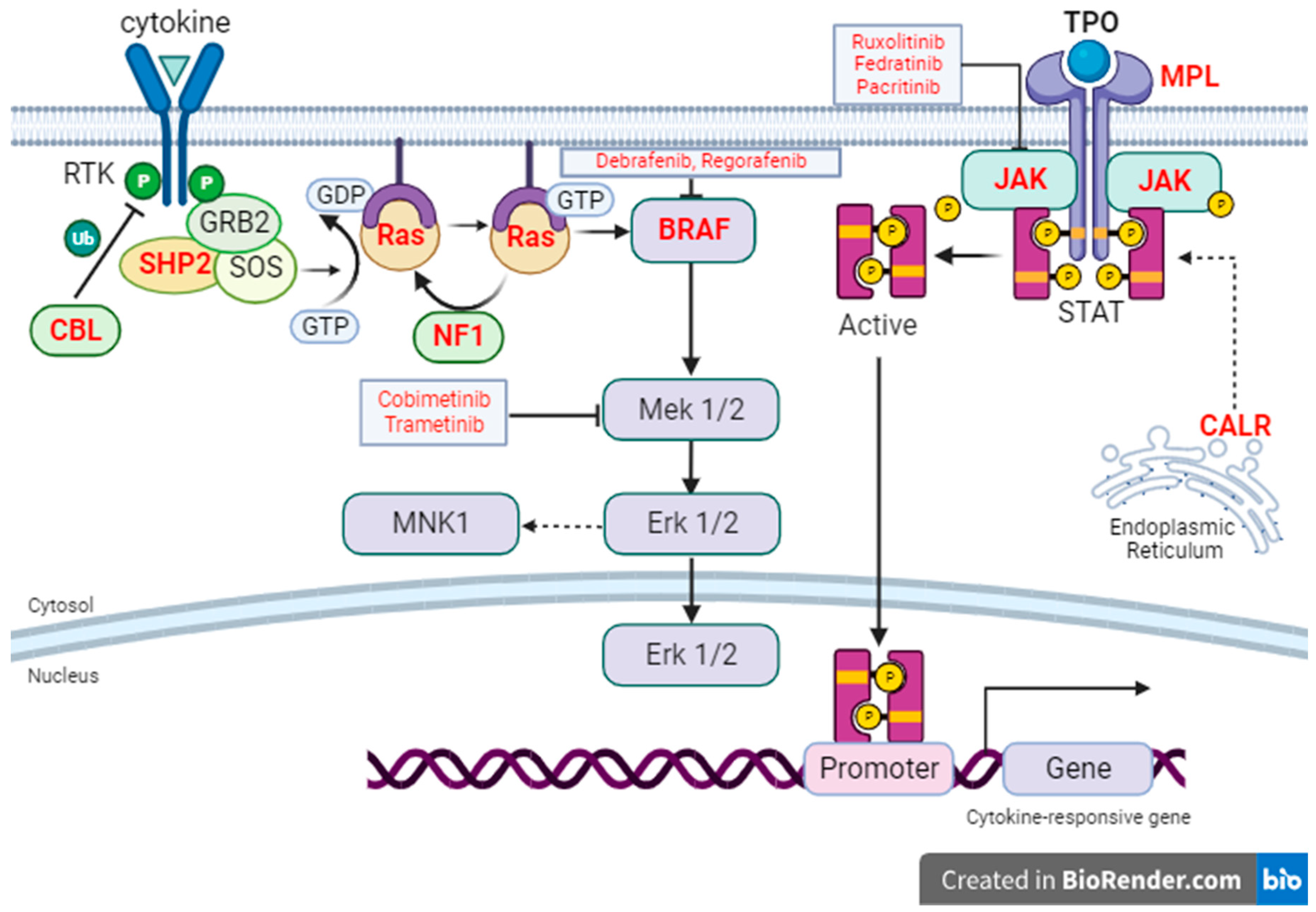
Janus kinase 2 (JAK2) is a member of the family of Jak nonreceptor tyrosine kinases and is an essential component of normal hematopoiesis [10]. Activation of receptor tyrosine kinases by growth factors, cytokines, or chemokines results in phosphorylation and activation of Jak2, which leads to Stat phosphorylation and activation of downstream targets of Stat transcription factors (Figure 1). Jak2 activation also leads to the activation of a signaling cascade through effector pathways including Ras/MAP Kinase and PI3K/Akt/mTOR, which drive cell growth. JAK2 mutations, specifically V617F, are common in myelofibrosis (35–57%), ubiquitous in polycythemia vera, occurring in 96–99% of patients, and are also the most commonly mutated gene in essential thrombocythemia (50–60%) [11,12,13,14,15,16,17].
The majority of JAK2 mutations identified in myelofibrosis are V617F. The JH2 pseudokinase domain of Jak2 protein has a negative regulatory effect on the JH1 kinase domain and maintains inactive Jak2 conformation [18]. Upon V617F mutation, however, this inhibitory effect is lost, resulting in constitutively active Jak2. The early reporting of JAK2V617F mutational presence in patients with myelofibrosis was through a series of publications in 2005, which ranged in number of evaluated patients (16–46) but generally identified a propensity for the mutation (35–57%) [19,20,21]. Murine models of mutant JAK2V617F have supported these clinical findings [16,17]. JAK2V617F allele burden can also be used in MF prognostication. In one 2017 model, allele burden of >50% is correlated with the median overall survival of 80 months compared with 50 months for values <50%.
Mutations in JAK2 Exon 12 have also been identified in MPNs but are more common in polycythemia vera. Exon 12 resides between Jak2 domains JH2 and SH2 and codes for the amino acids from 505 to 547; mutations in this exon are present in approximately 3% of patients with polycythemia vera. Disease in patients with exon 12 mutations is characterized more frequently by isolated erythrocytosis rather than hyperplasia across all three lineages [22]. Its presence in myelofibrosis, however, is uncommon and therefore its prognostic impact has not been approximated in clinical studies.

2.2. MPL

MPL mutations, while much less common than JAK2, are enriched in a subset of patients with MF and appear to drive the disease phenotype at least partially. MPL is a tyrosine kinase receptor encoding for thrombopoietin (TPO) receptor, which serves as a hematopoietic stem cell (HSC) growth factor receptor (Figure 1). MPL is normally activated by binding of its ligand TPO. MPL mutations lead to constitutively active Mpl which turns on the downstream JAK pathways, prompting both HSC renewal and megakaryocyte development [23]. Its presence in myelofibrosis has varied across reported studies from as low as non-existent to up to 7% of patients [24]. Mutations in polycythemia vera and essential thrombocythemia are even less common and, respectively, have only been reported in up to 1% and 5% of patients in each disease state [13,14,24,25].
The role of mutated MPL in myelofibrosis first came to prominence in 2006 when Levine and colleagues performed exomal sequencing of the trans- and juxtamembrane domains of EPOR, GCSFR, and Mpl in comparison with germline DNA. Results were most notable for an activating mutation in the MPL (W515L) transmembrane domain in 4/45 (9%) patients with JAK2-negative myelofibrosis. A murine bone marrow transplant model with MPLW515L expression also induced thrombocytosis, reticulin fibrosis and splenomegaly, a phenotype not observed with wildtype MPL expression [26]. The estimated median overall survival in patients with MPL mutations in myelofibrosis is 221 months [17].

2.3. CALR

CALR mutations in myelofibrosis vary in reported frequency (25–53%) but are more common than in patients with PV (not reported) or ET (25–27%) [12,13,14,24,27,28]. The exact prognostic significance of mutated CALR in MF is under investigation but its presence has been correlated with increased overall survival in the absence of accompanying JAK2 mutations [29].
The CALR gene, located on chromosome 19’s short arm, contains 9 exons and encodes for calreticulin, which is a chaperone protein in the endoplasmic reticulum and functions to maintain calcium homeostasis and protein folding [30,31]. The identification of somatic mutations of CALR in MF came in the form of two primary studies, one of which was published in 2013 by Klampfl and authors, who observed that an estimated 30–45% of patients with either primary myelofibrosis (PMF) or essential thrombocythemia (ET) had no previously known identifiable clonal mutations, including JAK2 and MPL mutations [29]. Authors then performed whole-exome sequencing in six patients diagnosed with PMF without mutations in JAK2 or MPL and identified CALR mutations in all sequenced patients. An expanded screening of 1107 samples from patients with various MPNs identified CALR mutations in 88% of patients with PMF when JAK2 and MPL mutations were absent and confirmed that such patients had longer overall survival (OS) and lower risk of thromboses than those with JAK mutations.
A second study from the same year (and journal) further expanded sequencing analyses, including Sanger, whole-exome, and whole-genome sequencing, in 1397 patient samples, collectively identifying 32 patients with primary MF, 18/32 (56%) of whom were CALR mutated [32]. These findings, combined with those reported by Klampfl et al. strengthened the assertion that somatic mutations in CALR are common and are likely driver mutations in primary myelofibrosis in the absence of JAK2 and MPL. Frameshift mutations in CALR exon 9, in a separate study, were associated with a median overall survival of 131 months in patients with primary myelofibrosis [17]. Lastly, murine models of CALR, including a CALRdel52 conditional inducible knock-in mutation in an Mx-Cre model, have again yielded similar findings and revealed progression from essential thrombocythemia (ET) to MF in homozygous mutant mice [33].
It is also essential to understand the cumulative relationship between these classic driver mutations, which was explored by Araki and authors in a 2016 study published in Blood [34]. Here, the authors reported that in UT-7/TPO cells, Jak2-bound c-Mpl preferentially associated with mutant CALR compared with wildtype. Mutant CALR was also found to induce Jak2 phosphorylation and activation of Jak2 downstream signaling, an effect that can be blocked by Jak2 inhibitors. Furthermore, in hematopoietic stem cells harboring CALR mutations, c-Mpl was required for TPO-independent megakaryopoiesis. Although the mechanism by which CALR mutations lead to activation of JAK signaling remains to be fully elucidated, it can be concluded from this study and related reports that mutant CALR activates signaling molecules downstream of Jak2 via association with c-Mpl and ultimately drives the development of MPNs. A depiction summarizing the interconnectivity between these driver mutations is included in Figure 1.
Figure 1. Intracellular signaling through Ras/MAP Kinase and Jak/Stat pathways. Genes whose mutations have been found in MF are labeled with red bold font. Within this model, loss of function in NF1 and CBL; gain of function mutations in PTPN11 (encoding SHP2); and activating mutations in K/NRAS, lead to hyperactive Ras signaling [35,36]. JAK2 and MPL gain-of-function mutations result in hyperactive Jak/Stat signaling. Mutations in CALR, lastly, induce constitutive activation of Mpl and Jak/Stat signaling, particularly in myeloproliferative neoplasms [37]. Various small molecular inhibitors against targets in the Ras/MAPK pathway or JAK/Stat pathway are included in text boxes in red font [38].
Cancers 15 04654 g001

3. Triple-Negative Myelofibrosis

Triple-negative myelofibrosis (TN MF) is diagnosed in the absence of JAK2, MPL, and CALR mutations while otherwise meeting morphologic and laboratory criteria for myelofibrosis. One 2021 study reported on a single-center experience of 626 patients with MF over an 18-year period and identified triple-negative disease in 38 (6%) patients [39]. Higher risk disease by both the dynamic and genetically inspired international prognostic scoring systems was observed more frequently in TN MF than in non-TN disease (65.8 vs. 58.1% DIPSS, 62.5 vs. 47.1% GIPSS). Median overall survival was also shortened in TN compared with JAK2-, MPL-, or CALR-mutated diseases at 37.4 vs. 85.7 months. Interestingly, mutations in CBL, IDH2, GNAS, SETBP1, and SRSF2 were enriched in triple negative disease. A working theory in myelofibrosis is that the acquisition of additional somatic mutations drives a more high-risk phenotype and is associated with disease advancement. In the era of expanded molecular analyses early in the diagnosis of MF, reports on the impact of these mutations are growing in the MF literature. The mutations described in the following sub-sections commonly co-exist with JAK2, MPL, and CALR mutations in patients with myelofibrosis and, while they fall within broad categories (such as proteins affecting splicing and epigenetic regulation), each has its own unique mechanism. They are also enriched in patients with triple-negative disease. The impact of several of these mutations, specifically ASXL1, EZH2, IDH1/2, and SRSF2, directly enhances a patient’s molecular risk stratification. In the MIPSS70v2 system, a patient is assigned 2 points if one of these high molecular risk (HMR) mutations is present and 3 points if ≥2 HMR’s are present [7].
One proposed mechanism behind this risk enhancement is that more mutations create more disruption to intracellular signaling. Another theory is that when multiple clones are driving the disease, some respond to therapy while others are persistent and become more dominant drivers of myeloproliferation. A representative summary of the various mechanisms of these additional somatic mutations as well as their presence as well as impact in various myeloproliferative neoplasms as compared to classical driver mutations is summarized in Table 1.

4. Mutations in Epigenetic Regulators

As in other myeloid neoplasms, epigenetic regulators that modify chromatin structure to regulate gene expression play an important role in the pathogenesis of MF. Mutations of ASXL1 and EZH2, for instance, have identified in myelofibrosis. The presence of these mutations is often associated with an overall poorer outcome and therefore provides prognostic value in disease. By comparison with other myeloproliferative neoplasms in PV and ET as depicted in Table 1, both ASXL1 and EZH2 mutations are more common in myelofibrosis.

4.1. ASXL1

ASXL1 is an epigenetic regulator of genes involved in chromatin remodeling, specifically a polycomb repressive complex protein (PRC), deletion of which has been implicated in impaired hematopoiesis and acceleration of myeloid neoplasms [62]. Guglielmelli, Coltro, and investigators reported in 2022 an analysis of 523 patients with MF at various disease stages, including primary myelofibrosis (PMF), pre-MF, and post-polycythemia vera (PPV-MF), in which ASXL1 mutations were identified in 157 (30%) of patients [63]. ASXL1 mutations in PMF were associated with several phenotypic characteristics of higher-risk disease, including leukocytosis, lower hemoglobin and platelets, peripheral blast presence, and marrow fibrosis grade ≥ 2. Amongst patients formally diagnosed with high-risk disease, ASXL1mut were identified in 62% of patients and were associated with a shortened median overall survival compared with ASXL1WT at 47 and 102 months, respectively (p = 0.0240).

4.2. EZH2

EZH2, in mammals, is a subunit of the larger histone methyltransferase polycomb repressive complex 2 (PRC2) and is felt to play a role in the progression of various tumors [64]. Like ASXL1, it was also explored by Guglielmelli in 2011 in an analysis of 370 patients with primary myelofibrosis and 148 with post-PV/post-ET MF, where genotyping for EZH2 identified mutations in 5.9%, 1.2%, and 9.4% of patients with primary MF, post-PV MF and post-ET MF, respectively [65]. Clinically, elevations in peripheral blasts, leukocytes and splenomegaly were significantly higher in EZH2-mutated PMF. EZH2 mutations in PMF were also associated with significantly decreased leukemia-free and overall survival.

5. Mutations in Splicing Factors

Splicing factors are essential to pre-mRNA regulation in normal cellular functions. Mutations of two such factors in SRSF2 and U2AF1 have been identified as high molecular risk mutations in myelofibrosis as both are associated with lower median overall survival compared with wildtype [49,66,67]. Both SRSF2 and U2AF1 mutations are rare in polycythemia vera (both < 2%) and essential thrombocythemia (collectively 1–3% as shown in Table 1) [13,15].

5.1. SRSF2

Serine and arginine-rich splicing factor 2 (SRSF2) is multifaceted but appears to be an essential component of pre-mRNA alternating splicing machinery, mutations of which have been identified in approximately 6–17% of patients with myelofibrosis [46,47,68]. A recent investigation into the prognostic significance of SRSF2 in 187 patients with PMF identified significantly lower median overall survival in patients harboring SRSF2 mutations (24 months) compared with SRSF2 wildtype (65 months); 2-year leukemia risk was also higher with mutated SRSF2 (30 vs. 8%) [47]. Other significant associations with SRSF2 mutations included co-mutated IDH1, higher risk stratification by dynamic international prognostic scoring system (DIPSS) and advanced age.

5.2. U2AF1

U2AF1 is also a splicing factor which encodes RNA-binding protein and is involved in recognizing the 3′ splice site facilitating U2 snRNP recruitment in the process of pre-mRNA splicing; it was also recently reported to promote survival of hematopoietic stem/progenitor cells (HSPCs) [69]. Tefferi and others in 2018 examined 491 patients with PMF and identified U2AF1 mutations in 77 (16%) patients, 50 of which involved the Q157 variant and 26 involving the S34 or other variants [48]. Median overall survival was 2.9 years for patients with U2AF1, variant Q157, significantly shorter than patients with the S34 variants and unmutated U2AF1 (5.8 and 5.5 years, respectively). U2AF1 Q157 was also identified to independently contribute to prognosis in PMF by multivariate analysis and is now included in the MIPSS70+ version 2.0+ risk assessment tool [70].

6. Mutations in IDH-Regulators of Cellular Metabolism

Isocitrate dehydrogenase (IDH) mutations are relatively uncommon in patients with myelofibrosis but, when present, appear to be associated with lower overall survival compared with wildtype [50]. IDH mutations in PV and ET are even more rare, both occurring in <2% of patients [13,15]. Isocitrate dehydrogenase (IDH) normally catalyzes the conversion of isocitrate to alpha-ketoglutarate and in mammals, both isoenzymes IDH1 (cytosolic) and IDH2 (mitochondrial) are NADP-dependent [66,71]. Mutant IDH1/2 in leukemic cells instead yields a separate oncometabolite called 2-hydroxyglutarate (2-HG), leading to multiple effects that include hematopoietic differentiation and inhibition of the TET methylcytosine dioxygenases, which are instrumental in DNA demethylation [67]. IDH mutations have been identified (and are targeted) in numerous solid and hematologic neoplasms, including acute myeloid leukemia, cholangiocarcinoma, and glioma [72]. The presence and impact of IDH mutations in PMF was explored by Tefferi in an analysis of 301 patients with PMF, 12 of whom (4%) were identified to have the mutation (5 IDH1, 7 IDH2) [50]. Inferior overall survival (OS) was observed in multivariate analysis in IDH-mutated patients. Interestingly, 6 of these 12 patients also possessed JAK2V617 mutations, and a more pronounced impact on LFS and OS was observed in patients co-mutated with IDH and JAK2, which authors suggested may represent “leukemogenic collaboration” between the two.
Regarding the impact of IDH mutations on therapy, there have been no studies examining IDH 1 nor 2 inhibitors as monotherapy in MF, although there was a Phase II trial recently completed in March 2023 evaluating combination of ruxolitinib + enasidenib in patients with IDH2 mutations and either blast-phase MPNs or chronic myelofibrosis [73]. Results are forthcoming but further investigation into the role of IDH inhibitors in myelofibrosis beyond this study is warranted to continue expanding available therapies.

7. Mutations in TP53 Tumor Suppressor Gene

TP53 mutations vary by prevalence in myelofibrosis (1–13%) but are associated with both increased risk for transformation to leukemia and poorer overall survival when present [52,53]. Similar to mutations of epigenetic, splicing, and cellular metabolism regulators, TP53 mutations are comparatively less common in polycythemia vera (<2%) and essential thrombocythemia (1–4%) [13,15,45].
TP53 is a tumor suppressor gene whose wildtype function is well-established in cell cycle control, DNA damage repair, and apoptosis and whose mutated forms, both inherited (germline) or acquired (somatic), have been implicated in the pathogenesis of numerous hematologic and visceral neoplasms [51,74]. TP53 has been recently evaluated in a multicenter cohort of 349 patients with myelofibrosis, 13% (49) of whom were TP53-mutated, with a median variant allele frequency (VAF) of 50% [52]. Authors also evaluated for multi-hit TP53 configurations and identified 30 of the 49 patients (8.6% of the entire cohort) possessing multi-hit mutations of TP53. The outcome data revealed a marked difference between median survival in TP53-mutated (1.5 years) and TP53 wildtype (13.5 years) patients (p < 0.001). A similar trend was reported in leukemic transformation, seen in 20% versus 2% of TP53 mutant and wildtype patients, respectively (p-value again <0.001).
Taken together, while mutations that lead to hyperactivation of JAK/STAT signaling, mostly in JAK/CALR/MPL, are highly prevalent in both PV/ET and MF, MF patients often acquire additional mutations in genes encoding epigenetic regulators (ASXL1 and EZH2), splicing factors (SRSF2 and U2AF1), metabolic enzymes IDH1/2, and TP53 tumor suppressor gene. These additional mutations are often associated with poor outcome and provide important prognostic information in MF patients (Table 1 and Figure 2). Strategies targeting these mutations or mutated pathways may lead to improved response in MF and remain to be investigated.

8. Expanding the Molecular Horizon in Myelofibrosis

While additional mutations and/or signaling pathways implicated in the pathogenesis of both marrow fibrosis and HSPC proliferation are expanding our present understanding of molecular landscape of this myeloproliferative neoplasm, treatment for MF remains limited and resistance to Jak inhibitors is a common theme in clinical practice and is a likely driver of worsening disease. Effort has been focused on identifying mechanisms of resistance to Jak inhibitor therapy and developing strategies to overcome this resistance. One of the proposed mechanisms for this resistance in pre-clinical models is up-regulation in Ras signaling, and multiple related pathways are actively being explored preclinically to better characterize the relationship between Ras signaling and myelofibrosis pathogenesis.
Y-box binding protein (YBX1) is a multifaceted protein that binds to nucleic acid, with functions in transcription as well as mRNA splicing. It is proposed to regulate various genes associated with cancer resistance. YBX1 phosphorylation also leads to nuclear localization and activation of YBX1 target genes, which in a recent study was increased in JAK2V617F cells in an MEK/ERK-dependent manner [75]. In an RNA interference screen targeting proteins enriched in JAK2V617F cells compared with JAK2 wild type cells, a loss of YBX1 sensitized cells to Ruxolitinib-mediated apoptosis [76]. Pharmacologic inhibition of Jak combined with YBX1 inactivation also induced apoptosis in Jak-2-dependent human and mouse cells. Authors concluded that co-targeting YBX1 and Jak2 can be an effective strategy in eradicating JAK2-mutant hematopoietic cells in MPNs. This study also provides evidence that MEK/ERK signaling pathway may be involved in resistance to JAK inhibitors.
In addition, upstream cytokine signaling that activates MEK/ERK has been shown to carry potential for therapeutic targeting in myelofibrosis. One of the recent studies in this space published in Blood by Melo-Cardenas and others developed various models of MF to analyze cytokine signaling [77]. Their first set of experiments to this goal was an analysis of cytokine profiles in mice before and after fibrotic marrow development, which was coupled with single-cell RNA sequencing of marrow populations. Here, IL-13 was elevated in the marrow of mice with MF, which induced both collagen biosynthesis and transforming growth factor beta (TGFβ) surface expression and promoted mutant megakaryocytic growth. In a correlative study with samples from patients with MF, IL-13 levels were increased in plasma and the expression of its receptor was increased in marrow megakaryocytes. Authors ultimately theorized through these and related experiments that IL-13 was not only contributory to MF progression but also a potential target in developing MF-directed therapies.
On this note, cytokine signaling is implicated not only in the pathogenesis of myelofibrosis but also the downstream activation of Ras/MAP Kinase signaling, which has led to numerous clinical and pre-clinical evaluations as to the role of Ras/MAP Kinase in the disease state. It is worth noting that while it is well-known that Jak/Stat can act as an upstream regulator of Ras signaling, cytokines can also activate Ras via alternative tyrosine kinase receptor pathways.

9. Ras/MAP Kinase Signaling in Myelofibrosis

Ras/MAP Kinase signaling begins when a ligand (cytokine or growth factor) binds to a receptor tyrosine kinase, leading to receptor dimerization and autophosphorylation [78,79]. Various adaptor proteins, including GRB2 and SOS, then bind to the now-activated receptors and recruit guanine nucleotide exchange factors (GEFs), which facilitate the exchange on Ras of guanine diphosphate (GDP) for guanine triphosphate (GTP) and activates Ras proteins. GTP-bound Ras can then interact with the Raf kinase, leading to the sequential activation and phosphorylation of MEK and then ERK, a process better known as the MAP kinase cascade. Key regulators in this process are neurofibromin 1 (NF1), which as depicted in Figure 1 inactivates Ras-GTP by stimulating GTPase, and SHP2 (encoded by PTPN11), a Ras-activating tyrosine phosphatase [36,59]. Mutations in various proteins either directly or indirectly involved in this process can have various impacts on Ras activation. CBL, for instance, is a ubiquitin ligase that degrades receptor tyrosine kinase and when mutated leads to a loss of function of CBL and increased Ras activation [80]. Ras/MAP Kinase pathway signaling is also theorized in various models to be interconnected with a downstream effector PI3K/Akt/mTOR pathway, collectively then enacting various cellular processes that regulate cell growth and survival [81]. The relationship between Jak-Stat and Ras signaling is under investigation but Jak-Stat is likely contributory at least to an extent to Ras activation [82].
Despite the available therapies targeting Jak-Stat signaling, myelofibrosis frequently worsens or transforms in the presence of the high-risk molecular mutations, particularly those in the Ras/MAP Kinase pathway. A few recent studies investigated the role of Ras pathway mutations in MF. Coltro and investigators in 2020 examined 464 patients with MF, 59 of whom had RAS/CBL mutations (5.4% NRAS, 2.8% KRAS, and 5.6% CBL) [57]. These mutations were associated with inferior overall survival and a higher 5-year incidence of leukemic transformation. These investigators also reported that, of the 61 patients treated with Jak inhibitors, response in spleen size and symptoms were lower in RAS/CBL mutant cohort, which was confirmed on logistic regression analysis. A similar 2020 study reported next generation sequencing data of 16 target genes in 723 patients with either primary or secondary MF and identified subclonal N/KRAS mutations in 6% of patients [8]. RAS mutations were also associated with advanced MF features, including leukocytosis and high burden of high-risk mutations; RAS-mutated patients also had shortened 3-year overall survival.
The frequency of Ras pathway mutations in JAK inhibitor resistant MF patients was evaluated in a recent study which retrospectively evaluated 113 patients with MF with baseline molecular data, focusing specifically on patterns in Jak inhibitor failure [83]. Amongst 49 patients with new comparative molecular data available who had failed Jak inhibitors, 29 different emergent mutations were identified in 19 patients (39%), 9 of which (18%) were within the Ras/MAP Kinase pathway.
More recently, a study utilizing Mi-Oncoseq analysis investigated Ras pathway mutations in in 216 patients with either primary myelofibrosis or ET/PV pre-primary myelofibrosis [55]. Here, mutations in the Ras/MAP Kinase pathway, including K/NRAS, MAPK, CBL, and NF1 were present in up to 25% of patients and were associated with significantly higher platelet and leukocyte counts (p-valves of 0.035 and 0.015, respectively). Interestingly, KRAS and NRAS mutations were exclusive to the MF cohort.
In summary, there is emerging evidence that mutations activating Ras signaling pathways are frequently found in MF but not earlier stage pre-myelofibrosis such as PV and ET patients. These mutations likely confer a proliferative and high-risk phenotype. More importantly, Ras pathways mutations are enriched in MF patients who fail JAK inhibitor therapies, raising the possibility that hyperactive Ras signaling is a potential mechanism of resistance to JAK inhibitors. Treatment targeting the Ras/MAPK pathway therefore can provide a strategy to overcome JAK inhibitor resistance. A concise summary of commonly utilized Jak inhibitors in myelofibrosis and various small molecule inhibitors in the MAP Kinase pathway are summarized in Table 2.

10. Targeting Ras Signaling in Myeloid Neoplasms

Various studies have directly evaluated the effect of inhibiting individual components of Ras signaling pathway, specifically through Mek or Erk 1/2 inhibitors, as an attempt to overcome therapeutic resistance in myeloid neoplasms. Most of these studies were conducted in AML with disappointing results from monotherapies. One example was a Phase II study evaluating binimetinib in 19 patients with either relapsed/refractory acute myeloid leukemia (AML) or myelodysplastic syndrome (MDS), 14 of whom harbored RAS mutations [92]. Ultimately, only 13 patients were evaluable and 12 (92%) did not achieve complete response (CR); authors suggested combining such inhibitors with other therapies for future studies. Another Phase II trial evaluated the oral Mek 1/2 inhibitor selumetinib in 47 patients who were either ≥60 with untreated AML or any age with relapsed/refractory AML [93]. Responses were absent in the FLT3 ITD and NRAS-mutated cohorts and the only patient with a KRAS mutation had a minor response (which itself was unconfirmed).
These limited results with individual agents have prompted investigations into combination therapies in myeloid diseases. Trametinib, an oral Mek 1/2 inhibitor, for example, was examined in a Phase II study in combination with an Akt inhibitor in 23 patients with RAS-mutant AML, none of whom achieved CR [94]. Pre-clinical investigations, however, are expanding on combinational regimens. One study published in Nature Communications, for instance, argued that osteopontin (or ‘OPN’) promoted the proliferation of mesenchymal stromal cells, collagen production, and marrow fibrosis induced by TPO receptor agonist (romiplostim) [95]. Inhibition of ERK 1/2 was then observed to substantially reduce the production of OPN and, in turn, marrow fibrosis in vivo. Recognizing that Ras/MAP Kinase inhibition alone is likely inadequate, Ross and investigators developed a multi-kinase inhibitor targeting Mek and P13K termed LP-182, which was designed for preferential lymphatic absorption, and were successful in ameliorating the myelofibrosis phenotype as well as improving overall survival in their animal models [96].
Another strategy that is in line with the concept of synergistic targeting but is more relevant to the known existing therapeutic landscape in myelofibrosis is combining Mek (or other agents targeting MAP Kinase pathway signaling) with Jak inhibitors to manage resistance to Jak inhibitor monotherapies. Pandey and investigators in 2022, for instance, further investigated mRNA splicing alterations and identified intron retention in mRNAs encoding for Mek/Erk signaling. One observation was that the expression of MAPK-interactive kinase 1 (MNK1) was diminished in YBX1-depleted JAK2-V617F cells and was required for Erk signaling [75]. Moreover, MNK1 inhibition enhanced Jak2 inhibitor-mediated cell death and, together with Mek inhibition, induced apoptosis of CD34+ JAK-V617 cells. A correlative in vivo study in PDX mouse models using transplanted JAK-V617F-expressing marrow from patients with MPN compared the effects of trametinib + ruxolitinib to ruxolitinib alone [76]. Authors reported significant growth inhibition as well as molecular remission of transplanted cells in the ruxotlinib + trametinib group compared with ruxolitinib alone, suggesting a synergy between ruxolitinib and Ras/MAP kinase pathway inhibitors.
The observation that Mek/Erk targeting increases susceptibility of MPNs to JAK inhibitors has been explored by other groups as well. Stivala and investigators in 2019 reported in vitro suppression of Mek/Erk activation with Type I and II Jak2 inhibition in MPN cell lines and in an ex vivo model [97]. This suppression, however, was lost in vivo in MPLW515L and JAK2V617F mice after Jak2 inhibition alone but combined Mek/Jak inhibition was able to suppress activation of Mek/Erk. Binimetinib plus ruxolitinib exposure for 2 weeks was also superior to ruxolitinib alone in the reduction in reticulin fibrosis, hepatosplenomegaly and megakaryocytic hyperplasia in the bone marrow. The difference between ex vivo and in vivo MAPK activation is at least partially explained by the cytokines that the mutant cells are exposed to in vivo. PDGFRα notably remained activated and production of PDGF-AA/PDGF-BB (which are the subunits of PDGFR) persisted following Jak2 inhibition in vivo and PDGF-BB, on exposure to ruxolitinib, maintained activation of Erk [98]. This observation was consistent with the known function of PDGF-BB serving as a bypass for Erk activation, leading to diminished efficacy of Jak2 inhibition, and supported the utility for dual MAP Kinase pathway/Jak2 inhibition in the management of MPNs.
These studies collectively suggest that RAS mutations and Ras signaling activating mutations are frequently detected in MF and are associated with an adverse outcome. In addition, Ras signaling is activated despite mutational status or Jak inhibition, possibly through increased cytokine signaling in MF, raising the possibility that Ras pathway targeting strategies may offer therapeutic benefit to MF patients.

11. Conclusions and Future Directions

The pathogenesis of primary myelofibrosis is not determined solely by alterations in JAK2, CALR, and MPL and likely involves a much broader landscape of somatic mutations, which themselves affect both clinical features of disease and overall survival in various capacities. Of these, Ras/MAP Kinase pathway mutations represent an area of growing clinical interest, both mechanistically and as potential therapeutic targets in myelofibrosis. Expanded translational analyses and early phase clinical trials exploring the role of Ras/MAP Kinase signaling and manipulation are needed to further targeted therapies in this myeloproliferative neoplasm.

Author Contributions

Conceptualization; investigation; resources; data curation; writing—original draft preparation; writing—review and editing; visualization; supervision; project administration; S.B.R., K.P., M.K., M.T. and Q.L. All authors have read and agreed to the published version of the manuscript.

Funding

S.B.R. is supported by an NIH T32CA009357. Q.L. is supported by NIH R01HL150707.

Institutional Review Board Statement

Not applicable due to the retrospective nature of this study, which did not involve human nor animal participants.

Informed Consent Statement

Not applicable as this study did not involve human participants.

Data Availability Statement

No new data were created or analyzed in this study. Data sharing is not applicable to this article.

Acknowledgments

The authors do not wish to acknowledge administrative nor technical support, or donations in kind (e.g., materials used for experiments).

Conflicts of Interest

The authors declare no conflict of interest.

References

  1. Tefferi, A. Primary myelofibrosis: 2021 update on diagnosis, risk-stratification and management. Am. J. Hematol. 2021, 96, 145–162. [Google Scholar] [CrossRef]
  2. Zhou, T.; Georgeon, S.; Moser, R.; Moore, D.J.; Caflisch, A.; Hantschel, O. Specificity and mechanism-of-action of the JAK2 tyrosine kinase inhibitors ruxolitinib and SAR302503 (TG101348). Leukemia 2014, 28, 404–407. [Google Scholar] [CrossRef]
  3. Singer, J.W.; Al-Fayoumi, S.; Ma, H.; Komrokji, R.S.; Mesa, R.; Verstovsek, S. Comprehensive kinase profile of pacritinib, a nonmyelosuppressive Janus kinase 2 inhibitor. J. Exp. Pharmacol. 2016, 8, 11–19. [Google Scholar] [CrossRef]
  4. Verstovsek, S.; Gotlib, J.; Mesa, R.A.; Vannucchi, A.M.; Kiladjian, J.-J.; Cervantes, F.; Harrison, C.N.; Paquette, R.; Sun, W.; Naim, A.; et al. Long-term survival in patients treated with ruxolitinib for myelofibrosis: COMFORT-I and -II pooled analyses. J. Hematol. Oncol. 2017, 10, 156. [Google Scholar] [CrossRef]
  5. Verstovsek, S.; for the COMFORT-I Investigators; Mesa, R.A.; Gotlib, J.; Gupta, V.; DiPersio, J.F.; Catalano, J.V.; Deininger, M.W.N.; Miller, C.B.; Silver, R.T.; et al. Long-term treatment with ruxolitinib for patients with myelofibrosis: 5-year update from the randomized, double-blind, placebo-controlled, phase 3 COMFORT-I trial. J. Hematol. Oncol. 2017, 10, 55. [Google Scholar]
  6. Harrison, C.N.; Vannucchi, A.M.; Kiladjian, J.J.; Al-Ali, H.K.; Gisslinger, H.; Knoops, L.; Cervantes, F.; Jones, M.M.; Sun, K.; McQuitty, M.; et al. Long-term findings from COMFORT-II, a phase 3 study of ruxolitinib vs. best available therapy for myelofibrosis. Leukemia 2016, 30, 1701–1707. [Google Scholar] [CrossRef]
  7. Tefferi, A.; Gangat, N.; Pardanani, A.; Crispino, J.D. Myelofibrosis: Genetic Characteristics and the Emerging Therapeutic Landscape. Cancer Res. 2022, 82, 749–763. [Google Scholar] [CrossRef]
  8. Santos, F.P.S.; Getta, B.; Masarova, L.; Famulare, C.; Schulman, J.; Datoguia, T.S.; Puga, R.D.; Paiva, R.d.M.A.; Arcila, M.E.; Hamerschlak, N.; et al. Prognostic impact of RAS-pathway mutations in patients with myelofibrosis. Leukemia 2020, 34, 799–810. [Google Scholar] [CrossRef]
  9. Gianelli, U.; Thiele, J.; Orazi, A.; Gangat, N.; Vannucchi, A.M.; Tefferi, A.; Kvasnicka, H.M. International Consensus Classification of myeloid and lymphoid neoplasms: Myeloproliferative neoplasms. Virchows Arch. Int. J. Pathol. 2023, 482, 53–68. [Google Scholar] [CrossRef]
  10. Yamaoka, K.; Saharinen, P.; Pesu, M.; Holt, V.E., 3rd; Silvennoinen, O.; O’Shea, J.J. The Janus kinases (Jaks). Genome Biol. 2004, 5, 253. [Google Scholar] [CrossRef]
  11. Moliterno, A.R.; Kaizer, H.; Reeves, B.N. JAK2V617F allele burden in polycythemia vera: Burden of proof. Blood 2023, 141, 1934–1942. [Google Scholar] [CrossRef]
  12. Broséus, J.; Park, J.-H.; Carillo, S.; Hermouet, S.; Girodon, F. Presence of calreticulin mutations in JAK2-negative polycythemia vera. Blood 2014, 124, 3964–3966. [Google Scholar] [CrossRef]
  13. Guglielmelli, P.; Gangat, N.; Coltro, G.; Lasho, T.L.; Loscocco, G.G.; Finke, C.M.; Morsia, E.; Sordi, B.; Szuber, N.; Hanson, C.A.; et al. Mutations and thrombosis in essential thrombocythemia. Blood Cancer J. 2021, 11, 77. [Google Scholar] [CrossRef] [PubMed]
  14. Elsayed, A.G.; Ranavaya, A.; Jamil, M.O. MPL Y252H anMd PL F126fs mutations in essential thrombocythemia: Case series and review of literature. Hematol. Rep. 2019, 11, 7868. [Google Scholar] [CrossRef] [PubMed]
  15. Tefferi, A.; Lasho, T.L.; Finke, C.; Elala, Y.; Barraco, D.; Hanson, C.A.; Ketterling, R.P.; Pardanani, A.; Gangat, N. Targeted Next-Generation Sequencing in Polycythemia Vera and Essential Thrombocythemia. Blood 2015, 126, 354. [Google Scholar] [CrossRef]
  16. Shide, K.; Shimoda, H.K.; Kumano, T.; Karube, K.; Kameda, T.; Takenaka, K.; Oku, S.; Abe, H.; Katayose, K.S.; Kubuki, Y.; et al. Development of ET, primary myelofibrosis and PV in mice expressing JAK2 V617F. Leukemia 2008, 22, 87–95. [Google Scholar] [CrossRef]
  17. Rozovski, U.; Verstovsek, S.; Manshouri, T.; Dembitz, V.; Bozinovic, K.; Newberry, K.; Zhang, Y.; Bove, J.E.; Pierce, S.; Kantarjian, H.; et al. An accurate, simple prognostic model consisting of age, JAK2, CALR, and MPL mutation status for patients with primary myelofibrosis. Haematologica 2017, 102, 79–84. [Google Scholar] [CrossRef]
  18. Chen, E.; Mullally, A. How does JAK2V617F contribute to the pathogenesis of myeloproliferative neoplasms? Hematology 2014, 2014, 268–276. [Google Scholar] [CrossRef]
  19. Levine, R.L.; Wadleigh, M.; Cools, J.; Ebert, B.L.; Wernig, G.; Huntly, B.J.; Boggon, T.J.; Wlodarska, I.; Clark, J.J.; Moore, S.; et al. Activating mutation in the tyrosine kinase JAK2 in polycythemia vera, essential thrombocythemia, and myeloid metaplasia with myelofibrosis. Cancer Cell 2005, 7, 387–397. [Google Scholar] [CrossRef] [PubMed]
  20. Kralovics, R.; Passamonti, F.; Buser, A.S.; Teo, S.S.; Tiedt, R.; Passweg, J.R.; Tichelli, A.; Cazzola, M.; Skoda, R.C. A gain-of-function mutation of JAK2 in myeloproliferative disorders. N. Engl. J. Med. 2005, 352, 1779–1790. [Google Scholar] [CrossRef]
  21. Baxter, E.J.; Scott, L.M.; Campbell, P.J.; East, C.; Fourouclas, N.; Swanton, S.; Vassiliou, G.S.; Bench, A.J.; Boyd, E.M.; Curtin, N.; et al. Acquired mutation of the tyrosine kinase JAK2 in human myeloproliferative disorders. Lancet 2005, 365, 1054–1061. [Google Scholar] [CrossRef] [PubMed]
  22. Maddali, M.; Kulkarni, U.P.; Ravindra, N.; Jajodia, E.; Arunachalam, A.K.; Suresh, H.; Venkatraman, A.; George, B.; Mathews, V.; Balasubramanian, P. JAK2 exon 12 mutations in cases with JAK2V617F-negative polycythemia vera and primary myelofibrosis. Ann. Hematol. 2020, 99, 983–989. [Google Scholar] [CrossRef]
  23. Guglielmelli, P.; Calabresi, L. The MPL mutation. Int. Rev. Cell Mol. Biol. 2021, 365, 163–178. [Google Scholar]
  24. Mejía-Ochoa, M.; Acevedo Toro, P.A.; Cardona-Arias, J.A. Systematization of analytical studies of polycythemia vera, essential thrombocythemia and primary myelofibrosis, and a meta-analysis of the frequency of JAK2, CALR and MPL mutations: 2000–2018. BMC Cancer 2019, 19, 590. [Google Scholar] [CrossRef] [PubMed]
  25. Pardanani, A.; Lasho, T.L.; Finke, C.M.; Tefferi, A. Infrequent occurrence of MPL exon 10 mutations in polycythemia vera and post-polycythemia vera myelofibrosis. Am. J. Hematol. 2011, 86, 701–702. [Google Scholar] [CrossRef]
  26. Pikman, Y.; Lee, B.H.; Mercher, T.; McDowell, E.; Ebert, B.L.; Gozo, M.; Cuker, A.; Wernig, G.; Moore, S.; Galinsky, I.; et al. MPLW515L is a novel somatic activating mutation in myelofibrosis with myeloid metaplasia. PLoS Med. 2006, 3, e270. [Google Scholar] [CrossRef]
  27. Tefferi, A.; Lasho, T.L.; Tischer, A.; Wassie, E.A.; Finke, C.M.; Belachew, A.A.; Ketterling, R.P.; Hanson, C.A.; Pardanani, A.D. The prognostic advantage of calreticulin mutations in myelofibrosis might be confined to type 1 or type 1-like CALR variants. Blood 2014, 124, 2465–2466. [Google Scholar] [CrossRef]
  28. Li, N.; Yao, Q.M.; Gale, R.P.; Li, J.L.; Li, L.D.; Zhao, X.S.; Jiang, H.; Jiang, Q.; Jiang, B.; Shi, H.X.; et al. Frequency and allele burden of CALR mutations in Chinese with essential thrombocythemia and primary myelofibrosis without JAK2(V617F) or MPL mutations. Leuk. Res. 2015, 39, 510–514. [Google Scholar] [CrossRef]
  29. Klampfl, T.; Gisslinger, H.; Harutyunyan, A.S.; Nivarthi, H.; Rumi, E.; Milosevic, J.D.; Them, N.C.C.; Berg, T.; Gisslinger, B.; Pietra, D.; et al. Somatic mutations of calreticulin in myeloproliferative neoplasms. N. Engl. J. Med. 2013, 369, 2379–2390. [Google Scholar] [CrossRef]
  30. Luo, W.; Yu, Z. Calreticulin (CALR) mutation in myeloproliferative neoplasms (MPNs). Stem Cell Investig. 2015, 2, 16. [Google Scholar]
  31. How, J.; Hobbs, G.S.; Mullally, A. Mutant calreticulin in myeloproliferative neoplasms. Blood 2019, 134, 2242–2248. [Google Scholar] [CrossRef]
  32. Nangalia, J.; Massie, C.E.; Baxter, E.J.; Nice, F.L.; Gundem, G.; Wedge, D.C.; Avezov, E.; Li, J.; Kollmann, K.; Kent, D.G.; et al. Somatic CALR Mutations in Myeloproliferative Neoplasms with Nonmutated JAK2. N. Engl. J. Med. 2013, 369, 2391–2405. [Google Scholar] [CrossRef]
  33. Li, J.; Prins, D.; Park, H.J.; Grinfeld, J.; Gonzalez-Arias, C.; Loughran, S.; Dovey, O.M.; Klampfl, T.; Bennett, C.; Hamilton, T.L.; et al. Mutant calreticulin knockin mice develop thrombocytosis and myelofibrosis without a stem cell self-renewal advantage. Blood 2018, 131, 649–661. [Google Scholar] [CrossRef] [PubMed]
  34. Araki, M.; Yang, Y.; Masubuchi, N.; Hironaka, Y.; Takei, H.; Morishita, S.; Mizukami, Y.; Kan, S.; Shirane, S.; Edahiro, Y.; et al. Activation of the thrombopoietin receptor by mutant calreticulin in CALR-mutant myeloproliferative neoplasms. Blood 2016, 127, 1307–1316. [Google Scholar] [CrossRef] [PubMed]
  35. Yzaguirre, A.D.; Padmanabhan, A.; de Groh, E.D.; Engleka, K.A.; Li, J.; Speck, N.A.; Epstein, J.A. Loss of neurofibromin Ras-GAP activity enhances the formation of cardiac blood islands in murine embryos. eLife 2015, 4, e07780. [Google Scholar] [CrossRef]
  36. Chou, Y.-T.; Bivona, T.G. Chapter Seven—Inhibition of SHP2 as an approach to block RAS-driven cancers. In Advances in Cancer Research; O’Bryan, J.P., Piazza, G.A., Eds.; Academic Press: Cambridge, MA, USA, 2022; Volume 153, pp. 205–236. [Google Scholar]
  37. Merlinsky, T.R.; Levine, R.L.; Pronier, E. Unfolding the Role of Calreticulin in Myeloproliferative Neoplasm Pathogenesis. Clin. Cancer Res. 2019, 25, 2956–2962. [Google Scholar] [CrossRef]
  38. Reynolds, S.B.; Pettit, K. New approaches to tackle cytopenic myelofibrosis. Hematology 2022, 2022, 235–244. [Google Scholar] [CrossRef]
  39. Aguirre, L.E.; Jain, A.G.; Ball, S.; Al Ali, N.; Tinsley-Vance, S.M.; Sallman, D.A.; Sweet, K.; Lancet, J.E.; Padron, E.; Kuykendall, A.T.; et al. Triple-Negative Myelofibrosis: Disease Features, Response to Treatment and Outcomes. Blood 2021, 138 (Suppl. 1), 1494. [Google Scholar] [CrossRef]
  40. Antonioli, E.; Guglielmelli, P.; Poli, G.; Bogani, C.; Pancrazzi, A.; Longo, G.; Ponziani, V.; Tozzi, L.; Pieri, L.; Santini, V.; et al. Influence of JAK2V617F allele burden on phenotype in essential thrombocythemia. Haematologica 2008, 93, 41–48. [Google Scholar] [CrossRef]
  41. Song, J.; Hussaini, M.; Zhang, H.; Shao, H.; Qin, D.; Zhang, X.; Ma, Z.; Hussnain Naqvi, S.M.; Zhang, L.; Moscinski, L.C. Comparison of the Mutational Profiles of Primary Myelofibrosis, Polycythemia Vera, and Essential Thrombocytosis. Am. J. Clin. Pathol. 2017, 147, 444–452. [Google Scholar] [CrossRef]
  42. Wang, Z.; Liu, W.; Wang, M.; Li, Y.; Wang, X.; Yang, E.; Ming, J.; Quan, R.; Hu, X. Prognostic value of ASXL1 mutations in patients with primary myelofibrosis and its relationship with clinical features: A meta-analysis. Ann. Hematol. 2021, 100, 465–479. [Google Scholar] [CrossRef]
  43. Andréasson, B.; Pettersson, H.; Wasslavik, C.; Johansson, P.; Palmqvist, L.; Asp, J. ASXL1 mutations, previous vascular complications and age at diagnosis predict survival in 85 WHO-defined polycythaemia vera patients. Br. J. Haematol. 2020, 189, 913–919. [Google Scholar] [CrossRef] [PubMed]
  44. Yang, Y.; Akada, H.; Nath, D.; Hutchison, R.E.; Mohi, G. Loss of Ezh2 cooperates with Jak2V617F in the development of myelofibrosis in a mouse model of myeloproliferative neoplasm. Blood 2016, 127, 3410–3423. [Google Scholar] [CrossRef] [PubMed]
  45. Marneth, A.E.; Mullally, A. The Molecular Genetics of Myeloproliferative Neoplasms. Cold Spring Harb. Perspect. Med. 2020, 10, a034876. [Google Scholar] [CrossRef] [PubMed]
  46. Tefferi, A.; Lasho, T.L.; Hanson, C.A.; Ketterling, R.P.; Gangat, N.; Pardanani, A. Screening for ASXL1 and SRSF2 mutations is imperative for treatment decision-making in otherwise low or intermediate-1 risk patients with myelofibrosis. Br. J. Haematol. 2018, 183, 678–681. [Google Scholar] [CrossRef]
  47. Lasho, T.L.; Jimma, T.; Finke, C.M.; Patnaik, M.; Hanson, C.A.; Ketterling, R.P.; Pardanani, A.; Tefferi, A. SRSF2 mutations in primary myelofibrosis: Significant clustering with IDH mutations and independent association with inferior overall and leukemia-free survival. Blood 2012, 120, 4168–4171. [Google Scholar] [CrossRef]
  48. Tefferi, A.; Finke, C.M.; Lasho, T.L.; Hanson, C.A.; Ketterling, R.P.; Gangat, N.; Pardanani, A. U2AF1 mutation types in primary myelofibrosis: Phenotypic and prognostic distinctions. Leukemia 2018, 32, 2274–2278. [Google Scholar] [CrossRef]
  49. Li, B.; Gale, R.P.; Xu, Z.; Qin, T.; Song, Z.; Zhang, P.; Bai, J.; Zhang, L.; Zhang, Y.; Liu, J.; et al. Non-driver mutations in myeloproliferative neoplasm-associated myelofibrosis. J. Hematol. Oncol. 2017, 10, 99. [Google Scholar] [CrossRef]
  50. Tefferi, A.; Jimma, T.; Sulai, N.H.; Lasho, T.L.; Finke, C.M.; Knudson, R.A.; McClure, R.F.; Pardanani, A. IDH mutations in primary myelofibrosis predict leukemic transformation and shortened survival: Clinical evidence for leukemogenic collaboration with JAK2V617F. Leukemia 2012, 26, 475–480. [Google Scholar] [CrossRef]
  51. Blagih, J.; Buck, M.D.; Vousden, K.H. p53, cancer and the immune response. J. Cell Sci. 2020, 133, jcs237453. [Google Scholar] [CrossRef]
  52. Gagelmann, N.; Badbaran, A.; Salit, R.B.; Schroeder, T.; Gurnari, C.; Pagliuca, S.; Panagiota, V.; Rautenberg, C.; Cassinat, B.; Thol, F.; et al. Impact of TP53 on outcome of patients with myelofibrosis undergoing hematopoietic stem cell transplantation. Blood 2023, 141, 2901. [Google Scholar] [CrossRef] [PubMed]
  53. Hussaini, M.O.; Song, J.; Komrokji, R.S.; Sallman, D.A.; Kuykendall, A.; Padron, E.; Mirza, S.; Zhang, L. TP53 Mutations Are a Rare Event in Primary Myelofibrosis, Associated with TET2 Mutations, and Suggest Poor Clinical Outcome. Blood 2017, 130 (Suppl. 1), 5270. [Google Scholar]
  54. Ogawa, F.; Walters, M.S.; Shafquat, A.; O’Beirne, S.L.; Kaner, R.J.; Mezey, J.G.; Zhang, H.; Leopold, P.L.; Crystal, R.G. Role of KRAS in regulating normal human airway basal cell differentiation. Respir. Res. 2019, 20, 181. [Google Scholar] [CrossRef]
  55. Malathi Kandarpa, T.Q.; Robinson, D.; Wu, Y.-M.; Pettit, K.; Li, Q.; Sartor, M.; Chinnaiyan, A.; Talpaz, M. Broad next generation integrated sequencing of myelofibrosis identifies disease-specific and age-related genomic alterations. Clin. Cancer Res. 2023. accepted for publication. [Google Scholar]
  56. Liyasova, M.S.; Ma, K.; Lipkowitz, S. Molecular Pathways: Cbl Proteins in Tumorigenesis and Antitumor Immunity—Opportunities for Cancer Treatment. Clin. Cancer Res. 2015, 21, 1789–1794. [Google Scholar] [CrossRef] [PubMed]
  57. Coltro, G.; Rotunno, G.; Mannelli, L.; Mannarelli, C.; Fiaccabrino, S.; Romagnoli, S.; Bartalucci, N.; Ravenda, E.; Gelli, E.; Sant’Antonio, E.; et al. RAS/CBL mutations predict resistance to JAK inhibitors in myelofibrosis and are associated with poor prognostic features. Blood Adv. 2020, 4, 3677–3687. [Google Scholar] [CrossRef]
  58. Singh, N.R. Genomic diversity in myeloproliferative neoplasms: Focus on myelofibrosis. Transl. Pediatr. 2015, 4, 107–115. [Google Scholar] [PubMed]
  59. Molosh, A.I.; Shekhar, A. Chapter 2—Neurofibromatosis type 1 as a model system to study molecular mechanisms of autism spectrum disorder symptoms. In Progress in Brain Research; Shekhar, A., Ed.; Elsevier: Amsterdam, The Netherlands, 2018; Volume 241, pp. 37–62. [Google Scholar]
  60. Hussain, M.R.M.; Baig, M.; Mohamoud, H.S.A.; Ulhaq, Z.; Hoessli, D.C.; Khogeer, G.S.; Al-Sayed, R.R.; Al-Aama, J.Y. BRAF gene: From human cancers to developmental syndromes. Saudi J. Biol. Sci. 2015, 22, 359–373. [Google Scholar] [CrossRef]
  61. Wan, Z.; Han, B. Comparison and Implications of Mutational Profiles of Myelodysplastic Syndromes, Myeloproliferative Neoplasms, and Myelodysplastic/Myeloproliferative Neoplasms: A Meta-Analysis. Front. Oncol. 2020, 10, 579221. [Google Scholar] [CrossRef]
  62. Asada, S.; Fujino, T.; Goyama, S.; Kitamura, T. The role of ASXL1 in hematopoiesis and myeloid malignancies. Cell. Mol. Life Sci. CMLS 2019, 76, 2511–2523. [Google Scholar] [CrossRef]
  63. Guglielmelli, P.; Coltro, G.; Mannelli, F.; Rotunno, G.; Loscocco, G.G.; Mannarelli, C.; Maccari, C.; Paoli, C.; Romagnoli, S.; Bartalucci, N.; et al. ASXL1 mutations are prognostically significant in PMF, but not MF following essential thrombocythemia or polycythemia vera. Blood Adv. 2022, 6, 2927–2931. [Google Scholar] [CrossRef]
  64. Tan, J.-Z.; Yan, Y.; Wang, X.-X.; Jiang, Y.; Xu, H.E. EZH2: Biology, disease, and structure-based drug discovery. Acta Pharmacol. Sin. 2014, 35, 161–174. [Google Scholar] [CrossRef] [PubMed]
  65. Guglielmelli, P.; Biamonte, F.; Score, J.; Hidalgo-Curtis, C.; Cervantes, F.; Maffioli, M.; Fanelli, T.; Ernst, T.; Winkelman, N.; Jones, A.V.; et al. EZH2 mutational status predicts poor survival in myelofibrosis. Blood 2011, 118, 5227–5234. [Google Scholar] [CrossRef] [PubMed]
  66. Reitman, Z.J.; Yan, H. Isocitrate dehydrogenase 1 and 2 mutations in cancer: Alterations at a crossroads of cellular metabolism. J. Natl. Cancer Inst. 2010, 102, 932–941. [Google Scholar] [CrossRef]
  67. Kropp, E.M.; Li, Q. Mechanisms of resistance to targeted therapies for relapsed or refractory acute myeloid leukemia. Exp. Hematol. 2022, 111, 13–24. [Google Scholar] [CrossRef]
  68. Li, K.; Wang, Z. Splicing factor SRSF2-centric gene regulation. Int. J. Biol. Sci. 2021, 17, 1708–1715. [Google Scholar] [CrossRef]
  69. Dutta, A.; Yang, Y.; Le, B.T.; Zhang, Y.; Abdel-Wahab, O.; Zang, C.; Mohi, G. U2af1 is required for survival and function of hematopoietic stem/progenitor cells. Leukemia 2021, 35, 2382–2398. [Google Scholar] [CrossRef] [PubMed]
  70. Tefferi, A.; Guglielmelli, P.; Lasho, T.L.; Gangat, N.; Ketterling, R.P.; Pardanani, A.; Vannucchi, A.M. MIPSS70+ Version 2.0: Mutation and Karyotype-Enhanced International Prognostic Scoring System for Primary Myelofibrosis. J. Clin. Oncol. 2018, 36, 1769–1770. [Google Scholar] [CrossRef]
  71. Wang, Z.; Li, Z.X.; Zhao, W.C.; Huang, H.B.; Wang, J.Q.; Zhang, H.; Lu, J.Y.; Wang, R.N.; Li, W.; Cheng, Z.; et al. Identification and characterization of isocitrate dehydrogenase 1 (IDH1) as a functional target of marine natural product grincamycin B. Acta Pharmacol. Sin. 2021, 42, 801–813. [Google Scholar] [CrossRef]
  72. Salati, M.; Caputo, F.; Baldessari, C.; Galassi, B.; Grossi, F.; Dominici, M.; Ghidini, M. IDH Signalling Pathway in Cholangiocarcinoma: From Biological Rationale to Therapeutic Targeting. Cancers 2020, 12, 3310. [Google Scholar] [CrossRef]
  73. Bar-Natan, M. Ruxolitinib and Enasidenib for the Treatment of Accelerated or Blast-Phase Myeloproliferative Neoplasm or Chronic-Phase Myelofibrosis with an IDH2 Mutation. ClinicalTrialsgov Identifier: NCT04281498. 2023. Available online: https://www.cancer.gov/about-cancer/treatment/clinical-trials/search/v?id=NCI-2021-00636&r=1#:~:text=Ruxolitinib%20is%20a%20treatment%20that,grow%20normal%20mature%20blood%20cells (accessed on 18 September 2023).
  74. Aubrey, B.J.; Strasser, A.; Kelly, G.L. Tumor-Suppressor Functions of the TP53 Pathway. Cold Spring Harb. Perspect. Med. 2016, 6, a026062. [Google Scholar] [CrossRef] [PubMed]
  75. Pandey, G.; Kuykendall, A.T.; Reuther, G.W. JAK2 inhibitor persistence in MPN: Uncovering a central role of ERK activation. Blood Cancer J. 2022, 12, 13. [Google Scholar] [CrossRef] [PubMed]
  76. Jayavelu, A.K.; Schnöder, T.M.; Perner, F.; Herzog, C.; Meiler, A.; Krishnamoorthy, G.; Huber, N.; Mohr, J.; Edelmann-Stephan, B.; Austin, R.; et al. Splicing factor YBX1 mediates persistence of JAK2-mutated neoplasms. Nature 2020, 588, 157–163. [Google Scholar] [CrossRef] [PubMed]
  77. Melo-Cardenas, J.; Bezavada, L.; Crawford, J.C.; Gurbuxani, S.; Cotton, A.; Kang, G.; Gossett, J.; Marinaccio, C.; Weinberg, R.; Hoffman, R.; et al. IL-13/IL-4 signaling contributes to fibrotic progression of the myeloproliferative neoplasms. Blood 2022, 140, 2805–2817. [Google Scholar] [CrossRef]
  78. Thatcher, J.D. The Ras-MAPK signal transduction pathway. Sci. Signal 2010, 3, tr1. [Google Scholar] [CrossRef]
  79. Pudewell, S.; Wittich, C.; Kazemein Jasemi, N.S.; Bazgir, F.; Ahmadian, M.R. Accessory proteins of the RAS-MAPK pathway: Moving from the side line to the front line. Commun. Biol. 2021, 4, 696. [Google Scholar] [CrossRef]
  80. Mohapatra, B.; Ahmad, G.; Nadeau, S.; Zutshi, N.; An, W.; Scheffe, S.; Dong, L.; Feng, D.; Goetz, B.; Arya, P.; et al. Protein tyrosine kinase regulation by ubiquitination: Critical roles of Cbl-family ubiquitin ligases. Biochim. Biophys. Acta 2013, 1833, 122–139. [Google Scholar] [CrossRef]
  81. Castellano, E.; Downward, J. RAS Interaction with PI3K: More Than Just Another Effector Pathway. Genes. Cancer 2011, 2, 261–274. [Google Scholar] [CrossRef]
  82. Satou, R.; Gonzalez-Villalobos, R.A. JAK-STAT and the renin-angiotensin system: The role of the JAK-STAT pathway in blood pressure and intrarenal renin-angiotensin system regulation. Jakstat 2012, 1, 250–256. [Google Scholar] [CrossRef]
  83. England, J.T.; McNamara, C.J.; Kennedy, J.A.; Capo-Chichi, J.-M.; Huang, J.; Arruda, A.; Nye, T.; Cheung, V.; Claudio, J.O.; Maze, D.; et al. Clinical and molecular correlates of JAK-inhibitor therapy failure in myelofibrosis: Long-term data from a molecularly annotated cohort. Leukemia 2022, 36, 1689–1692. [Google Scholar] [CrossRef]
  84. Tridente, G. (Ed.) Chapter 17—Ruxolitinib. In Adverse Events and Oncotargeted Kinase Inhibitors; Academic Press: Cambridge, MA, USA, 2017; pp. 375–393. [Google Scholar]
  85. Zeiser, R.; von Bubnoff, N.; Butler, J.; Mohty, M.; Niederwieser, D.; Or, R.; Szer, J.; Wagner, E.M.; Zuckerman, T.; Mahuzier, B.; et al. Ruxolitinib for Glucocorticoid-Refractory Acute Graft-versus-Host Disease. N. Engl. J. Med. 2020, 382, 1800–1810. [Google Scholar] [CrossRef] [PubMed]
  86. Bowyer, S.; Lee, R.; Fusi, A.; Lorigan, P. Dabrafenib and its use in the treatment of metastatic melanoma. Melanoma Manag. 2015, 2, 199–208. [Google Scholar] [CrossRef] [PubMed]
  87. Khunger, A.; Khunger, M.; Velcheti, V. Dabrafenib in combination with trametinib in the treatment of patients with BRAF V600-positive advanced or metastatic non-small cell lung cancer: Clinical evidence and experience. Ther. Adv. Respir. Dis. 2018, 12, 1753466618767611. [Google Scholar] [CrossRef]
  88. Gentry, L.; Samatar, A.A.; Der, C.J. Chapter Four—Inhibitors of the ERK Mitogen-Activated Protein Kinase Cascade for Targeting RAS Mutant Cancers. In The Enzymes; Tamanoi, F., Der, C.J., Eds.; Academic Press: Cambridge, MA, USA, 2013; Volume 34, pp. 67–106. [Google Scholar]
  89. Diamond, E.L.; Durham, B.H.; Ulaner, G.A.; Drill, E.; Buthorn, J.; Ki, M.; Bitner, L.; Cho, H.; Young, R.J.; Francis, J.H.; et al. Efficacy of MEK inhibition in patients with histiocytic neoplasms. Nature 2019, 567, 521–524. [Google Scholar] [CrossRef] [PubMed]
  90. Singh, P.K. Chapter 11—Molecular modeling studies of fused pyrimidine derivatives at various receptors. In Fused Pyrimidine-Based Drug Discovery; Kumar, R., Vardanyan, R., Eds.; Elsevier: Amsterdam, The Netherlands, 2023; pp. 273–332. [Google Scholar]
  91. Janku, F.; Patel, H.; Raghavan, V.K.; Goodman, A.; Barnes, T.G.; Kurzrock, R. MEK Inhibition with Trametinib in Patients with Non-Langerhans Cell Histiocytosis. Blood 2019, 134 (Suppl. 1), 2319. [Google Scholar] [CrossRef]
  92. Maiti, A.; Naqvi, K.; Kadia, T.M.; Borthakur, G.; Takahashi, K.; Bose, P.; Daver, N.G.; Patel, A.; Alvarado, Y.; Ohanian, M.; et al. Phase II Trial of MEK Inhibitor Binimetinib (MEK162) in RAS-mutant Acute Myeloid Leukemia. Clin. Lymphoma Myeloma Leuk. 2019, 19, 142–148.e1. [Google Scholar] [CrossRef] [PubMed]
  93. Jain, N.; Curran, E.; Iyengar, N.M.; Diaz-Flores, E.; Kunnavakkam, R.; Popplewell, L.; Kirschbaum, M.H.; Karrison, T.; Erba, H.P.; Green, M.; et al. Phase II study of the oral MEK inhibitor selumetinib in advanced acute myelogenous leukemia: A University of Chicago phase II consortium trial. Clin. Cancer Res. 2014, 20, 490–498. [Google Scholar] [CrossRef] [PubMed]
  94. Ragon, B.K.; Odenike, O.; Baer, M.R.; Stock, W.; Borthakur, G.; Patel, K.; Han, L.; Chen, H.; Ma, H.; Joseph, L.; et al. Oral MEK 1/2 Inhibitor Trametinib in Combination with AKT Inhibitor GSK2141795 in Patients with Acute Myeloid Leukemia with RAS Mutations: A Phase II Study. Clin. Lymphoma Myeloma Leuk. 2019, 19, 431–440.e13. [Google Scholar] [CrossRef]
  95. Bianchi, E.; Rontauroli, S.; Tavernari, L.; Mirabile, M.; Pedrazzi, F.; Genovese, E.; Sartini, S.; Dall’Ora, M.; Grisendi, G.; Fabbiani, L.; et al. Inhibition of ERK1/2 signaling prevents bone marrow fibrosis by reducing osteopontin plasma levels in a myelofibrosis mouse model. Leukemia 2023, 37, 1068–1079. [Google Scholar] [CrossRef]
  96. Ross, B.D.; Jang, Y.; Welton, A.; Bonham, C.A.; Palagama, D.S.; Heist, K.; Boppisetti, J.; Imaduwage, K.P.; Robison, T.; King, L.R.; et al. A lymphatic-absorbed multi-targeted kinase inhibitor for myelofibrosis therapy. Nat. Commun. 2022, 13, 4730. [Google Scholar] [CrossRef]
  97. Stivala, S.; Codilupi, T.; Brkic, S.; Baerenwaldt, A.; Ghosh, N.; Hao-Shen, H.; Dirnhofer, S.; Dettmer, M.S.; Simillion, C.; Kaufmann, B.A.; et al. Targeting compensatory MEK/ERK activation increases JAK inhibitor efficacy in myeloproliferative neoplasms. J. Clin. Investig. 2019, 129, 1596–1611. [Google Scholar] [CrossRef] [PubMed]
  98. Mao, H.; Ito, Y. 4.19 Growth Factors and Protein-Modified Surfaces and Interfaces. In Comprehensive Biomaterials II; Ducheyne, P., Ed.; Elsevier: Oxford, UK, 2017; pp. 321–359. [Google Scholar]
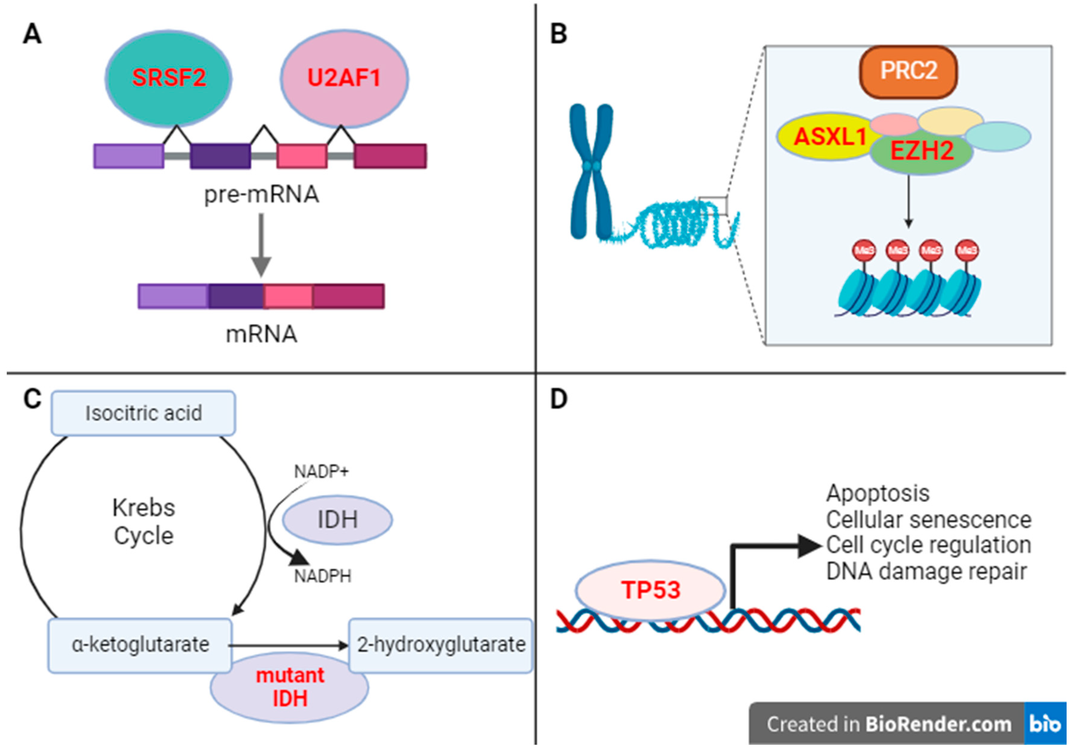
Figure 2. Functions of mutations other than JAK/MPL/CALR commonly found in MF. (A) Loss-of-function mutations in splicing factors SRSF2 and U2AF1 lead to altered mRNA splicing in myelofibrosis [49,66]. (B) Loss-of-function mutations in ASXL1 and EZH2 lead to impaired epigenetic regulation and are resultantly associated with high-risk MF disease features, including elevation of blasts in the peripheral blood and shortened overall survival [65,70]. (C) IDH 1/2 mutations have been studied in myeloid neoplasms and other malignancies, and when present, yield an oncometabolite termed 2-hyroxyglutarate and are correlated with inferior overall survival in myelofibrosis [73]. (D) TP53 loss-of-function mutations, lastly, are also identified in MF and are associated with both shortened median overall survival and increased risk for leukemic transformation [53].
Cancers 15 04654 g002
Table 1. Mutations implicated in the pathogenesis and/or molecular characterization of myelofibrosis (MF), polycythemia vera (PV), and essential thrombocythemia (ET). “Triple negative” refers to the designation of JAK2, CALR, and MPL wildtype myelofibrosis. The references for each incidence listed above are denoted by brackets.
MutationNormal FunctionIncidence in MF (%)Incidence in PV (%)Incidence in ET (%)
Driver mutations in myeloproliferative neoplasms
JAK2Nonreceptor tyrosine kinase mediating normal hematopoiesis, cell growth [19,20,21]35–57 [19,20,21]96–99 [11,12]50–60 [13,14,40]
MPLProtooncogene encoding for thrombopoietin receptor (TPOR) [23]0–7 [24,27]0–1 [24,25]5 [13,14]
CALRChaperone ER protein maintain calcium homeostasis and protein folding [31,32]25–53 [27,28]0 [12,24]25–27 [13,14]
“Triple negative”-10–15 [41]Rare35–52 [15]
Mutations in Regulators of Epigenetic Regulators
ASXL1Epigenetic regulator of genes involved in chromatin remodeling [42]13–38 [42]8.2 [43]7–20 [13]
EZH2Epigenetic regulator of posttranslational histone modification [44]6–13 [44]0–3 [45]2–4 [13,15]
Mutations in Regulators of Splicing Factors
SRSF2Splicing of pre-mRNA [46]3–17 [46,47]<2 [15]2–3 [13]
U2AF1Splicing factor involved in pre-mRNA splicing, promotion of HSPC survival [48]16–22 [48,49]<2 [15]1 [13]
Mutations in Regulators of Cellular Metabolism
IDHCatalyzation of isocitrate decarboxylation to yield 2-oxoglutarate (2-OG) [50]4 [50]<2 [15]1 [13]
Mutations in Tumor Suppressor Genes
TP53Tumor suppression, cell cycle regulation [51]1–13 [52,53]<2 [15]1–4 [13,45]
Ras/MAP Kinase Pathway Mutations
KRAS/NRASProto-oncogene with GTPase activity; K/Nras are two different Ras isoforms [8,54]2–15/4.4–14 [8,55]<2 (NRAS) [15]<2 (NRAS) [15]
CBLE3 ubiquitin ligase; negatively regulates various receptor tyrosine kinases [56]5–13 [55,57,58]<2 [15]1–2 [13]
NF1Inactivation of Ras-GTP through stimulation of constitutional GTPase [59]0–6 [55,58]RareRare
BRAFCytosolic serine/threonine kinase regulating MEK, ERK [60]0–1 [61]RareRare
PTPN11 (SHP2)PTPN11 encodes for SHP2, a tyrosine phosphatase activating Ras/MAP Kinase [36]1 [8]<2 [15]<2 [15]
Table 2. JAK and Ras/map Kinase Signaling inhibitors common used as cancer therapies. Abbreviations: GVHD: graft-versus-host disease; NSCLC: non-small cell lung cancer; MF: myelofibrosis. References are denoted by brackets.
InhibitorMechanism/TargetsFDA-Approved Treatment Indications
RuxolitinibJak1; Jak2 [84]Proliferative myelofibrosis;
Glucocorticoid-refractory GVHD [38,85]
FedratinibJak2; FLT3; BRD4 [38]Symptomatic, severe cytopenic MF [38]
PacritinibJak2; IRAK1 [38]Proliferative and moderately cytopenic MF [38]
DabrafenibBRAF, CRAF [86]BRAF V600E-mutated NSCLC; melanoma [87]
VemurafenibBRAF [86]BRAF V600E-mutated NSCLC; melanoma [87]
CobimetinibMek 1/2 [88]Melanoma; histiocytic disorders [87,89]
TrametinibMek 1/2 [90]Melanoma; histiocytic disorders [87,91]
Disclaimer/Publisher’s Note: The statements, opinions and data contained in all publications are solely those of the individual author(s) and contributor(s) and not of MDPI and/or the editor(s). MDPI and/or the editor(s) disclaim responsibility for any injury to people or property resulting from any ideas, methods, instructions or products referred to in the content.