

In Vivo Application of Carboranes for Boron Neutron Capture Therapy (BNCT): Structure, Formulation and Analytical Methods for Detection


Abstract
:Simple Summary
Abstract
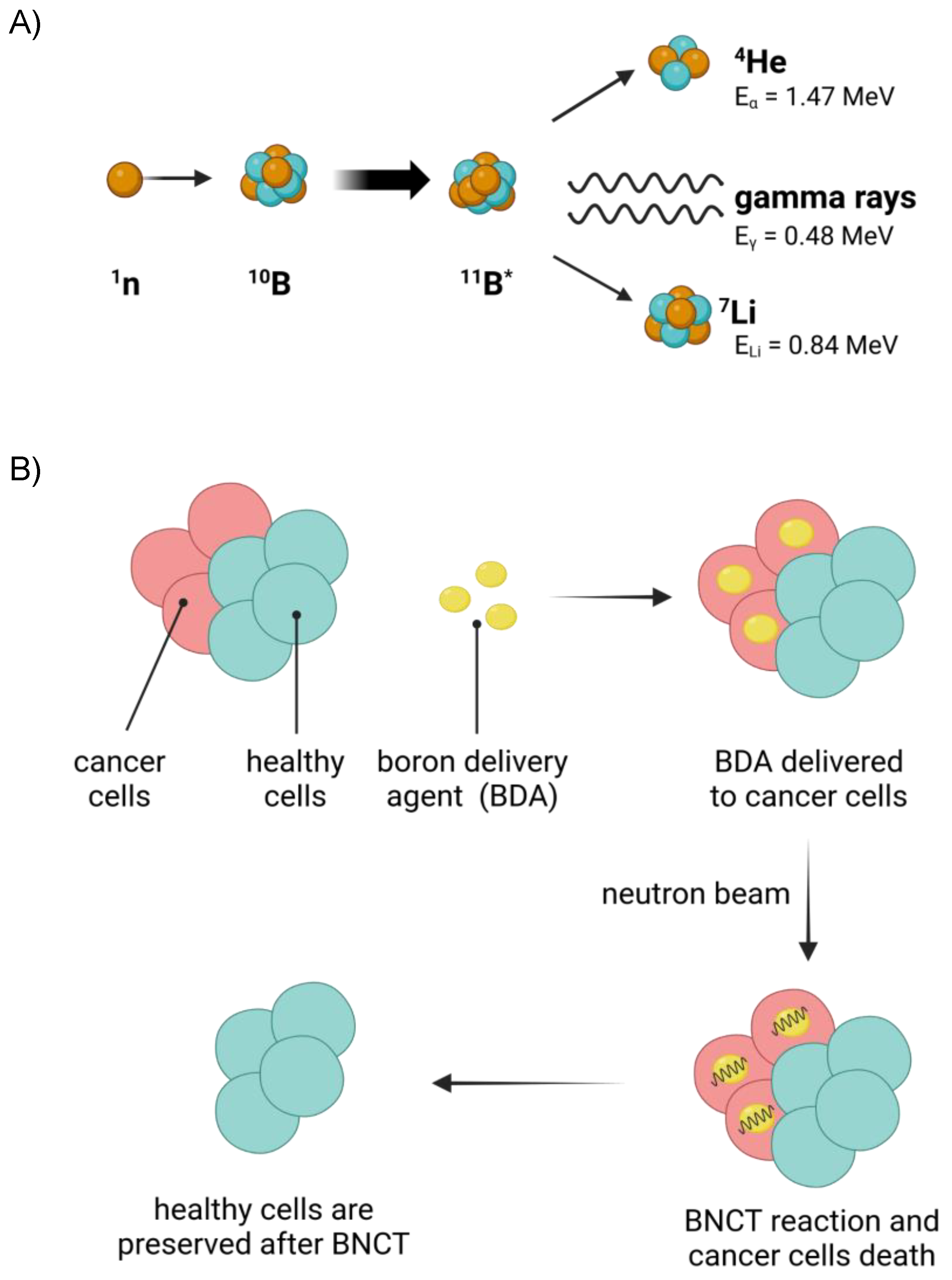
1. Introduction
2. In Vivo Studies of Carboranes for BNCT
2.1. Chemical Derivatization of Carboranes for In Vivo Studies
2.1.1. Carbohydrate Derivatives of Carboranes
2.1.2. Nucleoside Derivatives of Carboranes
2.1.3. Drug Derivatives of Carboranes
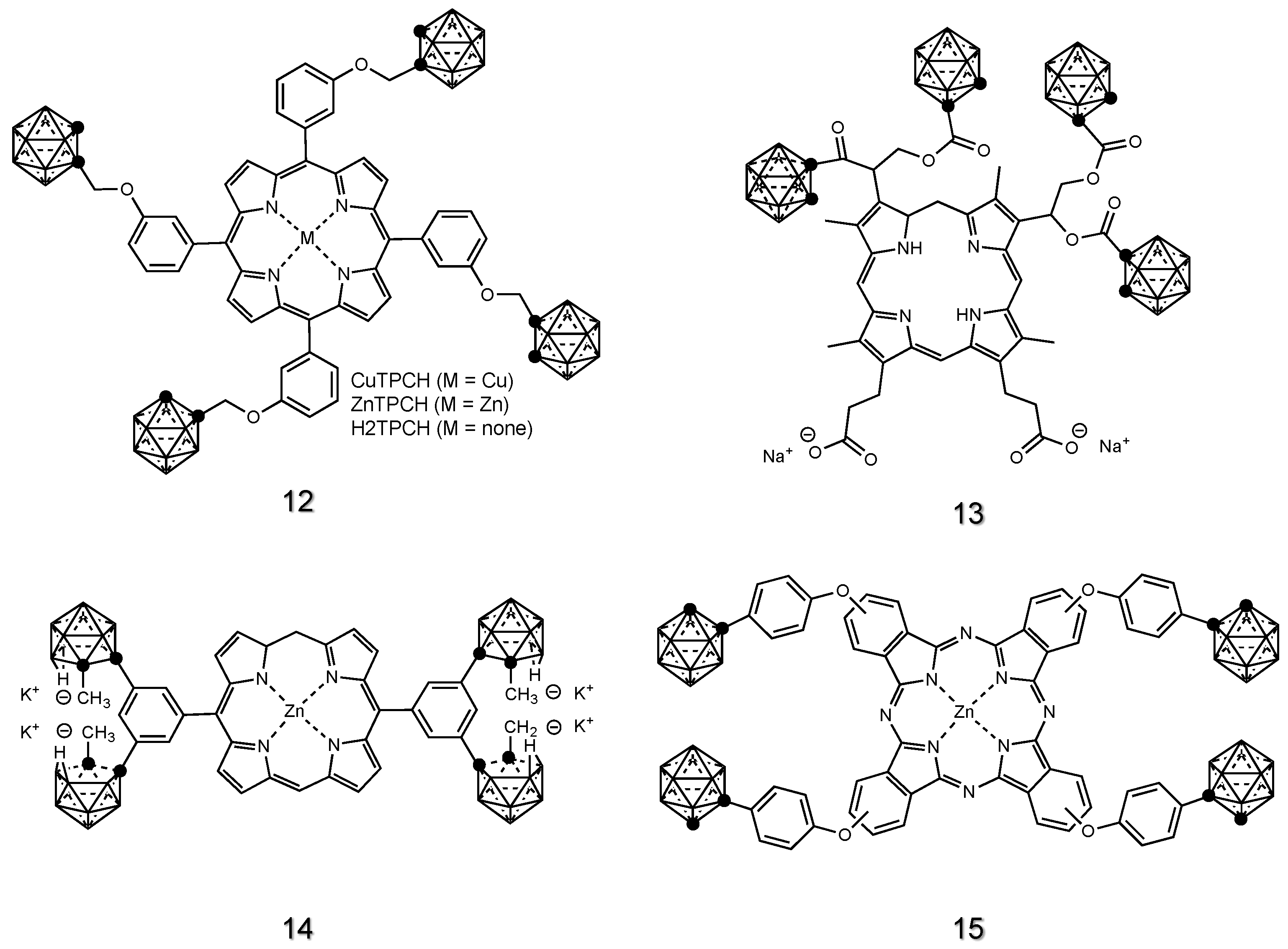
2.1.4. Porphyrin Derivatives of Carboranes
2.1.5. Imaging Agent Derivatives of Carboranes
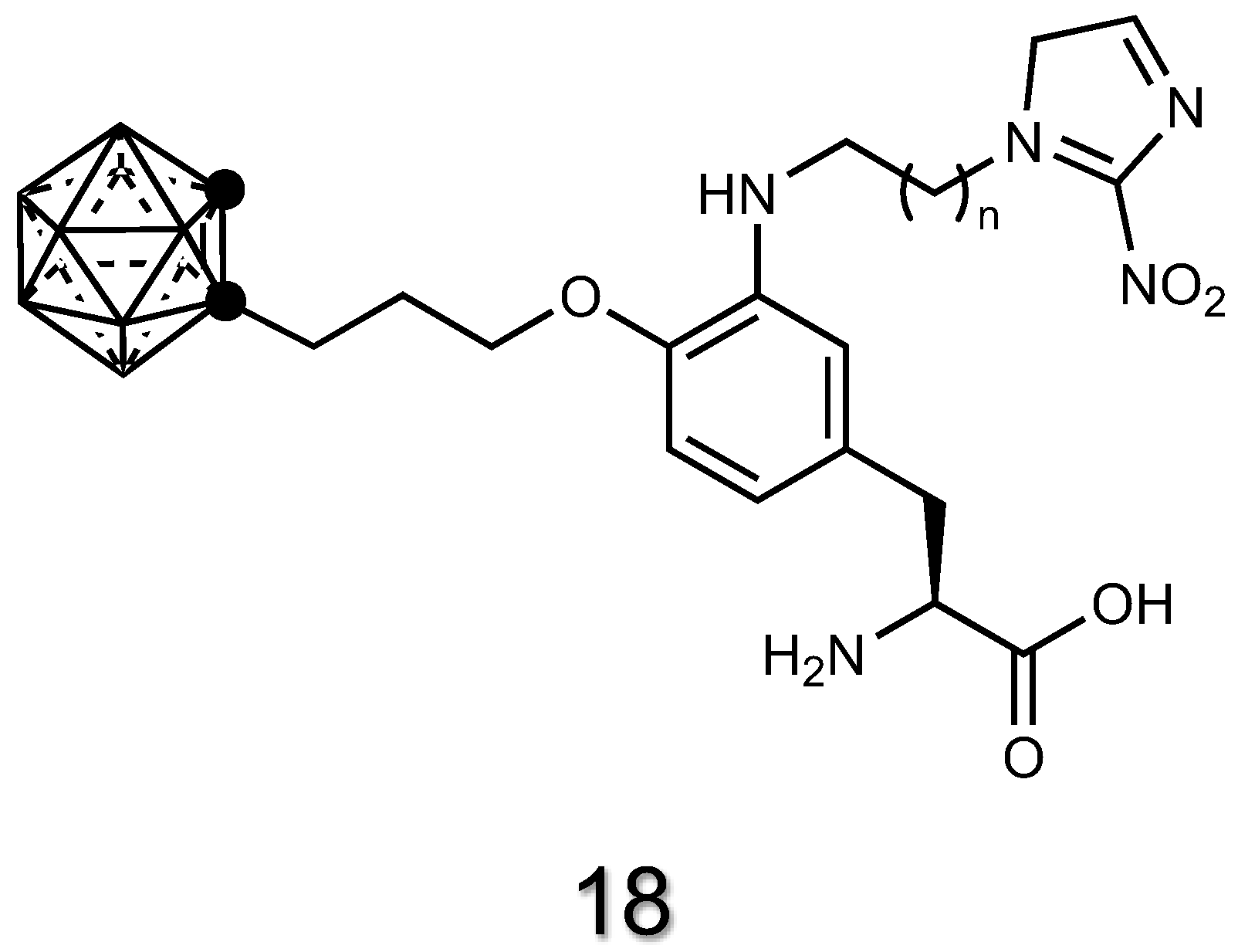
2.1.6. Amino Acid Derivatives of Carboranes
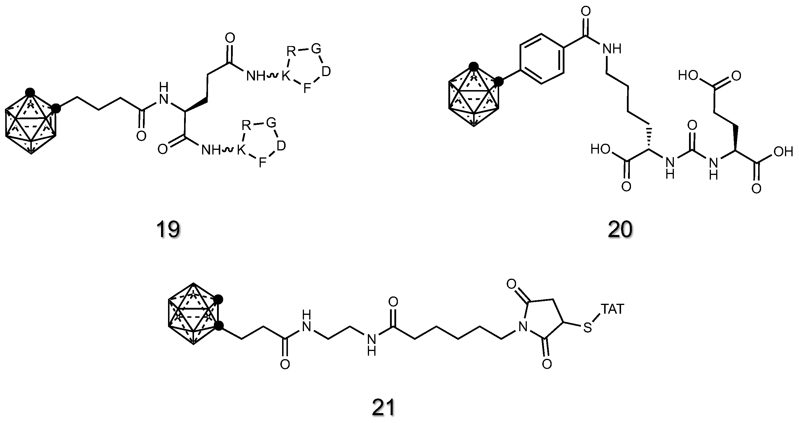
2.1.7. Peptide Derivatives of Carboranes
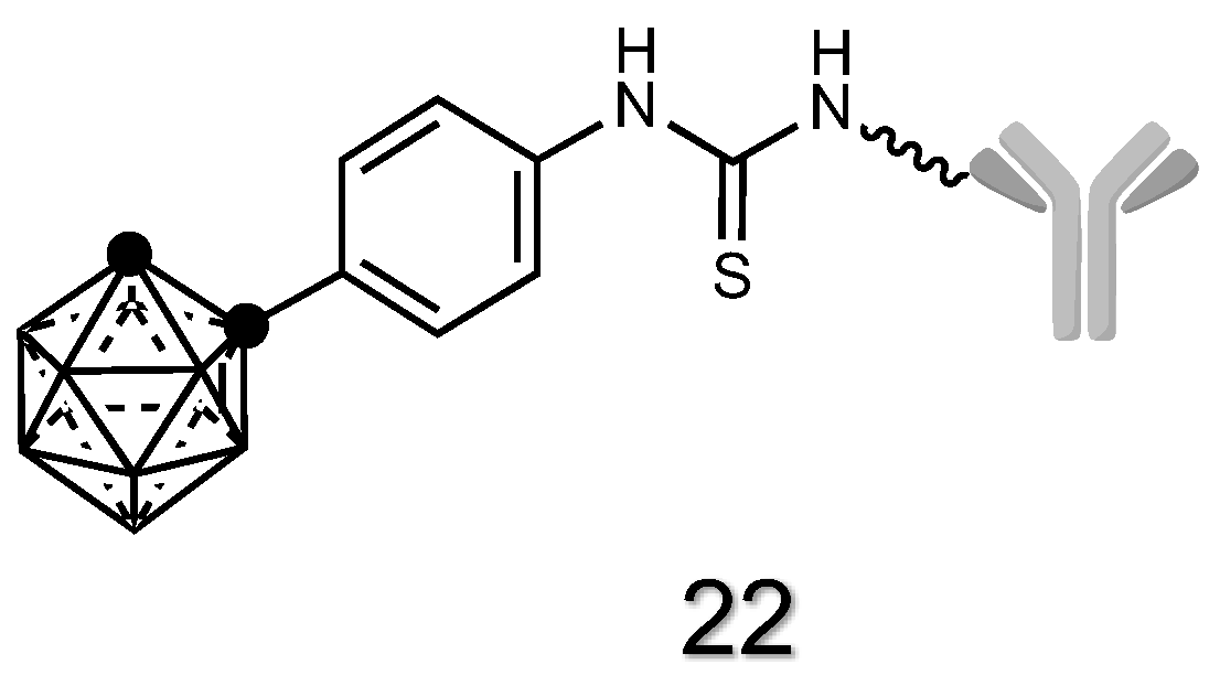
2.1.8. Antibody Derivatives of Carboranes
2.2. Incorporation of Carboranes in Drug Delivery Systems for In Vivo Studies
2.2.1. Supramolecular Carriers
2.2.2. Self-Assembled Supramolecular Carriers

2.2.3. Nanoparticles

2.3. Formulation and Administration of Carboranes for In Vivo Studies
2.4. Cancer Models Used for In Vivo Studies Using Carboranes in BNCT
3. Analytical Methodologies Used for Carborane Quantification in In Vivo Studies
3.1. Gamma-Ray Measurements
3.1.1. Prompt Gamma-Ray Neutron Activation Analysis (PGAA)
3.1.2. Carborane Labeling with Gamma-Ray Emitters
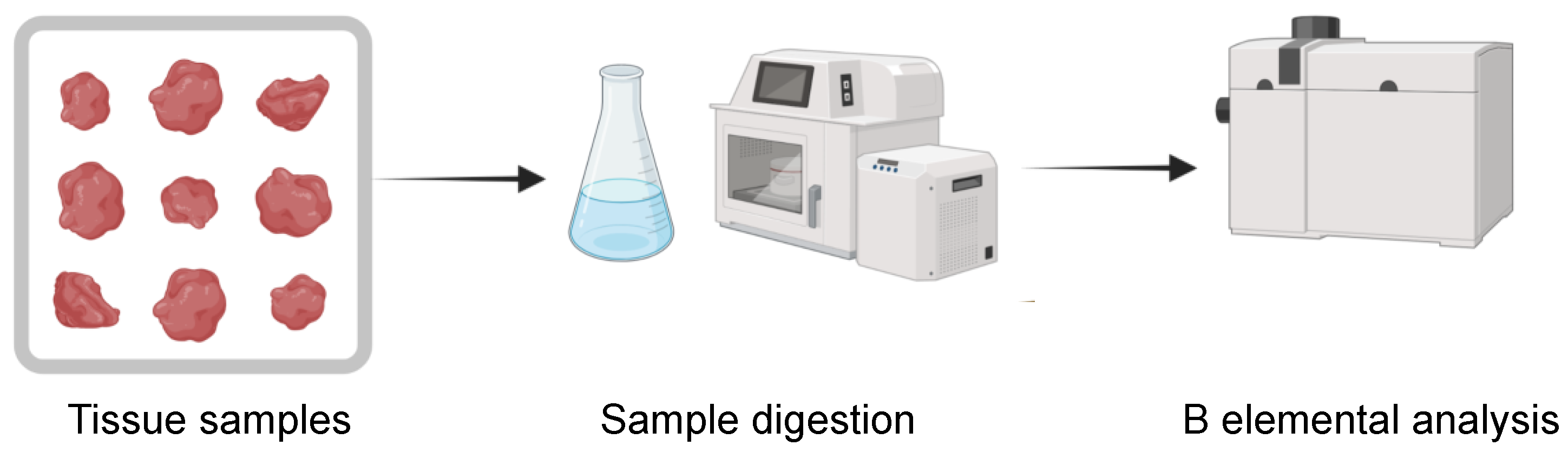
3.2. Boron Elemental Analysis Techniques
3.3. Fluorescence-Spectroscopy/Microscopy
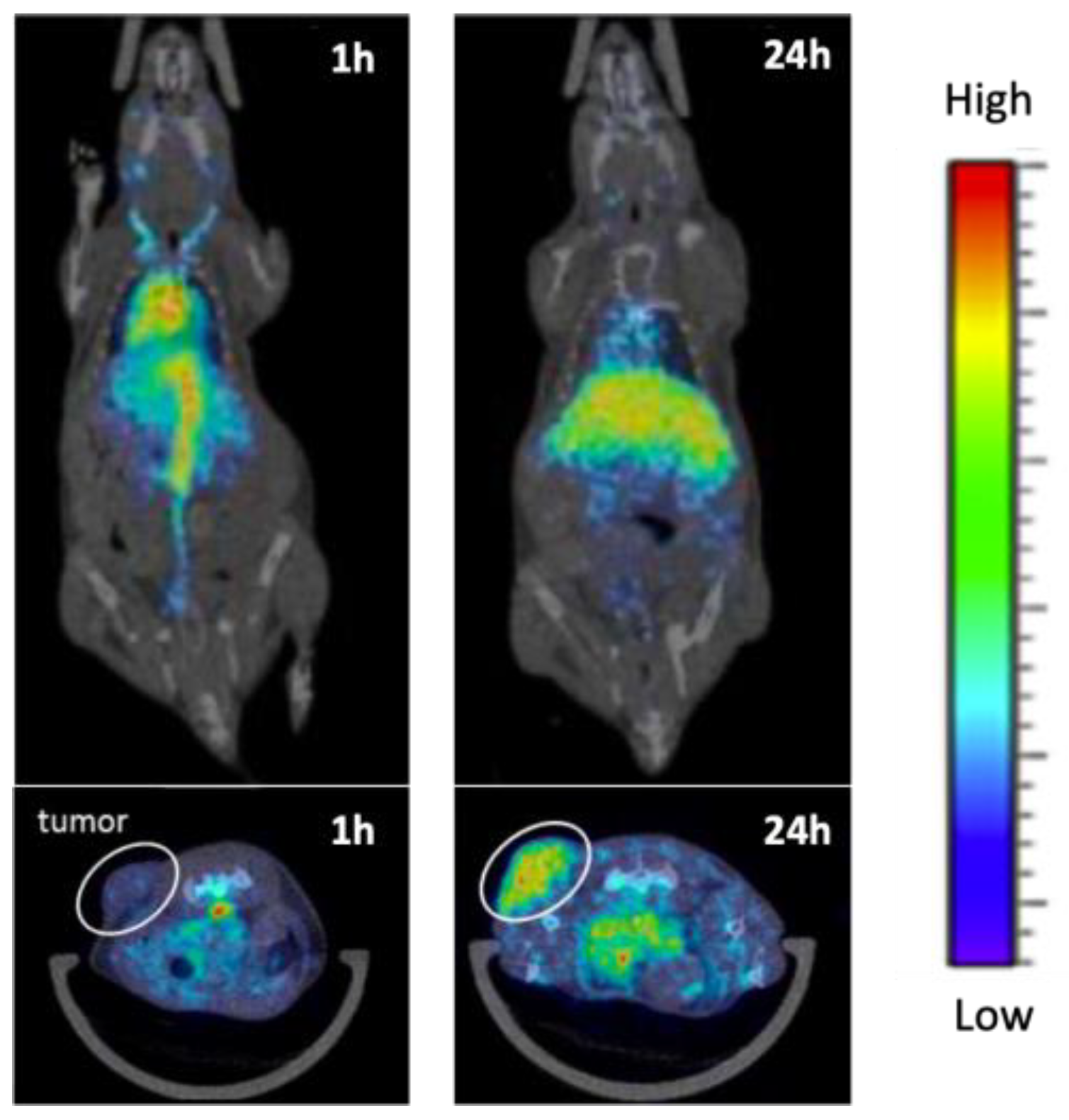
3.4. PET/SPECT
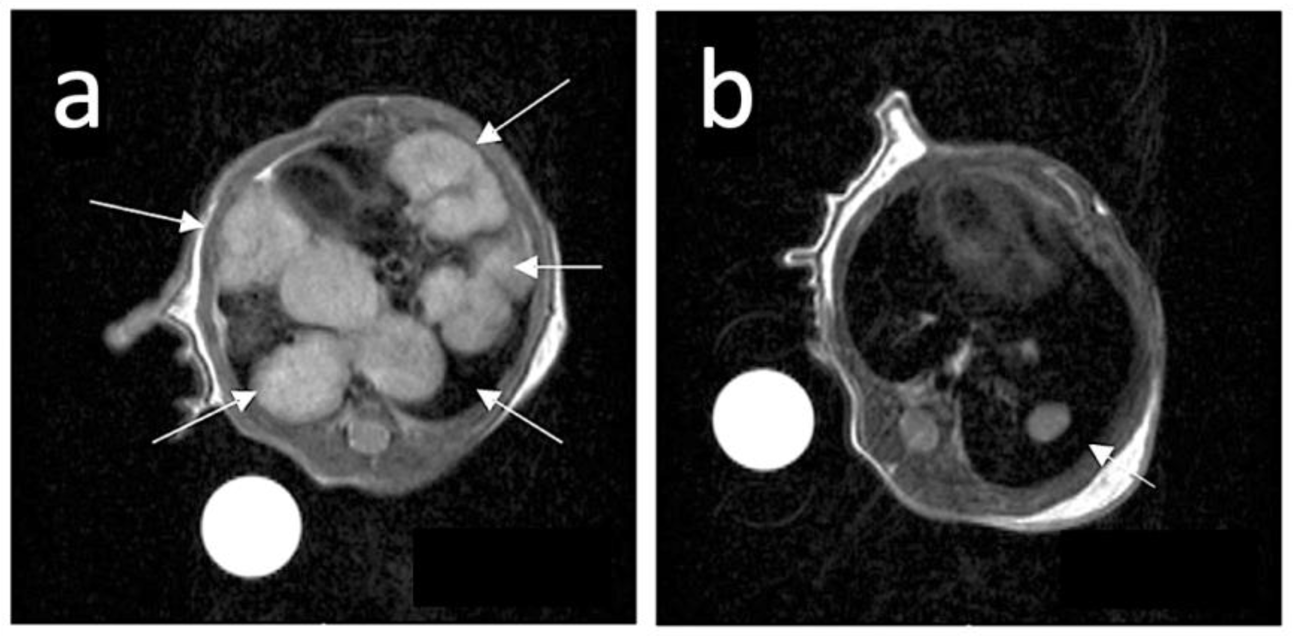
3.5. Magnetic Resonance Imaging (MRI)
3.6. Other Methodologies for Carborane In Vivo Analysis
4. Conclusions
Supplementary Materials
Author Contributions
Funding
Institutional Review Board Statement
Informed Consent Statement
Data Availability Statement
Conflicts of Interest
References
- Seneviratne, D.S.; Saifi, O.; Mackeyev, Y.; Malouff, T.; Krishnan, S. Next-Generation Boron Drugs and Rational Translational Studies Driving the Revival of BNCT. Cells 2023, 12, 1398. [Google Scholar] [CrossRef]
- Skwierawska, D.; López-Valverde, J.A.; Balcerzyk, M.; Leal, A. Clinical Viability of Boron Neutron Capture Therapy for Personalized Radiation Treatment. Cancers 2022, 14, 2865. [Google Scholar] [CrossRef] [PubMed]
- Jin, W.H.; Seldon, C.; Butkus, M.; Sauerwein, W.; Giap, H.B. A Review of Boron Neutron Capture Therapy: Its History and Current Challenges. Int. J. Part. Ther. 2022, 9, 71–82. [Google Scholar] [CrossRef] [PubMed]
- Miyatake, S.I.; Wanibuchi, M.; Hu, N.; Ono, K. Boron Neutron Capture Therapy for Malignant Brain Tumors. J. Neurooncol. 2020, 149, 1–11. [Google Scholar] [CrossRef] [PubMed]
- Wang, L.W.; Liu, Y.W.H.; Chu, P.Y.; Liu, H.M.; Peir, J.J.; Lin, K.H.; Huang, W.S.; Lo, W.L.; Lee, J.C.; Lin, T.Y.; et al. Boron Neutron Capture Therapy Followed by Image-Guided Intensity-Modulated Radiotherapy for Locally Recurrent Head and Neck Cancer: A Prospective Phase I/II Trial. Cancers 2023, 15, 2762. [Google Scholar] [CrossRef] [PubMed]
- Kanno, H.; Nagata, H.; Ishiguro, A.; Tsuzuranuki, S.; Nakano, S.; Nonaka, T.; Kiyohara, K.; Kimura, T.; Sugawara, A.; Okazaki, Y.; et al. Designation Products: Boron Neutron Capture Therapy for Head and Neck Carcinoma. Oncologist 2021, 26, e1250–e1255. [Google Scholar] [CrossRef]
- Malouff, T.D.; Seneviratne, D.S.; Ebner, D.K.; Stross, W.C.; Waddle, M.R.; Trifiletti, D.M.; Krishnan, S. Boron Neutron Capture Therapy: A Review of Clinical Applications. Front. Oncol. 2021, 11, 601820. [Google Scholar] [CrossRef]
- Wang, S.; Zhang, Z.; Miao, L.; Li, Y. Boron Neutron Capture Therapy: Current Status and Challenges. Front. Oncol. 2022, 12, 788770. [Google Scholar] [CrossRef]
- Fukuda, H. Boron Neutron Capture Therapy (BNCT) for Cutaneous Malignant Melanoma Using 10B-p-Boronophenylalanine (BPA) with Special Reference to the Radiobiological Basis and Clinical Results. Cells 2021, 10, 2881. [Google Scholar] [CrossRef]
- Koivunoro, H.; Kankaanranta, L.; Seppälä, T.; Haapaniemi, A.; Mäkitie, A.; Joensuu, H. Boron Neutron Capture Therapy for Locally Recurrent Head and Neck Squamous Cell Carcinoma: An Analysis of Dose Response and Survival. Radiother. Oncol. 2019, 137, 153–158. [Google Scholar] [CrossRef]
- Hu, K.; Yang, Z.; Zhang, L.; Xie, L.; Wang, L.; Xu, H.; Josephson, L.; Liang, S.H.; Zhang, M.R. Boron Agents for Neutron Capture Therapy. Coord. Chem. Rev. 2020, 405, 213139. [Google Scholar] [CrossRef]
- Matsumura, A.; Asano, T.; Hirose, K.; Igaki, H.; Kawabata, S.; Kumada, H. Initiatives Toward Clinical Boron Neutron Capture Therapy in Japan. Cancer Biother. Radiopharm. 2023, 38, 201–207. [Google Scholar] [CrossRef] [PubMed]
- Zavjalov, E.; Zaboronok, A.; Kanygin, V.; Kasatova, A.; Kichigin, A.; Mukhamadiyarov, R.; Razumov, I.; Sycheva, T.; Mathis, B.J.; Maezono, S.E.B.; et al. Accelerator-Based Boron Neutron Capture Therapy for Malignant Glioma: A Pilot Neutron Irradiation Study Using Boron Phenylalanine, Sodium Borocaptate and Liposomal Borocaptate with a Heterotopic U87 Glioblastoma Model in SCID Mice. Int. J. Radiat. Biol. 2020, 96, 868–878. [Google Scholar] [CrossRef]
- Wang, S.; Igawa, K.; Ogawara, R.; Suda, M.; Hamano, T.; Ibaragi, S.; Hanafusa, T.; Ichikawa, Y.; Sasaki, A. The Accelerator-Based Boron Neutron Capture Reaction Evaluation System for Head and Neck Cancer. Appl. Radiat. Isot. 2020, 165, 109271. [Google Scholar] [CrossRef] [PubMed]
- Sato, E.; Zaboronok, A.; Yamamoto, T.; Nakai, K.; Taskaev, S.; Volkova, O.; Mechetina, L.; Taranin, A.; Kanygin, V.; Isobe, T.; et al. Radiobiological Response of U251MG, CHO-K1 and V79 Cell Lines to Accelerator-Based Boron Neutron Capture Therapy. J. Radiat. Res. 2018, 59, 101–107. [Google Scholar] [CrossRef]
- Nuez-Martínez, M.; Queralt-Martín, M.; Muñoz-Juan, A.; Aguilella, V.M.; Laromaine, A.; Teixidor, F.; Viñas, C.; Pinto, C.G.; Pinheiro, T.; Guerreiro, J.F.; et al. Boron clusters (ferrabisdicarbollides) shaping the future as radiosensitizers for multimodal (chemo/radio/PBFR) therapy of glioblastoma. J. Mater. Chem. B 2022, 10, 9794–9815. [Google Scholar] [CrossRef]
- Boron Agents. Japanese Society of Neutron Capture Therapy. Available online: http://www.jsnct.jp/e/about_nct/houso.html (accessed on 29 August 2023).
- Lamba, M.; Goswami, A.; Bandyopadhyay, A. A Periodic Development of BPA and BSH Based Derivatives in Boron Neutron Capture Therapy (BNCT). Chem. Commun. 2021, 57, 827. [Google Scholar] [CrossRef]
- Fukuda, H.; Hiratsuka, J. Pharmacokinetics of 10B-p-Boronophenylalanine (BPA) in the Blood and Tumors in Human Patients: A Critical Review with Special Reference to Tumor-to-Blood (T/B) Ratios Using Resected Tumor Samples. Appl. Radiat. Isot. 2020, 166, 109308. [Google Scholar] [CrossRef]
- Hideghéty, K.; Sauerwein, W.; Wittig, A.; Götz, C.; Paquis, P.; Grochulla, F.; Haselsberger, K.; Wolbers, J.; Moss, R.; Huiskamp, R.; et al. Tissue Uptake of BSH in Patients with Glioblastoma in the EORTC 11961 Phase I BNCT Trial. J. NeuroOncol. 2003, 62, 145–156. [Google Scholar] [CrossRef]
- Puris, E.; Gynther, M.; Auriola, S.; Huttunen, K.M. L-Type Amino Acid Transporter 1 as a Target for Drug Delivery. Pharm. Res. 2020, 37, 88. [Google Scholar] [CrossRef]
- Wongthai, P.; Hagiwara, K.; Miyoshi, Y.; Wiriyasermkul, P.; Wei, L.; Ohgaki, R.; Kato, I.; Hamase, K.; Nagamori, S.; Kanai, Y. Boronophenylalanine, a Boron Delivery Agent for Boron Neutron Capture Therapy, Is Transported by ATB0,+, LAT1 and LAT2. Cancer Sci. 2015, 106, 279–286. [Google Scholar] [CrossRef] [PubMed]
- Zaidlewicz, M.; Sokół, W.; Wolan, A.; Cytarska, J.; Tafelska-Kaczmarek, A.; Dzieleńdziak, A.; Prewysz-Kwinto, A. Enolboration of Conjugated Ketones and Synthesis of β-Amino Alcohols and Boronated α-Amino Acids. Pure Appl. Chem. 2003, 75, 1349–1355. [Google Scholar] [CrossRef]
- Nakamura, H.; Fujiwara, M.; Yamamoto, Y. A Practical Method for the Synthesis of Enantiomerically Pure 4-Borono-L-Phenylalanine. Bull. Chem. Soc. Jpn. 2001, 73, 231–235. [Google Scholar] [CrossRef]
- Nakamura, H.; Fujiwara, M.; Yamamoto, Y. A Concise Synthesis of Enantiomerically Pure L-(4-Boronophenyl)Alanine from L-Tyrosine. J. Org. Chem. 1998, 63, 7529–7530. [Google Scholar] [CrossRef] [PubMed]
- Snyder, H.R.; Reedy, A.J.; Lennarz, W.J. Synthesis of Aromatic Boronic Acids. Aldehydo Boronic Acids and a Boronic Acid Analog of Tyrosine. J. Am. Chem. Soc. 1958, 80, 835–838. [Google Scholar] [CrossRef]
- Zhang, Z.; Yong, Z.; Jin, C.; Song, Z.; Zhu, S.; Liu, T.; Chen, Y.; Chong, Y.; Chen, X.; Zhou, Y. Biodistribution Studies of Boronophenylalanine in Different Types of Skin Melanoma. Appl. Radiat. Isot. 2020, 163, 109215. [Google Scholar] [CrossRef]
- Kortesniemi, M.; Seppälä, T.; Auterinen, I.; Savolainen, S. Enhanced Blood Boron Concentration Estimation for BPA-F Mediated BNCT. Appl. Radiat. Isot. 2004, 61, 823–827. [Google Scholar] [CrossRef]
- Henriksson, R.; Capala, J.; Michanek, A.; Lindahl, S.Å.; Salford, L.G.; Franzén, L.; Blomquist, E.; Westlin, J.E.; Bergenheim, A.T. Boron Neutron Capture Therapy (BNCT) for Glioblastoma Multiforme: A Phase II Study Evaluating a Prolonged High-Dose of Boronophenylalanine (BPA). Radiother. Oncol. 2008, 88, 183–191. [Google Scholar] [CrossRef]
- Kulvik, M.; Kallio, M.; Laakso, J.; Vähätalo, J.; Hermans, R.; Järviluoma, E.; Paetau, A.; Rasilainen, M.; Ruokonen, I.; Seppälä, M.; et al. Biodistribution of Boron after Intravenous 4-Dihydroxyborylphenylalanine-Fructose (BPA-F) Infusion in Meningioma and Schwannoma Patients: A Feasibility Study for Boron Neutron Capture Therapy. Appl. Radiat. Isot. 2015, 106, 207–212. [Google Scholar] [CrossRef]
- De Simone, U.; Manzo, L.; Ferrari, C.; Bakeine, J.; Locatelli, C.; Coccini, T. Short and Long-Term Exposure of CNS Cell Lines to BPA-f a Radiosensitizer for Boron Neutron Capture Therapy: Safety Dose Evaluation by a Battery of Cytotoxicity Tests. Neurotoxicology 2013, 35, 84–90. [Google Scholar] [CrossRef]
- Heikkinen, S.; Savolainen, S.; Melkko, P. In Vitro Studies on Stability of L-p-Boronophenylalanine–Fructose Complex (BPA-F). J. Radiat. Res. 2011, 52, 360–364. [Google Scholar] [CrossRef] [PubMed]
- Kankaanranta, L.; Seppälä, T.; Koivunoro, H.; Välimäki, P.; Beule, A.; Collan, J.; Kortesniemi, M.; Uusi-Simola, J.; Kotiluoto, P.; Auterinen, I.; et al. L-Boronophenylalanine-Mediated Boron Neutron Capture Therapy for Malignant Glioma Progressing After External Beam Radiation Therapy: A Phase I Study. Int. J. Radiat. Oncol.* Biol.* Phys. 2011, 80, 369–376. [Google Scholar] [CrossRef] [PubMed]
- Sköld, K.; H-Stenstam, B.; Diaz, A.Z.; Giusti, V.; Pellettieri, L.; Hopewell, J.W. Boron Neutron Capture Therapy for Glioblastoma Multiforme: Advantage of Prolonged Infusion of BPA-f. Acta Neurol. Scand. 2010, 122, 58–62. [Google Scholar] [CrossRef] [PubMed]
- Tsurubuchi, T.; Shirakawa, M.; Kurosawa, W.; Matsumoto, K.; Ubagai, R.; Umishio, H.; Suga, Y.; Yamazaki, J.; Arakawa, A.; Maruyama, Y.; et al. Evaluation of a Novel Boron-Containing α-d-Mannopyranoside for BNCT. Cells 2020, 9, 1277. [Google Scholar] [CrossRef]
- Schaffran, T.; Lissel, F.; Samatanga, B.; Karlsson, G.; Burghardt, A.; Edwards, K.; Winterhalter, M.; Peschka-Süss, R.; Schubert, R.; Gabel, D. Dodecaborate Cluster Lipids with Variable Headgroups for Boron Neutron Capture Therapy: Synthesis, Physical–Chemical Properties and Toxicity. J. Organomet. Chem. 2009, 694, 1708–1712. [Google Scholar] [CrossRef]
- Kikuchi, S.; Kanoh, D.; Sato, S.; Sakurai, Y.; Suzuki, M.; Nakamura, H. Maleimide-Functionalized Closo-Dodecaborate Albumin Conjugates (MID-AC): Unique Ligation at Cysteine and Lysine Residues Enables Efficient Boron Delivery to Tumor for Neutron Capture Therapy. J. Control. Release 2016, 237, 160–167. [Google Scholar] [CrossRef]
- Nishimura, K.; Harrison, S.; Kawai, K.; Morita, T.; Miura, K.; Okada, S.; Nakamura, H. Iodophenyl-Conjugated Closo-Dodecaborate as a Promising Small Boron Molecule That Binds to Serum Albumin and Accumulates in Tumor. Bioorg. Med. Chem. Lett. 2022, 72, 128869. [Google Scholar] [CrossRef]
- Kashiwagi, H.; Hattori, Y.; Kawabata, S.; Kayama, R.; Yoshimura, K.; Fukuo, Y.; Kanemitsu, T.; Shiba, H.; Hiramatsu, R.; Takami, T.; et al. Multi-Targeted Neutron Capture Therapy Combined with an 18 KDa Translocator Protein-Targeted Boron Compound Is an Effective Strategy in a Rat Brain Tumor Model. Cancers 2023, 15, 1034. [Google Scholar] [CrossRef]
- Tsujino, K.; Kashiwagi, H.; Nishimura, K.; Kayama, R.; Yoshimura, K.; Fukuo, Y.; Shiba, H.; Hiramatsu, R.; Nonoguchi, N.; Furuse, M.; et al. Improved Boron Neutron Capture Therapy Using Integrin Avβ3-Targeted Long-Retention-Type Boron Carrier in a F98 Rat Glioma Model. Biology 2023, 12, 377. [Google Scholar] [CrossRef]
- Nishimura, K.; Kashiwagi, H.; Morita, T.; Fukuo, Y.; Okada, S.; Miura, K.; Matsumoto, Y.; Sugawara, Y.; Enomoto, T.; Suzuki, M.; et al. Efficient Neutron Capture Therapy of Glioblastoma with Pteroyl-Closo-Dodecaborate-Conjugated 4-(p-Iodophenyl)Butyric Acid (PBC-IP). J. Control. Release 2023, 360, 249–259. [Google Scholar] [CrossRef]
- Nakamura, H.; Ueda, N.; Ban, H.S.; Ueno, M.; Tachikawa, S. Design and Synthesis of Fluorescence-Labeled Closo-Dodecaborate Lipid: Its Liposome Formation and in Vivo Imaging Targeting of Tumors for Boron Neutron Capture Therapy. Org. Biomol. Chem. 2012, 10, 1374–1380. [Google Scholar] [CrossRef]
- Asano, R.; Nagami, A.; Fukumoto, Y.; Miura, K.; Yazama, F.; Ito, H. Bioorganic & Medicinal Chemistry Letters Synthesis and Biological Evaluation of New Boron-Containing Chlorin Derivatives as Agents for Both Photodynamic Therapy and Boron Neutron Capture Therapy of Cancer. Bioorg. Med. Chem. Lett. 2014, 24, 1339–1343. [Google Scholar] [CrossRef] [PubMed]
- Takeuchi, K.; Hattori, Y.; Kawabata, S.; Futamura, G.; Hiramatsu, R.; Wanibuchi, M.; Tanaka, H.; Masunaga, S.I.; Ono, K.; Miyatake, S.I.; et al. Synthesis and Evaluation of Dodecaboranethiol Containing Kojic Acid (KA-BSH) as a Novel Agent for Boron Neutron Capture Therapy. Cells 2020, 9, 1551. [Google Scholar] [CrossRef] [PubMed]
- Monti Hughes, A. Importance of Radiobiological Studies for the Advancement of Boron Neutron Capture Therapy (BNCT). Expert. Rev. Mol. Med. 2022, 24, e14. [Google Scholar] [CrossRef] [PubMed]
- Kimura, S.; Masunaga, S.I.; Harada, T.; Kawamura, Y.; Ueda, S.; Okuda, K.; Nagasawa, H. Synthesis and Evaluation of Cyclic RGD-Boron Cluster Conjugates to Develop Tumor-Selective Boron Carriers for Boron Neutron Capture Therapy. Bioorg. Med. Chem. 2011, 19, 1721–1728. [Google Scholar] [CrossRef]
- Iguchi, Y.; Michiue, H.; Kitamatsu, M.; Hayashi, Y.; Takenaka, F.; Nishiki, T.i.; Matsui, H. Tumor-Specific Delivery of BSH-3R for Boron Neutron Capture Therapy and Positron Emission Tomography Imaging in a Mouse Brain Tumor Model. Biomaterials 2015, 56, 10–17. [Google Scholar] [CrossRef]
- Coninx, S.; Kalot, G.; Godard, A.; Bodio, E.; Goze, C.; Sancey, L.; Auzély-Velty, R. Tailored Hyaluronic Acid-Based Nanogels as Theranostic Boron Delivery Systems for Boron Neutron Cancer Therapy. Int. J. Pharm. X 2022, 4, 100134. [Google Scholar] [CrossRef]
- Michiue, H.; Sakurai, Y.; Kondo, N.; Kitamatsu, M.; Bin, F.; Nakajima, K.; Hirota, Y.; Kawabata, S.; Nishiki, T.i.; Ohmori, I.; et al. The Acceleration of Boron Neutron Capture Therapy Using Multi-Linked Mercaptoundecahydrododecaborate (BSH) Fused Cell-Penetrating Peptide. Biomaterials 2014, 35, 3396–3405. [Google Scholar] [CrossRef]
- Kalot, G.; Godard, A.; Busser, B.; Pliquett, J.; Broekgaarden, M.; Motto-Ros, V.; Wegner, K.D.; Resch-Genger, U.; Köster, U.; Denat, F.; et al. Aza-BODIPY: A New Vector for Enhanced Theranostic Boron Neutron Capture Therapy Applications. Cells 2020, 9, 1953. [Google Scholar] [CrossRef]
- Hawthorne, M.F. The Role of Chemistry in the Development of Boron Neutron Capture Therapy of Cancer. Angew. Chem. Int. Ed. Engl. 1993, 32, 950–984. [Google Scholar] [CrossRef]
- Soloway, A.H.; Tjarks, W.; Barnum, B.A.; Rong, F.G.; Barth, R.F.; Codogni, I.M.; Wilson, J.G. The Chemistry of Neutron Capture Therapy. Chem. Rev. 1998, 98, 1515–1562. [Google Scholar] [CrossRef] [PubMed]
- Hosmane, N.S. Boron Science: New Technologies and Applications; CRC Press: Boca Raton, FL, USA, 2016. [Google Scholar] [CrossRef]
- Hosmane, N.S.; Eagling, R. Handbook of Boron Science: With Applications in Organometallics, Catalysis, Materials and Medicine; World Scientific Publishing Company: Singapore, 2018; Volume 1–4, pp. 1–1051. [Google Scholar] [CrossRef]
- Hey-Hawkins, E. Boron-Based Compounds: Potential and Emerging Applications in Medicine; John Wiley & Sons: Hoboken, NJ, USA, 2018; 470p. [Google Scholar]
- Grimes, R.N. Carboranes; Academic Press: London, UK, 2016; ISBN 9780128018941. [Google Scholar]
- Mingos, D.M.P. Structure and Bonding; Springer: Cham, Switzerland, 2021; Volume 187. [Google Scholar] [CrossRef]
- Poater, J.; Viñas, C.; Bennour, I.; Escayola, S.; Solà, M.; Teixidor, F. Too Persistent to Give Up: Aromaticity in Boron Clusters Survives Radical Structural Changes. J. Am. Chem. Soc. 2020, 142, 9396–9407. [Google Scholar] [CrossRef] [PubMed]
- Scholz, M.; Hey-Hawkins, E. Carbaboranes as Pharmacophores: Properties, Synthesis, and Application Strategies. Chem. Rev. 2011, 111, 7035–7062. [Google Scholar] [CrossRef] [PubMed]
- Marfavi, A.; Kavianpour, P.; Rendina, L.M. Carboranes in Drug Discovery, Chemical Biology and Molecular Imaging. Nat. Rev. Chem. 2022, 6, 486–504. [Google Scholar] [CrossRef]
- Komarnicka, K.; Lesiów, M.; Jaros, S.; Messner, K.; Vuong, B.; Tranmer, G.K. The Boron Advantage: The Evolution and Diversification of Boron’s Applications in Medicinal Chemistry. Pharmaceuticals 2022, 15, 264. [Google Scholar] [CrossRef]
- Fink, K.; Uchman, M. Boron Cluster Compounds as New Chemical Leads for Antimicrobial Therapy. Coord. Chem. Rev. 2021, 431, 213684. [Google Scholar] [CrossRef]
- Issa, F.; Kassiou, M.; Rendina, L.M. Boron in Drug Discovery: Carboranes as Unique Pharmacophores in Biologically Active Compounds. Chem. Rev. 2011, 111, 5701–5722. [Google Scholar] [CrossRef]
- Fujii, S. Expanding the Chemical Space of Hydrophobic Pharmacophores: The Role of Hydrophobic Substructures in the Development of Novel Transcription Modulators. Medchemcomm 2016, 7, 1082–1092. [Google Scholar] [CrossRef]
- Valliant, J.F.; Guenther, K.J.; King, A.S.; Morel, P.; Schaffer, P.; Sogbein, O.O.; Stephenson, K.A. The Medicinal Chemistry of Carboranes. Coord. Chem. Rev. 2002, 232, 173–230. [Google Scholar] [CrossRef]
- Chen, Y.; Du, F.; Tang, L.; Xu, J.; Zhao, Y.; Wu, X.; Li, M.; Shen, J.; Wen, Q.; Cho, C.H.; et al. Carboranes as Unique Pharmacophores in Antitumor Medicinal Chemistry. Mol. Ther. Oncolytics 2022, 24, 400–416. [Google Scholar] [CrossRef]
- Qiu, Z. Recent Advances in Transition Metal-Mediated Functionalization of o-Carboranes. Tetrahedron Lett. 2015, 56, 963–971. [Google Scholar] [CrossRef]
- Qiu, Z.; Xie, Z. Functionalization of O-Carboranes via Carboryne Intermediates. Chem. Soc. Rev. 2022, 51, 3164. [Google Scholar] [CrossRef] [PubMed]
- Zhang, X.; Yan, H. Transition Metal-Induced B–H Functionalization of o-Carborane. Coord. Chem. Rev. 2019, 378, 466–482. [Google Scholar] [CrossRef]
- Baek, Y.; Cheong, K.; Ko, G.H.; Han, G.U.; Han, S.H.; Kim, D.; Kim, D.; Lee, K.; Lee, P.H. Iridium-Catalyzed Cyclative Indenylation and Dienylation through Sequential B(4)-C Bond Formation, Cyclization, and Elimination from o-Carboranes and Propargyl Alcohols. J. Am. Chem. Soc. 2020, 142, 9890–9895. [Google Scholar] [CrossRef] [PubMed]
- Gazvoda, M.; Dhanjee, H.H.; Rodriguez, J.; Brown, J.S.; Farquhar, C.E.; Truex, N.L.; Loas, A.; Buchwald, S.L.; Pentelute, B.L. Palladium-Mediated Incorporation of Carboranes into Small Molecules, Peptides, and Proteins. J. Am. Chem. Soc. 2022, 144, 7852–7860. [Google Scholar] [CrossRef]
- Li, C.X.; Yan, H. Recent Trends and Tactics in Facile Functionalization of Neutral Icosahedral Carboranes (C2B10H12) and Nido-Carborane (7,8-C2B9H12−). Adv. Catal. 2022, 71, 201–283. [Google Scholar] [CrossRef]
- Imperio, D.; Panza, L. Sweet Boron: Boron-Containing Sugar Derivatives as Potential Agents for Boron Neutron Capture Therapy. Symmetry 2022, 14, 182. [Google Scholar] [CrossRef]
- Satapathy, R.; Dash, B.P.; Mahanta, C.S.; Swain, B.R.; Jena, B.B.; Hosmane, N.S. Glycoconjugates of Polyhedral Boron Clusters Dedicated to Professor Russell Grimes on the Occasion of His 80th Birthday. J. Organomet. Chem. 2015, 798, 13–23. [Google Scholar] [CrossRef]
- Orlova, A.V.; Kononov, L.O. Synthesis of conjugates of polyhedral boron compounds with carbohydrates. Russian Chem. Rev. 2009, 78, 629–642. [Google Scholar] [CrossRef]
- Wang, J.; Zhang, Y.; Lu, Q.; Xing, D.; Zhang, R. Exploring Carbohydrates for Therapeutics: A Review on Future Directions. Front. Pharmacol. 2021, 12, 756724. [Google Scholar] [CrossRef]
- Cao, X.; Du, X.; Jiao, H.; An, Q.; Chen, R.; Fang, P.; Wang, J.; Yu, B. Carbohydrate-Based Drugs Launched during 2000–2021. Acta Pharm Sin B 2022, 12, 3783–3821. [Google Scholar] [CrossRef] [PubMed]
- Di, X.; Liang, X.; Shen, C.; Pei, Y.; Wu, B.; He, Z. Carbohydrates Used in Polymeric Systems for Drug Delivery: From Structures to Applications. Pharmaceutics 2022, 14, 739. [Google Scholar] [CrossRef] [PubMed]
- Ekholm, F.S.; Matovic, J.; Jarvinen, J.; Bland, H.C.; Sokka, I.K.; Imlimthan, S.; Huttunen, K.M.; Timonen, J.; Peraniemi, S.; Aitio, O.; et al. Addressing the Biochemical Foundations of a Glucose-Based “Trojan Horse”-Strategy to Boron Neutron Capture Therapy: From Chemical Synthesis to in Vitro Assessment. Mol. Pharm. 2020, 17, 3885–3899. [Google Scholar] [CrossRef]
- Matović, J.; Järvinen, J.; Sokka, I.K.; Imlimthan, S.; Raitanen, J.E.; Montaser, A.; Maaheimo, H.; Huttunen, K.M.; Peräniemi, S.; Airaksinen, A.J.; et al. Exploring the Biochemical Foundations of a Successful GLUT1-Targeting Strategy to BNCT: Chemical Synthesis and in Vitro Evaluation of the Entire Positional Isomer Library of Ortho-Carboranylmethyl-Bearing Glucoconjugates. Mol. Pharm. 2021, 18, 285–304. [Google Scholar] [CrossRef] [PubMed]
- Matović, J.; Bahrami, K.; Stockmann, P.; Sokka, I.K.; Khng, Y.C.; Sarparanta, M.; Hey-Hawkins, E.; Rautio, J.; Ekholm, F.S. Sweet Battle of the Epimers—Continued Exploration of Monosaccharide-Derived Delivery Agents for Boron Neutron Capture Therapy. Mol. Pharm. 2023, 20, 3127–3139. [Google Scholar] [CrossRef]
- Hoppenz, P.; Els-Heindl, S.; Kellert, M.; Kuhnert, R.; Saretz, S.; Lerchen, H.G.; Köbberling, J.; Riedl, B.; Hey-Hawkins, E.; Beck-Sickinger, A.G. A Selective Carborane-Functionalized Gastrin-Releasing Peptide Receptor Agonist as Boron Delivery Agent for Boron Neutron Capture Therapy. J. Org. Chem. 2020, 85, 1446–1457. [Google Scholar] [CrossRef] [PubMed]
- Tietze, L.F.; Bothe, U.; Griesbach, U.; Nakaichi, M.; Hasegawa, T.; Nakamura, H.; Yamamoto, Y. Ortho-Carboranyl Glycosides for the Treatment of Cancer by Boron Neutron Capture Therapy. Bioorganic Med. Chem. 2001, 9, 1747–1752. [Google Scholar] [CrossRef]
- Yang, Q.; Dai, Q.; Bao, X.; Zhou, Y.; Lu, Y.; Zhong, H.; Wu, L.; Guo, Y.; Liu, L.; Tan, X.; et al. Evaluation of a Tumor-Targeting Oligosaccharide Nanosystem in BNCT on an Orthotopic Hepatocellular Carcinoma Model. Mol. Pharm. 2023, 20, 1025–1038. [Google Scholar] [CrossRef]
- Kawasaki, R.; Sasaki, Y.; Akiyoshi, K. Intracellular Delivery and Passive Tumor Targeting of a Self-Assembled Nanogel Containing Carborane Clusters for Boron Neutron Capture Therapy. Biochem. Biophys. Res. Commun. 2017, 483, 147–152. [Google Scholar] [CrossRef]
- Kawasaki, R.; Hirano, H.; Yamana, K.; Isozaki, H.; Kawamura, S.; Sanada, Y.; Bando, K.; Tabata, A.; Yoshikawa, K.; Azuma, H.; et al. Carborane Bearing Pullulan Nanogel-Boron Oxide Nanoparticle Hybrid for Boron Neutron Capture Therapy. Nanomedicine 2023, 49, 102659. [Google Scholar] [CrossRef]
- Tietze, L.F.; Bothe, U. Ortho -Carboranyl Glycosides of Glucose, Mannose, Maltose and Lactose For. Chem.—A Eur. J. 1998, 4, 1179–1183. [Google Scholar] [CrossRef]
- Tietze, L.F.; Griesbach, U.; Schuberth, I.; Bothe, U.; Marra, A.; Dondoni, A. Novel Carboranyl C-Glycosides for the Treatment of Cancer by Boron Neutron Capture Therapy. Chem.—A Eur. J. 2003, 9, 1296–1302. [Google Scholar] [CrossRef] [PubMed]
- Druzina, A.A.; Bregadze, V.I.; Mironov, A.F.; Semioshkin, A.A. Synthesis of Conjugates of Polyhedral Boron Hydrides with Nucleosides. Russian Chem. Rev. 2016, 85, 1229. [Google Scholar] [CrossRef]
- Jarugula, V.R.; Schinazi, R.F.; Fulcrand, G.; El Kattan, Y.; Liotaii, D.C.; Douglas Boudinot, F. Pharmacokinetics of 5-carboranyl-2′-deoxyuridine in rats. J. Pharm. Sci. 1994, 86, 1697–1699. [Google Scholar] [CrossRef]
- Murayama, M.; Murai, H.; Senpuku, K.; Kadokura, T.; Ozaki, M. NII-Electronic Library Service. Chem. Pharm. Bull. 1970, 20, 2091–2095. [Google Scholar] [CrossRef]
- Schinazi, R.F.; Hurwitz, S.J.; Liberman, I.; Juodawlkis, A.S.; Tharnish, P.; Shi, J.; Liotta, D.C.; A Coderre, J.; Olson, J. Treatment of Isografted 9L Rat Brain Tumors with-5-o-Carboranyl-2′-Deoxyuridine Neutron Capture Therapy. Clin. Cancer Res. 2000, 6, 725–730. [Google Scholar] [PubMed]
- Schinazi, R.F.; Hurwitz, S.J.; Liberman, I.; Glazkova, Y.; Mourier, N.S.; Olson, J.; Keane, T. Tissue Disposition of 5-o-Carboranyluracil—A Novel Agent for the Boron Neutron Capture Therapy of Prostate Cancer. Nucleosides Nucleotides Nucleic Acids 2004, 23, 291–306. [Google Scholar] [CrossRef]
- Barth, R.F.; Yang, W.; Al-Madhoun, A.S.; Johnsamuel, J.; Byun, Y.; Chandra, S.; Smith, D.R.; Tjarks, W.; Eriksson, S. Boron-Containing Nucleosides as Potential Delivery Agents for Neutron Capture Therapy of Brain Tumors. Cancer Res. 2004, 64, 6287–6295. [Google Scholar] [CrossRef]
- Binello, E.; Mitchell, R.N.; Harling, O.K. T Cell Uptake for the Use of Boron Neutron Capture as an Immunologic Research Tool. Appl. Radiat. Isot. 2004, 61, 959–962. [Google Scholar] [CrossRef]
- Byun, Y.; Thirumamagal, B.T.S.; Yang, W.; Eriksson, S.; Barth, R.F.; Tjarks, W. Preparation and Biological Evaluation of 10B-Enriched 3-[5-{2-(2,3-Dihydroxyprop-1-Yl)-o-Carboran-1-Yl}pentan-1-Yl]Thymidine (N5-2OH), a New Boron Delivery Agent for Boron Neutron Capture Therapy of Brain Tumors. J. Med. Chem. 2006, 49, 5513–5523. [Google Scholar] [CrossRef]
- Barth, R.F.; Yang, W.; Wu, G.; Swindall, M.; Byun, Y.; Narayanasamy, S.; Tjarks, W.; Tordoff, K.; Moeschberger, M.L.; Eriksson, S.; et al. Thymidine Kinase 1 as a Molecular Target for Boron Neutron Capture Therapy of Brain Tumors. Proc. Natl. Acad. Sci. USA 2008, 105, 17493–17497. [Google Scholar] [CrossRef]
- Goudgaon, N.M.; El-Kattan, Y.A.; Xia, X.; McAtee, J.; Soria, J.; Wey, S.J.; Liotta, D.C.; Schinazi, R.F. A General Synthetic Method of 5-Carboranyluracil Nucleosides with Potential Antiviral Activity and Use in Neutron Capture Therapy. Nucleosides Nucleotides 1997, 16, 2133–2150. [Google Scholar] [CrossRef]
- Reynolds, R.C.; Campbell, S.R.; Fairchild, R.G.; Kisliuk, R.L.; Micca, P.L.; Queener, S.F.; Riordan, J.M.; Sedwick, W.D.; Waud, W.R.; Leung, A.K.W.; et al. Novel Boron-Containing, Nonclassical Antifolates: Synthesis and Preliminary Biological and Structural Evaluation. J. Med. Chem. 2007, 50, 3283–3289. [Google Scholar] [CrossRef] [PubMed]
- Huang, L.; Jiang, S.; Shi, Y. Tyrosine Kinase Inhibitors for Solid Tumors in the Past 20 Years (2001–2020). J. Hematol. Oncol. 2020, 13, 143. [Google Scholar] [CrossRef] [PubMed]
- Moran, M.; Nickens, D.; Adcock, K.; Bennetts, M.; Desscan, A.; Charnley, N.; Fife, K. Sunitinib for Metastatic Renal Cell Carcinoma: A Systematic Review and Meta-Analysis of Real-World and Clinical Trials Data. Target. Oncol. 2019, 14, 405–416. [Google Scholar] [CrossRef] [PubMed]
- Truong, D.H.; Le, V.K.H.; Pham, T.T.; Dao, A.H.; Pham, T.P.D.; Tran, T.H. Delivery of Erlotinib for Enhanced Cancer Treatment: An Update Review on Particulate Systems. J. Drug Deliv. Sci. Technol. 2020, 55, 101348. [Google Scholar] [CrossRef]
- Couto, M.; Alamón, C.; Sánchez, C.; Dávila, B.; Fernández, M.; Lecot, N.; Cabral, P.; Teixidor, F.; Viñas, C.; Cerecetto, H. Carboranylanilinoquinazoline EGFR-Inhibitors: Toward “lead-to-Candidate” Stage in the Drug-Development Pipeline. Future Med. Chem. 2019, 11, 2273–2285. [Google Scholar] [CrossRef]
- Greco, G.; Ulfo, L.; Turrini, E.; Marconi, A.; Costantini, P.E.; Marforio, T.D.; Mattioli, E.J.; Di Giosia, M.; Danielli, A.; Fimognari, C.; et al. Light-Enhanced Cytotoxicity of Doxorubicin by Photoactivation. Cells 2023, 12, 392. [Google Scholar] [CrossRef]
- Chen, J.; Dai, Q.; Yang, Q.Y.; Bao, X.; Zhou, Y.; Zhong, H.; Wu, L.; Wang, T.; Zhang, Z.; Lu, Y.; et al. Therapeutic Nucleus-Access BNCT Drug Combined CD47-Targeting Gene Editing in Glioblastoma. J. Nanobiotechnol. 2022, 20, 102. [Google Scholar] [CrossRef]
- Wondrak, G.T. Redox-Directed Cancer Therapeutics: Molecular Mechanisms and Opportunities. Antioxid. Redox Signal 2009, 11, 3013–3069. [Google Scholar] [CrossRef]
- Morris, J.H.; Peters, G.S.; Koldaeva, E.; Spryshkova, R.; Borisov, G. Synthesis and Characterization of 7-(CH3)3N-4-{2,4-(NO2)2C6H3S}-nido-7-CB10H11 and Its Biodistribution in C57B16 Mice Bearing B16 Melanoma. Appl. Organomet. Chem. 1995, 9, 323–325. [Google Scholar] [CrossRef]
- Wood, P.J.; Scobie, M.; Threadgill, M.D. U Ptake and Retention of Nitroim Idazole-Carboranes Designed for Boron Neutron Capture Therapy in Experim Ental m Urine Tum Ours: Detection by 11 B m Agnetic Resonance Spectroscopy. Int. J. Radiat. Biol. 1996, 70, 587–592. [Google Scholar] [PubMed]
- Ward, C.; Meehan, J.; Gray, M.; Kunkler, I.H.; Langdon, S.P.; Argyle, D.J. Carbonic Anhydrase IX (CAIX), Cancer, and Radiation Responsiveness. Metabolites 2018, 8, 13. [Google Scholar] [CrossRef] [PubMed]
- Alberti, D.; Michelotti, A.; Lanfranco, A.; Protti, N.; Altieri, S.; Deagostino, A.; Geninatti Crich, S. In Vitro and in Vivo BNCT Investigations Using a Carborane Containing Sulfonamide Targeting CAIX Epitopes on Malignant Pleural Mesothelioma and Breast Cancer Cells. Sci. Rep. 2020, 10, 19274. [Google Scholar] [CrossRef]
- Xue, X.; Lindstrom, A.; Li, Y. Porphyrin-Based Nanomedicines for Cancer Treatment. Bioconjug. Chem. 2019, 30, 1585–1603. [Google Scholar] [CrossRef]
- Montaseri, H.; Kruger, C.A.; Abrahamse, H. Recent Advances in Porphyrin-Based Inorganic Nanoparticles for Cancer Treatment. Int. J. Mol. Sci. 2020, 21, 3358. [Google Scholar] [CrossRef]
- Miura, M.; Micca, P.L.; Heinrichs, J.C.; Gabel, D.; Fairchild, R.G.; Slatkin, D.N. Biodistribution and Toxicity of 2,4-Divinyl-Nido-o-Carboranyldeuteroporphyrin IX in Mice. Biochem. Pharmacol. 1992, 43, 467–476. [Google Scholar] [CrossRef]
- Miura, M.; Micca, P.L.; Donaldson, J.A.; Heinrichs, J.C.; Shelnutt, J.A.; Finkel, G.C.; Slatkin, D.N. Synthesis, Tissue Uptake, and Toxicity of a Nickel Tetracarboranylphenylporphyrin. Cancer Neutron. Capture Ther. 1996, 119, 137–141. [Google Scholar] [CrossRef]
- Miura, M.; Micca, P.L.; Fisher, C.D.; Gordon, C.R.; Heinrichs, J.C.; Slatkin, D.N. Evaluation of Carborane-Containing Porphyrins as Tumour Targeting Agents for Boron Neutron Capture Therapy. Br. J. Radiol. 1998, 71, 773–781. [Google Scholar] [CrossRef]
- Miura, M.; Morris, G.M.; Micca, P.L.; Lombardo, D.T.; Youngs, K.M.; Kalef-Ezra, J.A.; Hoch, D.A.; Slatkin, D.N.; Ma, R.; Coderre, J.A. Boron Neutron Capture Therapy of a Murine Mammary Carcinoma Using a Lipophilic Carboranyltetraphenylporphyrin. Radiat. Res. 2001, 155, 603–610. [Google Scholar] [CrossRef]
- Miura, M.; Joel, D.D.; Smilowitz, H.M.; Nawrocky, M.M.; Micca, P.L.; Hoch, D.A.; Coderre, J.A.; Slatkin, D.N. Biodistribution of Copper Carboranyltetraphenylporphyrins in Rodents Bearing an Isogeneic or Human Neoplasm. J. Neurooncol. 2001, 52, 111–117. [Google Scholar] [CrossRef] [PubMed]
- Miura, M.; Morris, G.M.; Micca, P.L.; Nawrocky, M.M.; Makar, M.S.; Cook, S.P.; Slatkin, D.N. Synthesis of Copper Octabromotetracarboranylphenylporphyrin for Boron Neutron Capture Therapy and Its Toxicity and Biodistribution in Tumour-Bearing Mice. Br. J. Radiol. 2004, 77, 573–580. [Google Scholar] [CrossRef] [PubMed]
- Miura, M.; Morris, G.M.; Hopewell, J.W.; Micca, P.L.; Makar, M.S.; Nawrocky, M.M.; Renner, M.W. Enhancement of the Radiation Response of EMT-6 Tumours by a Copper Octabromotetracarboranylphenylporphyrin. Br. J. Radiol. 2012, 85, 443–450. [Google Scholar] [CrossRef] [PubMed]
- Miura, M.; Gabel, D.; Oenbrink, G.; Fairchild, R.G. Preparation of Carboranyl Porphyrins for Boron Neutron Capture Therapy. Tetrahedron Lett. 1990, 31, 2247–2250. [Google Scholar] [CrossRef]
- Wu, H.; Micca, P.L.; Makar, M.S.; Miura, M. Total Syntheses of Three Copper (II) Tetracarboranylphenylporphyrins Containing 40 or 80 Boron Atoms and Their Biological Properties in EMT-6 Tumor-Bearing Mice. Bioorg. Med. Chem. 2006, 14, 5083–5092. [Google Scholar] [CrossRef]
- Smilowitz, H.M.; Slatkin, D.N.; Micca, P.L.; Miura, M. Microlocalization of Lipophilic Porphyrins: Non-Toxic Enhancers of Boron Neutron-Capture Therapy. Int. J. Radiat. Biol. 2013, 89, 611–617. [Google Scholar] [CrossRef]
- Asano, R.; Nagami, A.; Fukumoto, Y.; Miura, K.; Yazama, F.; Ito, H.; Sakata, I.; Tai, A. Synthesis and Biological Evaluation of New BSH-Conjugated Chlorin Derivatives as Agents for Both Photodynamic Therapy and Boron Neutron Capture Therapy of Cancer. J. Photochem. Photobiol. B 2014, 140, 140–149. [Google Scholar] [CrossRef]
- Morris, G.M.; Coderre, J.A.; Hopewell, J.W.; Micca, P.L.; Nawrocky, M.; Miura, M. Porphyrin-Mediated Boron Neutron Capture Therapy: Evaluation of the Reactions of Skin and Central Nervous System. Int. J. Radiat. Biol. 2003, 79, 149–158. [Google Scholar] [CrossRef]
- Vicente, M.G.H.; Wickramasinghe, A.; Nurco, D.J.; Wang, H.J.H.; Nawrocky, M.M.; Makar, M.S.; Miura, M. Synthesis, Toxicity and Biodistribution of Two 5,15-Di[3,5-(Nido-Carboranylmethyl)Phenyl]Porphyrins in EMT-6 Tumor Bearing Mice. Bioorg. Med. Chem. 2003, 11, 3101–3108. [Google Scholar] [CrossRef]
- Kreimann, E.L.; Miura, M.; Itoiz, M.E.; Heber, E.; Garavaglia, R.N.; Batistoni, D.; Jiménez Rebagliati, R.; Roberti, M.J.; Micca, P.L.; Coderre, J.A.; et al. Biodistribution of a Carborane-Containing Porphyrin as a Targeting Agent for Boron Neutron Capture Therapy of Oral Cancer in the Hamster Cheek Pouch. Arch. Oral. Biol. 2003, 48, 223–232. [Google Scholar] [CrossRef]
- Renner, M.W.; Miura, M.; Easson, M.W.; Vicente, M.G.H. Recent Progress in the Syntheses and Biological Evaluation of Boronated Porphyrins for Boron Neutron-Capture Therapy. Anticancer. Agents Med. Chem. 2008, 6, 145–157. [Google Scholar] [CrossRef] [PubMed]
- Pietrangeli, D.; Rosa, A.; Ristori, S.; Salvati, A.; Altieri, S.; Ricciardi, G. Carboranyl-Porphyrazines and Derivatives for Boron Neutron Capture Therapy: From Synthesis to in Vitro Tests. Coord. Chem. Rev. 2013, 257, 2213–2231. [Google Scholar] [CrossRef]
- Özçelik, Ş.; Gül, A. Boron-Containing Tetrapyrroles. Turk. J. Chem. 2014, 38, 950–979. [Google Scholar] [CrossRef]
- Stoliar, P.; Kreiner, A.J.; Debray, M.E.; Caraballo, M.E.; Valda, A.A.; Davidson, J.; Davidson, M.; Kesque, J.M.; Somacal, H.; DiPaolo, H.; et al. Microdistributions of Prospective BNCT-Compound CuTCPH in Tissue Sections with a Heavy Ion Microbeam. Appl. Radiat. Isot. 2004, 61, 771–774. [Google Scholar] [CrossRef] [PubMed]
- Shahbazi-Gahrouei, D.; Williams, M.; Rizvi, S.; Allen, B.J. In Vivo Studies of Gd-DTPA-Monoclonal Antibody and Gd-Porphyrins: Potential Magnetic Resonance Imaging Contrast Agents for Melanoma. J. Magn. Reson. Imaging 2001, 14, 169–174. [Google Scholar] [CrossRef]
- Soncin, M.; Friso, E.; Jori, G.; Hao, E.; Vicente, M.G.H.; Miotto, G.; Colautti, P.; Moro, D.; Esposito, J.; Rosi, G.; et al. Tumor-Localizing and Radiosensitizing Properties of Meso—Tetra(4-Nido-Carboranylphenyl)Porphyrin(H2 TCP). J. Porphyr. Phthalocyanines 2008, 12, 866–873. [Google Scholar] [CrossRef]
- Fabris, C.; Vicente, M.G.H.; Hao, E.; Friso, E.; Borsetto, L.; Jori, G.; Miotto, G.; Colautti, P.; Moro, D.; Esposito, J.; et al. Tumour-Localizing and -Photosensitising Properties of Meso-Tetra(4-Nido-Carboranylphenyl)Porphyrin (H2TCP). J. Photochem. Photobiol. B 2007, 89, 131–138. [Google Scholar] [CrossRef]
- Jori, G.; Soncin, M.; Friso, E.; Vicente, M.G.H.; Hao, E.; Miotto, G.; Colautti, P.; Moro, D.; Esposito, J.; Rosi, G.; et al. A Novel Boronated-Porphyrin as a Radio-Sensitizing Agent for Boron Neutron Capture Therapy of Tumours: In Vitro and in Vivo Studies. Appl. Radiat. Isot. 2009, 67, S321–S324. [Google Scholar] [CrossRef]
- Kawabata, S.; Yang, W.; Barth, R.F.; Wu, G.; Huo, T.; Binns, P.J.; Riley, K.J.; Ongayi, O.; Gottumukkala, V.; Vicente, M.G.H. Convection Enhanced Delivery of Carboranylporphyrins for Neutron Capture Therapy of Brain Tumors. J. Neurooncol. 2011, 103, 175–185. [Google Scholar] [CrossRef]
- Viaggi, M.; Dagrosa, M.A.; Longhino, J.; Blaumann, H.; Calzetta, O.; Kahl, S.B.; Juvenal, G.J.; Pisarev, M.A. Boron Neutron Capture Therapy for Undifferentiated Thyroid Carcinoma: Preliminary Results with the Combined Use of BPA and BOPP. Appl. Radiat. Isot. 2004, 61, 905–909. [Google Scholar] [CrossRef]
- Dagrosa, M.A.; Viaggi, M.; Rebagliati, R.J.; Batistoni, D.; Kahl, S.B.; Juvenal, G.J.; Pisarev, M.A. Biodistribution of Boron Compounds in an Animal Model of Human Undifferentiated Thyroid Cancer for Boron Neutron Capture Therapy. Mol. Pharm. 2005, 2, 151–156. [Google Scholar] [CrossRef] [PubMed]
- Friso, E.; Roncucci, G.; Dei, D.; Soncin, M.; Fabris, C.; Chiti, G.; Colautti, P.; Esposito, J.; De Nardo, L.; Riccardo Rossi, C.; et al. A Novel 10B-Enriched Carboranyl-Containing Phthalocyanine as a Radio- and Photo-Sensitising Agent for Boron Neutron Capture Therapy and Photodynamic Therapy of Tumours: In Vitro and in Vivo Studies. Photochem. Photobiol. Sci. 2006, 5, 39–50. [Google Scholar] [CrossRef] [PubMed]
- Hiramatsu, R.; Kawabata, S.; Tanaka, H.; Sakurai, Y.; Suzuki, M.; Ono, K.; Miyatake, S.I.; Kuroiwa, T.; Hao, E.; Vicente, M.G.H. Tetrakis(p-Carboranylthio-Tetrafluorophenyl)Chlorin (TPFC): Application for Photodynamic Therapy and Boron Neutron Capture Therapy. J. Pharm. Sci. 2015, 104, 962–970. [Google Scholar] [CrossRef]
- Geninatti-Crich, S.; Alberti, D.; Szabo, I.; Deagostino, A.; Toppino, A.; Barge, A.; Ballarini, F.; Bortolussi, S.; Bruschi, P.; Protti, N.; et al. MRI-Guided Neutron Capture Therapy by Use of a Dual Gadolinium/Boron Agent Targeted at Tumour Cells through Upregulated Low-Density Lipoprotein Transporters. Chem.—A Eur. J. 2011, 17, 8479–8486. [Google Scholar] [CrossRef] [PubMed]
- Alberti, D.; Protti, N.; Toppino, A.; Deagostino, A.; Lanzardo, S.; Bortolussi, S.; Altieri, S.; Voena, C.; Chiarle, R.; Geninatti Crich, S.; et al. A Theranostic Approach Based on the Use of a Dual Boron/Gd Agent to Improve the Efficacy of Boron Neutron Capture Therapy in the Lung Cancer Treatment. Nanomedicine 2015, 11, 741–750. [Google Scholar] [CrossRef]
- Nakamura, H.; Fukuda, H.; Girald, F.; Kobayashi, T.; Hiratsuka, J.; Akaizawa, T.; Nemoto, H.; Cai, J.; Yoshida, K.; Yamamoto, Y. In Vivo Evaluation of Carborane Gadolinium-DTPA Complex as an MR Imaging Boron Carrier. Chem. Pharm. Bull. 2000, 48, 1034–1038. [Google Scholar] [CrossRef] [PubMed]
- Gona, K.B.; Zaulet, A.; Gómez-Vallejo, V.; Teixidor, F.; Llop, J.; Viñas, C. COSAN as a Molecular Imaging Platform: Synthesis and “in Vivo” Imaging. Chem. Commun. 2014, 50, 11415–11417. [Google Scholar] [CrossRef] [PubMed]
- Genady, A.R.; Tan, J.; El-Zaria, M.E.; Zlitni, A.; Janzen, N.; Valliant, J.F. Synthesis, Characterization and Radiolabeling of Carborane-Functionalized Tetrazines for Use in Inverse Electron Demand Diels-Alder Ligation Reactions. J. Organomet. Chem. 2015, 791, 204–213. [Google Scholar] [CrossRef]
- Chen, C.J.; Primus, F.J.; Szalai, G.; Shively, J.E.; Kane, R.R.; Hawthorne, M.F. Synthesis and Characterization of Oligomeric Nido-Carboranyl Phosphate Diester Conjugates to Antibody and Antibody Fragments for Potential Use in Boron Neutron Capture Therapy of Solid Tumors. Bioconjug. Chem. 1994, 5, 557–564. [Google Scholar] [CrossRef]
- Wilbur, D.S.; Chyan, M.K.; Hamlin, D.K.; Vessella, R.L.; Wedge, T.J.; Hawthorne, M.F. Reagents for Astatination of Biomolecules. 2. Conjugation of Anionic Boron Cage Pendant Groups to a Protein Provides a Method for Direct Labeling That Is Stable to in Vivo Deastatination. Bioconjug. Chem. 2007, 18, 1226–1240. [Google Scholar] [CrossRef]
- El-Zaria, M.E.; Genady, A.R.; Janzen, N.; Petlura, C.I.; Beckford Vera, D.R.; Valliant, J.F. Preparation and Evaluation of Carborane-Derived Inhibitors of Prostate Specific Membrane Antigen (PSMA). Dalton Trans. 2014, 43, 4950–4961. [Google Scholar] [CrossRef]
- Gruzdev, D.A.; Levit, G.L.; Krasnov, V.P.; Charushin, V.N. Carborane-Containing Amino Acids and Peptides: Synthesis, Properties and Applications. Coord. Chem. Rev. 2021, 433, 213753. [Google Scholar] [CrossRef]
- Li, R.; Zhang, J.; Guo, J.; Xu, Y.; Duan, K.; Zheng, J.; Wan, H.; Yuan, Z.; Chen, H. Application of Nitroimidazole-Carbobane-Modified Phenylalanine Derivatives as Dual-Target Boron Carriers in Boron Neutron Capture Therapy. Mol. Pharm. 2020, 17, 202–211. [Google Scholar] [CrossRef]
- Pitto-Barry, A. Polymers and Boron Neutron Capture Therapy (BNCT): A Potent Combination. Polym. Chem. 2021, 12, 2035–2044. [Google Scholar] [CrossRef]
- Quan, H.; Fan, L.; Huang, Y.; Xia, X.; He, Y.; Liu, S.; Yu, J. Hyaluronic Acid-Decorated Carborane-TAT Conjugation Nanomicelles: A Potential Boron Agent with Enhanced Selectivity of Tumor Cellular Uptake. Colloids Surf. B Biointerfaces 2021, 204, 111826. [Google Scholar] [CrossRef] [PubMed]
- Wang, S.; Blaha, C.; Santos, R.; Huynh, T.; Hayes, T.R.; Beckford-Vera, D.R.; Blecha, J.E.; Hong, A.S.; Fogarty, M.; Hope, T.A.; et al. Synthesis and Initial Biological Evaluation of Boron-Containing Prostate-Specific Membrane Antigen Ligands for Treatment of Prostate Cancer Using Boron Neutron Capture Therapy. Mol. Pharm. 2019, 16, 3831–3841. [Google Scholar] [CrossRef]
- Jin, S.; Sun, Y.; Liang, X.; Gu, X.; Ning, J.; Xu, Y.; Chen, S.; Pan, L. Emerging New Therapeutic Antibody Derivatives for Cancer Treatment. Signal Transduct. Target. Ther. 2022, 7, 39. [Google Scholar] [CrossRef] [PubMed]
- Varadarajan, A.; Sharkey, R.M.; Goldenberg, D.M.; Frederick Hawthorne, M. Conjugation of Phenyl Isothiocyanate Derivatives of Carborane to Antitumor Antibody and in Vivo Localization of Conjugates in Nude Mice. Bioconjug. Chem. 1991, 2, 102–110. [Google Scholar] [CrossRef]
- Paxton, R.J.; Beatty, B.G.; Varadarajan, A.; Hawthorne, M.F. Carboranyl Peptide-Antibody Conjugates for Neutron-Capture Therapy: Preparation, Characterization, and in Vivo Evaluation. Bioconjug. Chem. 1992, 3, 241–247. [Google Scholar] [CrossRef] [PubMed]
- Di Giosia, M.; Bomans, P.H.H.; Bottoni, A.; Cantelli, A.; Falini, G.; Franchi, P.; Guarracino, G.; Friedrich, H.; Lucarini, M.; Paolucci, F.; et al. Proteins as Supramolecular Hosts for C60: A True Solution of C60 in Water. Nanoscale 2018, 10, 9908–9916. [Google Scholar] [CrossRef]
- Di Giosia, M.; Zerbetto, F.; Calvaresi, M. Incorporation of Molecular Nanoparticles Inside Proteins: The Trojan Horse Approach in Theranostics. Acc. Mater. Res. 2021, 2, 594–605. [Google Scholar] [CrossRef]
- Marforio, T.D.; Mattioli, E.J.; Zerbetto, F.; Calvaresi, M. Exploiting Blood Transport Proteins as Carborane Supramolecular Vehicles for Boron Neutron Capture Therapy. Nanomaterials 2023, 13, 1770. [Google Scholar] [CrossRef]
- Calvaresi, M.; Zerbetto, F. In Silico Carborane Docking to Proteins and Potential Drug Targets. J. Chem. Inf. Model. 2011, 51, 1882–1896. [Google Scholar] [CrossRef] [PubMed]
- Liu, P.; Chen, G.; Zhang, J. A Review of Liposomes as a Drug Delivery System: Current Status of Approved Products, Regulatory Environments, and Future Perspectives. Molecules 2022, 27, 1372. [Google Scholar] [CrossRef] [PubMed]
- Feakes, D.A.; Shelly, K.; Hawthornet, M.F. Selective Boron Delivery to Murine Tumors by Lipophilic Species Incorporated in the Membranes of Unilamellar Liposomes (Boron Neutron Capture Therapy/Borane/Vesicle). Proc. Natl. Acad. Sci. USA 1995, 92, 1367–1370. [Google Scholar] [CrossRef] [PubMed]
- Li, T.; Hamdi, J.; Hawthorne, M.F. Unilamellar Liposomes with Enhanced Boron Content. Bioconjug. Chem. 2006, 17, 15–20. [Google Scholar] [CrossRef]
- Heber, E.M.; Kueffer, P.J.; Lee, M.W.; Hawthorne, M.F.; Garabalino, M.A.; Molinari, A.J.; Nigg, D.W.; Bauer, W.; Hughes, A.M.; Pozzi, E.C.C.; et al. Boron Delivery with Liposomes for Boron Neutron Capture Therapy (BNCT): Biodistribution Studies in an Experimental Model of Oral Cancer Demonstrating Therapeutic Potential. Radiat. Environ. Biophys. 2012, 51, 195–204. [Google Scholar] [CrossRef]
- Miyajima, Y.; Nakamura, H.; Kuwata, Y.; Lee, J.D.; Masunaga, S.; Ono, K.; Maruyama, K. Transferrin-Loaded Nido-Carborane Liposomes: Tumor-Targeting Boron Delivery System for Neutron Capture Therapy. Bioconjug. Chem. 2006, 17, 1314–1320. [Google Scholar] [CrossRef]
- Kueffer, P.J.; Maitz, C.A.; Khan, A.A.; Schuster, S.A.; Shlyakhtina, N.I.; Jalisatgi, S.S.; Brockman, J.D.; Nigg, D.W.; Hawthorne, M.F. Boron Neutron Capture Therapy Demonstrated in Mice Bearing EMT6 Tumors Following Selective Delivery of Boron by Rationally Designed Liposomes. Proc. Natl. Acad. Sci. USA 2013, 110, 6512–6517. [Google Scholar] [CrossRef]
- Heber, E.M.; Hawthorne, M.F.; Kueffer, P.J.; Garabalino, M.A.; Thorp, S.I.; Pozzi, E.C.C.; Hughes, A.M.; Maitz, C.A.; Jalisatgi, S.S.; Nigg, D.W.; et al. Therapeutic Efficacy of Boron Neutron Capture Therapy Mediated by Boron-Rich Liposomes for Oral Cancer in the Hamster Cheek Pouch Model. Proc. Natl. Acad. Sci. USA 2014, 111, 16077–16081. [Google Scholar] [CrossRef]
- Chen, G.; Yang, J.; Lu, G.; Liu, P.C.; Chen, Q.; Xie, Z.; Wu, C. One Stone Kills Three Birds: Novel Boron-Containing Vesicles for Potential Bnct, Controlled Drug Release, and Diagnostic Imaging. Mol. Pharm. 2014, 11, 3291–3299. [Google Scholar] [CrossRef] [PubMed]
- Takeuchi, I.; Tomoda, K.; Matsumoto, K.; Uchiro, H.; Makino, K. PEGylated Liposomes Prepared with Polyborane Instead of Cholesterol for BNCT: Characteristics and Biodistribution Evaluation. Colloid. Polym. Sci. 2016, 294, 1679–1685. [Google Scholar] [CrossRef]
- Xiong, H.; Wei, X.; Zhou, D.; Qi, Y.; Xie, Z.; Chen, X.; Jing, X.; Huang, Y. Amphiphilic Polycarbonates from Carborane-Installed Cyclic Carbonates as Potential Agents for Boron Neutron Capture Therapy. Bioconjug. Chem. 2016, 27, 2214–2223. [Google Scholar] [CrossRef] [PubMed]
- Tsygankova, A.R.; Gruzdev, D.A.; Kanygin, V.V.; Guselnikova, T.Y.; Telegina, A.A.; Kasatova, A.I.; Kichigin, A.I.; Levit, G.L.; Mechetina, L.V.; Mukhamadiyarov, R.A.; et al. Liposomes Loaded with Lipophilic Derivative of Closo-Carborane as a Potential Boron Delivery System for Boron Neutron Capture Therapy of Tumors. Mendeleev Commun. 2021, 31, 659–661. [Google Scholar] [CrossRef]
- Li, J.; Sun, Q.; Lu, C.; Xiao, H.; Guo, Z.; Duan, D.; Zhang, Z.; Liu, T.; Liu, Z. Boron Encapsulated in a Liposome Can Be Used for Combinational Neutron Capture Therapy. Nat. Commun. 2022, 13, 2143. [Google Scholar] [CrossRef]
- Takeuchi, I.; Kishi, N.; Shiokawa, K.; Uchiro, H.; Makino, K. Polyborane Encapsulated Liposomes Prepared Using PH Gradient and Reverse-Phase Evaporation for Boron Neutron Capture Therapy: Biodistribution in Tumor-Bearing Mice. Colloid. Polym. Sci. 2018, 296, 1137–1144. [Google Scholar] [CrossRef]
- Lee, W.; Sarkar, S.; Ahn, H.; Kim, J.Y.; Lee, Y.J.; Chang, Y.; Yoo, J. PEGylated Liposome Encapsulating Nido-Carborane Showed Significant Tumor Suppression in Boron Neutron Capture Therapy (BNCT). Biochem. Biophys. Res. Commun. 2020, 522, 669–675. [Google Scholar] [CrossRef]
- Sumitani, S.; Oishi, M.; Yaguchi, T.; Murotani, H.; Horiguchi, Y.; Suzuki, M.; Ono, K.; Yanagie, H.; Nagasaki, Y. Pharmacokinetics of Core-Polymerized, Boron-Conjugated Micelles Designed for Boron Neutron Capture Therapy for Cancer. Biomaterials 2012, 33, 3568–3577. [Google Scholar] [CrossRef]
- Zhang, T.; Li, G.; Li, S.; Wang, Z.; He, D.; Wang, Y.; Zhang, J.; Li, J.; Bai, Z.; Zhang, Q.; et al. Asialoglycoprotein Receptor Targeted Micelles Containing Carborane Clusters for Effective Boron Neutron Capture Therapy of Hepatocellular Carcinoma. Colloids Surf. B Biointerfaces 2019, 182, 110397. [Google Scholar] [CrossRef]
- Fithroni, A.B.; Kobayashi, K.; Uji, H.; Ishimoto, M.; Akehi, M.; Ohtsuki, T.; Matsuura, E. Novel Self-Forming Nanosized DDS Particles for BNCT: Utilizing A Hydrophobic Boron Cluster and Its Molecular Glue Effect. Cells 2022, 11, 3307. [Google Scholar] [CrossRef]
- Feng, Y.; Guo, J. Biodegradable Polydepsipeptides. Int. J. Mol. Sci. 2009, 10, 589–615. [Google Scholar] [CrossRef] [PubMed]
- Yin, Y.; Hu, B.; Yuan, X.; Cai, L.; Gao, H.; Yang, Q. Nanogel: A Versatile Nano-Delivery System for Biomedical Applications. Pharmaceutics 2020, 12, 290. [Google Scholar] [CrossRef]
- Setiawan, Y.; Moore, D.E.; Allen, B.J. Selective Uptake of Boronated Low-Density Lipoprotein in Melanoma Xenografts Achieved by Diet Supplementation. Br. J. Cancer 1996, 74, 1705–1708. [Google Scholar] [CrossRef] [PubMed]
- Xiong, H.; Zhou, D.; Qi, Y.; Zhang, Z.; Xie, Z.; Chen, X.; Jing, X.; Meng, F.; Huang, Y. Doxorubicin-Loaded Carborane-Conjugated Polymeric Nanoparticles as Delivery System for Combination Cancer Therapy. Biomacromolecules 2015, 16, 3980–3988. [Google Scholar] [CrossRef] [PubMed]
- Takeuchi, I.; Nomura, K.; Makino, K. Hydrophobic Boron Compound-Loaded Poly(L-Lactide-Co-Glycolide) Nanoparticles for Boron Neutron Capture Therapy. Colloids Surf. B Biointerfaces 2017, 159, 360–365. [Google Scholar] [CrossRef]
- Shi, Y.; Guo, Z.; Fu, Q.; Shen, X.; Zhang, Z.; Sun, W.; Wang, J.; Sun, J.; Zhang, Z.; Liu, T.; et al. Localized Nuclear Reaction Breaks Boron Drug Capsules Loaded with Immune Adjuvants for Cancer Immunotherapy. Nat. Commun. 2023, 14, 1884. [Google Scholar] [CrossRef]
- Yinghuai, Z.; Peng, A.T.; Carpenter, K.; Maguire, J.A.; Hosmane, N.S.; Takagaki, M. Substituted Carborane-Appended Water-Soluble Single-Wall Carbon Nanotubes: New Approach to Boron Neutron Capture Therapy Drug Delivery. J. Am. Chem. Soc. 2005, 127, 9875–9880. [Google Scholar] [CrossRef]
- Ferrer-Ugalde, A.; Sandoval, S.; Pulagam, K.R.; Muñoz-Juan, A.; Laromaine, A.; Llop, J.; Tobias, G.; Núñez, R. Radiolabeled Cobaltabis(Dicarbollide) Anion-Graphene Oxide Nanocomposites for in Vivo Bioimaging and Boron Delivery. ACS Appl. Nano Mater. 2021, 4, 1613–1625. [Google Scholar] [CrossRef]
- Wang, Y.; Xu, Y.; Yang, J.; Qiu, X.; Li, N.; Zhu, Y.; Yan, L.; Li, W.; Huang, X.; Liang, K.; et al. Carborane Based Mesoporous Nanoparticles as a Potential Agent for BNCT. Mater. Chem. Front. 2021, 5, 2771–2776. [Google Scholar] [CrossRef]
- Zhang, T.; Xu, D.; Yi, Y.; Wang, Y.; Cui, Z.; Chen, X.; Ma, Q.; Song, F.; Zhu, B.; Zhao, Z.; et al. Chitosan-Lactobionic Acid-Thioctic Acid-Modified Hollow Mesoporous Silica Composite Loaded with Carborane for Boron Neutron Capture Therapy of Hepatocellular Carcinoma. Mater. Des. 2022, 223, 111196. [Google Scholar] [CrossRef]
- Farzin, A.; Etesami, S.A.; Quint, J.; Memic, A.; Tamayol, A. Magnetic Nanoparticles in Cancer Therapy and Diagnosis. Adv. Healthc. Mater. 2020, 9, e1901058. [Google Scholar] [CrossRef] [PubMed]
- Zhu, Y.; Lin, Y.; Zhu, Y.Z.; Lu, J.; Maguire, J.A.; Hosmane, N.S. Boron Drug Delivery via Encapsulated Magnetic Nanocomposites: A New Approach for BNCT in Cancer Treatment. J. Nanomater. 2010, 2010, 409320. [Google Scholar] [CrossRef]
- Wang, J.; Chen, L.; Ye, J.; Li, Z.; Jiang, H.; Yan, H.; Stogniy, M.Y.; Sivaev, I.B.; Bregadze, V.I.; Wang, X. Carborane Derivative Conjugated with Gold Nanoclusters for Targeted Cancer Cell Imaging. Biomacromolecules 2017, 18, 1466–1472. [Google Scholar] [CrossRef]
- Pulagam, K.R.; Gona, K.B.; Gomez-Vallejo, V.; Meijer, J.; Zilberfain, C.; Estrela-lopis, I.; Baz, Z.; Cossio, U.; Llop, J. Gold Nanoparticles as Boron Carriers for Boron and In Vivo Evaluation. Molecules 2019, 24, 3609. [Google Scholar] [CrossRef]
- Pulagam, K.R.; Henriksen-Lacey, M.; Uribe, K.B.; Renero-Lecuna, C.; Kumar, J.; Charalampopoulou, A.; Facoetti, A.; Protti, N.; Gómez-Vallejo, V.; Baz, Z.; et al. In Vivo Evaluation of Multifunctional Gold Nanorods for Boron Neutron Capture and Photothermal Therapies. ACS Appl. Mater. Interfaces 2021, 13, 49589–49601. [Google Scholar] [CrossRef] [PubMed]
- Shi, Y.; Fu, Q.; Li, J.; Liu, H.; Zhang, Z.; Liu, T.; Liu, Z. Covalent Organic Polymer as a Carborane Carrier for Imaging-Facilitated Boron Neutron Capture Therapy. ACS Appl. Mater. Interfaces 2020, 12, 55564–55573. [Google Scholar] [CrossRef] [PubMed]
- Agarwal, H.K.; Khalil, A.; Ishita, K.; Yang, W.; Nakkula, R.J.; Wu, L.C.; Ali, T.; Tiwari, R.; Byun, Y.; Barth, R.F.; et al. Synthesis and Evaluation of Thymidine Kinase 1-Targeting Carboranyl Pyrimidine Nucleoside Analogs for Boron Neutron Capture Therapy of Cancer. Eur. J. Med. Chem. 2015, 100, 197–209. [Google Scholar] [CrossRef]
- Gelderblom, H.; Verweij, J.; Nooter, K.; Sparreboom, A. Cremophor EL: The Drawbacks and Advantages of Vehicle Selection for Drug Formulation. Eur. J. Cancer 2001, 37, 1590–1598. [Google Scholar] [CrossRef]
- Fuentes, I.; García-Mendiola, T.; Sato, S.; Pita, M.; Nakamura, H.; Lorenzo, E.; Teixidor, F.; Marques, F.; Viñas, C. Metallacarboranes on the Road to Anticancer Therapies: Cellular Uptake, DNA Interaction, and Biological Evaluation of Cobaltabisdicarbollide [COSAN]−. Chem.—A Eur. J. 2018, 24, 17239–17254. [Google Scholar] [CrossRef]
- Ailuno, G.; Balboni, A.; Caviglioli, G.; Lai, F.; Barbieri, F.; Dellacasagrande, I.; Florio, T.; Baldassari, S. Boron Vehiculating Nanosystems for Neutron Capture Therapy in Cancer Treatment. Cells 2022, 11, 4029. [Google Scholar] [CrossRef]
- Wittig, A.; Michel, J.; Moss, R.L.; Stecher-Rasmussen, F.; Arlinghaus, H.F.; Bendel, P.; Mauri, P.L.; Altieri, S.; Hilger, R.; Salvadori, P.A.; et al. Boron Analysis and Boron Imaging in Biological Materials for Boron Neutron Capture Therapy (BNCT). Crit. Rev. Oncol. Hematol. 2008, 68, 66–90. [Google Scholar] [CrossRef] [PubMed]
- Dai, Q.; Yang, Q.; Bao, X.; Chen, J.; Han, M.; Wei, Q. The Development of Boron Analysis and Imaging in Boron Neutron Capture Therapy (BNCT). Mol. Pharm. 2022, 19, 363–377. [Google Scholar] [CrossRef] [PubMed]
- Fairchild, R.G.; Gabel, D.; Laster, B.H.; Greenberg, D.; Kiszenick, W.; Micca, P.L. Microanalytical Techniques for Boron Analysis Using the 10B(n,α)7Li Reaction. Med. Phys. 1986, 13, 50–56. [Google Scholar] [CrossRef] [PubMed]
- Probst, T.U.; Berryman, N.G.; Lemmen, P.; Weissfloch, L.; Auberger, T.; Gabel, D.; Carlsson, J.; Larsson, B. Comparison of Inductively Coupled Plasma Atomic Emission Spectrometry and Inductively Coupled Plasma Mass Spectrometry with Quantitative Neutron Capture Radiography for the Determination of Boron in Biological Samples from Cancer Therapy. J. Anal. At. Spectrom. 1997, 12, 1115–1122. [Google Scholar] [CrossRef]
- Verlinden, B.; Van Hoecke, K.; Aerts, A.; Daems, N.; Dobney, A.; Janssens, K.; Cardinaels, T. Quantification of Boron in Cells for Evaluation of Drug Agents Used in Boron Neutron Capture Therapy. J. Anal. At. Spectrom. 2021, 36, 598–606. [Google Scholar] [CrossRef]
- Wang, D.; Meng, Y.; Wang, X.; Xia, G.; Zhang, Q. The Endocytic Mechanism and Cytotoxicity of Boron-Containing Vesicles. Chem. Pharm. Bull. 2020, 68, 618–627. [Google Scholar] [CrossRef]
- Feiner, I.V.J.; Pulagam, K.R.; Gómez-Vallejo, V.; Zamacola, K.; Baz, Z.; Caffarel, M.M.; Lawrie, C.H.; Ruiz-de-Angulo, A.; Carril, M.; Llop, J. Therapeutic Pretargeting with Gold Nanoparticles as Drug Candidates for Boron Neutron Capture Therapy. Part. Part. Syst. Charact. 2020, 37, 3–5. [Google Scholar] [CrossRef]
- Zharkov, D.O.; Yudkina, A.V.; Riesebeck, T.; Loshchenova, P.S.; Mostovich, E.A.; Dianov, G.L. Boron-Containing Nucleosides as Tools for Boron-Neutron Capture Therapy. Am. J. Cancer Res. 2021, 11, 4668–4682. [Google Scholar]
- Moldovan, R.P.; Wenzel, B.; Teodoro, R.; Neumann, W.; Dukic-Stefanovic, S.; Kraus, W.; Rong, P.; Deuther-Conrad, W.; Hey-Hawkins, E.; Krügel, U.; et al. Studies towards the Development of a PET Radiotracer for Imaging of the P2Y1 Receptors in the Brain: Synthesis, 18F-Labeling and Preliminary Biological Evaluation. Eur. J. Med. Chem. 2019, 165, 142–159. [Google Scholar] [CrossRef]
- Kaniowski, D.; Suwara, J.; Ebenryter-Olbińska, K.; Jakóbik-Kolon, A.; Nawrot, B. EGFR-Targeted Cellular Delivery of Therapeutic Nucleic Acids Mediated by Boron Clusters. Int. J. Mol. Sci. 2022, 23, 14793. [Google Scholar] [CrossRef]
- Kwiatkowska, A.; Sobczak, M.; Mikolajczyk, B.; Janczak, S.; Olejniczak, A.B.; Sochacki, M.; Lesnikowski, Z.J.; Nawrot, B. SiRNAs Modified with Boron Cluster and Their Physicochemical and Biological Characterization. Bioconjug. Chem. 2013, 24, 1017–1026. [Google Scholar] [CrossRef]
- Vorobyeva, M.A.; Dymova, M.A.; Novopashina, D.S.; Kuligina, E.V.; Timoshenko, V.V.; Kolesnikov, I.A.; Taskaev, S.Y.; Richter, V.A.; Venyaminova, A.G. Tumor Cell-Specific 2′-Fluoro RNA Aptamer Conjugated with Closo-Dodecaborate as A Potential Agent for Boron Neutron Capture Therapy. Int. J. Mol. Sci. 2021, 22, 7326. [Google Scholar] [CrossRef]
- Kaniowski, D.; Kulik, K.; Suwara, J.; Ebenryter-Olbińska, K.; Nawrot, B. Boron Clusters as Enhancers of RNase H Activity in the Smart Strategy of Gene Silencing by Antisense Oligonucleotides. Int. J. Mol. Sci. 2022, 23, 12190. [Google Scholar] [CrossRef] [PubMed]
- Cantelli, A.; Malferrari, M.; Solda, A.; Simonetti, G.; Forni, S.; Toscanella, E.; Mattioli, E.J.; Zerbetto, F.; Zanelli, A.; Di Giosia, M.; et al. Human Serum Albumin–oligothiophene Bioconjugate: A Phototheranostic Platform for Localized Killing of Cancer Cells by Precise Light Activation. JACS Au 2021, 1, 625–635. [Google Scholar] [CrossRef] [PubMed]
- Mattioli, E.J.; Ulfo, L.; Marconi, A.; Pellicioni, V.; Costantini, P.E.; Marforio, T.D.; Di Giosia, M.; Danielli, A.; Fimognari, C.; Turrini, E.; et al. Carrying Temoporfin with Human Serum Albumin: A New Perspective for Photodynamic Application in Head and Neck Cancer. Biomolecules 2023, 13, 68. [Google Scholar] [CrossRef] [PubMed]
- Marconi, A.; Mattioli, E.J.; Ingargiola, F.; Giugliano, G.; Marforio, T.D.; Prodi, L.; Di Giosia, M.; Calvaresi, M. Dissecting the Interactions between Chlorin E6 and Human Serum Albumin. Molecules 2023, 28, 2348. [Google Scholar] [CrossRef] [PubMed]
- Marconi, A.; Giugliano, G.; Di Giosia, M.; Marforio, T.D.; Trivini, M.; Turrini, E.; Fimognari, C.; Zerbetto, F.; Mattioli, E.J.; Calvaresi, M. Identification of Blood Transport Proteins to Carry Temoporfin: A Domino Approach from Virtual Screening to Synthesis and In Vitro PDT Testing. Pharmaceutics 2023, 15, 919. [Google Scholar] [CrossRef] [PubMed]
- Raskolupova, V.I.; Wang, M.; Dymova, M.A.; Petrov, G.O.; Shchudlo, I.M.; Taskaev, S.Y.; Abramova, T.V.; Godovikova, T.S.; Silnikov, V.N.; Popova, T.V. Design of the New Closo-Dodecarborate-Containing Gemcitabine Analogue for the Albumin-Based Theranostics Composition. Molecules 2023, 28, 2672. [Google Scholar] [CrossRef]
- Gozzi, M.; Schwarze, B.; Hey-Hawkins, E. Preparing (Metalla)Carboranes for Nanomedicine. ChemMedChem 2021, 16, 1533–1565. [Google Scholar] [CrossRef]


Disclaimer/Publisher’s Note: The statements, opinions and data contained in all publications are solely those of the individual author(s) and contributor(s) and not of MDPI and/or the editor(s). MDPI and/or the editor(s) disclaim responsibility for any injury to people or property resulting from any ideas, methods, instructions or products referred to in the content. |
© 2023 by the authors. Licensee MDPI, Basel, Switzerland. This article is an open access article distributed under the terms and conditions of the Creative Commons Attribution (CC BY) license (https://creativecommons.org/licenses/by/4.0/).
Share and Cite
Marforio, T.D.; Carboni, A.; Calvaresi, M. In Vivo Application of Carboranes for Boron Neutron Capture Therapy (BNCT): Structure, Formulation and Analytical Methods for Detection. Cancers 2023, 15, 4944. https://doi.org/10.3390/cancers15204944
Marforio TD, Carboni A, Calvaresi M. In Vivo Application of Carboranes for Boron Neutron Capture Therapy (BNCT): Structure, Formulation and Analytical Methods for Detection. Cancers. 2023; 15(20):4944. https://doi.org/10.3390/cancers15204944
Chicago/Turabian StyleMarforio, Tainah Dorina, Andrea Carboni, and Matteo Calvaresi. 2023. "In Vivo Application of Carboranes for Boron Neutron Capture Therapy (BNCT): Structure, Formulation and Analytical Methods for Detection" Cancers 15, no. 20: 4944. https://doi.org/10.3390/cancers15204944
APA StyleMarforio, T. D., Carboni, A., & Calvaresi, M. (2023). In Vivo Application of Carboranes for Boron Neutron Capture Therapy (BNCT): Structure, Formulation and Analytical Methods for Detection. Cancers, 15(20), 4944. https://doi.org/10.3390/cancers15204944