Article

Intracranial Solitary Fibrous Tumour Management: A French Multicentre Retrospective Study

by
Marine Lottin
1,*,
Alexandre Escande
2,3,4,
Luc Bauchet
5,6,7,
Marie Albert-Thananayagam
8,
Maël Barthoulot
8,
Matthieu Peyre
9,
Mathieu Boone
1,
Sonia Zouaoui
5,7,
Jacques Guyotat
10,
Guillaume Penchet
11,
Johan Pallud
12,
Henry Dufour
13,
Evelyne Emery
14,
Michel Lefranc
15,
Sébastien Freppel
16,
Houman Namaki
17,
Edouard Gueye
18,
Jean-Jacques Lemaire
19,
Bertrand Muckensturm
20,
Robin Srour
21,
Stéphane Derrey
22,
Apolline Monfilliette
23,
Jean-Marc Constans
24,
Claude-Alain Maurage
25,
Bruno Chauffert
26 and
Nicolas Penel
8,27
add Show full author list remove Hide full author list
1
Department of Oncology, Amiens University Hospital, 80054 Amiens, France
2
Department of Radiotherapy, Lille Oscar Lambret Centre, 59000 Lille, France
3
School of Medicine H. Warembourg, Lille University, 59000 Lille, France
4
Lab CRIStAL, Lille University, UMR 9189, 59655 Villeneuve d’Ascq, France
5
Department of Neurosurgery, Montpellier University Hospital, 34295 Montpellier, France
6
Institute of Functional Genomics, Montpellier University, 34000 Montpellier, France
7
Unit of French Brain Tumour DataBase, (Registre des Tumeurs de l’Hérault), Montpellier Cancer Insitute, 34000 Montpellier, France
8
Clinical Research Department, Oscar Lambret Centre, 59000 Lille, France
9
Department of Neurosurgery, Sorbonne University, La-Pitié-Salpêtrière University Hospital, 75651 Paris, France
10
Department of Neurosurgery, Lyon University Hospital, 69500 Bron, France
11
Department of Neurosurgery, Bordeaux University Hospital, 33076 Bordeaux, France
12
Department of Neurosurgery, Sainte-Anne University Hospital, 75014 Paris, France
13
Department of Neurosurgery, La Timone University Hospital, 13005 Marseille, France
14
Department of Neurosurgery, Caen University Hospital, 14033 Caen, France
15
Department of Neurosurgery, Amiens University Hospital, 80054 Amiens, France
16
Department of Neurosurgery, Saint-Pierre University Hospital, 97448 Saint Pierre, France
17
Department of Neurosurgery, Perpignan Hospital, 66046 Perpignan, France
18
Department of Neurosurgery, Limoges University Hospital, 87042 Limoges, France
19
Department of Neurosurgery, Clermont-Ferrand University Hospital, 63003 Clermont-Ferrand, France
20
Department of Neurosurgery, Orleans Hospital, 45067 Orleans, France
21
Department of Neurosurgery, Colmar Hospital, 68024 Colmar, France
22
Department of Neurosurgery, Rouen University Hospital, 76038 Rouen, France
23
Department of Medical Oncology, Lille University Hospital, 59000 Lille, France
24
Department of Radiology, Amiens University Hospital, 80054 Amiens, France
25
Department of Pathology, Lille University Hospital, 59000 Lille, France
26
Department of Oncology, Saint Quentin Hospital, 02321 Saint Quentin, France
27
Department of Medical oncology, Oscar Lambret Centre, 59000 Lille, France
*
Author to whom correspondence should be addressed.
Cancers 2023, 15(3), 704; https://doi.org/10.3390/cancers15030704
Submission received: 1 December 2022 / Revised: 9 January 2023 / Accepted: 20 January 2023 / Published: 24 January 2023
(This article belongs to the Special Issue Solitary Fibrous Tumor)

Abstract

:

Simple Summary

Intracranial solitary fibrous tumours (iSFTs) are exceptional mesenchymal tumours with a high relapse rate. We aimed to analyse the clinical outcome at each stage of the disease. We carried out a multicentre retrospective study including 88 patients from 16 French centres. Gross tumour resection was found to be a factor for good prognosis and significantly reduced local recurrence without impacting overall survival. High-grade tumours were a factor for poorer PFS and LRFS. More than 40% of our patients experienced local recurrence and were mostly treated with surgery and radiotherapy. The first relapse is a turning point in iSFT evolution, with reduced recurrence latency over the course of the disease. The management of repeated recurrence and disseminated diseases is challenging; these situations should be treated, if feasible, with local techniques considering the poor efficiency of systemic treatments.

Abstract

Background: Intracranial solitary fibrous tumour (iSFT) is an exceptional mesenchymal tumour with high recurrence rates. We aimed to analyse the clinical outcomes of newly diagnosed and recurrent iSFTs. Methods: We carried out a French retrospective multicentre (n = 16) study of histologically proven iSFT cases. Univariate and multivariate Cox models were used to estimate the prognosis value of the age, location, size, WHO grade, and surgical extent on overall survival (OS), progression-free survival (PFS), and local recurrence-free survival (LRFS). Results: Eighty-eight patients were included with a median age of 54.5 years. New iSFT cases were treated with gross tumour resection (GTR) (n = 75) or subtotal resection (STR) (n = 9) and postoperative radiotherapy (PORT) (n = 32, 57%). The median follow-up time was 7 years. The median OS, PFS, and LRFS were 13 years, 7 years, and 7 years, respectively. Forty-two patients experienced recurrence. Extracranial metastasis occurred in 16 patients. Median OS and PFS after the first recurrence were 6 years and 15.4 months, respectively. A higher histological grade was a prognosis factor for PFS (p = 0.04) and LRFS (p = 0.03). GTR influenced LRFS (p = 0.03). Conclusion: GTR provided benefits as a first treatment for iSFTs. However, approximately 40% of patients experienced relapse, which remains a challenging state.

1. Introduction

Intracranial solitary fibrous tumours (iSFTs) and hemangiopericytoma are rare primary tumours of the central nervous system (CNS) [1]. These mesenchymal tumours are considered sarcomas and represent less than 1% of primary CNS tumours and 2–5% of all meningeal tumours [1,2]. Their incidence in France is 0.061 per 100,000 inhabitants per year and 0.041 per 100,000 inhabitants worldwide [3]. ISFTs emerge from pericytes, a mural component of vessels that enables the function of the blood–brain barrier and the support of intracranial immunity [1,2]. Until recently, SFTs were distinguished from hemangiopericytoma (HPC) because of their difference in terms of aggressiveness. Even if SFTs were mostly considered benign tumours, some have a malignant evolution [1]. Conversely, HPC evolution may mimic benign tumours [1]. Thus, based on those characteristics and a common biological anomaly, i.e., the fusion of the NAB2-STAT6 gene arising from the chromosome 12q13 inversion, the 2016 WHO classification merged SFTs and HPC into the single entity “SFT/HPC” [4,5]. To align with the soft tissue nomenclature, this entity was renamed “SFT” in 2021.
Recurrences of iSFT are frequent, iterative, and more commonly located at the initial tumour site [6,7]. Because of their low occurrence, evidence for the general management of iSFTs still relies on few prospective data and retrospective studies [1,6,8,9]. Relapses are even less described, leading to heterogeneous practice between and within countries. Those relapses might also be metastatic with poor outcomes [10,11]. The aim of iSFT management must be to reduce the percentage of local and distant recurrences while considering practicable therapy [1,12]. Surgery is the universal mainstay of SFT treatment. A complete surgery or “gross tumour resection” (GTR) enables a significant gain in terms of survival and local control [1,2]. In addition, postoperative radiotherapy (PORT) provides good local control, especially after a “subtotal tumour resection” (STR) and high-grade cases. However, PORT’s impact on the overall survival (OS) of iSFTs is still discussed, leading to various approaches to management at international and national levels [1,10,13,14]. Given the difficulty of performing successive local interventions without considerable side effects, systemic treatments might be employed to treat local recurrences. Systemic treatments are mostly used in the metastatic stage, although the impact of survival remains inconclusive [8,11]. Thus, the treatment of local and distant relapses remains a serious source of concern, lacking sufficient guidelines.
Currently, very few studies have examined the survival outcome of iSFTs according to their treatment following diagnosis and relapse [9,15,16]. Firstly, we aimed to analyse iSFT outcomes at each stage of the disease in a large, multicentre, retrospective French cohort. Secondly, we aimed to analyse the associated clinicopathological factors to provide a better understanding and enhance future therapeutic decisions.

2. Materials and Methods
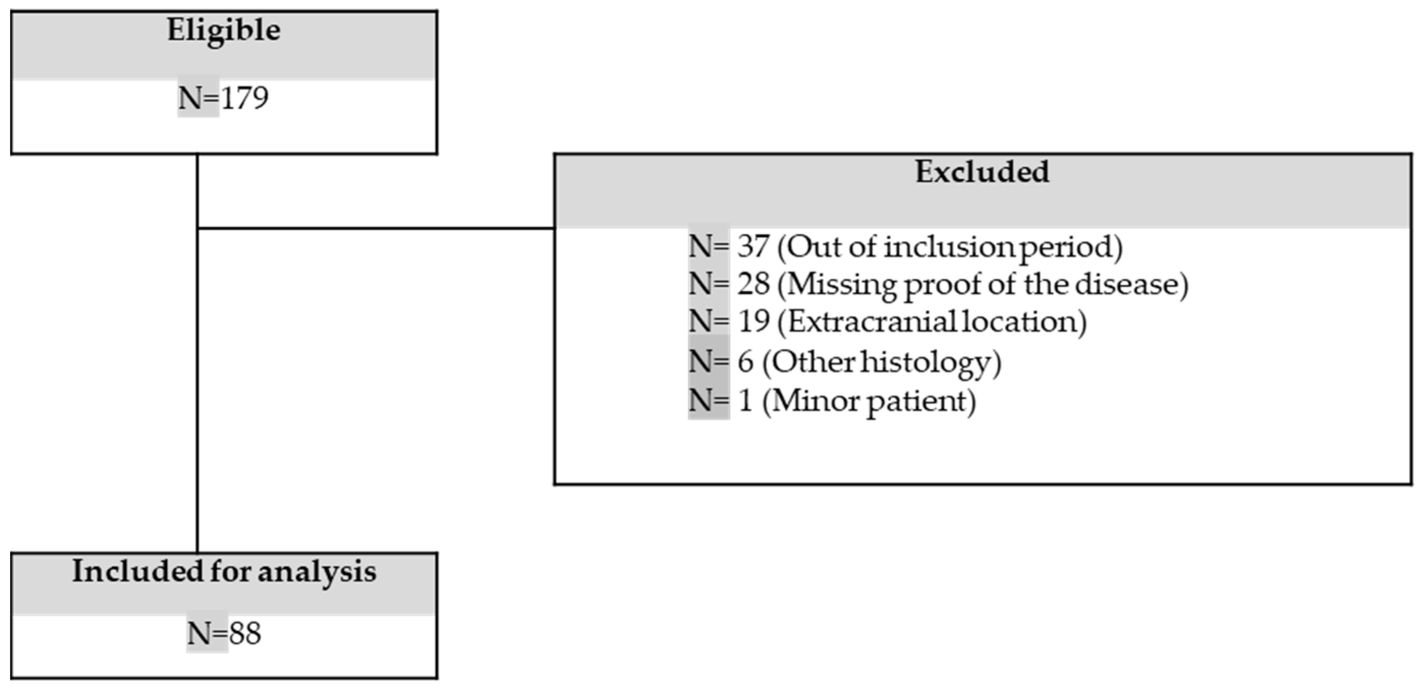
2.1. Case Selection

We collected the data of cases from the “French brain tumour database” (FBTDB) in collaboration with the French neuropathology network (RENOCLIP-LOC). The database combines information of patients with a confirmed pathological diagnosis of intracranial SFT, HPC, and anaplastic HPC in France from 2006 to 2015. Clinical and pathological records of the patients were retrospectively reviewed to extract the relevant clinical factors at 16 French centres (Amiens, Bordeaux, Caen, Clermont-Ferrand, Colmar/Strasbourg, Ile de la Réunion, Lille, Limoges, Lyon, Marseille, Montpellier, Orléans, Paris-La-pitié-salpêtrière, Paris-Sainte-Anne, Perpignan, and Rouen) from January 2006 to December 2015. The exclusion criteria were disease occurrence outside the inclusion period or at an extracranial level, a lack of histological proof of SFT, age <18 years at diagnosis, and a written refusal of consent to the study. Information on age, sex, clinical presentation of the disease, time to diagnosis, tumour location, tumour size, extent of resection, preoperative biopsy, preoperative embolization, pathology including immunohistochemistry, adjuvant radiotherapy, tumour recurrence or metastasis occurrence, and patient survival was collected.
Intracranial hypertension was defined by the association of headaches, nausea, and vomiting, and was distinguished from exclusive headaches in the clinical presentation of the disease. Tumour size corresponded to the maximal diameter measured by the radiologist on the preoperative radiologic sequences. Time to diagnosis was estimated as the period between the outbreak of evocative neurological symptoms and the date of the established histology. The extent of resection was classified as GTR or STR according to the postoperative MRI results. As meningiomas, GTR and STR are defined as Simpson grades I or II removal and grades III or IV removal, respectively [1,12]. Preoperative intervention through embolization guided with arteriography was conducted at the surgeon’s discretion. Tumour local recurrence was defined as the reappearance of the tumour within the cranial cavity or an increase in the size of the residual tumour according to RECIST1.1 criteria. Metastasis was defined as an extracranial appearance of SFT.

2.2. Pathology

All specimens of SFTs were confirmed by a pathologist with expertise in this disease. Specimens were graded according to the 2021 CNS WHO classification into grades 1, 2, or 3 [5]. According to these criteria, grade 1 is characterized by a highly collagenous, relatively low-cellularity spindle-cell lesion, previously diagnosed as SFT. Grade 2 corresponds to a more cellular, less collagenous tumour with plump cells, staghorn vasculature, and mitosis <5 per 10 high-power field (HPF), previously diagnosed as HPC. Grade 3 corresponds to ≥5 mitoses/10 HPF and/or the presence of necrosis, and it was previously named “anaplastic HPC”.

2.3. Statistical Analyses

Patient characteristics were described using the mean (standard deviation (SD)) or median (range) as appropriate for continuous variables and frequency (percentage) for categorical variables. Missing data were reported.
The median follow-up time was estimated by using the reverse Kaplan–Meier method, considering survival status at the end of the study as an event and death as a censored event. Survival analyses were performed using the Kaplan–Meier method. The survival rate, median, and confidence interval at 95% (95% confidence interval (95% CI)) were reported. Follow-up times were defined as the time from the date of the initial pathological diagnosis of SFT to the date of (1) death for OS; (2) recurrence or death if no recurrence occurred for progression-free survival (PFS); (3) local recurrence or death if no local recurrence occurred for local recurrence-free survival (LRFS). In cases of recurrence, follow-up times were defined as the time from the date of first recurrence of SFT to the date of (4) death for the second OS (OS2) and (5) the second recurrence or death if no second recurrence occurred for the second PFS (PFS2). Patients who remained alive or were lost-to-follow-up were censored.
The hazard ratio (HR) for recurrence and/or death (PFS) or for death (OS) associated with patients’ characteristics were estimated using Cox models—first, in the univariate analysis and then in the multivariate analysis adjusted for possible confounders. The initial multivariate model included all covariates with a p-value < 0.20 in univariate analyses except for adjuvant radiotherapy, which only concerned the subgroup of patients with the indication according to the literature [14]. Correlations between variables were searched. The backward selection gave the final multivariate model including only covariates with p-value < 0.05. Sensitivity multivariate analyses were performed with PORT as a covariate. All the point estimates were reported with their 95% CI. All tests were two-sided, and the threshold for statistical significance was set to p < 0.05.
The software used for the analyses was Stata version 17.0 (StataCorp. 2021. Stata Statistical Software: Release 17. College Station, TX: StataCorp LLC).

2.4. Ethical Approval

The study complied with the “reference methodology” MR004 adopted by the French Data Protection Authority (CNIL), and we checked that patients did not object to the use of their clinical data for the research purpose. Ethical local approval was obtained on 25 May 2022. The number of the ethical approval is N° 2022-013. All data were anonymized.

3. Results

3.1. Patient Characteristics

A query of the database revealed 179 patients with a primary iSFT from January 2006 to December 2015 in the 16 participating French centres. Ninety-one cases were excluded because they were diagnosed before 2006 or after 2015 (n = 37), had extracranial localization (n = 19), were missing proof of the disease (n = 28), had a revised histology by pathological review (n = 6), or were patients aged <18 years old (n = 1). Finally, we included 88 patients in the study (Figure 1).
There was a female predominance (n = 50, 57%), and the median age at diagnosis was 54.5 years (range: 19–88). The SFTs were mainly local (n = 84, 95%) and supratentorial (n = 71, 81%) at diagnosis. Tumours often had a large maximal diameter on brain MRI (≥5 cm for 32 (36%) patients-Table 1). All the clinical characteristics of the patients were similar regardless of tumour lateralization and are summarized in Table 1.

3.2. Clinical and Pathological Presentation

The symptoms leading to diagnosis were related to the tumour location and consisted mainly of headaches (n = 41, 47%) and cognitive disorders (n = 37, 42%) (see Supplementary Table S1).
Seventy-seven tumours (70%) were graded according to the 2021 CNS WHO classification. There were 1 (1%), 25 (28%), and 51 (58%) grade one, two, and three iSFT cases, respectively. The 34 samples (39%) that were tested for the nuclear expression of STAT6 using immunohistochemistry were all positive (Table 1). The characteristics of the population with STAT6 status results were similar to those with an unknown status.

3.3. Initial Treatment

Eighty-seven (99%) patients underwent surgical resection and one (1%) had a biopsy without an additional resection. GTR and STR were achieved in 75 (85%) and 9 (10%) patients, respectively. The surgical excision (GTR or STR) status of the three other patients was unknown (Table 1). Only eight patients (9%) received preoperative embolization. After initial surgery, 32 patients (57%) received adjuvant radiotherapy. One of the 32 patients received exclusive radiotherapy after a single biopsy without resection. Systemic treatments were not employed at the initial stage (Table 1).

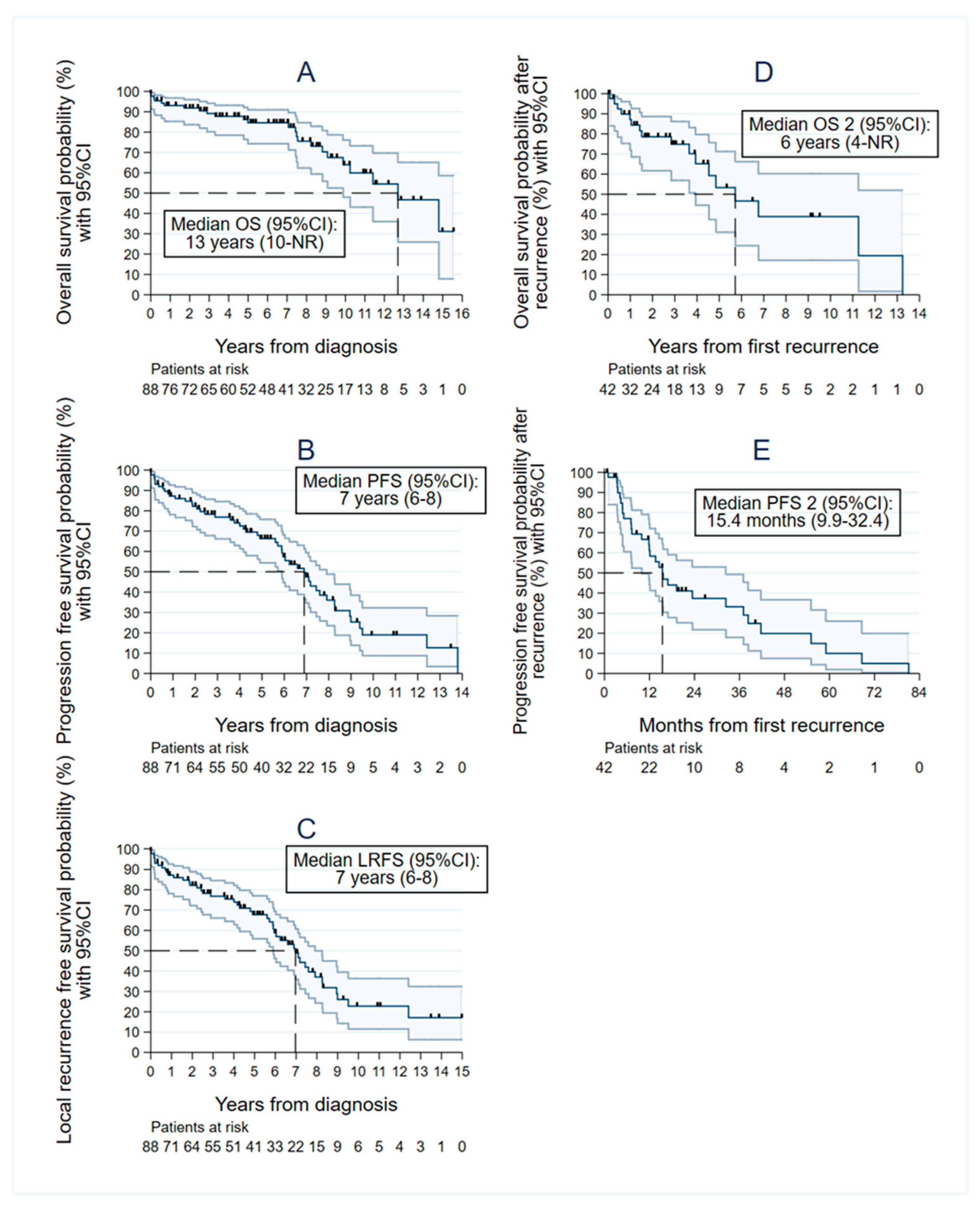
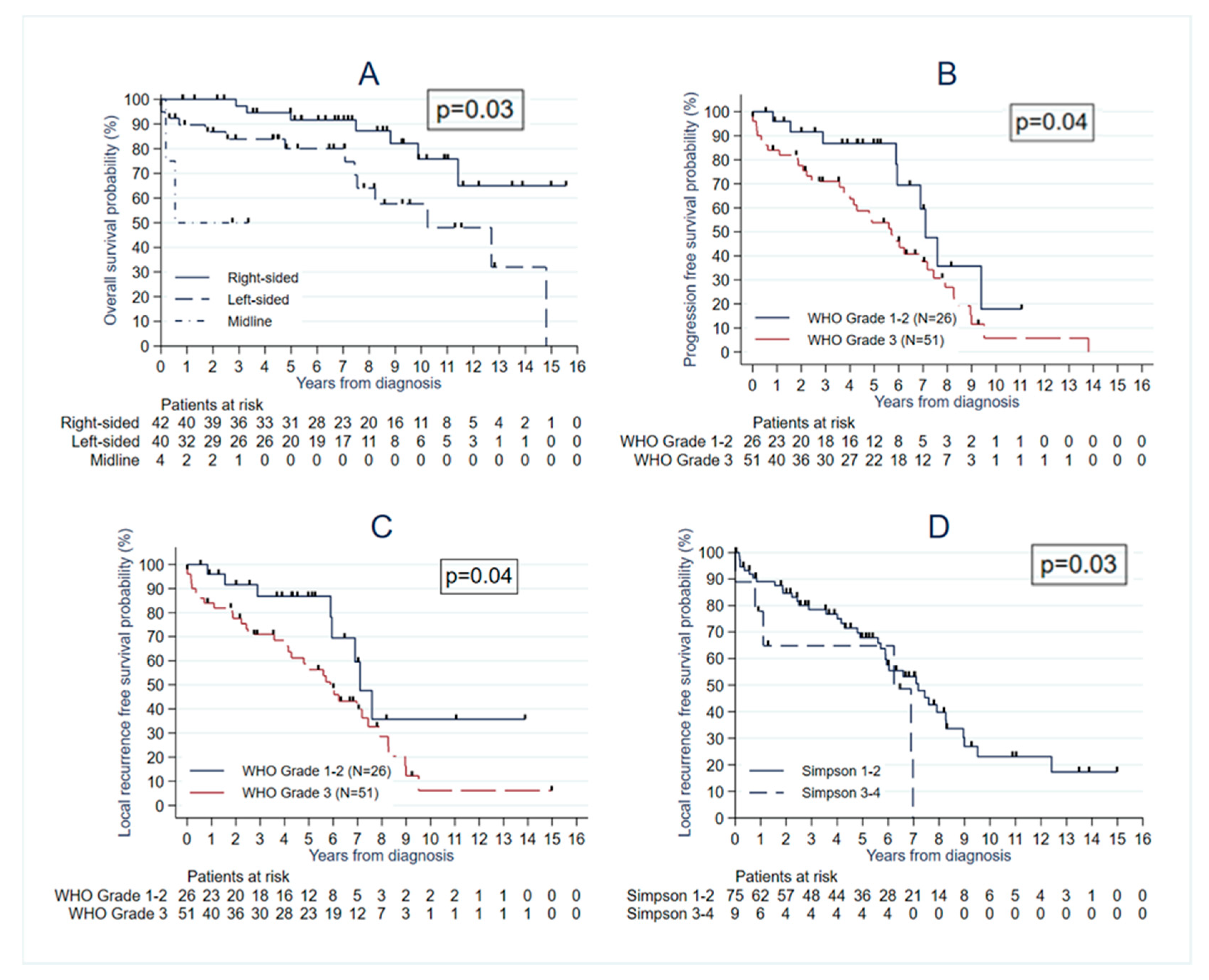
3.4. Survival Analyses

The median follow-up time was 7 years (range 0–16 years). Twenty-four patients had died by the retrospective analysis time, seven (29%) of whom had metastatic disease. The median OS time was 13 years (95% CI: 10–nonreached (NR)), with 1-, 5-, and 10-year OS rates of 93% (95% CI: 85–97), 85% (95% CI: 74–91), and 64% (95% CI: 48–76), respectively. The median PFS was 7 years (95% CI: 6–8 years) after the date of diagnosis, with 1-, 5-, and 10-year PFS rates of 87% (95% CI: 78–93), 66% (95% CI: 54–76), and 19% (95% CI: 9–32), respectively. The median LRFS was 7 years (95% CI: 6–8 years) after the date of diagnosis, with 1-, 5-, and 10-year LRFS rates of 87% (95% CI: 78–93), 68% (95% CI: 56–77), and 23% (95% CI: 11–36), respectively (Figure 2).
The median OS2 was 6 years (95% CI 4–NR), with 1-, 3-, and 5-year OS2 of 87% (95% CI 72–94), 75% (95% CI 57–86), and 53% (95% CI 31–71), respectively. The median PFS2 was 15.4 months (95% CI 9.9–32.4), with 1-, 3-, and 5-year PFS2 rates of 61% (95% CI 44–74), 33% (95% CI 18–49), and 10% (95% CI 2–26), respectively (Figure 2D,E).

3.5. Recurrences and Treatment at Recurrence

During the follow-up, 42 patients (48%) experienced at least one progression or recurrence and 16 (18%) patients had a distant recurrence. Among the 42 relapses, 35 (83%) were located at the initial site, 5 (12%) were distant, and 2 (5%) were simultaneously local and distant. The morphological and treatment characteristics of the first local recurrence are summarized in Table 2. Among the 37 patients presenting a local relapse, 17 (46%) received surgical treatment, of whom 10 (59%) underwent PORT. Exclusive radiotherapy was performed in 14 patients (38%) and systemic treatment in 2 (5%) (Table 2).
A second recurrence occurred in 25 patients, of whom 16 were local (64%) and treatment corresponded to surgery for 6 (38%) patients, radiotherapy for 6 (38%), and systemic treatment for 3 (19%) patients. The relapses were repetitive, and we observed fifth (n = 3), sixth (n = 2), and seventh (n = 1) recurrences in our series, which were all metastatic. Their treatment consisted of either surgery, radiotherapy, or systemic treatments. To our knowledge, no other solid or haematological cancer occurred during the time of surveillance.

3.6. Extracranial Metastasis

Sixteen patients (18%) developed extracranial metastases. The most frequent sites of extracranial metastases were the liver (five patients, 31%), bones (nine patients, 56%), and the lungs (six patients, 38%). Interestingly, only eight (50%) patients received a systemic treatment at this disseminated stage. The treatment consisted of chemotherapy (doxorubicin n = 2; temozolomide n = 2; doxorubicin + ifosfamide n = 1) in five (62.5%) patients and antiangiogenic targeted therapy in two patients (25%) (bevacizumab n = 1, pazopanib n = 1). One patient (12.5%) was treated with a combination of chemotherapy and vascular endothelial growth factor (VEGF) inhibitor (temozolomide and bevacizumab). A complete response was observed in five patients who underwent surgery or radiotherapy. The best response in the systemic group was a stable disease (see Supplementary Table S2).

3.7. Prognosis Factors

We observed that age was associated with poorer OS (HR = 1.04 (95% CI: 1.01–1.08); p = 0.01), PFS (HR = 1.03 (95% CI: 1.01–1.05); p = 0.008), and LRFS (HR = 1.04 (95% CI: 1.01–1.06); p = 0.002) (Table 3). Tumour topography (midline and left lateralization) was associated with worse OS in the multivariate analyses with HR = 2.69 (95% CI: 1.07–6.81), p = 0.03 for left tumours and HR = 8.29 (95% CI: 1.42-48.50), p = 0.03 for midline tumours, as compared with right-sided tumours (Table 3, Figure 3A). A higher histological grade was significantly associated with lower PFS and LRFS (HR = 2.14 (95% CI: 1.03–4.78; p = 0.04) and HR = 2.36 (95% CI: 1.08–5.16; p = 0.03), respectively) (Table 3).
We observed that treatment influenced the outcome. We observed a significantly lower LRFS in the STR group in the multivariate analyses with HR = 3.00 (95% CI: 1.09–8.24; p = 0.03) (Table 3, Figure 3D). However, performing STR was associated with significantly lower OS only in the univariate analysis with HR = 3.20 (95% CI: 1.04–9.83; p = 0.04) and tended to be associated with lower PFS (HR = 2.71 (95% CI: 0.99–7.41; p = 0.052)). Preoperative embolization was associated with a lower risk of PFS and LRFS in the univariate analysis, but this association was not significant in the multivariate analysis (HR = 0.08 (95% CI: 0.001–0.55; p = 0.08) and HR = 0.09 (95% CI: 0.001–0.60; p = 0.09), respectively). Adjuvant radiotherapy was significantly associated with greater PFS and LRFS in the univariate analysis (HR = 0.50 (95% CI: 0.26–0.97; p = 0.04) and HR = 0.56 [95% CI: 0.29–1.10]; p = 0.10)), respectively, but not with OS (HR = 0.88 (95% CI: 0.34–2.29); p = 0.80). Sensitivity multivariate analyses were performed with PORT as a covariate. We observed a significantly lower PFS with HR = 0.47 (95% CI: 0.24–0.91; p = 0.03), but the association was no more significant regarding LRFS.

4. Discussion

Intracranial SFTs are very rare mesenchymal tumours with a high risk of recurrence [9,15,17]. To the best of our knowledge, this was the largest clinical retrospective cohort describing data at the stage of diagnosis and recurrence in Europe and the USA. There are no established guidelines for the management of iSFTs, a hard-to-treat malignancy, especially at recurrence. Current evidence came from retrospective data and prospective studies with a limited number of cases [18]. We conducted a long-term study permitting the report of 42 patients in relapse and their characterization, which is usually difficult given the rarity of this entity [2,6,19]. Local treatments are often employed early and systemic treatment seems to present a restrained efficiency.
As reported by several authors, we observed that GTR provided a strong benefit in terms of tumour control as a first treatment for iSFTs [6,10,12,13,20]. Kim et al. reported a significant decrease in the 5-year recurrence risk in GTR cases compared to STR (20.8% vs. 72.7%; p = 0.006) cases [2]. However, the results of GTR on OS remain controversial. Consistent with our observation, Soyuer et al. found no association between survival and resection extent [10]. However, Rutkowski et al. observed an increased survival with GTR (13 vs. 9.7 years, p < 0.05), independent of the realisation of PORT [9].
Several studies have analysed the impact of PORT on iSFT recurrence and survival with conflicting results [7,14,21,22,23]. In our cohort, PORT seemed to be efficient only in terms of local control and PFS in patients who presented with an aggressive tumour or underwent an incomplete resection as, discussed in Stessin et al. Other authors, such as Coombs et al., even reported improved OS [7,12,14,21]. However, Xiao et al. found no association between PORT and survival or PFS irrespective of the quality of resection [24]. The low percentage of STR cases included in our cohort and missing data induced a lack of power that could explain our observations. Thus, our cohort could not engage the role of adjuvant radiotherapy in these situations, but the results remained promising. Comparison between different radiotherapy techniques and regimens or between stereotactic radiosurgery (SRS) and fractionated radiotherapy was not addressed in our series due to the small number of patients undergoing adjuvant SRS and missing data. Moreover, due to the retrospective nature of our cohort based on surgical specimens, no patient treated with exclusive SRS was included in the study. However, like meningiomas, this treatment strategy should be further explored.
Based on the rationale of the meningioma treatment, a preoperative embolization is sometimes conducted to perform a GTR and limit morbidity in these heavily vascularized tumours [19,25]. To date, its impact on clinical outcome has not been demonstrated in iSFTs. In our series, this technique tended to reduce recurrence. The nonsignificant association in the multivariate models might have been due to the lack of power because of the small number of patients.
Concerning survival predictive factors, age at diagnosis is heavily discussed [10,12,26]. However, as Wang et al., we found that a 1-year increase in age predicted a poor outcome [15]. We observe that the CNS WHO grade III was associated with poorer PFS and LRFS as Macagno et al. had described [4,20].
We reported an association between survival and tumour location. The results concerning midline locations should be interpreted with caution, given the very small number of patients (n = 4) with this type of location. To our knowledge, this information was not described in other studies of SFTs. Even if extra- and intra-axial tumours are not similar, some authors have described a poor prognosis in glioblastoma with central and left temporal localizations [27]. The impact of left lateralization on OS in our series might be explained by the greater preoperative risks due to the presence of language centres, frequently located on the left [12,13]. However, we did not observe a negative impact for tumours located at intrasinusal or cerebellar levels, which are usually reported to have worse prognoses [9,12,15].
Very few studies have reported on the treatment and characteristics of patients with iSFTs at diagnosis and at relapse [9,15,16]. To our knowledge, we carried out the first description of the clinical management of relapses in France and the largest one in Europe and in the USA. Recurrences occur mostly at the initial local site and are belated and repetitive [23]. Like Rutkowski et al., we observed that the treatment of the first relapse consisted of local techniques (radiotherapy and surgery) for most of our patients [9]. In concordance with the literature, we observed that the first relapse represented a turning point in iSFT evolution, with reduced recurrence latency over the course of the disease [9,12,15,25].
The specificity of iSFT is its potential for extracranial metastatic spreading in approximately 11% to 60% of cases [1,2,15]. The effectiveness of systemic treatments appears limited [28,29,30]. Nevertheless, antiangiogenic targeted therapy has shown promising OS and PFS results [8,28,31]. Interestingly, we noticed a long-term recurrence-free survival after the fourth local recurrence in one patient treated using an INF-alpha inhibitor. This observation strengthens the rationale of an influence of angiogenesis inhibition in treating unresectable iSFTs [11,32,33].
Although our cohort consisted of the largest description of iSFT cases in Europe and the USA, several limitations remained, especially those inherent in their retrospective nature. For example, some aspects could not be monitored in our study, especially those related to neuropsychiatric symptoms. Only one iSFT was declared to be revealed with psychiatric relapse, and some others with mental confusion regrouped in high-function disorders, but no psychiatric-specific tests were routinely performed during their surveillance. Nowadays, iSFT diagnosis requires the STAT6 immunohistochemistry status [4,5]. Another limitation of our study was the STAT6 status. The period of inclusion of our study was prior to the current use of this tool, explaining that we had less than 40% of patients with a known STAT6 status in our cohort, but all were positive. Nevertheless, our patients were included if there was a confirmed pathological review of iSFT specimens; otherwise, the patient was excluded (n = 6). At relapse, all specimens tested for STAT6 in immunochemistry were positive. Moreover, no difference for patients’ characteristics was seen in our cohort depending on a known STAT6 status. We conducted a long-term study that enabled extended surveillance to detect potential late recurrence. However, the time and period of surveillance were not consensual in France [1,9]. This might explain why some of our patients were lost-to-follow-up before 10 years of surveillance. An underestimation of recurrence and metastasis might have occurred.

5. Conclusions

Intracranial SFTs are rare sarcomas, which must be followed closely and for a long time after multimodal treatment because of their slow progression. From this study, there emerged the fact that iSFT management is heterogeneous within and between centres, particularly for the treatment of repeated recurrences.
Potential clinical, biological, and radiological factors predicting recurrence require further in-depth studies, especially at an international level. Moreover, prospective studies are necessary to evaluate new modalities of treatment after GTR or suboptimal resection, such as a new regimen of chemotherapy or refined modern techniques, such as proton therapy.

6. Suggested Recommendations Resulting from Our Clinical Experience

Intracranial SFTs should first be treated with surgical techniques. Adjuvant radiotherapy seemed to reduce recurrence in patients with a high histological grade and incomplete resection. Its indication could be discussed in multidisciplinary meetings. Local recurrence and oligometastatic diseases should be treated with local techniques (surgery, SRS) if feasible. Finally, systemic treatments lack efficiency, and clinical studies should be proposed.

Supplementary Materials

The following supporting information can be downloaded at: https://www.mdpi.com/article/10.3390/cancers15030704/s1, Table S1: initial symptoms in patients with newly diagnosed iSFTs; Table S2: clinical characteristics and medical strategies in patients with distant iSFTs.

Author Contributions

Conceptualization, M.L. (Marine Lottin), A.E., L.B., M.P. and N.P.; Data curation, M.L. (Marine Lottin), A.E., M.A.-T. and M.B. (Maël Barthoulot); Formal analysis, A.E., M.A.-T. and M.B. (Maël Barthoulot); Investigation, M.L. (Marine Lottin), A.E., M.P. and S.Z.; Methodology, M.L. (Marine Lottin), A.E., L.B., M.A.-T., M.B. (Maël Barthoulot), M.P. and N.P.; Project administration, A.E. and N.P.; Resources, L.B., M.B. (Mathieu Boone), S.Z., J.G., G.P., J.P., H.D., E.E., M.L. (Michel Lefranc), S.F., H.N., E.G., J.-J.L., B.M., R.S., S.D., A.M., J.-M.C., C.-A.M. and B.C.; Software, L.B., M.A.-T., M.B. (Maël Barthoulot), and S.Z.; Supervision, A.E., B.C. and N.P.; Validation, M.L. (Marine Lottin), A.E., L.B., M.B. (Mathieu Boone), J.G., G.P., J.P., H.D., E.E., M.L. (Michel Lefranc), S.F., H.N., E.G., J.-J.L., B.M., R.S., S.D., A.M., J.-M.C., C.-A.M., B.C. and N.P.; Visualization, N.P.; Writing—original draft, M.L. (Marine Lottin) and A.E.; Writing—review and editing, M.L. (Marine Lottin), A.E., L.B., M.A.-T., M.B. (Maël Barthoulot), M.P., M.B. (Mathieu Boone), J.G., G.P., J.P., H.D., E.E., M.L. (Michel Lefranc), S.F., H.N., E.G., J.-J.L., B.M., R.S., S.D., A.M., J.-M.C., C.-A.M., B.C. and N.P. All authors have read and agreed to the published version of the manuscript.

Funding

This research received no external funding.

Institutional Review Board Statement

The study was conducted in accordance with the Institutional Review Board of Centre Oscar Lambret. Ethical local approval was obtained on 25 May 2022. The number of the ethical approval is N° 2022-013.

Informed Consent Statement

Informed consent was obtained from all subjects involved in the study.

Data Availability Statement

The data presented in this study are available on request from the corresponding author.

Acknowledgments

This study was designed with the participation of the APOTAC “association picarde pour l’optimisation des thérapeutiques anti-cancéreuses“ and FBTDB “Recensement national histologique des Tumeurs Primitives du Système Nerveux Central” with the collaboration of the French associations “Association des Neuro-Oncologues d’Expression Française” (ANOCEF) and the “Club de Neuro-Oncologie de la Société Française de Neurochirurgie” (CNO-SFNC). The authors particularly thank the patient associations. The authors thank those who oversaw the FBTDB: Fabienne Bauchet (FBTDB, ICM Montpellier), Valérie Rigau (CHU Montpellier), Amélie Darlix (ICM Montpellier), Brigitte Trétarre (Registre Tumeur Hérault), Montpellier, France; and the nonprofit organizations supporting the FBTDB: Ligue Nationale Contre le Cancer and comité 34, Associations pour la Recherche sur les Tumeurs Cérébrales (ARTC) Nord et Sud, Des étoiles dans la Mer.

Conflicts of Interest

The authors declare no conflict of interest. The funders had no role in the design of the study; in the collection, analyses, or interpretation of data; in the writing of the manuscript, or in the decision to publish the results.

References

  1. Cohen-Inbar, O. Nervous System Hemangiopericytoma. Can. J. Neurol. Sci. J. Can. des Sci. Neurol. 2019, 47, 18–29. [Google Scholar] [CrossRef]
  2. Kim, B.S.; Kim, Y.; Kong, D.-S.; Nam, D.-H.; Lee, J.-I.; Suh, Y.-L.; Seol, H.J. Clinical outcomes of intracranial solitary fibrous tumor and hemangiopericytoma: Analysis according to the 2016 WHO classification of central nervous system tumors. J. Neurosurg. 2018, 129, 1384–1396. [Google Scholar] [CrossRef] [Green Version]
  3. Darlix, A.; Zouaoui, S.; Rigau, V.; Bessaoud, F.; Figarella-Branger, D.; Mathieu-Daudé, H.; Trétarre, B.; Bauchet, F.; Duffau, H.; Taillandier, L.; et al. Epidemiology for primary brain tumors: A nationwide population-based study. J. Neuro-Oncology 2016, 131, 525–546. [Google Scholar] [CrossRef] [PubMed]
  4. Macagno, N.; Vogels, R.; Appay, R.; Colin, C.; Mokhtari, K.; Küsters, B.; Wesseling, P.; Figarella-Branger, D.; Flucke, U.; Bouvier, C.; et al. Grading of meningeal solitary fibrous tumors/hemangiopericytomas: Analysis of the prognostic value of theMarseilleGradingSystem in a cohort of 132 patients. Brain Pathol. 2018, 29, 18–27. [Google Scholar] [CrossRef] [PubMed] [Green Version]
  5. Louis, D.N.; Perry, A.; Wesseling, P.; Brat, D.J.; Cree, I.A.; Figarella-Branger, D.; Hawkins, C.; Ng, H.K.; Pfister, S.M.; Reifenberger, G.; et al. The 2021 WHO Classification of Tumors of the Central Nervous System: A summary. Neuro-Oncology 2021, 23, 1231–1251. [Google Scholar] [CrossRef] [PubMed]
  6. Kim, J.H.; Jung, H.-W.; Kim, Y.-S.; Kim, C.J.; Hwang, S.-K.; Paek, S.H.; Kim, D.G.; Kwun, B.D. Meningeal hemangiopericytomas: Long-term outcome and biological behavior. Surg. Neurol. 2003, 59, 47–53, discussion 53. [Google Scholar] [CrossRef]
  7. Dufour, H.; Métellus, P.; Fuentes, S.; Murracciole, X.; Régis, J.; Figarella-Branger, D.; Grisoli, F. Meningeal Hemangiopericytoma: A Retrospective Study of 21 Patients with Special Review of Postoperative External Radiotherapy. Neurosurgery 2001, 48, 756–763. [Google Scholar] [CrossRef]
  8. Martin-Broto, J.; Cruz, J.; Penel, N.; Le Cesne, A.; Hindi, N.; Luna, P.; Moura, D.S.; Bernabeu, D.; de Alava, E.; Lopez-Guerrero, J.A.; et al. Pazopanib for treatment of typical solitary fibrous tumours: A multicentre, single-arm, phase 2 trial. Lancet Oncol. 2020, 21, 456–466. [Google Scholar] [CrossRef]
  9. Rutkowski, M.J.; Bloch, O.; Jian, B.J.; Chen, C.; Sughrue, M.E.; Tihan, T.; Barani, I.J.; Berger, M.S.; McDermott, M.W.; Parsa, A.T. Management of recurrent intracranial hemangiopericytoma. J. Clin. Neurosci. 2011, 18, 1500–1504. [Google Scholar] [CrossRef]
  10. Soyuer, S.; Chang, E.L.; Selek, U.; McCutcheon, I.E.; Maor, M.H. Intracranial meningeal hemangiopericytoma: The role of radiotherapy: Report of 29 cases and review of the literature. Cancer 2004, 100, 1491–1497. [Google Scholar] [CrossRef]
  11. Chamberlain, M.C.; Glantz, M.J. Sequential salvage chemotherapy for recurrent intracranial hemangiopericytoma. Neurosurgery 2008, 63, 720–727. [Google Scholar] [CrossRef]
  12. Guthrie, B.L.; Ebersold, M.J.; Scheithauer, B.W.; Shaw, E.G. Meningeal Hemangiopericytoma: Histopathological Features, Treatment, and Long-Term Follow-up of 44 Cases. Neurosurgery 1989, 25, 514–522. [Google Scholar] [CrossRef] [PubMed]
  13. Rutkowski, M.J.; Sughrue, M.E.; Kane, A.J.; Aranda, D.; Mills, S.A.; Barani, I.J.; Parsa, A.T. Predictors of mortality following treatment of intracranial hemangiopericytoma: Clinical article. J. Neurosurg. 2010, 113, 333–339. [Google Scholar] [CrossRef] [PubMed]
  14. Stessin, A.M.; Sison, C.; Nieto, J.; Raifu, M.; Li, B. The Role of Postoperative Radiation Therapy in the Treatment of Meningeal Hemangiopericytoma—Experience From the SEER Database. Int. J. Radiat. Oncol. Biol. Phys. Biol. Phys. 2013, 85, 784–790. [Google Scholar] [CrossRef]
  15. Wang, W.; Zhang, G.-J.; Zhang, L.-W.; Li, D.; Wu, Z.; Zhang, J.-T. Long-Term Outcome and Prognostic Factors After Repeated Surgeries for Intracranial Hemangiopericytomas. World Neurosurg. 2017, 107, 495–505. [Google Scholar] [CrossRef] [PubMed]
  16. Schiariti, M.; Goetz, P.; El-Maghraby, H.; Tailor, J.; Kitchen, N.D. Hemangiopericytoma: Long-term outcome revisited. Clinical article. J. Neurosurg. 2011, 114, 747–755. [Google Scholar] [CrossRef]
  17. Chen, L.-F.; Yang, Y.; Yu, X.-G.; Gui, Q.-P.; Xu, B.-N.; Zhou, D.-B. Multimodal treatment and management strategies for intracranial hemangiopericytoma. J. Clin. Neurosci. 2015, 22, 718–725. [Google Scholar] [CrossRef]
  18. Lottin, M.; Escande, A.; Peyre, M.; Sevestre, H.; Maurage, C.A.; Chauffert, B.; Penel, N. What’s new in the management of meningeal solitary fibrous tu-mor/hemoangioperctyoma? Bull. du Cancer 2020, 107, 1260–1273. [Google Scholar] [CrossRef]
  19. Jääskeläinen, J.; Servo, A.; Haltia, M.; Wahlström, T.; Valtonen, S. Intracranial hemangiopericytoma: Radiology, surgery, radiotherapy, and outcome in 21 patients. Surg. Neurol. 1985, 23, 227–236. [Google Scholar] [CrossRef]
  20. Melone, A.G.; D’Elia, A.; Santoro, F.; Salvati, M.; Delfini, R.; Cantore, G.; Santoro, A. Intracranial Hemangiopericytoma—Our Experience in 30 Years: A Series of 43 Cases and Review of the Literature. World Neurosurg. 2013, 81, 556–562. [Google Scholar] [CrossRef]
  21. Combs, S.E.; Thilmann, C.; Debus, J.; Schulz-Ertner, D. Precision radiotherapy for hemangiopericytomas of the central nervous system. Cancer 2005, 104, 2457–2465. [Google Scholar] [CrossRef] [PubMed]
  22. Cohen-Inbar, O.; Lee, C.-C.; Mousavi, S.H.; Kano, H.; Mathieu, D.; Meola, A.; Nakaji, P.; Honea, N.; Johnson, M.; Abbassy, M.; et al. Stereotactic radiosurgery for intracranial hemangiopericytomas: A multicenter study. J. Neurosurg. 2017, 126, 744–754. [Google Scholar] [CrossRef] [PubMed] [Green Version]
  23. Lee, J.H.; Jeon, S.H.; Park, C.K.; Park, S.H.; Yoon, H.I.; Chang, J.H.; Suh, C.O.; Kang, S.J.; Lim, D.H.; Kim, I.A.; et al. The Role of Postoperative Radiotherapy in Intracranial Solitary Fibrous Tumor/Hemangiopericytoma: A Multi-Institutional Ret-rospective Study (KROG 18-11). Cancer Res. Treat. 2022, 54, 65–74. [Google Scholar] [CrossRef] [PubMed]
  24. Xiao, J.; Xu, L.; Ding, Y.; Wang, W.; Chen, F.; Zhou, Y.; Zhang, F.; Zhou, Q.; Wu, X.; Li, J.; et al. Does post-operative radiotherapy improve the treatment outcomes of intracranial hemangiopericytoma? A retrospective study. BMC Cancer 2021, 21, 1–10. [Google Scholar] [CrossRef] [PubMed]
  25. Fountas, K.N.; Kapsalaki, E.; Kassam, M.; Feltes, C.H.; Dimopoulos, V.G.; Robinson, J.S.; Smith, J.R. Management of intracranial meningeal hemangiopericytomas: Outcome and experience. Neurosurg. Rev. 2006, 29, 145–153. [Google Scholar] [CrossRef] [PubMed]
  26. Kumar, N.; Kumar, R.; Kapoor, R.; Ghoshal, S.; Kumar, P.; Salunke, P.S.; Radotra, B.D.; Sharma, S.C. Intracranial meningeal hemangiopericytoma: 10 years experience of a tertiary care Institute. Acta Neurochir. 2012, 154, 1647–1651. [Google Scholar] [CrossRef]
  27. Fyllingen, E.H.; Bø, L.E.; Reinertsen, I.; Jakola, A.S.; Sagberg, L.M.; Berntsen, E.M.; Salvesen, Ø.; Solheim, O. Survival of glioblastoma in relation to tumor location: A statistical tumor atlas of a population-based cohort. Acta Neurochir. 2021, 163, 1895–1905. [Google Scholar] [CrossRef]
  28. Park, M.S.; Patel, S.R.; Ludwig, J.A.; Trent, J.C.; Conrad, C.A.; Lazar, A.J.; Wang, W.-L.; Boonsirikamchai, P.; Choi, H.; Wang, X.; et al. Activity of temozolomide and bevacizumab in the treatment of locally advanced, recurrent, and metastatic hemangiopericytoma and malignant solitary fibrous tumor. Cancer 2011, 117, 4939–4947. [Google Scholar] [CrossRef]
  29. Bastin, K.T.; Mehta, M.P. Meningeal hemangiopericytoma: Defining the role for radiation therapy. J. Neuro-Oncology 1992, 14, 277–287. [Google Scholar] [CrossRef]
  30. Beadle, C.F.; Hillcoat, B.L. Treatment of advanced malignant hemangiopericytoma with combination adriamycin and dtic: A report of four cases. J. Surg. Oncol. 1983, 22, 167–170. [Google Scholar] [CrossRef]
  31. De Lemos, M.L.; Kang, I.; Schaff, K. Efficacy of bevacizumab and temozolomide therapy in locally advanced, recurrent, and metastatic malignant solitary fibrous tumour: A population-based analysis. J. Oncol. Pharm. Pract. 2018, 25, 1301–1304. [Google Scholar] [CrossRef] [PubMed]
  32. Lackner, H.; Urban, C.; Schwinger, W.; Kerbl, R.; Sovinz, P. Interferon alfa-2a in recurrent metastatic hemangiopericytoma. Med Pediatr. Oncol. 2003, 40, 192–194. [Google Scholar] [CrossRef] [PubMed]
  33. Kirn, D.H.; Kramer, A. Long-Term Freedom From Disease Progression With Interferon Alfa Therapy in Two Patients With Malignant Hemangiopericytoma. Gynecol. Oncol. 1996, 88, 764–765. [Google Scholar] [CrossRef] [PubMed]
Figure 1. Flow chart of the study.
Cancers 15 00704 g001
Figure 2. Survival analysis in newly diagnosed and recurrent iSFT patients. (A) Survival curve according to the Kaplan–Meier method showing median OS, (B) PFS, and (C) LRFS in newly diagnosed iSFTs and (D) OS2 and (E) PFS2 after the first recurrence, with their respective 95% confidence intervals (95% CI). NR: nonreached.
Cancers 15 00704 g002
Figure 3. The survival curve obtained with the Kaplan–Meier method showing the difference in OS according to tumour location (A), PFS according to the WHO grade (B), LRFS according to the WHO grade (C), and LRFS according to the extent of resection with newly diagnosed iSFT (D) with their respective p value, significant if <0.05.
Cancers 15 00704 g003
Table 1. Clinical, pathological, morphological, and treatment characteristics for the 88 patients presenting a new diagnosis of iSFT.
Characteristics (N = 88)n (%)
Sex
Female50 (57%)
Male38 (43%)
Median age (range)54.5 (19–88)
Mean age (SD)51.5 (16.4)
STAT6 expression (immunohistochemistry)
Yes34 (39%)
No0 (0%)
Not available54 (61%)
The WHO histoprognostic grade
Grade 11 (1%)
Grade 225 (28%)
Grade 351 (58%)
Not available11 (13%)
Mitotic count
<536 (41%)
≥545 (51%)
Not available7 (8%)
Necrosis
Yes33 (38%)
No55 (63%)
Number of lesions
Unique84 (95%)
Multiple4 (5%)
Maximal diameter
< 3cm5 (6%)
3–5 cm24 (27%)
≥5 cm32 (36%)
Not available27 (30%)
Topography of the tumour
Right-sided42 (48%)
Left-sided40 (45%)
Midline *4 (4%)
Bilateral1 (1%)
Not available1 (1%)
Location
Supratentorial71 (81%)
Infratentorial17 (17%)
Supra- and infratentorial 2 (2%)
Preoperative embolization
Yes8 (9%)
No80 (91%)
Resection grade according to Simpson
Simpson 1–2 (gross tumour resection)75 (85%)
Simpson 3–4 (subtotal tumour resection)9 (10%)
Simpson 5 (biopsy)1 (1%)
Not available 3 (3%)
Postoperative radiotherapy (N = 56)
Yes32 (57%)
No24 (43%)
Technique of radiotherapy
Conformational radiotherapy
Radiosurgery
Not available
19 (50%)
3 (9%)
10 (31%)
Local control
Yes76 (86%)
No11 (13%)
Not available1 (1%)
Type of treatment (N = 65)
Surgery alone33 (51%)
Radiotherapy alone1 (1%)
Surgery plus postoperative radiotherapy31 (48%)
* Falx and pineal region tumours.
Table 2. Morphological and therapeutic management data in patients with at least one episode of localized recurrence of iSFT.
Characteristics (N = 37)n (%)
Unifocal recurrence
Yes32 (87%)
No5 (14%)
Maximal diameter of recurrence
<3 cm19 (51%)
3–5 cm5 (14%)
≥5 cm2 (5%)
Not available11 (30%)
Recurrence topography
Right-sided18 (49%)
Left-sided16 (43%)
Midline3 (8%)
Bilateral0 (0%)
Recurrence location
Supratentorial30 (81%)
Infratentorial7 (19%)
Supra- and infratentorial 0 (0%)
Preoperative embolization
Yes0 (0%)
No37 (100%)
Preoperative radiotherapy
Yes1 (3%)
No36 (97%)
Surgery
Yes17 (46%)
No20 (54%)
Resection grade according to Simpson (N = 17)
Simpson 1–2 (gross tumour resection)15 (88%)
Simpson 3–4 (subtotal tumour resection)1 (6%)
Not available1 (6%)
Postoperative radiotherapy (N = 17)
Yes10 (59%)
No7 (41%)
Type of radiotherapy
Conformational radiotherapy
Radiosurgery
Not available
4 (40%)
4 (40%)
2 (20%)
Exclusive radiotherapy
Yes14 (38%)
No23 (62%)
Technique of radiotherapy
Conformational radiotherapy
Radiosurgery
Not available
2 (14%)
8 (57%)
4 (29%)
Chemotherapy associated with radiotherapy
Yes1 (3%)
No36 (97%)
Postoperative chemotherapy
Yes1 (3%)
No36 (97%)
Local control after treatment
Yes26 (70%)
No10 (27%)
Not available1 (3%)
Type of treatment that led to control (N = 26)
Surgery alone4 (15%)
Radiotherapy alone 11 (42%)
Surgery and postoperative radiotherapy11 (42%)
Chemotherapy alone 0 (0%)
Radiotherapy and postoperative chemotherapy 0 (0%)
No treatment0 (0%)
Table 3. Univariate and multivariate analyses according to Cox models in patients presenting an initial diagnosis of iSFT.
CharacteristicsOverall SurvivalProgression-Free SurvivalLocal Recurrence-Free Survival
Univariate HR (95% CI)pMultivariate HR (95% CI)PUnivariate HR (95% CI)pMultivariate HR (95% CI)PUnivariate HR (95% CI)pMultivariate HR (95% CI)p
Sex 0.78 0.36 0.60
Female1 1 1
Male0.89(0.39–2.02) 0.76 (0.43–1.36) 0.85(0.48–1.53)
Age *1.05(1.02–1.09)0.0031.04(1.01–1.08)0.011.03 (1.01–1.05)0.0031.03(1.01–1.05)0.0081.03(1.01–1.05)0.0021.04(1.01–1.06)0.002
Signs of raised intracranial pressure $ (n = 85)1.30(0.53–3.17)0.57 0.95(0.48–1.89)0.89 0.99(0.50–1.97)0.98
Motor deficit $ (n = 85)1.56 (0.63–3.84)0.34 1.34 (0.71–2.51)0.37 1.28 (0.67–2.44)0.46
Sensory deficit $ (n = 85)1.11 (0.26–4.80)0.89 0.90 (0.32–2.51)0.84 0.95 (0.34–2.66)0.92
Epileptic seizures *$ (n = 85)0.22 (0.03–1.66)0.14 0.37 (0.13–1.03)0.056 0.40 (0.14–1.11)0.08
High-function disorder *$ (n = 85)1.65 (0.72–3.78)0.23 1.52 (0.86–2.70)0.15 1.52 (0.85–2.73)0.16
Visual disorder $ (n = 85)0.60 (0.20–1.77)0.35 0.95 (0.50–1.81)0.88 1.02 (0.53–1.95)0.95
Cerebellar syndrome *$ (n = 86)1.83 (0.67–5.00)0.24 1.78 (0.83–3.86)0.14 1.86 (0.86–4.02)0.12
Headache $ (n = 85)1.00 (0.43–2.33)1.00 0.97 (0.54–1.73)0.91 0.83 (0.46–1.50)0.54
Mitotic count ≥5 *$ (n = 81)1.47 (0.62–3.48)0.38 2.03 (1.07–3.85)0.03 2.36 (1.21–4.63)0.01
Necrosis $1.25 (0.56–2.81)0.59 1.15 (0.65–2.06)0.63 1.15 (0.64–2.07)0.65
WHO grade (n = 77) 0.29 0.047 0.04 0.03 0.03
Grade 1–21 1 1
Grade 31.76 (0.58–5.28) 2.05 (0.98–4.28) 2.14 (1.03–4.78) 2.21 (1.02–4.79) 2.36 (1.08–5.16)
Max. diameter (n = 61) 0.83 0.79 0.74
< 3 cm1 1 1
3–5 cm1.73(0.18–228.92) 1.96(0.25–15.16) 1.94(0.25–15.00)
≥5 cm2.20(0.27–285.32) 1.70(0.22–12.93) 1.55(0.20–11.85)
Topography *(n = 87) 0.004 0.03 0.003 0.002
Right-sided1 1 1 1
Left-sided *3.02(1.21–7.52) 2.69(1.07–6.81) 1.84 (1.01–3.37) 2.03 (1.10–3.76)
Midline *13.86(2.54–75.71) 8.29(1.42–48.50) 8.57(2.32–31.65) 9.08(2.44–33.73)
Location 0.94 0.70 0.62
Supratentorial1 1 1
Infratentorial0.96 (0.32–2.84) 1.15 (0.55–2.40) 1.20 (0.58–2.51)
Preoperative embolization *$0.25 (0.002–1.82)0.35 0.08 (0.001–0.55)0.08 0.09 (0.001–0.60)0.09
Resection grade * (n = 84) 0.04 0.12 0.10 0.03
Simpson 1–21 1 1 1 1
Simpson 3–43.20 (1.04–9.83) 2.02 (0.84–4.89) 2.11 (0.87–5.12) 3.00 (1.09–8.24)
PORT $ (n = 56)0.88 (0.34–2.29)0.80 0.50 (0.26–0.97)0.04 0.56 (0.29–1.10)0.10
* Univariate significant parameters (p < 0.20) were then tested in a multivariate model; they were significant if p < 0.05, indicated by bolded text. HR: hazard ratio; PORT: postoperative radiotherapy; $ For binary variables, the reference category was “No”.
Disclaimer/Publisher’s Note: The statements, opinions and data contained in all publications are solely those of the individual author(s) and contributor(s) and not of MDPI and/or the editor(s). MDPI and/or the editor(s) disclaim responsibility for any injury to people or property resulting from any ideas, methods, instructions or products referred to in the content.

