Systematic Review

CA125 for the Diagnosis of Advanced Urothelial Carcinoma of the Bladder: A Systematic Review and Meta-Analysis

1
Division of Nephrology, Asia University Hospital, Taichung 41354, Taiwan
2
Department of Bioinformatics and Medical Engineering, Asia University, Taichung 41354, Taiwan
3
College of Medicine, China Medical University, Taichung 404332, Taiwan
4
Division of Nephrology and Kidney Institute, China Medical University Hospital, Taichung 404332, Taiwan
5
Department of Post-baccalaureate Veterinary Medicine, Asia University, Taichung 41354, Taiwan
*
Author to whom correspondence should be addressed.
These authors contribute equally to this work.
Cancers 2023, 15(3), 813; https://doi.org/10.3390/cancers15030813
Submission received: 9 December 2022 / Revised: 23 January 2023 / Accepted: 26 January 2023 / Published: 28 January 2023
(This article belongs to the Special Issue New Advances in Urothelial Cancer: Diagnosis, Therapy and Prognosis)

Abstract

:

Simple Summary

Urothelial carcinoma of the bladder (UCB) is a common genitourinary cancer. Advanced UCB, defined as muscle-invasive and lymph node-involving, is linked to poor outcomes. CA125 is a well-known tumor marker for ovarian cancer and can be used as a diagnostic marker for advanced UCB. This meta-analysis reviewed published studies in English and Chinese on serum CA125 for advanced UCB. One thousand five hundred seventy patients from 14 studies were analyzed. The diagnostic odds ratio was 8.138, and the area under the receiver operating characteristic curve was 0.797. Serum CA125 may help distinguish advanced UCB from non-advanced UCB patients. CA125 may help clinicians plan the treatment of UCB.

Abstract

Background: Urothelial carcinoma of the bladder (UCB) is the second most common genitourinary cancer. This study aims to assess the diagnostic accuracy of CA125 in advanced UCB. Methods: We searched prevalent studies in PubMed, the Cochrane Library, Scopus, Embase, the Web of Science China National Knowledge Infrastructure database, and Wanfang data before October 2022. Pooled sensitivity, specificity, and summary receiver operating characteristics were used to assess the diagnostic value of CA125. Results: One thousand six hundred forty-one patients from 14 studies were analyzed. UCB stage T3–4N1 was defined as advanced UCB in ten studies; T2–4 was used in three studies; and N1M1 in one study. Patients’ age was between 21 to 92, and 21% to 48.6% of patients were female. The pooled sensitivity was 0.695 (95% confidence interval (CI): 0.426–0.875). The pooled specificity was 0.846 (95% CI: 0.713–0.924). The diagnostic odds ratio was 8.138 (95% CI: 4.559–14.526). The AUC was 0.797. Conclusion: CA125 may provide significant diagnostic accuracy in identifying muscle-invasive, lymph node-involved, and distant metastatic tumors in patients with urothelial carcinoma of the bladder. Limited studies have been conducted on the prognostic role of CA125. More studies are needed for a meta-analysis on the prognostic role of CA125 in UCB.

1. Introduction

Urothelial carcinoma of the bladder (UCB) is the 10th most common cancer, with approximately 573,000 new cases and 213,000 deaths worldwide. It is more common in men than in women. The incidence rate and mortality rate of UCB are four times higher in men than in women [1]. In the United States, an estimated 81,180 new cases of UCB were diagnosed, 61,700 were male and 19,480 were female. UCB is the 4th most common cancer in males. The estimated death was 17,100, including 12,120 males and 4980 females. UCB is the 8th most common cause of cancer death in the United States. Advanced age is the most significant risk factor for UCB, and the average age of diagnosis is between 70 and 84 years [2]. Smoking is an important modifiable exposure. The observed geographic patterns of UCB mirror the prevalence of smoking, with a population-attributable risk of approximately 50%. Occupational exposure to benzene dyes, factory chemicals, Schistosoma hematobium (particularly common in Northern Africa), cyclophosphamide, pelvic radiation, and arsenic contamination in drinking water also increases the risk of UCB [3]. In developing countries, the mortality rates of UCB have declined because of improvements in treatments such as endoscopic resection, adjuvant chemotherapy, and immunotherapy [4]. The incidence trends were stable in men from the 1990s to the 2020s, but the incidence trends were rising with the rising smoking prevalence in women [5]. As non-invasive cancers account for a large proportion of UCB, mortality rates may be of greater utility in assessing overall progress in controlling UCB [3].
The prognosis of UCB is strongly correlated with bladder muscle invasion. The 5-year survival is 93% in non-muscle-invasive, 36–48% in muscle-invasive, 36% in lymph nodes involved, and 5% in distant metastatic UCB [2]. Non-muscle-invasive UCB represents approximately 70% of organ-confined cancer and is treated differently from muscle-invasive UCB. Transurethral resection of the bladder tumor, a single perioperative dose of chemotherapy, and intravesical therapy are indicated in non-muscle-invasive UCB. Chemotherapy and radical cystectomy are indicated in muscle-invasive UCB. Adjuvant chemotherapy, cytotoxic chemotherapy, and immunotherapy, such as PD-L1, are indicated in metastatic UCB. Conventional staging modalities such as transurethral resection of bladder tumors (TURBT), computer tomography (CT), and magnetic resonance imaging (MRI) have a limited role in tumor staging and lymph node involvement in patients with muscle-invasive UCB. TURBT may diagnose UCB, resect the visible tumor, and allow for its staging. Repeat TURBT in 4–6 weeks is recommended in high-risk patients such as incomplete resection or pathological high-risk. 51% of patients may have residual disease, and 8% may have muscle-invasive UBC in the repeat TURBT [6]. MRI correctly staged 56% of patients, and CT correctly staged 50% of patients [7]. The Vesical Imaging-Reporting and Data System provides good diagnostic accuracy in identifying muscle-invasive UBC. The pooled sensitivity and specificity were 0.90 (95% CI: 0.86–0.94) and 0.86 (95% CI: 0.71–0.94) in a meta-analysis of more than 1000 patients from six studies [8]. However, the Vesical Imaging-Reporting and Data System is unable to identify the lymph nodes involved and metastatic UBC.
CA125 is a repeating peptide epitope of the mucin MUC16 and is involved in the glycosylation of MUC16 [9]. MUC16 overexpression promotes cancer cell proliferation [10] and cancer cell resistance to therapy [11]. CA125 is a well-known biomarker to monitor epithelial ovarian cancer and for the differential diagnosis of pelvic masses [12]. CA125 is routinely monitored in patients with ovarian cancer and is a prognostic indicator of cancer recurrence. CA125 may help identify advanced UCB because UCB is a type of epithelial cell, and MUC16 is overexpressed in advanced UCB [13]. CA125 expression in bladder epithelial cells is linked to the infiltration of immunosuppressive cells such as regulatory T cells and M2 macrophages. Clinical studies supported the applications of CA125, CA199, carcinoembryonic antigen (CEA) [14], vascular endothelial growth factors [15], and tumor-specific growth factor [16] in identifying advanced UCB. Monitoring the markers may help guide treatments such as TURBT or radical cystectomy. CA125 is the most extensively explored among these tumor markers in clinical studies. In this study, we defined muscle-invasive, lymph node-involved, and distant metastatic UCB as advanced UCB and non-muscle-invasive UCB as non-advanced UCB. In this meta-analysis, we assess the diagnostic accuracy of CA125 in advanced UCB among patients with UBC.

2. Methods

The systematic review followed the recommendations of the Preferred Reporting Items for Systematic Reviews and Meta-Analyses (PRISMA). The protocol has not been registered. We searched all published Chinese and English studies up to October 2022 that assessed the diagnostic values of serum CA125 for UCB in PubMed, Embase, Web of Science, Cochrane Library, Scopus, the China National Knowledge Infrastructure database (CNKI), and Wanfang data (Figure 1). The keywords were CA125 and bladder cancer or carcinoma, urothelial cancer or carcinoma, and transitional cell carcinoma. We selected articles using the following inclusion criteria: (1) patients with a pathological diagnosis of UCB, and the definition of advanced UCB was provided; (2) reporting the serum CA125 assay and cut-off; (3) reporting the number of patients according to the CA125 cut-off according to non-advanced and advanced UCB; (4) published in English or Chinese. Exclusion criteria were: (1) no serum CA125; (2) not advanced UCB; (3) studies using the same dataset; (4) case reports, letters, animal models, review articles, and meta-analyses; and (5) inadequate data, such as the number of the patients, according to the cut-off of CA125, was not available. We used the modified version of the quality assessment of diagnostic accuracy studies tool-2 (QUADAS-2) to assess the methodological quality of the selected studies.
Two researchers (HJ and HC) independently assessed the title, abstract, and full text to identify the relevant articles. Two researchers (CC and CY Lee) extracted the data. Disagreements were solved through discussions and by a third researcher (CY Chou).

3. Statistical Analysis

All analysis was performed using R Statistical Software (version 4.2.1, R Foundation for Statistical Computing, Vienna, Austria) with the mada and meta packages. A p < 0.05 was considered statistically significant. The true positive (TP), false positive (FP), false negative (FN), and true negative (TN) were extracted from the selected studies. The pooled sensitivity, specificity, diagnostic odds ratio, and summary receiver operating characteristics were obtained to assess the diagnostic value of CA125. The heterogeneity of studies was analyzed using the chi-square test. An I2 < 50% was considered as no observable heterogeneity, a fixed effects model was used, or a random effects model was utilized. The mada package did not provide a summary of sensitivity and specificity. We used the meta package to calculate the summary of sensitivity and specificity [17]. RevMan 5.2 software with QUADAS-2 was used to analyze the methodological quality.

4. Results

We identified 113 records from Pubmed, 22 from Embase, 70 from Scopus, 60 from Web of Science, 28 from CNKI, and 61 from WanFang data (Figure 1). The title and abstract of 242 records were screened after removing 311 duplicate records. The full text of 27 studies was obtained. Nine studies were excluded because the patient number, according to the cut-off for CA125, was unavailable. The data were extracted from 14 studies [14,18,19,20,21,22,23,24,25,26,27,28,29,30]. We summarized the cut-off of CA125, the patient number in each study, the age of the patients, and the percentage of female patients (Table 1). One thousand six hundred forty-one patients from 14 studies were analyzed. CA125 was measured using an enzyme immunoassay in nine studies [18,19,20,21,22,23,24,25,28]; five studies did not report the assay for CA125. A cut-off of 35 U/mL was used in 12 studies, 30 U/mL in one study, and 20 U/mL in another. UCB stage T3–4N1 was defined as advanced UCB in nine studies; T2–4 was used in two studies; and N1M1 was used in one study. Patients’ ages ranged from 21 to 92, and 10.9% to 49.1% were female.
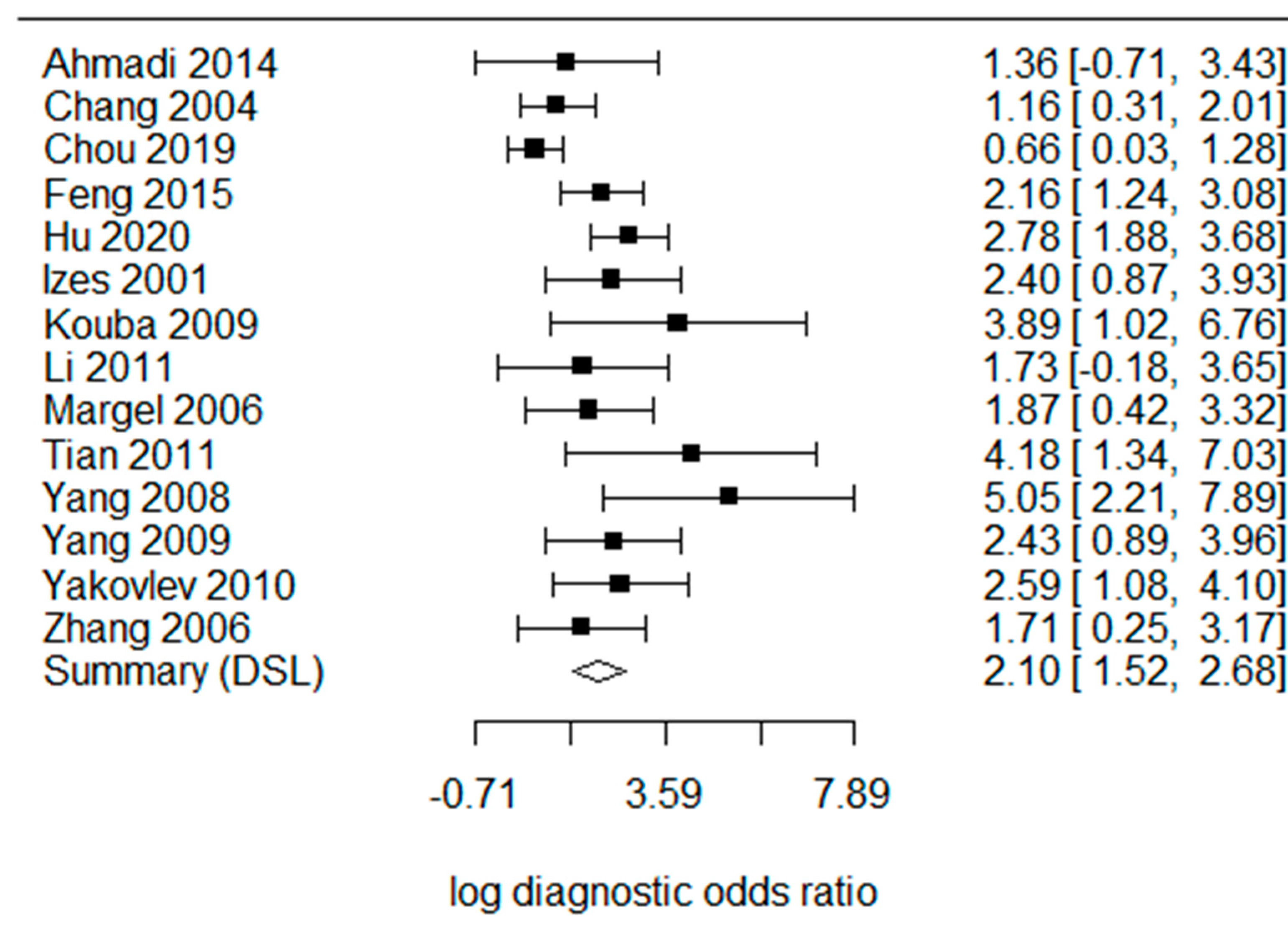
The pooled sensitivity (Figure 2) was 0.695 [95% confidence interval (CI) 0.426–0.875] in the random effects model because the heterogeneity I2 = 91%. The pooled specificity was 0.846 (95% CI 0.713–0.924, I2 = 89%). The log diagnostic odds ratio (DOR, Figure 3) was 2.10 (95% CI: 1.52–2.68). The area under the receiver operating characteristic (AUC) was 0.797 (I2 = 22.4%) in the summary ROC analysis (Figure 4).
The methodological quality of 14 studies is summarized in Figure 5. One study [28] considered a high risk of bias, and four studies [21,25,29,30] considered an unclear risk of bias in patient selection. Three studies [25,26,30] had a high risk of bias in the index test. Four studies [22,25,26,30] were unclear regarding reference standards. Two studies [23,30] had a high risk of bias, and two [20,27] had an unclear risk of bias in the flow and timing. One study [28] had an applicability concern, and six studies [14,21,25,27,29,30] had an applicability concern in patient selection. In the index test, one study [30] had a high risk of applicability concerns, and three studies [23,25,26] had an unclear risk of applicability concerns. Five studies [21,22,25,26,30] had an unclear risk of applicability concerns in the reference standard.

5. Discussions and Systematic Review

5.1. CA125 for Advanced UCB

The diagnostic odds ratio of CA125 was 8.1381 (95% CI: 4.559–14.526), and the AUC was 0.797 for advanced UCB. Patients in the selected studies were around 70–80 years old, and the prevalence of males was four times higher than that of females. These characteristics of patients are consistent with that those of the national data from UCB [1,31]. A DOR of 8.138 and an AUC of 0.797 indicate an acceptable diagnostic accuracy in clinical practice. For example, C-reactive protein is wildly used in clinical practice to identify sepsis, and a DOR and AUC of C-reactive protein were 6.89 and 0.73, respectively [32]. The diagnostic accuracy can be further improved using a combination of biomarkers, such as CA199 and CEA [14,33]. We did not explore the accuracy of the combination of tumor markers in this study because the results were presented in different ways. [34,35] Margel et al. [14] reported the prognostic role of CA125 in advanced UCB. The combination of CA125, CA199, and CEA may improve the diagnostic accuracy for advanced UCB. Margel et al. developed and validated an algorithm [33,36] for patients at risk of advanced UCB.
Asymptomatic microscopic or gross hematuria is the most common presentation of UCB. Approximately 4% of patients with microscopic hematuria and 16.5% of those with gross hematuria have UCB [37]. Urine cytology is used in the evaluation of gross hematuria and posttreatment surveillance. The pooled sensitivity and specificity of the urine cytology were 0.37 (95% CI, 0.35–0.39) and 0.95 (95% CI, 0.94–0.95) in non-muscle-invasive UCB [38]. Urinary biomarkers are attractive in clinical setting because of their non-invasive nature. BTA, nuclear matrix protein 22, immunoCyt/uCyt+, UroVysion fluorescence in situ hybridization (FISH) assay are the US Food and Drug Administration-approved urinary biomarkers for non-muscle-invasive UCB [38]. However, none of those mentioned above biomarkers were explored for advanced UCB. We analyzed the diagnostic accuracy of CA125 in the diagnosis of UCB using three studies [20,22,39]. The pooled sensitivity was 0.776 (95% CI: 0.730–0.817, I2 = 0%), and the pooled specificity was 0.821 (95% CI: 0.577–0.939, I2 = 99%). The DOR was 18.018 (95% CI: 7.831–41.453), and the I2 was 51%. The AUC was 0.906 for CA125 for the diagnosis of UCB. Disease control (chronic kidney disease) patients were used as the reference group in our previous study [20] and may decrease the diagnostic accuracy of CA125.
The circulating tumor cells of UCB are linked to tumor stage, histological grade, metastasis, overall survival, progression/disease-free survival, and cancer-specific survival. The DOR of circulating tumor cells was 4.60 (2.34–9.03) for advanced UCB [40] and is lower than the DOR of CA125. The detection of plasma cell-free tumor DNA may predict a metastatic relapse after chemotherapy [41]. Compared to circulating tumor cells and plasma cell-free DNA, CA125 is available in most clinical settings without the need for assay validation and may help identify patients at risk of advanced UCB and direct the optimal treatment (cystoscopy or cystectomy).

5.2. CA125 as a Prognostic Marker

Four studies reported the association between CA125 and survival [18,24,29,34]. Elevated CA125 is linked to overall survival in advanced UCB but not recurrence-free survival in Chang et al.’s study [19]. However, CA125 was not related to survival in Ahmadi et al.’s work [18]. Ahmadi et al. showed that elevated CA199 or CEA is linked to overall and recurrence-free survival [18]. Margel et al. studied patients with organ-confined UCB after radical cystectomy. CA125 greater than 35 U/mL was associated with a higher chance of being lymph node-positive, extravesical [14], metastatic disease [29], and worse overall survival [24,29]. CA125 and CA199 were associated with worse overall survival and disease-specific survival in univariate Kaplan-Meier analysis. CA 199 was an independent predictor of disease-specific survival in multivariate Cox regression [34]. We calculated the pooled hazard ratio of CA125 and CA199 using data from two studies. [18,34] The pooled hazard ratio of CA125 for overall survival was 0.90 (95% CI 0.59–1.39, I2 = 0%). The pooled hazard ratio of CA199 for overall survival was 2.08 (95% CI 0.91–4.78, I2 = 66%). CA199 might provide a better prediction of survival than CA125. More studies are needed to confirm the role of CA199 in the prognosis of UCB.
Bazargani et al. reported that elevated CA125, CA199, or CEA (31% of 337 patients) were linked to worse recurrence-free survival (hazard ratio = 2.81, p < 0.001) and overall survival (hazard ratio = 3.97, p < 0.001) in Kaplan-Meier analysis [35]. In Izes et al.’s study, increased CA125 was also linked to therapeutic failure, and decreased CA125 reflected disease regression. The CA125 was 9.9 ± 5.1 U/mL in the patients without disease progression and 140.9 ± 155.4 U/mL in patients with radiologically documented progression [25]. Cook et al. explored tumor markers (beta-HCG, CEA, CA125, and CA199) in assessing chemotherapy response in advanced UCB. Clinical response was strongly related to marker response [42]. Manvar et al. reviewed studies using a different cut-off for CA125 (15, 20, and 35 U/mL). A 15 or 20 U/mL threshold resulted in a higher proportion of upstaged UCB [43]. A lower cut-off of CA125 improves sensitivity for predicting advanced UCB at the expense of increasing the false-positive rate in patients with non-advanced UCB [28].

5.3. Correlation of CA125 with Tumor Stage and Grade

Ten of the 14 studies [18] reported a significant association between CA125 and tumor stage. Patients with a CA125 greater than 35 U/mL had a higher chance of developing muscle-invasive, lymph node-involved, and metastatic disease. [14,19,21,22,23,24,27,28,29]. The percentage of patients with a CA125 greater than 35 U/mL was 6–11% in T1–T2 UCB and 36–92% in T3–4 UCB. Three studies reported that CA125 was significantly higher in T3–4, lymph node-involved disease, and metastatic disease than in the non-advanced UCB [24,27,28,29]. Margel et al.’s study also supported the association between CA125 and tumor stage because the ratio of CA125 greater than 35 U/mL was significantly higher in extravesical disease and lymph node-involved disease [14]. No association between CA125 and tumor stage was found in Ahmadi et al.’s and Bazargani et al.’s works [18,35]. Patients with CA125 greater than 35 U/mL had a higher chance of having high-grade UCB [22,23,24]. CA125 was significantly higher in patients with high-grade UCB [21,24,27]. Higher grade UCB is linked to resistance to treatment and recurrence. Three studies reported the histological grade as G1-3 [22,26,27]. The percentage of patients with a CA125 greater than 35 U/mL was 5.9–26.3% in G1, 12.5–68.4% in G2, and 72–92% in G3.

5.4. CA125 for Metastatic UCB

Four studies supported the association between elevated CA125 and metastatic UCB [24,25,26,29]. Three studies (110 patients) reported a high number of patients with a CA125 greater than 35 U/mL and metastatic UCB [24,25,26,29]. The pooled sensitivity was 0.850 (95% CI 0.624–0.951, I2 = 0%), and the pooled specificity was 0.697 (95% CI: 0.553–0.811, I2 = 54%). The DOR was 8.516 (95% CI: 2.403–30.186), and the I2 was 0%. The AUC was 0.803 for CA125 in the diagnosis of metastatic UCB. CA125 was significantly higher in patients with metastatic UCB, with an average CA125 of 26.4 ± 6.4 U/mL (20.4 ± 7.0 in non-metastatic UCB) [24]. There were six patients with metastatic UCB in the Yang et al. study, and 100% of them had CA125 of more than 35 U/mL [23].

5.5. CA125 in Follow-Up

Three [23,25,27] of the fourteen studies explored changes in serum CA125 after treatment and recurrence. In Izes et al.’s study, CA125 was reduced by 42% after chemotherapy. 70% of patients with increased CA125 had radiologically documented progression. Five cases with increased CA125 had clinical deterioration during the next several months [25]. The average CA125 of 15 patients who underwent urinary diversion operations decreased from 398 U/mL to 368 U/mL after the surgery. The serum CA125 of 10 patients who underwent TURBT was reduced from 480 U/mL to 368 U/mL. The serum CA125 of 17 patients decreased from 475 to 237 U/mL after embolization. CA125 was increased in 16 patients with recurrence in a 3–12 month follow-up [27]. In Yang et al.’s study, 54.3% of patients had a recurrence in a 3–24 month follow-up. 63.7% of patients had a serum CA125 greater than 35 U/mL (compared with 28.5% in patients without recurrence) [23]. In Bazargani et al.’s study, 51% (30/59) of patients had elevated CA125, CA199, or CEA. 10% of patients with normal levels of CA125, CA199, and CEA had a lower rate and a longer median time to recurrence/progression [35].
We use QUADAS-2 to assess the methodological quality. In the patient selection bias, patients were enrolled for radical cystectomy, or TURBT, in the selected studies. No study reported other inclusion criteria. Because the serum CA125 was taken before the surgery in all patients, we did not consider the enrollment of the UCB patients as a patient selection bias. In the reference standard, two studies selected 30 and 20 U/mL as the optimal cut-off because the cut-off was not set based on the AUC analysis. We assigned an unclear risk of reference standard bias to the studies.
Some limitations may be considered when interpreting the conclusion of the study. Six [21,22,23,24,26,29] of the fourteen studies were published in Chinese, and an English abstract was available for four [21,22,24,26] of the six studies. We checked the quality of all studies, and these studies were suitable for data extraction. Because half of the included patients may be Chinese, the applicability of the results to different ethnicities and regions may be reduced. The incidence of UCB is highest in southern Europe (Greece (highest incidence rate in men), Spain, and Italy), followed by Western Europe (Belgium and the Netherlands), and Northern America. The highest rate is in Hungary among women. No reports are available from these countries or regions. The studies from China showed consistent results on the diagnostic accuracy of CA125 in advanced UCB. The studies that showed different results were from the United States. More studies are needed to explore if the diagnostic accuracy of CA125 is related to the incidence of UCB in different countries or regions. The meta-analysis was performed cross-sectionally, which may lead to an overestimation of the performance of serum CA125. A significant degree of performance heterogeneity was noted and may affect the accuracy of serum CA-125 in the diagnosis of UCB. Although we did not detect substantial publication bias, we cannot completely exclude the possibility of publication bias. Finally, we searched all the available databases for eligible studies. However, we might not include all studies on CA125 and UCB.

6. Conclusions

CA125 may provide significant diagnostic accuracy in identifying muscle-invasive, lymph node-involved, and distant metastatic tumors in patients with urothelial carcinoma of the bladder. Significant heterogenous results were found in the selected studies with regard to the diagnostic accuracy of CA125 in identifying advanced from non-advanced urothelial carcinoma of the bladder. A limited number of studies on the prognostic role of CA125 were available. More studies are needed to conduct a meta-analysis on the prognostic role of CA125 in UCB.

Author Contributions

Conceptualization: H.-J.L. and H.-C.C.; methodology: H.-C.C. and C.-Y.L.; software: H.-J.L.; validation: H.-J.L., H.-C.C., and C.-C.L.; investigation: C.-C.L.; data curation: C.-Y.L.; writing—original draft preparation: C.-Y.C.; writing—review and editing: R.-M.H.; supervision: R.-M.H.; funding acquisition: C.-Y.C. All authors have read and agreed to the published version of the manuscript.

Funding

This work is supported by the Asia University Hospital Research Grant (10951008, 10951021, 11051009, and 11151008). The funders had no role in study design, data collection, analysis, publication decision, or manuscript preparation.

Data Availability Statement

The data used to support this study are available from the corresponding author upon request.

Conflicts of Interest

The authors declare that they have no conflicts of interest.

References

  1. Sung, H.; Ferlay, J.; Siegel, R.L.; Laversanne, M.; Soerjomataram, I.; Jemal, A.; Bray, F. Global Cancer Statistics 2020: GLOBOCAN Estimates of Incidence and Mortality Worldwide for 36 Cancers in 185 Countries. CA Cancer J. Clin. 2021, 71, 209–249. [Google Scholar] [CrossRef] [PubMed]
  2. Lenis, A.T.; Lec, P.M.; Chamie, K.; Mshs, M.D. Bladder Cancer: A Review. JAMA 2020, 324, 1980–1991. [Google Scholar] [CrossRef] [PubMed]
  3. Antoni, S.; Ferlay, J.; Soerjomataram, I.; Znaor, A.; Jemal, A.; Bray, F. Bladder Cancer Incidence and Mortality: A Global Overview and Recent Trends. Eur. Urol. 2017, 71, 96–108. [Google Scholar] [CrossRef]
  4. Babjuk, M.; Böhle, A.; Burger, M.; Capoun, O.; Cohen, D.; Compérat, E.M.; Hernández, V.; Kaasinen, E.; Palou, J.; Rouprêt, M.; et al. EAU Guidelines on Non–Muscle-invasive Urothelial Carcinoma of the Bladder: Update 2016. Eur. Urol. 2016, 71, 447–461. [Google Scholar] [CrossRef]
  5. Islami, F.; Goding Sauer, A.; Miller, K.D.; Siegel, R.L.; Fedewa, S.A.; Jacobs, E.J.; McCullough, M.L.; Patel, A.V.; Ma, J.; Soerjomataram, I.; et al. Proportion and number of cancer cases and deaths attributable to potentially modifiable risk factors in the United States. CA A Cancer J. Clin. 2017, 68, 31–54. [Google Scholar] [CrossRef] [PubMed]
  6. Chang, S.S.; Boorjian, S.A.; Chou, R.; Clark, P.E.; Daneshmand, S.; Konety, B.R.; Pruthi, R.; Quale, D.Z.; Ritch, C.R.; Seigne, J.D.; et al. Diagnosis and Treatment of Non-Muscle Invasive Bladder Cancer: AUA/SUO Guideline. J. Urol. 2016, 196, 1021–1029. [Google Scholar] [CrossRef]
  7. Vargas, H.A.; Akin, O.; Schoder, H.; Olgac, S.; Dalbagni, G.; Hricak, H.; Bochner, B.H. Prospective evaluation of MRI, (1)(1)C-acetate PET/CT and contrast-enhanced CT for staging of bladder cancer. Eur. J. Radiol. 2012, 81, 4131–4137. [Google Scholar] [CrossRef]
  8. Luo, C.; Huang, B.; Wu, Y.; Chen, J.; Chen, L. Use of Vesical Imaging-Reporting and Data System (VI-RADS) for detecting the muscle invasion of bladder cancer: a diagnostic meta-analysis. Eur. Radiol. 2020, 30, 4606–4614. [Google Scholar] [CrossRef]
  9. Yin, B.W.T.; Lloyd, K.O. Molecular cloning of the CA125 ovarian cancer antigen: Identification as a new mucin, MUC16. J. Biol. Chem. 2001, 276, 27371–27375. [Google Scholar] [CrossRef] [Green Version]
  10. Rump, A.; Morikawa, Y.; Tanaka, M.; Minami, S.; Umesaki, N.; Takeuchi, M.; Miyajima, A. Binding of ovarian cancer antigen CA125/MUC16 to mesothelin mediates cell adhesion. J. Biol. Chem. 2004, 279, 9190–9198. [Google Scholar] [CrossRef]
  11. Comamala, M.; Pinard, M.; Theriault, C.; Matte, I.; Albert, A.; Boivin, M.; Beaudin, J.; Piche, A.; Rancourt, C. Downregulation of cell surface CA125/MUC16 induces epithelial-to-mesenchymal transition and restores EGFR signalling in NIH:OVCAR3 ovarian carcinoma cells. Br. J. Cancer 2011, 104, 989–999. [Google Scholar] [CrossRef] [Green Version]
  12. Sturgeon, C.M.; Duffy, M.J.; Stenman, U.H.; Lilja, H.; Brunner, N.; Chan, D.W.; Babaian, R.; Bast, R.C., Jr.; Dowell, B.; Esteva, F.J.; et al. National Academy of Clinical Biochemistry laboratory medicine practice guidelines for use of tumor markers in testicu-lar, prostate, colorectal, breast, and ovarian cancers. Clin. Chem. 2008, 54, e11–e79. [Google Scholar]
  13. Cotton, S.; Azevedo, R.; Gaiteiro, C.; Ferreira, D.; Lima, L.; Peixoto, A.; Fernandes, E.; Neves, M.; Neves, D.; Amaro, T.; et al. Targeted Oglycoproteomics explored increased sialylation and identified MUC16 as a poor prognosis biomarker in advanced-stage bladder tumours. Mol. Oncol. 2017, 11, 895–912. [Google Scholar] [CrossRef] [Green Version]
  14. Margel, D.; Tal, R.; Neuman, A.; Konichezky, M.; Sella, A.; Baniel, J. Prediction of Extravesical Disease by Preoperative Serum Markers in Patients With Clinically Organ Confined Invasive Bladder Cancer. J. Urol. 2006, 175, 1253–1257. [Google Scholar] [CrossRef]
  15. Benoit, T.; Keller, E.X.; Wolfsgruber, P.; Hermanns, T.; Gunthart, M.; Banzola, I.; Sulser, T.; Provenzano, M.; Poyet, C. High VEGF-D and Low MMP-2 Serum Levels Predict Nodal-Positive Disease in Invasive Bladder Cancer. Med. Sci. Monit. 2015, 21, 2266–2274. [Google Scholar]
  16. Lin, Y.; Zhang, Y.; Luo, L.; Zhang, X. Clinical effect of robot-assisted radical cystectomy in bladder cancer. Am. J. Transl. Res. 2021, 13, 10545–10553. [Google Scholar]
  17. Shim, S.R.; Kim, S.J.; Lee, J. Diagnostic test accuracy: application and practice using R software. Epidemiol. Health 2019, 41, e2019007. [Google Scholar] [CrossRef]
  18. Ahmadi, H.; Djaladat, H.; Cai, J.; Miranda, G.; Daneshmand, S. Precystectomy serum levels of carbohydrate antigen 19-9, carbohydrate antigen 125, and carcinoembryonic antigen: Prognostic value in invasive urothelial carcinoma of the bladder. Urol. Oncol. Semin. Orig. Investig. 2014, 32, 648–656. [Google Scholar] [CrossRef]
  19. Chang, A.; Cai, J.; Miranda, G.; Groshen, S.; Skinner, D.; Stein, J.P. USEFULNESS OF CA 125 AS A PREOPERATIVE PROGNOSTIC MARKER FOR TRANSITIONAL CELL CARCINOMA OF THE BLADDER. J. Urol. 2004, 172, 2182–2186. [Google Scholar] [CrossRef]
  20. Chou, C.-Y.; Shu, K.-H.; Chen, H.-C.; Wang, M.-C.; Chang, C.-C.; Hsu, B.-G.; Chen, T.-W.; Chen, C.-L.; Huang, C.-C. Development and validation of a nomogram for urothelial cancer in patients with chronic kidney disease. Sci. Rep. 2019, 9, 1–7. [Google Scholar] [CrossRef] [Green Version]
  21. Li, D.L.; Zhang, W. Clinical significance of VEGF and CA125. Med. Inf. (In Chinese). 2011, 24, 118. [Google Scholar]
  22. Feng, R.; Li, Z.X.; Shen, B.; Wang, X.; Ge, G.C.; Wu, D.; Jia, Y.J. Detection of serum CA125 in patients with bladder urothelial carci-noma and the clinical significance. Hainan Med. J. 2015, 26, 3018–3020. [Google Scholar]
  23. Yang, H.W.; Liu, L. Clinical significance of serum CA125 in patients with transitional cell carcinoma of the bladder. Shenyang Budui Yiyao 2009, 22, 391–392. (In Chinese) [Google Scholar]
  24. Hu, J.D.; Zhang, J.J.; Yan, H.Q.; Xu, Y.G.; Shi, R.J.; Cheng, Z.; Wang, S.H. Predictive value of serum VEGF, TSGF and CA125 for prognosis in patients with invasive bladder cancer. J. Minim. Invasive Urol. 2020, 9, 400–405. [Google Scholar]
  25. Izes, J.K.; Dyer, M.W.; Callum, M.G.; Bankes, P.; Libertino, J.A.; Caffrey, J.A. Ca 125 as a marker of tumor activity in advanced urothelial malignancy. J. Urol. 2001, 165, 1908–1913. [Google Scholar] [CrossRef] [PubMed]
  26. Zhang, J. Expression of E-cadherin in transitional cell carcinoma of bladder and its relationship with serum CA125. Xiandai Miniao Waike Zazhi 2006, 11, 83–85. [Google Scholar]
  27. Yang, J.Y.; Wei, W.; Liu, J.G.; Hu, X.Q.; Lin, Y.; Zhou, J.; Jiang, H.; Jiang, X.J. Clinical significance of elevation of serum CA125 level in patients with metastatic bladder carcinoma. Chin. Ger. J. Clin. Oncol. 2008, 7, 477–479. [Google Scholar] [CrossRef]
  28. Kouba, E.J.; Lentz, A.; Wallen, E.M.; Pruthi, R.S. Clinical use of serum CA-125 levels in patients undergoing radical cystectomy for transitional cell carcinoma of the bladder. Urol. Oncol. Semin. Orig. Investig. 2009, 27, 486–490. [Google Scholar] [CrossRef]
  29. Tian, X.Q.; Xing, N.X. Clinical significance of CA125 in advanced bladder cancer. Chin. J. Postgrad. Med. 2011, 34, 32–34. [Google Scholar]
  30. Yakovlev, P.G.; Sakalo, V.S.; Mrachkovskiy, V.V.; Kondratenko, A.V.; Hrygorenko, V.M.; Olijnichenko, G.P. Sensitivity and specificity of serum marker CA-125 in patients with urothelial carcinoma. Eur. Urol. Suppl. 2010, 9, 544. [Google Scholar] [CrossRef]
  31. Siegel, R.L.; Miller, K.D.; Fuchs, H.E.; Jemal, A. Cancer statistics, 2022. CA Cancer J. Clin. 2022, 72, 7–33. [Google Scholar] [CrossRef] [PubMed]
  32. Tan, M.; Lu, Y.; Jiang, H.; Zhang, L. The diagnostic accuracy of procalcitonin and C-reactive protein for sepsis: A systematic review and meta-analysis. J. Cell Biochem. 2019, 120, 5852–5859. [Google Scholar] [CrossRef] [PubMed]
  33. Margel, D.; Harel, A.; Yossepowitch, O.; Baniel, J. A novel algorithm to improve pathologic stage prediction of clinically or-gan-confined muscle-invasive bladder cancer. Cancer 2009, 115, 1459–1464. [Google Scholar] [CrossRef] [PubMed]
  34. Margel, D.; Tal, R.; Baniel, J. Serum tumor markers may predict overall and disease specific survival in patients with clinically organ confined invasive bladder cancer. J. Urol. 2007, 178, 2297–2300. [Google Scholar] [CrossRef] [PubMed]
  35. Bazargani, S.T.; Clifford, T.G.; Djaladat, H.; Schuckman, A.K.; Wayne, K.; Miranda, G.; Cai, J.; Sadeghi, S.; Dorff, T.; Quinn, D.I.; et al. Association between precystectomy epithelial tumor marker response to neoadjuvant chemotherapy and oncological out-comes in urothelial bladder cancer. Urol. Oncol. 2019, 37, 1–11. [Google Scholar] [CrossRef] [PubMed]
  36. Margel, D.; Bostrom, P.; Baniel, J.; Yossepowitch, O.; Zlotta, A.; Fleshner, N. External Validation of a Biomarker Based Pre-Cystectomy Algorithm to Predict Nonorgan Confined Urothelial Cancers. J. Urol. 2012, 187, 840–844. [Google Scholar] [CrossRef] [PubMed]
  37. Kumar, V. A PROSPECTIVE ANALYSIS OF THE DIAGNOSTIC YIELD RESULTING FROM THE ATTENDANCE OF 4020 PATIENTS AT A PROTOCOL-DRIVEN HAEMATURIA CLINIC. BJU Int. 2007, 99, 699. [Google Scholar] [CrossRef]
  38. Faiena, I.; Rosser, C.J.; Chamie, K.; Furuya, H. Diagnostic biomarkers in non-muscle invasive bladder cancer. World J. Urol. 2018, 37, 2009–2016. [Google Scholar] [CrossRef]
  39. Xie, Z.H.; Qi, H.G.; Zhang, S.W.; Yao, X.P. The study on the seven tumor markers including serum CK19 and AFP in the com-bined diagnosis of bladder cancer. China Mod. Dr. 2017, 55, 23–27. [Google Scholar]
  40. Zhang, Z.; Fan, W.; Deng, Q.; Tang, S.; Wang, P.; Xu, P.; Wang, J.; Yu, M. The prognostic and diagnostic value of circulating tumor cells in bladder cancer and upper tract urothelial carcinoma: A meta-analysis of 30 published studies. Oncotarget 2017, 8, 59527–59538. [Google Scholar] [CrossRef]
  41. Christensen, E.; Birkenkamp-Demtröder, K.; Sethi, H.; Shchegrova, S.; Salari, R.; Nordentoft, I.K.; Wu, H.-T.; Knudsen, M.; Lamy, P.; Lindskrog, S.V.; et al. Early Detection of Metastatic Relapse and Monitoring of Therapeutic Efficacy by Ultra-Deep Sequencing of Plasma Cell-Free DNA in Patients With Urothelial Bladder Carcinoma. J. Clin. Oncol. 2019, 37, 1547–1557. [Google Scholar] [CrossRef] [PubMed]
  42. Cook, A.M.; Huddart, R.A.; Jay, G.; Norman, A.; Dearnaley, D.P.; Horwich, A. Horwich, The utility of tumour markers in assessing the re-sponse to chemotherapy in advanced bladder cancer. Br. J. Cancer 2000, 82, 1952–1957. [Google Scholar] [PubMed]
  43. Manvar, A.M.; Wallen, E.M.; Pruthi, R.S.; E Nielsen, M. Prognostic value of CA 125 in transitional cell carcinoma of the bladder. Expert Rev. Anticancer Ther. 2010. 10, 1877–1881. [CrossRef]
Figure 1. Flow diagram for systematic review. WOS: web of science; CNKI: China National Knowledge Infrastructure.
Cancers 15 00813 g001
Figure 2. Forest plots of the pooled sensitivity and specificity of CA125 for advanced urothelial carcinoma of the bladder [14,18,19,20,21,22,23,24,25,26,27,28,29,30].
Cancers 15 00813 g002
Figure 3. Forest plots of the diagnostic odds ratio of CA125 in the diagnosis of advanced urothelial carcinoma of the bladder [14,18,19,20,21,22,23,24,25,26,27,28,29,30].
Cancers 15 00813 g003
Figure 4. The summary receiver operating characteristic (SROC) curve of CA125 in the diagnosis of advanced urothelial carcinoma of the bladder.
Cancers 15 00813 g004
Figure 5. Summary of methodological quality of all studies using the QUADAS-2 [14,18,19,20,21,22,23,24,25,26,27,28,29,30].
Cancers 15 00813 g005
Table 1. Characteristics of included studies reporting CA125 for advanced urothelial carcinoma of the bladder (UCB) [14,18,19,20,21,22,23,24,25,26,27,28,29,30].
StudyCut-OffAdvancedNumberAgeFemale (%)
Ahmadi 201435T3-4N118636–8936.6
Chang 200435T3-4N128734–8921.3
Chou 201935T3-4N116937–9035.5
Feng 201535T3-4N125121–9248.6
Hu 202035T3-4N111855–7949.1
Izes 200135T3-4N1M16843–86NA
Kouba 200935T3-4N192NA31.5
Li 201135T2-49031–8826.7
Margel 200635T3-4N19167 ± 915.4
Tian 201135T3-4N19247–8540.2
Yang 200835T3-4M15845–8622.4
Yang 200935T2-44643–7410.9
Yakovlev 201030N1M15345–7124.5
Zhang 200620T2-46041–7530.0
NA: Not available.
Disclaimer/Publisher’s Note: The statements, opinions and data contained in all publications are solely those of the individual author(s) and contributor(s) and not of MDPI and/or the editor(s). MDPI and/or the editor(s) disclaim responsibility for any injury to people or property resulting from any ideas, methods, instructions or products referred to in the content.