Article

Cumulative UV Exposure or a Modified SCINEXA™-Skin Aging Score Do Not Play a Substantial Role in Predicting the Risk of Developing Keratinocyte Cancers after Solid Organ Transplantation—A Case Control Study

by
Liliane Borik-Heil
1,†,
Georg Endler
2,†,
Walther Parson
3,4,
Andreas Zuckermann
5,
Lisa Schnaller
3,
Keziban Uyanik-Ünal
6,
Peter Jaksch
6,
Georg Böhmig
7,
Daniel Cejka
8,
Katharina Staufer
9,
Elisabeth Hielle-Wittmann
9,
Susanne Rasoul-Rockenschaub
9,
Peter Wolf
10,
Raute Sunder-Plassmann
2,‡ and
Alexandra Geusau
1,*,‡
1
Department of Dermatology, Medical University of Vienna, 1090 Vienna, Austria
2
Department of Laboratory Medicine, Medical University of Vienna, 1090 Vienna, Austria
3
Institute of Legal Medicine, Medical University of Innsbruck, 6020 Innsbruck, Austria
4
Forensic Science Program, The Pennsylvania State University, University Park, PA 16801, USA
5
Department of Cardiac Surgery, Medical University of Vienna, 1090 Vienna, Austria
6
Department of Thoracic Surgery, Medical University of Vienna, 1090 Vienna, Austria
7
Department of Medicine III, Division of Nephrology and Dialysis, Medical University of Vienna, 1090 Vienna, Austria
8
Department of Nephrology, Ordensklinikum Barmherzige Schwestern Linz, 4020 Linz, Austria
9
Department of General Surgery, Division of Transplantation, Medical University of Vienna, 1090 Vienna, Austria
10
Department of Dermatology, Medical University of Graz, 8036 Graz, Austria
*
Author to whom correspondence should be addressed.
These authors contributed equally to this work.
Cancers 2023, 15(3), 864; https://doi.org/10.3390/cancers15030864
Submission received: 21 December 2022 / Revised: 25 January 2023 / Accepted: 27 January 2023 / Published: 30 January 2023
(This article belongs to the Special Issue Views and Perspectives of Cutaneous Squamous Cell Carcinoma)

Abstract

:

Simple Summary

Due to life-long immunosuppression, organ transplant recipients are prone to skin cancer development. We evaluated the association of cumulative UV burden with skin aging and skin cancer in two groups of transplant patients with and without skin cancer, who were matched for gender, age, type of organ transplanted, post-transplantation period, and immunosuppressive therapy. Individuals with actinic keratoses had a 7.5-fold risk for skin cancer development, and those with green or blue eyes a 4.1- or 3.6-fold risk, respectively; carriers of particular MC1R genotypes associated with a diminished function had a 1.9-fold increased risk. The extent of skin aging was only connected with a higher number of tumors, but not with the development of skin cancer per se.

Abstract

The risk of keratinocyte cancer is determined by intrinsic and extrinsic factors, which also influence skin aging. Few studies have linked skin aging and UV exposure with the incidence of non-melanoma skin cancer (NMSC). We evaluated signs of actinic skin damage and aging, individual UV burden, and melanocortin-1 receptor (MC1R) variants. A total of 194 organ transplant recipients (OTR) who suffered from NMSC were compared to 194 tumor-free controls matched for gender, age, type of transplanted organ, post-transplantation (TX) period, and immunosuppressive therapy. Compared with the cases, the controls scored higher in all skin aging scores and there were no differences in UV burden except for intentional whole-body UV exposure for specific UV scenarios and periods of life in favor of cases. The number of NMSCs correlated with all types of skin aging scores, the extent of intentional sun exposure, older age, longer post-TX period, shorter interval from TX to first NMSC, and specific MC1R risk groups. Multivariable models revealed a 7.5-fold risk of developing NMSC in individuals with actinic keratosis; 4.1- or 3.6-fold in those with green or blue eyes, respectively; and a 1.9-fold increased risk in the MC1R medium- + high-risk group. In the absence of skin aging contributing to NMSC development, certain MC1R risk types may identify OTR at risk for high tumor burden.

1. Introduction

Due to long-term immunosuppression (IS), organ transplant recipients (OTR), particularly of European descent, are at increased risk for skin cancer, especially non-melanoma skin cancer (NMSC) [1]. Cutaneous squamous cell carcinoma (cSCC) and basal cell carcinoma (BCC) are the most common malignancies after transplantation (TX) and account for more than 90% of all skin cancers in these patients [2,3,4]. Because the development of skin cancer is accelerated in OTR, they represent an ideal ’model’ for accelerated tumor development and skin cancer studies [5,6]. The pathogenesis of keratinocyte cancer (KC) is complex and involves multiple mechanisms [7]. Ultraviolet radiation (UVR) is thought to be the main driving force causing oxidative stress, IS, an inflammatory response, and DNA damage [8,9,10,11,12,13]. Clinically visible signs of actinic skin damage are an important factor reflecting past sun exposure. The SCINEXA™score is a validated visual score that has been shown in cohort studies to be well suited to measure the intrinsic and extrinsic aging of facial skin [14,15]. The extrinsic skin aging score is considered an indicator of the age-related cumulative effect of environmental factors on human skin, particularly UVR [15], and is synonymously referred to as ‘photoaging′, which is reflected in the structural and morphological changes of the skin. The causes of intrinsic or so-called chronological skin aging are less clear and correlate mainly with age, but they are also influenced by genetic factors. Intrinsically aged skin appears to be thinner, more lax, and more finely lined [16].
One of the key proteins involved in regulating skin pigmentation and thus protecting the skin from UV damage is the melanocortin-1 receptor (MC1R). Mutations in the MC1R gene result in decreased eumelanin production, leading to red hair color and increased susceptibility to NMSC due to impaired UV protection, which may be of greater importance in OTR under IS therapy [17,18,19].
In our study, we aimed to investigate an association between environmental and genetic factors and skin cancer incidence in a carefully selected cohort of OTR. We assessed individual UV burden, quantified skin aging using a validated photoaging score, and determined the MC1R genotype of the participants.

2. Patients and Methods

2.1. Patients

We enrolled 388 OTR (cases/controls, n = 194 each), recruited between 2017 and 2019. Cases who had suffered from at least one histologically proven NMSC (cSCC, BCC, Bowen′s disease (BD), Bowen′s carcinoma (BowCa)) post-TX were matched with 194 KC-free controls who had the same gender, age, were in the same post-TX period, and received comparable IS therapy. More male (149 pairs, 77%) than female subjects (45 pairs, 23%) signed up for this prospective case control study, with a mean age of 68 years at first visit (SD ± 9.1). The mean age at TX was 54.7 years (SD ± 12.2), and on average, participants had been transplanted 13.5 years before enrolment into the study (SD ± 7.8). The majority of OTR received a heart (HTR; n = 164; 42%) or kidney transplant (KTR; n = 148; 38%). Liver transplant recipients (LTR; n = 38; 10%) and lung transplant recipients (LuTR; n = 38; 10%) together accounted for 20%. The mean time from TX to the occurrence of the first NMSC was 7.9 years (SD ± 7.1) (Table S1A). A subset of 87 cases, again paired with 87 controls, had suffered 5 or more NMSC after TX.
We documented hair and eye color and classified skin photo type clinically into four categories—I, II, III, and IV—according to the Fitzpatrick skin type (FST: type I always burns, never tans; type II usually burns, tans minimally; type III sometimes burns slightly, tans evenly; type IV burns minimally, always tans well) [20,21]; skin types V and VI were not represented in the study cohort (Table S1A). Skin phototype was verified by a non-invasive method using a colorimeter. The melanin content of the skin at two sun-exposed sites (forehead and dorsum of the hands) and UV-unirradiated skin of the buttocks were determined according to the manufacturer′s recommendations, using reflectance spectroscopy to obtain a numerical and thus, quantifiable, value for melanin (the ‘melanin Index′ = MI) (DSM II Colorimeter™, Cortex Technology, Hadsund, Denmark; Table S1B) [22,23].
We quantified skin aging using a modified version of the SCore of INtrinsic and EXtrinsic skin aging (SCINEXA™score, intrinsic (ISA), extrinsic (ESA) both combined in the total skin aging score (TSA), Table S2) [14,15,24] and assessed smoking history (Table S1A). This SCINEXA™score is a validated visual score for measuring intrinsic and extrinsic facial skin aging in case control studies [15]. Five signs characteristic of facial intrinsic skin aging (lax appearance, uneven pigmentation, fine wrinkles, decreased adipose tissue, and benign skin tumors, e.g., seborrheic warts or dermal nevi) and fifteen signs of extrinsic skin aging at defined locations were assessed and graded using ordinal scales as follows: 0 (none), 1 (mild), 2 (moderate), 3 (severe). This grading was applied to sunburn freckles (shoulders), solar lentigines (average score of 4 locations, i.e., neck, face, dorsum of hands, and forearms), pigment change, change in photo type of the skin, yellowness, pseudo-scars, coarse wrinkles (average value of the sites forehead, periorbital area, perioral area, nasolabial area), elastosis cutis, dryness, comedones, telangiectasias, and permanent erythema (neck/décolleté); for cutis rhomboidalis nuchae, Favre Racouchot′s disease, actinic keratosis, and the intrinsic item ‘uneven pigmentation′, a binary scale ‘present = 3’ and ‘absent = 0’ was used. The score was modified to exclude the variable ‘NMSC present′ because, per definition, only ‘cases′ but not ‘controls′ had tumors. The maximal score achievable for the ESA score was 45 and for the ISA score was 15 (Table S2). In addition, we re-assessed all signs of intrinsic and extrinsic skin damage using photographs taken from each participant. Two dermatologists (AG, LBH) then scored these photographs independently of each other.
The individual UV burden of each patient was assessed using a standardized study protocol that focused on variables shown in Table 1 and Table S3. Included were the place of residence during most of life taking into account altitude (<500 m, 500–1000 m, >1000 m; latitude was the same, as all study participants lived in Austria, and was therefore not included in further calculations) and ‘living >2 years outside Central Europe’ (north/south of Central Europe; in the subtropics/tropics).
Estimates of cumulative lifetime UV exposure are typically based on questionnaires of time spent outdoors, e.g., hours generally spent outdoors in sunlight during summer (May–August), i.e., between 9:00 AM and 3:00 PM, an approach that was also used in our study. To obtain reliable data on this topic, we carefully recorded the time spent outdoors on a typical weekday or weekend day. We additionally collected information on time spent outdoors for a specific activity (Table S3). It is known that this allows individuals to provide more precise information, leading to an improved estimate of their actual sun exposure, as shown in a comparative study [25].
In addition, outdoor occupation was assessed (yes, no; if yes—hours spent with outdoor occupation); time spent outdoors during the week in leisure time and at work (May–August, Monday–Friday (hours) per decade/life stage: ages 10–19, 20–39, 40–59, ≥60 years were referred to as ‘UV scenario I’; time spent outdoors on weekends during leisure (May–August, (hours) per decade/life stage: ages 10–19, 20–39, 40–59, ≥60 were referred to as ‘UV scenario II′; UV scenario I + II together represent ‘total time spent outdoors with unintentional sun exposure’ (leisure plus occupation). ‘UV scenario III’ (sun exposure in Central Europe–sunbathing between 9:00 AM and 3:00 PM; hours per decade/life stage: ages 10–19, 20–39, 40–59, ≥60 years) and ‘UV scenario IV′ (sun exposure in southern geographic regions–vacation with high sun exposure in Mediterranean or subtropical/tropical regions, e.g., beach holiday; between 9:00 AM and 3:00 PM; hours per decade/life-stage: ages 10–19, 20–39, 40–59, ≥60 years) were combined into ‘UV scenario III + IV’: total time spent outdoors with intentional sun exposure (Table 1).
Recreational activities independent of vacation included gardening and sunbed use–one unit sunbed use was calculated as 30 min (yes, no; if yes—total lifetime hours); recreational activities with body covered: mountaineering (>2000 m altitude), hiking, skiing/snowboarding (yes, no; if yes—total weeks of life); recreational activities wearing swimwear only: watersports, sunbathing (yes, no; if yes—total weeks of life); and recreational activities with uncovered body: nudist beach (yes, no; if yes—total weeks of life) (Table 2). The number of sunburns with skin peeling or blistering was assessed by frequency per decade (1–5, 6–10, 11–20, >20 times; ages 10–19, 20–39, 40–59, ≥60 years; Table S5).

2.2. Mitochondrial DNA Point Heteroplasmy

In a subset of 23 individuals, skin scales from the sun-exposed preauricular area and from non-UV-exposed buttocks were prepared for the evaluation of UVR-induced mitochondrial DNA (mtDNA) point heteroplasmy (PHP). Only superficial layers adjacent to the stratum corneum and lacking vasculature were used, as it was essential to avoid contamination with blood for the analysis of PHP in keratinocytes only. Details of the mtDNA isolation and analysis of PHP can be found in the Supplementary Materials.

2.3. Genetic Analyses

Genomic DNA was isolated from the patients′ peripheral blood using the QIAsymphony® (Qiagen, Hilden, Germany) according to the manufacturer′s suggestions. PCR and subsequent sequencing of the MC1R gene were performed using the following primers: for PCR, E1fw GCAGCACCATGAACTAAGCA, E1rv GTGGATGAAGCTTTCTGGTCA; for sequencing additionally, E1rva CTGCAGGTGATCACGTCAAT, E1rvb CAGGGTCACACAGGAACCA, and an Applied Biosystems 3130xl Genetic Analyzer (TFS) according to the manufacturer′s suggestions.
Participants who did not carry an MC1R variant and individuals who carried variants that were unlikely to affect receptor function (based on published data or as predicted by bioinformatic mutation analysis tools) were considered as the MC1R 0/0 risk group. Together with heterozygous carriers of MC1R r variants (0/r; reduced receptor function), these individuals were assigned to the MC1R low-risk group (0/0 plus 0/r). A medium genetic risk for MC1R dysfunction was defined by the presence of either heterozygous R (0/R; loss of function), compound heterozygous Rr, or homo- or compound heterozygous rr variants. Individuals assigned a high MC1R risk carried homozygous or compound heterozygous RR variants [17,26].

2.4. Statistical Analyses

A statistical analysis was performed with SPSS 20 statistical software package (IBM SPSS Analytics, New York, NY, USA). Categorical variables were given as numbers and percentages continuous variables as the means and standard deviation unless otherwise stated. A comparison between groups was performed with a Chi-square test or a univariate ANOVA where appropriate. Correlation coefficients were calculated with a non-parametric Spearman′s correlation coefficient. A two-sided p value < 0.05 was considered as statistically significant. Due to the exploratory nature of our case-control design, we did not correct for multiple comparison. After the Bonferroni correction, the level of significance would be p < 0.01.
For a multivariate analysis to assess the major risk factors, a multivariate binary logistic regression model was applied including all major covariates (type of tumor, Fitzpatric skin type, hair color, eye color, MC1R genotype, freckles, moles, presence of actinic keratosis, smoking status, type of organ transplanted, skin aging score, and all major environmental variables (sun exposure, etc.)) in a reverse conditional model. Variables not reaching statistical significance at a p value > 0.1 were excluded. Odds ratios and the respective 95% confidence intervals are presented in a forest plot.

3. Results

3.1. Participants’ Characteristics

The characteristics of the participants (n = 388; cases/controls each n = 194) in terms of gender and age distribution, type of transplanted organ, age at TX, post-TX period, time to first NMSC after TX, IS regimen, and skin phenotypes including colorimetric assessment, and smoking history are shown in Table S1A. The mean value of the MI corresponded to each skin type, with higher values in darker skin (Table S1B). Regarding the pigmentation phenotype, of 388 participants, 9 (2%) were classified as FST I, the majority had FST II (n = 177, 46%) or FST III (n = 199, 51%); only 3 individuals had FST IV (1%). The distribution of FST differed significantly between cases and controls: more cases (55%) than controls (36%) had FST II, while controls had a higher proportion of type III and IV than cases (43% vs. 60%; p < 0.001; Table S1A).
The frequency and extent of sunburns were equally distributed between the cases and controls (Table S5). Regarding smoking habits, there was no difference between cases/controls, neither in the whole study population (p = 0.056) nor in a subgroup of 97 FST-matched pairs (FSTII n = 43 pairs, FSTIII n = 54 pairs, p = 0.06; Tables S1A and S6).

3.2. Non-Melanoma Skin Cancer

Among the cases, a total of 1649 skin tumors were documented (1633 NMSC; 13 melanomas, 3 pleomorphic dermal sarcomas). Of the NMSC, 45% were BCC, 38% cSCC/BowCa, 17% were in situ carcinomas (BD); localizations and numbers of the different tumors per patient are shown in Figure 1, Table S7A–C. There was no correlation between the number of tumors and lighter skin and hair, smoking, sunburn freckles or moles, gender, or age at TX; however, there was a correlation with lighter eye color (p = 0.009), longer post-TX period (p = 0.01), and a shorter interval from TX to first NMSC (p = 0.019; (Table S8).

3.3. Skin Aging Score

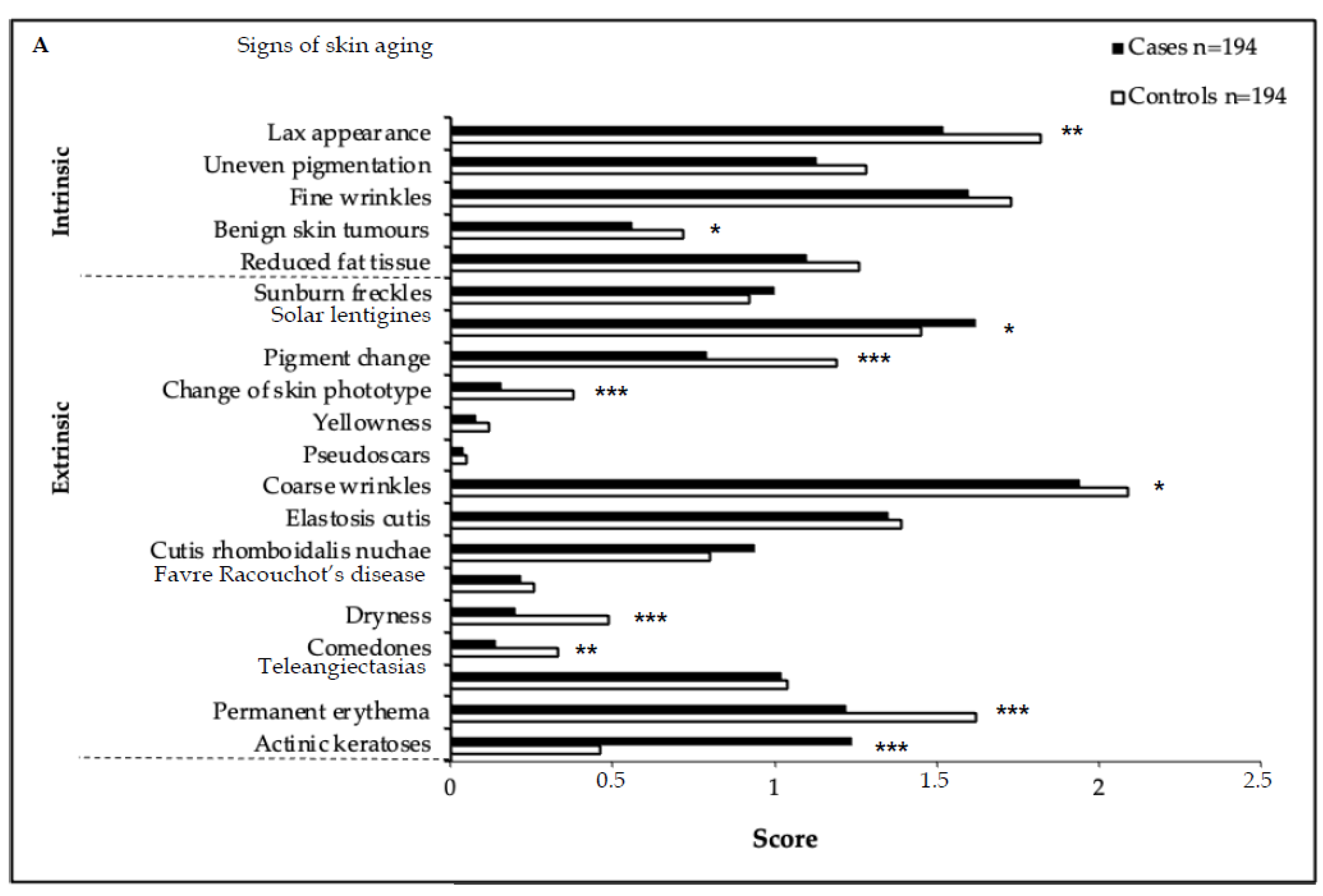
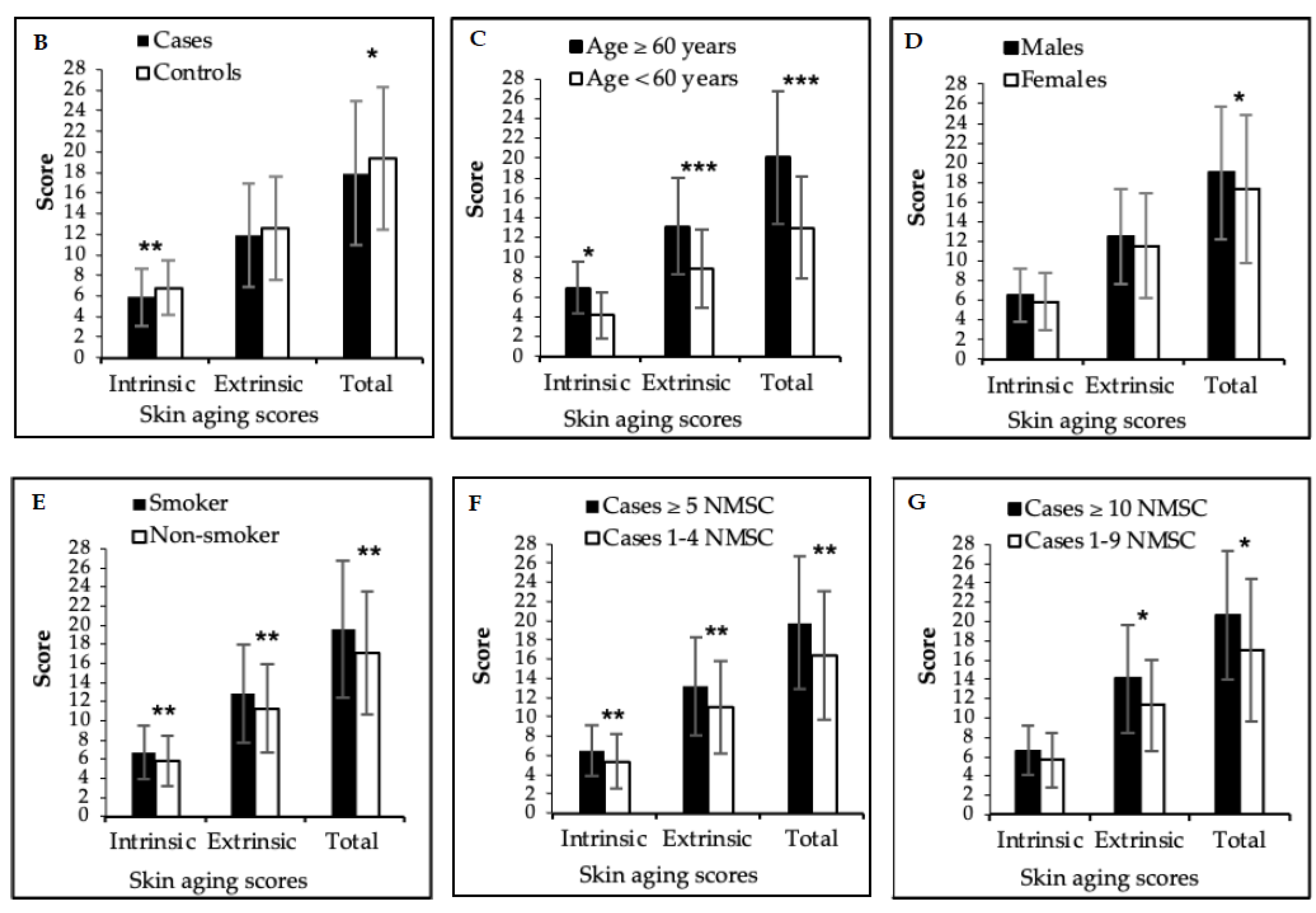
Cases had significantly lower ISA and TSA scores compared to the controls (p = 0.001 and p = 0.04, respectively; Figure 2A,B, Table S2). The presence of AKs, which apart from solar lentigines was the only ESA score feature observed more frequently in cases (p < 0.001), did not shift the ESA score into significance (p = 0.236; Table S2). However, all types of aging scores correlated significantly with the number of NMSC. Ninety-seven case–control pairs matched for FST (FST II n = 43 pairs, FST III n = 54 pairs), and thus having the same skin type was comparable with respect to age at examination, type of transplant, post-TX period, and smoking history (Tables S6 and S9). In these 97 pairs, all types of skin aging scores reached significance in favor of the controls (ISA score p = 0.034, ESA score p = 0.018, TSA score p = 0.016; Table S9), as was also the case in a subset of 51 FST-matched and AK-free pairs. When comparing skin aging scores between individuals with FSTII and FSTIII in the 97 FST-matched pairs, only the ESA score was significantly higher in individuals with FSTII (p = 0.036; Table S9), an effect not seen in the entire study population.
Higher values of all skin aging scores correlated with age >60 years (p < 0.001) and smoking (p < 0.005); male gender only correlated with the TSA score (p = 0.043). Cases/controls with smoking history did not differ in any of the aging scores, regardless of their FST (Figure 2C–E, Tables S10 and S11).

3.4. Mitochondrial DNA Point Heteroplasmy

In a pilot experiment, we obtained skin scales from the UV-exposed and -unexposed skin areas of 23 individuals. We found a statistically significant difference in the number of mutations in mitochondrial DNA between UV-unexposed and -exposed skin (p < 0.001; Table S12). No correlation was observed between the extent of PHP and the level of the modified skin aging score and the extent of cumulative UV exposure.

3.5. Ultraviolet Burden

Outdoor occupational activities were not associated with the development of NMSC: 37% of cases and 44% of controls reported an outdoor occupation in the past (p = 0.180). We found no difference in skin aging scores among cases/controls with comparable occupational sun exposure. Control subjects spent more time outdoors with unintentional sun exposure (UV scenario I + II, i.e., body partly covered, with UVR exposure only to uncovered body parts such as head/neck, face, or upper extremities; p = 0.665; Table 1). Pairs additionally matched for TSA score (n = 57) showed no difference with respect to UV scenarios (Table S13).
The total number of hours spent with intentional recreational and whole-body UV exposure (UV scenario III + IV) were significantly higher in cases (p = 0.033), due to increased UV exposure in different periods of life, e.g., sunbathing in Central Europe at ages 10–19 years (‘at home′, p = 0.027) and beach holidays in Southern/Mediterranean regions at ages 40–59 years (p = 0.017; Table 1).
The effect was more pronounced in 97 FST-matched pairs (UV scenario IV: p = 0.002; UV scenario III + IV: p = 0.015), and it remained significant in FST-matched pairs without AKs (n = 51 pairs; p = 0.023 and p = 0.037, respectively). When comparing 86 individuals with FSTII and 108 with FSTIII within the subgroup of 97 FST-matched pairs, we did not detect any difference in terms of UV exposure (Table S13). The detailed survey of the weeks of life recreational activities showed no substantial differences between cases and controls (Table S3).
In the whole study population, regardless of the development of NMSC, higher TSA scores correlated with higher unintentional (p < 0.001) as well as intentional (p = 0.007) sun exposure. Notably, unintentional sun exposure, including outdoor occupation, accounted for most of the total UV burden (Table 2). In the subgroup of 97 cases/control pairs additionally matched for FST, unintentional UV exposure was again associated with significantly higher TSA and ESA scores (p = 0.006 and p = 0.001, respectively; Table S14). A higher ESA, but not TSA score, correlated with outdoor occupation (p = 0.040). For intentional sun exposure, only sunbathing in Central Europe correlated with an increased ESA score (p = 0.042). Mean lifetime hours of sunbed use and gardening were strongly correlated with TSA score (p = 0.014, p = 0.001, respectively; Table S14). The total number of lifetime hours of UV exposure was twice as high in Central Europe as in the southern regions, explaining the association with higher skin aging scores (ESA and TSA p = 0.001, ISA score p = 0.036) (Table 1 and Table 2). Smoking had an additive effect on UV-related skin aging: in the subgroup of cases with a UV burden in the highest quartile (n = 97), we found a statistically significant higher ISA and TSA score in smokers compared to non-smokers (n = 62; ISA score: p = 0.003, TSA score p = 0.015; Table S11, Figure S1).

3.6. Analyses of Cofactors for Non-Melanoma Skin Cancer

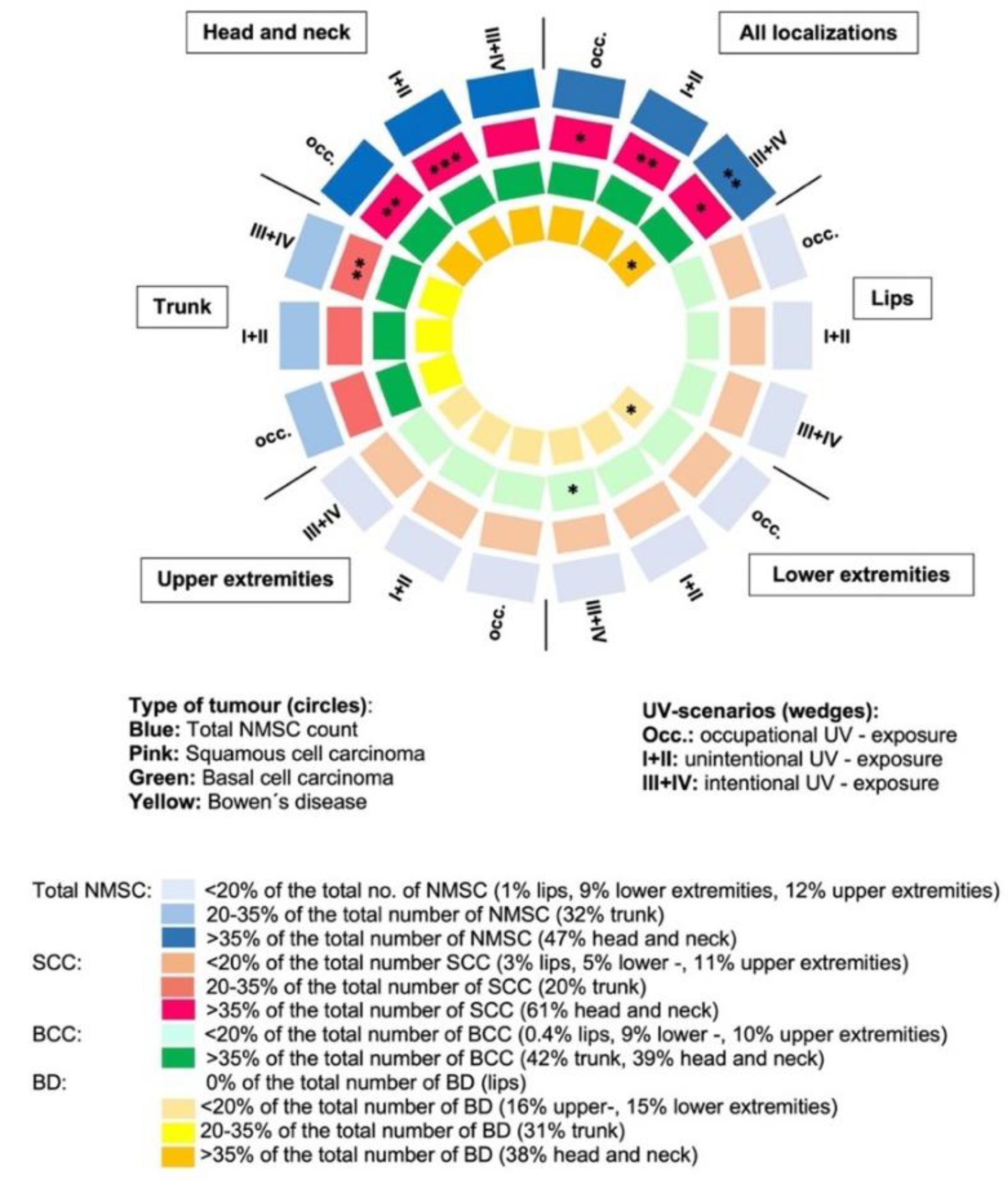
In the cases, the total numbers of NMSC (all locations), cSCC (total, head/neck, and trunk region), and BCC (total, head/neck region, upper extremities) correlated with all types of skin aging scores (range p < 0.001 to p = 0.025), although we evaluated skin aging in the head/neck region only (Table S15). When we linked the number and types of NMSC with UV exposure, occupational sun exposure correlated only with the total number of cSCC (p = 0.012), especially in the head/neck region (p = 0.001); no significant correlations were observed for BCC in this setting (Figure 3, Table S16).
The total time spent outdoors with unintentional sun exposure (UV scenario I + II) showed no correlation with the total number of NMSC, but it was highly and exclusively correlated with the total number of cSCC (p = 0.001) and the number of SCC in the head/neck location (p < 0.001). The total time spent outdoors with intentional sun exposure (uncovered body, wearing swimwear, UV scenario III + IV) was highly correlated with the total number of NMSC (p = 0.002), cSCC (p = 0.049), and BD (p = 0.025); with respect to the different tumor locations, only the trunk region reached significance (p = 0.009) for cSCC numbers, and for BCC, the lower extremities (p = 0.025; Figure 3, Table S16).
Outdoor recreational activities or gardening showed no correlation with cSCC or BCC, while mountaineering, hiking, or skiing correlated with the occurrence of BD in the head/neck area (p = 0.031), and sunbed use correlated with BD in the lower extremities (p = 0.045). When comparing the high-risk cases (≥5 NMSC) of FSTII (n = 48) with those of FSTIII (n = 35), no difference was observed regarding UV burden in the different UV scenarios. When comparing these two groups in terms of skin aging, only the ESA score was significantly higher in cases with FSTII compared to FSTIII (p = 0.042; Table S17).
In the subgroups of 87 cases with ≥5 NMSC and 44 cases with ≥10 NMSC—compared to those with a lower number of NMSC (1–4 NMSC, 1–9 NMSC, respectively)—higher values in all skin aging scores correlated with a higher number of NMSC, except for the ISA score in cases ≥10 NMSC (Figure 2F,G). Only intentional vacation-associated high UV exposure correlated with a higher number of NMSC (≥5 NMSC: p = 0.004; ≥10 NMSC: p = 0.002). The type of activity, full or partial sun exposure, or sunbed use made no difference (Table 3).

3.7. MC1R Analyses

The MC1R genotype was available for each patient included in this study (n = 388; Table S18A). The definition of MC1R risk variants/groups is described in the Supplementary Materials. Red hair was found exclusively in R/R individuals. Individuals with FST I predominantly (89%) carried R/R high-risk variants (n = 8), whereas the majority of participants with FST II and III carried no (0/0; 30% and 38%, respectively) or low-risk 0/r (32% and 34%, respectively) variants (Table S19).
The cases and controls differed significantly in their distribution within the low- and medium- + high-risk groups (p = 0.03). The only MC1R haplotype associated with NMSC was defined by the relatively rare variant c.464T > C (p = 0.03; Table S18B). Only the risk group 0/R was more common among cases (22%) compared to controls (14%; p = 0.044; Table S20), and also more common in cases with FSTII (27% cases versus 15% controls).
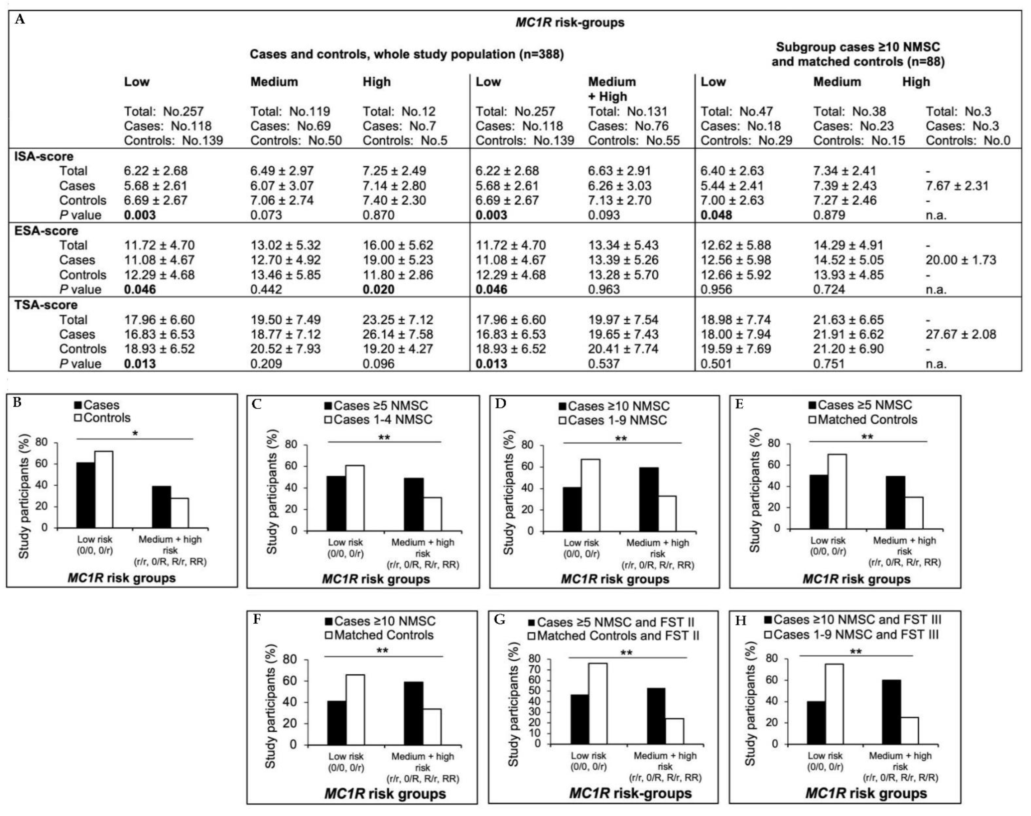
Higher NMSC counts clearly correlated with the MC1R medium- + high-risk group when cases with a history of ≥5 or ≥10 NMSC were compared with those with 1–4 (p = 0.004) or 1–9 (p = 0.001) NMSC, respectively, or with matched controls (p = 0.008 and p = 0.019, respectively). The associations of NMSC counts, MC1R risk groups, and FST are shown in Figure 4. In contrast to the overall study population (p = 0.325), in the subset of cases with a high tumor burden (≥10 NMSC), individuals with FSTIII or dark brown hair more frequently carried medium + high-risk variants than cases with ≤10 tumors (p = 0.008 and p = 0.001, respectively). This suggests a pigmentation-independent effect of MC1R on the tumor number. Only in blue-eyed individuals, did cases carry medium- + high-risk variants more frequently (43%) than controls (25%; p = 0.024), an observation that was more pronounced when cases of ≥5 or ≥10 tumors were compared with matched controls (p = 0.007 and p = 0.028, respectively; Tables S19–S23).
Regarding the numbers of different NMSC entities, only the number of cSCC was significantly and exclusively higher in the RR risk group (p = <0.001). However, due to the small number of patients, the informative value of this observation is limited.
Skin aging scores gradually increased from the low, medium, and high MC1R risk groups in the whole study population (ESA score p = 0.002; TSA score p = 0.01) and when calculated separately in cases and controls. Only the ESA score was significantly higher in cases in the high MC1R risk group (RR, p = 0.02; Figure 4), but again, there was only a small number of cases.
Within the different MC1R risk groups, self-reported UV exposure was comparable between the cases and controls (Table S24A). Therefore, an additional UV effect on the development of skin cancer in carriers of medium- and high-risk MC1R variants can be neglected.
A multivariate analysis revealed a 1.9-fold increased risk for subjects belonging to the medium- + high-risk MC1R group, and a 4.1- and 3.6-fold increased risk for individuals with green or blue eyes, respectively, while the presence of AKs conferred a 7.5-fold increased risk for the development of NMSC (Figure 5).

4. Discussion

Organ transplant recipients are at high risk for KC, particularly cSCC [2], which accounted for 55% of the 1633 NMSC in our study. The selection of two cohorts of patients —‘cases’ with NMSC and matched ‘controls′ who remained tumor-free after TX—provided us with an approach to evaluate the effect of skin aging and UV exposure on the development of NMSC in OTR. A valid assessment of lifetime UVR exposure and skin aging using a standardized questionnaire and a detailed survey of the signs of skin aging was a prerequisite for the evaluation of factors contributing to the development of NMSC.
Extrinsic skin aging, synonymously referred to as ‘photoaging’, i.e., structural and morphological changes of the skin including the clinical sign of AKs, serves as an indicator of age-related environmental effects on human skin, especially UVR [15]. Apart from solar lentigines, AKs were the only ESA score parameter that was significantly higher in cases: nearly 50% were affected, compared with 16% in the controls. All other ESA score parameters and the TSA score reached higher values in the controls. Concerning the whole study population, higher scores correlated with age, male gender, and history of smoking, independent of FST and/or the presence of AKs, demonstrating the reliability of our quantification of skin aging [27,28]. We observed a non-significant trend toward higher ESA score values in individuals with FSTII compared with FSTIII, with more than 50% of cases being FSTII compared with 36% of the controls: a lighter skin type seems to be associated with the development of NMCSs after TX. To avoid bias due to skin type, we examined cases/control pairs that were additionally matched on FST. Again, the controls scored higher on all skin aging scores, suggesting that skin aging per se does not necessarily predict a higher risk for KC. In addition to the exogenous factors, interindividual differences in skin aging can also be attributed to genetic factors that influence collagen degradation and elastin deposition [29,30]. The intrinsic skin aging score may not adequately reflect these factors, which could explain the lack of association between skin aging and KC development.
Only in the MC1R high-risk group (R/R) did cases have higher ESA and TSA scores. MC1R is an important determinant of skin phototype, as it regulates melanin production [26,31,32,33,34,35,36,37,38,39,40,41,42,43,44]. While FST appears to be the major factor predisposing to skin cancer in our collective, the MC1R genotype seems to predominate over FST: in a multivariate analysis, the MC1R genotype, but not the FST, remained significant. In our cohort and consistent with published data, FSTI correlated with ‘high-risk types’, i.e., R/R MC1R carriers [43]. Only in cases that had FSTII and/or blue eyes, did the medium- + high-risk MC1R group correlate with the development of NMSC, confirming a predisposition to KC in individuals with a lighter skin type. Nevertheless, a higher percentage of cases and the majority of controls carried low-risk variants, implying that additional genetic factors play a role. We found a correlation between MC1R risk types and the number of skin tumors in individuals with a history of more than 5 or 10 NMSCs. Those with a higher tumor burden more often carried medium- or high-risk MC1R variants. Interestingly, this was also true for cases with FSTIII and/or dark brown hair, indicating a pigmentation-independent effect of MC1R variants on KC development. Tagliabue et al. reported similar results [43].
While we assessed skin aging signs only in the head/neck area, skin aging scores correlated with higher numbers of NMSC at all locations, numbers of cSCC predominately in the head/neck/trunk area, and BCC in the head/neck/upper extremities region. This suggests that our quantification of skin aging provides a risk assessment for the occurrence of multiple NMSCs in the most commonly affected areas.
We applied established protocols for the reliable assessment of sun exposure and collected information on time spent outdoors and outdoor activities [25]. Comparing cases and controls, we found no difference in unintentional or occupational UV exposure—the latter possibly because of the small sample size—nor any association with different leisure activities. Sunbathing in Central Europe in adolescence, and beach vacations in the southern or Mediterranean regions in adulthood, appear to be associated with the development of NMSC and higher tumor count, independent of FST. This implies that high-intensity UVR is critical for the development of KC [45].
To date, there are no data on occupational sun exposure in OTR [46]. In our study, the number of cSCC—but not BCC—in the head/neck region correlated with occupational UV exposure. Studies focusing on NMSC in association with cumulative occupational UV exposure are conflicting [46,47]. They report an increased risk of NMSC in outdoor workers, mostly farmers, and differ for cSCC and BCC [46,48]. A meta-analysis found a stronger association with occupational UV exposure for BCC than for cSCC [46]. This is supported by the fact that in the general population the incidence of BCC is twice that of cSCC [49], whereas in OTR the ratio is inverse.
Higher intentional and unintentional UV exposure correlated with higher TSA scores [28]. In a subset of pairs matched for skin aging—30% of the cohort—we found an equal UV burden, demonstrating that UV exposure contributes to skin aging [12] without clear evidence of its effect on NMSC. A subanalysis of pairs additionally matched on FST confirmed this. The number of NMSCs correlated with aging scores and specific UV exposure. There was no correlation with lighter skin type or smoking history.
Our findings are consistent with the knowledge that UVR is a major factor in age-related skin changes that are accelerated by smoking [27]. In our study, all aging scores were higher in smokers, which remained significant in 97 FST-matched pairs. However, there was no significant difference between cases and controls in this subgroup of 97 FST-matched pairs with respect to smoking, suggesting that smoking plays a role in skin aging but does not appear to be associated with cancer development [27,50].
Consistent with the literature, AKs as precursors of cSCC were highly associated with UVR, but only in the context of intentional sun exposure in Central Europe [51]. Spending time outdoors, fully clothed but with the head and neck uncovered, correlated exclusively with high numbers of cSCC in this region. Intentional UV exposure correlated strongly with the total number of NMSC as well as cSCC on the trunk but not on the head/neck. In this UV scenario, we found a correlation between the total number of BD and BCC on the lower extremities. Outdoor recreational activities or gardening showed no particular association with NMSC.
To investigate an association between the extent of UV exposure and MC1R genotypes was of interest because there might be an additive effect on skin tumor development. However, we found no difference between our cases and the controls when we stratified MC1R genotypes and UV exposure with tumor development. A novel aspect was a possible association of MC1R risk types with the individual diversity of KC types; cases that developed a single tumor entity preferentially carried low-risk variants, whereas cases affected by the full spectrum of KC had equal distributions of low- and medium- + high-risk MC1R types. Finally, in multivariable models, we identified AKs, green or blue eyes, and the presence of medium- + high-risk MC1R types as factors associated with the development of NMSC. Skin aging, time spent outdoors, sunbed use, FST, and hair color had no significant association in these models.
To our knowledge, this is the only case control study in OTR with and without skin tumors in which skin aging, previous UV exposure, and MC1R genotypes were evaluated. The strength of this study lies in the fact that patients with and without skin tumors were maximally matched in patient characteristics, post-TX period, and immunosuppressive therapy. Some limitations should be considered: due to the case-control design, selection bias cannot be completely excluded, and because of the exploratory nature of the case-control design, our results should be confirmed in a cohort study.

5. Conclusions

In analyzing a substantial number of OTR who received IS therapy and were therefore at risk for developing a skin tumor, we found that in our study population, the actual risk for developing KC was not adequately explained by the extent of skin aging, UV-related skin damage, or cumulative UV exposure. However, skin aging does influence the individual number of tumors. Previous intensive UV exposure was relevant to the development of KC. Unintentional sun exposure accounted for most of the total UV burden: twice as high in Central Europe as in the southern regions, explaining the association with higher skin aging scores. Whole-body UV exposure was associated with a higher number of NMSCs. The same was observed for certain MC1R variants, even in individuals with FSTIII, suggesting a pigmentation-independent effect (Table 4).
To summarize, in OTR, skin aging increases with age, smoking history, and high UVR exposure. Men have higher skin aging scores. Risk factors for developing NMSC include lighter phenotype, holiday-related high UVR, specific MC1R variants, and the presence of AK, but not skin aging per se. The number of NMSCs increases with longer time after TX period, shorter interval between TX and first NMSC, older age, higher skin aging scores, the presence of AK, specific MC1R variants, and severe sunburns as young adults.
Independently of the known proactive measures in the dermatologic follow-up of OTRs, to better assess the risk for developing skin tumors, a history regarding previous high-intensity UVR exposure could be performed. The inclusion of genetic factors could be an additional facet of risk assessment in the future.

Supplementary Materials

The following supporting information can be downloaded at: https://www.mdpi.com/article/10.3390/cancers15030864/s1, References [52,53,54,55,56] are cited in the Supplementary Materials. Table S1. Patient Characteristics (a) and Correlation of Melanin Index and Fitzpatrick Skin Type (I-IV) (b). Table S2. Modified Skin Aging Scores Including the Intrinsic and Extrinsic Signs of Skin Aging in Cases and Controls. Table S3. Recreational Activities in Total Weeks of Life in Cases and Controls. Table S4. Correlation between NMSC Count and Clinical/UV Associated Parameters. Table S5. Reaction to Sun Exposure and Sunburns in Different Periods of Life. Table S6. Characteristics of a Subset of Pairs Additionally Matched for Fitzpatrick Skin Type. Table S7. The Number and Percentage of the Different Tumor Entities in Different Regions (a), Distribution of the Tumors in Different Localizations (b), Mean Number of Tumors per Patient and tumor entity (c). Table S8. Correlation between NMSC Count and Age, Gender, Aging Score, Time to First NMSC, Number of AKs, and MC1R Risk Groups. Table S9. Skin Aging Scores in a Subset of Cases and Controls Additionally Matched for Fitzpatrick Skin Type and in the Whole Study Population. Table S10. Skin Aging Score Values in Association with Age and Gender. Table S11. Skin Aging Scores in Association with Smoking History in the Whole Study Population, Subsets of FST-matched Cases/Controls Pairs and Cases with the Highest UV-burden. Table S12. Mitochondrial DNA Point Heteroplasmy (PHP): Number of mtDNA PHP in UV-exposed Versus UV-unexposed Skin and in Cases Versus Controls. Table S13. Quantification of UV-Exposure in Different Scenarios and Different Subsets (cases versus controls). Table S14. Correlations of the Skin Aging Scores with UV-exposure Scenarios in a Subset of 97 Cases/Control Pairs Matched for Fitzpatrick Skin Type. Table S15. Correlations between Skin Aging Scores and NMSCs Located in Different Regions. Table S16. Correlation of NMSC (localization) With UV Exposure. Table S17. Comparison of The Skin Aging Scores and UV- Scenarios in Cases ≥5 NMSC: FST II Versus FST III Subsets. Table S18. MC1R Variants and their Frequency in Cases Versus Controls (a), MC1R Haplotypes and Their Frequency in Cases and Controls (b), MC1R Diplotypes and Their Frequency in Cases and Controls (c). Table S19. Association of MC1R Risk Groups with Skin Type, Hair and Eye Color in cases versus controls. Table S20. MC1R Risk Groups in Different Subsets of Cases and Controls. Table S21. Association of MC1R Risk Groups with Skin Pigmentation in Different Subgroups of OTR. Table S22. Association of MC1R Risk Groups with Hair Color in Different Subgroups of OTR (≥5 and ≥10 NMSC vs. matched controls). Table S23. Association of MC1R Risk Groups with Eye Color in Different Subgroups of OTR (≥5 and ≥10 NMSC vs. matched controls). Table S24 Association of MC1R Risk Groups with Different UV Scenarios in Cases versus Controls (a, Part I and II), Association of MC1R Risk Groups with Different UV scenarios in Cases with ≥ 10 NMSC (n = 44) versus Matched Controls (b, Part I and II). Table S25. Association of MC1R Risk Groups with Hair Color in Different Subgroups of OTR (≥5 vs. 1–4 and ≥10 vs. 1–9 NMSC). Table S26. Association of MC1R Risk Groups with Different NMSC Entities. Figure S1. Hours of High UV-exposure (9 am–3 pm) in Mediterranean or Southern Regions in Cases versus Controls.

Author Contributions

Conceptualization, A.G. and R.S.-P.; acquisition of the data, L.B.-H., A.G., L.S., A.Z., K.U.-Ü., P.J., G.B., D.C., K.S., E.H.-W. and S.R.-R.; data analysis, G.E., W.P., R.S.-P. and L.S.; interpretation of the data, A.G. and R.S.-P.; drafting the work, A.G. and R.S.-P.; revising the work, A.G., R.S.-P., G.B. and P.W. All authors have read and agreed to the published version of the manuscript.

Funding

Jubilee Fund of the Austrian National Bank Fond (OeNB, grant number 17221 for Alexandra Geusau). This research was supported in part by a clinician scientist grant of the Austrian Society of Dermatology and Venereology (‘ÖGDV Forschungsförderung Klinische ForscherInnen in der Dermatovenerologie′). The funders were not involved in the study design, data collection, data analysis, and manuscript preparation or publication decisions.

Institutional Review Board Statement

This study was conducted according to the guidelines of the Declaration of Helsinki and approved by the Ethics Committee of the Medical University of Vienna (protocol number 1233/2016, approved 12 April 2016).

Informed Consent Statement

Informed consent was obtained from all subjects involved in the study.

Data Availability Statement

The data that support the findings of the study are available upon request.

Acknowledgments

We thank Ulrike Sattler and Andreas Ebner for the clinical photography; Sabina Plumer, C. Amory, A. Heidegger, A. Kaindl-Lindinger, and the Forensic Genetics and Casework section of the Institute of Legal Medicine in Innsbruck for technical assistance; Tina Breckwoldt for editing the manuscript.

Conflicts of Interest

The authors declare no conflict of interest, except for Staufer (Chief Medical Officer at Versantis AG, received speaker honoraria from Vifor and Chiesi, and received travel grants from Vifor).

References

  1. Plasmeijer, E.I.; Sachse, M.M.; Gebhardt, C.; Geusau, A.; Bouwes Bavinck, J.N. Cutaneous squamous cell carcinoma (cSCC) and immunosurveillance—The impact of immunosuppression on frequency of cSCC. J. Eur. Acad. Derm. Venereol. 2019, 33 (Suppl. S8), 33–37. [Google Scholar] [CrossRef] [PubMed] [Green Version]
  2. Euvrard, S.; Kanitakis, J.; Claudy, A. Skin cancers after organ transplantation. N. Engl. J. Med. 2003, 348, 1681–1691. [Google Scholar] [CrossRef] [PubMed] [Green Version]
  3. Grulich, A.E.; van Leeuwen, M.T.; Falster, M.O.; Vajdic, C.M. Incidence of cancers in people with HIV/AIDS compared with immunosuppressed transplant recipients: A meta-analysis. Lancet 2007, 370, 59–67. [Google Scholar] [CrossRef] [PubMed]
  4. Krynitz, B.; Edgren, G.; Lindelof, B.; Baecklund, E.; Brattstrom, C.; Wilczek, H.; Smedby, K.E. Risk of skin cancer and other malignancies in kidney, liver, heart and lung transplant recipients 1970 to 2008—A Swedish population-based study. Int. J. Cancer 2013, 132, 1429–1438. [Google Scholar] [CrossRef]
  5. Billups, K.; Neal, J.; Salyer, J. Immunosuppressant-driven de novo malignant neoplasms after solid-organ transplant. Prog. Transpl. 2015, 25, 182–188. [Google Scholar] [CrossRef]
  6. Mudigonda, T.; Levender, M.M.; O’Neill, J.L.; West, C.E.; Pearce, D.J.; Feldman, S.R. Incidence, risk factors, and preventative management of skin cancers in organ transplant recipients: A review of single- and multicenter retrospective studies from 2006 to 2010. Derm. Surg. 2013, 39, 345–364. [Google Scholar] [CrossRef] [PubMed]
  7. Ratushny, V.; Gober, M.D.; Hick, R.; Ridky, T.W.; Seykora, J.T. From keratinocyte to cancer: The pathogenesis and modeling of cutaneous squamous cell carcinoma. J. Clin. Investig. 2012, 122, 464–472. [Google Scholar] [CrossRef] [Green Version]
  8. Didona, D.; Paolino, G.; Bottoni, U.; Cantisani, C. Non Melanoma Skin Cancer Pathogenesis Overview. Biomedicines 2018, 6, 6. [Google Scholar] [CrossRef] [Green Version]
  9. Geusau, A.; Dunkler, D.; Messeritsch, E.; Sandor, N.; Heidler, G.; Rodler, S.; Ankersmit, J.; Zuckermann, A.; Tschachler, E. Non-melanoma skin cancer and its risk factors in an Austrian population of heart transplant recipients receiving induction therapy. Int. J. Derm. 2008, 47, 918–925. [Google Scholar] [CrossRef]
  10. Harwood, C.A.; Toland, A.E.; Proby, C.M.; Euvrard, S.; Hofbauer, G.F.L.; Tommasino, M.; Bouwes Bavinck, J.N.; KeraCon, C. The pathogenesis of cutaneous squamous cell carcinoma in organ transplant recipients. Br. J. Derm. 2017, 177, 1217–1224. [Google Scholar] [CrossRef]
  11. Hofbauer, G.F.; Bouwes Bavinck, J.N.; Euvrard, S. Organ transplantation and skin cancer: Basic problems and new perspectives. Exp. Derm. 2010, 19, 473–482. [Google Scholar] [CrossRef] [PubMed]
  12. Krutmann, J.; Bouloc, A.; Sore, G.; Bernard, B.A.; Passeron, T. The skin aging exposome. J. Derm. Sci. 2017, 85, 152–161. [Google Scholar] [CrossRef] [PubMed] [Green Version]
  13. Mancebo, S.E.; Wang, S.Q. Skin cancer: Role of ultraviolet radiation in carcinogenesis. Rev. Environ. Health 2014, 29, 265–273. [Google Scholar] [CrossRef]
  14. Fuks, K.B.; Huls, A.; Sugiri, D.; Altug, H.; Vierkotter, A.; Abramson, M.J.; Goebel, J.; Wagner, G.G.; Demuth, I.; Krutmann, J.; et al. Tropospheric ozone and skin aging: Results from two German cohort studies. Environ. Int. 2019, 124, 139–144. [Google Scholar] [CrossRef]
  15. Vierkotter, A.; Ranft, U.; Kramer, U.; Sugiri, D.; Reimann, V.; Krutmann, J. The SCINEXA: A novel, validated score to simultaneously assess and differentiate between intrinsic and extrinsic skin ageing. J. Derm. Sci. 2009, 53, 207–211. [Google Scholar] [CrossRef]
  16. Yaar, M.; Gilchrest, B.A. Aging versus photoaging: Postulated mechanisms and effectors. J. Investig. Derm. Symp. Proc. 1998, 3, 47–51. [Google Scholar]
  17. Manganelli, M.; Guida, S.; Ferretta, A.; Pellacani, G.; Porcelli, L.; Azzariti, A.; Guida, G. Behind the Scene: Exploiting MC1R in Skin Cancer Risk and Prevention. Genes 2021, 12, 1093. [Google Scholar] [CrossRef]
  18. Nasti, T.H.; Timares, L. MC1R, eumelanin and pheomelanin: Their role in determining the susceptibility to skin cancer. Photochem. Photobiol. 2015, 91, 188–200. [Google Scholar] [CrossRef] [Green Version]
  19. Wendt, J.; Rauscher, S.; Burgstaller-Muehlbacher, S.; Fae, I.; Fischer, G.; Pehamberger, H.; Okamoto, I. Human Determinants and the Role of Melanocortin-1 Receptor Variants in Melanoma Risk Independent of UV Radiation Exposure. JAMA Derm. 2016, 152, 776–782. [Google Scholar] [CrossRef] [PubMed] [Green Version]
  20. Fitzpatrick, T.B. The validity and practicality of sun-reactive skin types I through VI. Arch. Derm. 1988, 124, 869–871. [Google Scholar] [CrossRef] [PubMed]
  21. Sitek, A.; Rosset, I.; Zadzinska, E.; Kasielska-Trojan, A.; Neskoromna-Jedrzejczak, A.; Antoszewski, B. Skin color parameters and Fitzpatrick phototypes in estimating the risk of skin cancer: A case-control study in the Polish population. J. Am. Acad. Derm. 2016, 74, 716–723. [Google Scholar] [CrossRef] [PubMed]
  22. Dwyer, T.; Muller, H.K.; Blizzard, L.; Ashbolt, R.; Phillips, G. The use of spectrophotometry to estimate melanin density in Caucasians. Cancer Epidemiol. Biomark. Prev. 1998, 7, 203–206. [Google Scholar] [CrossRef] [Green Version]
  23. Eilers, S.; Bach, D.Q.; Gaber, R.; Blatt, H.; Guevara, Y.; Nitsche, K.; Kundu, R.V.; Robinson, J.K. Accuracy of self-report in assessing Fitzpatrick skin phototypes I through VI. JAMA Derm. 2013, 149, 1289–1294. [Google Scholar] [CrossRef] [PubMed] [Green Version]
  24. Buranasirin, P.; Pongpirul, K.; Meephansan, J. Development of a Global Subjective Skin Aging Assessment score from the perspective of dermatologists. BMC Res. Notes 2019, 12, 364. [Google Scholar] [CrossRef]
  25. Yu, C.L.; Li, Y.; Freedman, D.M.; Fears, T.R.; Kwok, R.; Chodick, G.; Alexander, B.; Kimlin, M.G.; Kricker, A.; Armstrong, B.K.; et al. Assessment of lifetime cumulative sun exposure using a self-administered questionnaire: Reliability of two approaches. Cancer Epidemiol. Biomark. Prev. 2009, 18, 464–471. [Google Scholar] [CrossRef] [PubMed] [Green Version]
  26. Garcia-Borron, J.C.; Sanchez-Laorden, B.L.; Jimenez-Cervantes, C. Melanocortin-1 receptor structure and functional regulation. Pigment. Cell. Res. 2005, 18, 393–410. [Google Scholar] [CrossRef]
  27. Morita, A. Tobacco smoke causes premature skin aging. J. Derm. Sci. 2007, 48, 169–175. [Google Scholar] [CrossRef] [PubMed]
  28. Vierkotter, A.; Krutmann, J. Environmental influences on skin aging and ethnic-specific manifestations. Dermato-Endocrinology 2012, 4, 227–231. [Google Scholar] [CrossRef] [Green Version]
  29. Wigmann, C.; Huls, A.; Krutmann, J.; Schikowski, T. Estimating the Relative Contribution of Environmental and Genetic Risk Factors to Different Aging Traits by Combining Correlated Variables into Weighted Risk Scores. Int. J. Environ. Res. Public Health 2022, 19, 16746. [Google Scholar] [CrossRef]
  30. Zhang, Y.; Zhang, M.; Yao, A.; Xie, Y.; Lin, J.; Sharifullah, F.; Hong, Y.; Chen, H.; Cheng, F.; Lai, W. Circ_0011129 Encapsulated by the Small Extracellular Vesicles Derived from Human Stem Cells Ameliorate Skin Photoaging. Int. J. Mol. Sci. 2022, 23, 15390. [Google Scholar] [CrossRef]
  31. Bastiaens, M.T.; ter Huurne, J.A.; Kielich, C.; Gruis, N.A.; Westendorp, R.G.; Vermeer, B.J.; Bavinck, J.N.; Leiden Skin Cancer Study Team. Melanocortin-1 receptor gene variants determine the risk of nonmelanoma skin cancer independently of fair skin and red hair. Am. J. Hum. Genet. 2001, 68, 884–894. [Google Scholar] [CrossRef]
  32. Beaumont, K.A.; Shekar, S.N.; Newton, R.A.; James, M.R.; Stow, J.L.; Duffy, D.L.; Sturm, R.A. Receptor function, dominant negative activity and phenotype correlations for MC1R variant alleles. Hum. Mol. Genet. 2007, 16, 2249–2260. [Google Scholar] [CrossRef]
  33. Binstock, M.; Hafeez, F.; Metchnikoff, C.; Arron, S.T. Single-nucleotide polymorphisms in pigment genes and nonmelanoma skin cancer predisposition: A systematic review. Br. J. Derm. 2014, 171, 713–721. [Google Scholar] [CrossRef] [PubMed]
  34. Duffy, D.L.; Box, N.F.; Chen, W.; Palmer, J.S.; Montgomery, G.W.; James, M.R.; Hayward, N.K.; Martin, N.G.; Sturm, R.A. Interactive effects of MC1R and OCA2 on melanoma risk phenotypes. Hum. Mol. Genet. 2004, 13, 447–461. [Google Scholar] [CrossRef] [PubMed] [Green Version]
  35. Garcia-Borron, J.C.; Olivares, C. Melanocortin 1 receptor and skin pathophysiology: Beyond colour, much more than meets the eye. Exp. Derm. 2014, 23, 387–388. [Google Scholar] [CrossRef] [PubMed] [Green Version]
  36. Han, J.; Kraft, P.; Colditz, G.A.; Wong, J.; Hunter, D.J. Melanocortin 1 receptor variants and skin cancer risk. Int. J. Cancer 2006, 119, 1976–1984. [Google Scholar] [CrossRef] [PubMed]
  37. Muller, C.; Wendt, J.; Rauscher, S.; Burgstaller-Muehlbacher, S.; Sunder-Plassmann, R.; Scheurecker, C.; Richtig, E.; Fae, I.; Fischer, G.; Pehamberger, H.; et al. Characterization of patients at high risk of melanoma in Austria. Br. J. Derm. 2016, 174, 1308–1317. [Google Scholar] [CrossRef] [PubMed]
  38. Pasquali, E.; Garcia-Borron, J.C.; Fargnoli, M.C.; Gandini, S.; Maisonneuve, P.; Bagnardi, V.; Specchia, C.; Liu, F.; Kayser, M.; Nijsten, T.; et al. MC1R variants increased the risk of sporadic cutaneous melanoma in darker-pigmented Caucasians: A pooled-analysis from the M-SKIP project. Int. J. Cancer 2015, 136, 618–631. [Google Scholar] [CrossRef] [Green Version]
  39. Raimondi, S.; Sera, F.; Gandini, S.; Iodice, S.; Caini, S.; Maisonneuve, P.; Fargnoli, M.C. MC1R variants, melanoma and red hair color phenotype: A meta-analysis. Int. J. Cancer 2008, 122, 2753–2760. [Google Scholar] [CrossRef]
  40. Scherer, D.; Bermejo, J.L.; Rudnai, P.; Gurzau, E.; Koppova, K.; Hemminki, K.; Kumar, R. MC1R variants associated susceptibility to basal cell carcinoma of skin: Interaction with host factors and XRCC3 polymorphism. Int. J. Cancer 2008, 122, 1787–1793. [Google Scholar] [CrossRef]
  41. Scherer, D.; Rachakonda, P.S.; Angelini, S.; Mehnert, F.; Sucker, A.; Egberts, F.; Hauschild, A.; Hemminki, K.; Schadendorf, D.; Kumar, R. Association between the germline MC1R variants and somatic BRAF/NRAS mutations in melanoma tumors. J. Investig. Derm. 2010, 130, 2844–2848. [Google Scholar] [CrossRef]
  42. Tagliabue, E.; Fargnoli, M.C.; Gandini, S.; Maisonneuve, P.; Liu, F.; Kayser, M.; Nijsten, T.; Han, J.; Kumar, R.; Gruis, N.A.; et al. MC1R gene variants and non-melanoma skin cancer: A pooled-analysis from the M-SKIP project. Br. J. Cancer 2015, 113, 354–363. [Google Scholar] [CrossRef] [PubMed] [Green Version]
  43. Tagliabue, E.; Gandini, S.; Garcia-Borron, J.C.; Maisonneuve, P.; Newton-Bishop, J.; Polsky, D.; Lazovich, D.; Kumar, R.; Ghiorzo, P.; Ferrucci, L.; et al. Association of Melanocortin-1 Receptor Variants with Pigmentary Traits in Humans: A Pooled Analysis from the M-Skip Project. J. Investig. Derm. 2016, 136, 1914–1917. [Google Scholar] [CrossRef] [PubMed] [Green Version]
  44. Valverde, P.; Healy, E.; Jackson, I.; Rees, J.L.; Thody, A.J. Variants of the melanocyte-stimulating hormone receptor gene are associated with red hair and fair skin in humans. Nat. Genet. 1995, 11, 328–330. [Google Scholar] [CrossRef]
  45. Martens, M.C.; Seebode, C.; Lehmann, J.; Emmert, S. Photocarcinogenesis and Skin Cancer Prevention Strategies: An Update. Anticancer Res. 2018, 38, 1153–1158. [Google Scholar]
  46. Loney, T.; Paulo, M.S.; Modenese, A.; Gobba, F.; Tenkate, T.; Whiteman, D.C.; Green, A.C.; John, S.M. Global evidence on occupational sun exposure and keratinocyte cancers: A systematic review. Br. J. Derm. 2021, 184, 208–218. [Google Scholar] [CrossRef] [PubMed]
  47. Schmitt, J.; Diepgen, T.; Bauer, A. Occupational exposure to non-artificial UV-light and non-melanocytic skin cancer—A systematic review concerning a new occupational disease. J. Dtsch. Derm. Ges. 2010, 8, 250–264. [Google Scholar] [CrossRef]
  48. Paulo, M.S.; Adam, B.; Akagwu, C.; Akparibo, I.; Al-Rifai, R.H.; Bazrafshan, S.; Gobba, F.; Green, A.C.; Ivanov, I.; Kezic, S.; et al. WHO/ILO work-related burden of disease and injury: Protocol for systematic reviews of occupational exposure to solar ultraviolet radiation and of the effect of occupational exposure to solar ultraviolet radiation on melanoma and non-melanoma skin cancer. Environ. Int. 2019, 126, 804–815. [Google Scholar] [CrossRef] [PubMed]
  49. Lomas, A.; Leonardi-Bee, J.; Bath-Hextall, F. A systematic review of worldwide incidence of nonmelanoma skin cancer. Br. J. Derm. 2012, 166, 1069–1080. [Google Scholar] [CrossRef] [PubMed]
  50. Goodman, G.D.; Kaufman, J.; Day, D.; Weiss, R.; Kawata, A.K.; Garcia, J.K.; Santangelo, S.; Gallagher, C.J. Impact of Smoking and Alcohol Use on Facial Aging in Women: Results of a Large Multinational, Multiracial, Cross-sectional Survey. J. Clin. Aesthet. Derm. 2019, 12, 28–39. [Google Scholar]
  51. Rangwala, S.; Tsai, K.Y. Roles of the immune system in skin cancer. Br. J. Derm. 2011, 165, 953–965. [Google Scholar]
  52. Heidegger, A.; Pisarek, A.; de la Puente, M.; Niederstatter, H.; Pospiech, E.; Wozniak, A.; Schury, N.; Unterlander, M.; Sidstedt, M.; Junker, K.; et al. Development and inter-laboratory validation of the VISAGE enhanced tool for age estimation from semen using quantitative DNA methylation analysis. Forensic Sci. Int. Genet. 2022, 56, 102596. [Google Scholar] [CrossRef]
  53. Xavier, C.; Eduardoff, M.; Strobl, C.; Parson, W. SD quants-Sensitive detection tetraplex-system for nuclear and mitochondrial DNA quantification and degradation inference. Forensic Sci. Int. Genet. 2019, 42, 39–44. [Google Scholar] [CrossRef] [PubMed]
  54. Strobl, C.; Churchill Cihlar, J.; Lagace, R.; Wootton, S.; Roth, C.; Huber, N.; Schnaller, L.; Zimmermann, B.; Huber, G.; Lay Hong, S.; et al. Evaluation of mitogenome sequence concordance, heteroplasmy detection, and haplogrouping in a worldwide lineage study using the Precision ID mtDNA Whole Genome Panel. Forensic Sci. Int. Genet. 2019, 42, 244–251. [Google Scholar] [CrossRef]
  55. Andrews, R.M.; Kubacka, I.; Chinnery, P.F.; Lightowlers, R.N.; Turnbull, D.M.; Howell, N. Reanalysis and revision of the Cambridge reference sequence for human mitochondrial DNA. Nat. Genet. 1999, 23, 147. [Google Scholar] [CrossRef] [PubMed]
  56. Thorvaldsdottir, H.; Robinson, J.T.; Mesirov, J.P. Integrative Genomics Viewer (IGV): High-performance genomics data visualization and exploration. Brief Bioinform. 2013, 14, 178–192. [Google Scholar] [CrossRef]
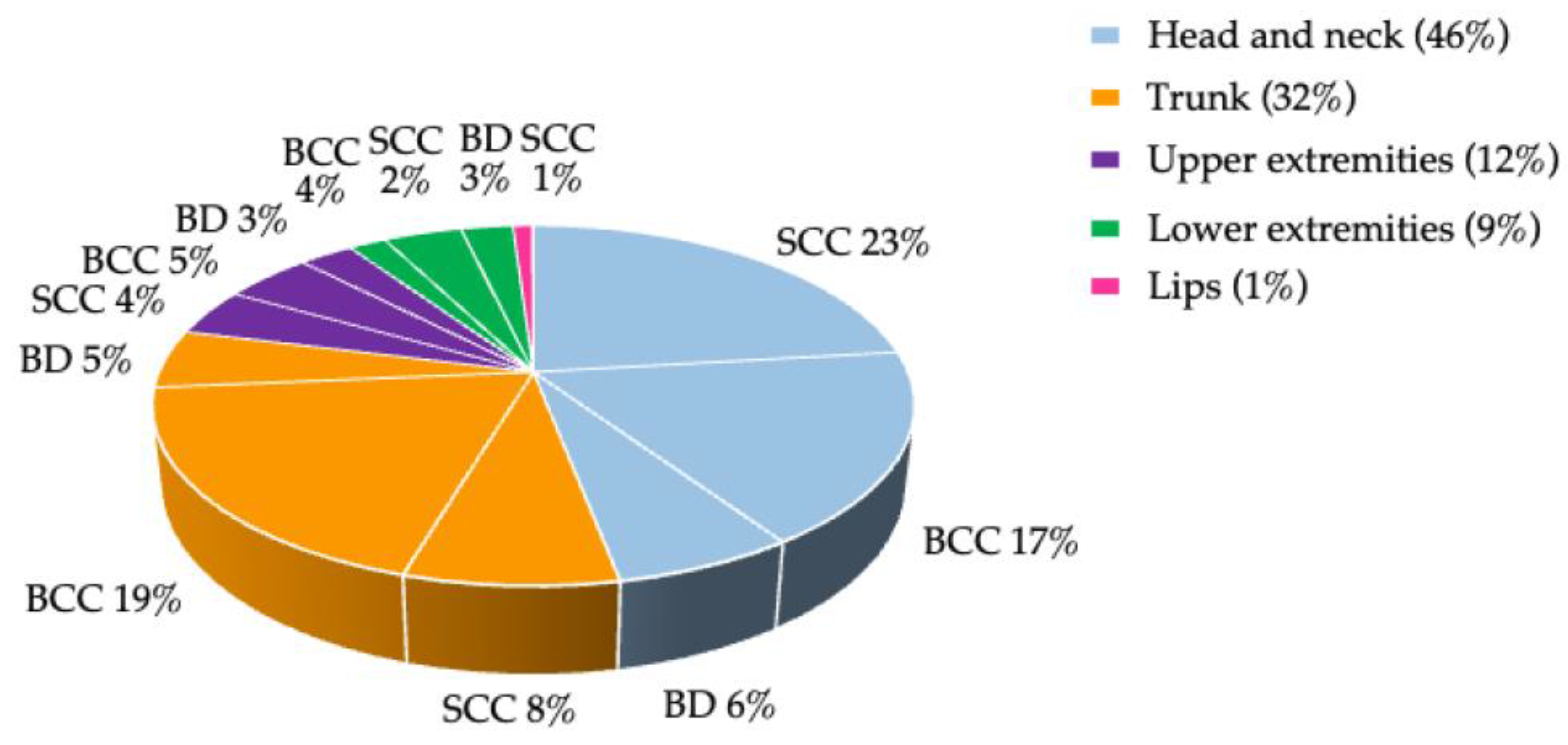
Figure 1. Anatomical localization of NMSC subtypes.
Cancers 15 00864 g001
Figure 2. Signs of skin aging and skin aging scores. Significant p values are indicated by asterisks: * p < 0.05, ** p < 0.01, *** p < 0.001. (A) Signs of skin aging in cases and controls; (B) skin aging scores (mean ± standard deviation) in cases and controls; (C) in age groups ≥60 (n = 310) and <60 (n = 78) years at time of evaluation; (D) in males (n = 298) and females (n = 90); (E) correlated with smoking history: smokers (n = 243) and non-smokers (n = 145); (F) in cases with ≥5 NMSCs (n = 87) and cases with 1–4 NMSCs (n = 107); (G) in cases with ≥10 NMSCs (n = 44) and cases with 1–9 NMSCs (n = 150).
Cancers 15 00864 g002aCancers 15 00864 g002b
Figure 3. Correlations between UV exposure, localization, and number of NMSCs. NMSC types are shown as colored circles and the percentage of NMSCs as colored wedges, with the intensity of the colors corresponding to the percentage of the total number of each tumor. Significant p values are indicated by asterisks, *: p < 0.05, **: p < 0.01, ***: p < 0.001.
Cancers 15 00864 g003
Figure 4. Distribution of MC1R risk groups in different cases/controls scenarios: (A) correlated with skin aging scores; significant p values (p < 0.05) calculated by Chi-square test are displayed in bold; n.a.: not available, ISA: intrinsic skin aging, ESA: extrinsic skin aging, TSA: total skin aging; (B) in the entire study population, *: p = 0.03; (C) in cases with ≥5 NMSC (n = 87) and cases with 1–4 NMSC (n = 107), **: p = 0.004; (D) in cases with ≥10 NMSC (n = 44) and cases with 1–9 NMSC (n = 150), **: p = 0.01; (E) in cases with ≥5 NMSC (n = 87) and matched controls (n = 87), **: p = 0.008; (F) in cases with ≥10 NMSC (n = 44) and matched controls (n = 44), **: p = 0.019; (G) in cases with ≥5 NMSC and FST II (n = 48) and matched controls and FST II (n = 34), **: p = 0.008; (H) in cases with ≥10 NMSC and FSTIII (n = 15) and cases with 1–9 NMSC and FSTIII (n = 68), **: p = 0.008.
Cancers 15 00864 g004
Figure 5. Multivariate logistic regression analysis for factors contributing to the risk of NMSC development. The x-axis represents the odds ratios (diamonds) and 95% confidence intervals (solid horizontal lines). The dashed vertical line indicates an OR value of 1 (no effect). Factors identified in multivariable models and significantly associated with NMSC development included blue eyes (OR: 3.6; 95% CI: 2.02–6.28; p < 0.001), green eyes (OR: 4.1; 95% CI: 1.88–8.81; p < 0.001), MC1R medium- + high-risk variants (OR: 1.9; 95% CI: 1.13–3.08; p = 0.015), and AKs (OR: 7.5; 95% CI: 4.08–13.68; p < 0.001).
Cancers 15 00864 g005
Table 1. UV exposure in lifetime hours and UV scenarios in cases and controls.
TotalCasesControlsp Value #
n = 388 n = 194 n = 194
Main place of residence (altitude; n (%)) 0.594
<500 m370 (95)187 (96)183 (94)
500–1000 m16 (4)6 (3)10 (5)
>1000 m2 (1)1 (1)1 (1)
Residence abroad > 2 years (n (%)) 0.041
Northern Europe 7 (2)4 (2)3 (2)
Southern Europe41 (11)15 (8)26 (13)
Subtropics 29 (8)10 (5)19 (10)
Tropics 7 (2)6 (3)1 (1)
Time spent outdoors with unintentional sun exposure(MayAugust)
Outdoor occupation
All study participants (hours, mean ± SD)12,257 ± 10,35512,246 ± 10,217 12,268 ± 10,5891.000
Participants with outdoor occupation
(n (%))
157 (40)72 (37)85 (44)0.180
Without outdoor occupation (n (%))231 (60)122 (63)109 (56)0.180
Recreation and occupation during the week (UV scenario I; hours, mean ± SD)
17,369 ± 12,86817,080 ± 12,90317,657 ± 12,8360.659
Ages 10–19 years3604 ± 1748 3513 ± 17353703 ± 17610.286
Ages 20–39 years5699 ± 62595629 ± 70335769 ± 49390.820
Ages 40–59 years5345 ± 76975285 ± 84975377 ± 68970.908
Ages ≥60 years2993 ± 24622950 ± 23303110 ± 25930.550
Recreation on weekends (UV scenario II; hours, mean ± SD)
10,827 ± 377810,782 ± 342810,871 ± 41280.814
Ages 10–19 years2475 ± 6072463 ± 5932487 ± 6210.688
Ages 20–39 years3962 ± 15223995 ± 14613929 ± 15820.670
Ages 40–59 years3172 ± 15553132 ± 14943196 ± 16470.680
Ages ≥60 years1337 ± 11091316 ± 10391390 ± 11780.540
Total time spent outdoors with unintentional sun exposure (UV scenario I + II; total hours mean ± SD)28,196 ± 15,16327,862 ± 14,57128,528 ± 15,7550.665
Time spent outdoors with intentional sun exposure(9:00 AM–3:00 PM,
MayAugust; mean ± SD)
Central Europe (UV scenario III;
hours, mean ± SD)
5678 ± 39466023 ± 36995333 ± 41920.087
Ages 10–19 years1951 ± 11972087 ± 11801816 ± 12130.027
Ages 20–39 years1875 ± 14431949 ± 13961801 ± 14890.313
Ages 40–59 years1341 ± 13541454 ± 13711221 ± 13370.092
Ages ≥60 years598 ± 1455610 ± 1068564 ± 13040.750
Southern geographic regions (UV scenario IV, Mediterranean, subtropical, tropical regions; hours, mean ± SD)
2774 ± 26102903 ± 25582586 ± 26220.228
Ages 10–19 years288 ± 647253 ± 543323 ± 7500.294
Ages 20–39 years1148 ± 10961189 ± 11581107 ± 10340.464
Ages 40–59 years995 ± 10541120 ± 1049863 ± 10580.017
Ages ≥60 years369 ± 718385 ± 663337 ± 7720.590
Total time spent outdoors with intentional sun exposure (UV scenario III + IV; total hours, mean ± SD)8423 ± 46459019 ± 47718228 ± 45190.033
Recreational activities independent of vacation (except gardening; hours,
mean ± SD)
3398 ± 40533136 ± 34713592 ± 46350.275
Gardening (hours, mean ± SD)
All study participants 3109 ± 49712992 ± 43623227 ± 55790.645
Gardeners only 4340 ± 38124206 ± 32504471 ± 43750.700
n (%)278 (72)138 (71)140 (72)
Sunbed use (hours, mean ± SD)
All study participants6 ± 237 ± 335 ± 120.131
Sunbed users only 34 ± 1033 ± 1236 ± 80.392
n (%)66 (17)40 (21)27 (14)
# Significant p values (p < 0.05) by two-sided Student’s t-test are displayed in bold. SD: standard deviation.
Table 2. Correlations of the skin aging scores with different UV scenarios in the whole study population.
Skin Aging Score
IntrinsicExtrinsicTotal
Time spent outdoors with unintentional sun exposure
(MayAugust; mean lifetime hours)
Outdoor occupationrs−0.0210.100*0.068
p value0.6780.0480.182
Total time spent outdoors with unintentional sun exposure
(Recreation and occupation; UV scenario I + II)
rs0.129*0.231 **0.217 **
p value0.011<0.001<0.001
Time spent outdoors with intentional sun exposure
(9AM–3PM, mean lifetime hours)
Central Europe (UV scenario III)rs0.107*0.167 **0.166 **
p value0.0360.0010.001
Southern geographic regions (UV scenario IV)rs0.015−0.031−0.026
p value0.7650.5380.611
Total time spent outdoors with intentional sun exposure
(UV scenario III + IV)
rs0.110*0.132 **0.137 **
p value0.0300.0090.007
Recreational activities during vacation
(mean weeks of life)
Body covered
(mountaineering, hiking, skiing)
rs0.0730.0830.091
p value0.1530.1010.073
Wearing swimwear only
(watersports, sunbathing)
rs0.0040.0740.056
p value0.9310.1480.271
Body uncovered (nudist beach)rs0.0410.0460.042
p value0.4210.3680.405
Sunbed use(mean lifetime hours, sunbed users only)rs0.318 **0.323 **0.362 **
p value0.0090.0080.003
Sunbed use(yes/no)rs−0.163 **−0.080−0.130 *
p value0.0010.1160.010
Gardening(mean lifetime hours)rs0.166 **0.195**0.213 **
p value0.0050.001<0.001
rs: Spearman′s rank correlation coefficient. *: correlation. **: strong correlation. Significant p values (p < 0.05) calculated by Spearman’s rank correlation coefficient are displayed in bold.
Table 3. Total hours of UV exposure in different UV scenarios in subgroups of cases.
Subgroups of Cases
1–4 NMSC≥5 NMSCp Value1–9 NMSC≥10 NMSCp Value
n = 107n = 87 n = 150n = 44
Time spent outdoors with unintentional sun exposure
(MayAugust; hours, mean ± SD)
Outdoor occupation4662 ± 89274402 ± 81780.8344949 ± 90463167 ± 66520.227
Total time spent outside with unintentional sun exposure (UV scenario I + II; total hours, mean ± SD)26,082 ± 10,99830,051 ± 17,8500.05927,843 ± 14,22527,927 ± 15,8690.973
Time spent outdoors with intentional sun exposure
(MayAugust; hours, mean ± SD)
Central Europe (UV scenario III)5277 ± 32616940 ± 40100.0025693 ± 35347147 ± 40620.022
Southern geographic regions
(UV scenario IV; Mediterranean, subtropical, tropical regions)
2762 ± 23053076 ± 28420.3972658 ± 22873739 ± 32100.013
Total time spent outside with intentional sun exposure
(UV scenario III + IV; total hours, mean ± SD)
8040 ± 404010,017 ± 54640.0048351 ± 429610,886 ± 59380.002
Gardening (hours, mean ± SD)4076 ± 48104386 ± 44670.7013963 ± 44875161 ± 52400.225
Holiday activities (weeks, mean ± SD)
Body covered (mountaineering, hiking, skiing)69 ± 10759 ± 730.47265 ± 9762 ± 780.808
Wearing swimwear (watersports, sunbathing)44 ± 7639 ± 610.60842 ± 7141 ± 670.899
Full body uncovered (nudist beach; weeks, mean ± SD) 3 ± 138 ± 310.1315 ± 246 ± 210.830
Significant p values (p < 0.05) by two-sided Student’s t-test are displayed in bold. SD: standard deviation.
Table 4. Factors associated with skin aging, NMSC development, and NMSC number.
Skin Aging
Whole Study Population
NMSC Development
Cases Versus Controls
NMSC Numbers
Cases
Demographic data>60 years
Smoking history
Male gender
Longer post-TX period
Shorter interval from TX to first NMSC
Older age at examination
Skin type, hair, and eye color FST II, lighter hair, and blue and green eyes #Lighter eye color
Skin aging +High aging scores
UVRIntentional and unintentional high UVR exposureVacation-related high intensity UVR * Sunburns with blistering at age 20–39
Outdoor occupation:
cSCC in the head/neck region
BD on the lower extremities
MC1R Specific MC1R variantsSpecific MC1R variants
Actinic keratosis AKAK
# in FST-matched cases and controls, no difference; + ISA and TSA higher in controls, ESA comparable; * no difference in the cumulative UVR between cases and controls.
Disclaimer/Publisher’s Note: The statements, opinions and data contained in all publications are solely those of the individual author(s) and contributor(s) and not of MDPI and/or the editor(s). MDPI and/or the editor(s) disclaim responsibility for any injury to people or property resulting from any ideas, methods, instructions or products referred to in the content.