Simple Summary
The present study evaluates the reliability of depapillation surrounding oral tongue squamous cell carcinomas (OTSCC) as a perineural invasion predictor and how it could affect narrow-band imaging performance. Our retrospective study was conducted on seventy-six patients affected by OTSCC submitted to radical surgery. The presence of depapillation, clinicopathological variables and narrow-band imaging vascular patterns were analyzed. Our results report that peritumoral depapillation is a reliable feature for perineural invasion in OTSCC, and narrow-band imaging margin detection is not impaired by depapillation.
Abstract
A recent study reported that the occurrence of depapillated mucosa surrounding oral tongue squamous cell carcinomas (OTSCC) is associated with perineural invasion (PNI). The present study evaluates the reliability of depapillation as a PNI predictor and how it could affect narrow-band imaging (NBI) performance. This is thus a retrospective study on patients affected by OTSCC submitted to radical surgery. The preoperative endoscopy was evaluated to identify the presence of depapillation. Differences in distribution between depapillation and clinicopathological variables were analyzed. NBI vascular patterns were reported, and the impact of depapillation on those was studied. We enrolled seventy-six patients. After evaluation of the preoperative endoscopies, 40 (53%) patients had peritumoral depapillation, while 59 (78%) had a positive NBI pattern. Depapillation was strongly correlated to PNI, 54% vs. 28% (p = 0.022). Regarding the NBI pattern, there was no particular association with depapillation-associated tumors. The presence of depapillation did not affect the intralesional pattern detected by the NBI, while no NBI-positive pattern was found in the depapillation area. Finally, the NBI-guided resection margins were not affected by depapillation. Peritumoral depapillation is a reliable feature for PNI in OTSCC. NBI margin detection is not impaired by depapillation.
1. Introduction
Oral cavity squamous cell carcinoma (OSCC) represents a considerable burden of global neoplastic disease. OSCC accounts for more than fifty thousand new diagnoses per year in the United States [1], with a reported cancer-specific mortality of around 30% [2,3,4,5]. The tongue is affected in about half of all cases [6], demonstrating a tendency to local tissue and regional lymphatic spread early in the course of the disease.
Despite the progress made in multidisciplinary care, oncologic outcomes in patients affected by oral tongue squamous cell carcinoma (OTSCC) did not show significant changes over the last two decades, and surgery remains the mainstay of treatment [7,8]. Currently, the Union for International Cancer Control (UICC)/American Joint Committee on Cancer (AJCC) 8th edition TNM staging system for oral cancer identifies several pathologic risk factors that warrant adjuvant treatment [9,10].
Among those, perineural invasion (PNI) is known to be an independent prognostic factor for overall survival (OS), disease-specific survival (DSS), and locoregional recurrence [4,11]. Moreover, this histological feature correlates with the presence of lymph node metastasis, representing a strong prognosticator for poor outcomes, especially in early-stage tongue neoplasms [12,13,14,15]. Therefore, PNI could be considered a surrogate histological marker that defines a tumor’s aggressive behavior and could guide the surgeon in the therapeutic management and adjuvant approach [16].
Depapillation is a clinical feature that may be encountered on the tongue epithelium. It appears as a strip of mucosa that is smoother than the normal tongue surface due to the papilla dysfunction. Recently, the presence of an evident strip of depapillated mucosa around OTSCCs was described for both early- and advanced-stage tumors [17]. It is believed that the local neoplastic invasion and inflammation cause the tongue’s papilla dysfunction, leading to the clinical appearance of depapillation [17]. This dysfunction would lead to the accumulation of neuro-promoters and the presence of a favorable environment for the neural infiltration of neoplastic cells. In their work, Singh and colleagues reported that peritumoral depapillation is indeed strongly associated with PNI [17].
A useful tool in the preoperative characterization of cancers of the oral cavity is narrow-band imaging (NBI). Thanks to the ability of NBI to reveal the submucosal vascular abnormalities induced by a tumor, Takano et al. suggested a classification of oral lesions into four categories according to the shape and size of the typical neoplastic intrapapillary capillary loops (IPCL) [18]. This classification proved to be effective in the evaluation and clinical diagnosis of OSCC [19,20]. Moreover, NBI was proven to be an important tool in guiding the surgeon during tumor resection, leading to a significant in superficial positive margins [21,22]. Over the years, the main issue described in the literature of this endoscopic technique has been its value on different oral cavity subsites. According to Lin et al., where keratinized mucosa shows a greater thickness than 1300 μm, capillary loops are difficult to evaluate, such as in the dorsal tongue [23]. However, recently, Piazza et al. addressed this topic and described how epithelial thickness and keratinization do not hinder the penetration of blue and green wavelengths since the papillae of the lamina propria and IPCLs usually reach a more superficial layer, confirming NBI’s diagnostic value to be comparable in every oral cavity subsite [24]. In their work on 128 patients, a comparison of the diagnostic value of NBI did not show statistically significant differences among different oral cancer subsites characterized by three different types of epithelia: thick keratinized, thin non-keratinized, and thick non-keratinized. Furthermore, epithelial thickness is usually thought to be the distance from the epithelial surface to the basal membrane, but the papillae of the lamina propria with IPCLs reach a more superficial layer. Therefore, the actual depth of penetration needed to assess IPCLs is within the limit of NBI light penetration across all three different types of epithelia above-mentioned [24]. In the case of depapillation, a consideration needs to be addressed: potential changes in epithelium thickness in this scenario could prevent the identification of submucosal vascularization. The concern that depapillation could be linked to a change in epithelium thickness was described in a review by Picciani et al. in 2016 [25]. On the surface of the tongue mucosa, papillary epithelial atrophy could suggest parakeratosis and acanthosis underneath [25]. In literature, NBI’s efficacy in detecting the specific neoangiogenic patterns characteristic of premalignant and neoplastic transformation has been widely described [20,24,26,27,28,29]: well-demarcated brownish or darker areas in the context of a green–blue-appearing normal mucosa with thick dark spots, increased microvascular density, and the arrangement of vessels in intraepithelial papillary capillary loops (IPCLs) inside and surrounding the lesion. Additionally, if a lesion is characterized by a leukoplastic appearance, this typical microvascular arrangement may only be detected in the mucosa surrounding the cancer. Furthermore, the presence of afferent perpendicular thick vessels pointing towards the lesion and branching out in IPCLs within its context can be frequently observed. Moreover, neoplastic-induced neoangiogenesis leads to the elongation of IPCLs and the development of additional intraepithelial vessels that are not usually seen in normal mucosa. These superficial vascular abnormalities are easily detected by NBI light, even in oral subsites with thicker mucosa [24].
Our work aims to further investigate the relation between PNI and the presence of depapillation in OTSCC to potentially predict neural spreading during the preoperative clinical evaluation to help in the decision of the most appropriate therapeutic management for the patient. The secondary endpoint is to assess NBI’s diagnostic value in depapillated mucosa and evaluate if resection margin status guided by NBI is affected by depapillation.
2. Materials and Methods
2.1. Study Design
A single-center retrospective study was carried out at the Unit of Otolaryngology, Head and Neck Surgery, IRCCS Ospedale Policlinico San Martino, University of Genova (Genova, Italy), after approval by the Institutional Review Board that waived the need for informed consent due to the study’s retrospective nature. The study was carried out in accordance with the principles of the Helsinki Declaration.
The primary objective was to determine whether the presence of depapillation was associated with other clinical or histopathological factors in oral tongue cancers. The secondary objective was to study the correlation between depapillation and NBI. All patients who underwent surgery with curative intent from September 2013 to March 2022 were reviewed. The inclusion criteria were as follows: (1) biopsy-proven oral cavity invasive squamous cell carcinoma; (2) the presence of a preoperative endoscopic recording with a white light (WL) and NBI examination; (3) the presence of a pathological report after definitive excision; (4) a primary lesion centered on or extending to the lateral border/dorsum of the mobile tongue. Exclusion criteria were: (1) previous surgeries in the oral cavity; (2) the previous administration of RT in the oral cavity; (3) the absence of tumor extension to the tongue papillae; (4) the presence of any comorbidity or associated potentially malignant disorder that could possibly influence the presence of papillary atrophy on the tongue.
2.2. Treatment Protocol
A preoperative panendoscopy was conducted in all cases in the office with a flexible digital endoscope through the nose and rigid telescope through the mouth with an Olympus CV-170 ENT digital platform together with a video-endoscope ENF-VT2 (Olympus Medical System Corporation, Tokyo, Japan). All the endoscopic examinations were carried out in WL and then switched to NBI to better define the local extension of the tumor and to find possible satellite lesions or second primaries. Primary tumor excision was planned based on the endoscopic extension and the preoperative imaging (CT scan and/or MRI). Distant metastases were ruled out preoperatively either with a PET CT or chest CT coupled with an abdomen ultrasound examination. All patients had been submitted to surgery after multidisciplinary team (MDT) discussion and preoperative counseling between head and neck surgeons and radiation and medical oncologists. Therapeutic neck dissection was performed simultaneously according to the presence of nodal metastasis at presentation or electively if the preoperative depth of invasion (DOI) measured at the imaging was ≥4 mm. If not already performed, elective neck dissection was carried on after the primary excision in case of a pathological DOI ≥ 4 mm. Adjuvant RT was started 4–6 weeks after surgery in the presence of adverse pathological features and/or pathological DOI of 4 mm or more.
2.3. Data Collection
The following data were collected: age, gender, smoking and alcohol status, tumor appearance (exophytic, plane, or ulcerated) at the preoperative endoscopy, clinical TNM staging, pathological TNM staging, surgical margin status, grading, DOI, presence of lymphovascular invasion (LVI) or perineural invasion (PNI), presence of tumor-infiltrating lymphocytes (TILs), tumor budding [30], the worst pattern of invasion (WPOI) [30,31] at the primary specimen, and the presence of positive nodes during the neck dissection. The presence of lymph node metastasis was defined based on the final pathology report for those patients who underwent neck dissection and on the preoperative imaging for those who did not. A definitive histopathological report was used to define the surgical margin status, labeled as positive if microscopic invasive carcinoma was found at the inked margin of the specimen and close if at a distance inferior to 5 mm. All tumors were staged according to the 8th edition of the American Joint Committee on Cancer (AJCC) staging manual [32]. The above-mentioned risk factors, such as PNI and LVI, were gathered by blind data collectors who did not know if a lesion was characterized by the presence of depapillation.
Depapillation was defined according to the International Agency for Research on Cancer as “atrophy or absence of papilla” on the tongue [33], and it was assessed on the oral endoscopy video recordings where the primary tumor interface with the tongue’s papillae was clearly visible in WL (Figure 1).
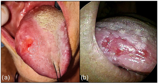
Figure 1.
(a) Lesion of the right border and dorsum of tongue. An evident strip of depapillated mucosa surrounds the lesion; (b) Tongue cancer of the border of tongue on WL, with depapillated mucosa at the superior interface.
The NBI vascular pattern was assessed only by the senior surgeon according to the classification proposed by Takano et al. based on IPCL, which is reported here [18]. The type I pattern comprises normal IPCL running parallel to the surface of the mucosa and appearing as a wavy line. In the Type II pattern, the IPCL have a similar shape to type I, but their caliber is notably increased and dilated. The IPCL of Type III are tangled lines (due to the severe increase in length) running perpendicular to the surface of the mucosa and therefore appearing as dark brown dots from a distance. Type IV is characterized by a chaotic representation of the IPCL that appear as large vessels with no loops at the terminal branches due to the progression of carcinogenesis, which leads to the dilation and elongation of the loops and finally to their destruction. Type IIIa and IV IPCLs are considered to be strongly associated with carcinoma in situ and invasive carcinoma in the oral cavity. Takano’s classification is depicted in Figure 2. Both depapillation and NBI patterns were assessed by a senior head and neck surgeon with special experience in oral cavity surgery and NBI (Figure 3). To assess if the identification of the depapillation and NBI pattern were reproducible and reliable, the senior surgeon trained a young fellow to recognize them. The young fellow assessed those independently and was blinded to the senior surgeon’s evaluation. For the univariable analysis, only the senior’s assessment was considered.
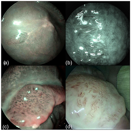
Figure 2.
Takano’s classification of tongue vascular patterns on NBI filter. (a) Type I; (b) Type II; (c) Type III; (d) Type IV.
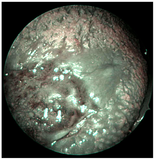
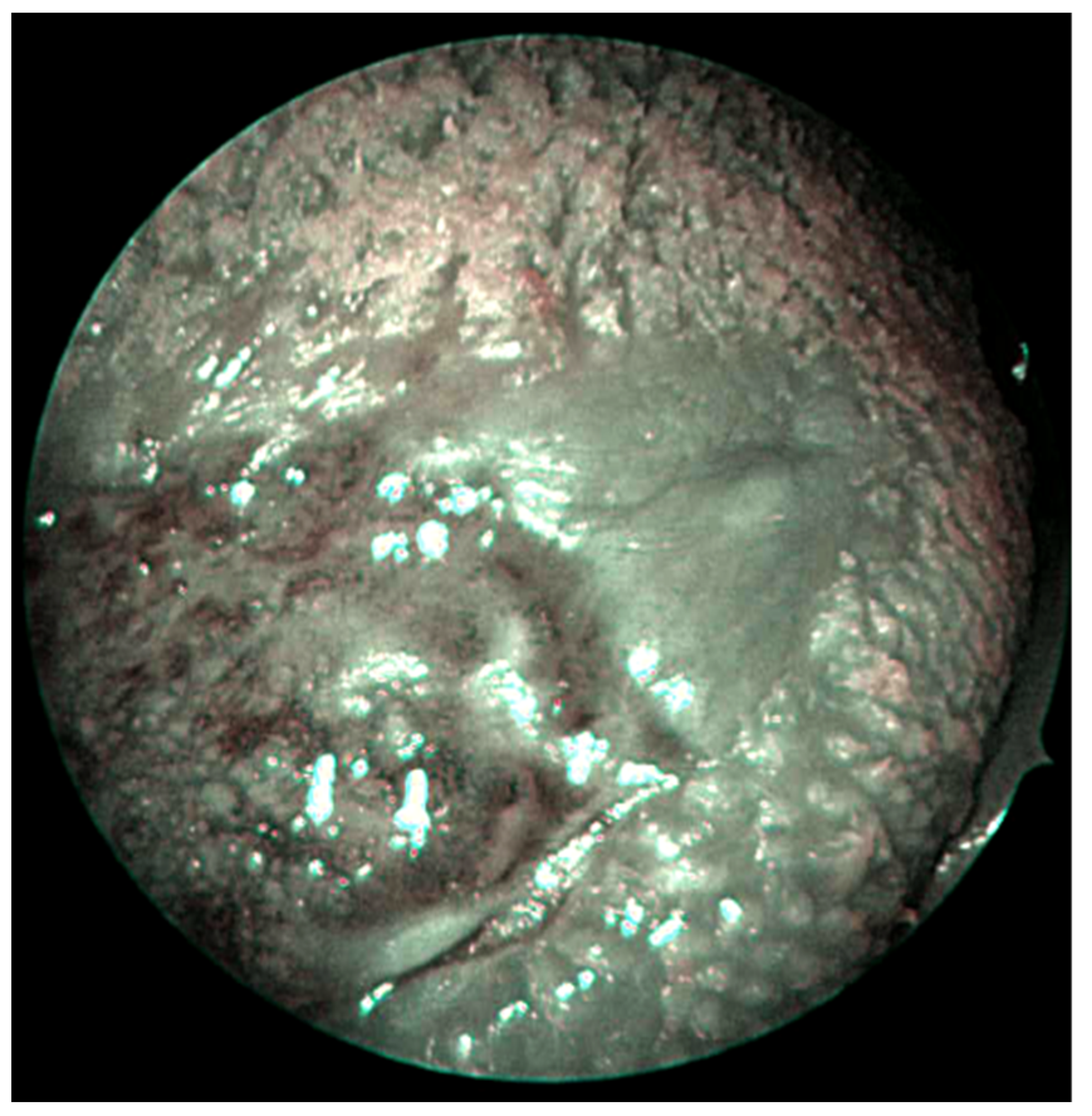
Figure 3.
Dorsal tongue cancer on NBI filter (Takano’s classification Type III), with depapillated mucosa on the interface of the posterior margin.
2.4. Statistical Analysis
Categorical variables were summarized as counts and percentages, while continuous variables were reported as medians and interquartile ranges (IQR: 25th and 75th). Frequencies and distribution for all demographic, clinical and pathological variables of the cohort were reported. The difference in the distribution of categorical variables was compared using the chi-squared test and Fisher’s exact test, as appropriate. Differences in continuous variables between groups were tested using the Mann–Whitney U test. The influence of depapillation on recurrence-free survival (RFS), defined as the time from surgery to death or tumor recurrence, was also considered a secondary endpoint. Calculations were made using the Kaplan–Meier method, and the survivals among the two groups were compared with the log-rank test. To assess the inter-rater agreement, the Cohen’s Kappa coefficient was computed based on the evaluations made by the senior and the young authors, and it was interpreted as follows: values 0–0.20 as no agreement, 0.21–0.40 as fair, 0.41–0.60 as moderate, 0.61–0.80 as substantial, and 0.81–1.00 as (almost) perfect agreement. Finally, for all tests, a value of p < 0.05 was considered to indicate statistical significance. Statistical analyses were performed using the R software for statistical computing (R version 4.0.1). Data were analyzed in November 2022.
3. Results
Seventy-six patients met the inclusion criteria and were included in the analysis. The whole cohort had a median age of 70.00 (IQR: 56.0–80.0), and the majority of patients were females (n = 40, 53%). The main features of the study population are summarized in Table 1. After the evaluation of the preoperative endoscopies by the senior specialist, 40 (53%) patients had depapillation at the WL examination, while 59 (78%) had a Takano’s Type III or IV pattern with NBI.

Table 1.
Patient demographics and tumor characteristics.
When measuring the inter-rater agreements, both the accordance between the physicians in assessing the presence of depapillation and rating the NBI pattern were substantial and greater than would be expected by chance (κ = 0.63, Z = 5.59, p < 0.001 and κ = 0.80, Z = 7.01, p < 0.001, respectively).
The presence of depapillation was strongly correlated to PNI, which was found in 54% of the cases in the depapillation group vs. 28% of the cases in the non-depapillated group (p = 0.022). No other factor differed significantly between the two groups (Table 2). Notably, no statistically significant difference was found in the number of cases with positive margins between the depapillated lesions group and the group of lesions that was not characterized by depapillation. Regarding the NBI pattern, according to Takano et al., there was no correlation with depapillation.
Table 2.
Univariable analysis according to the presence of depapillation.
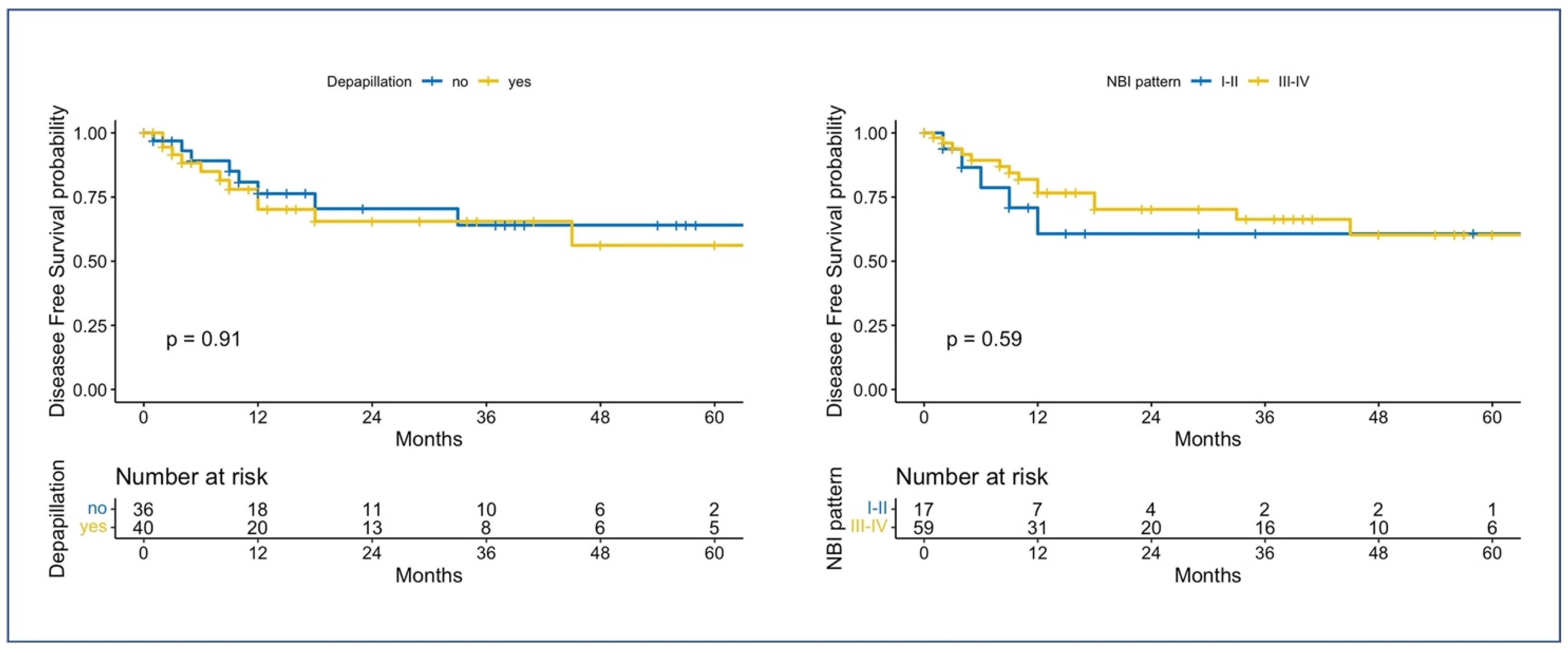
The median follow-up time was 13.0 months (IQR: 3.00–35.50). For the whole cohort, the 2-year and 5-year RFS (95% CI; number still at risk) were 67% (0.55–0.82; 24) and 59% (0.45–0.77; 7), respectively. Figure 4 shows the Kaplan–Meier curves when comparing the RFS of different groups based on the presence of depapillation or the NBI pattern: no statistical differences were found with the log-rank test for both comparisons (p = 0.91 and p = 0.59, respectively).
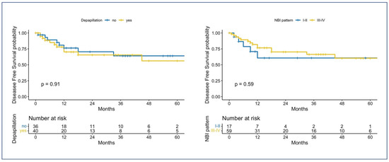
Figure 4.
Kaplan–Meier survival curves for RFS. On the left, the RFS is compared based on the presence of depapillation; on the right, the RFS is compared according to the NBI vascular pattern. The p-values are computed using the log-rank tests.
4. Discussion
Histological adverse pathological factors (APF) can reveal tumor aggressiveness, and they impact local and regional control [34,35]. Predicting, preoperatively, the biological behavior of OTSCC would allow the surgeon to tailor the tumor excision or the neck treatment. This would be valuable, especially in early-stage OTSCC, as, due to histopathological heterogeneity, there are important variations in the outcome even with identical clinical stages. In fact, although stage I/II OTSCC has been shown to have favorable outcomes, an estimated 30–35% of patients have locoregional recurrences [36,37]. Nowadays, among the APF, the only one preoperatively available is the DOI predicted by imaging [38]; in fact, combining different instrumental methodologies, such as magnetic resonance imaging and transoral ultrasound, can adequately estimate the measurement of tumor infiltration [39,40]. Thus, most of the surgical decision-making processes are based on this value. This measure can be considered a surrogate marker for tumor biology, as a more extensive deep infiltration reveals a more aggressive neoplasm, which is more prone to produce lymph node metastases than thick exophytic tumors [41]. Nevertheless, the DOI alone is insufficient to accurately predict the behavior of early lesions. Larson et al. [42] demonstrated how the simultaneous combination of different APFs, rather than the DOI value alone, affects DSS and OS in both univariate and multivariate analyses. Subramanian et al. [43] obtained comparable results, suggesting that even pT1N0 with free margins but presenting with simultaneous APFs should still be considered for adjuvant treatment. Furthermore, the reliability of DOI in predicting occult metastasis might vary widely according to the subsites involved [44]. In fact, among OTSCC patients, salient differences exist considering different subsites. In particular, those with OTSCC are more likely to experience cancer-specific mortality than patients with floor of the mouth, upper gum, and retromolar trigone cancer [45]. Several authors underlined how there are distinct anatomic barriers, local spreading pathways [46], and different nodal drainage patterns [47,48] that might justify this inferior survival outcomes for OTSCC. Accordingly, we can observe heterogeneity among the oral cavity subsites, even regarding APFs. Kim et al. [49] reported that PNI was more likely for tumors located in the oral tongue or floor of the mouth. Moreover, Liu et al. [50] demonstrated how the prognostic impact of pathological features was subsite-dependent, as PNI was associated with poor survival, especially in patients with OTSCC, where it was found to be the only significant factor associated with DSS of stage I and II. OTSCC’s tendency to present with PNI and its anatomical proximity with major cranial nerves might justify a more aggressive surgical approach, even in early small lesions [51]. PNI act as a passive conduit, providing an additional route for dissemination [52]. Therefore, PNI are among the worst prognosticators approved by the majority of investigators and are associated with locoregional recurrence, distant metastasis and decreased 5-year OS probability [53]. In addition, it has been demonstrated to be correlated with such major risk factors as extranodal extension [49]. All together, these findings may lead to planning a more aggressive therapeutic approach, even in early small OTSCCs where PNI is suspected [51]. Accordingly, the current National Comprehensive Cancer Network Panel (NCCN) Guidelines suggest the addition of adjuvant radiotherapy even for patients with surgically resected T1–T2 and N0 OTSCC when PNI is described in the pathology report [54].
In this scenario, observing the loss of tongue dorsum papillae in the area surrounding the tumor during the clinical examination has been emerging as an interesting predictor for APFs. To date in the literature, only Singh et al. investigated this topic, finding a significant relation between OTSCC with depapillation and pathologically documented PNI. Our study obtained similar results concerning depapillation correlation with PNI (p = 0.022). Observing this finding in a different population, heterogenic in terms of ethnicity and staging, reinforces the possible role of depapillation as a clinical predictive feature. This feature has the advantage of being cost effective, as it can be detected during a standard physical investigation. Particularly, despite the novelty of this feature, which physicians are not routinely trained to detect, in our study, the inter-rater agreement between the otolaryngologists was substantial, underlining how even less experienced specialists can identify it. Since our center is well-trained in applying NBI, an equivalent score has been obtained regarding Takano’s classification. Nevertheless, we did not observe any correlation between the NBI pattern and the depapillation group; therefore, we can state that the presence of depapillation does not hinder the effectiveness of NBI in detecting the typical perilesional vascular changes that occur during neoplastic development. Accordingly, also, no differences were found for what concerns surgical margins between the group of depapillated lesions and the group without atrophy of papillae. As the NBI evaluation was not affected by depapillation, we expected the same for margins, considering that in our center, we routinely tailor our resections based on NBI due to its established role in the whole oral cavity [20,55,56,57]. The automated delineation of tumor boundaries with this technique was recently initiated, and the effect of depapillation on that might be worth investigating [58].
Modern studies have demonstrated that PNI is a deliberate, molecularly mediated process that results from reciprocal interactions between cancer and nerves. Although depapillation is associated with PNI, it represents the mirror of the changes in the peritumoral microenvironment rather than an event driven purely by the progress of cancer alone. The first step of tumor infiltration and distant dissemination is a set of changes named epithelial-mesenchymal transition (EMT). EMT is a dynamic process allowing a polarized epithelial cell to undergo multiple biochemical changes leading to a mesenchymal cell phenotype, such as enhanced migratory capacity and invasiveness [59]; once the tumor infiltrates the basement membrane, it invades the adjacent cells first, which are the sustentacular cells. Since these cells clear the neurotransmitters synthesized by the taste receptor cells, there is a subsequent accumulation of neuronal promoters, leading to neurotropism and neural spread. This might explain why depapillation could be a clinical feature mirroring EMT, which leads to PNI and subsequent dissemination.
Promising efforts have been made to anticipate APFs in a preoperative setting. Notwithstanding, unfavorable histologic parameters in preoperative biopsy specimens showed poor correlation with the subsequent resection specimen. Likewise, in early OSCC, the differentiation grade determined by biopsy was demonstrated to be of little predictive value for the grading of the resection specimen. Poor differentiation grade, revealed at the biopsy, could not be related to the presence of nodal metastasis or survival and seems not to have any prognostic value concerning outcome [60]. Regarding PNI and LVI, the preoperative biopsy specimens did not represent the final post-surgical specimen and the subsequent risk of occult metastasis [61]. Finally, the feasibility of predicting WPOI, a negative prognosticator of nodal metastases and oncological outcomes [31], in the preoperative biopsy specimen has been assessed by Pu et al. [31]. Despite the promising results, the authors did not obtain an adequate overlap between the intermediate biopsy pattern and the final WPOI of Type 4 and 5, whose clinical outcomes are relatively poor, making the biopsy pattern unreliable in predicting prognosis preoperatively. Although peritumoral depapillation is not ubiquitous when PNI is present, its assessment is time and cost-effective, and it could be easily integrated into standard clinical examination; furthermore, it is reproducible. The routine use of high-definition intraoperative endoscopy for NBI evaluation would allow the surgeon to better inspect the tumor’s surrounding areas and obtain magnified and more reliable images that allow an improved detection of depapillation. The retrospective nature of our study represents its main limitation together with the small study cohort. Lastly, NBI evaluation of the lesions is subjective, but we reduced this bias by letting different senior surgeons judge the appearance of neoplastic lesions on NBI filters.
5. Conclusions
In our study, clinical peritumoral depapillation is associated with PNI on the pathology report. Its presence does not affect the NBI’s ability to detect perilesional neoangiogenesis and delineate resection margins. In the future, combining different techniques and clinical features might allow us to depict an endoscopic signature of the more aggressive tumors and, eventually, tailor the surgical treatment. We hope that further studies will corroborate our findings and fully understand the process that leads to depapillation in roughly half of the OTSCC population.
Author Contributions
Conceptualization, A.I. and C.S.; Data curation, C.S., A.P. and P.L.C.; Formal analysis, C.S.; Funding acquisition, G.P. (Giorgio Peretti); Investigation, A.I., A.P. and P.L.C.; Methodology, A.I. and C.S.; Project administration, G.P. (Giorgio Peretti); Resources, A.I. and P.L.C.; Software, C.S.; Supervision, G.P. (Giorgio Peretti); Validation, A.I. and G.P. (Giorgio Peretti); Writing—original draft, A.I., C.S. and A.P.; Writing—review & editing, A.I., C.S., F.M., A.L.C.C., M.F. and G.P. (Giampiero Parrinello). All authors have read and agreed to the published version of the manuscript.
Funding
This research received no external funding.
Institutional Review Board Statement
The study was conducted in accordance with the Declaration of Helsinki and approved by the Regional Ethics Committee (Registry number CER Liguria: 133/2021—DB id 11296).
Informed Consent Statement
Informed consent was obtained from all subjects involved in the study.
Data Availability Statement
The data can be shared up on request.
Conflicts of Interest
The authors declare no conflict of interest.
References
- Henley, S.J.; Ward, E.M.; Scott, S.; Ma, J.; Anderson, R.N.; Firth, A.U.; Thomas, C.C.; Islami, F.; Weir, H.K.; Lewis, D.R.; et al. Annual report to the nation on the status of cancer, part I: National cancer statistics. Cancer 2020, 126, 2225–2249. [Google Scholar] [CrossRef]
- Xu, B.; Salama, A.M.; Valero, C.; Yuan, A.; Khimraj, A.; Saliba, M.; Zanoni, D.K.; Ganly, I.; Patel, S.G.; Katabi, N.; et al. The prognostic role of histologic grade, worst pattern of invasion, and tumor budding in early oral tongue squamous cell carcinoma: A comparative study. Virchows Arch. 2021, 479, 597–606. [Google Scholar] [CrossRef]
- Tsai, T.Y.; Iandelli, A.; Marchi, F.; Huang, Y.; Tai, S.F.; Hung, S.Y.; Kao, H.; Chang, K. The Prognostic Value of Lymph Node Burden in Oral Cavity Cancer: Systematic Review and Meta-Analysis. Laryngoscope 2022, 132, 88–95. [Google Scholar] [CrossRef] [PubMed]
- Zanoni, D.K.; Montero, P.H.; Migliacci, J.C.; Shah, J.P.; Wong, R.J.; Ganly, I.; Patel, S.G. Survival outcomes after treatment of cancer of the oral cavity (1985–2015). Oral Oncol. 2019, 90, 115–121. [Google Scholar] [CrossRef]
- Balasubramanian, D.; Thankappan, K.; Battoo, A.J.; Rajapurkar, M.; Kuriakose, M.A.; Iyer, S. Isolated skip nodal metastasis is rare in T1 and T2 oral tongue squamous cell carcinoma. Otolaryngol. –Head Neck Surg. 2012, 147, 275–277. [Google Scholar] [CrossRef] [PubMed]
- Marur, S.; Forastiere, A.A. Head and Neck Squamous Cell Carcinoma: Update on Epidemiology, Diagnosis, and Treatment. Mayo Clin. Proc. 2016, 91, 386–396. [Google Scholar] [CrossRef] [PubMed]
- Riemann, M.; Knipfer, C.; Rohde, M.; Adler, W.; Schuster, M.; Noeth, E.; Oetter, N.; Shams, N.; Neukam, F.-W.; Stelzle, F. Oral squamous cell carcinoma of the tongue: Prospective and objective speech evaluation of patients undergoing surgical therapy. Head Neck 2016, 38, 993–1001. [Google Scholar] [CrossRef]
- Rogers, S.N.; Brown, J.S.; Woolgar, J.A.; Lowe, D.; Magennis, P.; Shaw, R.J.; Sutton, D.; Errington, D.; Vaughan, D. Survival following primary surgery for oral cancer. Oral Oncol. 2009, 45, 201–211. [Google Scholar] [CrossRef]
- Brierley, J.; Gospodarowicz, M.; Wittekind, C. TNM Classification of Malignant Tumours, 8th ed.; Union for International Cancer Control: Geneva, Switzerland, 2016. [Google Scholar]
- Iandelli, A.; Marchi, F.; Chen, A.-C.; Young, C.-K.; Liao, C.-T.; Tsao, C.-K.; Kang, C.-J.; Wang, H.-M.; Chang, T.-C.J.; Huang, S.-F. Adequacy of Disease Control by Supraomohyoid Neck Dissection in cT1/T2 Tongue Cancer. J. Pers. Med. 2022, 12, 1535. [Google Scholar] [CrossRef] [PubMed]
- Quintana, D.M.V.O.; Dedivitis, R.A.; Kowalski, L.P. Prognostic impact of perineural invasion in oral cancer: A systematic review. Acta Otorhinolaryngol. Ital. 2022, 42, 17–25. [Google Scholar] [CrossRef] [PubMed]
- Brown, I.S. Pathology of Perineural Spread. J. Neurol.Surg. Part B Skull Base 2016, 77, 124–130. [Google Scholar] [CrossRef] [PubMed]
- Tai, S.-K.; Li, W.-Y.; Chu, P.-Y.; Chang, S.-Y.; Tsai, T.-L.; Wang, Y.-F.; Huang, J.-L. Risks and clinical implications of perineural invasion in T1-2 oral tongue squamous cell carcinoma. Head Neck 2012, 34, 994–1001. [Google Scholar] [CrossRef] [PubMed]
- Erriu, M.; Pili, F.M.G.; Cadoni, S.; Garau, V. Diagnosis of Lingual Atrophic Conditions: Associations with Local and Systemic Factors. A Descriptive Review. Open Dent. J. 2016, 10, 619–635. [Google Scholar]
- Rahima, B.; Shingaki, S.; Nagata, M.; Saito, C. Prognostic significance of perineural invasion in oral and oropharyngeal carcinoma. Oral Surg. Oral Med. Oral Pathol. Oral Radiol. Endodontology 2004, 97, 423–431. [Google Scholar] [CrossRef] [PubMed]
- Nair, D.; Mair, M.; Singhvi, H.; Mishra, A.; Nair, S.; Agrawal, J.; Chaturvedi, P. Perineural invasion: Independent prognostic factor in oral cancer that warrants adjuvant treatment. Head Neck 2018, 40, 1780–1787. [Google Scholar] [CrossRef]
- Singh, A.; Singhavi, H.; Sathe, P.; Mair, M.; Qayyumi, B.; Shetty, R.; Bal, M.; Joshi, P.; Nair, S.V.; Chaturvedi, P. The impact of peritumoral depapillation in cancers of the tongue. Oral Surg. Oral Med. Oral Pathol. Oral Radiol. 2020, 129, 369–376. [Google Scholar] [CrossRef] [PubMed]
- Takano, J.H.; Yakushiji, T.; Kamiyama, I.; Nomura, T.; Katakura, A.; Takano, N.; Shibahara, T. Detecting early oral cancer: Narrowband imaging system observation of the oral mucosa microvasculature. Int. J. Oral Maxillofac. Surg. 2010, 39, 208–213. [Google Scholar] [CrossRef] [PubMed]
- Vu, A.; Farah, C.S. Narrow band imaging: Clinical applications in oral and oropharyngeal cancer. Oral Dis. 2016, 22, 383–390. [Google Scholar] [CrossRef]
- Piazza, C.; Cocco, D.; del Bon, F.; Mangili, S.; Nicolai, P.; Majorana, A.; Villaret, A.B.; Peretti, G. Narrow band imaging and high definition television in evaluation of oral and oropharyngeal squamous cell cancer: A prospective study. Oral Oncol. 2010, 46, 307–310. [Google Scholar] [CrossRef]
- Farah, C.S.; Dalley, A.J.; Nguyen, P.; Batstone, M.; Kordbacheh, F.; Perry-Keene, J.; Fielding, D. Improved surgical margin definition by narrow band imaging for resection of oral squamous cell carcinoma: A prospective gene expression profiling study. Head Neck 2016, 38, 832–839. [Google Scholar] [CrossRef]
- Tirelli, G.; Piovesana, M.; Gatto, A.; Tofanelli, M.; Biasotto, M.; Boscolo Nata, F. Narrow band imaging in the intra-operative defini-tion of resection margins in oral cavity and oropharyngeal cancer. Oral Oncol. 2015, 51, 908–913. [Google Scholar] [CrossRef]
- Lin, Y.C.; Wang, W.H.; Lee, K.F.; Tsai, W.C.; Weng, H.H. Value of narrow band imaging endoscopy in early mucosal head and neck cancer. Head Neck 2012, 34, 1574–1579. [Google Scholar] [CrossRef]
- Piazza, C.; del Bon, F.; Paderno, A.; Grazioli, P.; Perotti, P.; Barbieri, D.; Majorana, A.; Bardellini, E.; Peretti, G.; Nicolai, P. The diagnostic value of narrow band imaging in different oral and oropharyngeal subsites. Eur. Arch. Oto-Rhino-Laryngol. 2016, 273, 3347–3353. [Google Scholar] [CrossRef]
- Picciani, B.L.; Domingos, T.A.; Teixeira-Souza, T.; Santos Vde, C.; Gonzaga, H.F.; Cardoso-Oliveira, J.; Gripp, A.C.; Dias, E.P.; Carneiro, S. Geographic tongue and psoriasis: Clinical, histopathological, immunohistochemical and genetic correlation—A literature review. An. Bras. Dermatol. 2016, 91, 410–421. [Google Scholar] [CrossRef]
- Piazza, C.; Del Bon, F.; Peretti, G.; Nicolai, P. “Biologic endoscopy”: Optimization of upper aerodigestive tract cancer evaluation. Curr. Opin. Otolaryngol. Head Neck Surg. 2011, 19, 67–76. [Google Scholar] [CrossRef]
- Watanabe, A.; Taniguchi, M.; Tsujie, H.; Hosokawa, M.; Fujita, M.; Sasaki, S. The value of narrow band imaging endoscope for early head and neck cancers. Otolaryngol. Head Neck Surg. 2018, 138, 446–451. [Google Scholar] [CrossRef]
- Muto, M.; Katada, C.; Sano, Y.; Yoshida, S. Narrow band imaging: A new diagnostic approach to visualize angiogenesis in superficial neoplasia. Clin. Gastroenterol. Hepatol. 2005, 3 (Suppl. S1), 16–20. [Google Scholar] [CrossRef] [PubMed]
- Fuji, S.; Yamazaki, M.; Muto, M.; Ochiai, A. Microvascular irregularities are associated with composition of squamous epithelial lesions and correlate with subepithelial invasion of superficial-type pharyngeal squamous cell carcinoma. Histopathology 2010, 56, 510–522. [Google Scholar] [CrossRef]
- Chatterjee, D.; Bansal, V.; Malik, V.; Bhagat, R.; Punia, R.S.; Handa, U.; Gupta, A.; Dass, A. Tumor Budding and Worse Pattern of Invasion Can Predict Nodal Metastasis in Oral Cancers and Associated with Poor Survival in Early-Stage Tumors. Ear Nose Throat J. 2019, 98, E112–E119. [Google Scholar] [CrossRef] [PubMed]
- Pu, Y.; Ding, L.; Wang, Y.; Wang, Y.; Chen, S.; Huang, X.; He, Z.; Ni, Y.; Hu, Q. Biopsy pattern of invasion type to determine the surgical approach in early-stage oral squamous cell carcinoma. Virchows Arch. 2021, 479, 109–119. [Google Scholar] [CrossRef] [PubMed]
- Amin, M.B.; Greene, F.L.; Edge, S.B.; Compton, C.C.; Gershenwald, J.E.; Brookland, R.K.; Meyer, L.; Gress, D.M.; Byrd, D.R.; Winchester, D.P. The Eighth Edition AJCC Cancer Staging Manual: Continuing to build a bridge from a population-based to a more “personalized” approach to cancer staging. CA Cancer J. Clin. 2017, 67, 93–99. [Google Scholar] [CrossRef]
- International Agency for Research on Cancer. Available online: https://screening.iarc.fr/atlasoralglossdef.php?key=depapillation&img (accessed on 7 November 2022).
- Subramaniam, N.; Balasubramanian, D.; Hubert Low, T.-H.; Murthy, S.; Anand, A.; Prasad, C.; Vijayan, S.N.; Thankappan, K.; Iyer, S. Role of adverse pathological features in surgically treated early oral cavity carcinomas with adequate margins and the development of a scoring system to predict local control. Head Neck 2018, 40, 2329–2333. [Google Scholar] [CrossRef] [PubMed]
- Balasubramanian, D.; Subramaniam, N.; Missale, F.; Marchi, F.; Dokhe, Y.; Vijayan, S.; Nambiar, A.; Mattavelli, D.; Calza, S.; Bresciani, L.; et al. Predictive nomograms for oral tongue squamous cell carcinoma applying the American Joint Committee on Cancer/Union Internationale Contre le Cancer 8th edition staging system. Head Neck 2021, 43, 1043–1055. [Google Scholar] [CrossRef] [PubMed]
- Liao, C.-T.; Chang, J.T.-C.; Wang, H.-M.; Ng, S.-H.; Hsueh, C.; Lee, L.-Y.; Lin, C.-H.; Chen, I.-H.; Huang, S.-F.; Cheng, A.-J.; et al. Salvage therapy in relapsed squamous cell carcinoma of the oral cavity: How and when? Cancer 2008, 112, 94–103. [Google Scholar] [CrossRef]
- Missale, F.; Marchi, F.; Iandelli, A.; Subramaniam, N.; Dokhe, Y.; Sampieri, C.; Mattavelli, D.; Bresciani, L.; Carobbio, A.L.C.; Grammatica, A.; et al. Oncological outcomes of compartmental surgery and wide local excision in oral tongue and floor of the mouth cancer. Oral Oncol. 2022, 135, 106210. [Google Scholar] [CrossRef] [PubMed]
- Marchi, F.; Filauro, M.; Iandelli, A.; Luigi, A.; Carobbio, C.; Mazzola, F.; Santori, G.; Parrinello, G.; Canevari, F.R.M.; Piazza, C.; et al. Magnetic Resonance vs. Intraoral Ultrasonography in the Preoperative Assessment of Oral Squamous Cell Carcinoma : A Systematic Review and Meta-Analysis. Front. Oncol. 2020, 9, 1571. [Google Scholar] [CrossRef] [PubMed]
- Caprioli, S.; Casaleggio, A.; Tagliafico, A.S.; Conforti, C.; Borda, F.; Fiannacca, M.; Filauro, M.; Iandelli, A.; Marchi, F.; Parrinello, G.; et al. High-Frequency Intraoral Ultrasound for Preoperative Assessment of Depth of Invasion for Early Tongue Squamous Cell Carcinoma : Radiological–Pathological Correlations. Int. J. Environ. Res. Public Health 2022, 19, 14900. [Google Scholar] [CrossRef] [PubMed]
- Filauro, M.; Missale, F.; Marchi, F.; Iandelli, A.; Carobbio, A.L.C.; Mazzola, F.; Parrinello, G.; Barabino, E.; Cittadini, G.; Farina, D.; et al. Intraoral ultrasonography in the assessment of DOI in oral cavity squamous cell carcinoma: A comparison with magnetic resonance and histopathology. Eur. Arch. Oto-Rhino-Laryngol. 2020, 278, 2943–2952. [Google Scholar] [CrossRef]
- Ebrahimi, A.; Gil, Z.; Amit, M.; Yen, T.-C.; Liao, C.-t.; Chaturvedi, P.; Agarwal, J.P.; Kowalski, L.P.; Köhler, H.F.; Kreppel, M.; et al. Depth of invasion alone as an indication for postoperative radiotherapy in small oral squamous cell carcinomas: An International Collaborative Study. Head Neck 2019, 41, 1935–1942. [Google Scholar] [CrossRef]
- Larson, A.R.; Kemmer, J.; Formeister, E.; El-sayed, I.; Ha, P.; George, J.; Ryan, W.; Chan, E.; Heaton, C. Beyond Depth of Invasion: Adverse Pathologic Tumor Features in Early Oral Tongue Squamous Cell Carcinoma. Laryngoscope 2020, 130, 1715–1720. [Google Scholar] [CrossRef]
- Subramaniam, N.; Balasubramanian, D.; Murthy, S.; Thankappan, K.; Iyer, S. Predictors of locoregional control in stage I/II oral squamous cell carcinoma classi fi ed by AJCC 8th edition. Eur. J. Surg. Oncol. 2019, 45, 2126–2130. [Google Scholar] [CrossRef] [PubMed]
- Brockhoff, H.C.; Kim, R.Y.; Braun, T.M.; Skouteris, C.; Helman, J.I.; Ward, B.B. Correlating the depth of invasion at specific anatomic locations with the risk for regional metastatic disease to lymph nodes in the neck for oral squamous cell carcinoma Hans. Head Neck 2017, 39, 974–979. [Google Scholar] [CrossRef] [PubMed]
- Farhood, Z.; Simpson, M.; Ward, G.M.; Walker, R.J.; Osazuwa-Peters, N. Does anatomic subsite influence oral cavity cancer mortality? A SEER database analysis. Laryngoscope 2019, 129, 1400–1406. [Google Scholar] [CrossRef]
- Calabrese, L.; Bizzoca, M.E.; Grigolato, R.; Maffini, F.A.; Tagliabue, M.; Negro, R.; Leuci, S.; Mignogna, M.D.; Muzio, L.L. From bench to bedside in tongue muscle cancer invasion and back again: Gross anatomy, microanatomy, surgical treatments and basic research. Life 2020, 10, 197. [Google Scholar] [CrossRef] [PubMed]
- Deganello, A.; Rampinelli, V.; Gualtieri, T.; Paderno, A.; Bonomo, P.; di Monale e Bastia, M.B. Letter to “Medial lingual lymph node metastasis in carcinoma of the tongue”. Auris Nasus Larynx 2020, 47, 1091–1092. [Google Scholar] [CrossRef]
- Gvetadze, S.R.; Ilkaev, K.D. Lingual lymph nodes: Anatomy, clinical considerations, and oncological significance. World J. Clin. Oncol. 2020, 11, 337–347. [Google Scholar] [CrossRef]
- Kim, R.Y.; Helman, J.I.; Braun, T.M.; Ward, B.B. Increased Presence of Perineural Invasion in the Tongue and Floor of the Mouth: Could It Represent a More Aggressive Oral Squamous Cell Carcinoma, or Do Larger Aggressive Tumors Cause Perineural Invasion? J. Oral Maxillofac. Surg. 2019, 77, 852–858. [Google Scholar] [CrossRef]
- Liu, S.A.; Wang, C.C.; Jiang, R.S.; Lee, F.Y.; Lin, W.J.; Lin, J.C. Pathological features and their prognostic impacts on oral cavity cancer patients among different subsites—A singe institute’s experience in Taiwan. Sci. Rep. 2017, 7, 7451. [Google Scholar] [CrossRef]
- Tai, S.K.; Li, W.Y.; Yang, M.H.; Chu, P.Y.; Wang, Y.F. Perineural invasion in T1 oral squamous cell carcinoma indicates the need for aggressive elective neck dissection. Am. J. Surg. Pathol. 2013, 37, 1164–1172. [Google Scholar] [CrossRef]
- Bakst, R.L.; Glastonbury, C.M.; Parvathaneni, U.; Katabi, N.; Hu, K.S.; Yom, S.S. Perineural Invasion and Perineural Tumor Spread in Head and Neck Cancer. Int. J. Radiat. Oncol. Biol. Phys. 2019, 103, 1109–1124. [Google Scholar] [CrossRef]
- Li, Y.; Bai, S.; Carroll, W.; Dayan, D.; Dort, J.C.; Heller, K.; Jour, G.; Lau, H.; Penner, C.; Prystowsky, M.; et al. Validation of the Risk Model: High-Risk Classification and Tumor Pattern of Invasion Predict Outcome for Patients with Low-Stage Oral Cavity Squamous Cell Carcinoma. Head Neck Pathol. 2013, 7, 211–223. [Google Scholar] [CrossRef] [PubMed]
- National Comprehensive Cancer Network. Head and Neck Cancers. In NCCN Guidelines Version 3; National Comprehensive Cancer Network: Plymouth Meeting, PA, USA, 2019. [Google Scholar] [CrossRef]
- Nair, D.; Qayyumi, B.; Sharin, F.; Mair, M.; Bal, M.; Pimple, S.; Mishra, G.; Nair, S.; Chaturvedi, P. Narrow band imaging observed oral mucosa microvasculature as a tool to detect early oral cancer: An Indian experience. Eur. Arch. Oto-Rhino-Laryngol. 2021, 278, 3965–3971. [Google Scholar] [CrossRef] [PubMed]
- Deganello, A.; Paderno, A.; Morello, R.; Fior, M.; Berretti, G.; Del Bon, F.; Alparone, M.; Bardellini, E.; Majorana, A.; Nicolai, P. Diagnostic Accuracy of Narrow Band Imaging in Patients with Oral Lichen Planus: A Prospective Study. Laryngoscope 2021, 131, E1156–E1161. [Google Scholar] [CrossRef] [PubMed]
- Guida, A.; Maglione, M.; Crispo, A.; Perri, F.; Villano, S.; Pavone, E.; Aversa, C.; Longo, F.; Feroce, F.; Botti, G.; et al. Oral lichen planus and other confounding factors in narrow band imaging (NBI) during routine inspection of oral cavity for early detection of oral squamous cell carcinoma: A retrospective pilot study. BMC Oral Health 2019, 19, 70. [Google Scholar] [CrossRef]
- Azam, M.A.; Sampieri, C.; Ioppi, A.; Benzi, P.; Giordano, G.G.; De Vecchi, M.; Campagnari, V.; Li, S.; Guastini, L.; Paderno, A.; et al. Videomics of the Upper Aero-Digestive Tract Cancer: Deep Learning Applied to White Light and Narrow Band Imaging for Automatic Segmentation of Endoscopic Images. Front. Oncol. 2022, 12, 900451. [Google Scholar] [CrossRef]
- Ling, Z.; Cheng, B.; Tao, X. Epithelial-to-mesenchymal transition in oral squamous cell carcinoma: Challenges and opportunities. Int. J. Cancer 2021, 148, 1548–1561. [Google Scholar] [CrossRef]
- Dik, E.A.; Ipenburg, N.A.; Kessler, P.A.; van Es, R.J.J.; Willems, S.M. The value of histological grading of biopsy and resection specimens in early stage oral squamous cell carcinomas. J. Cranio-Maxillofac. Surg. 2018, 46, 1001–1006. [Google Scholar] [CrossRef]
- Dik, E.A.; Ipenburg, N.A.; Adriaansens, S.O.; Kessler, P.A.; Van Es, R.J.; Willems, S.M. Poor correlation of histologic parameters between biopsy and resection specimen in early stage oral squamous cell carcinoma. Am. J. Clin. Pathol. 2015, 144, 659–666. [Google Scholar] [CrossRef] [PubMed]


Disclaimer/Publisher’s Note: The statements, opinions and data contained in all publications are solely those of the individual author(s) and contributor(s) and not of MDPI and/or the editor(s). MDPI and/or the editor(s) disclaim responsibility for any injury to people or property resulting from any ideas, methods, instructions or products referred to in the content. |
© 2023 by the authors. Licensee MDPI, Basel, Switzerland. This article is an open access article distributed under the terms and conditions of the Creative Commons Attribution (CC BY) license (https://creativecommons.org/licenses/by/4.0/).