Article

Impact of Novel Treatments in Patients with Melanoma Brain Metastasis: Real-World Data

by
Sophie H. A. E. Derks
1,2,3,
Joost L. M. Jongen
1,
Edgar L. van der Meer
1,
Li Shen Ho
1,
Cleo Slagter
4,
Arjen Joosse
2,
Maja J. A. de Jonge
2,
Joost W. Schouten
5,
Esther Oomen-de Hoop
2,
Martin J. van den Bent
1 and
Astrid A. M. van der Veldt
2,3,*
1
Department of Neuro-Oncology, Erasmus MC Cancer Institute, 3015 GD Rotterdam, The Netherlands
2
Department of Medical Oncology, Erasmus MC Cancer Institute, 3015 GD Rotterdam, The Netherlands
3
Department of Radiology & Nuclear Medicine, Erasmus Medical Center, 3015 GD Rotterdam, The Netherlands
4
Department of Radiotherapy, Erasmus MC Cancer Institute, 3015 GD Rotterdam, The Netherlands
5
Department of Neurosurgery, Erasmus Medical Center, 3015 GD Rotterdam, The Netherlands
*
Author to whom correspondence should be addressed.
Cancers 2023, 15(5), 1461; https://doi.org/10.3390/cancers15051461
Submission received: 1 February 2023 / Revised: 20 February 2023 / Accepted: 22 February 2023 / Published: 25 February 2023
(This article belongs to the Special Issue Advances in Research, Diagnosis and Treatment of Brain Metastases)

Abstract

:

Simple Summary

Cutaneous melanoma is highly prone to metastasize to the brain, with a historically poor overall survival of only 4–5 months. Over the past decade, novel drugs such as targeted therapies and immune checkpoint inhibitors (ICIs) have revolutionized the treatment of advanced melanoma. However, most prospective studies have excluded patients with melanoma brain metastasis (MBM) or included only patients with stable (steroid-free) or asymptomatic MBM. To assess the impact of novel treatments in patients with MBM in a real-world setting, we studied a cohort of consecutive patients over a period of sixteen years (2005 to 2021) in a large, tertiary referral center for melanoma (Erasmus MC, Rotterdam, the Netherlands). We found a significant improvement in overall survival in the modern (post-2015) era, associated with stereotactic radiotherapy and especially ICIs. These findings support that ICIs, if clinically feasible, should be considered first in clinical practice after a diagnosis of MBM.

Abstract

Background: Melanoma brain metastasis (MBM) is associated with poor outcome, but targeted therapies (TTs) and immune checkpoint inhibitors (ICIs) have revolutionized treatment over the past decade. We assessed the impact of these treatments in a real-world setting. Methods: A single-center cohort study was performed at a large, tertiary referral center for melanoma (Erasmus MC, Rotterdam, the Netherlands). Overall survival (OS) was assessed before and after 2015, after which TTs and ICIs were increasingly prescribed. Results: There were 430 patients with MBM included; 152 pre-2015 and 278 post-2015. Median OS improved from 4.4 to 6.9 months (HR 0.67, p < 0.001) after 2015. TTs and ICIs prior to MBM diagnosis were associated with poorer median OS as compared to no prior systemic treatment (TTs: 2.0 vs. 10.9 and ICIs: 4.2 vs. 7.9 months, p < 0.001). ICIs directly after MBM diagnosis were associated with improved median OS as compared to no direct ICIs (21.5 vs. 4.2 months, p < 0.001). Stereotactic radiotherapy (SRT; HR 0.49, p = 0.013) and ICIs (HR 0.32, p < 0.001) were independently associated with improved OS. Conclusion: After 2015, OS significantly improved for patients with MBM, especially with SRT and ICIs. Demonstrating a large survival benefit, ICIs should be considered first after MBM diagnosis, if clinically feasible.

1. Introduction

Cutaneous melanoma has the highest risk of brain metastasis (MBM) of all solid tumors, spreading to the brain in over 50% of patients with metastatic disease [1,2,3]. Overall survival (OS) used to be <5 months, but several novel treatments have become available in the last decade, which have changed the perspectives of patients with MBM [2].
Whole brain radiotherapy (WBRT) and chemotherapy have limited efficacy in MBM, and WBRT is associated with cognitive impairment in the few long-term survivors [4,5,6]. These days, stereotactic radiotherapy (SRT) and especially targeted therapies (TTs; BRAF-MEK inhibitors) and immune checkpoint inhibitors (ICIs; anti-PD[L]1 and anti-CTLA4) have revolutionized treatment for patients with MBM [7,8,9,10,11,12,13,14]. Combined dabrafenib–trametinib showed an intracranial response rate (IRR) of 58%, but with limited duration of response (6.5 months) [7]. More durable responses are reported for ICIs, especially for combined nivolumab–ipilimumab. The ABC trial reported an IRR of 51% with a 5-year OS of 51% [8,9,10], and the CHECKMATE-204 trial had an IRR of 57% with a 1-year OS of 81.5% for this combination [11,15]. However, only a few phase II and III trials included limited numbers of patients with previously treated and/or symptomatic MBM, and these reported considerably lower IRRs (between 6% and 17% for ICIs) [8,11,15,16].
To assess the eligibility of individual patients for these novel treatments, the Melanoma-molGPA is often used in clinical practice as a prognostic scoring tool. This index is based on age, Karnofsky performance status (KPS), presence of extracranial metastasis (ECM), number of MBMs and, more recently, BRAF-mutation status [17,18,19]. However, since the index was derived from a retrospective cohort (n = 823) of patients between 2006 and 2015, its validity for modern cohorts is uncertain [17,20].
In the Netherlands, TTs and ICIs have been implemented in clinical practice for patients with MBM since their reimbursement almost a decade ago [21]. To assess the real-world impact of these treatments on MBM, we studied OS in a cohort of consecutive patients diagnosed with MBM over a period of sixteen years (2005 to 2021). Additionally, we examined the validity of the Melanoma-molGPA index for patients diagnosed with MBM in the modern treatment era.

2. Materials and Methods

2.1. Patient Selection

This retrospective cohort study was performed at Erasmus MC Cancer Institute (Rotterdam, The Netherlands), a large, tertiary referral center for patients with melanoma. In this center, the treatment plan for patients with MBM is routinely discussed in a local multidisciplinary board which includes experienced medical oncologists, neuro-oncologists, radiotherapists, and neurosurgeons. We identified all consecutive patients with a diagnosis of cutaneous or mucosal melanoma and MBM referred to the Erasmus MC between 1 January 2005 and 1 July 2021 (Figure A1). The study was approved by the local Institutional Review Board (MEC-2020-0681).

2.2. Data Collection and Definitions

Two trained data managers (E.L.v.d.M., L.S.H.) retrieved data from the electronic patient records. All data were reassessed by two clinical physicians (S.H.A.E.D., J.L.M.J.). The following baseline characteristics were collected: age, sex, KPS, lactate dehydrogenase (LDH) level, BRAF-status, presence of symptoms of MBM (e.g., headache, nausea, epilepsy, focal deficits), number of MBMs, and status of ECM. Diagnosis of MBM was the date of first brain imaging (magnetic resonance imaging or computed tomography (CT)) that confirmed parenchymal MBM. Diagnosis of ECM was the date of first imaging (CT or 2-deoxy-2-[18F]fluoro-D-glucose-positron emission tomography-CT) of at least thorax and abdomen that confirmed metastasis (M1-disease). Melanoma brain metastasis was considered synchronous when diagnosed within one month of diagnosis of ECM and metachronous when diagnosed at least one month after diagnosis of ECM. The Melanoma-molGPA was calculated for each individual patient.
In addition, details of previous systemic treatments (i.e., given at any time prior to diagnosis of MBM) and treatments given directly after diagnosis of MBM (before the first physician assessed progression of MBM) were collected. Local treatments included surgical resection, SRT, and WBRT. Systemic treatments included chemotherapy (e.g., dacarbazine), TTs (e.g., vemurafenib, dabrafenib, encorafenib, alone or combined with cobimetinib, trametinib, binimetinib, respectively) and ICIs (e.g., pembrolizumab, atezolizumab, nivolumab, ipilimumab, or nivolumab–ipilimumab).

2.3. Statistical Analysis

Data cut-off was 4 March 2022. We created a total cohort and two consecutive time cohorts split 1 January 2015, as novel systemic treatments were increasingly prescribed after 2015 in the Netherlands (Table A1, Figure A3). The primary outcome was OS, defined as the time between diagnosis of MBM and death of any cause.
Continuous variables were described with medians (interquartile range (IQR)) and categorical variables with frequencies. The Mann–Whitney U test was used for continuous, and the Chi-squared test for categorical variables. For categorical variables with groups < 10 patients, the Fisher exact test was used. Missing data were omitted from analysis.
Kaplan–Meier (KM) and Cox Proportional Hazards (CPH) methods were used for time-to-event analysis and assessed with the Logrank and Likelihood ratio tests, respectively. For multivariate CPH modeling, backward elimination (i.e., including variables with p < 0.2 in univariate analysis) was applied, and interaction testing was performed. No correction for multiple testing was performed in these exploratory analyses. A two-sided p-value of 0.05 was taken as statistically significant. All analyses were performed using R version 4.0.2 (the R-Project, Auckland, New Zealand).

3. Results

3.1. Baseline Characteristics

We included 430 patients in total; 152 (35.3%) patients were categorized in the pre-2015 cohort and 278 (64.7%) in the post-2015 cohort (Figure A1). The majority of patients had a follow-up of at least 12 months (420 of 430 patients, 97.6%). Baseline characteristics at diagnosis of MBM are shown in Table 1. Overall, 133 (30.9%) patients had a KPS ≤ 70 and 321 of 430 (74.6%) patients had symptomatic MBM, with fewer symptomatic patients post-2015 (n = 193 of 278, 69.4%) as compared to pre-2015 (n = 128 of 152, 84.4%; p = 0.001).

3.2. Treatments over Time

Prior to the diagnosis of MBM, 104 of 430 (24.2%) patients had received one or more systemic treatments (Table 2). Fewer patients had received previous chemotherapy post-2015 compared to pre-2015 (1.4% vs. 6.6%; p = 0.008), whereas TTs and ICIs prior to MBM diagnosis had been administered more often after 2015 (3.3% to 13.3%, p < 0.001; 2.0% to 25.2%, p < 0.001, respectively).
Directly after diagnosis of MBM, 358 of 430 (83.3%) patients received one or more systemic and/or local treatments (Table 2). The frequency of SRT and surgical resection directly after diagnosis of MBM did not significantly change over time, whereas the frequency of WBRT and chemotherapy significantly decreased after 2015 (55.3% to 12.6%, p < 0.001; 14.5% to 1.4%, p < 0.001; respectively). The prescription of TTs and ICIs directly after diagnosis of MBM significantly increased after 2015 (10.5% to 33.8%, p < 0.001; 2.6% to 35.3%, p < 0.001; respectively).

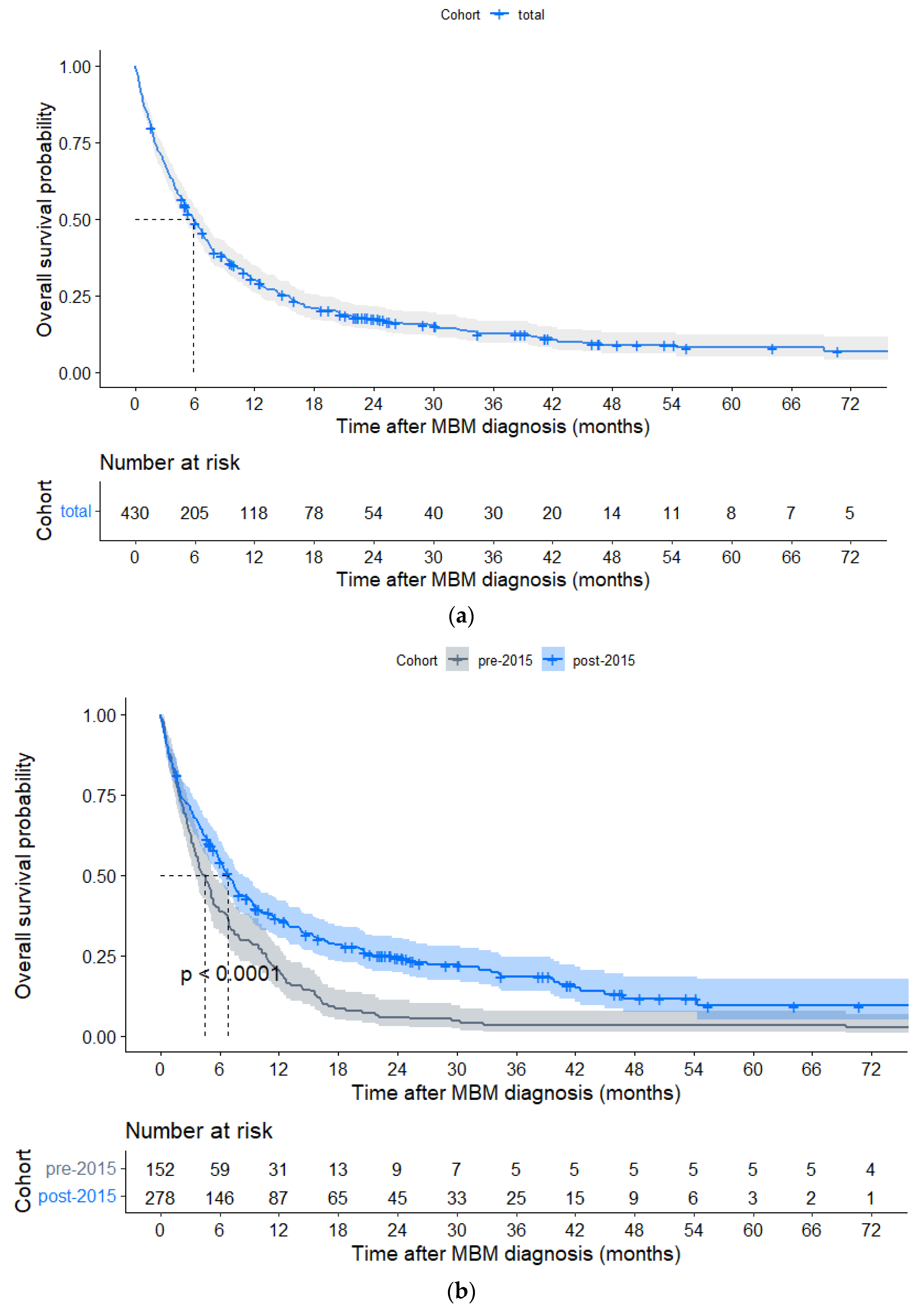
3.3. Overall Survival

In the total cohort, median OS was 5.9 months (IQR 2.07–15.41), with 1- and 3-year OS rates of 30.2% and 12.5%, respectively (Figure 1a). Here, 21 of 430 (4.9%) patients had a survival time of at least 5 years since diagnosis of MBM, of whom 17 (81.0%) were diagnosed with MBM post-2015. At data cut-off, 65 of 430 (15.1%) patients, of whom 61 were diagnosed with MBM post-2015, were alive with a median follow-up of 23.8 (IQR 11.6–41.2) months. Median OS was significantly longer post-2015 as compared to pre-2015 (6.9 (IQR 2.07–23.39) vs. 4.4 (IQR 1.92–10.73) months, HR0.63, p < 0.001), especially in subgroups of patients with synchronous MBM, LDH levels > ULN and a KPS > 70 (Figure 1b and Figure 2, Table A2).
For patients receiving SRT and surgical resection directly after diagnosis of MBM, OS improved after 2015 (Table A3). Systemic treatments were not analyzed over time since their frequencies significantly changed over time (Table 2). To assess the impact of treatments in the modern era, we analyzed treatment subgroups in the post-2015 cohort (n = 278) only.
In these univariate analyses, a specific treatment before the diagnosis of MBM or directly after was compared to not having that specific treatment at that time. In addition, patients with symptomatic MBM were analyzed separately.

3.3.1. Local Treatments Post-2015

Forty-two patients received SRT directly after diagnosis of MBM, of whom 39 (92.9%) had <4 MBMs. In these patients, SRT significantly improved OS as compared to patients without SRT (median OS 30.3 (IQR 9.0–NA) vs. 7.6 (IQR 2.6–32.3) months, HR0.46, p < 0.001; Figure 3a, Table A4). In patients with <4 MBMs and symptomatic MBM, SRT remained associated with improved OS (Figure A4a).
In patients with <4 MBMs and symptomatic MBM, surgical resection directly after diagnosis of MBM was associated with improved OS as compared to no surgical resection (median OS 21.5 (IQR 11.3–29.6) vs. 5.7 (IQR 2.3–25.9) months, HR0.58, p = 0.046; Figure A4b).

3.3.2. Systemic Treatments Post-2015

In patients with BRAF V600E+/K-mutated melanoma (n = 157), TTs prior to the diagnosis of MBM were associated with a shorter median OS as compared to no prior TTs (2.0 (IQR 0.8–7.1) vs. 10.9 (IQR 5.2–27.0) months, HR2.67, p < 0.001; Figure 4a, Table A4). In patients with prior TTs, 58.3% of patients had also been previously treated with ICIs (Table A5a). Of all patients with TTs directly after diagnosis of MBM (n = 89), 64 (71.9%) had symptomatic MBM (Figure A5a). No significantly different OS was found between patients with and without TTs directly after MBM diagnosis (7.8 (IQR 4.8–17.0) vs. 7.4 (IQR 1.5–33.4) months, HR1.15, p = 0.43; Figure 4c, Table A4).
Patients treated with ICIs prior to the diagnosis of MBM had a poorer median OS as compared to patients without prior ICIs (4.2 (IQR 1.0–10.2) vs. 7.9 (IQR 3.1–27.0) months, HR 1.67, p < 0.001) and 30.0% of patients with prior ICIs had also been previously treated with TTs (Figure 4b, Table A4 and Table A5b). Patients with ICIs directly after diagnosis of MBM had a better median OS as compared to patients without ICIs directly after MBM (21.5 (IQR 9.4–NA) vs. 4.2 (IQR 1.4–8.0) months, HR 0.28, p < 0.001; Figure 4d, Table A4), which was also confirmed in a subgroup of symptomatic patients (18.5 (IQR 9.0–34.4) vs. 4.0 (IQR 1.1–7.6) months, HR 0.33, p < 0.001; Figure A5b). Of all patients (n = 98) with ICIs directly after MBM diagnosis, 44 (44.9%) were alive at data cut-off, and 17 (17.3%) patients had a survival time of at least 5 years after diagnosis of MBM. In 23 of 98 (23.5%) patients, ICIs were combined with SRT.

3.4. Independent Prognostic Variables

In multivariate analysis of the post-2015 cohort (n = 278), symptomatic MBM (HR 1.74 [1.21–2.50], p = 0.003) and metachronous MBM (HR 2.73 [1.50–4.95], p < 0.001) were independently associated with poorer OS, whereas a KPS of >70 (HR 0.51 [0.37–0.70], p < 0.001), SRT (HR 0.49 [0.28–0.86], p = 0.013) and ICIs directly after diagnosis of MBM (HR 0.32 [0.22–0.47], p < 0.001) were independently associated with an improved OS (Table 3).

3.5. Melanoma-molGPA

The Melanoma-molGPA could be assessed for 268 of 278 (96.4%) patients post-2015. The melanoma-molGPA subclasses (I, II, III, and IV, respectively) showed a subsequent improvement of OS (median OS 3.0, 6.9, 24.5, and 30.3 months, respectively, p < 0.001; Figure 5, Table A6).

3.6. Switching from Targeted Therapy to Immune Checkpoint Inhibition

Here, 18 of 430 (4.2%) patients initiated with TTs directly after diagnosis of MBM and switched to ICIs when their performance status improved and/or imaging showed tumor response at a median time of 6.1 months (IQR 3.9–7.0) after the start of TTs (Table A7). Three of eighteen (16.7%) patients remained stable since the switch to ICIs and were alive at data cut-off, with a median survival time of 25.5 months (IQR 21.7–27.2) since ICI initiation. Three of eighteen (16.7%) patients had progressive disease after ICI initiation but received no further treatment due to poor clinical condition, with a median OS of 11.9 months (IQR 7.6–16.3) since ICI initiation. Twelve of eighteen (66.7%) patients had progressive disease on ICIs and switched back to TTs, with a median OS of 11.6 months (IQR 6.5–25.5) since ICI initiation.

4. Discussion

Since 2015, novel systemic therapies (TTs and ICIs) and SRT have replaced chemotherapy and WBRT for patients with MBM, resulting in a significantly improved OS. Compared to clinical trials, real-world patients with MBM usually have a worse clinical condition, which is illustrated by the high number of patients with poor KPS and symptomatic MBM in this cohort.
Surgical resection and SRT remained important treatment options after 2015, although their frequency directly after diagnosis of MBM did not significantly increase over time. Especially SRT was associated with a beneficial impact on survival (median OS 30.5 months) and remained independently associated with improved OS. Although the efficacy of SRT has been demonstrated in patients with up to 10 MBMs [22,23], SRT was almost exclusively reserved for patients with <4 MBMs in the current cohort.
The increased use of TTs (10.5% to 33.8%) and ICIs (2.6% to 35.3%) post-2015, directly after diagnosis of MBM, reflects the clinical approval of different novel drugs by the European Medicines Agency (EMA) and their reimbursement in the Netherlands since 2015 [24]. Bander et al. showed an even higher use of ICIs (77%) in their American cohort, likely resulting from the earlier approval of these drugs by the Food and Drug Administration (FDA) and from differences in therapeutic approaches between centers [25,26].
Patients who received TTs or ICIs to treat ECM, before the diagnosis of MBM, had poor survival (median OS of 2.0 and 4.2 months, respectively) as they had secondary resistance with intracranial disease progression on/after these treatments. Other real-world studies confirm the poor OS of patients with systemic treatments prior to the diagnosis of MBM [26,27]. Most importantly, a significantly favorable OS was seen with ICIs directly after the diagnosis of MBM (median OS 21.5 months), even in symptomatic patients (18.5 months). Although median follow-up is limited, the tail of the OS curve (Figure 4d) showed a considerable group of patients with long-term survival, as illustrated by >17% of patients with ICIs who lived >5 years after diagnosis of MBM. This confirms in a real-world setting the efficacy of ICIs in MBM, as reported by the benchmark phase II and III trials [10,15]. Therefore, ICIs should be considered first after diagnosis of MBM, if clinically feasible.
The Melanoma-molGPA, based on an MBM cohort between 2006 and 2015, remained a valid prognostic tool in patients diagnosed with MBM between 2015 and 2021 [17,18]. Therefore, clinicians may continue to use this tool to predict prognosis and assess the eligibility of individual patients for specific treatments.
Since TTs can induce rapid tumor responses, these drugs were frequently administered (>70%) to patients with symptomatic MBM and a poor performance status. However, TTs lack durable responses and it was recently demonstrated that first-line nivolumab–ipilimumab followed by BRAF/MEK-inhibition on progression had the most favorable OS in metastatic melanoma [28]. Nevertheless, in patients with a BRAF V600E+/K-mutation and an initial poor performance status, induction with TTs provides an opportunity for effective ICI treatment after performance has improved [29]. Although our study was not designed to compare treatment strategies, we assessed the outcome of 18 patients who were first treated with induction TTs after the diagnosis of MBM, followed by ICIs. Only three (16.7%) patients benefitted from the switch to ICIs, with stable disease and a median OS of 25.5 months since ICIs. Although (pre-)clinical studies have shown that an immune-resistant phenotype might arise after progression on BRAF/MEK-inhibition, it is unknown whether the switch to ICIs before progression on TTs could be effective, and it is worthwhile to further investigate this prospectively [8,30].
This study was designed to assess the potential survival gain in the brain for patients with MBM in a real-world setting after the introduction of novel systemic treatments. The retrospective design is a limitation, as is the lack of information on steroid use. The inclusion of a single center might provide additional bias. However, this tertiary center receives referrals from a large region in the Netherlands, and each consecutive patient is treated and followed in this center until end-of-life care or death. Therefore, this data set provides detailed information over a large period of time, fully capturing the changing treatment landscape.

5. Conclusions

Overall survival has improved for patients with MBM after 2015 and is associated with the use of SRT and ICIs directly after diagnosis of MBM. Immune checkpoint inhibitors (ICIs) showed an important survival benefit and should be considered first after the diagnosis of MBM, if clinically feasible. The Melanoma-molGPA remains a valid prognostic tool for clinicians in the modern treatment era for patients with MBM. To further improve the prognosis of patients with MBM, future research should focus on optimizing treatment sequencing, such as switching from TTs to ICIs, especially in patients with an initial poor performance.

Author Contributions

Conceptualization, S.H.A.E.D., J.L.M.J., M.J.v.d.B. and A.A.M.v.d.V.; data curation, E.L.v.d.M. and L.S.H.; formal analysis, S.H.A.E.D.; investigation, E.L.v.d.M. and L.S.H.; methodology, S.H.A.E.D. and E.O.-d.H.; project administration, M.J.v.d.B. and A.A.M.v.d.V.; software, S.H.A.E.D., E.L.v.d.M. and E.O.-d.H.; supervision, J.L.M.J., M.J.v.d.B. and A.A.M.v.d.V.; validation, S.H.A.E.D., J.L.M.J. and E.O.-d.H.; visualization, S.H.A.E.D.; writing—original draft, S.H.A.E.D.; writing—review and editing, J.L.M.J., C.S., A.J., M.J.A.d.J., J.W.S., E.O.-d.H., M.J.v.d.B. and A.A.M.v.d.V. All authors have read and agreed to the published version of the manuscript.

Funding

This research was funded by the Daniël den Hoed Foundation and the Erasmus MC Foundation.

Institutional Review Board Statement

The study was conducted in accordance with the Declaration of Helsinki and approved by the Institutional Review Board (or Ethics Committee) of Erasmus MC (MEC-2020-0681, 4 February 2020).

Informed Consent Statement

Informed consent was waived due to the retrospective nature of the study.

Data Availability Statement

The data presented in this study are available within the article or as Appendix A.

Conflicts of Interest

A.A.M.v.d.V.: consultancy boards (fees paid to the institution) for BMS, MSD, Merck, Sanofi, Pierre Fabre, Roche, Novartis, Pfizer, Eisai, Ipsen. These funders had no role in the design of the study; in the collection, analyses, or interpretation of data; in the writing of the manuscript; or in the decision to publish the results. All other authors declare no conflict of interest.

Appendix A

Figure A1. Flow chart of patient inclusion. Patients were subdivided into a pre-2015 cohort (diagnosis of melanoma brain metastasis (MBM) before 1 January 2015) and a post-2015 cohort (diagnosis of MBM after 1 January 2015). CT: computed tomography. MRI: magnetic resonance imaging.
Cancers 15 01461 g0a1
Table A1. Yearly incidence of patients with newly diagnosed melanoma brain metastasis (MBM) referred to the Erasmus MC, and yearly treatment prescriptions (at any time after MBM diagnosis).
Year of PrescriptionNo. of PatientsChemo-therapyTargeted TherapyImmune Checkpoint InhibitionStereotactic RadiotherapyWhole Brain RadiotherapySurgical Resection
20051100101
20073100213
20084100221
2009228000194
201012300172
2011201205134
2012305814233
2013212319133
2014377637197
20153301295172
20163501175111
201747017179129
201840018231548
2019600384017211
2020450202412314
2021 *2001613613
* Only the first 6 months of 2021 were included.
Figure A2. Number of patients referred each year to the Erasmus MC with newly diagnosed melanoma brain metastasis.
Cancers 15 01461 g0a2
Figure A3. Number of started treatments each year at the Erasmus MC, at any time after diagnosis of melanoma brain metastasis.
Cancers 15 01461 g0a3
Table A2. Overall survival for subgroups of patients diagnosed with melanoma brain metastasis (MBM) in the total cohort (n = 430), pre-2015 cohort (n = 152) and post-2015 cohort (n = 278). Significance testing was performed to compare the pre-2015 and post-2015 cohorts, with significant p-values (<0.05) in italic.
Total CohortPre-2015 CohortPost-2015 CohortHR [95%CI]
(Ref. = Pre-2015 Cohort)
p-Value between Time Cohorts
VariablesMedian OS (Months)Median OS (Months)Median OS (Months)
5.884.446.870.626
[0.507–0.773]
<0.001
Symptoms of MBM
  Asymptomatic10.054.5714.130.418
[0.255–0.684]
<0.001
  Symptomatic5.034.375.680.768
[0.607–0.971]
0.027
KPS
  ≤702.072.601.810.956
[0.659–1.389]
0.816
  >709.046.7711.300.516
[0.392–0.679]
<0.001
LDH level
  ≤ULN9.367.0010.050.720
[0.488–1.061]
0.097
  >ULN3.552.734.670.627
[0.447–0.880]
0.007
ECM status
  None11.5311.2020.730.635
[0.374–1.078]
0.092
  Synchronous MBM6.444.178.970.456
[0.329–0.634]
<0.001
  Metachronous MBM4.013.554.220.798
[0.563–1.130]
0.210
Number of MBMs
  <49.666.9312.710.597
[0.439–0.813]
0.001
  ≥43.942.995.190.621
[0.464–0.832]
0.001
Abbreviations: KPS: Karnofsky performance status, LDH: lactate dehydrogenase, ULN: upper limit of normal (247 U/L), ECM: extracranial metastasis.
Table A3. Overall survival (OS) for local treatment directly after MBM diagnosis, in patients with <4 melanoma brain metastases (MBMs) in the total cohort (n = 216), pre-2015 cohort (n = 75) and post-2015 cohort (n = 141).
Total CohortPre-2015 CohortPost-2015 CohortHR [95%CI]
(Ref. = Pre-2015 Cohort)
p-Value between Time Cohorts
Median OS (Months)Median OS (Months)Median OS (Months)
Local treatments, <4 MBMs (n = 216)
SRT
  Yes20.412.830.30.515
[0.272–0.974]
0.04
  No6.86.07.60.620
[0.436–0.883]
0.007
Surgical resection
  Yes11.59.921.50.470
[0.249–0.890]
0.02
  No7.66.59.40.617
[0.432–0.882]
0.007
Abbreviations: SRT: stereotactic radiotherapy.
Table A4. Overall survival (OS) for treatment subgroups of patients with melanoma brain metastasis (MBM) in the post-2015 cohort (n = 278) and for local treatments in the post-2015 cohort of patients with <4 MBMs (n = 141).
Median OS (Months)1-Year Probability OS (%)HR [95%CI]p-Value
Treatments prior to diagnosis of MBM
Systemic treatments (n = 278)
  No prior TTs 110.940.47ref.
  Prior TTs 11.950.112.670
[1.793–3.974]
<0.001
  No prior ICIs7.890.40ref.
  Prior ICIs4.240.241.665
[1.232–2.250]
<0.001
Treatments directly after diagnosis of MBM
Local treatments, <4 MBMs (n = 141)
  No SRT7.620.42ref.
  SRT30.330.720.456
[0.284–0.734]
<0.001
  No surgical resection9.360.47ref.
  Surgical resection21.490.660.769
[0.466–1.268]
0.303
Systemic treatments (n = 278)
  No TTs 17.410.42ref.
  TTs 17.790.361.154
[0.804–1.653]
0.435
  No ICIs4.240.16ref.
  ICIs21.490.720.280
[0.205–0.384]
<0.001
1 In patients with a targetable BRAF-mutation (n = 157). Abbreviations: TTs: targeted therapies, ICIs: immune checkpoint inhibitors, SRT: stereotactic radiotherapy.
Table A5. (a) Baseline characteristics of patients with a BRAF V600E+/K mutated melanoma and melanoma brain metastasis (MBM) from the post-2015 cohort (n = 157) treated with and without targeted therapies (TTs) prior to MBM diagnosis. (b) Baseline characteristics of patients from the post-2015 cohort (n = 278) treated with or without immune checkpoint inhibitors (ICIs) prior to MBM diagnosis.
(a)
VariablesPrevious TTs (%)No Previous TTs (%)p-Value
36 (100)121 (100)
Number of MBMs 0.003
  1–37 (19.4)57 (47.1)
  ≥429 (80.6)64 (52.9)
ECM status <0.001
  None1 (2.8)19 (15.7)
  Synchronous MBM1 (2.8)69 (57.0)
  Metachronous MBM33 (91.7)31 (25.6)
  Unknown 11 (2.8)2 (1.7)
LDH status 0.099
  ≤ULN15 (41.7)57 (47.1)
  >ULN20 (55.6)48 (39.7)
  Unknown 11 (2.8)16 (13.2)
Symptomatic MBM 0.040
  No6 (16.7)43 (35.5)
  Yes30 (83.3)78 (64.5)
Previous ICIs <0.001
  No15 (41.7)103 (85.1)
  Yes21 (58.3)18 (14.9)
First line TTs <0.001
  No28 (77.8)40 (33.1)
  Yes8 (22.2)81 (66.9)
First line ICIs 0.013
  No31 (86.1)77 (63.6)
  Yes5 (13.9)44 (36.4)
Median time between primary diagnosis of ECM and MBM (months) 28.0 IQR (5.0–19.0)7.0 IQR (4.0–10.0)0.40
(b)
VariablesPrevious ICI (%)No previous ICI (%)p-Value
70 (100)208 (100)
Number of MBMs 0.10
  1–329 (41.4)112 (53.8)
  ≥441 (58.6)96 (46.2)
ECM status <0.001
  None4 (5.7)40 (19.2)
  Synchronous MBM6 (8.6)116 (55.8)
  Metachronous MBM58 (82.9)46 (22.1)
  Unknown 12 (2.8)6 (2.9)
LDH status 0.018
  ≤ULN26 (37.1)96 (46.2)
  >ULN38 (54.3)75 (36.1)
  Unknown 16 (8.6)37 (17.8)
Symptomatic MBM 0.57
  No19 (27.1)66 (31.7)
  Yes51 (72.9)142 (68.3)
Previous TTs <0.001
  No49 (70.0)192 (92.3)
  Yes21 (30.0)16 (7.7)
First line TTs 0.22
  No51 (72.9)133 (63.9)
  Yes19 (27.1)75 (36.1)
First line ICIs <0.001
  No59 (84.3)121 (58.2)
  Yes11 (15.7)87 (41.8)
Median time between primary diagnosis of ECM and primary diagnosis of MBM (months) 28.0 (IQR 6.0–19.0)7.5 (IQR 3.0–17.0)0.50
1 The “unknown” category was omitted from statistical testing. 2 Analyzed for patients with metachronous MBM only (previous TT, n = 33; no previous TT, n = 71). Abbreviations: ECM: extracranial metastasis, LDH: lactate dehydrogenase, ULN: upper limit of normal (247 U/L), ICIs: immune checkpoint inhibitors, TTs: targeted therapies. Significant p-values (<0.05) are in italic.
Figure A4. Kaplan–Meier curves in symptomatic patients with <4 melanoma brain metastases (MBMs) of the post-2015 cohort (n = 85) and local treatments given directly after diagnosis of MBM. (a) Stereotactic radiotherapy (SRT) or no SRT directly after MBM diagnosis. (b) Surgical resection or no surgical resection directly after MBM diagnosis.
Cancers 15 01461 g0a4aCancers 15 01461 g0a4b
Figure A5. Kaplan–Meier curves in symptomatic patients with melanoma brain metastasis (MBM) in the post-2015 cohort (n = 193) and systemic treatments given directly after diagnosis of MBM. (a) targeted therapies (TTs) or no TTs directly after diagnosis of MBM in patients with a positive BRAF V600E+/K-status (n = 108). (b) immune checkpoint inhibitors (ICIs) or no ICIs directly after diagnosis of MBM.
Cancers 15 01461 g0a5aCancers 15 01461 g0a5b
Table A6. Median overall survival of the melanoma-molGPA classes of patients with melanoma brain metastasis (MBM) in the post-2015 cohort with available model input parameters (n = 268).
ClassNumber of Patients%Median OSHRp-Value
I (0–1)9033.62.99ref.<0.001
II (1,5–2)11643.36.870.627
[0.463–0.849]
III (2,5–3)4717.524.510.290
[0.189–0.446]
IV (3,5–4)155.630.330.203
[0.097–0.423]
Unknown *10
* Patients where the score could not be determined (unknown) were excluded from percentage calculation and analysis.
Table A7. A subgroup of 18 patients with melanoma brain metastasis (MBM) who switched from initial targeted therapies (TTs) to immune checkpoint inhibitors (ICIs) after MBM diagnosis.
VariableAt Initiation of TTs
n = 18 (100%)
At Initiation of ICIs
(TTs Switched to ICIs)
n = 18 (100%)
At First Progression 2 after Initiation of ICIs
n = 18 (100%)
Symptomatic MBM 1
  Yes16 (88.9)2 (11.1)7 (38.9)
  No2 (11.1)13 (72.2)4 (22.2)
  Unknown 0 (0)3 (16.7)7 (38.9)
Performance status 1
  KPS ≤706 (33.3)2 (11.1)6 (33.3)
  KPS >7012 (66.7)15 (83.3)4 (22.2)
  Unknown0 (0)1 (5.6)8 (44.4)
LDH 1
  ≤ULN13 (72.2)10 (55.6)3 (16.7)
  >ULN3 (16.7)5 (27.8)9 (69.2)
  Unknown2 (11.1)1 (5.6)6 (33.3)
BM status 1
  New/progressive18 (100)2 (11.1)13 (72.2)
  Stable/response0 (0)12 (66.7)3 (16.7)
  Mixed response0 (0)3 (16.7)1 (5.6)
  Unknown0 (0)1 (5.6)1 (5.6)
ECM status 1
  New/progressive15 (83.3)4 (22.2)4 (22.2)
  Stable/response1 (5.6)8 (44.4)9 (50.0)
  Mixed response0 (0)2 (11.1)3 (16.7)
  No ECM2 (11.1)2 (11.1)1 (5.6)
  Unknown0 (0)2 (11.1)1 (5.6)
Systemic treatment
  Dabrafenib
  + trametinib
8 (44.4)NA7 (38.9)
  Vemurafenib
  + cobimetinib
7 (38.9)NA2 (11.1)
  Dabrafenib2 (11.1)NA0 (0)
  Vemurafenib1 (5.6)NA0 (0)
  Encorafenib
  + binimetinib
0 (0)NA3 (16.7)
  Nivolumab
  + ipilimumab
NA12 (66.7)NA
  PembrolizumabNA4 (22.2)NA
  NivolumabNA2 (11.1)NA
  No systemic treatmentNANA6 (33.3)
1 Data were collected when found within +/− two weeks of initiation of TTs, initiation of ICIs, and first progression after start of ICIs, respectively. If not within that time frame, data were regarded as ‘unknown’. 2 First progression was defined as first imaging during treatment follow-up with clinician-assessed progression. Abbreviations: LDH: lactate dehydrogenase, ULN: upper limit of normal (247 U/L), BM: brain metastasis, ECM: extracranial metastasis.

Detailed Description Table A6: Switching from TT to ICI Treatment

Eighteen of four hundred and thirty (4.2%) patients of the total cohort, all with a diagnosis of melanoma brain metastasis (MBM) post-2015, initiated with targeted therapies (TTs) directly after diagnosis of MBM and later switched to immune checkpoint inhibitors (ICIs), after a generally improved clinical condition and/or improving disease status on imaging (Table A6). In 8 of these 18 (44.4%) patients, TTs were combined with surgical resection plus radiotherapy (SRT n = 2, WBRT n = 1), surgical resection alone (n = 2), and radiotherapy alone (SRT n = 2, WBRT n = 1). Median time between diagnosis of MBM and the start of TTs was 0.76 months. Patients switched to ICIs after a median time of 6.11 months (IQR 3.9–7.0) since the start of TTs. In all 18 patients, TTs were discontinued within one week prior to the start of ICIs.
In three (16.7%) patients, the switch from TTs to ICIs was successful since they had stable disease since ICI initiation until at least data cut-off. These three patients had a median survival time of 25.5 months (IQR 21.7–27.2) since ICI initiation.
Thirteen of eighteen (72.2%) patients had intracranial disease progression after ICI initiation, and four of eighteen (22.2%) patients had extracranial disease progression. Ultimately, 12 of 18 (66.7%) patients switched back from ICIs to TTs at a median time of 2.6 months (IQR 2.5–4.8) since ICI initiation. These 12 patients had a median OS of 11.6 months (IQR 6.5–25.5) since ICI initiation.
Three of eighteen (16.7%) patients progressed after ICI initiation, receiving no further treatment due to poor clinical condition: two patients died of progressive ECM (stable MBM) and one patient of combined progressive MBM and ECM. These three patients had a median OS of 11.9 months (IQR 7.6–16.3) since ICI initiation.
Figure A6. Kaplan–Meier estimates for overall survival in patients with melanoma brain metastasis (MBM) in the pre-2015 cohort of patients with available model input parameters (n = 136) according to the different prognostic classes (I to IV) of melanoma-molGPA score.
Cancers 15 01461 g0a6

References

  1. Cagney, D.N.; Martin, A.M.; Catalano, P.J.; Redig, A.J.; Lin, N.U.; Lee, E.Q.; Wen, P.Y.; Dunn, I.F.; Bi, W.L.; Weiss, S.E.; et al. Incidence and prognosis of patients with brain metastases at diagnosis of systemic malignancy: A population-based study. Neuro. Oncol. 2017, 19, 1511–1521. [Google Scholar] [CrossRef] [PubMed] [Green Version]
  2. Davies, M.A.; Liu, P.; McIntyre, S.; Kim, K.B.; Papadopoulos, N.; Hwu, W.J.; Hwu, P.; Bedikian, A. Prognostic factors for survival in melanoma patients with brain metastases. Cancer 2011, 117, 1687–1696. [Google Scholar] [CrossRef]
  3. Cohen, J.V.; Tawbi, H.; Margolin, K.A.; Amravadi, R.; Bosenberg, M.; Brastianos, P.K.; Chiang, V.L.; de Groot, J.; Glitza, I.C.; Herlyn, M.; et al. Melanoma central nervous system metastases: Current approaches, challenges, and opportunities. Pigment. Cell Melanoma Res. 2016, 29, 627–642. [Google Scholar] [CrossRef] [PubMed] [Green Version]
  4. Hauswald, H.; Dittmar, J.O.; Habermehl, D.; Rieken, S.; Sterzing, F.; Debus, J.; Combs, S.E. Efficacy and toxicity of whole brain radiotherapy in patients with multiple cerebral metastases from malignant melanoma. Radiat. Oncol. 2012, 7, 130. [Google Scholar] [CrossRef] [Green Version]
  5. Chang, E.L.; Wefel, J.S.; Hess, K.R.; Allen, P.K.; Lang, F.F.; Kornguth, D.G.; Arbuckle, R.B.; Swint, J.M.; Shiu, A.S.; Maor, M.H.; et al. Neurocognition in patients with brain metastases treated with radiosurgery or radiosurgery plus whole-brain irradiation: A randomised controlled trial. Lancet Oncol. 2009, 10, 1037–1044. [Google Scholar] [CrossRef] [PubMed]
  6. Chang, J.; Atkinson, H.; A’Hern, R.; Lorentzos, A.; Gore, M.E. A phase II study of the sequential administration of dacarbazine and fotemustine in the treatment of cerebral metastases from malignant melanoma. Eur. J. Cancer 1994, 30, 2093–2095. [Google Scholar] [CrossRef]
  7. Davies, M.A.; Saiag, P.; Robert, C.; Grob, J.J.; Flaherty, K.T.; Arance, A.; Chiarion-Sileni, V.; Thomas, L.; Lesimple, T.; Mortier, L.; et al. Dabrafenib plus trametinib in patients with BRAF(V600)-mutant melanoma brain metastases (COMBI-MB): A multicentre, multicohort, open-label, phase 2 trial. Lancet Oncol. 2017, 18, 863–873. [Google Scholar] [CrossRef]
  8. Long, G.V.; Atkinson, V.; Lo, S.; Sandhu, S.; Guminski, A.D.; Brown, M.P.; Wilmott, J.S.; Edwards, J.; Gonzalez, M.; Scolyer, R.A.; et al. Combination nivolumab and ipilimumab or nivolumab alone in melanoma brain metastases: A multicentre randomised phase 2 study. Lancet Oncol. 2018, 19, 672–681. [Google Scholar] [CrossRef]
  9. Long, G.V.; Atkinson, V.G.; Lo, S.; Sandhu, S.K.; Brown, M.; Gonzalez, M.; Guminski, A.; Scolyer, R.A.; Emmett, L.; Menzies, A.M.; et al. 1311O—Long-term outcomes from the randomized phase II study of nivolumab (nivo) or nivo+ipilimumab (ipi) in patients (pts) with melanoma brain metastases (mets): Anti-PD1 brain collaboration (ABC). Ann. Oncol. 2019, 30, v534. [Google Scholar] [CrossRef]
  10. Georgina, V.L.; Victoria, A.; Serigne, L.; Alexander David, G.; Shahneen Kaur, S.; Michael Paul, B.; Maria, G.; Richard, A.S.; Louise, E.; Grant, A.M.; et al. Five-year overall survival from the anti-PD1 brain collaboration (ABC Study): Randomized phase 2 study of nivolumab (nivo) or nivo+ipilimumab (ipi) in patients (pts) with melanoma brain metastases (mets). J. Clin. Oncol. 2021, 39, 9508. [Google Scholar] [CrossRef]
  11. Tawbi, H.A.; Forsyth, P.A.; Algazi, A.; Hamid, O.; Hodi, F.S.; Moschos, S.J.; Khushalani, N.I.; Lewis, K.; Lao, C.D.; Postow, M.A.; et al. Combined Nivolumab and Ipilimumab in Melanoma Metastatic to the Brain. N. Engl. J. Med. 2018, 379, 722–730. [Google Scholar] [CrossRef]
  12. Long, G.V.; Trefzer, U.; Davies, M.A.; Kefford, R.F.; Ascierto, P.A.; Chapman, P.B.; Puzanov, I.; Hauschild, A.; Robert, C.; Algazi, A.; et al. Dabrafenib in patients with Val600Glu or Val600Lys BRAF-mutant melanoma metastatic to the brain (BREAK-MB): A multicentre, open-label, phase 2 trial. Lancet Oncol. 2012, 13, 1087–1095. [Google Scholar] [CrossRef]
  13. Di Giacomo, A.M.; Chiarion-Sileni, V.; Del Vecchio, M.; Ferrucci, P.F.; Guida, M.; Quaglino, P.; Guidoboni, M.; Marchetti, P.; Cutaia, O.; Amato, G.; et al. Primary Analysis and 4-Year Follow-Up of the Phase III NIBIT-M2 Trial in Melanoma Patients With Brain Metastases. Clin. Cancer Res. 2021, 27, 4737–4745. [Google Scholar] [CrossRef] [PubMed]
  14. Kluger, H.M.; Chiang, V.; Mahajan, A.; Zito, C.R.; Sznol, M.; Tran, T.; Weiss, S.A.; Cohen, J.V.; Yu, J.; Hegde, U.; et al. Long-Term Survival of Patients With Melanoma With Active Brain Metastases Treated With Pembrolizumab on a Phase II Trial. J. Clin. Oncol. 2019, 37, 52–60. [Google Scholar] [CrossRef] [PubMed]
  15. Tawbi, H.A.; Forsyth, P.A.; Hodi, F.S.; Algazi, A.P.; Hamid, O.; Lao, C.D.; Moschos, S.J.; Atkins, M.B.; Lewis, K.; Postow, M.A.; et al. Long-term outcomes of patients with active melanoma brain metastases treated with combination nivolumab plus ipilimumab (CheckMate 204): Final results of an open-label, multicentre, phase 2 study. Lancet Oncol. 2021, 22, 1692–1704. [Google Scholar] [CrossRef]
  16. Margolin, K.; Ernstoff, M.S.; Hamid, O.; Lawrence, D.; McDermott, D.; Puzanov, I.; Wolchok, J.D.; Clark, J.I.; Sznol, M.; Logan, T.F.; et al. Ipilimumab in patients with melanoma and brain metastases: An open-label, phase 2 trial. Lancet Oncol. 2012, 13, 459–465. [Google Scholar] [CrossRef] [PubMed]
  17. Sperduto, P.W.; Jiang, W.; Brown, P.D.; Braunstein, S.; Sneed, P.; Wattson, D.A.; Shih, H.A.; Bangdiwala, A.; Shanley, R.; Lockney, N.A.; et al. Estimating Survival in Melanoma Patients With Brain Metastases: An Update of the Graded Prognostic Assessment for Melanoma Using Molecular Markers (Melanoma-molGPA). Int. J. Radiat. Oncol. Biol. Phys. 2017, 99, 812–816. [Google Scholar] [CrossRef] [Green Version]
  18. Sperduto, P.W.; Jiang, W.; Brown, P.D.; Braunstein, S.; Sneed, P.; Wattson, D.A.; Shih, H.A.; Bangdiwala, A.; Shanley, R.; Lockney, N.A.; et al. The Prognostic Value of BRAF, C-KIT, and NRAS Mutations in Melanoma Patients With Brain Metastases. Int. J. Radiat. Oncol. Biol. Phys. 2017, 98, 1069–1077. [Google Scholar] [CrossRef]
  19. Sperduto, P.W.; Kased, N.; Roberge, D.; Xu, Z.; Shanley, R.; Luo, X.; Sneed, P.K.; Chao, S.T.; Weil, R.J.; Suh, J.; et al. Summary report on the graded prognostic assessment: An accurate and facile diagnosis-specific tool to estimate survival for patients with brain metastases. J. Clin. Oncol. 2012, 30, 419–425. [Google Scholar] [CrossRef] [Green Version]
  20. Steindl, A.; Brunner, T.J.; Heimbach, K.; Schweighart, K.; Moser, G.M.; Niziolek, H.M.; Moor, E.; Kreminger, J.; Starzer, A.M.; Dieckmann, K.; et al. Changing characteristics, treatment approaches and survival of patients with brain metastasis: Data from six thousand and thirty-one individuals over an observation period of 30 years. Eur. J. Cancer 2022, 162, 170–181. [Google Scholar] [CrossRef]
  21. van Breeschoten, J.; van den Eertwegh, A.J.M.; de Wreede, L.C.; Hilarius, D.L.; van Zwet, E.W.; Haanen, J.B.; Blank, C.U.; Aarts, M.J.B.; van den Berkmortel, F.W.P.J.; de Groot, J.W.B.; et al. Hospital Variation in Cancer Treatments and Survival OutComes of Advanced Melanoma Patients: Nationwide Quality Assurance in The Netherlands. Cancers 2021, 13, 5077. [Google Scholar] [CrossRef]
  22. Yamamoto, M.; Serizawa, T.; Shuto, T.; Akabane, A.; Higuchi, Y.; Kawagishi, J.; Yamanaka, K.; Sato, Y.; Jokura, H.; Yomo, S.; et al. Stereotactic radiosurgery for patients with multiple brain metastases (JLGK0901): A multi-institutional prospective observational study. Lancet Oncol. 2014, 15, 387–395. [Google Scholar] [CrossRef]
  23. Susko, M.S.; Garcia, M.A.; Ma, L.; Nakamura, J.L.; Raleigh, D.R.; Fogh, S.; Theodosopoulos, P.; McDermott, M.; Sneed, P.K.; Braunstein, S.E. Stereotactic Radiosurgery to More Than 10 Brain Metastases: Evidence to Support the Role of Radiosurgery for Ideal Hippocampal Sparing in the Treatment of Multiple Brain Metastases. World Neurosurg. 2020, 135, e174–e180. [Google Scholar] [CrossRef]
  24. da Rocha Dias, S.; Salmonson, T.; van Zwieten-Boot, B.; Jonsson, B.; Marchetti, S.; Schellens, J.H.; Giuliani, R.; Pignatti, F. The European Medicines Agency review of vemurafenib (Zelboraf®) for the treatment of adult patients with BRAF V600 mutation-positive unresectable or metastatic melanoma: Summary of the scientific assessment of the Committee for Medicinal Products for Human Use. Eur. J. Cancer 2013, 49, 1654–1661. [Google Scholar]
  25. Kim, G.; McKee, A.E.; Ning, Y.M.; Hazarika, M.; Theoret, M.; Johnson, J.R.; Xu, Q.C.; Tang, S.; Sridhara, R.; Jiang, X.; et al. FDA approval summary: Vemurafenib for treatment of unresectable or metastatic melanoma with the BRAFV600E mutation. Clin. Cancer Res. 2014, 20, 4994–5000. [Google Scholar] [CrossRef] [Green Version]
  26. Bander, E.D.; Yuan, M.; Carnevale, J.A.; Reiner, A.S.; Panageas, K.S.; Postow, M.A.; Tabar, V.; Moss, N.S. Melanoma brain metastasis presentation, treatment, and outcomes in the age of targeted and immunotherapies. Cancer 2021, 127, 2062–2073. [Google Scholar] [CrossRef]
  27. Pedersen, S.; Møller, S.; Donia, M.; Persson, G.F.; Svane, I.M.; Ellebaek, E. Real-world data on melanoma brain metastases and survival outcome. Melanoma Res. 2022, 32, 173–182. [Google Scholar] [CrossRef]
  28. Atkins, M.B.; Lee, S.J.; Chmielowski, B.; Tarhini, A.A.; Cohen, G.I.; Truong, T.G.; Moon, H.H.; Davar, D.; O’Rourke, M.; Stephenson, J.J.; et al. Combination Dabrafenib and Trametinib Versus Combination Nivolumab and Ipilimumab for Patients With Advanced BRAF-Mutant Melanoma: The DREAMseq Trial-ECOG-ACRIN EA6134. J. Clin. Oncol. 2022, JCO2201763. [Google Scholar] [CrossRef]
  29. Paolo Antonio, A.; Mario, M.; Pier Francesco, F.; Massimo, G.; Piotr, R.; Virginia, F.; Ana Maria, A.; Michele, G.; Evaristo, M.; Helen, G.; et al. Phase II study SECOMBIT (sequential combo immuno and target therapy study): A subgroup analysis with a longer follow-up. J. Clin. Oncol. 2022, 40, 9535. [Google Scholar] [CrossRef]
  30. Kakavand, H.; Wilmott, J.S.; Menzies, A.M.; Vilain, R.; Haydu, L.E.; Yearley, J.H.; Thompson, J.F.; Kefford, R.F.; Hersey, P.; Long, G.V.; et al. PD-L1 Expression and Tumor-Infiltrating Lymphocytes Define Different Subsets of MAPK Inhibitor-Treated Melanoma Patients. Clin. Cancer Res. 2015, 21, 3140–3148. [Google Scholar] [CrossRef] [Green Version]
Figure 1. (a) Kaplan–Meier curve of overall survival in the total cohort (n = 430) of patients with melanoma brain metastasis (MBM). (b) Kaplan–Meier curves of overall survival of patients diagnosed with MBM pre-2015 (n = 152) and post-2015 (n = 278).
Cancers 15 01461 g001
Figure 2. Forrest plot reflecting the hazard ratios (with 95% confidence interval) for patients diagnosed with melanoma brain metastasis (MBM) post-2015 (versus pre-2015) in several subgroups. Abbreviations: LDH: lactate dehydrogenase, ULN: upper limit of normal (247 U/L), KPS: Karnofsky performance status.
Cancers 15 01461 g002
Figure 3. Kaplan–Meier estimates for local treatments in patients with melanoma brain metastasis (MBM) of the post-2015 cohort and with <4 MBMs (n = 141). (a) Stereotactic radiotherapy (SRT) versus no stereotactic radiotherapy (no SRT) directly after diagnosis of MBM. (b) Surgical resection versus no surgical resection directly after diagnosis MBM.
Cancers 15 01461 g003
Figure 4. Kaplan–Meier estimates of systemic treatments given to patients with melanoma brain metastasis (MBM) of the post-2015 cohort (n = 278). (a) TTs or no TTs prior to diagnosis of MBM in patients with a targetable BRAF V600E or K+ mutation (n = 157). (b) ICIs or no ICIs prior to diagnosis of MBM. (c) TTs or no TTs directly after diagnosis of MBM in patients with a BRAF V600E or K+ mutation (n = 157). (d) ICIs or no ICIs directly after diagnosis of MBM.
Cancers 15 01461 g004aCancers 15 01461 g004b
Figure 5. Kaplan–Meier estimates for overall survival of patients diagnosed with melanoma brain metastases (MBM) in the post-2015 cohort of patients with available model input parameters (n = 268) according to the different prognostic classes (I to IV) of melanoma-molGPA score.
Cancers 15 01461 g005
Table 1. Baseline characteristics of patients at diagnosis of melanoma brain metastasis (MBM).
Patients (%)
TotalPre-2015Post-2015p-Value 1
Variable430152278
Age at diagnosis of MBM, years 0.005
  Median (interquartile range)63.1
(52.3–71.7)
60.6
(49.2–69.1)
64.5
(53.4–72.9)
Sex 0.192
  Women173 (40.2)68 (44.7)105 (37.8)
  Men257 (59.8)84 (55.3)173 (62.2)
Karnofsky performance status (KPS) 0.722
  ≤70133 (30.9)43 (28.3)90 (32.4)
  80141 (32.8)51 (33.6)90 (32.4)
  90–100131 (30.5)42 (27.6)89 (32.0)
  Unknown 225 (5.8)16 (10.5)9 (3.2)
LDH at MBM diagnosis 0.136
  ≤ULN162 (37.7)40 (26.3)122 (43.9)
  >ULN168 (39.1)55 (36.2)113 (47.8)
  Unknown 2100 (23.3)57 (37.5)43 (15.5)
BRAF mutational status 0.013
  Wildtype145 (33.7)45 (29.6)100 (36.0)
  V600E+ or K192 (44.7)35 (23.0)157 (56.5)
  Other15 (3.5)2 (1.3)13 (4.7)
  Unknown 278 (18.1)70 (46.1)8 (2.9)
Time between first diagnosis of melanoma and diagnosis of MBM, months
  Median (interquartile range)37.0
(11.0–68.0)
36.5
(12.0–59.3)
37.0
(10.0–70.0)
0.720
Time between first diagnosis of ECM and diagnosis of MBM, months 3
  Median (interquartile range)8.0
(5.0–17.0)
9.0
(4.0–15.0)
8.0
(5.0–19.0)
0.40
Symptomatic MBM 0.001
  Yes321 (74.6)128 (84.2)193 (69.4)
  No109 (25.3)24 (15.8)85 (30.6)
Number of MBMs 0.533
  1123 (28.6)38 (25.0)86 (30.9)
  257 (13.3)23 (15.1)34 (12.2)
  335 (8.1)14 (9.2)21 (7.6)
  ≥4215 (50.0)77 (50.7)137 (49.3)
Status of ECM 0.391
  No ECM75 (17.4)31 (20.4)44 (15.8)
  MBM synchronous with ECM187 (43.5)65 (42.8)122 (43.9)
   MBM metachronous with ECM153 (35.6)49 (32.2)104 (37.4)
  Unknown 215 (3.5)7 (4.6)8 (2.9)
1p-values are given for the comparison between pre- and post-2015 time cohorts; significant p-values (<0.05) are italic. 2 The “unknown” category was omitted from statistical testing. 3 Analysed in patients with metachronous MBM (n = 153). Abbreviations: LDH: lactate dehydrogenase, ULN: upper limit of normal (247 U/L), ECM: extracranial metastasis.
Table 2. Treatment patterns before and directly after diagnosis of melanoma brain metastasis (MBM).
Patients (%)
TotalPre-2015Post-2015p-Value 1
Variable430152278
Treatments prior to MBM diagnosis
Systemic treatments
Chemotherapy 0.008
  Yes14 (3.3)10 (6.6)4 (1.4)
  No416 (96.7)142 (93.4)274 (98.6)
Targeted therapy <0.001
  Yes42 (9.8)5 (3.3)37 (13.3)
  No388 (90.2)147 (96.7)241 (86.7)
Immune checkpoint inhibition <0.001
  Yes73 (17.0)3 (2.0)70 (25.2)
  No357 (83.0)149 (98.0)208 (74.8)
Treatments directly after MBM diagnosis
Local treatments
SRT 0.684
  Yes62 (14.4)20 (13.2)42 (15.1)
  No368 (85.6)132 (86.8)236 (84.9)
WBRT
  Yes120 (27.9)84 (55.3)35 (12.6)<0.001
  No310 (72.1)68 (44.7)243 (87.4)
Surgical resection 0.203
  Yes52 (12.1)23 (15.1)29 (10.4)
  No378 (87.9)129 (84.9)249 (89.6)
Systemic treatments
Chemotherapy <0.001
  Yes22 (5.1)22 (14.5)4 (1.4)
  No408 (94.9)130 (85.5)274 (98.6)
Targeted therapy <0.001
  Yes110 (25.6)16 (10.5)94 (33.8)
  No320 (74.4)136 (89.5)184 (66.2)
Immune checkpoint inhibition <0.001
  Yes101 (23.5)4 (2.6)98 (35.3)
  No329 (76.5)148 (97.4)180 (64.7)
Best supportive care 0.726
  Yes73 (17.0)24 (15.8)49 (17.6)
  No357 (83.0)128 (84.2)229 (82.4)
1p-Values were calculated to compare pre- and post-2015 time cohorts; significant p-values (<0.05) are in italic. Abbreviations: SRT: stereotactic radiotherapy, WBRT: whole brain radiotherapy.
Table 3. Cox proportional hazards model for overall survival of patients diagnosed with melanoma brain metastasis (MBM) in the post-2015 cohort (n = 278).
Univariate AnalysisMultivariate Analysis
VariableHR [95%CI]p-ValueHR [95%CI]p-Value
Age at MBM diagnosis1.012
[1.002–1.023]
0.017--
Time between primary diagnosis melanoma and MBM1.001
[0.999–1.003]
0.244--
Female sex (ref. male)0.945
[0.717–1.245]
0.686--
LDH > ULN (ref. ≤ ULN)1.701
[1.269–2.279]
<0.0011.305
[0.941–1.808]
0.110
KPS > 70 (ref. ≤ 70)0.331
[0.250–0.440]
<0.0010.511
[0.371–0.703]
<0.001
≥4 MBMs (ref. 1–3)1.983
[1.509–2.606]
<0.001--
BRAF V600E+/K mutation (ref. wildtype)0.913
[0.686–1.215]
0.546--
Symptomatic MBM (ref. no)1.918
[1.410–2.609]
<0.0011.741
[1.210–2.504]
0.003
MBM synchronous with ECM (ref. no ECM)1.499
[0.978–2.298]
0.0631.412
[0.790–2.524]
0.244
MBM metachronous to ECM (ref. no ECM)2.815
[1.830–4.330]
<0.0012.726
[1.501–4.951]
<0.001
Previous TTs (ref. no)2.247
[1.565–3.225]
<0.001--
Previous ICIs (ref. no)1.665
[1.232–2.250]
<0.0010.687
[0.456–1.035]
0.072
TTs after MBM diagnosis
(ref. no)
1.062
[0.803–1.404]
0.673--
ICIs after MBM diagnosis
(ref. no)
0.280
[0.205–0.384]
<0.0010.323
[0.221–0.472]
<0.001
Surgical resection
(ref. no)
0.566
[0.357–0.899]
0.0090.656
[0.348–1.237]
0.192
SRT (ref. no)0.412
[0.271–0.626]
<0.0010.493
[0.283–0.860]
0.013
Significant p-values (<0.05) in multivariate analysis are in italic. Abbreviations: LDH: lactate dehydrogenase, ULN: upper limit of normal (247 U/L), KPS: Karnofsky performance status, ECM: extracranial metastasis, TTs: targeted therapies, ICIs: immune checkpoint inhibitors, SRT: stereotactic radiotherapy.
Disclaimer/Publisher’s Note: The statements, opinions and data contained in all publications are solely those of the individual author(s) and contributor(s) and not of MDPI and/or the editor(s). MDPI and/or the editor(s) disclaim responsibility for any injury to people or property resulting from any ideas, methods, instructions or products referred to in the content.

