Article

Epigenetic Silencing of LRP2 Is Associated with Dedifferentiation and Poor Survival in Multiple Solid Tumor Types

by
Martin Q. Rasmussen
1,
Gitte Tindbæk
1,2,
Morten Muhlig Nielsen
3,4,
Camilla Merrild
1,
Torben Steiniche
5,
Jakob Skou Pedersen
3,4,6,
Søren K. Moestrup
1,7,
Søren E. Degn
1 and
Mette Madsen
1,*
1
Department of Biomedicine, Aarhus University, 8000 Aarhus, Denmark
2
Department of Clinical Biochemistry, Horsens Regional Hospital, 8700 Horsens, Denmark
3
Department of Molecular Medicine (MOMA), Aarhus University Hospital, 8200 Aarhus, Denmark
4
Department of Clinical Medicine, Aarhus University, 8200 Aarhus, Denmark
5
Department of Pathology, Aarhus University Hospital, 8200 Aarhus, Denmark
6
Bioinformatics Research Center (BiRC), Aarhus University, 8000 Aarhus, Denmark
7
Department of Cancer and Inflammation Research, University of Southern Denmark, 5230 Odense, Denmark
*
Author to whom correspondence should be addressed.
Cancers 2023, 15(6), 1830; https://doi.org/10.3390/cancers15061830
Submission received: 11 January 2023 / Revised: 7 March 2023 / Accepted: 9 March 2023 / Published: 17 March 2023
(This article belongs to the Special Issue The Role of Genetic and Epigenetic Aberrations in Cancer)

Abstract

:

Simple Summary

Epithelial tissues are the most common sites for the development of cancer. Loss of epithelial cell characteristics and dedifferentiation are hallmarks of cancer. A specialized and complex function in epithelial cells is receptor-mediated endocytosis. LRP2 (megalin) is the largest known endocytic membrane receptor of absorptive epithelia and mediates uptake of numerous ligands. However, its role and regulation in cancer has not been delineated. Therefore, we examined LRP2 expression across 33 cancer types in The Cancer Genome Atlas. As expected, we found the highest LRP2 expression in cancers arising from LRP2-expressing epithelia. However, in a subset of these tumors, we observed epigenetic silencing of LRP2. Interestingly, low LRP2 expression was associated with tumor cell dedifferentiation and poorer patient outcome, suggesting LRP2 is a potential cancer biomarker. Based on this, we warrant further studies on the functional role of LRP2 in tumors of epithelial origin and the implications of LRP2 downregulation.

Abstract

More than 80% of human cancers originate in epithelial tissues. Loss of epithelial cell characteristics are hallmarks of tumor development. Receptor-mediated endocytosis is a key function of absorptive epithelial cells with importance for cellular and organismal homeostasis. LRP2 (megalin) is the largest known endocytic membrane receptor and is essential for endocytosis of various ligands in specialized epithelia, including the proximal tubules of the kidney, the thyroid gland, and breast glandular epithelium. However, the role and regulation of LRP2 in cancers that arise from these tissues has not been delineated. Here, we examined the expression of LRP2 across 33 cancer types in The Cancer Genome Atlas. As expected, the highest levels of LRP2 were found in cancer types that arise from LRP2-expressing absorptive epithelial cells. However, in a subset of tumors from these cancer types, we observed epigenetic silencing of LRP2. LRP2 expression showed a strong inverse correlation to methylation of a specific CpG site (cg02361027) in the first intron of the LRP2 gene. Interestingly, low expression of LRP2 was associated with poor patient outcome in clear cell renal cell carcinoma, papillary renal cell carcinoma, mesothelioma, papillary thyroid carcinoma, and invasive breast carcinoma. Furthermore, loss of LRP2 expression was associated with dedifferentiated histological and molecular subtypes of these cancers. These observations now motivate further studies on the functional role of LRP2 in tumors of epithelial origin and the potential use of LRP2 as a cancer biomarker.

1. Introduction

Epithelial tissues are the most common sites for the development of cancer. Loss of epithelial cell characteristics, such as cell–cell adhesion, cellular polarization, and cell anchorage are hallmarks of tumor development and occur as part of a coordinated epigenetic process named epithelial-to-mesenchymal transition (EMT). EMT is driven by the activation of EMT transcription factors causing global repression of epithelial gene expression [1,2]. Cancer cells can exist along a spectrum of EMT cell states, and this variation contributes to tumor heterogeneity [3]. The acquisition of a more mesenchymal phenotype often results in increased cell migration and invasion; therefore, it has a major impact on tumor development, tumor progression, and metastasis [4].
Receptor-mediated endocytosis is a key function of epithelial cells [5]. During receptor-mediated endocytosis, extracellular ligands are internalized following binding to endocytic transmembrane receptors. Upon internalization and detachment of endocytic vesicles with their cargo, receptors and ligands traffic intracellularly in endosomal vesicles. Endosomal trafficking is controlled by a variety of adaptor proteins [6]. Most often, the receptors recycle back to the cell surface in recycling endosomes, and ligands are routed towards lysosomes for protein degradation. The panel of ligands for endocytic membrane receptors include cell adhesion molecules, signaling molecules, and nutrients. Therefore, the endocytic apparatus is involved in regulating cell adhesion and migration, cell signaling, and cell metabolism [5,6]. Multiple endocytic proteins are dysregulated in cancer [7]. In particular, studies have highlighted the importance of adaptor proteins (e.g., AP2, clathrin, dynamin, and the Rab subfamily of proteins) in regulating cancer cell migration and invasion [7,8,9,10]. However, less is known about the role of endocytic membrane receptors in cancer [7].
LRP2 (megalin) is a large endocytic receptor (600 kDa) highly expressed in specialized absorptive epithelia, such as the proximal tubules of the kidney, the thyroid gland, and glandular epithelium of the breast. LRP2 has more than 40 identified ligands, including lipoproteins, albumin, vitamin carrier proteins, hormones and signaling molecules, enzymes and enzyme inhibitors, and immunoglobulins [11,12,13,14,15,16]. LRP2 is most studied in the proximal tubules of the kidney, where it is essential for the recovery of proteins, nutrients, and minerals from glomerular ultrafiltrate [11,12]. Accordingly, patients diagnosed with multifaceted Donnai–Barrow/Facio-Ocular-Acustico-Renal Syndrome, a rare autosomal recessive disorder resulting from mutations in the LRP2 gene, display low-molecular-weight proteinuria, along with a panel of other disorders [17,18].
In contrast to the restricted expression pattern of LRP2 in adults, LRP2 is more widely expressed during embryonic development [19,20]. LRP2 is required for proper development of numerous fetal tissues, including the brain, as is evident from the severe brain malformations observed in LRP2 knockout mice and in patients diagnosed with Donnai–Barrow syndrome [17,18,21,22,23,24].
Despite the established importance of LRP2 in many tissues throughout embryonic development and in absorptive epithelia in adults, the role of LRP2 in tumors that arise from these tissues is not well understood, and only a few studies have addressed this. LRP2 has been proposed as a potential target for selective anti-cancer drug delivery, with studies showing that kidney-targeting multimodal micelles [25] and light-chain conjugated nanoparticles [26] can target renal cell carcinoma cells through their interaction with LRP2. LRP2 expression has been observed in breast cancer cell lines, where LRP2 has been suggested to facilitate vitamin D uptake [27,28]. Finally, it has been reported that LRP2 is expressed in a subset of melanoma cell lines and melanoma tumors, and that LRP2 in melanoma cells is involved in receptor-mediated endocytosis [29].
In the present study, we examine the gene expression of LRP2 across 33 cancer types in The Cancer Genome Atlas. As expected, LRP2 was highly expressed in tumors that arise from absorptive epithelia, such as clear cell renal cell carcinoma, papillary renal cell carcinoma, mesothelioma, invasive breast carcinoma, and papillary thyroid cancer. However, in a subset of tumors from these cancer types, we observed epigenetic silencing of LRP2. LRP2 expression was inversely correlated to methylation of a specific CpG site in the first intron (cg02361027) of the LRP2 gene. Interestingly, low LRP2 expression was associated with tumor cell dedifferentiation and poor outcome in clear cell renal cell carcinoma, papillary renal cell carcinoma, mesothelioma, papillary thyroid carcinoma, and invasive breast carcinoma.

2. Materials and Methods

2.1. Tissue

Sections of formalin-fixed, paraffin-embedded (FFPE) breast cancer samples from patients diagnosed with invasive ductal carcinoma from 2010 to 2014 were collected from the archives at Department of Pathology, Aarhus University Hospital, Denmark. Following surgical resection, breast cancer samples were fixed immediately in 4% formalin for 24 to 48 h, dehydrated in graded ethanol, and embedded in paraffin.
Invasive ductal carcinoma samples were included based on receptor status, proliferation rate, histological grade, and TNM classification. Inclusion criteria for the 12 Luminal A subtype samples included in this study were as follows: estrogen receptor (ER) positivity ≥ 80%; normal human epidermal growth factor receptor (HER2) status; and Ki67 ≤ 10%. The clinical-pathological parameters were as follows: histological grade = I for all; metastases = 0 for all; average tumor size = 14.4 mm; and age = 53–81 years (average 64 years). All samples were obtained from patients that had not been assigned neoadjuvant therapy.
Healthy human breast gland epithelium samples were obtained from breast reduction surgeries performed from 2012 to 2014 at Aarhus University Hospital, Denmark and were treated the same way as the breast cancer samples. Samples from patients between 24–45 years of age (average 35 years) at the time of reductive plastic surgery and without previously known cancer were included.
Human kidney cortex samples were collected from the archives at the Department of Pathology, Aarhus University Hospital. These tissue samples were isolated during postmortem autopsies and originated from individuals with no sign of kidney disease. These tissue samples were treated the same way as the breast tissue samples in terms of fixation and paraffin embedding.
Before the use of any biological material, each patient’s wish regarding the use of his or her tissue sample for research was inspected using the Tissue Application Registry in Denmark. All identifiers were removed after selection of tissue samples from the archives according to the guidelines from the Regional Ethical Committee (Region of Central Denmark). This study was approved by the Regional Ethical Committee (case#: 1-10-72-341-15) and by the Danish Data Protection Agency (case#: 1-16-02-678-15).

2.2. Antibodies

A previously characterized polyclonal rabbit anti-human LRP2 antibody [30,31,32] was protein G purified and used for immunohistochemistry (working concentration 16.5 μg/mL). In addition, we generated a novel monoclonal mouse anti-human LRP2 antibody against ligand-affinity purified full-length human LRP2 of renal origin using classical hybridoma technology. Hybridoma culture supernatant containing the monoclonal mouse anti-human LRP2 antibody was used for immunohistochemistry (diluted 1:100).

2.3. Immunohistochemistry

Sections of 2.5 µm from the FFPE samples of healthy human kidney cortex, healthy breast gland epithelium, and invasive ductal carcinoma samples were cut using a Leica RM2165 rotary microtome (Leica, Ballerup, Denmark), mounted on positive charged Superfrost® glass slides (Thermo Scientific, Slangerup, Denmark), and dried for 1 h at 60 °C.
Sections of healthy human kidney cortex were mounted alongside sections of breast tissue on all slides. Kidney cortex was included as a positive control for LRP2 labeling, as the expression of LRP2 in human kidney cortex is well characterized [33].
Immunohistochemical staining was performed using a Ventana XT Benchmark automated staining system (Ventana Medical Systems, Roche, Tucson, AZ, USA). Heat-induced epitope retrieval (HIER) was performed at pH 9. Visualization of LRP2 labeled with monoclonal mouse anti-human LRP2 was performed using the OptiView diaminobenzidine (DAB) detection kit (#760-700, Ventana Medical Systems, Roche), and visualization of LRP2 labeled with polyclonal rabbit anti-human LRP2 was performed using the UltraView DAB detection kit (#760-500, Ventana Medical Systems, Roche), including an additional UltraWash step. Sections were incubated with primary antibodies for 30 min. All sections were counterstained with hematoxylin. Sections were dehydrated and mounted with xylene and film using a Tissue-Tek SCA film coverslipper (Sakura Finetek, Zoeterwoude, The Netherlands). Whole slide images were captured using a Nanozoomer 1.0-HT scanner (Hamamatsu Photonics K.K., Hamamatsu City, Japan) with a magnification of 20×. Slides were analyzed using NDP viewer (Hamamatsu Photonics K.K.).

2.4. Data Availability

The Cancer Genome Atlas (TCGA) Pan-Cancer datasets were downloaded from the UCSC Xena data portal (https://xena.ucsc.edu/, accessed on 4 November 2021). TCGA Pan-Cancer RNA sequencing data were processed using the Toil pipeline [34], which uses STAR [35] to generate alignments (reference genome GRCh38) and performs quantification using RSEM [36] and Kallisto [37]. RNA sequencing data were downloaded as RSEM-based normalized gene quantifications and then log2-transformed with an offset of 1. TCGA Pan-Cancer Illumina450K methylation data were available as CpG β values. TCGA Pan-Cancer survival data were reviewed and made available by the TCGA Pan-Cancer Clinical Data Resource [38]. Single-cell data from renal cell carcinoma [39] and breast cancer [40] were accessed and visualized using the Broad Single Cell Portal (https://singlecell.broadinstitute.org/single_cell, accessed on 1 October 2022). Proteomics data from Clinical Proteomic Tumor Analysis Consortium (CPTAC) were retrieved as processed gene level values using the Python package cptac [41]. Processed data from the Molecular Taxonomy of Breast Cancer International Consortium (METABRIC) [42] were available from cBioportal (https://www.cbioportal.org/study/summary?id=brca_metabric, accessed on 19 November 2021). Mesothelioma gene-expression data from Bott et al. is available at the Gene Expression Omnibus (GEO) under accession number GSE29354 and clinical data are available from the original publication [43]. Mesothelioma gene-expression data from Gordon et al. is available from the GEO under accession number GSE2549 and clinical data are available from the original publication [44].

2.5. Single-Cell RNA Sequencing Analysis

Single-cell data from renal cell carcinoma [39] and breast cancer [40] were accessed using the Broad Single Cell Portal. UMAPs color-coded for cell annotations or normalized LRP2 expression values were generated in the portal (subsampling 10.000 cells). Violin plots and dot plots of LRP2 expression across major and minor cell types in breast cancer were generated using Seurat [45].

2.6. LRP2 Survival Analysis

For TCGA survival analyses, we used the recommended survival parameters from the TCGA Pan-Cancer Clinical Data Resource [38]. Survival analysis was not performed for seven cancer types flagged as having insufficient data for robust analysis. Univariate and multivariate Cox proportional-hazards modelling was performed using the coxph function from the survival R package (https://cran.r-project.org/web/packages/survival/index.html, accessed on 8 January 2023). Statistical analysis for the model was performed using likelihood-ratio test. Survival curves for LRP2high and LRP2low groups were plotted using the Kaplan–Meier method. For each cancer type, the cohort was stratified based on the lower quartile of LRP2 expression. Statistical analysis of survival curve differences between groups was performed using the log-rank test. We also performed survival analysis using the KMplotter online tool (https://kmplot.com/analysis/, accessed on 29 October 2022), which is an integrated database for transcriptomic datasets [46]. LRP2-based survival analysis was performed on the breast cancer microarray dataset, using a lower quartile cutoff for stratification and the overall survival parameter.

2.7. LRP2 Methylation Analysis

TCGA Pan-Cancer Illumina450K methylation β values were downloaded from the UCSC Xena data portal. Illumina450K probes mapping to the LRP2 gene were extracted from the Illumina450K manifest file (HM450.hg38.manifest.gencode.v36), available from the Genomic Data Commons portal (https://gdc.cancer.gov/about-data/gdc-data-processing/gdc-reference-files, accessed on 18 February 2023). There were 19 CpG sites with available data mapped to the LRP2 gene. The location of CpG sites in relation to the full-length LRP2 transcript (ENST00000649046) were determined using exon and intron intervals available from ENSEMBL GRCh38.

2.8. Differential Gene Expression and Gene Set Enrichment Analysis

Differential gene-expression analysis between groups was performed using limma-voom [47,48] within the Xena Differential Gene Expression Analysis Pipeline (analysis.xenahubs.net, accessed on 20 February 2023). LRP2high and LRP2low groups were defined using the lower quartile cutoff of LRP2 expression within each cancer type. Gene set enrichment analysis (GSEA) was performed on the ranked gene list from differential gene-expression analysis (p value x direction of fold change) using the fgsea (biorxiv.org/content/10.1101/060012v3, accessed on 8 January 2023) package in R. Gene sets included HALLMARKS, GO Biological Process (BP), GO Cellular Component (CC), and GO Molecular Function (MF). Enrichment plots were generated using clusterProfiler [49,50].

2.9. LRP2 Tumor Differentiation Analysis

For thyroid cancer, the tumor differentiation score was calculated as the average expression of 16 genes related to thyroid function and metabolism (DIO1, DIO2, DUOX1, DUOX2, FOXE1, GLIS3, NKX2-1, PAX8, SLC26A4, SLC5A5, SLC5A8, TG, THRA, THRB, TPO and TSHR) as previously defined [51]. Breast cancer PAM50 subtypes were available from the METABRIC sample annotation file (cBioPortal). Mesothelioma histological subtypes were available from the TCGA MESO sample annotation file (UCSC Xena data portal).

2.10. Statistical Analysis and Data Visualization

Data handling, statistical analysis, and data visualization were performed in Prism version 9.2.0 and R version 4.0.4. Heatmaps were generated using the ComplexHeatmap package (https://github.com/jokergoo/ComplexHeatmap, accessed on 8 January 2023) in R. Boxplots and scatterplots were generated using the ggplot2 [52] and ggpubr [53] packages in R.

3. Results

3.1. LRP2 Expression across Human Cancers

To characterize the expression of LRP2 across human cancers, we accessed RNA sequencing data from TCGA, consisting of 10,534 samples across 33 cancer types. LRP2 expression was variable across cancer types with the highest expression in cancers arising from tissues known to express LRP2 in the healthy state (Figure 1). This included renal cell carcinoma (KIRC) and papillary renal cell carcinoma (KIRP) originating from proximal tubule epithelial cells, invasive breast carcinoma (BRCA) with origin in breast epithelial cells, and papillary thyroid carcinoma (THCA) that derives from thyroid epithelial cells. Lung cancer types, including mesothelioma (MESO), lung adenocarcinoma (LUAD), and lung squamous cell carcinoma (LUSC), also showed mid-to-high levels of LRP2, which is consistent with the reported expression of LRP2 in pleural mesothelial cells in the embryo [19] and in type II pneumocytes in the adult lung [54]. Moderate levels of LRP2 were detected in low-grade glioma (LGG) and glioblastoma multiforme (GBM), although there have only been a few reports of LRP2 expression in the brain and spinal cord [55,56,57,58]. Furthermore, these data confirmed mid-to-high LRP2 expression in a subset of cutaneous melanoma (SKCM), which is consistent with our previous study [29].

3.2. Tumor LRP2 Expression Is Restricted to Malignant Cells and Correlates with LRP2 Protein Levels

LRP2 expression in bulk RNA sequencing samples could potentially be derived from multiple cell types in the tumor microenvironment. Single-cell RNA sequencing data from clear cell renal cell carcinoma [39] and breast cancer [40], two cancer types with high expression of LRP2, revealed that LRP2 expression is restricted to epithelial cancer cells (and residual normal epithelial cells) in the tumor microenvironment (Figure 2 and Figure S1A–D). Lack of LRP2 expression in stromal and immune cells is consistent with our previous immunohistochemical evaluation of melanoma tumors [29].
Gene expression and corresponding protein levels correlate to varying degrees in cancer cells and tumors [59]. We compared gene-level paired transcriptomic and proteomic data for clear cell renal cell carcinoma and invasive breast carcinoma in the Clinical Proteomic Tumor Analysis Consortium (CPTAC) [60,61]. In both KIRC (Pearson’s r = 0.83, p < 0.0001, Figure S2A) and BRCA (Pearson’s r = 0.58, p < 0.0001, Figure S2B), we observed a strong correlation between LRP2 gene and protein levels. Thus, LRP2 levels measured in tumor bulk RNA sequencing data reflect tumor-specific expression and correlate with protein abundance.

3.3. Confirmation of LRP2 Protein Expression in Luminal Invasive Breast Carcinoma

To confirm protein expression of LRP2 in a cancer of epithelial origin, we performed immunohistochemical analysis of sections of 12 formalin-fixed paraffin-embedded luminal A invasive breast carcinomas.
We used a previously characterized polyclonal rabbit anti-human LRP2 antibody [30,31,32] and a novel monoclonal mouse anti-human LRP2 antibody. We confirmed that the monoclonal anti-human LRP2 antibody reacted with LRP2 by Western blotting and by immunohistochemical staining of sections of kidney cortex, which revealed a staining pattern similar to that of the polyclonal anti-human LRP2 antibody (Figure S3).
First, immunohistochemical examination of sections from four different healthy glandular breast tissue samples showed that LRP2 was expressed exclusively in breast epithelial cells, where it localizes to the luminal surface (Figures S4 and S5), similar to its expression pattern in kidney proximal tubule cells.
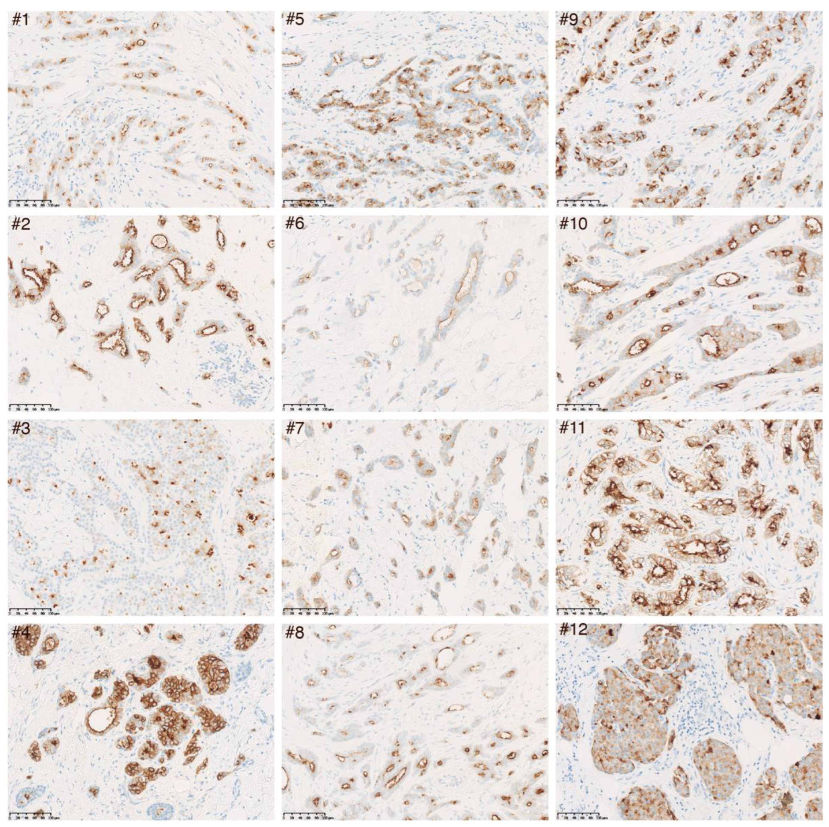
Next, immunohistochemical analysis of 12 luminal A invasive breast cancer samples was performed using either the monoclonal mouse anti-human LRP2 antibody (Figure 3) or the polyclonal rabbit anti-human LRP2 antibody (Figure S6). LRP2 was detectable, although to a varying degree, across all 12 tumor samples. LRP2 protein expression appeared confined to malignant cells (and potentially residual normal epithelial cells), which is consistent with the single-cell RNA sequencing data presented in Figure 2C,D. LRP2 was observed both at the plasma membrane and in cytoplasmic compartments distributed throughout malignant cells.

3.4. Low LRP2 Expression Is Associated with Poor Survival in Multiple Cancers

Investigation of LRP2 levels across cancer types showed considerable variation in LRP2 expression within individual cancer types (Figure 1), which could suggest that LRP2 is deregulated in a subset of tumors. This observation prompted us to test the association between LRP2 expression and clinical outcome. To do so, we performed univariate Cox proportional-hazards modelling of LRP2 expression and survival within individual TCGA cancer types (Figure 4). Survival parameters were chosen based on the recommendations from the TCGA Pan-Cancer Clinical Data Resource and analysis was not performed for seven tumor types flagged as having an insufficient event number [38]. Breast cancer survival analysis was performed using the METABRIC cohort, which includes long-term clinical follow-up data for > 2,000 patients [42]. Interestingly, low levels of LRP2 were associated with poor patient outcome in seven of the eight cancer types that express the highest levels of LRP2 on average (Figure 4, Table S2), including kidney renal clear cell carcinoma (KIRC) (HR = 0.91, p < 0.001), kidney renal papillary cell carcinoma (KIRP) (HR = 0.88, p < 0.01), mesothelioma (MESO) (HR = 0.87, p < 0.01), thyroid carcinoma (THCA) (HR = 0.82, p < 0.05), breast cancer (METABRIC) (HR = 0.90, p < 0.0001), lung adenocarcinoma (LUAD) (HR = 0.95, p < 0.05), and skin cutaneous melanoma (SKCM) (HR = 0.95, p < 0.05).
Based on the observation that a subset of tumors within these cancer types showed lower LRP2 expression, we stratified samples into LRP2high or LRP2low based on a lower quartile cutoff and performed Kaplan–Meier analysis. As expected, this provided similar results as the Cox proportional-hazards model and showed significantly poorer survival of patients with LRP2low tumors in KIRC (p = 0.00024), KIRP (p = 0.035), MESO (p = 0.01), THCA (p = 0.048), and METABRIC (p < 0.0001) (Figure 5A–E). However, at this particular cutoff, there were no significant differences between the survival of patients with LRP2high or LRP2low tumors in LUAD (p = 0.59) and SKCM (p = 0.29)(Figure 5F,G).
We sought to validate the association between low LRP2 and poor outcome in additional cohorts. In a previous unbiased analysis of primary clear cell renal cell carcinomas, low LRP2 was identified as one of 259 genes that predicted poor outcome after surgery [62]. Low LRP2 was also associated with shorter overall survival in the KMplotter database (lower quartile cutoff Kaplan–Meier analysis, HR = 0.64, p < 0.0001, Figure S7A), which is an integrated database of breast cancer gene-expression data [46]. Likewise, low LRP2 was associated with poor outcome in the Bott et al. microarray cohort for mesothelioma (univariate Cox proportional-hazards model, HR = 0.81, p = 0.038) and trended towards significance in the Gordon et al. microarray cohort for mesothelioma (univariate Cox proportional-hazards model, HR = 0.88, p = 0.108) (Figure S7C) [43,44].
Based on these analyses, we considered a framework where LRP2 expression is generally maintained in the cancer setting, but where its expression is lost in a subset of tumors, and that this is associated with poor patient outcome.

3.5. Low LRP2 Expression in Human Cancer Is Associated with First Intron CpG Methylation

To characterize the mechanisms that lead to putative loss of LRP2 expression in some tumors, we next explored the mutation profile of LRP2 in human cancers. Loss-of-function mutations and gene deletions in LRP2 are rare and cannot not explain the extent of lost LRP2 expression observed across cancer types. Therefore, we hypothesized that epigenetic events could account for loss of LRP2 expression in cancer.
To investigate this, we accessed TCGA Pan-Cancer methylation data from the Illumina 450K array, which provides a fraction of methylation values (β values) for 450,000 CpG sites in the human genome [63]. There were 19 probes available in the LRP2 promoter and gene body in the dataset. Heatmap visualization showed that CpG sites in the promoter region of LRP2 were demethylated, while CpG sites in the gene body were largely methylated (Figure 6, Table 1). Consistent with the general understanding of how DNA methylation regulates gene expression, CpG methylation in the LRP2 promoter region was negatively correlated to LRP2 expression, while sites in the gene body mainly showed a positive correlation (Table 1).
Of interest, a single CpG site in the first intron of LRP2, cg023161027, showed differential methylation across tumors and a strong inverse correlation with LRP2 expression (Pearson’s R = −0.65, p < 1 × 10100, Table 1). cg02361027 is found at position 169360890 (human genome version hg38) on the + strand of chromosome 2 and is in close proximity to a CpG island, stretching from position 169361804-169363035. Its negative correlation to LRP2 expression is consistent with studies showing genome-wide inverse correlations between first intron methylation and gene expression [64,65].
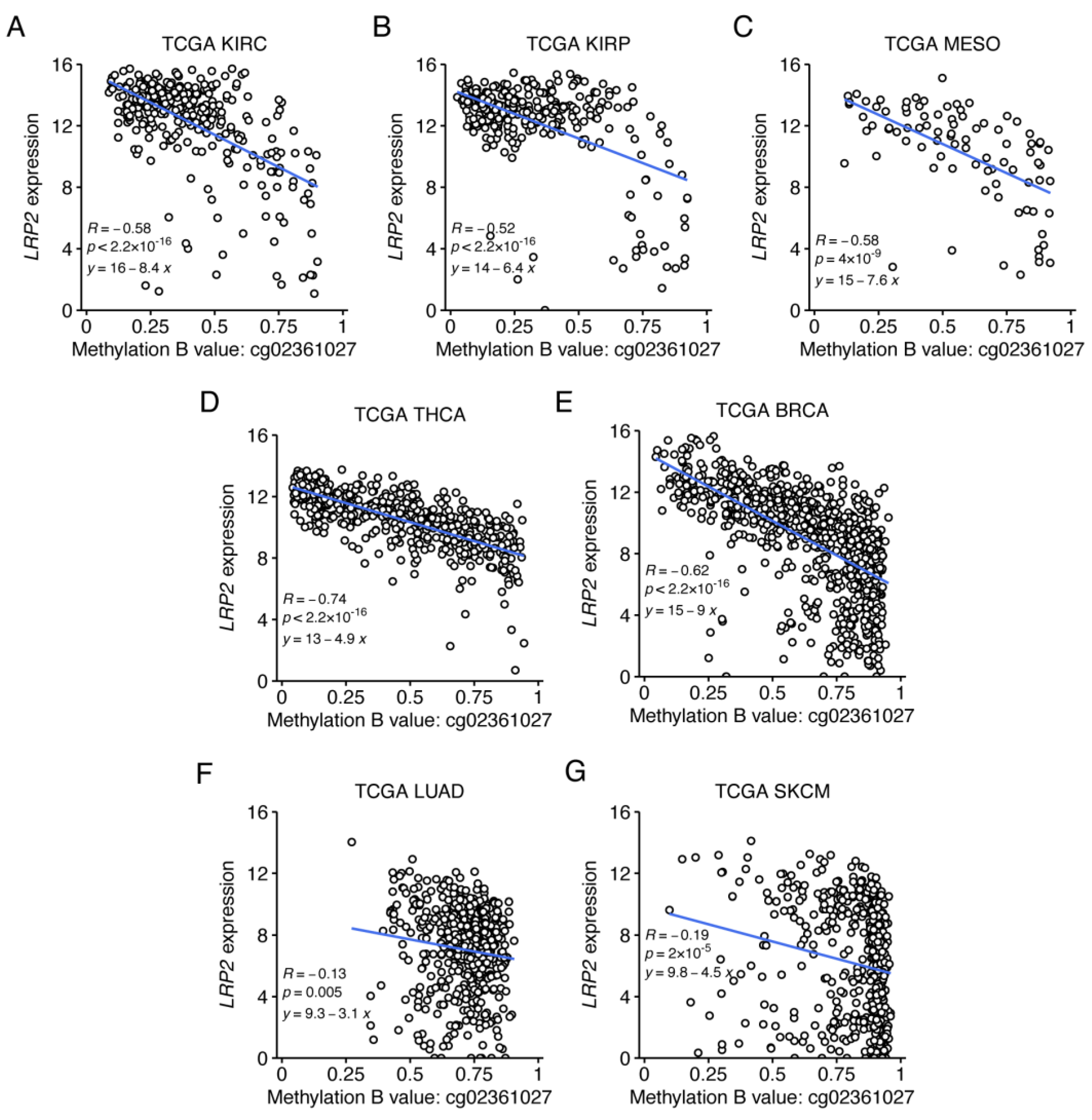
To further explore the relationship between LRP2 expression and cg02361027 methylation, we calculated their correlation within individual cancer types (Figure S8). This showed that the negative correlation is strongest in tumor types that on average express the highest levels of LRP2 (KIRC, KIRP, MESO, THCA and BRCA) (Figure 7A–E). On the other hand, the correlation is modest within tumor types that express mid-to-low levels of LRP2, including LUAD and SKCM (Figure 7F,G), suggesting that other factors contribute to differential LRP2 expression in these cancer types.

3.6. LRP2 Silencing Is Associated with Tumor Dedifferentiation

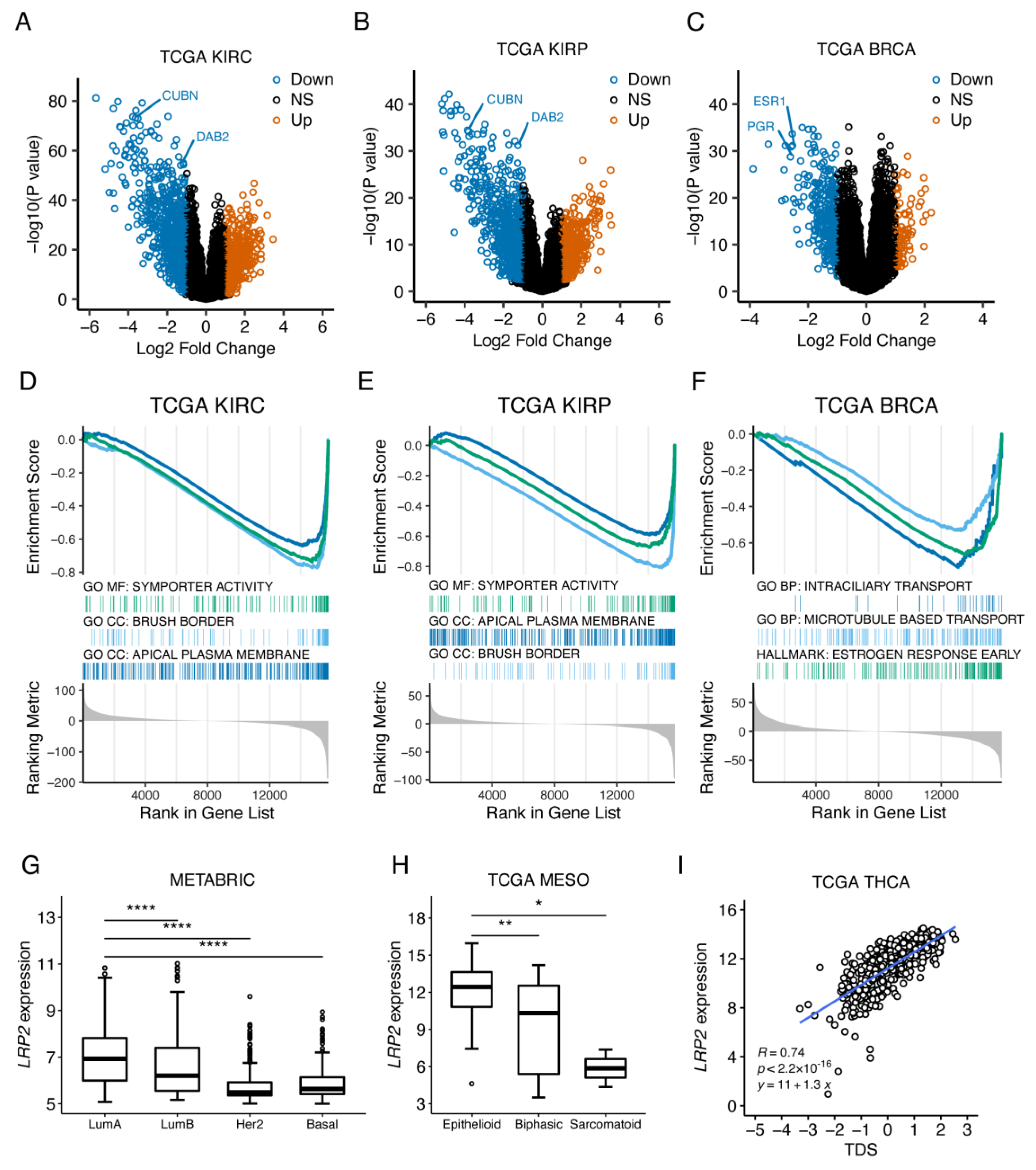
LRP2 expression in adults is restricted to absorptive epithelia, and LRP2 is considered a marker for proximal tubule epithelial cell-differentiation status. Based on this, we hypothesized that the loss of LRP2 expression observed in some tumors is associated with dedifferentiation. Differential gene-expression analysis between LRP2high and LRP2low tumors in KIRC and KIRP revealed that multiple genes (including CUBN and DAB2 encoding functional LRP2 partners in proximal tubule epithelial cells) (Figure 8A,B, Table S3) and pathways related to specialized proximal tubule epithelial cell functions (Figure 8D,E, Table S4) were also downregulated in LRP2low tumors. Similar analysis for BRCA revealed downregulation of multiple genes (including genes encoding hormone receptors ESR1 and PGR) (Figure 8C, Table S3) and pathways related to mammary gland epithelium differentiation and function (e.g., estrogen response) (Figure 8F, Table S4) in LRP2low tumors.
We further evaluated the relationship between LRP2 expression and molecular subtypes or signatures related to differentiation in breast cancer, mesothelioma, and thyroid cancer. In breast cancer, tumors are classified using the PAM50 gene signature into four molecular subtypes (luminal A, luminal B, HER2-enriched, and basal), which differ in their biological characteristics and prognosis [66]. Luminal tumors show a higher level of epithelial differentiation and estrogen receptor expression and signaling, while HER2 and basal tumors are more dedifferentiated [67]. Consistent with higher expression of LRP2 in more epithelial-like tumors, LRP2 expression was significantly higher in luminal A tumors compared to luminal B (p < 0.0001), HER2 (p < 0.0001), and basal (p < 0.0001) tumors in the METABRIC cohort (Figure 8G). In mesothelioma, histological subtypes are also classified as epithelioid, biphasic, or sarcomatoid based on their differentiation state [68]. In the TCGA MESO dataset, LRP2 expression was higher in epithelioid tumors compared to biphasic (p < 0.01) and sarcomatoid (p < 0.05) tumors (Figure 8H). In papillary thyroid cancer, a 16-gene tumor differentiation score (TDS) correlated to patient prognosis was defined by the TCGA THCA working group [51]. LRP2 showed a strong correlation to TDS in the TCGA THCA dataset (Pearson’s R = 0.76, p < 2.2 × 10¹⁶) (Figure 8I). These examples provide evidence that LRP2 silencing is related to a dedifferentiated tumor state in various epithelial cancer types.
Overall, this explorative analysis of the TCGA dataset is consistent with a model where epigenetic silencing of LRP2 is associated with tumor dedifferentiation and poor patient outcome in multiple cancer types arising from LRP2-expressing absorptive epithelia.

4. Discussion

To investigate the clinical and molecular correlates of LRP2 expression across human cancers, we performed an integrated analysis of publicly available data from The Cancer Genome Atlas. LRP2 was highly expressed in cancers that arise from absorptive epithelia, such as clear cell renal cell carcinoma, papillary renal cell carcinoma, mesothelioma, invasive breast carcinoma, and papillary thyroid cancer. A subset of tumors within these cancer types show downregulation of LRP2, which is correlated with epigenetic alterations in the LRP2 gene locus, tumor dedifferentiation, and poor patient outcome.
To our knowledge, there have been no earlier systematic studies of LRP2 in cancer. We have previously demonstrated that LRP2 gene and protein expression is acquired in a subset of melanoma tumors [29]. However, our pan-cancer analysis did not immediately reveal other cancer types with gained expression of LRP2. Instead, our data support a model where LRP2 is maintained in cancers arising from absorptive epithelial cells known to express high levels of LRP2 in the healthy state. Our data also highlight deregulation of LRP2 as a common event in these cancer types.
One limitation of our study is that it leveraged bulk RNA sequencing data, which could be confounded by expression of LRP2 in non-malignant cell types and might not reflect functional protein levels. To address this limitation, we queried single-cell RNA sequencing data and found that LRP2 expression is restricted to malignant cells. Furthermore, we used recent proteomics data from human tumors to demonstrate a strong correlation between LRP2 transcript and protein levels. Therefore, LRP2 levels in tumor bulk RNA measurements can be considered to reflect tumor-specific expression and to correlate with protein abundance.
Additionally, we confirmed LRP2 protein expression in healthy breast epithelial cells and malignant cells of 12 different invasive breast carcinomas. LRP2 expression was confined to the apical plasma membrane in healthy breast epithelial cells but was observed both at the plasma membrane and in cytoplasmic compartments distributed throughout malignant cells. We speculate that this could reflect increased endocytic activity and thus intracellular trafficking of LRP2 in malignant cells as opposed to healthy cells.
LRP2 in epithelial cancers was strongly and inversely correlated to methylation of a specific CpG site (cg02361027) in the first intron of the LRP2 locus. To our knowledge, the present study is the first identification of a robust correlation between LRP2 levels and DNA methylation in cancer. The inverse correlation between LRP2 expression and cg02361027 methylation could suggest that this region of the first intron is involved in transcriptional regulation, although this remains to be experimentally proven. Previous studies have demonstrated a genome-wide inverse correlation between first intron methylation and gene expression [64,65] and shown that conserved regions of the first intron are likely to regulate transcription [69]. Indeed, cg02361027 resides in a highly conserved region of the LRP2 first intron (UCSC Genome Browser, Figure S9). Further studies should determine the correlation between LRP2 expression and first intron methylation in other biological systems where LRP2 levels are variable, such as throughout embryonic development [19,20] or in the settings of acute and chronic kidney diseases [70,71,72]. Identification of transcription factors that interact with this region of the LRP2 locus will also be of interest.
We further observed that LRP2 downregulation was associated with dedifferentiated tumor subsets in multiple cancer types. We speculate that LRP2 loss in epithelial cancers might be part of a genome-wide epigenetic reprogramming in epithelial cancers, resulting in coordinated deactivation of epithelial gene expression. However, the results presented in the current study are correlative, and future studies are needed to dissect the exact mechanisms of LRP2 regulation in cancer.
In the context of cancer, loss of LRP2 protein could alter the cellular uptake and clearance of ligands from the tumor microenvironment. Putative tumor-suppressive functions of LRP2 could be exerted through the clearance of ligands that modulate proliferation, migration, and survival of cancer cells. More complex mechanisms are also possible, where LRP2-mediated clearance of certain molecules from the tumor microenvironment could modulate functions of immune or stromal cells.
Two studies support the role of LRP2 in mediating endocytic uptake of vitamin D (in complex with vitamin D binding protein) and in the activation of vitamin D receptor signaling in breast cancer cells [27,28]. In connection to this, it is important to mention that high serum levels of vitamin D [73,74] and high expression of the vitamin D receptor [75] are linked to better prognosis in breast cancer. In line with this, we find it tempting to speculate that LRP2 also plays a role in this context and that the presence or absence of LRP2 in breast cancer cells would have an important impact on vitamin D homeostasis and receptor signaling. However, this remains to be experimentally proven.
Given that more than 40 ligands have been identified for LRP2 to date [12,16], we hypothesize that multiple tissue-, context- and ligand-dependent cellular signaling pathways would be impacted by downregulation of this receptor in cancer cells.
We show that LRP2 downregulation correlates with poor patient outcome in multiple cancer types, including renal cell carcinoma, mesothelioma, thyroid cancer and invasive breast carcinoma. Interestingly, our analyses also demonstrate that the prognostic value of LRP2 appears to be independent of clinicopathologic variables, such as age, gender, tumor stage, and histological grade (Tables S5–S7). Accordingly, we suggest that future cancer-type-specific studies should investigate further the potential of LRP2 as an independent prognostic biomarker.
In line with the idea that LRP2 might be a potential novel biomarker in certain cancer types, a recent effort to identify genes related to metastasis in premenopausal patients with hormone receptor positive early breast cancer identified low LRP2 expression as the top independent factor in the Breast Cancer 360 TM panel [76]. This is consistent with our finding that low LRP2 predicts poor prognosis in the METABRIC cohort and supports that further studies should be conducted in order to evaluate LRP2 as biomarker in invasive breast carcinomas. It would be natural for such studies to focus on immunohistochemical detection of LRP2, which we previously demonstrated to be useful for the analysis of melanoma tumors [29] and, in the present study, for the analysis of invasive breast carcinomas.
Finally, the findings presented here might also have implications for the use of LRP2 as a target for anti-cancer drug delivery. LRP2 has several features that make it an attractive target for drug delivery. First, LRP2 is a long-lived fast-cycling and recycling endocytic receptor, presumably capable of delivering multiple cytotoxic payloads without being subject to cellular degradation itself [13]. Second, the tissue distribution of LRP2 in adults is limited, and to our knowledge, there are no instances where LRP2 is in direct contact with the bloodstream, limiting the risk of side effects from treatment. Previous studies established LRP2 as an effective target for drug delivery in clear cell renal cell carcinoma [25,26]. Because we show here that high levels of LRP2 are not only observed in clear cell renal cell carcinoma, but also in papillary renal cell carcinoma, mesothelioma, and in invasive breast carcinomas, we speculate that LRP2-targeted drug delivery might be effective in some of these cancer types as well.

5. Conclusions

We find that LRP2 expression is largely restricted to cancer types that arise from LRP2-expressing absorptive epithelia. However, we also highlight that a subset of tumors within these cancer types show epigenetic silencing of LRP2, which is associated with tumor dedifferentiation and poor survival. These observations now motivate further studies on the biological role of LRP2 in cancer, in particular epithelial cancers, and the potential use of LRP2 as a novel cancer-type-specific prognostic biomarker and target for anti-cancer therapy.

Supplementary Materials

The following supporting information can be downloaded at: https://www.mdpi.com/article/10.3390/cancers15061830/s1, Figure S1: LRP2 expression is restricted to epithelial cells in the breast tumor microenvironment. Figure S2: Scatter plot of LRP2 mRNA and LRP2 protein levels in CPTAC. Figure S3: Immunohistochemical detection of LRP2 in epithelial cells of proximal tubules in human kidney cortex. Figure S4: Immunohistochemical detection of LRP2 in ductal cells in healthy human glandular breast epithelium. Figure S5: High-magnification analyses of LRP2 expression and localization in human glandular breast epithelium sample #2. Figure S6: Immunohistochemical analysis of LRP2 expression in 12 different luminal A invasive ductal breast carcinomas. Figure S7: Low LRP2 is associated with poor survival in additional non-TCGA cohorts. Figure S8: Correlation between LRP2 expression and cg0263127 methylation across cancer types. Figure S9: cg02361027 is located within a highly conserved region of the LRP2 gene. Table S1: List of TCGA cancer type abbreviations. Table S2: Source data for TCGA Pan-Cancer LRP2 survival analysis. Table S3: Differential gene-expression analysis between LRP2low and LRP2high tumors in KIRC, KIRP and BRCA. Table S4: Gene Set Enrichment Analysis of differentially expressed genes between LRP2low and LRP2high tumors in KIRC, KIRP and BRCA. Table S5: Pairwise associations between LRP2 expression and clinicopathological variables. Table S6: Univariate Cox proportional-hazard survival analyses for LRP2 and clinicopathologic variables. Table S7: Multivariate Cox proportional-hazards analyses with LRP2 expression and clinicopathological variables as covariates.

Author Contributions

Conceptualization, M.Q.R., G.T., M.M.N., T.S., J.S.P., S.K.M., S.E.D., and M.M.; Data curation, M.Q.R., G.T., T.S., and M.M.; Formal analysis, M.Q.R., G.T., C.M., T.S., and M.M.; Funding acquisition, M.Q.R., J.S.P., and M.M.; Methodology, M.Q.R., G.T., M.M.N., C.M., T.S., J.S.P., S.K.M., S.E.D., and M.M.; Project administration, M.M.; Supervision, S.E.D. and M.M.; Writing—original draft, M.Q.R. and M.M.; Writing—review and editing, M.Q.R., G.T., M.M.N., C.M., T.S., J.S.P., S.K.M., S.E.D., and M.M. All authors have read and agreed to the published version of the manuscript.

Funding

This project was supported by grants from The Max Wørzner and wife Inger Wørzner’s Memorial Grant (Aarhus University reference number: 30109), The Foundation of Holger Hjortenberg and wife Dagmar Hjortenberg (Aarhus University reference number: 29877), The Eva and Henry Fraenkel’s Memorial Fund (Aarhus University reference number: 32535), The Dagmar Marshall Foundation (Aarhus University reference numbers: 33449 and 37155), The AP Moeller Foundation (19-L-0255), The Einar Willumsen’s Memorial Grant (Aarhus University reference number: 34604), the Novo Nordisk Foundation; grants NNF20OC0065103 and NNF18OC0053222, the Independent Research Fund Denmark; Medical Sciences; grant number: 8021-00419B, and Aarhus University; 4-year PhD fellowship for Martin Qvist Rasmussen.

Institutional Review Board Statement

Ethical review and approval from an institutional review board were waived for this study. All human material included in this study was collected from the archives at Aarhus University Hospital (formalin-fixed paraffin-embedded tissue samples) with all identifiers removed (totally anonymized) or post-surgery from breast reduction surgeries with all identifiers removed (totally anonymized) and in respect of each patient’s wishes regarding use of tissue for research.

Informed Consent Statement

Patient consent was waived for this study. All human material included in this study was collected from the archives at Aarhus University Hospital with all identifiers removed or post-surgery from breast reduction surgeries with all identifiers removed. Before the use of any human material, each patient’s wish regarding the use of his or her tissue sample for research was inspected using the Tissue Application Registry in Denmark.

Data Availability Statement

TCGA Pan-Cancer datasets including RNA sequencing, Illumina 450K methylation and survival data are available at the UCSC Xena data portal (https://xena.ucsc.edu/, accessed on 4 November 2021). Single-cell data from renal cell carcinoma and breast cancer can be accessed and visualized using the Broad Single Cell Portal (https://singlecell.broadinstitute.org/single_cell, accessed on 1 October 2022). Processed gene-level proteomics data from CPTAC can be accessed using the Python package cptac (https://paynelab.github.io/cptac/, accessed on 3 October 2022). Processed data from METABRIC are available from cBioportal (https://www.cbioportal.org/study/summary?id=brca_metabric, accessed on 19 November 2021). Mesothelioma gene-expression data from Bott et al. (GSE29354) and Gordon et al. (GSE2549) are available from GEO. All datasets used in this study are publicly available.

Acknowledgments

We thank Jeanette Bæhr Georgsen (Department of Pathology, Aarhus University Hospital) for technical assistance.

Conflicts of Interest

The authors declare no conflict of interest.

References

  1. Dongre, A.; Weinberg, R.A. New insights into the mechanisms of epithelial-mesenchymal transition and implications for cancer. Nat. Rev. Mol. Cell Biol. 2019, 20, 69–84. [Google Scholar] [CrossRef] [PubMed]
  2. Kalluri, R.; Weinberg, R.A. The basics of epithelial-mesenchymal transition. J. Clin. Investig. 2009, 119, 1420–1428. [Google Scholar] [CrossRef] [PubMed] [Green Version]
  3. Pastushenko, I.; Brisebarre, A.; Sifrim, A.; Fioramonti, M.; Revenco, T.; Boumahdi, S.; Van Keymeulen, A.; Brown, D.; Moers, V.; Lemaire, S.; et al. Identification of the tumour transition states occurring during EMT. Nature 2018, 556, 463–468. [Google Scholar] [CrossRef] [PubMed]
  4. Brabletz, T.; Kalluri, R.; Nieto, M.A.; Weinberg, R.A. EMT in cancer. Nat. Rev. Cancer 2018, 18, 128–134. [Google Scholar] [CrossRef]
  5. McMahon, H.T.; Boucrot, E. Molecular mechanism and physiological functions of clathrin-mediated endocytosis. Nat. Rev.. Mol. Cell Biol. 2011, 12, 517–533. [Google Scholar] [CrossRef] [PubMed]
  6. Murphy, J.E.; Padilla, B.E.; Hasdemir, B.; Cottrell, G.S.; Bunnett, N.W. Endosomes: A legitimate platform for the signaling train. Proc. Natl. Acad. Sci. USA 2009, 106, 17615–17622. [Google Scholar] [CrossRef] [Green Version]
  7. Khan, I.; Steeg, P.S. Endocytosis: A pivotal pathway for regulating metastasis. Br. J. Cancer 2021, 124, 66–75. [Google Scholar] [CrossRef]
  8. Raman, D.; Sai, J.; Hawkins, O.; Richmond, A. Adaptor protein2 (AP2) orchestrates CXCR2-mediated cell migration. Traffic 2014, 15, 451–469. [Google Scholar] [CrossRef] [Green Version]
  9. Chen, P.H.; Bendris, N.; Hsiao, Y.J.; Reis, C.R.; Mettlen, M.; Chen, H.Y.; Yu, S.L.; Schmid, S.L. Crosstalk between CLCb/Dyn1-Mediated Adaptive Clathrin-Mediated Endocytosis and Epidermal Growth Factor Receptor Signaling Increases Metastasis. Dev. Cell 2017, 40, 278–288.e275. [Google Scholar] [CrossRef] [Green Version]
  10. Reis, C.R.; Chen, P.H.; Srinivasan, S.; Aguet, F.; Mettlen, M.; Schmid, S.L. Crosstalk between Akt/GSK3beta signaling and dynamin-1 regulates clathrin-mediated endocytosis. EMBO J. 2015, 34, 2132–2146. [Google Scholar] [CrossRef] [Green Version]
  11. Christensen, E.I.; Birn, H. Megalin and cubilin: Multifunctional endocytic receptors. Nat. Rev. Mol. Cell Biol. 2002, 3, 256–266. [Google Scholar] [CrossRef] [PubMed]
  12. Christensen, E.I.; Birn, H.; Storm, T.; Weyer, K.; Nielsen, R. Endocytic receptors in the renal proximal tubule. Physiology 2012, 27, 223–236. [Google Scholar] [CrossRef] [PubMed] [Green Version]
  13. Perez Bay, A.E.; Schreiner, R.; Benedicto, I.; Paz Marzolo, M.; Banfelder, J.; Weinstein, A.M.; Rodriguez-Boulan, E.J. The fast-recycling receptor Megalin defines the apical recycling pathway of epithelial cells. Nat. Commun. 2016, 7, 11550. [Google Scholar] [CrossRef] [PubMed] [Green Version]
  14. Klassen, R.B.; Allen, P.L.; Batuman, V.; Crenshaw, K.; Hammond, T.G. Light chains are a ligand for megalin. J. Appl. Physiol. (1985) 2005, 98, 257–263. [Google Scholar] [CrossRef] [Green Version]
  15. Bryniarski, M.A.; Zhao, B.; Chaves, L.D.; Mikkelsen, J.H.; Yee, B.M.; Yacoub, R.; Shen, S.; Madsen, M.; Morris, M.E. Immunoglobulin G Is a Novel Substrate for the Endocytic Protein Megalin. AAPS J. 2021, 23, 40. [Google Scholar] [CrossRef]
  16. Nielsen, R.; Christensen, E.I.; Birn, H. Megalin and cubilin in proximal tubule protein reabsorption: From experimental models to human disease. Kidney Int. 2016, 89, 58–67. [Google Scholar] [CrossRef] [PubMed] [Green Version]
  17. Donnai, D.; Barrow, M. Diaphragmatic hernia, exomphalos, absent corpus callosum, hypertelorism, myopia, and sensorineural deafness: A newly recognized autosomal recessive disorder? Am. J. Med. Genet. 1993, 47, 679–682. [Google Scholar] [CrossRef]
  18. Kantarci, S.; Al-Gazali, L.; Hill, R.S.; Donnai, D.; Black, G.C.; Bieth, E.; Chassaing, N.; Lacombe, D.; Devriendt, K.; Teebi, A.; et al. Mutations in LRP2, which encodes the multiligand receptor megalin, cause Donnai-Barrow and facio-oculo-acoustico-renal syndromes. Nat. Genet. 2007, 39, 957–959. [Google Scholar] [CrossRef] [Green Version]
  19. Assemat, E.; Chatelet, F.; Chandellier, J.; Commo, F.; Cases, O.; Verroust, P.; Kozyraki, R. Overlapping expression patterns of the multiligand endocytic receptors cubilin and megalin in the CNS, sensory organs and developing epithelia of the rodent embryo. Gene Expr. Patterns 2005, 6, 69–78. [Google Scholar] [CrossRef]
  20. Fisher, C.E.; Howie, S.E. The role of megalin (LRP-2/Gp330) during development. Dev. Biol. 2006, 296, 279–297. [Google Scholar] [CrossRef] [Green Version]
  21. Willnow, T.E.; Hilpert, J.; Armstrong, S.A.; Rohlmann, A.; Hammer, R.E.; Burns, D.K.; Herz, J. Defective forebrain development in mice lacking gp330/megalin. Proc. Natl. Acad. Sci. USA 1996, 93, 8460–8464. [Google Scholar] [CrossRef] [PubMed] [Green Version]
  22. Christ, A.; Herzog, K.; Willnow, T.E. LRP2, an auxiliary receptor that controls sonic hedgehog signaling in development and disease. Dev. Dyn. Off. Publ. Am. Assoc. Anat. 2016, 245, 569–579. [Google Scholar] [CrossRef] [PubMed] [Green Version]
  23. Spoelgen, R.; Hammes, A.; Anzenberger, U.; Zechner, D.; Andersen, O.M.; Jerchow, B.; Willnow, T.E. LRP2/megalin is required for patterning of the ventral telencephalon. Development 2005, 132, 405–414. [Google Scholar] [CrossRef] [PubMed] [Green Version]
  24. Kowalczyk, I.; Lee, C.; Schuster, E.; Hoeren, J.; Trivigno, V.; Riedel, L.; Gorne, J.; Wallingford, J.B.; Hammes, A.; Feistel, K. Neural tube closure requires the endocytic receptor Lrp2 and its functional interaction with intracellular scaffolds. Development 2021, 148, dev195008. [Google Scholar] [CrossRef] [PubMed]
  25. Wang, J.; Poon, C.; Chin, D.; Milkowski, S.; Lu, V.; Hallows, K.R.; Chung, E.J. Design and in vivo characterization of kidney-targeting multimodal micelles for renal drug delivery. Nano Res. 2018, 11, 5584–5595. [Google Scholar] [CrossRef]
  26. Ordikhani, F.; Kasinath, V.; Uehara, M.; Akbarzadeh, A.; Yilmam, O.A.; Dai, L.; Aksu, H.; Jung, S.; Jiang, L.; Li, X.; et al. Selective Trafficking of Light Chain-Conjugated Nanoparticles to the Kidney and Renal Cell Carcinoma. Nano Today 2020, 35, 100990. [Google Scholar] [CrossRef]
  27. Chlon, T.M.; Taffany, D.A.; Welsh, J.; Rowling, M.J. Retinoids modulate expression of the endocytic partners megalin, cubilin, and disabled-2 and uptake of vitamin D-binding protein in human mammary cells. J. Nutr. 2008, 138, 1323–1328. [Google Scholar] [CrossRef] [Green Version]
  28. Rowling, M.J.; Kemmis, C.M.; Taffany, D.A.; Welsh, J. Megalin-mediated endocytosis of vitamin D binding protein correlates with 25-hydroxycholecalciferol actions in human mammary cells. J. Nutr. 2006, 136, 2754–2759. [Google Scholar] [CrossRef] [Green Version]
  29. Andersen, R.K.; Hammer, K.; Hager, H.; Christensen, J.N.; Ludvigsen, M.; Honore, B.; Thomsen, M.B.; Madsen, M. Melanoma tumors frequently acquire LRP2/megalin expression, which modulates melanoma cell proliferation and survival rates. Pigment. Cell Melanoma Res. 2015, 28, 267–280. [Google Scholar] [CrossRef]
  30. Jensen, L.L.; Andersen, R.K.; Hager, H.; Madsen, M. Lack of megalin expression in adult human terminal ileum suggests megalin-independent cubilin/amnionless activity during vitamin B12 absorption. Physiol. Rep. 2014, 2, e12086. [Google Scholar] [CrossRef]
  31. Storm, T.; Christensen, E.I.; Christensen, J.N.; Kjaergaard, T.; Uldbjerg, N.; Larsen, A.; Honore, B.; Madsen, M. Megalin Is Predominantly Observed in Vesicular Structures in First and Third Trimester Cytotrophoblasts of the Human Placenta. J. Histochem. Cytochem. 2016, 64, 769–784. [Google Scholar] [CrossRef] [PubMed] [Green Version]
  32. Storm, T.; Tranebjaerg, L.; Frykholm, C.; Birn, H.; Verroust, P.J.; Neveus, T.; Sundelin, B.; Hertz, J.M.; Holmstrom, G.; Ericson, K.; et al. Renal phenotypic investigations of megalin-deficient patients: Novel insights into tubular proteinuria and albumin filtration. Nephrol. Dial. Transplant. 2013, 28, 585–591. [Google Scholar] [CrossRef] [PubMed] [Green Version]
  33. Christensen, E.I.; Nielsen, S.; Moestrup, S.K.; Borre, C.; Maunsbach, A.B.; de Heer, E.; Ronco, P.; Hammond, T.G.; Verroust, P. Segmental distribution of the endocytosis receptor gp330 in renal proximal tubules. Eur. J. Cell. Biol. 1995, 66, 349–364. [Google Scholar] [PubMed]
  34. Vivian, J.; Rao, A.A.; Nothaft, F.A.; Ketchum, C.; Armstrong, J.; Novak, A.; Pfeil, J.; Narkizian, J.; Deran, A.D.; Musselman-Brown, A.; et al. Toil enables reproducible, open source, big biomedical data analyses. Nat. Biotechnol. 2017, 35, 314–316. [Google Scholar] [CrossRef] [Green Version]
  35. Dobin, A.; Davis, C.A.; Schlesinger, F.; Drenkow, J.; Zaleski, C.; Jha, S.; Batut, P.; Chaisson, M.; Gingeras, T.R. STAR: Ultrafast universal RNA-seq aligner. Bioinformatics 2013, 29, 15–21. [Google Scholar] [CrossRef]
  36. Li, B.; Dewey, C.N. RSEM: Accurate transcript quantification from RNA-Seq data with or without a reference genome. BMC Bioinform. 2011, 12, 323. [Google Scholar] [CrossRef] [Green Version]
  37. Bray, N.L.; Pimentel, H.; Melsted, P.; Pachter, L. Near-optimal probabilistic RNA-seq quantification. Nat. Biotechnol. 2016, 34, 525–527. [Google Scholar] [CrossRef]
  38. Liu, J.; Lichtenberg, T.; Hoadley, K.A.; Poisson, L.M.; Lazar, A.J.; Cherniack, A.D.; Kovatich, A.J.; Benz, C.C.; Levine, D.A.; Lee, A.V.; et al. An Integrated TCGA Pan-Cancer Clinical Data Resource to Drive High-Quality Survival Outcome Analytics. Cell. 2018, 173, 400–416.e411. [Google Scholar] [CrossRef] [Green Version]
  39. Bi, K.; He, M.X.; Bakouny, Z.; Kanodia, A.; Napolitano, S.; Wu, J.; Grimaldi, G.; Braun, D.A.; Cuoco, M.S.; Mayorga, A.; et al. Tumor and immune reprogramming during immunotherapy in advanced renal cell carcinoma. Cancer Cell 2021, 39, 649–661.e645. [Google Scholar] [CrossRef]
  40. Wu, S.Z.; Al-Eryani, G.; Roden, D.L.; Junankar, S.; Harvey, K.; Andersson, A.; Thennavan, A.; Wang, C.; Torpy, J.R.; Bartonicek, N.; et al. A single-cell and spatially resolved atlas of human breast cancers. Nat. Genet. 2021, 53, 1334–1347. [Google Scholar] [CrossRef]
  41. Lindgren, C.M.; Adams, D.W.; Kimball, B.; Boekweg, H.; Tayler, S.; Pugh, S.L.; Payne, S.H. Simplified and Unified Access to Cancer Proteogenomic Data. J. Proteome Res. 2021, 20, 1902–1910. [Google Scholar] [CrossRef] [PubMed]
  42. Curtis, C.; Shah, S.P.; Chin, S.F.; Turashvili, G.; Rueda, O.M.; Dunning, M.J.; Speed, D.; Lynch, A.G.; Samarajiwa, S.; Yuan, Y.; et al. The genomic and transcriptomic architecture of 2,000 breast tumours reveals novel subgroups. Nature 2012, 486, 346–352. [Google Scholar] [CrossRef] [PubMed]
  43. Bott, M.; Brevet, M.; Taylor, B.S.; Shimizu, S.; Ito, T.; Wang, L.; Creaney, J.; Lake, R.A.; Zakowski, M.F.; Reva, B.; et al. The nuclear deubiquitinase BAP1 is commonly inactivated by somatic mutations and 3p21.1 losses in malignant pleural mesothelioma. Nat. Genet. 2011, 43, 668–672. [Google Scholar] [CrossRef] [PubMed]
  44. Gordon, G.J.; Rockwell, G.N.; Jensen, R.V.; Rheinwald, J.G.; Glickman, J.N.; Aronson, J.P.; Pottorf, B.J.; Nitz, M.D.; Richards, W.G.; Sugarbaker, D.J.; et al. Identification of novel candidate oncogenes and tumor suppressors in malignant pleural mesothelioma using large-scale transcriptional profiling. Am. J. Pathol. 2005, 166, 1827–1840. [Google Scholar] [CrossRef] [PubMed]
  45. Hao, Y.; Hao, S.; Andersen-Nissen, E.; Mauck, W.M., 3rd; Zheng, S.; Butler, A.; Lee, M.J.; Wilk, A.J.; Darby, C.; Zager, M.; et al. Integrated analysis of multimodal single-cell data. Cell. 2021, 184, 3573–3587.e3529. [Google Scholar] [CrossRef]
  46. Gyorffy, B. Survival analysis across the entire transcriptome identifies biomarkers with the highest prognostic power in breast cancer. Comput. Struct. Biotechnol. J. 2021, 19, 4101–4109. [Google Scholar] [CrossRef]
  47. Ritchie, M.E.; Phipson, B.; Wu, D.; Hu, Y.; Law, C.W.; Shi, W.; Smyth, G.K. limma powers differential expression analyses for RNA-sequencing and microarray studies. Nucleic Acids Res. 2015, 43, e47. [Google Scholar] [CrossRef]
  48. Law, C.W.; Chen, Y.; Shi, W.; Smyth, G.K. voom: Precision weights unlock linear model analysis tools for RNA-seq read counts. Genome Biol. 2014, 15, R29. [Google Scholar] [CrossRef] [Green Version]
  49. Wu, T.; Hu, E.; Xu, S.; Chen, M.; Guo, P.; Dai, Z.; Feng, T.; Zhou, L.; Tang, W.; Zhan, L.; et al. clusterProfiler 4.0: A universal enrichment tool for interpreting omics data. Innovation 2021, 2, 100141. [Google Scholar] [CrossRef]
  50. Yu, G.; Wang, L.G.; Han, Y.; He, Q.Y. clusterProfiler: An R package for comparing biological themes among gene clusters. OMICS 2012, 16, 284–287. [Google Scholar] [CrossRef]
  51. Integrated genomic characterization of papillary thyroid carcinoma. Cell. 2014, 159, 676–690. [CrossRef] [PubMed] [Green Version]
  52. Wickham, H. ggplot2: Elegant Graphics for Data Analysis; Springer: New York, NY, USA, 2016. [Google Scholar]
  53. Kassambara, A. ggpubr: ‘ggplot2’ Based Publication Ready Plots. 2020. Available online: https://cran.r-project.org/web/packages/ggpubr/index.html (accessed on 10 January 2023).
  54. Zheng, G.; Bachinsky, D.R.; Stamenkovic, I.; Strickland, D.K.; Brown, D.; Andres, G.; McCluskey, R.T. Organ distribution in rats of two members of the low-density lipoprotein receptor gene family, gp330 and LRP/alpha 2MR, and the receptor-associated protein (RAP). J. Histochem. Cytochem. 1994, 42, 531–542. [Google Scholar] [CrossRef] [PubMed]
  55. Chun, J.T.; Wang, L.; Pasinetti, G.M.; Finch, C.E.; Zlokovic, B.V. Glycoprotein 330/megalin (LRP-2) has low prevalence as mRNA and protein in brain microvessels and choroid plexus. Exp. Neurol. 1999, 157, 194–201. [Google Scholar] [CrossRef]
  56. Gajera, C.R.; Emich, H.; Lioubinski, O.; Christ, A.; Beckervordersandforth-Bonk, R.; Yoshikawa, K.; Bachmann, S.; Christensen, E.I.; Gotz, M.; Kempermann, G.; et al. LRP2 in ependymal cells regulates BMP signaling in the adult neurogenic niche. J. Cell Sci. 2010, 123, 1922–1930. [Google Scholar] [CrossRef] [PubMed] [Green Version]
  57. Wicher, G.; Larsson, M.; Fex Svenningsen, A.; Gyllencreutz, E.; Rask, L.; Aldskogius, H. Low density lipoprotein receptor-related protein-2/megalin is expressed in oligodendrocytes in the mouse spinal cord white matter. J. Neurosci. Res. 2006, 83, 864–873. [Google Scholar] [CrossRef]
  58. Wicher, G.; Larsson, M.; Rask, L.; Aldskogius, H. Low-density lipoprotein receptor-related protein (LRP)-2/megalin is transiently expressed in a subpopulation of neural progenitors in the embryonic mouse spinal cord. J. Comp. Neurol. 2005, 492, 123–131. [Google Scholar] [CrossRef]
  59. Zhang, Y.; Chen, F.; Chandrashekar, D.S.; Varambally, S.; Creighton, C.J. Proteogenomic characterization of 2002 human cancers reveals pan-cancer molecular subtypes and associated pathways. Nat. Commun. 2022, 13, 2669. [Google Scholar] [CrossRef]
  60. Krug, K.; Jaehnig, E.J.; Satpathy, S.; Blumenberg, L.; Karpova, A.; Anurag, M.; Miles, G.; Mertins, P.; Geffen, Y.; Tang, L.C.; et al. Proteogenomic Landscape of Breast Cancer Tumorigenesis and Targeted Therapy. Cell. 2020, 183, 1436–1456.e1431. [Google Scholar] [CrossRef]
  61. Clark, D.J.; Dhanasekaran, S.M.; Petralia, F.; Pan, J.; Song, X.; Hu, Y.; da Veiga Leprevost, F.; Reva, B.; Lih, T.M.; Chang, H.Y.; et al. Integrated Proteogenomic Characterization of Clear Cell Renal Cell Carcinoma. Cell. 2019, 179, 964–983.e931. [Google Scholar] [CrossRef] [Green Version]
  62. Zhao, H.; Ljungberg, B.; Grankvist, K.; Rasmuson, T.; Tibshirani, R.; Brooks, J.D. Gene expression profiling predicts survival in conventional renal cell carcinoma. PLoS Med. 2006, 3, e13. [Google Scholar] [CrossRef]
  63. Bibikova, M.; Barnes, B.; Tsan, C.; Ho, V.; Klotzle, B.; Le, J.M.; Delano, D.; Zhang, L.; Schroth, G.P.; Gunderson, K.L.; et al. High density DNA methylation array with single CpG site resolution. Genomics 2011, 98, 288–295. [Google Scholar] [CrossRef] [PubMed] [Green Version]
  64. Anastasiadi, D.; Esteve-Codina, A.; Piferrer, F. Consistent inverse correlation between DNA methylation of the first intron and gene expression across tissues and species. Epigenet. Chromatin 2018, 11, 37. [Google Scholar] [CrossRef] [PubMed]
  65. Brenet, F.; Moh, M.; Funk, P.; Feierstein, E.; Viale, A.J.; Socci, N.D.; Scandura, J.M. DNA methylation of the first exon is tightly linked to transcriptional silencing. PLoS ONE 2011, 6, e14524. [Google Scholar] [CrossRef]
  66. Parker, J.S.; Mullins, M.; Cheang, M.C.; Leung, S.; Voduc, D.; Vickery, T.; Davies, S.; Fauron, C.; He, X.; Hu, Z.; et al. Supervised risk predictor of breast cancer based on intrinsic subtypes. J. Clin. Oncol. 2009, 27, 1160–1167. [Google Scholar] [CrossRef]
  67. Prat, A.; Parker, J.S.; Karginova, O.; Fan, C.; Livasy, C.; Herschkowitz, J.I.; He, X.; Perou, C.M. Phenotypic and molecular characterization of the claudin-low intrinsic subtype of breast cancer. Breast Cancer Res. 2010, 12, R68. [Google Scholar] [CrossRef] [PubMed] [Green Version]
  68. Hmeljak, J.; Sanchez-Vega, F.; Hoadley, K.A.; Shih, J.; Stewart, C.; Heiman, D.; Tarpey, P.; Danilova, L.; Drill, E.; Gibb, E.A.; et al. Integrative Molecular Characterization of Malignant Pleural Mesothelioma. Cancer Discov. 2018, 8, 1548–1565. [Google Scholar] [CrossRef] [Green Version]
  69. Park, S.G.; Hannenhalli, S.; Choi, S.S. Conservation in first introns is positively associated with the number of exons within genes and the presence of regulatory epigenetic signals. BMC Genom. 2014, 15, 526. [Google Scholar] [CrossRef] [PubMed] [Green Version]
  70. Mahadevappa, R.; Nielsen, R.; Christensen, E.I.; Birn, H. Megalin in acute kidney injury: Foe and friend. Am. J. Physiol. Ren. Physiol. 2014, 306, F147–F154. [Google Scholar] [CrossRef] [PubMed] [Green Version]
  71. Schreiber, A.; Theilig, F.; Schweda, F.; Hocherl, K. Acute endotoxemia in mice induces downregulation of megalin and cubilin in the kidney. Kidney Int. 2012, 82, 53–59. [Google Scholar] [CrossRef] [Green Version]
  72. Kim, H.J.; Moradi, H.; Yuan, J.; Norris, K.; Vaziri, N.D. Renal mass reduction results in accumulation of lipids and dysregulation of lipid regulatory proteins in the remnant kidney. Am. J. Physiol. Ren. Physiol. 2009, 296, F1297–F1306. [Google Scholar] [CrossRef] [Green Version]
  73. Yao, S.; Kwan, M.L.; Ergas, I.J.; Roh, J.M.; Cheng, T.D.; Hong, C.C.; McCann, S.E.; Tang, L.; Davis, W.; Liu, S.; et al. Association of Serum Level of Vitamin D at Diagnosis With Breast Cancer Survival: A Case-Cohort Analysis in the Pathways Study. JAMA Oncol. 2017, 3, 351–357. [Google Scholar] [CrossRef] [PubMed]
  74. Rose, A.A.; Elser, C.; Ennis, M.; Goodwin, P.J. Blood levels of vitamin D and early stage breast cancer prognosis: A systematic review and meta-analysis. Breast Cancer Res. Treat. 2013, 141, 331–339. [Google Scholar] [CrossRef] [PubMed]
  75. Huss, L.; Butt, S.T.; Borgquist, S.; Elebro, K.; Sandsveden, M.; Rosendahl, A.; Manjer, J. Vitamin D receptor expression in invasive breast tumors and breast cancer survival. Breast Cancer Res. 2019, 21, 84. [Google Scholar] [CrossRef] [PubMed]
  76. Ni, H.; Kumbrink, J.; Mayr, D.; Seiler, A.; Hagemann, F.; Degenhardt, T.; Sagebiel, S.; Würstlein, R.; Kates, R.; Harbeck, N.; et al. Molecular Prognostic Factors for Distant Metastases in Premenopausal Patients with HR+/HER2- Early Breast Cancer. J. Pers. Med. 2021, 11, 835. [Google Scholar] [CrossRef] [PubMed]
Figure 1. LRP2 expression across human cancers. Boxplot of LRP2 expression (RSEM norm_count log2 transformed with offset of 1) across each cancer type in the TCGA Pan-Cancer dataset. Boxplot lines represent lower quartile, median, and upper quartile. Whiskers represent 1.5 times above or below interquartile range. Points reflect outliers. Full list of cancer type abbreviations is provided in Table S1.
Cancers 15 01830 g001
Figure 2. LRP2 expression is largely restricted to malignant cells in the tumor microenvironment. (A) UMAP visualization of single-cell RNA sequencing data integrated across 8 advanced renal cell carcinoma tumors (subsampling 10.000 cells) and colored by lineage. (B) Log-normalized expression of LRP2 overlaid on UMAP from panel A. (C) UMAP visualization of single-cell RNA sequencing data integrated across 26 primary breast tumors (subsampling 10.000 cells) and colored by major cell type. (D) Log-normalized expression of LRP2 overlaid on UMAP from panel C. CAF: cancer associated fibroblasts.
Cancers 15 01830 g002
Figure 3. Immunohistochemical analysis of LRP2 protein expression in 12 luminal A invasive ductal carcinomas. Representative images of sections from 12 different invasive ductal carcinomas labeled with a monoclonal mouse anti-human LRP2 antibody (1:100) are shown. Magnification: 20×. Scale bar: 100 µm. Similar images from labeling of sections from the same 12 luminal A invasive ductal carcinomas with a polyclonal rabbit anti-human LRP2 antibody are shown in Figure S6.
Cancers 15 01830 g003
Figure 4. Low LRP2 expression is associated with poor survival in multiple cancers. LRP2 expression hazard ratio with 95% confidence intervals is shown for each cancer type. Hazard ratio < 1 indicates decreased risk with higher LRP2. Hazard ratio > 1 indicates increased risk with higher LRP2. Cancer types are ranked (top to bottom) according to their median expression of LRP2. p value from the likelihood-ratio test is shown for significant cancer types. No data are shown for seven cancer types where analysis was not performed due to insufficient event number. Full list of cancer-type abbreviations is provided in Table S1. Source data provided in Table S2.
Cancers 15 01830 g004
Figure 5. Low LRP2 expression is associated with poor outcome in multiple cancers. (AG) Kaplan–Meier curve of LRP2high and LRP2low groups stratified based on the lower quartile of LRP2 expression in each dataset (upper panel) and risk table (lower panel). p value from the log-rank test is shown.
Cancers 15 01830 g005
Figure 6. CpG methylation in the LRP2 gene locus. Heatmap of CpG site values from the 19 CpG sites in the LRP2 gene included in the Illumina450K TCGA Pan-Cancer dataset. LRP2 β values for each CpG site in KIRC, KIRP, MESO, THCA, BRCA, LUAD, and SKCM are shown. CpG ID, CpG position on chromosome 2 (GRCh38) and location relative to the full-length LRP2 transcript (ENST00000649046) are shown below the heatmap. cg02361027 at position 169360890, which shows a strong inverse correlation to LRP2 expression, is highlighted in red.
Cancers 15 01830 g006
Figure 7. LRP2 silencing is associated with methylation of a specific CpG site in the first intron. (AG) Scatter plots of LRP2 expression (RSEM norm_count log2 transformed with offset of 1) versus methylation β values for the CpG site cg02361027. Pearson’s R and corresponding p values are shown together with the linear regression line and equation.
Cancers 15 01830 g007
Figure 8. LRP2 silencing is associated with tumor dedifferentiation. (AC) Volcano plot of differential gene-expression analysis of LRP2low versus LRP2high tumors (lower quartile cutoff) in KIRC (panel A), KIRP (panel B), and BRCA (panel C). Genes downregulated in LRP2low tumors (FDR < 0.01 and log2 fold change < −1) are shown in blue. Genes upregulated in LRP2low tumors (FDR < 0.01 and log2 fold change > 1) are shown in orange. Non-significant genes (FDR > 0.01) are shown in black. (DF) Enrichment plots from gene set enrichment analysis showing selected top pathways downregulated in LRP2low tumors in KIRC (panel D), KIRP (panel E), and BRCA (panel F). (G) Boxplot of LRP2 expression across PAM50 molecular subtypes in breast cancer cohort METABRIC. (H) Boxplot of LRP2 expression (RSEM norm_count log2 transformed with offset of 1) across histological subtypes in TCGA MESO. Boxplot lines represent lower quartile, median, and upper quartile. Whiskers represent 1.5 times above or below interquartile range. Points reflect outliers. (I) Scatter plot of correlation between LRP2 expression (RSEM norm_count log2 transformed with offset of 1) and 16-gene tumor differentiation score in TCGA THCA. Pearson’s R and corresponding p values are shown together with the linear regression line and equation. Wilcox rank-sum test: * p ≤ 0.05, ** p ≤ 0.01, **** p ≤ 0.0001. TDS: tumor differentiation score.
Cancers 15 01830 g008
Table 1. CpG methylation in the LRP2 gene locus. CpG IDs and related information from the Illumina450K manifest file. CpG IDs 1 to 2 (from top) are related to a CpG island located on chromosome 2 position 169364262-169364604. CpG IDs 6 to 13 (from top) are related to a CpG island located on chromosome 2 position 169361804-169363035. Pearson’s R and corresponding p values between LRP2 expression and β values for each CpG site in KIRC, KIRP, MESO, THCA, BRCA, LUAD, and SKCM are shown. cg02361027 at position 169360890, which shows a strong inverse correlation to LRP2 expression, is highlighted in red.
CpG IDChrCpG Position
(Start)
StrandLocation Relative to
CpG Island
Location Relative to
Gene
Pearson
r
p Value
cg085806312169363877-N_ShoreTSS1500−0.294.92 × 10−61
cg013290102169363731+N_ShoreTSS1500−0.282.49 × 10−57
cg255549172169363618+S_ShoreTSS1500−0.331.91 × 10−79
cg030190332169363552-S_ShoreTSS1500−0.301 × 10−63
cg027140652169362894+IslandTSS1500−0.304.53 × 10−65
cg216458642169362508+IslandExon 1−0.362.48 × 10−95
cg134367992169362486+IslandExon 1−0.348.43 × 10−83
cg056601792169362179+IslandIntron 1-2−0.38<×10−100
cg114038742169362083+IslandIntron 1-2−0.41<1×10−100
cg007261742169361839-IslandIntron 1-2−0.335.3 × 10−78
cg014004772169361747+N_ShoreIntron 1-2−0.42<1 × 10−100
cg023610272169360890+N_ShoreIntron 1-2−0.65<1 × 10−100
cg033285712169358292-N_ShelfIntron 1-20.134.95 × 10−14
cg232386772169300823+OpenSeaIntron 4-50.38<1 × 10−100
cg070690902169292323-OpenSeaExon 70.312.10 × 10−79
cg053360562169285877+OpenSeaIntron 9-10−0.033.25 × 10−2
cg199623042169278848+OpenSeaIntron 12-130.229.24 × 10−34
cg268214332169246977+OpenSeaExon 210.351.63 × 10−90
cg086459802169128218+OpenSeaExon 790.324.27 × 10−75
Disclaimer/Publisher’s Note: The statements, opinions and data contained in all publications are solely those of the individual author(s) and contributor(s) and not of MDPI and/or the editor(s). MDPI and/or the editor(s) disclaim responsibility for any injury to people or property resulting from any ideas, methods, instructions or products referred to in the content.