Article

Comparative Study of the Effect of Radiation Delivered by Lutetium-177 or Actinium-225 on Anti-GD2 Chimeric Antigen Receptor T Cell Viability and Functions

1
Department of Human Oncology, University of Wisconsin School of Medicine and Public Health, Madison, WI 53792, USA
2
Department of Pediatrics, University of Wisconsin School of Medicine and Public Health, Madison, WI 53792, USA
3
Carbone Cancer Center, University of Wisconsin-Madison, Madison, WI 53792, USA
4
Department of Biomedical Engineering, University of Wisconsin-Madison, Madison, WI 53706, USA
5
Department of Radiology, University of Wisconsin School of Medicine and Public Health, Madison, WI 53792, USA
6
Department of Medical Physics, University of Wisconsin School of Medicine and Public Health, Madison, WI 53792, USA
7
Department of Biostatistics and Medical Informatics, University of Wisconsin School of Medicine and Public Health, Madison, WI 53792, USA
8
Department of Obstetrics and Gynecology, University of Wisconsin School of Medicine and Public Health, Madison, WI 53792, USA
*
Author to whom correspondence should be addressed.
Current address: Department of Radiation Oncology, The Sidney Kimmel Comprehensive Cancer Center, Johns Hopkins University, Baltimore, MD 21224, USA.
These authors contributed equally to this work.
Cancers 2024, 16(1), 191; https://doi.org/10.3390/cancers16010191
Submission received: 12 December 2023 / Revised: 27 December 2023 / Accepted: 28 December 2023 / Published: 30 December 2023
(This article belongs to the Section Cancer Immunology and Immunotherapy)

Abstract

:

Simple Summary

Low-dose radiation delivered by radionuclides stimulates an immune response against cancer. We hypothesize that this type of low-dose radiation can potentiate chimeric antigen receptor (CAR) T cell therapy against solid tumors. Before evaluating the combination of these therapies in vivo, we aim to determine the impacts of this type of low-dose radiation on CAR T cell viability and functions to guide the selection of the type of radionuclide (actinium-225 or lutetium-177), the dose of radiation (1, 2 or 6 Gy) and how to best sequence their administration. It follows that increasing the radiation dose results in lower CAR T cell viability while enhancing their killing potential to the same extent. At a similar dose, radiation delivered by actinium-225 is more toxic to CAR T cells than lutetium-177. This suggests that 1 or 2 Gy delivered by lutetium-177 may be optimal for in vivo combination studies with CAR T cells.

Abstract

Background and purpose. Chimeric antigen receptor (CAR) T cells have been relatively ineffective against solid tumors. Low-dose radiation which can be delivered to multiple sites of metastases by targeted radionuclide therapy (TRT) can elicit immunostimulatory effects. However, TRT has never been combined with CAR T cells against solid tumors in a clinical setting. This study investigated the effects of radiation delivered by Lutetium-177 (177Lu) and Actinium-225 (225Ac) on the viability and effector function of CAR T cells in vitro to evaluate the feasibility of such therapeutic combinations. After the irradiation of anti-GD2 CAR T cells with various doses of radiation delivered by 177Lu or 225Ac, their viability and cytotoxic activity against GD2-expressing human CHLA-20 neuroblastoma and melanoma M21 cells were determined by flow cytometry. The expression of the exhaustion marker PD-1, activation marker CD69 and the activating receptor NKG2D was measured on the irradiated anti-GD2 CAR T cells. Both 177Lu and 225Ac displayed a dose-dependent toxicity on anti-GD2 CAR T cells. However, radiation enhanced the cytotoxic activity of these CAR T cells against CHLA-20 and M21 irrespective of the dose tested and the type of radionuclide. No significant changes in the expression of PD-1, CD69 and NKG2D was noted on the CAR T cells following irradiation. Given a lower CAR T cell viability at equal doses and an enhancement of cytotoxic activity irrespective of the radionuclide type, 177Lu-based TRT may be preferred over 225Ac-based TRT when evaluating a potential synergism between these therapies in vivo against solid tumors.

1. Introduction

Chimeric antigen receptor (CAR) T cell therapy represents one of the three forms of adoptive T cell transfer therapy that has revolutionized cancer immunotherapy [1]. Structurally, a CAR is composed of an extracellular domain which includes an antigen-binding domain typically taken from the single-chain variable fragment (scFv) of an antibody, a transmembrane domain which traverses the cell membrane, costimulatory domains needed to enhance CAR T cell functions such as OX40, 4-1BB or CD28, and an intracellular domain comprised a T cell signaling domain (CD3-zeta) (see Figure 1A) [2]. CAR T cells targeting CD19 or B cell maturation antigen (BCMA) are FDA approved for the treatment of relapsed/refractory B cell lymphoma/leukemia and multiple myeloma, respectively, following high rates of complete responses during clinical trials [3,4,5,6,7,8,9,10]. Despite these successes against hematological malignancies, CAR T cell therapy has remained relatively ineffective against non-hematological solid tumors for numerous reasons including lack of persistence, exhaustion, lack of infiltration into the immunosuppressive tumor microenvironment (TME), and decreased antigen expression by tumor cells [11].
Targeted radionuclide therapy (TRT), also referred to as radiopharmaceutical therapy, selectively delivers radioactive compounds to malignant cells using tumor-homing ligands [12,13,14]. TRT enables the systemic and relatively selective delivery of radiation to all sites of disease in patients with metastatic disease and has demonstrated a survival benefit in patients with midgut neuroendocrine tumors and castration-resistant prostate cancer [15,16]. Furthermore, emerging data suggest that low-dose radiation delivered by TRT elicits a favorable immune response by activating and enhancing the infiltration of endogenous T cells into the TME [17,18]. Thus, we hypothesize that TRT may help overcome some of the shortcomings of CAR T cell therapy against solid tumors by inducing a pro-inflammatory TME. However, prior to testing this hypothesis in vivo, numerous questions must be addressed. Of particular importance is the type of radionuclide, alpha (α) versus beta (β) particle emitters, and how the timing and sequence of TRT relative to CAR T cell administration can impact the therapeutic efficacy, such that tumor susceptibility to CAR T cells can be augmented while not abrogating CAR T cell function.
Herein, we evaluated in vitro the impact of the α-particle emitter actinium-225 (225Ac) and the β-particle emitter lutetium-177 (177Lu) on the viability, cytotoxic function, and phenotype of third-generation anti-GD2 CAR T cells. These third-generation anti-GD2 CAR T cells were produced using a virus-free CRISPR-based approach, with the CAR construct (Figure 1B) knocked into the T cell receptor alpha constant gene (TRAC) locus, resulting in anti-GD2 CAR T cells devoid of their endogenous T cell receptors (Figure 1C) [19].

2. Materials and Methods

2.1. Cell Lines

The GD2-expressing CHLA-20 cell line (human neuroblastoma) and M21 cell line (human melanoma) were kindly donated by Dr. Mario Otto and Dr. Ralph Reisfeld, respectively, and grown in Dulbecco’s Modified Eagle Medium (DMEM) with high glucose (ThermoFisher Scientific, Waltham, MA, USA) supplemented with 10% fetal bovine serum (FBS) (Avantor, Radnor Township, PA, USA) and 1% penicillin–streptomycin (ThermoFisher Scientific, Waltham, MA, USA). These cells were maintained at 37 °C in 5% CO2. Cell line authentication was completed using genomic short-tandem repeat analysis (Idexx BioAnalytics, Columbia, MO, USA) and by cell morphology per ATCC guidelines. Mycoplasma testing was performed on a regular basis to rule out contamination using the Mycoplasma Detection Kit MycoStrip™ (InvivoGen, San Diego, CA, USA).

2.2. Anti-GD2 CAR T Cells

The CAR transgene from a pSFG.iCasp9.2A.14G2A-CD28-OX40-CD3ζ retroviral CAR plasmid (gift from Dr. Malcom Brenner, Baylor College of Medicine, Houston, TX, USA) was used to generate virus-free CRISPR anti-GD2 CAR T cells as previously described [19]. Primary human T cells were isolated from peripheral blood mononuclear cells from healthy donors using an Institutional Review Board-approved protocol from the University of Wisconsin-Madison (#2018-0103, Madison, WI, USA). The anti-GD2 CAR T cells were cultured in ImmunoCult-XF T cell Expansion Medium (Stemcell™ Technologies, Vancouver, BC, Canada) supplemented with 500 U/mL of IL-2 (Peprotech, Cranbury, NJ, USA) and maintained 37 °C in 5% CO2.

2.3. In Vitro Dosimetry

All in vitro dosimetry studies were performed in 6-well cell culture plates. Serial dilutions of various activity of 177Lu or 225Ac were made in the cell culture medium and added to each well. To determine the amount of activity needed to deliver a desired absorbed dose, Monte Carlo simulations were performed in representative geometries as previously reported [20]. The mean absorbed dose to cells was calculated using an extension of RAPID, which is a radiopharmaceutical therapy dosimetry platform that runs on the Geant4 Monte Carlo toolkit [21]. A model of a flat-bottom 6-well plate was developed in RAPID using manufacturing specifications where the diameter and height of each well were, respectively, 36 mm and 10.7 mm. The cell volume can be defined as a thin water-equivalent layer at the bottom of the well. To confirm the validity of these Monte Carlo dosimetry calculations, absorbed doses in the wells were also measured using thermoluminescent dosimeters (TLDs) placed at the bottom of wells containing cell culture medium with various activities of β particle emitter 90Y. The TLDs were harvested after 1 half-life and analyzed by the University of Wisconsin-Madison Radiation Calibration Laboratory (Calibration Cert # 1664.01). A calibration curve was obtained and used to determine the activity needed to deliver a given absorbed dose to the cells.

2.4. In Vitro Anti-GD2 CAR T Cell Irradiation

Anti-GD2 CAR T cells (106 per well) were irradiated with various doses of radiation delivered over 3 days by 177Lu or 225Ac diluted in 3 mL of ImmunoCult-XF T cell Expansion Medium per well on a 6-well plate. Doses of radiation delivered by 177Lu were 1, 2 and 6 Gy, corresponding to activity concentrations of 13.1, 26.3 and 78.7 μCi/mL, respectively. With 225Ac, 1 and 2 Gy were delivered, corresponding to activity concentrations of 0.088 and 0.176 μCi/mL, respectively.

2.5. In Vitro Anti-GD2 CAR T Cells and Tumor Cells Co-Culture

After the anti-GD2 CAR T cells were irradiated, they were harvested and washed 3 times with phosphate buffer saline (PBS) to remove residual radionuclide. A subsequent trypan blue viability assay was performed on the irradiated anti-GD2 CAR T cells, and viable CAR T cells were co-cultured with tumor cells with an effector to target (E:T) ratio of 10:1 (100,000 anti-GD2 CAR T cells to 10,000 tumor cells) in 100 µL of DMEM in a 96-well for 24 h.

2.6. Flow Cytometric Analysis

Cells were harvested, washed with PBS, and resuspended into single-cell solution in PBS as previously reported [22]. Fc blocking (Biolegend, San Diego, CA, USA) and Live/Dead staining with Ghost Red Dye 780 (Tonbo Biosciences, San Diego, CA, USA) were performed for 10 min at 4 °C. The fluorophore-conjugated antibodies including anti-CD45-APC (Biolegend), anti-GD2-PE-Dazzle584 (Biolegend), anti-PD-1-PE (Biolegend), anti-CD69-BV510 (Biolegend), and anti-NKG2D-BV605 (Biolegend) were incubated for 20 min at 4 °C and washed with 2% FBS in PBS. The analysis of the sample was performed using the Attune NxT Flow Cytometer (ThermoFisher Scientific, Waltham, MA, USA), and the collected data were analyzed using FlowJo software v10.

2.7. Statistical Analysis

The anti-GD2 CAR T cells’ viability, cytotoxic activity, and marker expression were summarized for each group in terms of means (triplicate measurements) and standard deviations. The effect of 225Ac and 177Lu on CAR T cells’ viability, cytotoxic activity, and marker expression were analyzed using one-way analysis of variance (ANOVA). Tukey’s Honestly Significant Difference (HSD) method was used to control the type I error when conducting multiple pairwise comparisons between the dose level group and the no radiation control group. Statistical analyses were performed using Prism 9 (GraphPad) software. All reported p-values were two-sided, and p < 0.05 was used to define statistical significance.

3. Results

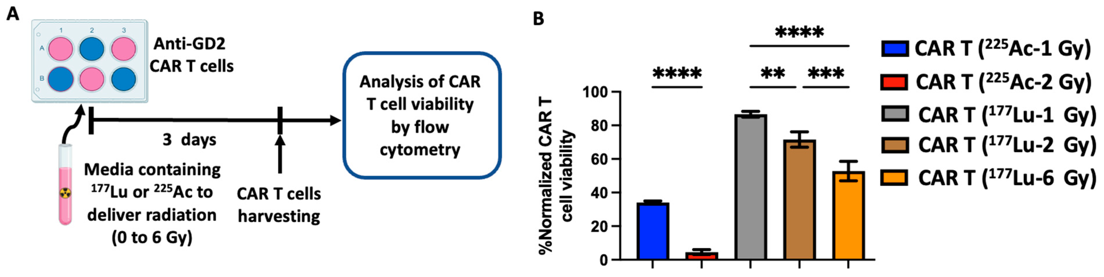
3.1. Radiation Delivered by 177Lu and 225Ac Results in a Dose-Dependent Anti-GD2 CAR T Cell Death

Combining in vivo TRT and CAR T cell therapy will undeniably lead to the exposure of CAR T cells to radiation, which could be detrimental to their viability. As such, to evaluate the effect of radiation delivered by TRT on CAR T cells, third-generation anti-GD2 CAR T cells were exposed to various doses of radiation delivered by 177Lu or 225Ac. Six-well plates containing anti-GD2 CAR T cells were incubated in media containing the specific activity of 177Lu or 225Ac needed to deliver radiation doses of 0, 1, 2 or 6 Gy by day 3. Following the delivery of the radiation, the anti-GD2 CAR T cells were harvested, and their viability was evaluated by flow cytometry (Figure 2A) using live/dead staining of the CD45+ cells (Figure 2B). A dose-dependent anti-GD2 CAR T cell death was observed with both radionuclides. However, 1 and 2 Gy of 177Lu appeared to be less cytotoxic than comparable doses of 225Ac. Compared to the non-irradiated anti-GD2 CAR T cells, 1 and 2 Gy of radiation delivered by 225Ac reduced the viability of anti-GD2 CAR T cells to 34% and 5%, respectively (Figure 2B). In contrast, after the delivery of 1, 2 and 6 Gy of radiation by 177Lu, the viability of anti-GD2 CAR T cells decreased to 87%, 72% and 53%, respectively, compared to non-irradiated CAR T cells (Figure 2B).

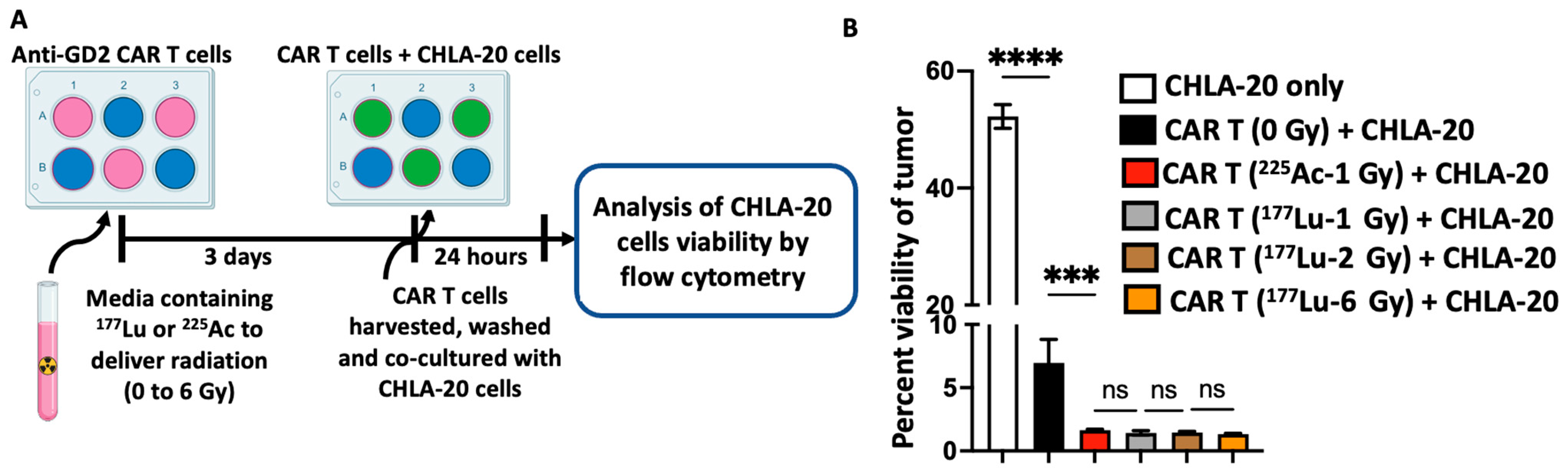
3.2. Radiation Delivered by 177Lu and 225Ac Enhances the Cytotoxic Activity of Anti-GD2 CAR T Cells against the GD2-Expressing Neuroblastoma Cell Line CHLA-20

In addition to evaluating the effect of radionuclide-delivered radiation on the viability of the anti-GD2 CAR T cells, we were also interested in determining the impact of such radiation on the effector function of the anti-GD2 CAR T cells without exposing the tumor cells to radiation. To this end, following irradiation, the anti-GD2 CAR T cells were washed and then co-cultured with the GD2-expressing neuroblastoma cell line CHLA-20 for 24 h. The killing potential of the irradiated anti-GD2 CAR T cells was determined by measuring the viability of CHLA-20 cells by flow cytometry (Figure 3A). Viable CHLA-20 cells were identified as CD45 negative and live/dead stain (Ghost red) negative (CD45-/Ghost red). As expected, non-irradiated anti-GD2 CAR T cells displayed a potent cytotoxicity activity against CHLA-20 cells. The viability of the CHLA-20 cells decreased to 7.0% after 24 h co-culture with non-irradiated anti-GD2 CAR T cells compared to 52% in the absence of anti-GD2 CAR T cells (Figure 3B). The irradiation of anti-GD2 CAR T cells prior to co-culture with CHLA-20 cells significantly enhanced their cytotoxic activity of the CAR T cells, resulting in a near complete eradication of CHLA-20 cells, as evidenced by tumor viabilities ranging from 1.3 to 1.6% (Figure 3B). For the type and doses of radionuclides used in this study, the enhanced cytotoxicity of the anti-GD2 CAR T cell was similar for all conditions tested (Figure 3B). The radiation-induced enhancement of anti-GD2 CAR T cell cytotoxic activity was also observed when these same CAR T cells were tested against the human melanoma cell line M21, which also expresses GD2 (Supplementary Figure S1).

3.3. Radiation Delivered by 177Lu and 225Ac Does Not Impact the Expression of Exhaustion and Activation Markers on Anti-GD2 CAR T Cells

To further characterize the effect of 225Ac or 177Lu-delivered radiation on anti-GD2 CAR T cells and understand the underlying mechanism of the radiation-induced cytotoxic enhancement of anti-GD2 CAR T cells, the expression of the T cell exhaustion marker PD-1, activation marker CD69, and activating receptor NKG2D were evaluated by flow cytometry. As previously described, following the irradiation of the anti-GD2 CAR T cells with the various radiation doses of 225Ac or 177Lu, the anti-GD2 CAR T cells were harvested, and the expression of cell surface markers was determined by flow cytometry (Figure 4A). Compared to non-irradiated anti-GD2 CAR T cells, 1 Gy delivered by 225Ac resulted in a slight increase in expression of PD-1 that was not significant. In contrast, radiation (1, 2, 6 Gy) delivered by 177Lu resulted in the decrease in PD-1 expression, which was also not significant (Figure 4B). CD69 and NKG2D expressions were increased following the delivery of radiation by both radionuclides, albeit this was non-significant (Figure 4C,D).

4. Discussion

Due to the increasing therapeutic role of TRT in the clinical management of cancers and the immunostimulatory effects at low-dose TRT, the combination of TRT with CAR T cell therapy may represent a therapeutic approach for selected patients with metastatic solid tumors [17,20]. As such, we investigated the in vitro effects of TRT on CAR T cells’ viability and potency to evaluate the influence of different doses of 225Ac and 177Lu (α vs. β-particle emitters). We tested whether selected doses of radiation delivered by these radionuclides can enhance the effector function of CAR T cells while minimizing the deleterious effects on CAR T cell viability.
The dose-dependent death of anti-GD2 CAR T cells observed in response to radiation delivered by 177Lu and 225Ac is expected because radiation is cytotoxic to lymphocytes, especially CD8+ T cells from which anti-GD2 CAR T cells are manufactured [23]. Although such dose-dependent death was observed with radiation delivered by either radionuclide, the biological effect of a similar dose of radiation (1 Gy) delivered by the α-emitter 225Ac was higher than that of the same dose delivered by the β-emitter 177Lu. This difference in biological effect is likely attributable to the linear energy transfer (LET) difference between these two radionuclides, which has been previously reported [24]. Despite having a shorter range in tissue (0.05–0.08 mm) compared to β-emitters (1–5 mm), α-emitters, due to their higher LET (50–230 keV/μm), induce 10–20 double-strand DNA breaks (DSBs) per 10 μm, resulting in potent cytotoxicity, whereas β-emitters induce more single-strand DNA breaks, which are easily repaired [25,26,27]. α-particle emitters induce multiple DSBs that are in close vicinity, resulting in the depletion of p53-binding protein 1 (53BP1), which is a DNA damage response protein required for double-strand DNA breaks repair. Such depletion leads to an insufficient amount of 53BP1 to adequately repair all DSBs, further accentuating the biological effectiveness of high LET α-particle emitters [28].
Using a prostate cancer in vitro model, the in vitro therapeutic efficacy of 225Ac-PSMA was found to be 4.2 times that of 177Lu-PSMA, and such a difference was solely attributed to the higher relative biological effectiveness (RBE) of 225Ac compared to 177Lu [29]. However, in this study, the in vitro CAR T cell death induced by 1 Gy of 225Ac is only 2.2 times that of 177Lu (Figure 2). This discrepancy could be attributed to the different in vitro models used in this study, the method used to access the therapeutic efficacy and/or to the fact that our in vitro dosimetry enabled the comparison of a similar dose of radiation (Gy) which mediates the biological effects delivered by a radionuclide rather than comparing activity. It is also possible that the mechanism of radionuclide-induced cell death of T cells is different from that of tumor cells.
Although radiation delivered by external beam radiation therapy (EBRT) upregulates the expression of PD-1 on CD8+ T cells, which can be detrimental to CAR T cell function, surprisingly, we did not observe such upregulation with 177Lu or 225Ac in the conditions tested [30]. This may be due to the inherent design of this third-generation anti-GD2 CAR with the CRISPR knock-in of the CAR construct into the TRAC locus, yielding CAR T cells devoid of T cell receptors, and it may represent another therapeutic benefit of combining CAR T cell therapy with TRT instead of EBRT [19].
The lack of significant impact of TRT on the expression of the activating receptor NKG2D appears to be in contradiction to the findings by others who demonstrated that low-dose radiation (1 and 3 Gy) delivered by EBRT induces an upregulation of the NKG2D receptors on T and NK cells [30]. Such a discrepancy could be attributed to the difference in the form of radiation delivered (EBRT vs. TRT) and/or in the method used to detect the NKG2D receptor protein expression. EBRT was previously shown to upregulate NKG2D expression by quantifying mRNA expression, whereas in this study, NKG2D receptor expression was quantified at the protein level by flow cytometry [31]. While the increase in mRNA expression may be used to suggest an upregulation of a gene of interest, this may not necessarily translate into a change in protein expression in the proper cellular compartment such as lysosome, mitochondrial or cell surface [32,33].
Radiation delivered by EBRT enhances the cytotoxic activity of CAR T cells including against antigen-negative malignant cells [34]. However, prior to this study, such enhancement has not been demonstrated with TRT. While 177Lu or 225Ac did not significantly decrease the expression of PD-1 or increase the expression of the NKG2D receptor or activation marker CD69, it is possible that the presence of the TRT in the milieu may have selected for more radioresistant CAR T cells subclones with potent cytolytic activity. It is also plausible that the low-dose radiation delivered by 177Lu or 225Ac stimulated ATP production (mitochondrial excitation), which can boost the anti-oxidative capacity of CAR T cells and enhance their radiosensitivity and survival [35].
Based on our in vitro studies, the low dose of radiation tested herein (1 or 2 Gy) delivered by α-emitters might not be ideal for a potential therapeutic combination with CAR T cells. Instead, β-emitters may be the preferred candidates, given that the 1 Gy delivered by a β-particle emitter enhanced CAR T cell cytotoxic function to the same extent as all other radiation doses evaluated (2 or 6 Gy) while inducing lower rates of anti-GD2 CAR T cell death compared to those higher doses. These observations suggest that with regard to the sequencing between TRT and CAR T cells combination, the infusion of CAR T cells may optimally be performed after the administration of TRT to minimize the exposure of CAR T cells to radiation while enabling the activation of inflammatory effects in the TME and phenotypic effects on tumor cells that maximize the sensitivity to CAR T cells killing, which are activated optimally at higher doses. While our in vitro study suggests that 1 Gy or lower delivered by the β-emitter 177Lu might be well tolerated by CAR T cells, the higher tested doses of 2 or 6 Gy may be considered if there is an adequate delay between the TRT delivery and CAR T cell infusion. This delay should enable the radioactive decay of the radionuclide such that when the CAR T cells are infused, the remaining activity of the radionuclide in the TME may only deliver a radiation dose of 1 Gy or less. In vivo testing is sorely needed. We speculate that using a radiation dose higher than 1 Gy may be an attractive option, as it will enable the tumoricidal effect of TRT on malignant cells to be harnessed, subsequently reducing the tumor volume against which CAR T cells have to mount an effective therapeutic response.
Aside from the significantly higher cytotoxicity of the α-emitter 225Ac on the CAR T cell compared to the β-emitter 177Lu, additional factors support the preference of 177Lu over 225Ac. To achieve the adequate delivery of the radionuclide in vivo, the incorporation of a radiometal chelating moiety in the pharmacophoric model of the TRT is needed. To date, 1,4,7,10-tetraazacyclododecane-1,4,7,10-tetraacetic acid (DOTA) is the commonly used chelator [36]. However, the stable retention of 225Ac and its daughters by this chelator has been challenging, resulting in it a non-specific release of the radionuclide from the TRT targeting vector and thus toxicities [37]. Furthermore, because of the relatively longer range in tissue of 177Lu compared to 225Ac, 177Lu may be advantageous in irradiating uniformly heterogeneous tumors in vivo [24].
Various limitations can be highlighted in this study, including the in vitro character of the study, the evaluation of only a select number of exhaustion or activation markers, the relatively limited numbers of radionuclide and doses evaluated. Nevertheless, this study provides critical insights on key questions regarding the optimal doses of radiation, type of radionuclide, and timing and sequence of TRT and CAR T cell combinations to enable the rational design of in vivo studies evaluating the combination of CAR T cell therapy and TRT in solid tumor models.

5. Conclusions

While the immunostimulatory effects of both α and β-emitter radionuclides have been reported and shown to potentiate immune checkpoint inhibitors, they also have the potential to be useful in overcoming some of the shortcomings of CAR T cell therapy against solid tumors. However, proper selection of the radionuclide is critical for in vivo studies to efficiently harness the immunostimulatory effect while minimizing the deleterious effects on CAR T cell viability and potency. Such selection should be guided by the properties of the radionuclides including LET, half-life, and range in tissue and should lead to establishing an optimal sequencing and timing between the delivery of TRT and CAR T cells. Lastly, because TRT selectively delivers radiation to all sites of disease, it may enable the immunostimulatory effect of radiation in combination with CAR T cells to be exploited in patients with widely metastatic solid tumors in whom treatment-related toxicities render EBRT to all sites of disease infeasible.

Supplementary Materials

The following supporting information can be downloaded at https://www.mdpi.com/article/10.3390/cancers16010191/s1, Figure S1: 225Ac and 177Lu enhance the cytotoxicity of anti-GD2 CAR T cells against the GD-2 expressing melanoma cell line M21.

Author Contributions

Conceptualization, Q.H.S., C.M.C., Z.S.M.; methodology, Q.H.S., C.M.C., Z.S.M.; software, Q.H.S.; validation, Q.H.S., M.H.F., D.C., C.P.K., L.S., A.S., D.P.A.; formal analysis, Q.H.S., M.H.F., J.C.E., I.M.O., C.M.C., Z.S.M.; investigation, Q.H.S., M.H.F., D.C., L.S., A.S., D.P.A.; resources, Q.H.S., R.H., J.W., B.P.B., K.S., P.M.S., C.M.C., Z.S.M.; data curation, Q.H.S., C.M.C., Z.S.M.; writing—original draft preparation, Q.H.S.; writing—review and editing, All authors; visualization, Q.H.S., M.H.F., C.M.C., Z.S.M.; supervision, Q.H.S., C.M.C., Z.S.M.; project administration, Q.H.S.; funding acquisition, C.M.C., Z.S.M. All authors have read and agreed to the published version of the manuscript.

Funding

We acknowledge generous support from the National Cancer Institute (NCI)/National Institute of Health (NIH) P01 CA250972 (R.H., J.W., J.C.E., I.M.O., B.P.B., P.M.S., Z.S.M.), University of Wisconsin (UW)-Madison Office of the Vice Chancellor for Research and Graduate Education with funding from the Wisconsin Alumni Research Foundation (R.H., J.W., K.S., P.M.S., C.M.C., Z.S.M.), St. Baldrick’s Empowering Pediatric Immunotherapy for Childhood Cancers Team Grant (K.S., P.M.S., C.M.C., Z.S.M.), the Midwest Athletes Against Childhood Cancer (MACC) Fund (P.M.S. and C.M.C.), the Grainger Institute for Engineering (K.S. and C.M.C.), and Hyundai Hope on Wheels (K.S. and C.M.C.). UW-Madison Radiology MD-PhD Graduate Student Fellowship and NIH awards F30CA26878 and T32GM140935 (C.P.K.). CMaT-Wisconsin Institute for Discovery (D.C.). The authors also thank the UWCCC Flow Cytometry core facility and Small Animal Imaging and Radiotherapy core facility, who are supported in part through NCI/NIH P30 CA014520. The project was supported by the Clinical and Translational Science Award (CTSA) program through the NIH National Center for Advancing Translational Sciences (NCATS), grant UL1TR002373. We thank the staff of the University of Wisconsin Carbone Cancer Center (UWCCC) Biostatistics Shared Resource for their valuable contributions to this research. Shared research services at the UWCCC are supported by Cancer Center Support Grant P30 CA014520. The contents of this article do not necessarily reflect the views or policies of the Department of Health and Human Services, nor does the mention of trade names, commercial products, or organizations imply endorsement by the US Government.

Institutional Review Board Statement

Peripheral blood from healthy donor was obtain in accordance with the Declaration of Helsinki and using a protocol approved by an Institutional Review Board from the University of Wisconsin-Madison (#2018-0103).

Informed Consent Statement

Informed consent was obtained from all human blood donors.

Data Availability Statement

The data presented in this study are available upon request to the corresponding author.

Acknowledgments

Tracy Berg for editorial review of this manuscript.

Conflicts of Interest

Q.H.S. is an inventor on patent applications related to this publication. MHF is an inventor on patent applications related to this publication. D.C.: None; C.P.K.: None; L.S.: None; A.S.: None; D.A.: None; J.C.E.: None; I.M.O.: None; R.H. received patent royalties from the Wisconsin Alumni Research Foundation; consulting fees from Archeus Technologies Inc. and Monopar Therapeutics. J.W.: is the cofounder of Archeus Technologies, which owns the licensing rights to NM600 and related technologies and holds stock in Archeus; B.P.B. received stock/stock options from Voximetry Inc.; K.S. received grant supports from Synthego and Spotlight Therapeutics; patent royalties from Wisconsin Alumni Research Foundation; honoraria from ISCT, is an inventor on patent applications related to this publication; Scientific Advisory Board Member for Notch Therapeutics and Andson Biotech. P.M.S. received support from the University of Wisconsin, Midwest Athletes For Childhood Cancer and the National Cancer Institute. C.M.C. received honoraria from Bayer, Nektar Therapeutics, Novartis, WiCell Research Institute, consulting fees from Elephas and is an inventor on patent applications related to this publication. Z.S.M. is a member of the Scientific Advisory Boards for Archeus Technologies, Seneca Therapeutics, and NorthStar Medical Isotopes; received royalties from patents held by the Wisconsin Alumni Research Foundation; received stock/stock options from Archeus Technologies Scientific Advisory board and Seneca Therapeutics Scientific Advisory Board; received research support from Point Biopharma, Telix Pharmaceuticals and XRD Therapeutics and is an inventor on patent applications related to this publication.

Abbreviations

CAR: Chimeric Antigen Receptor; CRISPR: Clustered Regularly Interspaced Short Palindromic Repeats; TME: Tumor Microenvironment; TRT: Targeted Radionuclide Therapy.

References

  1. June, C.H.; O’Connor, R.S.; Kawalekar, O.U.; Ghassemi, S.; Milone, M.C. CAR T cell immunotherapy for human cancer. Science 2018, 359, 1361–1365. [Google Scholar] [CrossRef] [PubMed]
  2. Feins, S.; Kong, W.; Williams, E.F.; Milone, M.C.; Fraietta, J.A. An introduction to chimeric antigen receptor (CAR) T-cell immunotherapy for human cancer. Am. J. Hematol. 2019, 94, S3–S9. [Google Scholar] [CrossRef] [PubMed]
  3. Neelapu, S.S.; Locke, F.L.; Bartlett, N.L.; Lekakis, L.J.; Miklos, D.B.; Jacobson, C.A.; Braunschweig, I.; Oluwole, O.O.; Siddiqi, T.; Lin, Y.; et al. Axicabtagene Ciloleucel CAR T-Cell Therapy in Refractory Large B-Cell Lymphoma. N. Engl. J. Med. 2017, 377, 2531–2544. [Google Scholar] [CrossRef] [PubMed]
  4. Abramson, J.S.; Palomba, M.L.; Gordon, L.I.; Lunning, M.A.; Wang, M.; Arnason, J.; Mehta, A.; Purev, E.; Maloney, D.G.; Andreadis, C.; et al. Lisocabtagene maraleucel for patients with relapsed or refractory large B-cell lymphomas (TRANSCEND NHL 001): A multicentre seamless design study. Lancet 2020, 396, 839–852. [Google Scholar] [CrossRef] [PubMed]
  5. Schuster, S.J.; Bishop, M.R.; Tam, C.S.; Waller, E.K.; Borchmann, P.; McGuirk, J.P.; Jäger, U.; Jaglowski, S.; Andreadis, C.; Westin, J.R.; et al. Tisagenlecleucel in Adult Relapsed or Refractory Diffuse Large B-Cell Lymphoma. N. Engl. J. Med. 2019, 380, 45–56. [Google Scholar] [CrossRef] [PubMed]
  6. Shah, B.D.; Ghobadi, A.; Oluwole, O.O.; Logan, A.C.; Boissel, N.; Cassaday, R.D.; Leguay, T.; Bishop, M.R.; Topp, M.S.; Tzachanis, D.; et al. KTE-X19 for relapsed or refractory adult B-cell acute lymphoblastic leukaemia: Phase 2 results of the single-arm, open-label, multicentre ZUMA-3 study. Lancet 2021, 398, 491–502. [Google Scholar] [CrossRef] [PubMed]
  7. Park, J.H.; Rivière, I.; Gonen, M.; Wang, X.; Sénéchal, B.; Curran, K.J.; Sauter, C.; Wang, Y.; Santomasso, B.; Mead, E.; et al. Long-Term Follow-up of CD19 CAR Therapy in Acute Lymphoblastic Leukemia. N. Engl. J. Med. 2018, 378, 449–459. [Google Scholar] [CrossRef] [PubMed]
  8. Roddie, C.; Dias, J.; O’Reilly, M.A.; Abbasian, M.; Cadinanos-Garai, A.; Vispute, K.; Bosshard-Carter, L.; Mitsikakou, M.; Mehra, V.; Roddy, H.; et al. Durable Responses and Low Toxicity after Fast Off-Rate CD19 Chimeric Antigen Receptor-T Therapy in Adults with Relapsed or Refractory B-Cell Acute Lymphoblastic Leukemia. J. Clin. Oncol. 2021, 39, 3352–3363. [Google Scholar] [CrossRef]
  9. Martin, T.; Usmani, S.Z.; Berdeja, J.G.; Agha, M.; Cohen, A.D.; Hari, P.; Avigan, D.; Deol, A.; Htut, M.; Lesokhin, A.; et al. Ciltacabtagene Autoleucel, an Anti–B-cell Maturation Antigen Chimeric Antigen Receptor T-Cell Therapy, for Relapsed/Refractory Multiple Myeloma: CARTITUDE-1 2-Year Follow-Up. J. Clin. Oncol. 2023, 41, 1265–1274. [Google Scholar] [CrossRef]
  10. Munshi, N.C.; Anderson, L.D., Jr.; Shah, N.; Madduri, D.; Berdeja, J.; Lonial, S.; Raje, N.; Lin, Y.; Siegel, D.; Oriol, A.; et al. Idecabtagene Vicleucel in Relapsed and Refractory Multiple Myeloma. N. Engl. J. Med. 2021, 384, 705–716. [Google Scholar] [CrossRef]
  11. Marofi, F.; Motavalli, R.; Safonov, V.A.; Thangavelu, L.; Yumashev, A.V.; Alexander, M.; Shomali, N.; Chartrand, M.S.; Pathak, Y.; Jarahian, M.; et al. CAR T cells in solid tumors: Challenges and opportunities. Stem Cell Res. Ther. 2021, 12, 81. [Google Scholar] [CrossRef] [PubMed]
  12. Artigas, C.; Mileva, M.; Flamen, P.; Karfis, I. Targeted radionuclide therapy: An emerging field in solid tumours. Curr. Opin. Oncol. 2021, 33, 493–499. [Google Scholar] [CrossRef] [PubMed]
  13. Gudkov, S.V.; Shilyagina, N.Y.; Vodeneev, V.A.; Zvyagin, A.V. Targeted Radionuclide Therapy of Human Tumors. Int. J. Mol. Sci. 2016, 17, 33. [Google Scholar] [CrossRef] [PubMed]
  14. Pomykala, K.L.; Hadaschik, B.A.; Sartor, O.; Gillessen, S.; Sweeney, C.J.; Maughan, T.; Hofman, M.S.; Herrmann, K. Next generation radiotheranostics promoting precision medicine. Ann. Oncol. 2023, 34, 507–519. [Google Scholar] [CrossRef] [PubMed]
  15. Strosberg, J.; El-Haddad, G.; Wolin, E.; Hendifar, A.; Yao, J.; Chasen, B.; Mittra, E.; Kunz, P.L.; Kulke, M.H.; Jacene, H.; et al. Phase 3 Trial of 177Lu-Dotatate for Midgut Neuroendocrine Tumors. N. Engl. J. Med. 2017, 376, 125–135. [Google Scholar] [CrossRef] [PubMed]
  16. Sartor, O.; de Bono, J.; Chi, K.N.; Fizazi, K.; Herrmann, K.; Rahbar, K.; Tagawa, S.T.; Nordquist, L.T.; Vaishampayan, N.; El-Haddad, G.; et al. Lutetium-177-PSMA-617 for Metastatic Castration-Resistant Prostate Cancer. N. Engl. J. Med. 2021, 385, 1091–1103. [Google Scholar] [CrossRef]
  17. Patel, R.B.; Hernandez, R.; Carlson, P.; Grudzinski, J.; Bates, A.M.; Jagodinsky, J.C.; Erbe, A.; Marsh, I.R.; Arthur, I.; Aluicio-Sarduy, E.; et al. Low-dose targeted radionuclide therapy renders immunologically cold tumors responsive to immune checkpoint blockade. Sci. Transl. Med. 2021, 13, eabb3631. [Google Scholar] [CrossRef]
  18. Hernandez, R.; Walker, K.L.; Grudzinski, J.J.; Aluicio-Sarduy, E.; Patel, R.; Zahm, C.D.; Pinchuk, A.N.; Massey, C.F.; Bitton, A.N.; Brown, R.J.; et al. 90Y-NM600 targeted radionuclide therapy induces immunologic memory in syngeneic models of T-cell Non-Hodgkin’s Lymphoma. Commun. Biol. 2019, 2, 79. [Google Scholar] [CrossRef]
  19. Mueller, K.P.; Piscopo, N.J.; Forsberg, M.H.; Saraspe, L.A.; Das, A.; Russell, B.; Smerchansky, M.; Cappabianca, D.; Shi, L.; Shankar, K.; et al. Production and characterization of virus-free, CRISPR-CAR T cells capable of inducing solid tumor regression. J. Immunother. Cancer 2022, 10, e004446. [Google Scholar] [CrossRef]
  20. Jagodinsky, J.C.; Jin, W.J.; Bates, A.M.; Hernandez, R.; Grudzinski, J.J.; Marsh, I.R.; Chakravarty, I.; Arthur, I.S.; Zangl, L.M.; Brown, R.J.; et al. Temporal analysis of type 1 interferon activation in tumor cells following external beam radiotherapy or targeted radionuclide therapy. Theranostics 2021, 11, 6120–6137. [Google Scholar] [CrossRef]
  21. Besemer, A.E.; Yang, Y.M.; Grudzinski, J.J.; Hall, L.T.; Bednarz, B.P. Development and Validation of RAPID: A Patient-Specific Monte Carlo Three-Dimensional Internal Dosimetry Platform. Cancer Biother. Radiopharm. 2018, 33, 155–165. [Google Scholar] [CrossRef] [PubMed]
  22. Sodji, Q.H.; Nambiar, D.K.; Viswanathan, V.; von Eyben, R.; Colburg, D.; Binkley, M.S.; Li, C.G.; Olcina, M.M.; Chang, D.T.; Le, Q.T.; et al. The Combination of Radiotherapy and Complement C3a Inhibition Potentiates Natural Killer cell Functions against Pancreatic Cancer. Cancer Res. Commun. 2022, 2, 725–738. [Google Scholar] [CrossRef] [PubMed]
  23. Heylmann, D.; Rödel, F.; Kindler, T.; Kaina, B. Radiation sensitivity of human and murine peripheral blood lymphocytes, stem and progenitor cells. Biochim. Biophys. Acta 2014, 1846, 121–129. [Google Scholar] [CrossRef] [PubMed]
  24. Sgouros, G.; Bodei, L.; McDevitt, M.R.; Nedrow, J.R. Radiopharmaceutical therapy in cancer: Clinical advances and challenges. Nat. Rev. Drug Discov. 2020, 19, 589–608. [Google Scholar] [CrossRef] [PubMed]
  25. Aten, J.A.; Stap, J.; Krawczyk, P.M.; van Oven, C.H.; Hoebe, R.A.; Essers, J.; Kanaar, R. Dynamics of DNA double-strand breaks revealed by clustering of damaged chromosome domains. Science 2004, 303, 92–95. [Google Scholar] [CrossRef] [PubMed]
  26. Kassis, A.I. Therapeutic Radionuclides: Biophysical and Radiobiologic Principles. Semin. Nucl. Med. 2008, 38, 358–366. [Google Scholar] [CrossRef]
  27. Aghevlian, S.; Boyle, A.J.; Reilly, R.M. Radioimmunotherapy of cancer with high linear energy transfer (LET) radiation delivered by radionuclides emitting α-particles or Auger electrons. Adv. Drug Deliv. Rev. 2017, 109, 102–118. [Google Scholar] [CrossRef]
  28. Roobol, S.J.; van den Bent, I.; van Cappellen, W.A.; Abraham, T.E.; Paul, M.W.; Kanaar, R.; Houtsmuller, A.B.; van Gent, D.C.; Essers, J. Comparison of High- and Low-LET Radiation-Induced DNA Double-Strand Break Processing in Living Cells. Int. J. Mol. Sci. 2020, 21, 6602. [Google Scholar] [CrossRef]
  29. Ruigrok, E.A.M.; Tamborino, G.; de Blois, E.; Roobol, S.J.; Verkaik, N.; De Saint-Hubert, M.; Konijnenberg, M.W.; van Weerden, W.M.; de Jong, M.; Nonnekens, J. In vitro dose effect relationships of actinium-225- and lutetium-177-labeled PSMA-I&T. Eur. J. Nucl. Med. Mol. Imaging 2022, 49, 3627–3638. [Google Scholar]
  30. Rodriguez-Ruiz, M.E.; Rodriguez, I.; Garasa, S.; Barbes, B.; Solorzano, J.L.; Perez-Gracia, J.L.; Labiano, S.; Sanmamed, M.F.; Azpilikueta, A.; Bolaños, E.; et al. Abscopal Effects of Radiotherapy Are Enhanced by Combined Immunostimulatory mAbs and Are Dependent on CD8 T Cells and Crosspriming. Cancer Res. 2016, 76, 5994–6005. [Google Scholar] [CrossRef]
  31. Öztürk, N.; Karlıtepe, A.; Depboylu, B.; Kılıç Eren, M. Immunomodulatory effects of ionizing radiation on peripheral blood mononuclear cells. Int. J. Radiat. Res. 2023, 21, 73–78. [Google Scholar]
  32. Wang, D. Discrepancy between mRNA and protein abundance: Insight from information retrieval process in computers. Comput. Biol. Chem. 2008, 32, 462–468. [Google Scholar] [CrossRef] [PubMed]
  33. Greenbaum, D.; Colangelo, C.; Williams, K.; Gerstein, M. Comparing protein abundance and mRNA expression levels on a genomic scale. Genome Biol. 2003, 4, 117. [Google Scholar] [CrossRef] [PubMed]
  34. DeSelm, C.; Palomba, M.L.; Yahalom, J.; Hamieh, M.; Eyquem, J.; Rajasekhar, V.K.; Sadelain, M. Low-Dose Radiation Conditioning Enables CAR T Cells to Mitigate Antigen Escape. Mol. Ther. 2018, 26, 2542–2552. [Google Scholar] [CrossRef]
  35. Hietanen, T.; Pitkanen, M.; Kapanen, M.; Kellokumpu-Lehtinen, P.L. Effects of Single and Fractionated Irradiation on Natural Killer Cell Populations: Radiobiological Characteristics of Viability and Cytotoxicity In Vitro. Anticancer Res. 2015, 35, 5193–5200. [Google Scholar]
  36. Yang, H.; Wilson, J.J.; Orvig, C.; Li, Y.; Wilbur, D.S.; Ramogida, C.F.; Radchenko, V.; Schaffer, P. Harnessing α-Emitting Radionuclides for Therapy: Radiolabeling Method Review. J. Nucl. Med. 2022, 63, 5–13. [Google Scholar] [CrossRef]
  37. Thiele, N.A.; Wilson, J.J. Actinium-225 for Targeted α Therapy: Coordination Chemistry and Current Chelation Approaches. Cancer Biother. Radiopharm. 2018, 33, 336–348. [Google Scholar] [CrossRef]
Figure 1. Third-generation virus-free CRISPR anti-GD2 CAR T cells. (A) Schematic of the domains of the third generation CAR. The extracellular domain includes the scFV (VH and VL chains connected by a linker) and a hinge. The intracellular domain is comprised of 2 costimulatory domains (CD28 and OX40) and a signaling domain (CD3-Zeta). These 2 domains are connected by a transmembrane domain (CD28 transmembrane domain). (B) Schematic of the third generation anti-GD2 CAR construct inserted into the human T cell receptor alpha constant gene (TRAC). (C) Schematic of the anti-GD2 CAR T cell used experimentally. It expresses the anti-GD2 CAR but is devoid of T cell receptor due to the CRISPR knockout of the human TRAC gene. scFv: single-chain variable fragment; VH: heavy chain; VL: light chain.
Cancers 16 00191 g001
Figure 2. Dose-dependent effect of 225Ac and 177Lu on the viability of anti-GD2 CAR T cells. (A) Experimental scheme: anti-GD2 CAR T cells were incubated in cell culture medium containing free 225Ac or 177Lu with activities calculated to deliver a radiation dose between 1 and 6 Gy by day 3. The CAR T cells were harvested, and their viability was analyzed by flow cytometry using a live/dead staining. Viable CAR T cells were determined as CD45+ and live/Dead-Ghost red-. (B) Both 225Ac and 177Lu led to a dose-dependent CAR T cell death. One-way ANOVA with Tukey’s multiple comparisons correction ** p = 0.0024; *** p = 0.0001; **** p < 0.0001. Each bar represents the mean (triplicate measurements) and error bar represents the standard deviation.
Cancers 16 00191 g002
Figure 3. Dose-independent effect of 225Ac and 177Lu on anti-GD2 CAR T cell cytotoxicity. (A) Experimental scheme: After irradiation of CAR T cells by 225Ac or 177Lu, the CAR T cells were harvested, washed and trypan blue viability assay was performed. The viable CAR T cells after irradiation were co-cultured with the GD2-expressing human neuroblastoma cell line CHLA-20 for 24 h at an E:T ratio of 10:1 (viable anti-GD2 CAR T cells: tumor cells). (B) The viability of the CHLA-20 cells was analyzed by flow cytometry. The exposure of anti-GD2 CAR T cells to radiation delivered by radionuclide enhances their cytotoxicity against CHLA-20 cells to a similar degree for all doses tested for 225Ac or 177Lu. One-way ANOVA with Tukey’s multiple comparisons correction *** p < 0.001; **** p < 0.0001; ns: not significant. Each bar represents the mean (triplicate measurements), and the error bar represents the standard deviation. The cytotoxic activity of CAR T cells irradiated with 2 Gy of radiation delivered by 225Ac was not performed because of very low CAR T cell viability after irradiation.
Cancers 16 00191 g003
Figure 4. 225Ac or 177Lu does not impact the expression of exhaustion and activation markers on anti-GD2 CAR T cells. (A) Experimental scheme: anti-GD2 CAR T cells were incubated in cell culture medium containing 225Ac or 177Lu with activities calculated to deliver a radiation dose between 1 and 6 Gy by day 3. CAR T cells were harvested, and the expression of exhaustion and activation markers was analyzed by flow cytometry. (B) The expression of the exhaustion marker PD-1 does not change following irradiation by 225Ac or 177Lu. (C) Similarly, no major impact is seen on the expression of the T cell activation marker CD69. (D) Irradiation does not result in a major impact on the activation marker NKG2D. One-way ANOVA with Tukey’s multiple comparisons correction; ns: not significant. Each bar represents the mean (triplicate measurements), and the error bar represents the standard deviation. The effects of 2 Gy irradiation delivered by 225Ac on the receptors’ expression was not evaluated because of very low CAR T cell viability after irradiation. ns: not significant.
Cancers 16 00191 g004
Disclaimer/Publisher’s Note: The statements, opinions and data contained in all publications are solely those of the individual author(s) and contributor(s) and not of MDPI and/or the editor(s). MDPI and/or the editor(s) disclaim responsibility for any injury to people or property resulting from any ideas, methods, instructions or products referred to in the content.

