Article

Novel Pathogenic Variants in Hereditary Cancer Syndromes in a Highly Heterogeneous Cohort of Patients: Insights from Multigene Analysis

1
Institute of Fundamental Medicine and Biology, Kazan Federal University, 420008 Kazan, Russia
2
SBHI Moscow Clinical Scientific Center Named after Loginov MHD, 111123 Moscow, Russia
3
The Federal State Budgetary Scientific Institution “Izmerov Research Institute of Occupational Health”, 105275 Moscow, Russia
4
Ministry of Health Kursk Region, 305000 Kursk, Russia
5
Kursk Regional Scientific and Clinical Center Named after G. Y. Ostroverkhov, 305524 Kursk, Russia
6
Centre for Strategic Planning and Management of Biomedical Health Risks, Federal Medical and Biological Agency, 119435 Moscow, Russia
7
Nizhny Novgorod Regional Oncologic Hospital, 603163 Nizhny Novgorod, Russia
8
Life Improvement by Future Technologies (LIFT) Center, 121205 Moscow, Russia
*
Author to whom correspondence should be addressed.
Cancers 2024, 16(1), 85; https://doi.org/10.3390/cancers16010085
Submission received: 4 December 2023 / Revised: 21 December 2023 / Accepted: 22 December 2023 / Published: 23 December 2023

Abstract

:

Simple Summary

This study addresses the global healthcare challenge of cancer by investigating hereditary cancer syndromes (HCS) and their genetic underpinnings. Using a multigene hereditary cancer panel, we examined Russian patients with suspected HCS, revealing that 21.6% had pathogenic or likely pathogenic genetic variants. Predominant mutations were found in BRCA1/BRCA2, CHEK2, and ATM genes, and we identified 16 previously undescribed variants in MUTYH, GALNT12, MSH2, MLH1, MLH3, EPCAM, and POLE genes. Our findings underscore the importance of comprehensive genetic testing for personalized cancer prevention and treatment. This research contributes essential genetic insights, particularly in regions like Russia where epidemiological data are limited, establishing the way for improved understanding and management of hereditary cancer syndromes.

Abstract

Cancer is a major global public health challenge, affecting both quality of life and mortality. Recent advances in genetic research have uncovered hereditary cancer syndromes (HCS) that predispose individuals to malignant neoplasms. While traditional single-gene testing has focused on high-penetrance genes, the past decade has seen a shift toward multigene panels, which facilitate the analysis of multiple genes associated with specific HCS. This approach reveals variants in less-studied gene regions and improves our understanding of cancer predisposition. In a study composed of Russian patients with clinical signs of HCS, we used a multigene hereditary cancer panel and revealed 21.6% individuals with pathogenic or likely pathogenic genetic variants. BRCA1/BRCA2 mutations predominated, followed by the CHEK2 and ATM variants. Of note, 16 previously undescribed variants were identified in the MUTYH, GALNT12, MSH2, MLH1, MLH3, EPCAM, and POLE genes. The implications of the study extend to personalized cancer prevention and treatment strategies, especially in populations lacking extensive epidemiological data, such as Russia. Overall, our research provides valuable genetic insights that give the way for further investigation and advances in the understanding and management of hereditary cancer syndromes.

1. Introduction

Cancer is a global unresolved healthcare challenge that is currently responsible for a significant decreased quality of life and mortality worldwide [1]. In recent years, significant progress has been made in understanding the genetic reasons for cancer development. One of the most important discoveries is the identification of hereditary cancer syndromes (HCS) characterized by an increased predisposition to the development of malignant neoplasms [2].
Traditionally, the identification of HCS relied on targeted single-gene testing, an approach primarily focused on well-established high-penetrance genes associated with specific cancer types. For example, when hereditary breast and ovarian cancer is suspected, testing for the BRCA1/BRCA2 genes is used effectively in clinical practice [3]. However, many hereditary cancer syndromes have similar clinical symptoms, which create difficulties in their differential diagnosis.
In the last decade, multigene panels have begun to be actively used in medical genetics for diagnosing diseases. This approach allows the simultaneous analysis of multiple genes whose pathogenic variants lead to the development of a specific hereditary cancer syndrome [4,5,6]. These panels can also reveal genetic variants in less-explored regions of genes, including noncoding intronic regions, potentially elucidating previously unrecognized contributors to cancer predisposition [7,8,9].
Furthermore, the application of hybrid targeted gene panels enables the simultaneous assessment of various characteristics, including the identification of germline variants. These panels provide the capability to determine the methylation status of oncogenes, detect gene fusions, assess tumor mutational burden (TMB), and evaluate microsatellite instability (MSI). The research focused on multigene panels is gaining traction and actively being implemented within the medical systems of several countries. This implementation allows for a comprehensive analysis of multiple genetic features in a more efficient and integrated manner [10,11,12,13]. Brian H. Shirts et al. and Holly LaDuca et al. have previously investigated the outcomes of employing multigene panels in cancer studies, establishing a foundation for exploring the genetic landscape of cancer via comprehensive gene panels [14,15]. In this study, we build upon their approach by extending our investigation to the relatively unexplored Russian population, with the goal of providing valuable insights to the field.
Understanding the genetic basis of HCS should inform the choice of strategies for clinical observation in the long-term. This has the potential to improve the prevention of cancer recurrence and secondary tumor development. Identification of genetic variants associated with HCS is particularly important in countries like Russia where there is a lack of population-based epidemiologic studies. Such studies not only expand our knowledge of the genetic epidemiology of HCS, but also provide a better genotype–phenotype correlation understanding of a certain hereditary cancer.
In our research, we performed genetic testing on Russian individuals with clinical evidence of HCS and/or a family history of cancer. The primary objective was to gain insights into the prevalence of various clinically significant genetic variants among cancer patients.

2. Materials and Methods
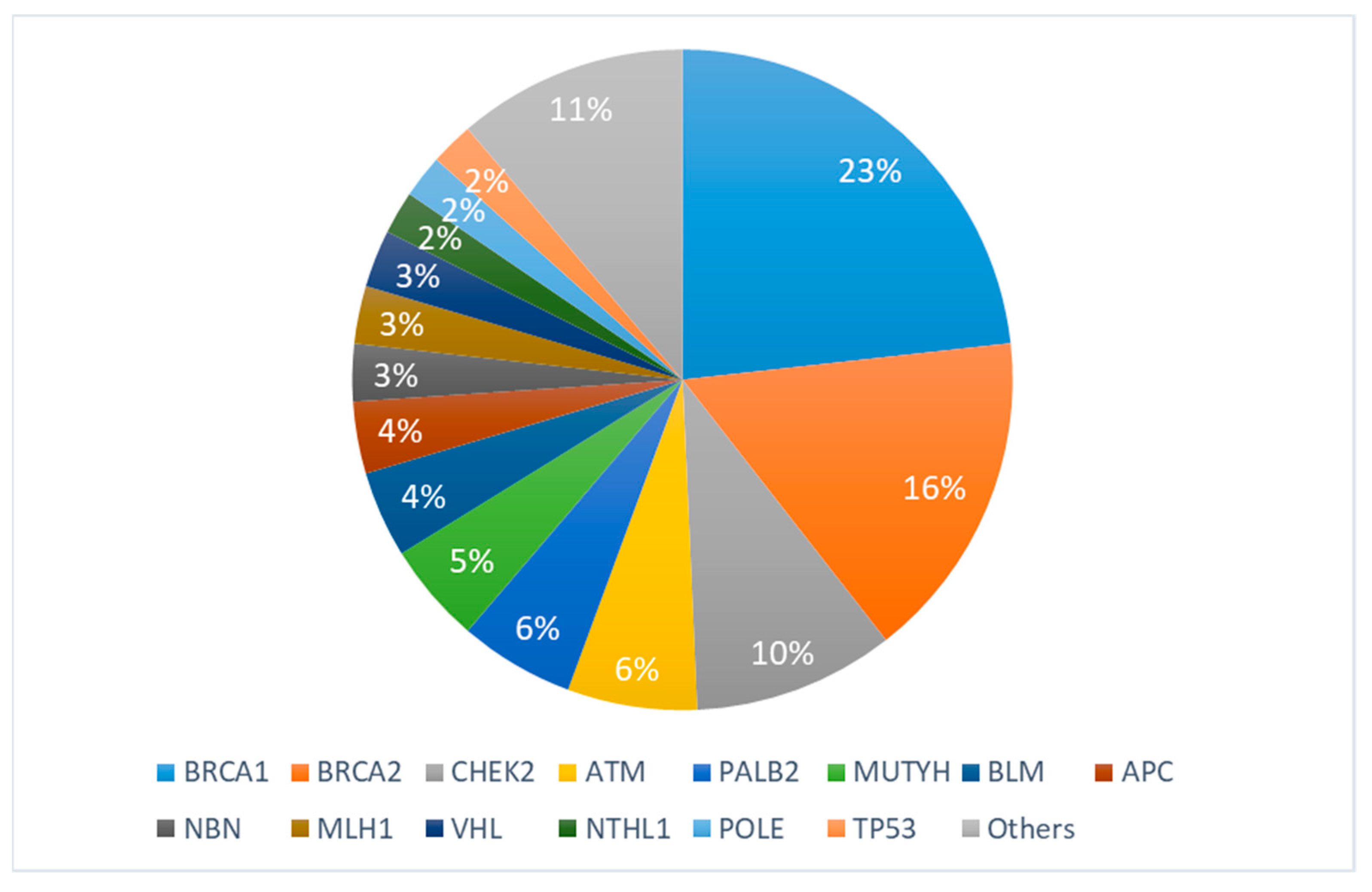
The study cohort included 657 patients from Russia divided into two groups: 632 (96.2%) cancer patients with clinical signs of cancer and 25 (3.8%) patients with benign tumors. These individuals were selected based on criteria established in a prior study and underwent consultations with geneticists [16]. Participation in the study involved molecular genetic testing, for which all participants provided detailed information regarding their personal and familial cancer histories. Additionally, they consented to the use of their anonymized data for the research and academic purposes. For the testing process, each participant contributed two blood samples, with each sample collected in an EDTA tube of 5 mL capacity.
To design the panel, we selected a total of 44 genes known to be involved in hereditary tumor syndromes. The full panel is presented in Table S1. The panel was designed using the HyperDesign online service provided by Roche (Roche, Basel, Switzerland)), which incorporates the coding regions, splicing sites, and 5’-UTR regions of the selected genes into the probe design.
For sample preparation, DNA was isolated from lymphocytes using the QIAamp DNA Blood Mini Kit from Qiagen (Hilden, Germany). Library preparation was performed using the KAPA HyperPrep Kit from Roche, following the standard protocol. The prepared libraries were then subjected to hybridization with the custom panel according to the Hyper protocol from Roche.
Sequencing was performed on the MiSeq platform from Illumina (San Diego, CA, USA) using the MiSeq Reagent Kit v2 with 500 cycles, achieving coverage of up to 1000×. This allowed for the simultaneous analysis of up to 96 libraries in a single run, ensuring efficient and high-throughput sequencing of the captured coding sequences.
Paired-end reads were aligned against the reference genome (hg38) using the BWA-MEM2 algorithm [17]. Following this, duplicate sequences were identified and removed via Picard MarkDuplicates [18]. Subsequently, recalibration of base quality scores and the identification of genetic variants were performed using Genome Analysis Toolkit (GATK) tools: BQSR for score recalibration and HaplotypeCaller for variant calling [19]. The uniformity of base coverage exceeded 98% for all samples All the samples with mean coverage ≤70× were excluded from the study. Germline variants were reported if they passed all the HaplotypeCaller filters and the total number of reads covering it was ≥40. Annotation and interpretation of all identified variants were carried out using proprietary software, which utilizes interpretation standards and guidelines of the American College of Medical Genetics and Genomics and the Association of Molecular Pathology [20]. In this study, we primarily focused on pathogenic and likely pathogenic genetic variants.
Overall, this targeted sequencing approach using a custom panel of probes and the MiSeq Illumina platform provided comprehensive coverage of the coding regions in the selected genes, enabling accurate and efficient analysis of potential genetic variants associated with tumor development [21].

3. Results

In our comprehensive analysis, we observed that 21.6% (142 out of 657) of the total participants had pathogenic (P) or likely pathogenic (LP) genetic variants, as outlined in Table 1. The mean age of manifestation in our study was 44.5 ± 11 years. Additionally, we observed that among the individuals with pathogenic or likely pathogenic genetic variants, there were 26 males and 116 females, providing valuable insights into the gender distribution of these variants within our study cohort.
Full information is presented in Table S2: Frequency of pathogenic and likely pathogenic variants.
Notably, the majority of these mutations (56, representing 39.4%) was identified in the BRCA1/BRCA2 genes, primarily associated with breast (26.7%) and ovarian cancer (8.4%) syndromes. In second place were variants of CHEK2 (14, 9.8%), which associated with breast cancer (7.7%). ATM (9, 6.3%), the third most common variant, was found in pancreatic (2.1%) and breast cancer (3.5%). All the variants are presented in Table 1 and Figure 1. We identified (16, 11.2%) variants that have not been found in any published studies according to the genomic databases, and are also absent from the gnomAD genomes (Table 2).
Most identified variants in our study were frameshift variants with 63 (44.4%) cases, followed by nonsense and missense variants, comprising 23.9% and 19.7%, respectively. Splicing genetic variants were found in 13 cases and accounted for 9.2% of all identified genetic alterations.
The median age for cancer diagnosis across the BRCA1/2 mutation carriers was 46 years, in the CHEK2 group it was 42.5 years, 44.8 years for ATM, 48.6 years for PALB2, 44 years for MUTYH, and 45.6 years for BLM.
Single mutations were identified in MEN1 for gastric cancer, MSH6 for colorectal cancer, CDKN2A for pancreatic cancer, and EPCAM/TSC2/GALNT12 for breast cancer.

4. Discussion

The use of multigene panels in the genetic analysis of hereditary cancer syndromes offers significant advantages, especially in diverse populations such as those in Russia. A major advantage is the comprehensive inclusion of the diverse genetic landscape inherent in such populations. Considering the vast heterogeneity in genetics and ethnicity across Russia’s regions, multigene panels enable the examination of potential genetic contributors to hereditary cancer in a more comprehensive manner. Multigene panels are preferable to traditional single-gene testing, as the latter often misses less common variants or those specific to certain ethnic groups. By contrast, multigene panels cast a wide net and encompass a range of genes linked to different hereditary cancer syndromes.
Multigene panels enable the assessment of multiple genes in one test, simplifying the diagnostic process and providing a comprehensive understanding of an individual’s genetic predisposition to cancer. This efficiency is particularly vital in diverse populations, where varying genetic profiles can result in distinct patterns of hereditary cancer syndromes.
Additionally, multigene panels have versatile utility beyond identifying pathogenic variants in coding regions. They enable the investigation of noncoding intronic regions, unveiling potential regulatory elements that could impact cancer predisposition. This well-rounded approach is especially critical in heterogeneous populations, where distinct genetic variants may contribute to the risk of hereditary cancer.
The application of multigene panels also addresses challenges posed by the clinical overlap of symptoms among various hereditary cancer syndromes. In heterogeneous populations, the diversity of genetic factors contributing to cancer predisposition can result in overlapping clinical presentations. Multigene panels provide a refined diagnostic approach by allowing for the simultaneous analysis of genes linked to distinct syndromes. This improves the accuracy of diagnosis, delivering a thorough comprehension of the genetic factors involved.

4.1. Novel Undescribed Variants

In our study, we identified 16 previously undescribed likely pathogenic variants in genes included in our multigene panel (Table 2). Most of these variants (12/16) are nonsense or frameshift variants, which lead to the formation of a premature stop codon. As a consequence, the resulting mRNA of this gene will be degraded by the well-studied mechanism of nonsense-mediated decay (NMD). All of these variants are not present in population genetic databases and previously were not described. Variants were characterized as likely pathogenic according to ACMG criteria. The discovered mutations also include variants located in canonical splicing sites. Variants leading to changes in the canonical splice site nucleotides (±1 or ±2) are referred to as loss-of-function (LOF) variants. Functional studies involving mRNA and protein analysis could confirm the molecular mechanism of pathogenicity.

4.1.1. Mutations in MLH1 Gene

MLH1 refers to mismatch repair (MMR) genes, which participate in recognizing and repairing DNA damage. Pathogenic LOF variants in MLH1 lead to the development of Lynch hereditary cancer syndrome, which is characterized by clinical and genetic heterogeneity [22,23]. Lynch syndrome is recognized for its predisposition to colorectal, endometrial, and various other cancers. This genetic condition is attributed to inherited pathogenic variants present in a heterozygous state within the MLH1, MSH2, MSH6, PMS2, and EPCAM genes. Cancer risk and survival correlates with mutations in the specific gene responsible for the development of Lynch syndrome. Pål Møller et al. reported cumulative risks at 75 years for various cancers associated with heterozygous mutations in the MLH1, MSH2, and MSH6 genes. The findings revealed the following cumulative risks: colorectal cancer—46%, 43%, and 15% for MLH1, MSH2, and MSH6 gene mutations carriers, respectively; endometrial cancer—43%, 57%, and 46%; ovarian cancer—10%, 17%, and 13%; upper gastrointestinal cancers—21%, 10%, and 7%; urinary tract cancers—8%, 25%, and 11%; prostate cancer—17%, 32%, and 18%; and brain tumors—1%, 5%, and 1% [24].
Hereditary nonpolyposis colorectal cancer is most common in patients with Lynch syndrome. In our research, pathogenic mutations in MLH1 were found in four patients, three of whom had colorectal cancer. We discovered the previously undescribed c.160_166del variant in the MLH1 gene, a 7-bp nucleotide deletion in exon 2, which leads to the formation of a premature stop codon. In our research, two patients with primary multiple tumors were also found to have likely pathogenic undescribed genetic variants in the MSH2 gene c.893del and c.1729del in a heterozygous state. Both variants also lead to a frameshift and a formation of a premature codon.

4.1.2. Mutations in EPCAM Gene

EPCAM (epithelial cell adhesion molecule) is a calcium-independent type I transmembrane glycoprotein. Initially identified as a tumor-associated antigen, EPCAM gained this recognition due to its elevated expression in rapidly proliferating epithelial tumors [25]. Extensive in vitro and in vivo studies have highlighted the critical role of EPCAM in migration, cell adhesion, proliferation, and signaling [26]. Notably, germline mutations in the human EPCAM gene have been identified as the underlying cause of congenital diarrhea with tufting enteropathy, a rare autosomal recessive disorder [27]. Ligtenberg et al. found deletions in the 3-prime end of the EPCAM gene, resulting in inactivation of the adjacent MSH2 gene. This inactivation occurred by induction of methylation in the MSH2 promoter in tissues expressing EPCAM [28]. In addition, Kuiper et al. performed an analysis of 45 Lynch syndrome families with EPCAM deletions. These included 27 families identified by targeted genomic screens in cohorts of unexplained Lynch-like families. Currently, it has been shown that 3’ EPCAM deletions lead to hypermethylation of the MSH2 promoter, resulting in Lynch syndrome [29]. The underlying mechanism for 3’ EPCAM deletion-mediated epigenetic silencing has not yet been clearly established. It is also unclear whether LOF mutations in other regions of the EPCAM gene are responsible for the development of Lynch syndrome, including splicing disorder mutations. In our study, we found only one likely pathogenic variant, c.184+1G>A, which is a single nucleotide substitution in the canonical splice donor site of the intron 2 EPCAM gene. This variant was found in one patient with breast cancer.

4.1.3. Mutations in MLH3 Gene

The MLH3 gene is a member of the MutL-homolog (MLH) family of DNA mismatch repair (MMR) genes. MLH genes maintain genomic integrity during DNA replication and after meiotic recombination. Several studies have been conducted on the possible relationship between the presence of germline mutations in MLH3 and the development of hereditary nonpolyposis colorectal cancer [30,31]. Researchers found no clear relationship between mutations in the MLH3 gene and the development of colorectal cancer. However, Liu et al. showed in their work that MLH3 is a low penetrant-risk gene for colorectal cancer. In the observed tumor samples, the presence of MLH3 mutations did not correspond with microsatellite instability. This suggests a lack of involvement of MLH3 in carcinogenesis through the disruption of DNA mismatch repair mechanisms [32]. However, Taylor et al. proposed that mutations in MLH3 might be implicated in the pathogenesis of certain endometrial cancer cases [33]. In our study, only one likely pathogenic frameshift variant c.1544del in the MLH3 gene was detected in a patient with ovarian cancer. Honglin Song et al. performed a large study to investigate associations of common variants in MMR genes, including MLH3 and ovarian cancer, using a single nucleotide polymorphism tagging approach [34]. They concluded that two common variants, rs7303 and rs175080, are unlikely to cause ovarian cancer [34]. The relationship between germline mutations in MLH3 and ovarian cancer risks remains unclear.

4.1.4. Mutations in ATM Gene

The ATM serine/threonine kinase (ATM) is a member of the phosphoinositide 3-kinase-related protein kinase (PIKK) family and plays a critical role in the DNA damage response [35]. Pathogenic loss-of-function (LOF) variants in the ATM gene are responsible for ataxia–telangiectasia, a rare autosomal recessive disorder characterized by neurodegeneration, increased sensitivity to radiation, immunodeficiency, and a predisposition to cancer [36,37].
Individuals who are heterozygous carriers of pathogenic germline variants in ATM are at increased risk of developing several types of cancer. This increased susceptibility includes hematopoietic, breast, pancreatic, and gastric cancers [38,39,40]. The 2-bp nucleotide deletion c.2227_2228del in exon 14 and 5-bp nucleotide insertion c.6060_6064dup in exon 41 that we detected both result in a frameshift, which causes the formation of a premature stop codon. The variants were found in patients with pancreatic cancer and breast cancer (Table 2). Fang-Chi Hsu et al. showed results that the cumulative risk of pancreatic cancer among individuals with a germline pathogenic ATM variant was estimated to be 1.1% (95% CI, 0.8–1.3%) by age 50 years; 6.3% (95% CI, 3.9–8.7%) by age 70 years; and 9.5% (95% CI, 5.0–14.0%) by age 80 years [41]. Neha Nanda et al. also described the role of ATM germline mutations in the development of pancreatic cancer [42]. Their research demonstrated a correlation between ATM variants and the susceptibility to breast cancer [43]. Based on seven adjusted case-control studies, the odds ratio (OR) for this association was calculated to be 1.67 (95% CI: 0.73–3.82). In nine unadjusted case-control studies, the crude OR was 2.27 (95% CI: 1.17–4.40), and in two cohort studies, the relative risk (RR) was estimated at 1.68 (95% CI: 1.17–2.40) [43].

4.1.5. Mutations in GALNT12 Gene

The GALNT12 gene encodes a member of the UDP-GalNAc:polypeptide N-acetylgalactosaminyltransferase family. These enzymes play a critical role in catalyzing the transfer of N-acetylgalactosamine (GalNAc) from UDP-GalNAc to a serine or threonine residue on a polypeptide acceptor. This process marks the first step in O-linked protein glycosylation [44]. In their study, Guda et al. suggested that germinal LOF variants in GALNT12 lead to increased susceptibility to colorectal cancer [45]. Further clinical studies have shown a correlation between pathogenic mutations in GALNT12 and colorectal cancer [46]. In our cohort, we identified a 14-bp nucleotide deletion c.171_184del in GALNT12 in a patient with breast cancer. The potential impact of LOF mutations in the GALNT12 gene on breast cancer risk has not yet been studied. However, Banu Arun et al., in their multi-gene panel testing of breast cancer patients, also found pathogenic germline variants in GALNT12 [47]. We hope that our study and others will contribute to a more thorough investigation about the relationship between mutations in the GALNT12 gene and cancer susceptibility.

4.1.6. Mutations in MUTYH Gene

The MUTYH gene encodes a base excision repair DNA glycosylase that helps protect cells against the mutagenic effects of guanine oxidation [48]. A series of clinical observations have shown that biallelic and heterozygous germline pathogenic variants in MUTYH are probably associated with the development of familial adenomatous polyposis [40,41,42,43,44,45,46,47,48,49,50,51]. Farrington and colleagues conducted a comprehensive study revealing that biallelic MUTYH mutations result in a 93-fold increase in the risk of colorectal cancer [52]. For heterozygous carriers, there was also a 1.68-fold increased risk for those over the age of 55 years. In our work, P/LP heterozygous mutations were found in three patients with breast cancer, two patients with pancreatic cancer, one with ovarian cancer, and one with colorectal cancer (Table 1). Some studies demonstrate an increased risk of breast cancer in patients with pathogenic mutations in MUTYH [53,54,55], but large international clinical trials have not yet been conducted. We found a previously undescribed likely pathogenic c.705G>T (p.Trp235Cys) MUTYH variant in a patient with pancreatic cancer. The detected variant is a missense substitution in the first nucleotide of exon 10. It results in the amino acid replacement of tryptophan at position 235 to cysteine. Replacing an aromatic amino acid to an aliphatic sulfur-containing one in a protein may lead to disruption of its function. The close position of this missense variant to the splice acceptor site of intron 9 may lead to splicing disruption, as confirmed by in silico splicing prediction tools [56]. Germline MUTYH mutations have previously been identified in patients with pancreatic cancer [57,58]; however, further clinical studies are necessary to determine the risks in cancer development.

4.1.7. Mutations in POLE Gene

The POLE gene encodes the central catalytic subunit of DNA polymerase epsilon, one of the four nuclear DNA polymerases in eukaryotic cells, which is involved in DNA repair [59]. Biallelic pathogenic genetic variants in POLE lead to the development of autosomal recessive diseases: FILS syndrome (OMIM #615139) and IMAGE-I syndrome (OMIM #618336) [60,61,62]. In the study by Claire Palles et al., it was first identified that heterozygous variants in the POLE, which lead to disruption of the exonuclease domain, cause an increased risk of colorectal cancer development [63]. Further clinical studies confirmed this relationship and also identified many different pathogenic variants in POLE [64,65]. We reported three novel likely pathogenic mutations in POLE: c.802-2A>G, c.6665_6666del, and c.799C>T in patients with colorectal cancer, ovarian cancer, and pancreatic cancer, respectively. Likely pathogenic variants c.6665_6666del and c.802-2A>G are a frameshift deletion and a single nucleotide substitution in the canonical splice site, respectively. The variant c.799C>T (p.Pro267Ser) is a missense that is predicted to disrupt splicing according to in silico prediction tools [56]. All variants we found potentially lead to a loss of function in the exonuclease domain or the whole protein. However, it is worth performing functional mRNA studies to confirm the molecular mechanism of pathogenicity in c.802-2A>G, c.6665_6666del, and c.799C>T. Cases of extracolonic tumors have been reported, including endometrial, ovarian, pancreatic tumors [66,67]. In study by Pilar Mur et al., it was shown that pathogenic germline mutations in the POLE and POLD1 genes most commonly associated with colorectal, endometrial and ovarian cancer tumor types [68].
NGS has not only facilitated the identification of known cancer-associated mutations, but has also played a critical role in the discovery of novel undescribed variants. By sequencing large numbers of genes, NGS enables researchers to identify previously unknown genetic alterations that may contribute to the development of cancer. These novel variants can provide valuable insights into the underlying mechanisms of tumorigenesis and potentially uncover new therapeutic targets. The ability of NGS to detect and characterize these novel variants has greatly expanded our understanding of cancer genetics and holds great promise for advances in cancer diagnosis and treatment.

5. Conclusions

Our study contributes valuable genetic data to the field of hereditary cancer syndromes, particularly in the context of the Russian population. The identification of novel pathogenic variants and their associations with specific cancer types facilitates further research and underscores the importance of comprehensive genetic testing in clinical practice. These insights have significant implications for the development of personalized approaches to cancer prevention and treatment. As we analyze the complex genetic data we have collected, our research aims to make a significant contribution to the worldwide comprehension of hereditary cancer syndromes. At the same time, we strive to address the specific needs and challenges presented by the genetic diversity found within the Russian population. Our goal is to lay the groundwork for more effective and individualized methodologies for the prevention and management of hereditary cancers.

Supplementary Materials

The following supporting information can be downloaded at: https://www.mdpi.com/article/10.3390/cancers16010085/s1, Table S1: List of genes, Table S2: Frequency of pathogenic and likely pathogenic variants.

Author Contributions

Conceptualization, A.D., A.B., and S.N.; methodology, S.N., N.V., and I.A.; validation, E.P., S.T., Y.K., and A.P.; formal analysis, N.V., I.A., and E.P.; data curation, S.G., E.K., A.P., U.S., T.L., G.S. and S.G.; writing—original draft preparation, A.B., S.N., A.D., and N.V.; writing—review and editing, A.B., S.N., A.D., and I.A.; supervision, I.K., O.G., and N.B.; project administration, I.K. and N.B. All authors have read and agreed to the published version of the manuscript.

Funding

This study was sponsored by the Moscow Center for Innovative Technologies in Healthcare.

Institutional Review Board Statement

The local ethics committee of the Moscow Clinical Scientific Center, named after A. S. Loginov, approved this research on 21 May 2022; approval code: 46457/15.1.

Informed Consent Statement

Informed consent was obtained from all subjects involved in the study.

Data Availability Statement

The data are not publicly available due to restrictions; these data contain information that could compromise the privacy of research participants. Requests to access the additional data should be addressed to the following email: [email protected].

Conflicts of Interest

The authors declare no conflicts of interest.

References

  1. Sung, H.; Ferlay, J.; Siegel, R.L.; Laversanne, M.; Soerjomataram, I.; Jemal, A.; Bray, F. Global Cancer Statistics 2020: GLOBOCAN Estimates of Incidence and Mortality Worldwide for 36 Cancers in 185 Countries. CA Cancer J. Clin. 2021, 71, 209–249. [Google Scholar] [CrossRef]
  2. Garber, J.E.; Offit, K. Hereditary cancer predisposition syndromes. J. Clin. Oncol. 2005, 23, 276–292. [Google Scholar] [CrossRef] [PubMed]
  3. Miki, Y.; Swensen, J.; Shattuck-Eidens, D.; Futreal, P.A.; Harshman, K.; Tavtigian, S.; Liu, Q.; Cochran, C.; Bennett, L.M.; Ding, W. A strong candidate for the breast and ovarian cancer susceptibility gene BRCA1. Science 1994, 266, 66–71. [Google Scholar] [CrossRef] [PubMed]
  4. Tsaousis, G.N.; Papadopoulou, E.; Apessos, A.; Agiannitopoulos, K.; Pepe, G.; Kampouri, S.; Diamantopoulos, N.; Floros, T.; Iosifidou, R.; Katopodi, O.; et al. Analysis of hereditary cancer syndromes by using a panel of genes: Novel and multiple pathogenic mutations. BMC Cancer 2019, 19, 535. [Google Scholar] [CrossRef] [PubMed]
  5. Tung, N.; Lin, N.U.; Kidd, J.; Allen, B.A.; Singh, N.; Wenstrup, R.J.; Hartman, A.R.; Winer, E.P.; Garber, J.E. Frequency of Germline Mutations in 25 Cancer Susceptibility Genes in a Sequential Series of Patients with Breast Cancer. J. Clin. Oncol. 2016, 34, 1460–1468. [Google Scholar] [CrossRef] [PubMed]
  6. Guindalini, R.S.C.; Viana, D.V.; Kitajima, J.P.F.W.; Rocha, V.M.; López, R.V.M.; Zheng, Y.; Freitas, É.; Monteiro, F.P.M.; Valim, A.; Schlesinger, D.; et al. Detection of germline variants in Brazilian breast cancer patients using multigene panel testing. Sci. Rep. 2022, 12, 4190. [Google Scholar] [CrossRef] [PubMed]
  7. Montalban, G.; Bonache, S.; Moles-Fernández, A.; Gisbert-Beamud, A.; Tenés, A.; Bach, V.; Carrasco, E.; López-Fernández, A.; Stjepanovic, N.; Balmaña, J.; et al. Screening of BRCA1/2 deep intronic regions by targeted gene sequencing identifies the first germline BRCA1 variant causing pseudoexon activation in a patient with breast/ovarian cancer. J. Med. Genet. 2019, 56, 63–74. [Google Scholar] [CrossRef]
  8. Singh, J.; Thota, N.; Singh, S.; Padhi, S.; Mohan, P.; Deshwal, S.; Sur, S.; Ghosh, M.; Agarwal, A.; Sarin, R.; et al. Screening of over 1000 Indian patients with breast and/or ovarian cancer with a multi-gene panel: Prevalence of BRCA1/2 and non-BRCA mutations. Breast Cancer Res. Treat. 2018, 170, 189–196. [Google Scholar] [CrossRef]
  9. Bono, M.; Fanale, D.; Incorvaia, L.; Cancelliere, D.; Fiorino, A.; Calò, V.; Dimino, A.; Filorizzo, C.; Corsini, L.R.; Brando, C.; et al. Impact of deleterious variants in other genes beyond BRCA1/2 detected in breast/ovarian and pancreatic cancer patients by NGS-based multi-gene panel testing: Looking over the hedge. ESMO Open 2021, 6, 100235. [Google Scholar] [CrossRef]
  10. Dámaso, E.; González-Acosta, M.; Vargas-Parra, G.; Navarro, M.; Balmaña, J.; Ramon, Y.; Cajal, T.; Tuset, N.; Thompson, B.A.; Marín, F.; et al. Comprehensive Constitutional Genetic and Epigenetic Characterization of Lynch-Like Individuals. Cancers 2020, 12, 1799. [Google Scholar] [CrossRef]
  11. Pearlman, R.; Frankel, W.L.; Swanson, B.; Zhao, W.; Yilmaz, A.; Miller, K.; Bacher, J.; Bigley, C.; Nelsen, L.; Goodfellow, P.J. Prevalence and Spectrum of Germline Cancer Susceptibility Gene Mutations among Patients with Early-Onset Colorectal Cancer. JAMA Oncol. 2017, 3, 464–471. [Google Scholar] [CrossRef] [PubMed]
  12. Sun, L.; Wu, A.; Bean, G.R.; Hagemann, I.S.; Lin, C.Y. Molecular Testing in Breast Cancer: Current Status and Future Directions. J. Mol. Diagn. 2021, 23, 1422–1432. [Google Scholar] [CrossRef] [PubMed]
  13. Kitazawa, S.; Chiyoda, T.; Nakamura, K.; Sakai, K.; Yoshihama, T.; Nishio, H.; Kobayashi, Y.; Iwata, T.; Banno, K.; Yamagami, W.; et al. Clinical availability and characteristics of multigene panel testing for recurrent/advanced gynecologic cancer. Int. J. Clin. Oncol. 2023, 28, 1554–1562. [Google Scholar] [CrossRef] [PubMed]
  14. Shirts, B.H.; Casadei, S.; Jacobson, A.L.; Lee, M.K.; Gulsuner, S.; Bennett, R.L.; Miller, M.; Hall, S.A.; Hampel, H.; Hisama, F.M.; et al. Improving performance of multigene panels for genomic analysis of cancer predisposition. Genet. Med. 2016, 18, 974–981. [Google Scholar] [CrossRef] [PubMed]
  15. LaDuca, H.; Stuenkel, A.J.; Dolinsky, J.S.; Keiles, S.; Tandy, S.; Pesaran, T.; Chen, E.; Gau, C.L.; Palmaer, E.; Shoaepour, K.; et al. Utilization of multigene panels in hereditary cancer predisposition testing: Analysis of more than 2,000 patients. Genet. Med. 2014, 16, 830–837. [Google Scholar] [CrossRef] [PubMed]
  16. Bilyalov, A.; Nikolaev, S.; Shigapova, L.; Khatkov, I.; Danishevich, A.; Zhukova, L.; Smolin, S.; Titova, M.; Lisica, T.; Bodunova, N.; et al. Application of Multigene Panels Testing for Hereditary Cancer Syndromes. Biology 2022, 11, 1461. [Google Scholar] [CrossRef] [PubMed]
  17. Vasimuddin, M.; Misra, S.; Li, H.; Aluru, S. Efficient Architecture-Aware Acceleration of BWA-MEM for Multicore Systems. In Proceedings of the 2019 IEEE International Parallel and Distributed Processing Symposium (IPDPS), Rio de Janeiro, Brazil, 20–24 May 2019; pp. 314–324. [Google Scholar]
  18. Broadinstitute/Picard: A Set of Command Line Tools (in Java) for Manipulating High-Throughput Sequencing (HTS) Data and Formats Such as SAM/BAM/CRAM and VCF. 2019. Available online: https://github.com/broadinstitute/picard (accessed on 1 November 2023).
  19. Poplin, R.; Ruano-Rubio, V.; DePristo, M.A.; Fennell, T.; Carneiro, M.O.; Van der Auwera, G.A.; Kling, D.E.; Gauthier, L.D.; Levy-Moonshine, A.; Roazen, D.; et al. Scaling accurate genetic variant discovery to tens of thousands of samples. bioRxiv 2017, 201178. [Google Scholar] [CrossRef]
  20. Kurian, A.W.; Li, Y.; Hamilton, A.S.; Ward, K.C.; Hawley, S.T.; Morrow, M.; McLeod, M.C.; Jagsi, R.; Katz, S.J. Gaps in Incorporating Germline Genetic Testing into Treatment Decision-Making for Early-Stage Breast Cancer. J. Clin. Oncol. 2017, 35, 2232–2239. [Google Scholar] [CrossRef]
  21. Jaganathan, K.; Kyriazopoulou Panagiotopoulou, S.; McRae, J.F.; Darbandi, S.F.; Knowles, D.; Li, Y.I.; Kosmicki, J.A.; Arbelaez, J.; Cui, W.; Schwartz, G.B.; et al. Predicting Splicing from Primary Sequence with Deep Learning. Cell 2019, 176, 535–548. [Google Scholar] [CrossRef]
  22. Bonadona, V.; Bonaïti, B.; Olschwang, S.; Grandjouan, S.; Huiart, L.; Longy, M.; Guimbaud, R.; Buecher, B.; Bignon, Y.J.; Caron, O.; et al. Cancer risks associated with germline mutations in MLH1, MSH2, and MSH6 genes in Lynch syndrome. JAMA 2011, 305, 2304–2310. [Google Scholar] [CrossRef]
  23. Idos, G.; Valle, L. Lynch Syndrome. In GeneReviews®; Adam, M.P., Feldman, J., Mirzaa, G.M., Pagon, R.A., Wallace, S.E., Wallace, S.E., Bean, L.J.H., Eds.; University of Washington: Seattle, WA, USA, 2004. [Google Scholar]
  24. Møller, P.; Seppälä, T.T.; Bernstein, I.; Holinski-Feder, E.; Sala, P.; Gareth Evans, D.; Lindblom, A.; Macrae, F.; Blanco, I.; Sijmons, R.H.; et al. Cancer risk and survival in path_MMR carriers by gene and gender up to 75 years of age: A report from the Prospective Lynch Syndrome Database. Gut 2018, 67, 1306–1316. [Google Scholar] [CrossRef] [PubMed]
  25. Herlyn, M.; Steplewski, Z.; Herlyn, D.; Koprowski, H. Colorectal carcinoma-specific antigen: Detection by means of monoclonal antibodies. Proc. Natl. Acad. Sci. USA 1979, 76, 1438–1442. [Google Scholar] [CrossRef] [PubMed]
  26. Schnell, U.; Cirulli, V.; Giepmans, B.N. EpCAM: Structure and function in health and disease. Biochim. Biophys. Acta 2013, 1828, 1989–2001. [Google Scholar] [CrossRef] [PubMed]
  27. Sivagnanam, M.; Mueller, J.L.; Lee, H.; Chen, Z.; Nelson, S.F.; Turner, D.; Zlotkin, S.H.; Pencharz, P.B.; Ngan, B.Y.; Libiger, O.; et al. Identification of EpCAM as the gene for congenital tufting enteropathy. Gastroenterology 2008, 135, 429–437. [Google Scholar] [CrossRef] [PubMed]
  28. Ligtenberg, M.J.; Kuiper, R.P.; Chan, T.L.; Goossens, M.; Hebeda, K.M.; Voorendt, M.; Lee, T.Y.; Bodmer, D.; Hoenselaar, E.; Hendriks-Cornelissen, S.J.; et al. Heritable somatic methylation and inactivation of MSH2 in families with Lynch syndrome due to deletion of the 3’ exons of TACSTD1. Nat. Genet. 2009, 41, 112–117. [Google Scholar] [CrossRef]
  29. Ligtenberg, M.J.; Kuiper, R.P.; Geurts van Kessel, A.; Hoogerbrugge, N. EPCAM deletion carriers constitute a unique subgroup of Lynch syndrome patients. Fam. Cancer 2013, 12, 169–174. [Google Scholar] [CrossRef]
  30. Wu, Y.; Berends, M.J.; Sijmons, R.H.; Mensink, R.G.; Verlind, E.; Kooi, K.A.; van der Sluis, T.; Kempinga, C.; van dDer Zee, A.G.; Hollema, H.; et al. A role for MLH3 in hereditary nonpolyposis colorectal cancer. Nat. Genet. 2001, 29, 137–138. [Google Scholar] [CrossRef]
  31. Ou, J.; Rasmussen, M.; Westers, H.; Andersen, S.D.; Jager, P.O.; Kooi, K.A.; Niessen, R.C.; Eggen, B.J.; Nielsen, F.C.; Kleibeuker, J.H.; et al. Biochemical characterization of MLH3 missense mutations does not reveal an apparent role of MLH3 in Lynch syndrome. Genes Chromosomes Cancer 2009, 48, 340–350. [Google Scholar] [CrossRef]
  32. Liu, H.X.; Zhou, X.L.; Liu, T.; Werelius, B.; Lindmark, G.; Dahl, N.; Lindblom, A. The role of hMLH3 in familial colorectal cancer. Cancer Res. 2003, 63, 1894–1899. [Google Scholar]
  33. Taylor, N.P.; Powell, M.A.; Gibb, R.K.; Rader, J.S.; Huettner, P.C.; Thibodeau, S.N.; Mutch, D.G.; Goodfellow, P.J. MLH3 mutation in endometrial cancer. Cancer Res. 2006, 66, 7502–7508. [Google Scholar] [CrossRef]
  34. Song, H.; Ramus, S.J.; Quaye, L.; DiCioccio, R.A.; Tyrer, J.; Lomas, E.; Shadforth, D.; Hogdall, E.; Hogdall, C.; McGuire, V.; et al. Common variants in mismatch repair genes and risk of invasive ovarian cancer. Carcinogenesis 2006, 27, 2235–2242. [Google Scholar] [CrossRef] [PubMed]
  35. Lavin, M.F.; Scott, S.; Gueven, N.; Kozlov, S.; Peng, C.; Chen, P. Functional consequences of sequence alterations in the ATM gene. DNA Repair 2004, 3, 1197–1205. [Google Scholar] [CrossRef] [PubMed]
  36. Swift, M.; Morrell, D.; Cromartie, E.; Chamberlin, A.R.; Skolnick, M.H.; Bishop, D.T. The incidence and gene frequency of ataxia-telangiectasia in the United States. Am. J. Hum. Genet. 1986, 39, 573–583. [Google Scholar] [PubMed]
  37. Taylor, A.M.; Byrd, P.J. Molecular pathology of ataxia telangiectasia. J. Clin. Pathol. 2005, 58, 1009–1015. [Google Scholar] [CrossRef] [PubMed]
  38. Mavrou, A.; Tsangaris, G.T.; Roma, E.; Kolialexi, A. The ATM gene and ataxia telangiectasia. Anticancer Res. 2008, 28, 401–405. [Google Scholar] [PubMed]
  39. Choi, M.; Kipps, T.; Kurzrock, R. ATM Mutations in Cancer: Therapeutic Implications. Mol. Cancer Ther. 2016, 15, 1781–1791. [Google Scholar] [CrossRef]
  40. Bernstein, J.L.; Teraoka, S.; Southey, M.C.; Jenkins, M.A.; Andrulis, I.L.; Knight, J.A.; John, E.M.; Lapinski, R.; Wolitzer, A.L.; Whittemore, A.S.; et al. Population-based estimates of breast cancer risks associated with ATM gene variants c.7271T>G and c.1066-6T>G (IVS10-6T>G) from the Breast Cancer Family Registry. Hum. Mutat. 2006, 27, 1122–1128. [Google Scholar] [CrossRef]
  41. Hsu, F.C.; Roberts, N.J.; Childs, E.; Porter, N.; Rabe, K.G.; Borgida, A.; Ukaegbu, C.; Goggins, M.G.; Hruban, R.H.; Zogopoulos, G.; et al. Risk of Pancreatic Cancer Among Individuals With Pathogenic Variants in the ATM Gene. JAMA Oncol. 2021, 7, 1664–1668. [Google Scholar] [CrossRef]
  42. Nanda, N.; Roberts, N.J. ATM Serine/Threonine Kinase and its Role in Pancreatic Risk. Genes 2020, 11, 108. [Google Scholar] [CrossRef]
  43. Moslemi, M.; Moradi, Y.; Dehghanbanadaki, H.; Afkhami, H.; Khaledi, M.; Sedighimehr, N.; Fathi, J.; Sohrabi, E. The association between ATM variants and risk of breast cancer: A systematic review and meta-analysis. BMC Cancer 2021, 21, 27. [Google Scholar] [CrossRef]
  44. Guo, J.M.; Zhang, Y.; Cheng, L.; Iwasaki, H.; Wang, H.; Kubota, T.; Tachibana, K.; Narimatsu, H. Molecular cloning and characterization of a novel member of the UDP-GalNAc:polypeptide N-acetylgalactosaminyltransferase family, pp-GalNAc-T12. FEBS Lett. 2002, 524, 211–218. [Google Scholar] [CrossRef] [PubMed]
  45. Guda, K.; Moinova, H.; He, J.; Jamison, O.; Ravi, L.; Natale, L.; Lutterbaugh, J.; Lawrence, E.; Lewis, S.; Willson, J.K.; et al. Inactivating germ-line and somatic mutations in polypeptide N-acetylgalactosaminyltransferase 12 in human colon cancers. Proc. Natl. Acad. Sci. USA 2009, 106, 12921–12925. [Google Scholar] [CrossRef] [PubMed]
  46. Evans, D.R.; Venkitachalam, S.; Revoredo, L.; Dohey, A.T.; Clarke, E.; Pennell, J.J.; Powell, A.E.; Quinn, E.; Ravi, L.; Gerken, T.A.; et al. Evidence for GALNT12 as a moderate penetrance gene for colorectal cancer. Hum. Mutat. 2018, 39, 1092–1101. [Google Scholar] [CrossRef] [PubMed]
  47. Arun, B.; Schnabel, F.R.; Chun, J.; Heeke, A.L.; Smith, J.A.; Roses, D.F.; Kurz, E.; Landry, K.; Barrera, A.G.; Wood, M.; et al. Non-BRCA hereditary gene mutations and breast cancer phenotype: An ISC-RAM Consortia study. J. Clin. Oncol. 2018, 36, 1540. [Google Scholar] [CrossRef]
  48. Cheadle, J.P.; Sampson, J.R. Exposing the MYtH about base excision repair and human inherited disease. Hum. Mol. Genet. 2003, 12, R159–R165. [Google Scholar] [CrossRef] [PubMed]
  49. Sieber, O.M.; Lipton, L.; Crabtree, M.; Heinimann, K.; Fidalgo, P.; Phillips, R.K.; Bisgaard, M.L.; Orntoft, T.F.; Aaltonen, L.A.; Hodgson, S.V.; et al. Multiple colorectal adenomas, classic adenomatous polyposis, and germ-line mutations in MYH. N. Engl. J. Med. 2003, 348, 791–799. [Google Scholar] [CrossRef] [PubMed]
  50. Jones, S.; Emmerson, P.; Maynard, J.; Best, J.M.; Jordan, S.; Williams, G.T.; Sampson, J.R.; Cheadle, J.P. Biallelic germline mutations in MYH predispose to multiple colorectal adenoma and somatic G:C-->T:A mutations. Hum. Mol. Genet. 2002, 11, 2961–2967. [Google Scholar] [CrossRef]
  51. Sampson, J.R.; Dolwani, S.; Jones, S.; Eccles, D.; Ellis, A.; Evans, D.G.; Frayling, I.; Jordan, S.; Maher, E.R.; Mak, T.; et al. Autosomal recessive colorectal adenomatous polyposis due to inherited mutations of MYH. Lancet 2003, 362, 39–41. [Google Scholar] [CrossRef]
  52. Farrington, S.M.; Tenesa, A.; Barnetson, R.; Wiltshire, A.; Prendergast, J.; Porteous, M.; Campbell, H.; Dunlop, M.G. Germline susceptibility to colorectal cancer due to base-excision repair gene defects. Am. J. Hum. Genet. 2005, 77, 112–119. [Google Scholar] [CrossRef]
  53. Rennert, G.; Lejbkowicz, F.; Cohen, I.; Pinchev, M.; Rennert, H.S.; Barnett-Griness, O. MutYH mutation carriers have increased breast cancer risk. Cancer 2012, 118, 1989–1993. [Google Scholar] [CrossRef]
  54. Wasielewski, M.; Out, A.A.; Vermeulen, J.; Nielsen, M.; van den Ouweland, A.; Tops, C.M.; Wijnen, J.T.; Vasen, H.F.; Weiss, M.M.; Klijn, J.G.; et al. Increased MUTYH mutation frequency among Dutch families with breast cancer and colorectal cancer. Breast Cancer Res. Treat. 2010, 124, 635–641. [Google Scholar] [CrossRef] [PubMed]
  55. Win, A.K.; Reece, J.C.; Dowty, J.G.; Buchanan, D.D.; Clendenning, M.; Rosty, C.; Southey, M.C.; Young, J.P.; Cleary, S.P.; Kim, H.; et al. Risk of extracolonic cancers for people with biallelic and monoallelic mutations in MUTYH. Int. J. Cancer 2016, 139, 1557–1563. [Google Scholar] [CrossRef] [PubMed]
  56. Kopanos, C.; Tsiolkas, V.; Kouris, A.; Chapple, C.E.; Albarca Aguilera, M.; Meyer, R.; Massouras, A. VarSome: The human genomic variant search engine. Bioinformatics 2019, 35, 1978–1980. [Google Scholar] [CrossRef] [PubMed]
  57. Thibodeau, M.L.; Zhao, E.Y.; Reisle, C.; Ch’ng, C.; Wong, H.L.; Shen, Y.; Jones, M.R.; Lim, H.J.; Young, S.; Cremin, C.; et al. Base excision repair deficiency signatures implicate germline and somatic MUTYH aberrations in pancreatic ductal adenocarcinoma and breast cancer oncogenesis. Cold Spring Harb. Mol. Case Stud. 2019, 5, a003681. [Google Scholar] [CrossRef] [PubMed]
  58. Chaffee, K.G.; Oberg, A.L.; McWilliams, R.R.; Majithia, N.; Allen, B.A.; Kidd, J.; Singh, N.; Hartman, A.R.; Wenstrup, R.J.; Petersen, G.M. Prevalence of germ-line mutations in cancer genes among pancreatic cancer patients with a positive family history. Genet. Med. 2018, 20, 119–127. [Google Scholar] [CrossRef] [PubMed]
  59. Li, Y.; Asahara, H.; Patel, V.S.; Zhou, S.; Linn, S. Purification, cDNA cloning, and gene mapping of the small subunit of human DNA polymerase epsilon. J. Biol. Chem. 1997, 272, 32337–32344. [Google Scholar] [CrossRef] [PubMed]
  60. Pachlopnik Schmid, J.; Lemoine, R.; Nehme, N.; Cormier-Daire, V.; Revy, P.; Debeurme, F.; Debré, M.; Nitschke, P.; Bole-Feysot, C.; Legeai-Mallet, L.; et al. Polymerase ε1 mutation in a human syndrome with facial dysmorphism, immunodeficiency, livedo, and short stature (“FILS syndrome”). J. Exp. Med. 2012, 209, 2323–2330. [Google Scholar] [CrossRef]
  61. Thiffault, I.; Saunders, C.; Jenkins, J.; Raje, N.; Canty, K.; Sharma, M.; Grote, L.; Welsh, H.I.; Farrow, E.; Twist, G.; et al. A patient with polymerase E1 deficiency (POLE1): Clinical features and overlap with DNA breakage/instability syndromes. BMC Med. Genet. 2015. [CrossRef]
  62. Logan, C.V.; Murray, J.E.; Parry, D.A.; Robertson, A.; Bellelli, R.; Tarnauskaitė, Ž.; Challis, R.; Cleal, L.; Borel, V.; Fluteau, A.; et al. DNA Polymerase Epsilon Deficiency Causes IMAGe Syndrome with Variable Immunodeficiency. Am. J. Hum. Genet. 2018, 103, 1038–1044. [Google Scholar] [CrossRef]
  63. Palles, C.; Cazier, J.B.; Howarth, K.M.; Domingo, E.; Jones, A.M.; Broderick, P.; Kemp, Z.; Spain, S.L.; Guarino, E.; Salguero, I.; et al. Germline mutations affecting the proofreading domains of POLE and POLD1 predispose to colorectal adenomas and carcinomas. Nat. Genet. 2013, 45, 136–144. [Google Scholar] [CrossRef]
  64. Elsayed, F.A.; Kets, C.M.; Ruano, D.; van den Akker, B.; Mensenkamp, A.R.; Schrumpf, M.; Nielsen, M.; Wijnen, J.T.; Tops, C.M.; Ligtenberg, M.J.; et al. Germline variants in POLE are associated with early onset mismatch repair deficient colorectal cancer. Eur. J. Hum. Genet. 2015, 23, 1080–1084. [Google Scholar] [CrossRef] [PubMed]
  65. Bellido, F.; Pineda, M.; Aiza, G.; Valdés-Mas, R.; Navarro, M.; Puente, D.A.; Pons, T.; González, S.; Iglesias, S.; Darder, E.; et al. POLE and POLD1 mutations in 529 kindred with familial colorectal cancer and/or polyposis: Review of reported cases and recommendations for genetic testing and surveillance. Genet. Med. 2016, 18, 325–332. [Google Scholar] [CrossRef] [PubMed]
  66. Rohlin, A.; Zagoras, T.; Nilsson, S.; Lundstam, U.; Wahlström, J.; Hultén, L.; Martinsson, T.; Karlsson, G.B.; Nordling, M. A mutation in POLE predisposing to a multi-tumour phenotype. Int. J. Oncol. 2014, 45, 77–81. [Google Scholar] [CrossRef] [PubMed]
  67. Hansen, M.F.; Johansen, J.; Bjørnevoll, I.; Sylvander, A.E.; Steinsbekk, K.S.; Sætrom, P.; Sandvik, A.K.; Drabløs, F.; Sjursen, W. A novel POLE mutation associated with cancers of colon, pancreas, ovaries and small intestine. Fam. Cancer 2015, 14, 437–448. [Google Scholar]
  68. Mur, P.; García-Mulero, S.; Del Valle, J.; Magraner-Pardo, L.; Vidal, A.; Pineda, M.; Cinnirella, G.; Martín-Ramos, E.; Pons, T.; López-Doriga, A.; et al. Role of POLE and POLD1 in familial cancer. Genet. Med. 2020, 22, 2089–2100. [Google Scholar] [CrossRef]
Figure 1. Spectrum of identified genetic variants. “Others” include the following genes: PMS2, MSH, BARD1, MSH2, MEN1, MSH6, CDKN2A, EPCAM, TSC2, GALNT12, MLH3, and BRIP1.
Cancers 16 00085 g001
Table 1. Frequency of P/LP variants among tested individuals.
GenesGastric CancerColorectal CancerPancreatic CancerBreast CancerOvarian CancerMultiple Primary TumorsP/LPSumGender
(Male/Female)
Age of Manifestation (Mean ± SD)
BRCA1 1247130/3332/3143.9 ± 11.2
BRCA2 2145221/2231/2248.4 ± 11.6
CHEK2 11111 8/6142/1242.5 ± 12.4
ATM1 35 7/292/743.6 ± 10.2
PALB21 7 6/282/648.6 ± 8.2
MUTYH 1231 3/472/541.6 ± 10
BLM 5 17/062/544 ± 12.7
APC 3 2 5/054/537.1 ± 14.7
NBN1 2 14/041/357 ± 8
MLH1 3 13/140/439.2 ± 6.1
VHL 13 0/440/441 ± 12.6
NTHL1 1 11 3/030/346.6 ± 6
POLE 111 0/330/332.3 ± 12.5
TP53 3 2/130/337.6 ± 15.9
PMS2 11 2/022/049 ± 5.6
MSH3 11 2/021/137.5 ± 3.5
BARD1 2 2/020/244 ± 1.4
MSH2 1 11/121/150 ± 4.2
MEN11 0/111/047
MSH6 1 0/111/031
CDKN2A 1 1/011/062
EPCAM 1 0/110/149
TSC2 1 0/111/042
GALNT12 1 0/110/140
MLH3 1 0/110/154
BRIP1 1 1/010/137
Table 2. Novel undescribed variants.
GeneTranscriptChromosomal Change CodingProteinACMGDiagnosis
ATMNM_000051.4chr11:108256317delTCc.2227_2228delp.Ser743ArgfsTer21LPPancreatic cancer
chr11:108315875insGCTGTc.6060_6064dupp.Gly2022AlafsTer27LPBreast cancer
BRCA1NM_007294chr17:43070934insTc.4980dupp.Glu1661ArgfsTer18LPBreast cancer
chr17:43094515delTTc.1015_1016delp.Lys339GlyfsTer6LPBreast cancer
BRCA2NM_000059chr13:32336925insTTc.2570_2571insTTp.Arg858TerLPPancreatic cancer
chr13:32340800delATTAc.6446_6449delp.Ile2149LysfsTer18LPBreast cancer
EPCAMNM_002354chr2:47373571G>Ac.184+1G>A-LPBreast cancer
GALNT12NM_024642chr9:98807865delCGCGCCCCGGGCGGc.171_184delp.Pro58AlafsTer42LPBreast cancer
MLH1NM_000249chr3:36996662delGGAGGCCc.160_166delp.Gly54TerLPColorectal cancer
MLH3NM_001040108chr14:75048112delGc.1544delp.Pro515HisfsTer11LPOvarian cancer
MSH2NM_000251chr2:47414369delAc.893delp.Gln298ArgfsTer3LPOvarian cancer
chr2:47471032delAc.1729delp.Ile577LeufsTer13LPMultiple primary tumors
MUTYHNM_001048174chr1:45332310C>Ac.705G>Tp.Trp235CysLPPancreatic cancer
POLENM_006231chr12:132624986delCAc.6665_6666delp.Leu2222GlnfsTer81LPOvarian cancer
chr12:132676655T>Cc.802-2A>G-LPColorectal cancer
chr12:132677365G>Ac.799C>Tp.Pro267SerLPPancreatic cancer
LP—likely pathogenic.
Disclaimer/Publisher’s Note: The statements, opinions and data contained in all publications are solely those of the individual author(s) and contributor(s) and not of MDPI and/or the editor(s). MDPI and/or the editor(s) disclaim responsibility for any injury to people or property resulting from any ideas, methods, instructions or products referred to in the content.

