Simple Summary
We developed an attention-based whole slide image (WSI)-level classification deep learning model employing surgically and endoscopically resected specimens to predict LNM in T1 CRC. Our AI model with H&E-stained WSIs and without annotations showed good performance power with the validation of an independent cohort in a single center. The area under the curve of our model was 0.781–0.824, higher than that of previous artificial intelligence (AI) studies with only WSIs. Our AI model, which showed the highest sensitivity (92.9%), reduced unnecessary additional surgery by 14.2% more than using the current JSCCR guidelines (68.3% vs. 82.5%). This revealed the feasibility of using an AI model with only H&E-stained WSIs to predict LNM in T1 CRC.
Abstract
According to the current guidelines, additional surgery is performed for endoscopically resected specimens of early colorectal cancer (CRC) with a high risk of lymph node metastasis (LNM). However, the rate of LNM is 2.1–25.0% in cases treated endoscopically followed by surgery, indicating a high rate of unnecessary surgeries. Therefore, this study aimed to develop an artificial intelligence (AI) model using H&E-stained whole slide images (WSIs) without handcrafted features employing surgically and endoscopically resected specimens to predict LNM in T1 CRC. To validate with an independent cohort, we developed a model with four versions comprising various combinations of training and test sets using H&E-stained WSIs from endoscopically (400 patients) and surgically resected specimens (881 patients): Version 1, Train and Test: surgical specimens; Version 2, Train and Test: endoscopic and surgically resected specimens; Version 3, Train: endoscopic and surgical specimens and Test: surgical specimens; Version 4, Train: endoscopic and surgical specimens and Test: endoscopic specimens. The area under the curve (AUC) of the receiver operating characteristic curve was used to determine the accuracy of the AI model for predicting LNM with a 5-fold cross-validation in the training set. Our AI model with H&E-stained WSIs and without annotations showed good performance power with the validation of an independent cohort in a single center. The AUC of our model was 0.758–0.830 in the training set and 0.781–0.824 in the test set, higher than that of previous AI studies with only WSI. Moreover, the AI model with Version 4, which showed the highest sensitivity (92.9%), reduced unnecessary additional surgery by 14.2% more than using the current guidelines (68.3% vs. 82.5%). This revealed the feasibility of using an AI model with only H&E-stained WSIs to predict LNM in T1 CRC.
1. Introduction
Colorectal cancer (CRC) is the second most fatal and the third most commonly diagnosed cancer worldwide [1,2]. However, CRC incidence and mortality have decreased due to colonoscopy screening, surveillance, and high-quality endoscopic treatment [3,4,5]. Endoscopic resection is recommended as the first-line treatment for early CRC without distant or lymph node metastasis (LNM). Although intramucosal CRC is not associated with LNM, submucosal CRC exhibits LNM in approximately 10% of cases [6,7,8,9,10]. Therefore, additional surgical resection is performed only when endoscopically resected specimens show high-risk features (deep submucosal (SM) invasion, lymphovascular invasion (LVI), tumor budding, or poorly differentiated histology) related to LNM [7,11,12,13,14,15]. However, even though additional surgery is recommended based on the current guidelines, LNM occurs in 2.1–25.0% of cases treated endoscopically and then surgically. In other words, 75–98% of additional surgeries are unnecessary [9,16,17,18,19]. The main challenge is to figure out LNM before undergoing surgery. Several studies have attempted to identify a method to predict LNM in patients with T1 CRC (tumor-invaded submucosa, according to the Japanese Society for Cancer of the Colon and Rectum (JSCCR) and American Joint Committee on Cancer) to reduce the number of unnecessary surgeries and minimize the risk of LNM. However, since low inter-observer agreement and limited indications of current guidelines, it is nearly impossible to predict LNM through pathologic examination based on Hematoxylin and eosin(H&E)-stained endoscopically resected specimen [20,21,22].
Recent studies have attempted to solve these problems using artificial intelligence (AI) [23,24,25,26,27]. The two strategical approaches for AI-assisted assessment of the risk of LNM in T1 CRC were pathologist-dependent and independent [28,29]. The pathologist-dependent strategy used text-based data, which included the histologic features obtained by a pathologist, such as depth of SM invasion, tumor differentiation, and LVI [23,25,30]. These test data-based AI models proved sufficient evidence with large cohorts and external validation, outperforming the current guidelines. Nevertheless, there were still limitations, such as varying pathologic criteria and standards among different guidelines and diagnostic disagreement among pathologists. To address these issues, a pathologist-independent AI model utilizing whole slide images (WSIs) has been reported. WSI-based AI models with hematoxylin and eosin staining alone, including our previous study, were simplified and less disruptive than current clinical best practices [26,27,31]. This strategy appears to be ideal for overcoming inter-observer discrepancy, but a relatively low area under the curve (AUC) compared to the pathologist-dependent method and external validation remain a challenge.
Even though our previous study demonstrated the potential of using an AI with H&E-stained WSIs from endoscopically resected specimens without handcrafted features to predict LNM in patients with T1 CRC, our model had certain limitations [31]. WSIs from endoscopically resected specimens had high-risk histological features of LNM because they belonged to patients who underwent additional surgery after endoscopic treatment. So, the previous model was unsuitable for predicting LNM in low-risk patients with T1 CRC. Also, the study population was small (n = 400), and AUC was relatively low. To increase the number of patients and WSIs from patients with low risk of LNM, we conducted a study with AI training and testing by expanding the scope to include previously endoscopically resected specimens from patients who underwent additional surgery due to the high risk of LNM, as well as surgical specimens from patients who underwent surgery for T1 CRC. Since the previous model lacked external validation with an independent cohort, we wanted to perform extensive external validation with WSIs from multi-centers and apply it to WSIs from T1 CRC patients who underwent endoscopic treatment. However, it takes a lot of time to prepare WSI from multicenter and obtain a 5-year overall survival rate in patients who have only undergone endoscopic treatment for T1 CRC. So, we conducted a study using an alternative method instead of external validation. To validate with an independent cohort in a single center, we aimed to develop an AI model with four versions comprising various combinations of training and test sets using H&E-stained WSIs from surgical and endoscopically resected specimens. Because endoscopic resected specimens contained only part of the SM layer while surgical specimens contained the entire layer of the intestine, the two cohorts were independent of each other. Additionally, we aimed to apply our AI program to predict LNM in T1 CRC samples.
2. Materials and Methods
2.1. Study Population
The inclusion criteria were (1) patients who underwent surgical resection for newly diagnosed T1 CRC between 2003 and 2020 at the Samsung Medical Center or (2) patients who underwent endoscopic treatment including endoscopic mucosal resection (EMR) and endoscopic submucosal dissection (ESD) for newly diagnosed T1 CRC, and those who underwent additional surgery based on the JSCCR guidelines [14] due to high risk of LNM indicated by at least one of the following histologic features: positive resection margin, deep SM invasion (SM depth > 1 mm, Sm2/Sm3 for sessile T1, and Haggitt 4 for pedunculated T1 CRC), presence of LVI, poorly differentiated histology, or tumor budding: within 3 months after EMR/ESD from 2010 to 2018 at the Samsung Medical Center. The exclusion criteria were as follows: (1) unavailable H&E-stained slide, (2) unclear H&E-stained slide image for analysis, (3) no LN dissection, or (4) presence of synchronous invasive carcinoma. The study protocol was approved by the Institutional Review Board of Samsung Medical Center (2021-01-042-005). The requirement for informed consent from the patients was waived due to the use of de-identified data routinely collected during hospital visits.
2.2. Clinicopathologic Features and Preparation of Whole Slide Images for the Study Population
Clinical data such as age at diagnosis, sex, body mass index, family history of CRC, presence of comorbidities, smoking status, alcohol consumption, and tumor location were reviewed. Additionally, pathologic features, such as the tumor size (length of cancer component measured by excluding adenoma component), positive resection margin, depth of SM invasion, LVI, histologic differentiation (based on the least differentiated component), tumor budding, and microsatellite instability, were reviewed by a pathologist and used only for comparison with the predictive performance of our model. Assessment of histological differentiation was based on the least high-grade pattern of the carcinoma, which often co-exists with dominant elements of low-grade patterns. Immunostaining with D2–40 was occasionally performed for lymphatic vessel to determine whether it was a true lymphatic or an iatrogenic empty space caused by tissue being pushed in the process of specimen fixing in formaldehyde and making into slide.
Surgical and endoscopically resected specimens were fixed in formaldehyde and embedded in paraffin. Tissue specimens were cut into sections with 3 μm that were placed on the slides. During preparation, the artifact was removed from ethanol and a 50 °C floating hot water tank. H&E-stained specimen slides were scanned using a VENTANA iScan HT scanner (Roche Diagnostics, Basel, Switzerland) at ×20 magnification.
2.3. Deep Learning Artificial Intelligence Model Development
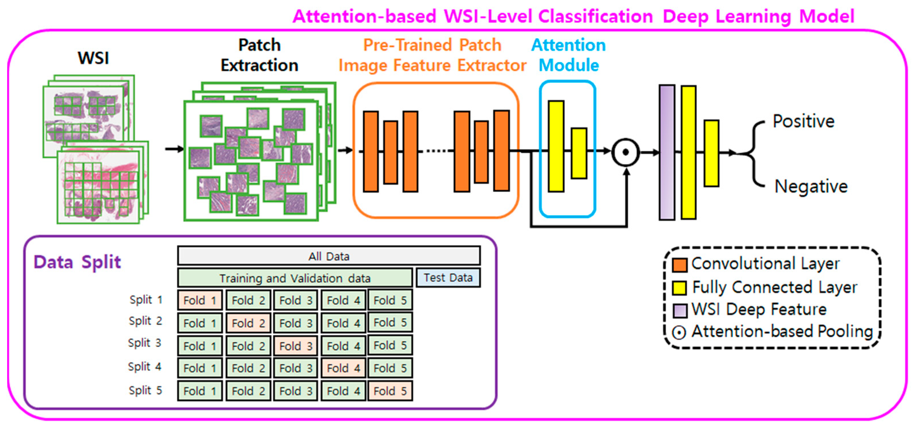
The deep learning method used in this study is the same as that employed in our previous study [31]. We developed an attention-based WSI-level classification deep learning model to predict whether a WSI is LNM positive or negative (Figure 1). The model was trained for a binary classification task, where the input was a WSI, and the output was the probability of the WSI being LNM-positive.
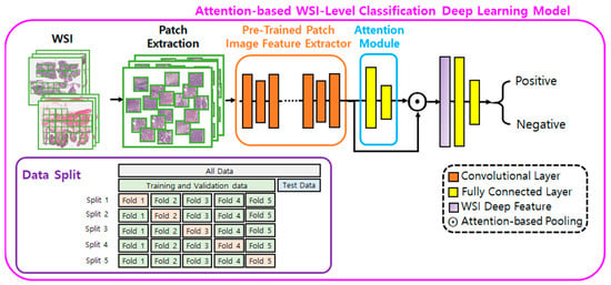
Figure 1.
Pipeline of the approach for classifying lymph node metastasis. WSI, whole slide image.
The model is an end-to-end neural network comprising a deep convolutional neural network (DCNN), attention module (AM), and classification module (CM) [32,33]. The DCNN was pre-trained with patches labeled as positive (patches from LNM positive WSIs) or negative (patches from LNM negative WSIs) to learn features in histopathological images in advance and to function as a patch image feature extractor (FE) [34]. The AM computes an attention score (AS), between 0 and 1, for each patch image in a WSI; the sum of these scores is equal to 1. An attention mechanism was used to visualize the spatial distribution of ASs of the WSIs. A higher AS indicates that the patch image is relatively more informative and has a greater influence on the final classification decision.
The model’s inferencing details are as follows: For a given WSI, all tissue regions are patched in a tiling manner and used as input for the DCNN FE, which compresses and encodes each patch image into a 512-dimensional feature vector (FV). The FVs are further aggregated into a single WSI-level FV (WSI deep feature) with 512 dimensions using their weighted average determined by the AS, computed by the AM [32]. The final WSI deep feature is then input into the CM to obtain the final prediction for LNM.
2.4. Statistical Analysis
Continuous variables are expressed as medians with interquartile ranges (IQRs) and analyzed using Student’s t-test and the Mann–Whitney U test. Statistical significance was set at p < 0.05. All statistical analyses were performed using SPSS software version 28 for Windows (SPSS Inc., Chicago, IL, USA).
The AI performance was evaluated using the AUC receiver operating characteristic curve (ROC). ROC is a probability curve, and AUC represents the degree or measure of separability. It showed how much the model was capable of distinguishing between classes.
To validate our model in case of a lack of other hospital WSI, we developed a model with four versions of training and test set combinations using endoscopically and surgically resected specimens: Version 1, Train and Test: surgical specimens; Version 2, Train and Test: endoscopic and surgically resected specimens; Version 3, Train: endoscopic and surgical specimens and Test: surgical specimens; and Version 4, Train: endoscopic and surgical specimens and Test: endoscopic specimens.
We performed a five-fold cross-validation (CV) on the training set, preserving the percentage of each class to determine how well our approach worked on each fold. Consequently, each of the five models trained in CV was applied to the held-out test set, and the results were obtained by averaging the output predictions. By comparing our AI model with a model using clinicopathological features, we trained a random forest (RF) classifier with 500 trees to predict LNM [35]. RF is a versatile and widely used machine learning algorithm that constructed multiple decision trees and combined their outputs for robust and accurate predictions.
The optimal cut-off sensitivity and specificity of each model were evaluated using the Youden index, the maximum potential effectiveness of a diagnostic biomarker, and a common summary measure of the ROC curve [36]. And we used McNemar’s tests, non-parametric test used to analyze paired nominal data, to compare predictive performances between our model and JSCCR guidelines, the most widely used guidelines in Asia.
3. Results
3.1. Baseline Characteristics of the Study Population
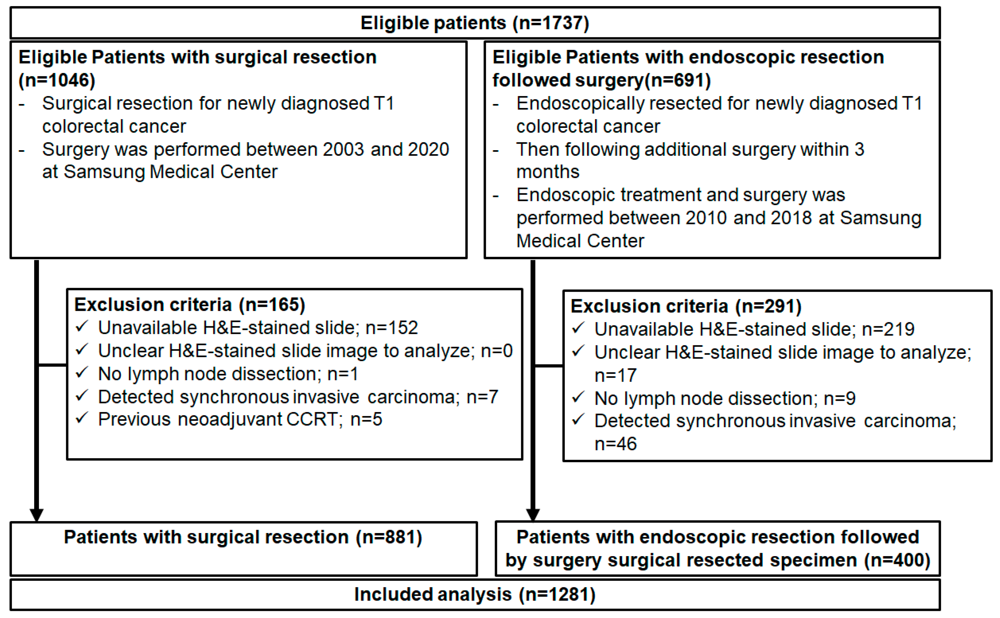
A total of 1737 patients with T1 CRCs (1046 surgical resections and 691 endoscopic resections followed by surgery) were eligible for this study, and 456 patients were excluded. Thus, 1281 patients (881 surgical resections and 400 endoscopic resections followed by surgery) were analyzed (Figure 2). Their baseline clinicopathological characteristics are presented in Table 1. The median age at CRC diagnosis was much younger in patients with endoscopic resection followed by additional surgery (59.0; IQR, 52.0–65.0) than in patients with surgical resection (60.0; IQR, 52.0–69.0). Men accounted for 59.6% of the total population. The percentage of patients with a family history of CRC, ex-/current smoker, or alcohol ex-/current drinker was higher in endoscopic resection followed by additional surgery. Patients without a family history of CRC accounted for 89.2% of the patients. High-risk pathologic features related to LNM, including LVI, tumor budding, positive resection margin, and microsatellite instability, were more in patients with endoscopic resection followed by additional surgery than surgical resection.
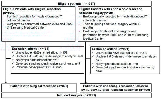
Figure 2.
Flow chart of study population. CCRT, concurrent chemoradiotherapy.

Table 1.
Baseline characteristics of study population.
Patients with CRC and LNM accounted for 6.6% (n = 58) of patients with surgical resection and 17.8% (n = 71) of patients with endoscopic resection followed by additional surgery. LN yield, the total number of LNs retrieved after surgery was 22,022. The LN ratio, the ratio of positive LNs out of the total removed, was 1.24% (273/22,022). In our study, an average of 17 LNs were retrieved in each surgery. When we compared the past group (patients who underwent surgery in 2003–2010) and the recent group (2011–2020), an average of 16 LNs were retrieved per surgery in the past group, and 18 LNs were retrieved per surgery in the recent group.
3.2. Train and Test Set in Model with Four Versions
A total of 2604 WSIs (184 positive LNM and 1139 negative LNM) from 881 surgical specimens (102 positive LNM and 791 negative LNM) and 400 endoscopically resected specimens (82 positive LNM and 348 negative LNM) were used to develop the model. A summary of the four versions is presented in Table 2.
Table 2.
Composition of number of patients and WSI according to lymph node metastasis in the training and test set of artificial intelligence model with four versions.
In Version 1, 893 WSIs (102 positive LNM and negative 791 LNM) from 881 surgical specimens were randomly split into training and test sets in a ratio of 4:1 at the patient level. Accordingly, 80 and 21 patients with positive LNM and 624 and 156 with negative LNM were assigned to the training and test sets, respectively.
In Version 2, 1323 WSIs (184 positive LNM and negative 1139 LNM) from 881 surgical specimens and 400 endoscopically resected specimens were randomly split into training and test sets in a ratio of 4:1 at the patient level. Accordingly, 137 and 35 patients with positive LNM and 887 and 222 patients with negative LNM were assigned to the training and test sets, respectively.
In version 3, 1144 WSIs (165 positive LNM and negative 1068 LNM) from 881 surgical specimens and 400 endoscopically resected specimens were randomly split into the training set and surgical specimens were randomly split into the test set in a ratio of 6:1 at the patient level. Accordingly, 137 and 21 patients with positive LNM and 887 and 156 patients with negative LNM were assigned to the training and test sets, respectively.
In Version 4, 1145 WSIs (163 positive LNM and 982 negative LNM) from 881 surgical specimens and 400 endoscopically resected specimens were randomly split into the training set and endoscopically resected specimens were randomly split into the test set in a ratio of 13:1 at the patient level. Accordingly, 137 and 14 patients with positive LNM and 887 and 66 patients with negative LNM were assigned to the training and test sets, respectively.
3.3. Area under the Curve for Predicting Lymph Node Metastasis
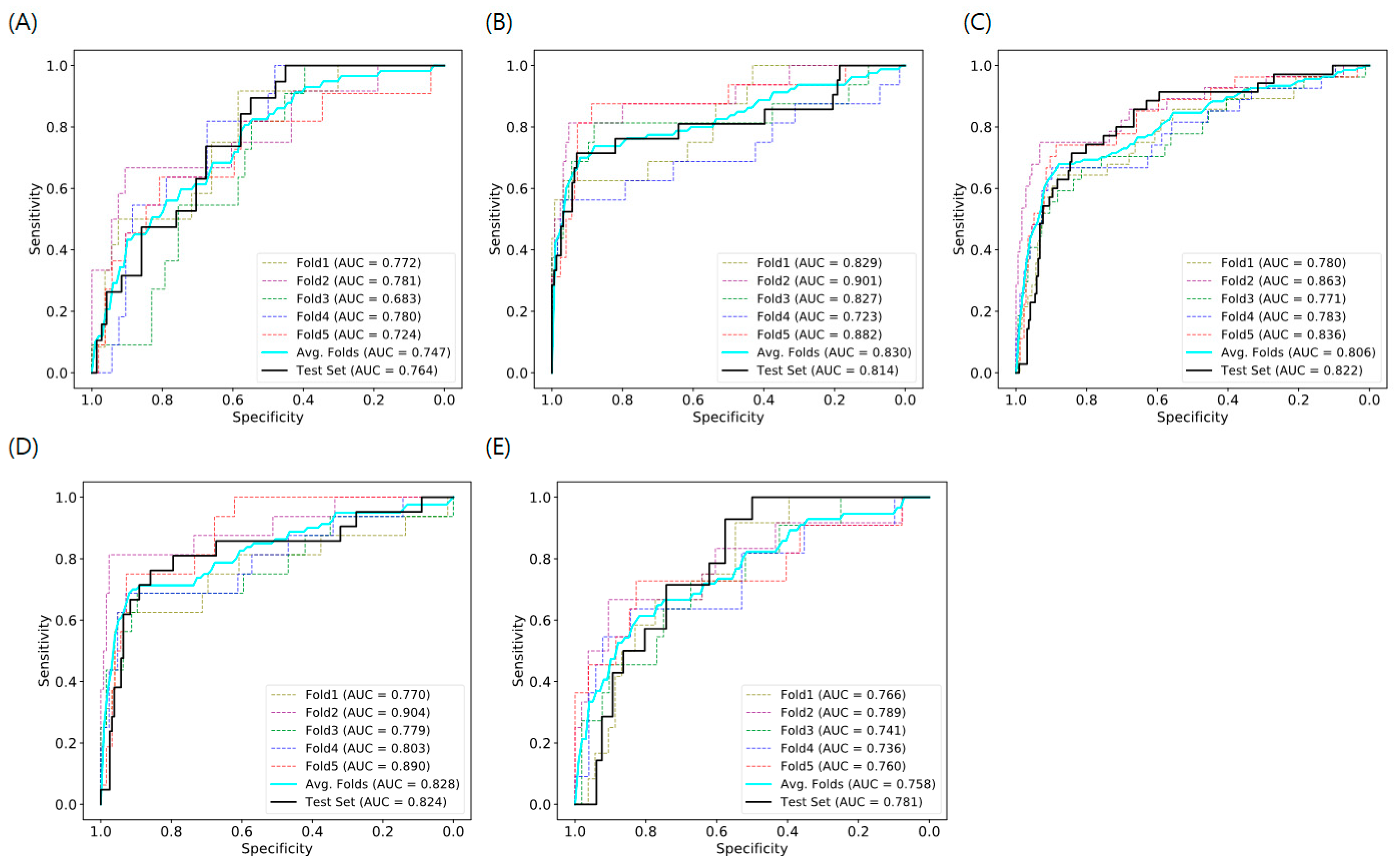
The AUCs for predicting LNM in T1 CRC using the AI model with histopathological images of endoscopic and surgical specimens and RF with clinicopathological features are shown in Table 3. Our model showed better prediction performance, with an AUC of 0.758–0.830 for the training set and 0.781–0.824 for the test set, than that of the model with clinicopathological features (AUC 0.516–0.683 in the test set). The ROC curves of the AI model are shown in Figure 3. The sensitivity and specificity of each version were 71.4% and 92.9% for version 1, 71.4% and 84.2% for version 2, 76.2% and 85.9% for version 3, and 92.9% and 57.6% for version 4 respectively.
Table 3.
Area under the curve of artificial intelligence model, compared to random forest with clinicopathologic features predicting of lymph node metastasis in in T1 CRC.
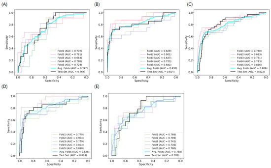
Figure 3.
Area under the ROC curve for attention-based WSI-level classification deep learning model for predicting lymph node metastasis in T1 colorectal cancer. (A) Previous model, (B) Version 1, (C) Version 2, (D) Version 3, (E) Version 4 ROC, receiver operating characteristic; WSI, whole-slide image; Version 1, train and test: surgical specimen; Version 2, train and test: endoscopic and surgical specimen; Version 3, train: endoscopic and surgical specimens and test: surgical specimen; Version 4, train: endoscopic and surgical specimens and test: endoscopic specimen; AUC: area under the curve; Avg: average.
3.4. Predictive Performance of Model with Four Versions vs. That of JSCCR Guidelines
We compared the performance of our model (four versions) with that of JSCCR guidelines using the test set (Table 4). JSCCR guidelines recommend that additional colorectal surgery be performed when endoscopically resected specimens show at least one of the high-risk features of LNM. JSCCR guideline did not allow any LNM of T1 CRC, which resulted in unnecessary additional surgery. It meant that this strategy showed 100% sensitivity and 0% specificity. On the other hand, to reflect reality as much as possible, we used the sensitivity and specificity of our model determined through the Youden index instead of setting it at 100% sensitivity to not allowing false negatives. The rate of unnecessary additional surgery attributable to misdiagnosing patients with negative LNM as having positive LNM was anticipated to be from 42.3% to 68.3% by our model with four versions and 82.5 to 88.1% by the JSCCR guidelines. Based on the results of the analysis, our model avoided at least 14.2% of unnecessary additional surgeries than predicted using the current JSCCR guidelines.
Table 4.
Predictive value of our artificial intelligence model with four versions and JSCCR guideline for lymph node metastasis in patients with T1 colorectal cancer.
3.5. Attention Score
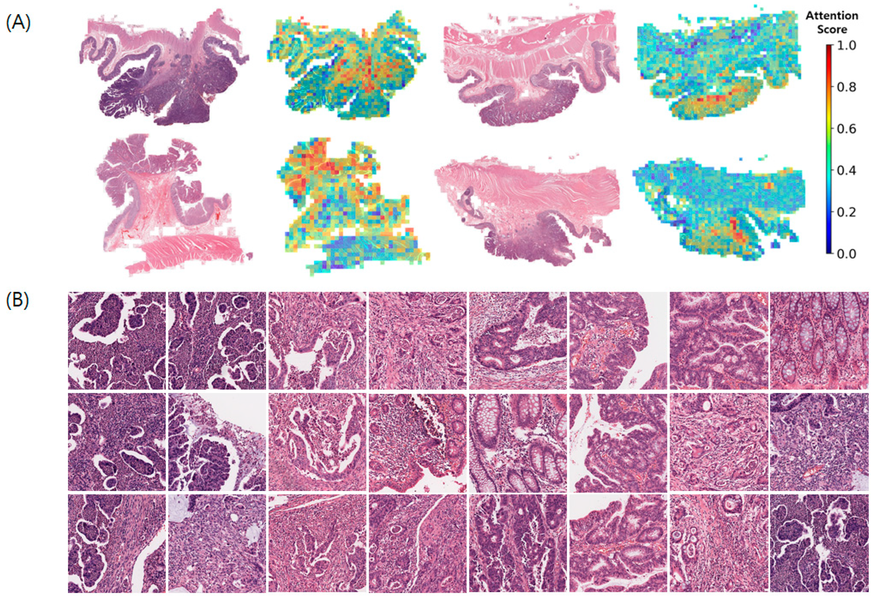
The attention mechanism interprets the effect of each patch on the final WSI-level decision using a scoring system. The calculated ASs of WSIs for positive LNM are displayed as a heatmap, highlighting regions of interest (ROIs), where ASs were normalized using a simple min-max normalization method (Figure 4). Sample patch images of LNM-positive WSIs are shown in Figure 4B. The prominent features of the sample patch images with high ASs were tumor budding and micropapillary patterns.
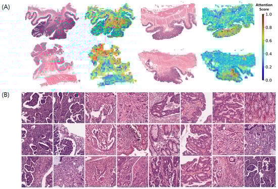
Figure 4.
Attention score visualization in WSIs of positive lymph node metastasis. (A) Spatial distribution of attention scores on the WSIs, (B) Patch-images stratified by high attention scores WSIs, whole slide images.
4. Discussion
The risk of LNM in T1 CRC is associated with the following histological risk factors in endoscopically resected specimens: LVI, tumor budding, histological grade, and depth of SM invasion. In cases of high-risk LNM, additional surgery is recommended based on the current guidelines. However, the risk of LNM in T1 CRC after additional surgery with LN dissection is estimated to be 6–14% [37]. Therefore, to avoid unnecessary surgery, it is important to predict LNM using endoscopically resected specimens before surgery. However, it is almost impossible to predict LNM based on pathological evaluation using only H&E-stained, endoscopically resected specimens. To address this issue, several recent studies have used AI models to determine the histological risk of LNM [23,24,26,38,39]. However, these DL models are still in the early stages of development and require extensive external validation [28,29,40].
In our previous study, we developed a prediction model analyzing H&E-stained WSIs for LNM in T1 CRC using DL without manual pixel-level annotation. Compared to existing studies, this study had the strengths of a relatively large target group that underwent additional surgery after endoscopic resection, resulting in better AUCs for predicting LNM with H&E-stained WSI information alone. Although the previous study included high-risk histological features of LNM, the absolute number of enrolled patients was small. Moreover, as it was difficult to perform an external validation using scanned WSIs from other hospitals in a relatively short time, no external validation was performed. Consequently, we planned to validate our model using not only endoscopic specimens but also surgical specimens. To increase the number of patients and WSIs from patients with low risk of LNM, we included patients who underwent surgery between 2003–2020, a longer study period than those who underwent endoscopic resection followed by surgery (2010–2018). Massive surgical specimens that were performed when surgery was the only treatment option for T1 CRC showed a lower risk of LNM compared to endoscopic resected specimens. Indeed, pathologic characteristics of patients with surgical resection showed a lower risk of LNM than in patients with endoscopic resection, followed by surgery. Therefore, as an alternative to the independent cohort, we trained and tested the model in four versions and assessed its performance.
We developed an AI model with a DL program to predict LNM in T1 CRC using surgical and endoscopic specimens. The AUCs of our DL model were 0.758–0.830 for the training set and 0.781–0.824 for the test set, which were improved compared with the previous model (AUC: 0.747 in training and 0.767 in testing sets). This could be explained by the fact that surgical specimens contained much more tissue and information than endoscopically resected specimens, and over 1000 WSIs were used for analysis compared with previous studies. Among four versions, Version 3 (train: endoscopic and surgical specimens, test: surgical specimens), which contained surgical specimens in both training and test sets, had the highest performance power. However, all versions of the AI model showed acceptable AUC ranges for predicting LNM in patients with T1 CRC. On the other hand, RF with clinicopathological features showed a lower AUC (0.516–0.701) than the AI model. Only Version 1 (training and test: surgical specimen) had an AUC barely greater than 0.7.
Among four versions, Version 4 (train: endoscopic and surgical specimens, test: endoscopic specimens) was the closest to the actual prediction target. Because the ultimate goal of the AI model was to predict LNM in patients who were only treated with endoscopic treatment for T1 CRC, however, considering the study results, it could be assumed that test with endoscopic resected specimen was difficult to predict LNM. The AUC of RF with clinicopathologic features was the lowest (0.516) in Version 4. The AUC of Version 4 was the lowest among the four versions, even though the difference was not significant. However, it was remarkable that the AI model with Version 4 had the greatest improvement of AUC (0.265), compared to RF with clinicopathologic features, among the four versions. Thus, our newly developed model showed the possibility of application in clinical practice for LNM prediction.
In a previous study, when we compared the AI model with JSCCR guidelines, we ideally set the cutoff threshold of the AI model at 100% sensitivity not to allow missed LNM, like JSCCR guidelines. As a result, the previous model reduced unnecessary additional surgeries by 15.1% than the current JSCCR guidelines. However, setting the cutoff threshold at 100% sensitivity might not reflect reality, so we used the Youden index for setting the cutoff in our present study. The present model avoided 14.2–45.8% of unnecessary additional surgeries than predicted using the current JSCCR guidelines while allowing missed LNM, which ranged from 7.1 to 28.6%. The AI model that reduced unnecessary additional surgery allowed more missed LNM. So, careful interpretation of results was needed. Considering, acceptable the lowest rate of missed LNM, the ultimate target population, and the improvement of AUC using the AI model, the AI model with Version 4 was compatible with clinical practice to predict LNM in T1 CRC. It reduced 14.2% of unnecessary additional surgeries than predicted using the current JSCCR guidelines while allowing 7% of missed LNM.
Previous studies using AI to determine the risk of LNM on histology showed AUC ranging between 0.567 and 0.938, consistent with our results (0.781–0.824) [26,27,31,38,39,41]. Most studies included clinicopathologic variables and/or additional immunohistochemistry performed by pathologists. However, several recent studies have demonstrated the potential of applying DL to predict LNM in T1 CRC using H&E-stained WSIs without a histological assessment [26,27,31]. This pathologist-independent strategy may be the focus of the next era of T1 CRC management [29]. Similarly, our AI model also used WSIs without annotation and included a large number of T1 CRC cases (n = 1281). The model was validated using an independent cohort at the same institution and showed the best AUC among the AI models using only WSI.
In contrast to previous studies, Kasahara et al. developed an AI model with 80–85% accuracy using biopsy specimens and mucosal layer of the surgical specimens in the absence of biopsy specimens to predict patients with T1 CRC without LNM and their LNM risk before treatment, and select appropriate procedure before treatment [41]. The study suggests that biopsy specimen characteristics are associated with LNM risk. However, they implemented a weakly supervised model with a small number of patients and images of the site of choice for each ROI selected by the pathologist. Our previous study was conducted using high-risk histological features of LNM and was unsuitable for predicting LNM in low-risk patients with T1 CRC. However, 10.1% of the present study population had LNM, consistent with previous Japanese studies on long-term outcomes of CRC (10.8–12.4%) [42,43]. Additionally, the sensitivity and specificity of the present model were improved compared to our previous study. Therefore, it might be acceptable to apply our AI model to predict LNM in patients with low-risk T1 CRC.
Even though depth of SM invasion and tumor budding were risk factors of LNM, the meaning of these is controversial, and inter-observer variation for measurement existed. According to JSCCR guidelines, SM depth >1000 μm was one of the risk factors for LNM, but several studies showed differences in SM invasion depth related to LNM [44,45,46]. The International Tumor Budding Consensus Conference guidelines recommended the use of a three-tier system for risk stratification: Bd 1, low budding (0–4 buds); Bd 2, intermediate budding (5–9 buds); Bd 3, high budding (10 or more buds) [47]. In p1 CRC, Bd 2 and Bd 3 were associated with an increased risk of LNM, whereas in stage II CRC, Bd 3 is associated with an increased risk of recurrence and mortality. Moreover, the current tumor bud assessment system focused only on the tumor bud count and did not account for other features [48]. So, we wanted to figure out the meaning of depth of invasion for LNM and tumor budding using an attention-based WSI-level classification deep learning model. Patch images with high ASs appeared to be located in the transformation zone, which is the boundary between normal and cancerous tissues. In our study, the prominent features of the patch images with high ASs were poorly differentiated histological grades, tumor budding and micropapillary patterns, well-known pathological factors associated with poor prognosis [49,50]. Additionally, Brockmoeller et al. demonstrated an association between inflamed fat in CRC and LNM and that AI had the potential to discover new mechanisms in cancer progression [26]. However, unlike their study, our patch images with high AS did not contain inflamed fat.
Nevertheless, our study had some limitations. First, it was a single tertiary center retrospective study, which has a potential for bias. Second, the AUC of our method was relatively low to use as a prediction model despite the improvement achieved by increasing the study population and using various training and test sets for endoscopic and surgical specimens. Additionally, the DL model was influenced by class balance, and intrinsic LNM in T1 CRC was low. Nonetheless, among the AI models that used only WSIs, our study showed the best performance. Third, extensive external validation was lacking. We performed validation with an independent cohort in a single center; however, staining using H&E, variations in WSI color, and clinicopathological characteristics in a single center were similar. Therefore, the discriminating power may have been overestimated [29,40]. However, our study enrolled a relatively large number of patients compared with previous studies. Moreover, the clinicopathological features did not seem to be important in predicting LN metastasis in our AI model with H&E-stained WSI. Finally, our model was not validated in a cohort that underwent endoscopic resection of T1 CRC without additional surgery, which is a part of the model’s real target.
5. Conclusions
In conclusion, our AI model with H&E-stained WSIs and without pathologists showed higher performance power (AUC, 0.782–0.824) with validation of an independent cohort in a single center than previous studies. Since WSIs from 1281 patients with low to high risk of LNM were used to develop the present AI model, it was suitable for predicting LNM even in low-risk patients with T1 CRC. Moreover, this model reduced 14.2% of unnecessary additional surgeries than predicted using the current JSCCR guidelines while allowing 7% of missed LNM. This revealed the feasibility of using an AI model with only H&E-stained WSIs to predict LNM in T1 CRC. However, to apply our model to real-world clinical practice, extensive external validation with WSI from multiple centers and patients who undergo only endoscopic treatment is warranted.
Author Contributions
Conceptualization, J.H.S., E.R.K. and Y.H.; methodology, J.H.S., E.R.K. and Y.H.; software, Y.H.; validation, Y.H. and I.S.; formal analysis, J.H.S.; investigation, J.H.S. and Y.H.; resources, J.H.S. and Y.H.; data curation, J.H.S.; writing—original draft preparation, J.H.S.; writing—review and editing, E.R.K. and Y.H.; visualization, J.H.S. and Y.H.; supervision, E.R.K., Y.H., I.S., S.A., S.-H.K. and K.-T.J.; funding acquisition, J.H.S. and K.-T.J. All authors have read and agreed to the published version of the manuscript.
Funding
This study was supported by the Research Supporting Program of the Korean Association for the Study of Intestinal Diseases for 2023 (2023-8). This research was supported by a grant of the Korea Health Technology R&D Project through the Korea Health Industry Development Institute (KHIDI), funded by the Ministry of Health and Welfare, Republic of Korea (grant number: HI21C1137).
Institutional Review Board Statement
The study was conducted in accordance with the Declaration of Helsinki and approved by the Institutional Review Board of Samsung Medical Center (2021-01-042-005, 17 November 2021).
Informed Consent Statement
Patient consent was waived due to the use of de-identified data routinely collected during hospital visits.
Data Availability Statement
The raw data supporting the conclusions of this article will be made available by the authors on request.
Acknowledgments
We thank all the patients who participated in this study.
Conflicts of Interest
Insuk Sohn was employed by the company Arontier Co., Ltd. The remaining authors declare no conflicts of interest.
Abbreviations
| CRC | colorectal cancer |
| LNM | lymph node metastasis |
| SM | submucosal |
| LVI | lymphovascular invasion |
| JSCCR | Japanese Society for Cancer of the Colon and Rectum |
| H&E | Hematoxylin and eosin |
| AI | artificial intelligence |
| WSI | whole slide images |
| AUC | area under the curve |
| EMR | endoscopic mucosal resection |
| ESD | endoscopic submucosal dissection |
| DCNN | a deep convolutional neural network |
| AM | attention module |
| CM | classification module |
| AS | attention score |
| FV | feature vector |
| IQR | interquartile ranges |
| ROC | receiver operating characteristic curve |
| CV | cross-validation |
| RF | random forest |
| ROI | regions of interest |
References
- Wong, M.C.S.; Ding, H.; Wang, J.; Chan, P.S.F.; Huang, J. Prevalence and risk factors of colorectal cancer in Asia. Intest. Res. 2019, 17, 317–329. [Google Scholar] [CrossRef]
- Sung, H.; Ferlay, J.; Siegel, R.L.; Laversanne, M.; Soerjomataram, I.; Jemal, A.; Bray, F. Global Cancer Statistics 2020: GLOBOCAN Estimates of Incidence and Mortality Worldwide for 36 Cancers in 185 Countries. CA Cancer J. Clin. 2021, 71, 209–249. [Google Scholar] [CrossRef] [PubMed]
- Winawer, S.J.; Zauber, A.G. The advanced adenoma as the primary target of screening. Gastrointest. Endosc. Clin. N. Am. 2002, 12, 1–9. [Google Scholar] [CrossRef] [PubMed]
- Wook, H.S.; Jeong-Sik, B. Endoscopic diagnosis and treatment of early colorectal cancer. Intest. Res. 2022, 20, 281–290. [Google Scholar]
- Kim, S.Y.; Kwak, M.S.; Yoon, S.M.; Jung, Y.; Kim, J.W.; Boo, S.-J.; Oh, E.H.; Jeon, S.R.; Nam, S.-J.; Park, S.-Y.; et al. Korean Guidelines for Postpolypectomy Colonoscopic Surveillance: 2022 revised edition. Intest. Res. 2023, 21, 20–42. [Google Scholar] [CrossRef] [PubMed]
- Fujimori, T.; Kawamata, H.; Kashida, H. Precancerous lesions of the colorectum. J. Gastroenterol. 2001, 36, 587–594. [Google Scholar] [CrossRef]
- Morson, B.C.; Whiteway, J.E.; Jones, E.A.; Macrae, F.A.; Williams, C.B. Histopathology and prognosis of malignant colorectal polyps treated by endoscopic polypectomy. Gut 1984, 25, 437–444. [Google Scholar] [CrossRef] [PubMed]
- Minamoto, T.; Mai, M.; Ogino, T.; Sawaguchi, K.; Ohta, T.; Fujimoto, T.; Takahashi, Y. Early invasive colorectal carcinomas metastatic to the lymph node with attention to their nonpolypoid development. Am. J. Gastroenterol. 1993, 88, 1035–1039. [Google Scholar] [PubMed]
- Kitajima, K.; Fujimori, T.; Fujii, S.; Takeda, J.; Ohkura, Y.; Kawamata, H.; Kumamoto, T.; Ishiguro, S.; Kato, Y.; Shimoda, T. Correlations between lymph node metastasis and depth of submucosal invasion in submucosal invasive colorectal carcinoma: A Japanese collaborative study. J. Gastroenterol. 2004, 39, 534–543. [Google Scholar] [CrossRef]
- Kyzer, S.; Begin, L.R.; Gordon, P.H.; Mitmaker, B. The care of patients with colorectal polyps that contain invasive adenocarcinoma. Endoscopic polypectomy or colectomy? Cancer 1992, 70, 2044–2050. [Google Scholar] [CrossRef]
- Nivatvongs, S.; Rojanasakul, A.; Reiman, H.M.; Dozois, R.R.; Wolff, B.G.; Pemberton, J.H.; Beart, R.W., Jr.; Jacques, L.F. The risk of lymph node metastasis in colorectal polyps with invasive adenocarcinoma. Dis. Colon Rectum 1991, 34, 323–328. [Google Scholar] [CrossRef] [PubMed]
- Netzer, P.; Forster, C.; Biral, R.; Ruchti, C.; Neuweiler, J.; Stauffer, E.; Schönegg, R.; Maurer, C.; Hüsler, J.; Halter, F.; et al. Risk factor assessment of endoscopically removed malignant colorectal polyps. Gut 1998, 43, 669–674. [Google Scholar] [CrossRef] [PubMed]
- Watanabe, T.; Muro, K.; Ajioka, Y.; Hashiguchi, Y.; Ito, Y.; Saito, Y.; Hamaguchi, T.; Ishida, H.; Ishiguro, M.; Ishihara, S.; et al. Japanese Society for Cancer of the Colon and Rectum (JSCCR) guidelines 2016 for the treatment of colorectal cancer. Int. J. Clin. Oncol. 2018, 23, 1–34. [Google Scholar] [CrossRef] [PubMed]
- Ramirez, M.; Schierling, S.; Papaconstantinou, H.T.; Thomas, J.S. Management of the malignant polyp. Clin. Colon Rectal Surg. 2008, 21, 286–290. [Google Scholar] [CrossRef] [PubMed]
- Aarons, C.B.; Shanmugan, S.; Bleier, J.I. Management of malignant colon polyps: Current status and controversies. World J. Gastroenterol. 2014, 20, 16178–16183. [Google Scholar] [CrossRef] [PubMed]
- Cooper, H.S. Surgical pathology of endoscopically removed malignant polyps of the colon and rectum. Am. J. Surg. Pathol. 1983, 7, 613–623. [Google Scholar] [CrossRef] [PubMed]
- Coverlizza, S.; Risio, M.; Ferrari, A.; Fenoglio-Preiser, C.M.; Rossini, F.P. Colorectal adenomas containing invasive carcinoma. Pathologic assessment of lymph node metastatic potential. Cancer 1989, 64, 1937–1947. [Google Scholar] [CrossRef]
- Colacchio, T.A.; Forde, K.A.; Scantlebury, V. Endoscopic Polypectomy: Inadequate Treatment for Invasive Colorectal Carcinoma. Ann. Surg. 1982, 194, 704–707. [Google Scholar] [CrossRef]
- Choi, Y.S.; Kim, W.S.; Hwang, S.W.; Park, S.H.; Yang, D.-H.; Ye, B.D.; Myung, S.-J.; Yang, S.-K.; Byeon, J.-S. Clinical outcomes of submucosal colorectal cancer diagnosed after endoscopic resection: A focus on the need for surgery. Intest. Res. 2020, 18, 96–106. [Google Scholar] [CrossRef]
- Kojima, M.; Puppa, G.; Kirsch, R.; Basturk, O.; Frankel, W.L.; Vieth, M.; Lugli, A.; Sheahan, K.; Yeh, M.; Lauwers, G.Y.; et al. Blood and lymphatic vessel invasion in pT1 colorectal cancer: An international concordance study. J. Clin. Pathol. 2015, 68, 628–632. [Google Scholar] [CrossRef]
- Kouyama, Y.; Kudo, S.E.; Miyachi, H.; Ichimasa, K.; Hisayuki, T.; Oikawa, H.; Matsudaira, S.; Kimura, Y.J.; Misawa, M.; Mori, Y.; et al. Practical problems of measuring depth of submucosal invasion in T1 colorectal carcinomas. Int. J. Color. Dis. 2016, 31, 137–146. [Google Scholar] [CrossRef]
- Barel, F.; Auffret, A.; Cariou, M.; Kermarrec, T.; Samaison, L.; Bourhis, A.; Badic, B.; Jézéquel, J.; Cholet, F.; Bail, J.P.; et al. High reproducibility is attainable in assessing histoprognostic parameters of pT1 colorectal cancer using routine histopathology slides and immunohistochemistry analyses. Pathology 2019, 51, 46–54. [Google Scholar] [CrossRef]
- Ichimasa, K.; Kudo, S.E.; Mori, Y.; Misawa, M.; Matsudaira, S.; Kouyama, Y.; Baba, T.; Hidaka, E.; Wakamura, K.; Hayashi, T.; et al. Artificial intelligence may help in predicting the need for additional surgery after endoscopic resection of T1 colorectal cancer. Endoscopy 2018, 50, 230–240. [Google Scholar] [PubMed]
- Takamatsu, M.; Yamamoto, N.; Kawachi, H.; Chino, A.; Saito, S.; Ueno, M.; Ishikawa, Y.; Takazawa, Y.; Takeuchi, K. Prediction of early colorectal cancer metastasis by machine learning using digital slide images. Comput. Methods Programs Biomed. 2019, 178, 155–161. [Google Scholar] [CrossRef]
- Kudo, S.E.; Ichimasa, K.; Villard, B.; Mori, Y.; Misawa, M.; Saito, S.; Hotta, K.; Saito, Y.; Matsuda, T.; Yamada, K. Artificial Intelligence System to Determine Risk of T1 Colorectal Cancer Metastasis to Lymph Node. Gastroenterology 2021, 160, 1075–1084.e2. [Google Scholar] [CrossRef]
- Brockmoeller, S.; Echle, A.; Ghaffari Laleh, N.; Eiholm, S.; Malmstrøm, M.L.; Plato Kuhlmann, T.; Levic, K.; Grabsch, H.I.; West, N.P.; Saldanha, O.L.; et al. Deep learning identifies inflamed fat as a risk factor for lymph node metastasis in early colorectal cancer. J. Pathol. 2022, 256, 269–281. [Google Scholar] [CrossRef]
- Takamatsu, M.; Yamamoto, N.; Kawachi, H.; Nakano, K.; Saito, S.; Fukunaga, Y.; Takeuchi, K. Prediction of lymph node metastasis in early colorectal cancer based on histologic images by artificial intelligence. Sci. Rep. 2022, 12, 2963. [Google Scholar] [CrossRef] [PubMed]
- Ichimasa, K.; Kudo, S.-e.; Lee, J.W.J.; Nemoto, T.; Yeoh, K.G. Artificial intelligence–assisted treatment strategy for T1 colorectal cancer after endoscopic resection. Gastrointest. Endosc. 2023, 97, 1148–1152. [Google Scholar] [CrossRef]
- Ichimasa, K.; Kudo, S.-E.; Lee, J.W.J.; Yeoh, K.G. “Pathologist-independent” strategy for T1 colorectal cancer after endoscopic resection. J. Gastroenterol. 2022, 57, 815–816. [Google Scholar] [CrossRef] [PubMed]
- Ahn, J.H.; Kwak, M.S.; Lee, H.H.; Cha, J.M.; Shin, H.P.; Jeon, J.W.; Yoon, J.Y. Development of a novel prognostic model for predicting lymph node metastasis in early colorectal cancer: Analysis based on the surveillance, epidemiology, and end results database. Front. Oncol. 2021, 11, 614398. [Google Scholar] [CrossRef]
- Song, J.H.; Hong, Y.; Kim, E.R.; Kim, S.-H.; Sohn, I. Utility of artificial intelligence with deep learning of hematoxylin and eosin-stained whole slide images to predict lymph node metastasis in T1 colorectal cancer using endoscopically resected specimens; prediction of lymph node metastasis in T1 colorectal cancer. J. Gastroenterol. 2022, 57, 654–666. [Google Scholar]
- Ilse, M.; Tomczak, J.; Welling, M. Attention-based deep multiple instance learning. In Proceedings of the International Conference on Machine Learning (PMLR), Stockholm, Sweden, 10–15 July 2018. [Google Scholar]
- Lu, M.Y.; Zhao, M.; Shady, M.; Lipkova, J.; Chen, T.Y.; Williamson, D.F.; Mahmood, F. Deep learning-based computational pathology predicts origins for cancers of unknown primary. arXiv 2020, arXiv:2006.13932. [Google Scholar]
- He, K.; Zhang, X.; Ren, S.; Sun, J. Deep residual learning for image recognition. In Proceedings of the IEEE Conference on Computer Vision and Pattern Recognition, Las Vegas, NV, USA, 27–30 June 2016. [Google Scholar]
- Pedregosa, F.; Varoquaux, G.; Gramfort, A.; Michel, V.; Thirion, B.; Grisel, O.; Blondel, M.; Prettenhofer, P.; Weiss, R.; Dubourg, V. Scikit-learn: Machine learning in Python. J. Mach. Learn. Res. 2011, 12, 2825–2830. [Google Scholar]
- Ruopp, M.D.; Perkins, N.J.; Whitcomb, B.W.; Schisterman, E.F. Youden Index and optimal cut-point estimated from observations affected by a lower limit of detection. Biom. J. 2008, 50, 419–430. [Google Scholar] [CrossRef] [PubMed]
- Ebbehøj, A.L.; Jørgensen, L.N.; Krarup, P.M.; Smith, H.G. Histopathological risk factors for lymph node metastases in T1 colorectal cancer: Meta-analysis. Br. J. Surg. 2021, 108, 769–776. [Google Scholar] [CrossRef] [PubMed]
- Kwak, M.S.; Lee, H.H.; Yang, J.M.; Cha, J.M.; Jeon, J.W.; Yoon, J.Y.; Kim, H.I. Deep Convolutional Neural Network-Based Lymph Node Metastasis Prediction for Colon Cancer Using Histopathological Images. Front. Oncol. 2020, 10, 619803. [Google Scholar] [CrossRef] [PubMed]
- Kang, J.; Choi, Y.J.; Kim, I.K.; Lee, H.S.; Kim, H.; Baik, S.H.; Kim, N.K.; Lee, K.Y. LASSO-Based Machine Learning Algorithm for Prediction of Lymph Node Metastasis in T1 Colorectal Cancer. Cancer Res. Treat. 2021, 53, 773–783. [Google Scholar] [CrossRef]
- Li, J.W.; Wang, L.M.; Ichimasa, K.; Lin, K.W.; Ngu, J.C.-Y.; Ang, T.L. Use of artificial intelligence in the management of T1 colorectal cancer: A new tool in the arsenal or is deep learning out of its depth? Clin. Endosc. 2023, 57, 24–35. [Google Scholar] [CrossRef]
- Kasahara, K.; Katsumata, K.; Saito, A.; Ishizaki, T.; Enomoto, M.; Mazaki, J.; Tago, T.; Nagakawa, Y.; Matsubayashi, J.; Nagao, T.; et al. Artificial intelligence predicts lymph node metastasis or risk of lymph node metastasis in T1 colorectal cancer. Int. J. Clin. Oncol. 2022, 27, 1570–1579. [Google Scholar] [CrossRef]
- Yoda, Y.; Ikematsu, H.; Matsuda, T.; Yamaguchi, Y.; Hotta, K.; Kobayashi, N.; Fujii, T.; Oono, Y.; Sakamoto, T.; Nakajima, T.; et al. A large-scale multicenter study of long-term outcomes after endoscopic resection for submucosal invasive colorectal cancer. Endoscopy 2013, 45, 718–724. [Google Scholar] [CrossRef]
- Ikematsu, H.; Yoda, Y.; Matsuda, T.; Yamaguchi, Y.; Hotta, K.; Kobayashi, N.; Fujii, T.; Oono, Y.; Sakamoto, T.; Nakajima, T.; et al. Long-term Outcomes After Resection for Submucosal Invasive Colorectal Cancers. Gastroenterology 2013, 144, 551–559. [Google Scholar] [CrossRef] [PubMed]
- Nakadoi, K.; Tanaka, S.; Kanao, H.; Terasaki, M.; Takata, S.; Oka, S.; Yoshida, S.; Arihiro, K.; Chayama, K. Management of T1 colorectal carcinoma with special reference to criteria for curative endoscopic resection. J. Gastroenterol. Hepatol. 2012, 27, 1057–1062. [Google Scholar] [CrossRef] [PubMed]
- Tanaka, S.; Haruma, K.; Oh, E.H.; Nagata, S.; Hirota, Y.; Furudoi, A.; Amioka, T.; Kitadai, Y.; Yoshihara, M.; Shimamoto, F. Conditions of curability after endoscopic resection for colorectal carcinoma with submucosally massive invasion. Oncol. Rep. 2000, 7, 783–788. [Google Scholar] [CrossRef] [PubMed]
- Egashira, Y.; Yoshida, T.; Hirata, I.; Hamamoto, N.; Akutagawa, H.; Takeshita, A.; Noda, N.; Kurisu, Y.; Shibayama, Y. Analysis of pathological risk factors for lymph node metastasis of submucosal invasive colon cancer. Mod. Pathol. 2004, 17, 503–511. [Google Scholar] [CrossRef] [PubMed]
- Lugli, A.; Kirsch, R.; Ajioka, Y.; Bosman, F.; Cathomas, G.; Dawson, H.; El Zimaity, H.; Fléjou, J.-F.; Hansen, T.P.; Hartmann, A.; et al. Recommendations for reporting tumor budding in colorectal cancer based on the International Tumor Budding Consensus Conference (ITBCC) 2016. Mod. Pathol. 2017, 30, 1299–1311. [Google Scholar] [CrossRef] [PubMed]
- Chen, L.; Yang, F.; Qi, Z.; Tai, J. Predicting lymph node metastasis and recurrence in patients with early stage colorectal cancer. Front. Med. 2022, 9, 991785. [Google Scholar] [CrossRef]
- Pyo, J.-S.; Park, M.J.; Kang, D.-W. The clinicopathological significance of micropapillary pattern in colorectal cancers. Hum. Pathol. 2018, 77, 159–165. [Google Scholar] [CrossRef]
- Zhang, S.; Zhang, D.; Yang, Z.; Zhang, X. Tumor Budding, Micropapillary Pattern, and Polyploidy Giant Cancer Cells in Colorectal Cancer: Current Status and Future Prospects. Stem Cells Int. 2016, 2016, 4810734. [Google Scholar] [CrossRef]
Disclaimer/Publisher’s Note: The statements, opinions and data contained in all publications are solely those of the individual author(s) and contributor(s) and not of MDPI and/or the editor(s). MDPI and/or the editor(s) disclaim responsibility for any injury to people or property resulting from any ideas, methods, instructions or products referred to in the content. |
© 2024 by the authors. Licensee MDPI, Basel, Switzerland. This article is an open access article distributed under the terms and conditions of the Creative Commons Attribution (CC BY) license (https://creativecommons.org/licenses/by/4.0/).