


Evolution of Liquid Biopsies for Detecting Pancreatic Cancer

Abstract
Simple Summary
Abstract

1. Introduction
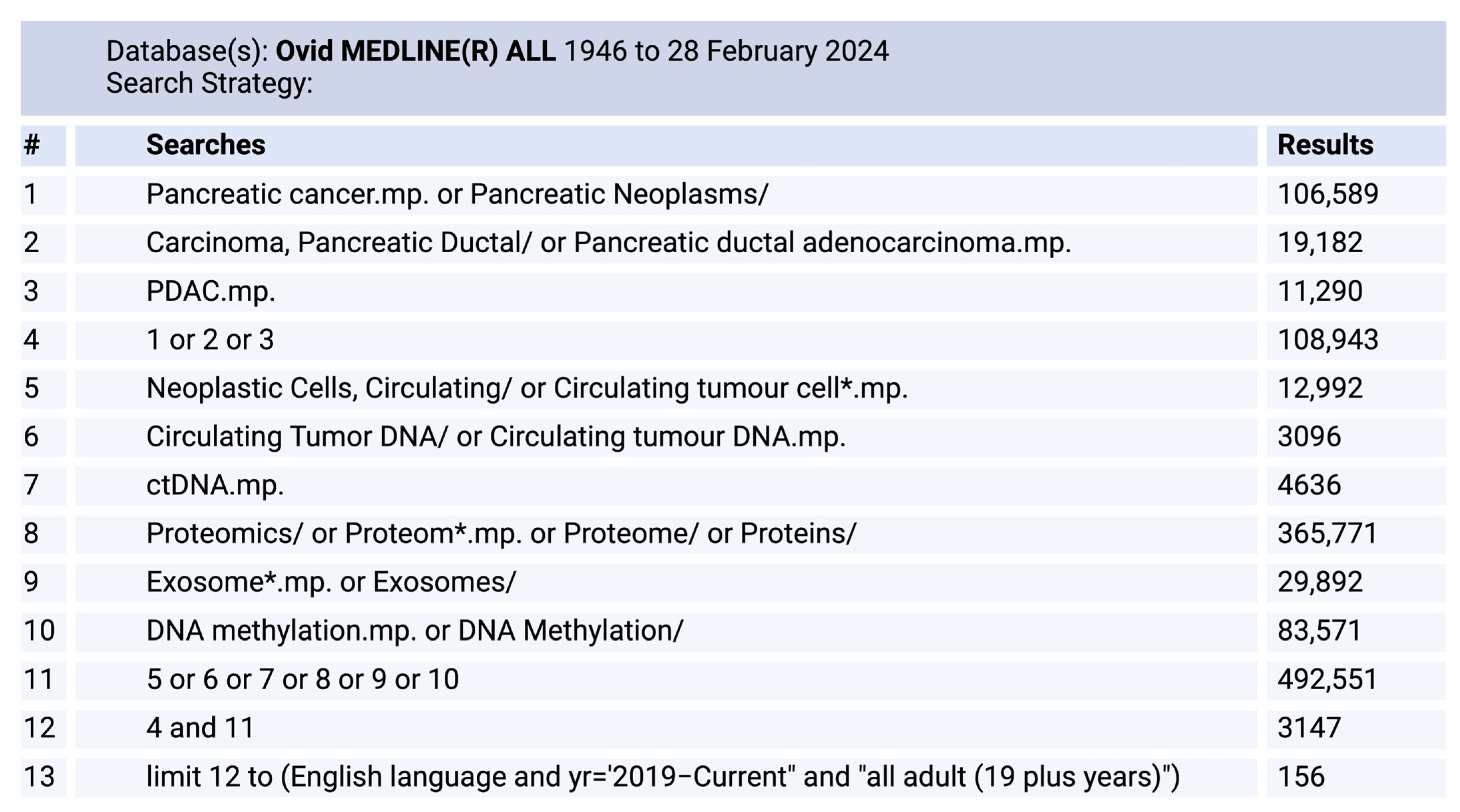
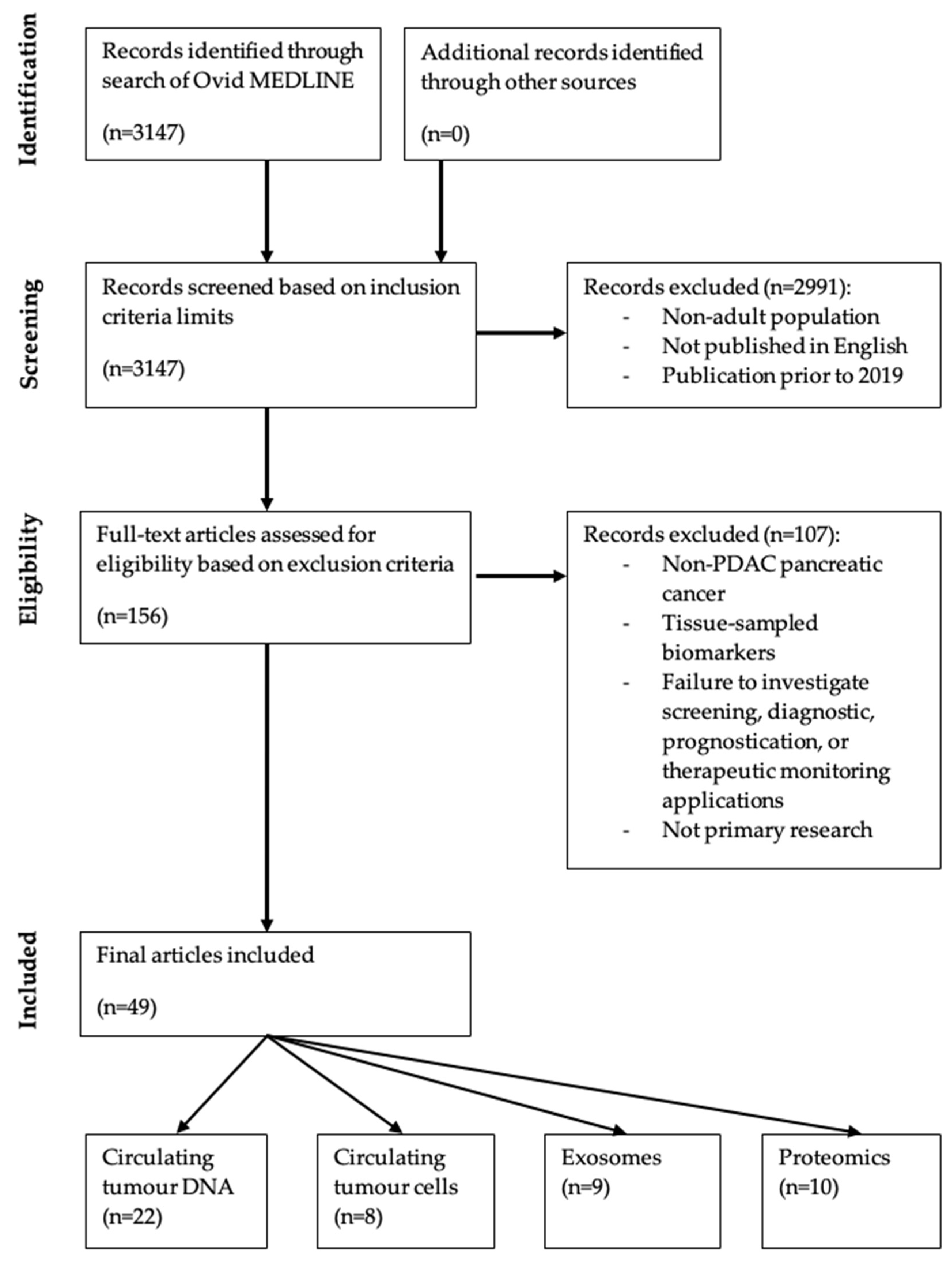
2. Methods
3. Discussion
3.1. Circulating Tumour DNA

| Citation | Patients | Applications Explored | Methodology | |||||||
|---|---|---|---|---|---|---|---|---|---|---|
| N | Disease Descriptor | Study Aim | Diagnosis (Sensitivity, Specificity) | Prognostic | Predictive | Agnostic/ Informed | Broad Method | Detection Technology | Gene(s) Examined | |
| Bachet et al., 2020 [46] | 113 | Stage IV | To evaluate the prognostic and predictive value of ctDNA from plasma samples during a randomised phase II trial which has assessed eryaspase efficacy in patients with advanced PDAC. | Yes (68.1, NR) | Yes | Yes | Agnostic | Mutation | AmpliSeq colon and lung cancer panel V2 (NGS) | 22 |
| Cheng et al., 2020 [58] | 210 | Stage III–IV | To evaluate the KRAS mutation status in ctDNA and circulating T cell subsets in a cohort of advanced pancreatic cancer patients. | No | Yes | No | Agnostic | Mutation | ddPCR | KRAS |
| Chung et al., 2021 [42] | 77 | Stage IV | To characterise the mutational landscape of patients with metastatic PDAC who received blood-based molecular profiling. | Yes (44.1, NR) | No | No | Agnostic | Mutation | Guardant-360 (NGS) | 83 |
| Fujimoto et al., 2021 [55] | 55 | Stage I–IV | To evaluate the sensitivity and specificity of serum DNA testing of methylated RUNX3 by the CORD assay for the detection of PDAC. | Yes (50.9, 93.5) | No | No | Agnostic | HM | CORD assay | RUNX3 |
| Groot et al., 2019 [39] | 59 | Pre- and post-operative | To perform analytical and clinical validation of a KRAS ctDNA assay in a certified clinical laboratory. | Yes (49.2, NR) | Yes | No | Agnostic | Mutation | ddPCR | KRAS |
| Guo et al., 2020 [41] | 113 | Pre-operative | To explore the clinical value of hotspot mutations in resectable PDAC patients. | Yes (38.1, NR) | Yes | No | Agnostic | Mutation | NGS | 50 |
| Hussung et al., 2021 [37] | 25 | Post-operative | To identify associations between mutant cfKRAS and CA19-9 dynamics and clinical outcome post resection. | Yes (48.0, NR) | Yes | No | Agnostic | Mutation | ddPCR | KRAS |
| Lee et al., 2019 [40] | 42 | Pre-operative | To evaluate the feasibility and clinical utility of ctDNA analysis to inform adjuvant therapy decision making. | Yes (62.2, NR) | Yes | No | Informed | Mutation | Safe-SeqS assay (PCR) | KRAS |
| Li et al., 2021 [56] | 105 | Pre-operative | To establish a scoring system for preoperative screening of resectable PDAC patients. | No | Yes | No | Agnostic | Mutation | qPCR | KRAS |
| Macgregor-Das et al., 2020 [36] | 67 | Screening and pre-operative | To utilise enzymatic pretreatment of plasma DNA followed by digital NGS to detect hotspot mutations in KRAS and GNAS in patients with pancreatic cancer. | Yes (36.5, 92.0) | No | No | Agnostic | Mutation | Digital NGS | KRAS GNAS |
| Miller et al., 2021 [53] | 17 | Stage I–IV | To assess the effectiveness of hypermethylation at the CpG island of ZNF154 for use in a blood-based cancer detection assay. | Yes (NR, NR) | No | No | Agnostic | HM | ddPCR | ZNF154 |
| Patel et al., 2019 [47] | 112 | Stage I–IV | To assess the genomic landscape of ctDNA in patients with PDAC, using clinical-grade NGS to investigate the clinical implications. | Yes (69.6, NR) | Yes | No | Agnostic | Mutation | NGS | 54–73 |
| Pietrasz et al., 2022 [51] | 372 | Stage IV; CTx-naïve | To determine whether ctDNA is an independent factor for the prognostication of metastatic PDAC. | Yes (56.8, NR) | Yes | No | Agnostic | HM | ddPCR | HOXD8 POU4F1 |
| Shinjo et al., 2020 [54] | 47 | Stage I–IV | To identify effective DNA methylation markers for the diagnosis of PDAC. | Yes (49.0, 86.0) | No | No | Informed | HM | MBD-ddPCR | HOXA1 PCDH10 ADAMTS2 SEMA5A SPSB4 |
| Singh et al., 2020 [52] | 65 | Stage I–IV | To conduct absolute quantification of methylation in SPARC, UCHL1, PENK, and NPTX2 genes and assess the respective methylation load for their ability to perform as non-invasive differentiating and prognostic marker(s) for PDAC. | Yes (NR, NR) | Yes | No | Agnostic | HM | qPCR | SPARC UCHL1 PENK NPTX2 |
| Strijker et al., 2020 [43] | 58 | Stage IV; CTx-naïve | To evaluate the capability of targeted sequencing using a custom pancreatobiliary NGS panel and of ddPCR to detect ctDNA in metastatic PDAC. | Yes (44.8, NR) | Yes | No | Agnostic | Mutation | NGS | 8 |
| Sugimori et al., 2020 [57] | 21 | Stage IV | To assess the dynamics of ctDNA in patients with advanced PDAC undergoing chemotherapy using dPCR. | No | Yes | No | Agnostic | Mutation | dPCR | KRAS |
| Terasawa et al., 2019 [38] | 56 | Stage II–IV | To analyse KRAS mutations in urine to investigate the potential of urine liquid biopsy in PDAC. | Yes (48.2, NR) | No | No | Agnostic | Mutation | ddPCR | KRAS |
| Uesato et al., 2020 [44] | 104 | Stage IV | To evaluate circulating tumor DNA as a tumor biomarker to prognosticate pancreatic cancer. | Yes (50.0, NR) | Yes | No | Agnostic | Mutation | Oncomine colon cfDNA assay (NGS) | 14 |
| Wang et al., 2021 [60] | 97 | Pre-operative | To evaluate the role of cfDNA in PDAC of the pancreatic head. | No | Yes | No | - | Total cfDNA | qPCR | - |
| Watanabe et al., 2019 [59] | 78 | Pre- and post- CTx and/or surgery | To evaluate the significance of sequentially assessing KRAS ctDNA levels through longitudinal monitoring. | No | Yes | Yes | Informed | Mutation | ddPCR | KRAS |
| Wei et al., 2019 [45] | 38 | Receiving palliative CTx | To explore the application of cfDNA profiling in monitoring tumor burden in patients with PDAC. | Yes (65.8, 100) | Yes | Yes | Agnostic | Mutation | NGS | 560 |
3.2. Circulating Tumour Cells
| Citation | Patients | Applications Explored | Methodology | |||||
|---|---|---|---|---|---|---|---|---|
| N | Disease Descriptor | Study Aim | Diagnosis [AUROC] or (Sensitivity, Specificity) | Prognostic | Total/ Subpops | Biomolecule/Physical | Isolation Technology | |
| Cheng et al., 2020 [68] | 45 a | Stage I–IV | To investigate the diagnostic value of folate receptor-positive CTCs in distinguishing pancreatic cancer from benign pancreatic disease. | Yes [0.837] | No | Folate-receptor positive | Biomolecule | LT-PCR |
| Gasparini-Junior et al., 2019 [70] | 21 | Stage II–IV | To correlate the number of CTCs in the peripheral blood of patients with locally advanced or metastatic pancreatic tumors, and the protein expression involved in EMT in CTCs with clinical characteristics, mPFS, and mOS. | No | Yes | Total and EMT subpops | Physical | ISET |
| Park et al., 2021 [67] | 36 | Post-operative | To determine whether the preoperative presence of CTCs is associated with the overall survival and recurrence-free survival in patients with PDAC. | Yes (33.3, NR) | Yes | Total only | Physical | CD-PRIME |
| Semaan et al., 2021 [65] | 74 | Stage I–IV | To perform comprehensive phenotypic characterisation of CTCs and their clinical significance in a longitudinal cohort of PDAC patients. | Yes (76.8, NR) | Yes | Total and EMT subpops | Physical | DEP-FFF |
| Wei at al., 2019 [64] | 100 | Stage I–IV | To determine whether cell-surface vimentin could be a biomarker to isolate CTCs in PDAC. | Yes (76.0, 97.4) | Yes | Total and EMT subpops | Biomolecule | CytoQuest CR |
| Xing et al., 2022 [62] | 106 | Stage II–IV | To explore the relationship between CTCs or T lymphocyte subsets and prognosis in patients with pancreatic cancer. | Yes (75.5, NR) | Yes | Total and EMT subpops | Physical | CanPatrol CTC |
| Zhao et al., 2019 [66] | 107 | Stage I–IV | To evaluate the clinical properties of three CTC subpopulations undergoing EMT in PDAC patients. | Yes (78.5, NR) | Yes | Total and EMT subpops | Physical | CanPatrol CTC |
| Zhu et al., 2021 [63] | 40 | Pre-operative | To determine the prognostic significance of CTCs expressing Krüppel-like factor 8 and vimentin in pancreatic cancer. | Yes (75.0, NR) | Yes | Total and EMT subpops | Biomolecule | Immuno-magnetic separation |
3.3. Circulating Tumour Exosomes
| Citation | Patients | Applications Explored | Methodology | |||||
|---|---|---|---|---|---|---|---|---|
| N | Disease Descriptor | Study Aim | Diagnosis [AUROC] or (Sensitivity, Specificity) | Prognostic | Biomolecule Class | Analysis Technique | Molecule(s) Examined | |
| Giampieri et al., 2019 [81] | 19 | Receiving palliative CTx | To analyse the contents of circulating exosomes in patients with pancreatic cancer who received palliative CTx. | No | Yes | Protein | ELISA | 10 |
| Kitagawa et al., 2019 [74] | 27 | Stage I–III | To assess the utility of several serum mRNAs and snoRNAs as diagnostic markers for differentiating PDAC patients from control patients without pancreatic disease. | Yes [0.883] a | Yes | mRNA snoRNA | RT-qPCR | 4 (mRNA) 5 (snoRNA) |
| Lux et al., 2019 [80] | 55 | Stage I–IV | To assess whether pancreatic carcinomas release exosomes which express c-Met and PD-L1, and whether the detection of such expression in serum has diagnostic or prognostic meaning for the affected patients. | Yes (70, 85) | Yes | Protein | Flow cytometry | c-Met PD-L1 |
| Takahashi et al., 2020 [73] | 20 | Stage II–IV | To identify lncRNAs involved in the EMT pathway and investigate their functional roles during PDAC cell invasion and migration. | Yes [0.92] b | Yes | lncRNA | dPCR | HULC |
| Tao et al., 2019 [76] | 22 | Post-operative | To identify the possible prognostic or diagnostic metabolite biomarkers in the serum exosome of PDAC patients. | Yes (NR, NR) | Yes | Lipid | LC-DDA-MS | 270 |
| Vicentini et al., 2020 [75] | 55 | Stage I–IV | To identify circulating miRNAs able to discriminate different histotypes of pancreatobiliary neoplasms. | Yes (NR, NR) | Yes | miRNA | NanoString | 22 |
| Wei et al., 2020 [77] | 40 | Stage I–IV | To understand the mechanism underlying pancreatic cancer metastasis to identify novel biomarkers. | Yes (NR, NR) | Yes | Protein | ELISA | EphA2 |
| Wei et al., 2020 [78] | 204 | Stage I–IV | To evaluate serum Exo-EphA2 as a potential diagnostic biomarker in pancreatic cancer. | Yes [0.94] | Yes | Protein | ELISA | EphA2 |
| Xiao et al., 2020 [79] | 27 | NR | To establish a simple and efficient standard method for the detection and analysis of exosomal GPC1 protein to explore screening value in Chinese patients with pancreatic cancer. | Yes [0.869] c | No | Protein | Flow cytometry | GPC1 CD82 |
3.4. Proteomics
| Citation | Patients | Applications Explored | Methodology | |||||
|---|---|---|---|---|---|---|---|---|
| N | Disease Descriptor | Study Aim | Diagnosis [AUROC] or (Sensitivity, Specificity) | Prognostic | Candidate Only | Analysis Technique | Protein(s) Examined | |
| Deutsch et al., 2020 [87] | 15 | Stage III–IV | To identify and develop an early detection assay for pancreatic cancer based on candidate biomarkers in oral fluids. | Yes [0.91] | No | No—comp (oral fluids) | DM-LCT-MS | Cytokeratin-14 Cytokeratin-16 Cytokeratin-17 Lacto-peroxidase Peptidyl-prolyl cis–trans isomerase B |
| Duan et al., 2019 [85] | 22 | Stage I–III | To identify differentially expressed peptides involved in pancreatic cancer as potential biomarkers. | Yes (93.0, 94.6) | No | No—comp | LC-ESI-MS | 7 |
| Holm et al., 2020 [89] | 21 | Stage I, II, IV | To analyse the serum proteome of a small cohort of PDAC patients and compare the protein expression between patients with short- and long-term survival, in order to discover proteins that could be of value as new candidates for prognostic biomarkers. | No | Yes | No—comp | UPLC-UDMS | 31 |
| Kartsonaki et al., 2022 [84] | 610 | NR | To examine the prospective associations of >90 protein biomarkers with development of pancreatic cancer and to assess the extent to which they could help predict risk of a future diagnosis. | Yes (C-statistic) a | No | Yes | Olink—immuno-oncology panel | 92 |
| Kim et al., 2021 [82] | 401 | Stage I–IV | To develop and validate a protein-based, multi-marker panel that provides superior PDAC detection abilities with sufficient diagnostic performance. | Yes [0.977] | No | Yes | MRM-MS | 14 |
| Rittmann et al., 2021 [91] | 14 | Pre-operative | To identify preoperative plasma protein biomarkers with the potential to predict early recurrence after resection of PDAC. | No | Yes | Yes | Olink—several panels | 23 |
| Sahni et al., 2020 [88] | 12 | Pre-operative | To identify potential prognostic biomarkers in plasma and isolated microparticles from PDAC patients. | No | Yes | No—comp | SWATH-MS | PTPRB PTPRM |
| Wu et al., 2021 [90] | 71 | Stage I–IV | To identify novel plasma glycobiomarkers of pancreatic cancer, with a view to analysing the glycoproteome of plasma samples from patients with non-metastatic and metastatic cancer and gallstones. | Yes (NR, NR) | Yes | No—comp (glycoproteome) | LC-MS | 22 |
| Wu et al., 2019 [83] | 183 | Stage I–IV | To identify proteomic changes in sera of pancreatic cancer patients, and subsequently evaluate the expression levels of these proteins to evaluate their potential as possible diagnostic markers. | Yes [0.966] | No | No—comp | LC-MS | PROZ TNFRSF6B |
| Yu et al., 2021 [86] | 135 | Stage I–IV | To identify plasma protein biomarkers for early detection of PDAC. | Yes [0.81–0.85] (2 cohorts) b | No | Yes | Olink—oncology II panel | 8 |
| Modality | Limitations | Strengths |
|---|---|---|
| ctDNA |
|
|
| CTCs |
|
|
| Exosomes |
|
|
| Proteomics |
|
|
3.5. Multi-Cancer Early Detection Tests
4. Conclusions
Author Contributions
Funding
Conflicts of Interest
Appendix A
References
- Hu, J.X.; Zhao, C.F.; Chen, W.B.; Liu, Q.C.; Li, Q.W.; Lin, Y.Y.; Gao, F. Pancreatic cancer: A review of epidemiology, trend, and risk factors. World J. Gastroenterol. 2021, 27, 4298–4321. [Google Scholar] [CrossRef] [PubMed]
- Kleeff, J.; Korc, M.; Apte, M.; La Vecchia, C.; Johnson, C.D.; Biankin, A.V.; Neale, R.E.; Tempero, M.; Tuveson, D.A.; Hruban, R.H.; et al. Pancreatic cancer. Nat. Rev. Dis. Primers 2016, 2, 16022. [Google Scholar] [CrossRef] [PubMed]
- Sarantis, P.; Koustas, E.; Papadimitropoulou, A.; Papavassiliou, A.G.; Karamouzis, M.V. Pancreatic ductal adenocarcinoma: Treatment hurdles, tumor microenvironment and immunotherapy. World J. Gastrointest. Oncol. 2020, 12, 173–181. [Google Scholar] [CrossRef] [PubMed]
- AIHW. Cancer Data in Australia; AIHW: Canberra, Australia, 2022. [Google Scholar]
- Meng, R.; Chen, J.W.; D’Onise, K.; Barreto, S.G. Pancreatic ductal adenocarcinoma survival in South Australia: Time trends and impact of tumour location. ANZ J. Surg. 2021, 91, 921–926. [Google Scholar] [CrossRef] [PubMed]
- Ushio, J.; Kanno, A.; Ikeda, E.; Ando, K.; Nagai, H.; Miwata, T.; Kawasaki, Y.; Tada, Y.; Yokoyama, K.; Numao, N.; et al. Pancreatic Ductal Adenocarcinoma: Epidemiology and Risk Factors. Diagnostics 2021, 11, 562. [Google Scholar] [CrossRef] [PubMed]
- Jiang, Y.; Sohal, D.P.S. Pancreatic Adenocarcinoma Management. JCO Oncol. Pract. 2022, 19, 19–32. [Google Scholar] [CrossRef]
- Nakaoka, K.; Ohno, E.; Kawabe, N.; Kuzuya, T.; Funasaka, K.; Nakagawa, Y.; Nagasaka, M.; Ishikawa, T.; Watanabe, A.; Tochio, T.; et al. Current Status of the Diagnosis of Early-Stage Pancreatic Ductal Adenocarcinoma. Diagnostics 2023, 13, 215. [Google Scholar] [CrossRef]
- Takikawa, T.; Kikuta, K.; Hamada, S.; Kume, K.; Miura, S.; Yoshida, N.; Tanaka, Y.; Matsumoto, R.; Ikeda, M.; Kataoka, F.; et al. Clinical features and prognostic impact of asymptomatic pancreatic cancer. Sci. Rep. 2022, 12, 4262. [Google Scholar] [CrossRef] [PubMed]
- Tempero, M.A.; Malafa, M.P.; Al-Hawary, M.; Asbun, H.; Bain, A.; Behrman, S.W.; Benson, A.B., III; Binder, E.; Cardin, D.B.; Cha, C.; et al. Pancreatic Adenocarcinoma, Version 2.2017, NCCN Clinical Practice Guidelines in Oncology. J. Natl. Compr. Cancer Netw. 2017, 15, 1028–1061. [Google Scholar] [CrossRef]
- Sharma, A.; Kandlakunta, H.; Nagpal, S.J.S.; Feng, Z.; Hoos, W.; Petersen, G.M.; Chari, S.T. Model to Determine Risk of Pancreatic Cancer in Patients with New-Onset Diabetes. Gastroenterology 2018, 155, 730–739.e733. [Google Scholar] [CrossRef] [PubMed]
- Tiwari, A.; Kumar, L. Pancreatic ductal adenocarcinoma: Role of chemotherapy & future perspectives. Indian J. Med. Res. 2018, 148, 254–257. [Google Scholar] [CrossRef] [PubMed]
- Yang, H.; Li, W.; Ren, L.; Yang, Y.; Zhang, Y.; Ge, B.; Li, S.; Zheng, X.; Liu, J.; Zhang, S.; et al. Progress on diagnostic and prognostic markers of pancreatic cancer. Oncol. Res. 2023, 31, 83–99. [Google Scholar] [CrossRef] [PubMed]
- Ballehaninna, U.K.; Chamberlain, R.S. The clinical utility of serum CA 19-9 in the diagnosis, prognosis and management of pancreatic adenocarcinoma: An evidence based appraisal. J. Gastrointest. Oncol. 2012, 3, 105–119. [Google Scholar] [CrossRef] [PubMed]
- Luo, G.; Jin, K.; Deng, S.; Cheng, H.; Fan, Z.; Gong, Y.; Qian, Y.; Huang, Q.; Ni, Q.; Liu, C.; et al. Roles of CA19-9 in pancreatic cancer: Biomarker, predictor and promoter. Biochim. Biophys. Acta BBA Rev. Cancer 2021, 1875, 188409. [Google Scholar] [CrossRef]
- Hartwig, W.; Strobel, O.; Hinz, U.; Fritz, S.; Hackert, T.; Roth, C.; Büchler, M.W.; Werner, J. CA19-9 in Potentially Resectable Pancreatic Cancer: Perspective to Adjust Surgical and Perioperative Therapy. Ann. Surg. Oncol. 2013, 20, 2188–2196. [Google Scholar] [CrossRef] [PubMed]
- Isaji, S.; Mizuno, S.; Windsor, J.A.; Bassi, C.; Fernández-Del Castillo, C.; Hackert, T.; Hayasaki, A.; Katz, M.H.G.; Kim, S.W.; Kishiwada, M.; et al. International consensus on definition and criteria of borderline resectable pancreatic ductal adenocarcinoma 2017. Pancreatology 2018, 18, 2–11. [Google Scholar] [CrossRef]
- Kang, M.J.; Kim, S.-W. Paradigm shandt for defining the resectability of pancreatic cancer. Ann. Hepatobiliary Pancreat. Surg. 2021, 25, 451–455. [Google Scholar] [CrossRef] [PubMed]
- Nong, M.Z.; Dove, D.; Fischer, D.A.; Hourdequin, K.C.; Ripple, G.H.; Amin, M.A.; McGrath, E.B.; Zaki, B.I.; Smith, K.D.; Brooks, G.A. Surveillance with Serial Imaging and CA 19-9 Tumor Marker Testing after Resection of Pancreatic Cancer: A Single-Center Retrospective Study. Am. J. Clin. Oncol. 2024, 47, 25–29. [Google Scholar] [CrossRef]
- Lee, T.; Teng, T.Z.J.; Shelat, V.G. Carbohydrate antigen 19-9—Tumor marker: Past, present, and future. World J. Gastrointest. Surg. 2020, 12, 468–490. [Google Scholar] [CrossRef]
- Watanabe, F.; Suzuki, K.; Noda, H.; Rikiyama, T. Liquid biopsy leads to a paradigm shift in the treatment of pancreatic cancer. World J. Gastroenterol. 2022, 28, 6478–6496. [Google Scholar] [CrossRef]
- Sherman, M.H.; Beatty, G.L. Tumor Microenvironment in Pancreatic Cancer Pathogenesis and Therapeutic Resistance. Ann. Rev. Pathol. 2023, 18, 123–148. [Google Scholar] [CrossRef] [PubMed]
- Elhanafi, S.; Mahmud, N.; Vergara, N.; Kochman, M.L.; Das, K.K.; Ginsberg, G.G.; Rajala, M.; Chandrasekhara, V. Comparison of endoscopic ultrasound tissue acquisition methods for genomic analysis of pancreatic cancer. J. Gastroenterol. Hepatol. 2019, 34, 907–913. [Google Scholar] [CrossRef] [PubMed]
- Wang, K.; Wang, X.; Pan, Q.; Zhao, B. Liquid biopsy techniques and pancreatic cancer: Diagnosis, monitoring, and evaluation. Mol. Cancer 2023, 22, 167. [Google Scholar] [CrossRef] [PubMed]
- Chudasama, D.; Katopodis, P.; Stone, N.; Haskell, J.; Sheridan, H.; Gardner, B.; Urnovitz, H.; Schuetz, E.; Beck, J.; Hall, M.; et al. Liquid Biopsies in Lung Cancer: Four Emerging Technologies and Potential Clinical Applications. Cancers 2019, 11, 331. [Google Scholar] [CrossRef]
- Stefanovic, S.; Deutsch, T.M.; Wirtz, R.; Hartkopf, A.; Sinn, P.; Schuetz, F.; Sohn, C.; Bohlmann, M.K.; Sütterlin, M.; Schneeweiss, A.; et al. Molecular Subtype Conversion between Primary and Metastatic Breast Cancer Corresponding to the Dynamics of Apoptotic and Intact Circulating Tumor Cells. Cancers 2019, 11, 342. [Google Scholar] [CrossRef] [PubMed]
- Lee, B.; Tie, J.; Wang, Y.; Cohen, J.D.; Shapiro, J.D.; Wong, R.; Aghmesheh, M.; Kiberu, A.D.; Francesconi, A.; Burge, M.E.; et al. The potential role of serial circulating tumor DNA (ctDNA) testing after upfront surgery to guide adjuvant chemotherapy for early stage pancreatic cancer: The AGITG DYNAMIC-Pancreas trial. J. Clin. Oncol. 2024, 42, 107. [Google Scholar] [CrossRef]
- Lee, J.; Ahn, J.; Jung, K.; Lee, J.-c.; Youn, Y.; Hwang, J.-H. Serial circulating tumor DNA monitoring for predicting treatment response and prognosis in patients with pancreatic cancer. J. Clin. Oncol. 2024, 42, 4154. [Google Scholar] [CrossRef]
- Buscail, E.; Maulat, C.; Muscari, F.; Chiche, L.; Cordelier, P.; Dabernat, S.; Alix-Panabières, C.; Buscail, L. Liquid Biopsy Approach for Pancreatic Ductal Adenocarcinoma. Cancers 2019, 11, 852. [Google Scholar] [CrossRef]
- Bronkhorst, A.J.; Ungerer, V.; Holdenrieder, S. The emerging role of cell-free DNA as a molecular marker for cancer management. Biomol. Detect. Quantif. 2019, 17, 100087. [Google Scholar] [CrossRef] [PubMed]
- Topham, J.T.; Renouf, D.J.; Schaeffer, D.F. Circulating tumor DNA: Toward evolving the clinical paradigm of pancreatic ductal adenocarcinoma. Ther. Adv. Med. Oncol. 2023, 15, 17588359231157651. [Google Scholar] [CrossRef]
- Cohen, R.; Platell, C.F.; McCoy, M.J.; Meehan, K.; Fuller, K. Circulating tumour DNA in colorectal cancer management. Br. J. Surg. 2023, 110, 773–783. [Google Scholar] [CrossRef] [PubMed]
- Honoré, N.; van Marcke, C.; Galot, R.; Helaers, R.; Ambroise, J.; van Maanen, A.; Mendola, A.; Dahou, H.; Marbaix, E.; Van Eeckhout, P.; et al. Tumor-agnostic plasma assay for circulating tumor DNA detects minimal residual disease and predicts outcome in locally advanced squamous cell carcinoma of the head and neck. Ann. Oncol. 2023, 34, 1175–1186. [Google Scholar] [CrossRef] [PubMed]
- Chan, H.T.; Nagayama, S.; Otaki, M.; Chin, Y.M.; Fukunaga, Y.; Ueno, M.; Nakamura, Y.; Low, S.K. Tumor-informed or tumor-agnostic circulating tumor DNA as a biomarker for risk of recurrence in resected colorectal cancer patients. Front. Oncol. 2022, 12, 1055968. [Google Scholar] [CrossRef] [PubMed]
- Huerta, M.; Roselló, S.; Sabater, L.; Ferrer, A.; Tarazona, N.; Roda, D.; Gambardella, V.; Alfaro-Cervelló, C.; Garcés-Albir, M.; Cervantes, A.; et al. Circulating Tumor DNA Detection by Digital-Droplet PCR in Pancreatic Ductal Adenocarcinoma: A Systematic Review. Cancers 2021, 13, 994. [Google Scholar] [CrossRef]
- Macgregor-Das, A.; Yu, J.; Tamura, K.; Abe, T.; Suenaga, M.; Shindo, K.; Borges, M.; Koi, C.; Kohi, S.; Sadakari, Y.; et al. Detection of Circulating Tumor DNA in Patients with Pancreatic Cancer Using Digital Next-Generation Sequencing. J. Mol. Diagn. JMD 2020, 22, 748–756. [Google Scholar] [CrossRef] [PubMed]
- Hussung, S.; Akhoundova, D.; Hipp, J.; Follo, M.; Klar, R.F.U.; Philipp, U.; Scherer, F.; von Bubnoff, N.; Duyster, J.; Boerries, M.; et al. Longitudinal analysis of cell-free mutated KRAS and CA 19-9 predicts survival following curative resection of pancreatic cancer. BMC Cancer 2021, 21, 49. [Google Scholar] [CrossRef] [PubMed]
- Terasawa, H.; Kinugasa, H.; Ako, S.; Hirai, M.; Matsushita, H.; Uchida, D.; Tomoda, T.; Matsumoto, K.; Horiguchi, S.; Kato, H.; et al. Utility of liquid biopsy using urine in patients with pancreatic ductal adenocarcinoma. Cancer Biol. Ther. 2019, 20, 1348–1353. [Google Scholar] [CrossRef] [PubMed]
- Groot, V.P.; Mosier, S.; Javed, A.A.; Teinor, J.A.; Gemenetzis, G.; Ding, D.; Haley, L.M.; Yu, J.; Burkhart, R.A.; Hasanain, A.; et al. Circulating Tumor DNA as a Clinical Test in Resected Pancreatic Cancer. Clin. Cancer Res. Off. J. Am. Assoc. Cancer Res. 2019, 25, 4973–4984. [Google Scholar] [CrossRef]
- Lee, B.; Lipton, L.; Cohen, J.; Tie, J.; Javed, A.A.; Li, L.; Goldstein, D.; Burge, M.; Cooray, P.; Nagrial, A.; et al. Circulating tumor DNA as a potential marker of adjuvant chemotherapy benefit following surgery for localized pancreatic cancer. Ann. Oncol. Off. J. Eur. Soc. Med. Oncol. 2019, 30, 1472–1478. [Google Scholar] [CrossRef]
- Guo, S.; Shi, X.; Shen, J.; Gao, S.; Wang, H.; Shen, S.; Pan, Y.; Li, B.; Xu, X.; Shao, Z.; et al. Preoperative detection of KRAS G12D mutation in ctDNA is a powerful predictor for early recurrence of resectable PDAC patients. Br. J. Cancer 2020, 122, 857–867. [Google Scholar] [CrossRef]
- Chung, C.; Galvin, R.; Achenbach, E.; Dziadkowiec, O.; Sen, S. Characterization of Blood-Based Molecular Profiling in Pancreatic Adenocarcinoma. Oncology 2021, 35, 794–803. [Google Scholar] [CrossRef] [PubMed]
- Strijker, M.; Soer, E.C.; de Pastena, M.; Creemers, A.; Balduzzi, A.; Beagan, J.J.; Busch, O.R.; van Delden, O.M.; Halfwerk, H.; van Hooft, J.E.; et al. Circulating tumor DNA quantity is related to tumor volume and both predict survival in metastatic pancreatic ductal adenocarcinoma. Int. J. Cancer 2020, 146, 1445–1456. [Google Scholar] [CrossRef] [PubMed]
- Uesato, Y.; Sasahira, N.; Ozaka, M.; Sasaki, T.; Takatsuki, M.; Zembutsu, H. Evaluation of circulating tumor DNA as a biomarker in pancreatic cancer with liver metastasis. PLoS ONE 2020, 15, e0235623. [Google Scholar] [CrossRef] [PubMed]
- Wei, T.; Zhang, Q.; Li, X.; Su, W.; Li, G.; Ma, T.; Gao, S.; Lou, J.; Que, R.; Zheng, L.; et al. Monitoring Tumor Burden in Response to FOLFIRINOX Chemotherapy via Profiling Circulating Cell-Free DNA in Pancreatic Cancer. Mol. Cancer Ther. 2019, 18, 196–203. [Google Scholar] [CrossRef]
- Bachet, J.-B.; Blons, H.; Hammel, P.; Hariry, I.E.; Portales, F.; Mineur, L.; Metges, J.-P.; Mulot, C.; Bourreau, C.; Cain, J.; et al. Circulating Tumor DNA is Prognostic and Potentially Predictive of Eryaspase Efficacy in Second-line in Patients with Advanced Pancreatic Adenocarcinoma. Clin. Cancer Res. Off. J. Am. Assoc. Cancer Res. 2020, 26, 5208–5216. [Google Scholar] [CrossRef]
- Patel, H.; Okamura, R.; Fanta, P.; Patel, C.; Lanman, R.B.; Raymond, V.M.; Kato, S.; Kurzrock, R. Clinical correlates of blood-derived circulating tumor DNA in pancreatic cancer. J. Hematol. Oncol. 2019, 12, 130. [Google Scholar] [CrossRef]
- Kim, M.P.; Li, X.; Deng, J.; Zhang, Y.; Dai, B.; Allton, K.L.; Hughes, T.G.; Siangco, C.; Augustine, J.J.; Kang, Y.; et al. Oncogenic KRAS Recruits an Expansive Transcriptional Network through Mutant p53 to Drive Pancreatic Cancer Metastasis. Cancer Discov. 2021, 11, 2094–2111. [Google Scholar] [CrossRef]
- Sivapalan, L.; Kocher, H.M.; Ross-Adams, H.; Chelala, C. Molecular profiling of ctDNA in pancreatic cancer: Opportunities and challenges for clinical application. Pancreatology 2021, 21, 363–378. [Google Scholar] [CrossRef]
- Alese, O.B.; Cook, N.; Ortega-Franco, A.; Ulanja, M.B.; Tan, L.; Tie, J. Circulating Tumor DNA: An Emerging Tool in Gastrointestinal Cancers. Am. Soc. Clin. Oncol. Educ. Book 2022, 40, 279–298. [Google Scholar] [CrossRef]
- Pietrasz, D.; Wang-Renault, S.; Taieb, J.; Dahan, L.; Postel, M.; Durand-Labrunie, J.; Le Malicot, K.; Mulot, C.; Rinaldi, Y.; Phelip, J.-M.; et al. Prognostic value of circulating tumour DNA in metastatic pancreatic cancer patients: Post-hoc analyses of two clinical trials. Br. J. Cancer 2022, 126, 440–448. [Google Scholar] [CrossRef]
- Singh, N.; Rashid, S.; Rashid, S.; Dash, N.R.; Gupta, S.; Saraya, A. Clinical significance of promoter methylation status of tumor suppressor genes in circulating DNA of pancreatic cancer patients. J. Cancer Res. Clin. Oncol. 2020, 146, 897–907. [Google Scholar] [CrossRef] [PubMed]
- Miller, B.F.; Petrykowska, H.M.; Elnitski, L. Assessing ZNF154 methylation in patient plasma as a multicancer marker in liquid biopsies from colon, liver, ovarian and pancreatic cancer patients. Sci. Rep. 2021, 11, 221. [Google Scholar] [CrossRef] [PubMed]
- Shinjo, K.; Hara, K.; Nagae, G.; Umeda, T.; Katsushima, K.; Suzuki, M.; Murofushi, Y.; Umezu, Y.; Takeuchi, I.; Takahashi, S.; et al. A novel sensitive detection method for DNA methylation in circulating free DNA of pancreatic cancer. PLoS ONE 2020, 15, e0233782. [Google Scholar] [CrossRef]
- Fujimoto, Y.; Suehiro, Y.; Kaino, S.; Suenaga, S.; Tsuyama, T.; Matsui, H.; Higaki, S.; Fujii, I.; Suzuki, C.; Hoshida, T.; et al. Combination of CA19-9 and Blood Free-Circulating Methylated RUNX3 May Be Useful to Diagnose Stage I Pancreatic Cancer. Oncology 2021, 99, 234–239. [Google Scholar] [CrossRef] [PubMed]
- Li, S.; Zhang, G.; Li, X.; Li, X.; Chen, X.; Xu, Y.; Ren, H. Role of the preoperative circulating tumor DNA KRAS mutation in patients with resectable pancreatic cancer. Pharmacogenomics 2021, 22, 657–667. [Google Scholar] [CrossRef]
- Sugimori, M.; Sugimori, K.; Tsuchiya, H.; Suzuki, Y.; Tsuyuki, S.; Kaneta, Y.; Hirotani, A.; Sanga, K.; Tozuka, Y.; Komiyama, S.; et al. Quantitative monitoring of circulating tumor DNA in patients with advanced pancreatic cancer undergoing chemotherapy. Cancer Sci. 2020, 111, 266–278. [Google Scholar] [CrossRef]
- Cheng, H.; Luo, G.; Jin, K.; Fan, Z.; Huang, Q.; Gong, Y.; Xu, J.; Yu, X.; Liu, C. Kras mutation correlating with circulating regulatory T cells predicts the prognosis of advanced pancreatic cancer patients. Cancer Med. 2020, 9, 2153–2159. [Google Scholar] [CrossRef]
- Watanabe, F.; Suzuki, K.; Tamaki, S.; Abe, I.; Endo, Y.; Takayama, Y.; Ishikawa, H.; Kakizawa, N.; Saito, M.; Futsuhara, K.; et al. Longitudinal monitoring of KRAS-mutated circulating tumor DNA enables the prediction of prognosis and therapeutic responses in patients with pancreatic cancer. PLoS ONE 2019, 14, e0227366. [Google Scholar] [CrossRef] [PubMed]
- Wang, S.-E.; Shyr, B.-U.; Shyr, B.-S.; Chen, S.-C.; Chang, S.-C.; Shyr, Y.-M. Circulating Cell-Free DNA in Pancreatic Head Adenocarcinoma Undergoing Pancreaticoduodenectomy. Pancreas 2021, 50, 214–218. [Google Scholar] [CrossRef]
- Thiele, J.A.; Pitule, P.; Hicks, J.; Kuhn, P. Single-Cell Analysis of Circulating Tumor Cells. Methods Mol. Biol. 2019, 1908, 243–264. [Google Scholar] [CrossRef]
- Xing, Y.; Zhang, X.; Qin, F.; Yang, J.; Ai, L.; Wang, Q.; Zhai, Y. The clinical significance of circulating tumor cells and T lymphocyte subtypes in pancreatic cancer patients. Bioengineered 2022, 13, 2130–2138. [Google Scholar] [CrossRef] [PubMed]
- Zhu, P.; Liu, H.-Y.; Liu, F.-C.; Gu, F.-M.; Yuan, S.-X.; Huang, J.; Pan, Z.-Y.; Wang, W.-J. Circulating Tumor Cells Expressing Kruppel-Like Factor 8 and Vimentin as Predictors of Poor Prognosis in Pancreatic Cancer Patients. Cancer Control J. Moffitt Cancer Cent. 2021, 28, 10732748211027163. [Google Scholar] [CrossRef] [PubMed]
- Wei, T.; Zhang, X.; Zhang, Q.; Yang, J.; Chen, Q.; Wang, J.; Li, X.; Chen, J.; Ma, T.; Li, G.; et al. Vimentin-positive circulating tumor cells as a biomarker for diagnosis and treatment monitoring in patients with pancreatic cancer. Cancer Lett. 2019, 452, 237–243. [Google Scholar] [CrossRef] [PubMed]
- Semaan, A.; Bernard, V.; Kim, D.U.; Lee, J.J.; Huang, J.; Kamyabi, N.; Stephens, B.M.; Qiao, W.; Varadhachary, G.R.; Katz, M.H.; et al. Characterisation of circulating tumour cell phenotypes identifies a partial-EMT sub-population for clinical stratification of pancreatic cancer. Br. J. Cancer 2021, 124, 1970–1977. [Google Scholar] [CrossRef] [PubMed]
- Zhao, X.-H.; Wang, Z.-R.; Chen, C.-L.; Di, L.; Bi, Z.-F.; Li, Z.-H.; Liu, Y.-M. Molecular detection of epithelial-mesenchymal transition markers in circulating tumor cells from pancreatic cancer patients: Potential role in clinical practice. World J. Gastroenterol. 2019, 25, 138–150. [Google Scholar] [CrossRef] [PubMed]
- Park, Y.; Jun, H.R.; Choi, H.W.; Hwang, D.W.; Lee, J.H.; Song, K.B.; Lee, W.; Kwon, J.; Ha, S.H.; Jun, E.; et al. Circulating tumour cells as an indicator of early and systemic recurrence after surgical resection in pancreatic ductal adenocarcinoma. Sci. Rep. 2021, 11, 1644. [Google Scholar] [CrossRef]
- Cheng, H.; He, W.; Yang, J.; Ye, Q.; Cheng, L.; Pan, Y.; Mao, L.; Chu, X.; Lu, C.; Li, G.; et al. Ligand-targeted polymerase chain reaction for the detection of folate receptor-positive circulating tumour cells as a potential diagnostic biomarker for pancreatic cancer. Cell Prolif. 2020, 53, e12880. [Google Scholar] [CrossRef]
- Aiello, N.M.; Kang, Y. Context-dependent EMT programs in cancer metastasis. J. Exp. Med. 2019, 216, 1016–1026. [Google Scholar] [CrossRef] [PubMed]
- Gasparini-Junior, J.L.; Fanelli, M.F.; Abdallah, E.A.; Chinen, L.T.D. Evaluating Mmp-2 and Tgfs-Ri Expression in Circulating Tumor Cells Of Pancreatic Cancer Patients and Their Correlation with Clinical Evolution. Arq. Bras. Cir. Dig. ABCD Braz. Arch. Dig. Surg. 2019, 32, e1433. [Google Scholar] [CrossRef]
- Stoecklein, N.H.; Fluegen, G.; Guglielmi, R.; Neves, R.P.L.; Hackert, T.; Birgin, E.; Cieslik, S.A.; Sudarsanam, M.; Driemel, C.; van Dalum, G.; et al. Ultra-sensitive CTC-based liquid biopsy for pancreatic cancer enabled by large blood volume analysis. Mol. Cancer 2023, 22, 181. [Google Scholar] [CrossRef]
- Kalluri, R.; LeBleu, V.S. The biology, function, and biomedical applications of exosomes. Science 2020, 367, eaau6977. [Google Scholar] [CrossRef] [PubMed]
- Takahashi, K.; Ota, Y.; Kogure, T.; Suzuki, Y.; Iwamoto, H.; Yamakita, K.; Kitano, Y.; Fujii, S.; Haneda, M.; Patel, T.; et al. Circulating extracellular vesicle-encapsulated HULC is a potential biomarker for human pancreatic cancer. Cancer Sci. 2020, 111, 98–111. [Google Scholar] [CrossRef] [PubMed]
- Kitagawa, T.; Taniuchi, K.; Tsuboi, M.; Sakaguchi, M.; Kohsaki, T.; Okabayashi, T.; Saibara, T. Circulating pancreatic cancer exosomal RNAs for detection of pancreatic cancer. Mol. Oncol. 2019, 13, 212–227. [Google Scholar] [CrossRef]
- Vicentini, C.; Calore, F.; Nigita, G.; Fadda, P.; Simbolo, M.; Sperandio, N.; Luchini, C.; Lawlor, R.T.; Croce, C.M.; Corbo, V.; et al. Exosomal miRNA signatures of pancreatic lesions. BMC Gastroenterol. 2020, 20, 137. [Google Scholar] [CrossRef] [PubMed]
- Tao, L.; Zhou, J.; Yuan, C.; Zhang, L.; Li, D.; Si, D.; Xiu, D.; Zhong, L. Metabolomics identifies serum and exosomes metabolite markers of pancreatic cancer. Metabolomics Off. J. Metabolomic Soc. 2019, 15, 86. [Google Scholar] [CrossRef]
- Wei, Q.; Wei, L.; Zhang, J.; Li, Z.; Feng, H.; Ren, L. EphA2-enriched exosomes promote cell migration and are a potential diagnostic serum marker in pancreatic cancer. Mol. Med. Rep. 2020, 22, 2941–2947. [Google Scholar] [CrossRef] [PubMed]
- Wei, Q.; Zhang, J.; Li, Z.; Wei, L.; Ren, L. Serum Exo-EphA2 as a Potential Diagnostic Biomarker for Pancreatic Cancer. Pancreas 2020, 49, 1213–1219. [Google Scholar] [CrossRef]
- Xiao, D.; Dong, Z.; Zhen, L.; Xia, G.; Huang, X.; Wang, T.; Guo, H.; Yang, B.; Xu, C.; Wu, W.; et al. Combined Exosomal GPC1, CD82, and Serum CA19-9 as Multiplex Targets: A Specific, Sensitive, and Reproducible Detection Panel for the Diagnosis of Pancreatic Cancer. Mol. Cancer Res. MCR 2020, 18, 300–310. [Google Scholar] [CrossRef]
- Lux, A.; Kahlert, C.; Grutzmann, R.; Pilarsky, C. c-Met and PD-L1 on Circulating Exosomes as Diagnostic and Prognostic Markers for Pancreatic Cancer. Int. J. Mol. Sci. 2019, 20, 3305. [Google Scholar] [CrossRef] [PubMed]
- Giampieri, R.; Piva, F.; Occhipinti, G.; Bittoni, A.; Righetti, A.; Pagliaretta, S.; Murrone, A.; Bianchi, F.; Amantini, C.; Giulietti, M.; et al. Clinical impact of different exosomes’ protein expression in pancreatic ductal carcinoma patients treated with standard first line palliative chemotherapy. PLoS ONE 2019, 14, e0215990. [Google Scholar] [CrossRef] [PubMed]
- Kim, Y.; Yeo, I.; Huh, I.; Kim, J.; Han, D.; Jang, J.-Y.; Kim, Y. Development and Multiple Validation of the Protein Multi-marker Panel for Diagnosis of Pancreatic Cancer. Clin. Cancer Res. Off. J. Am. Assoc. Cancer Res. 2021, 27, 2236–2245. [Google Scholar] [CrossRef] [PubMed]
- Wu, X.; Zhang, Z.-X.; Chen, X.-Y.; Xu, Y.-L.; Yin, N.; Yang, J.; Zhu, D.-M.; Li, D.-C.; Zhou, J. A Panel of Three Biomarkers Identified by iTRAQ for the Early Diagnosis of Pancreatic Cancer. Proteom. Clin. Appl. 2019, 13, e1800195. [Google Scholar] [CrossRef] [PubMed]
- Kartsonaki, C.; Pang, Y.; Millwood, I.; Yang, L.; Guo, Y.; Walters, R.; Lv, J.; Hill, M.; Yu, C.; Chen, Y.; et al. Circulating proteins and risk of pancreatic cancer: A case-subcohort study among Chinese adults. Int. J. Epidemiol. 2022, 51, 817–829. [Google Scholar] [CrossRef]
- Duan, B.; Hu, X.; Fan, M.; Xiong, X.; Han, L.; Wang, Z.; Tong, D.; Liu, L.; Wang, X.; Li, W.; et al. RNA-Binding Motif Protein 6 is a Candidate Serum Biomarker for Pancreatic Cancer. Proteom. Clin. Appl. 2019, 13, e1900048. [Google Scholar] [CrossRef] [PubMed]
- Yu, J.; Ploner, A.; Kordes, M.; Lohr, M.; Nilsson, M.; de Maturana, M.E.L.; Estudillo, L.; Renz, H.; Carrato, A.; Molero, X.; et al. Plasma protein biomarkers for early detection of pancreatic ductal adenocarcinoma. Int. J. Cancer 2021, 148, 2048–2058. [Google Scholar] [CrossRef] [PubMed]
- Deutsch, O.; Haviv, Y.; Krief, G.; Keshet, N.; Westreich, R.; Stemmer, S.M.; Zaks, B.; Navat, S.P.; Yanko, R.; Lahav, O.; et al. Possible proteomic biomarkers for the detection of pancreatic cancer in oral fluids. Sci. Rep. 2020, 10, 21995. [Google Scholar] [CrossRef]
- Sahni, S.; Krisp, C.; Molloy, M.P.; Nahm, C.; Maloney, S.; Gillson, J.; Gill, A.J.; Samra, J.; Mittal, A. PSMD11, PTPRM and PTPRB as novel biomarkers of pancreatic cancer progression. Biochim. Biophys. Acta Gen. Subj. 2020, 1864, 129682. [Google Scholar] [CrossRef]
- Holm, M.; Saraswat, M.; Joenvaara, S.; Seppanen, H.; Renkonen, R.; Haglund, C. Label-free proteomics reveals serum proteins whose levels differ between pancreatic ductal adenocarcinoma patients with short or long survival. Tumour Biol. J. Int. Soc. Oncodev. Biol. Med. 2020, 42, 1010428320936410. [Google Scholar] [CrossRef]
- Wu, C.-C.; Lu, Y.-T.; Yeh, T.-S.; Chan, Y.-H.; Dash, S.; Yu, J.-S. Identification of Fucosylated SERPINA1 as a Novel Plasma Marker for Pancreatic Cancer Using Lectin Affinity Capture Coupled with iTRAQ-Based Quantitative Glycoproteomics. Int. J. Mol. Sci. 2021, 22, 6079. [Google Scholar] [CrossRef]
- Rittmann, M.C.; Hussung, S.; Braun, L.M.; Klar, R.F.U.; Biesel, E.A.; Fichtner-Feigl, S.; Fritsch, R.; Wittel, U.A.; Ruess, D.A. Plasma biomarkers for prediction of early tumor recurrence after resection of pancreatic ductal adenocarcinoma. Sci. Rep. 2021, 11, 7499. [Google Scholar] [CrossRef]
- Cohen, J.D.; Li, L.; Wang, Y.; Thoburn, C.; Afsari, B.; Danilova, L.; Douville, C.; Javed, A.A.; Wong, F.; Mattox, A.; et al. Detection and localization of surgically resectable cancers with a multi-analyte blood test. Science 2018, 359, 926–930. [Google Scholar] [CrossRef] [PubMed]
- Nené, N.R.; Ney, A.; Nazarenko, T.; Blyuss, O.; Johnston, H.E.; Whitwell, H.J.; Sedlak, E.; Gentry-Maharaj, A.; Apostolidou, S.; Costello, E.; et al. Serum biomarker-based early detection of pancreatic ductal adenocarcinomas with ensemble learning. Commun. Med. 2023, 3, 10. [Google Scholar] [CrossRef]
- Klein, E.A.; Richards, D.; Cohn, A.; Tummala, M.; Lapham, R.; Cosgrove, D.; Chung, G.; Clement, J.; Gao, J.; Hunkapiller, N.; et al. Clinical validation of a targeted methylation-based multi-cancer early detection test using an independent validation set. Ann. Oncol. 2021, 32, 1167–1177. [Google Scholar] [CrossRef]
- Liu, M.C.; Oxnard, G.R.; Klein, E.A.; Swanton, C.; Seiden, M.V.; Liu, M.C.; Oxnard, G.R.; Klein, E.A.; Smith, D.; Richards, D.; et al. Sensitive and specific multi-cancer detection and localization using methylation signatures in cell-free DNA. Ann. Oncol. 2020, 31, 745–759. [Google Scholar] [CrossRef] [PubMed]
- Nadauld, L.D.; McDonnell, C.H., III; Beer, T.M.; Liu, M.C.; Klein, E.A.; Hudnut, A.; Whittington, R.A.; Taylor, B.; Oxnard, G.R.; Lipson, J.; et al. The PATHFINDER Study: Assessment of the Implementation of an Investigational Multi-Cancer Early Detection Test into Clinical Practice. Cancers 2021, 13, 3501. [Google Scholar] [CrossRef] [PubMed]
- Liu, M.C.; Cummings, S.; Vachon, C.M.; Kerlikowske, K.; Couch, F.J.; Morris, E.A.; Olson, J.E.; Polley, E.C.; Conners, A.L.; Ellis, R.L. Abstract OT3-02-01: Development of cell-free nucleic acid-based tests for early detection of breast cancer: The STRIVE study. Cancer Res. 2018, 78, OT3-02-01–OT03-02-01. [Google Scholar] [CrossRef]
- Janes, S.; Dickson, J.; Devaraj, A.; Horst, C.; Quaife, S.; Levermore, C.; Gyertson, K.; Mullin, A.; Farrelly, L.; Allen, B. P1. 11-19 trial in progress: Cancer screening study with or without low dose lung CT to validate a multi-cancer early detection blood test. J. Thorac. Oncol. 2019, 14, S523. [Google Scholar] [CrossRef]
- Schrag, D.; Beer, T.M.; McDonnell, C.H., III; Nadauld, L.; Dilaveri, C.A.; Reid, R.; Marinac, C.R.; Chung, K.C.; Lopatin, M.; Fung, E.T.; et al. Blood-based tests for multicancer early detection (PATHFINDER): A prospective cohort study. Lancet 2023, 402, 1251–1260. [Google Scholar] [CrossRef] [PubMed]
Disclaimer/Publisher’s Note: The statements, opinions and data contained in all publications are solely those of the individual author(s) and contributor(s) and not of MDPI and/or the editor(s). MDPI and/or the editor(s) disclaim responsibility for any injury to people or property resulting from any ideas, methods, instructions or products referred to in the content. |
© 2024 by the authors. Licensee MDPI, Basel, Switzerland. This article is an open access article distributed under the terms and conditions of the Creative Commons Attribution (CC BY) license (https://creativecommons.org/licenses/by/4.0/).
Share and Cite
Munnings, R.; Gibbs, P.; Lee, B. Evolution of Liquid Biopsies for Detecting Pancreatic Cancer. Cancers 2024, 16, 3335. https://doi.org/10.3390/cancers16193335
Munnings R, Gibbs P, Lee B. Evolution of Liquid Biopsies for Detecting Pancreatic Cancer. Cancers. 2024; 16(19):3335. https://doi.org/10.3390/cancers16193335
Chicago/Turabian StyleMunnings, Ryan, Peter Gibbs, and Belinda Lee. 2024. "Evolution of Liquid Biopsies for Detecting Pancreatic Cancer" Cancers 16, no. 19: 3335. https://doi.org/10.3390/cancers16193335
APA StyleMunnings, R., Gibbs, P., & Lee, B. (2024). Evolution of Liquid Biopsies for Detecting Pancreatic Cancer. Cancers, 16(19), 3335. https://doi.org/10.3390/cancers16193335