Article

Gut Microbiome Changes After Neoadjuvant Chemotherapy and Surgery in Patients with Gastric Cancer

by
Kristina Žukauskaitė
1,2,
Bernardas Baušys
1,3,
Angela Horvath
2,4,
Rasa Sabaliauskaitė
1,5,
Agnė Šeštokaitė
1,5,
Agata Mlynska
6,7,
Sonata Jarmalaitė
1,5,
Vanessa Stadlbauer
2,4,
Rimantas Baušys
8 and
Augustinas Baušys
1,8,9,*
1
Institute of Biosciences, Life Science Center, Vilnius University, 01513 Vilnius, Lithuania
2
Division for Gastroenterology and Hepatology, Department of Internal Medicine, Medical University of Graz, 8010 Graz, Austria
3
Institute of Clinical Medicine, Faculty of Medicine, Vilnius University, 03101 Vilnius, Lithuania
4
Division of Translational Precision Medicine, Center for Biomarker Research in Medicine (CBmed GmbH), 8010 Graz, Austria
5
Laboratory of Genetic Diagnostics, National Cancer Institute, 08406 Vilnius, Lithuania
6
Laboratory of Immunology, National Cancer Institute, 08406 Vilnius, Lithuania
7
Department of Chemistry and Bioengineering, Vilnius Gediminas Technical University, 10223 Vilnius, Lithuania
8
Department of General and Abdominal Surgery and Oncology, National Cancer Institute, 08406 Vilnius, Lithuania
9
Laboratory of Experimental Surgery and Oncology, Translational Health Research Institute, Faculty of Medicine, 03101 Vilnius, Lithuania
*
Author to whom correspondence should be addressed.
Cancers 2024, 16(23), 4074; https://doi.org/10.3390/cancers16234074
Submission received: 14 November 2024 / Revised: 30 November 2024 / Accepted: 3 December 2024 / Published: 5 December 2024
(This article belongs to the Special Issue Advances in Esophagogastric Cancer)

Simple Summary

Neoadjuvant chemotherapy (NAC) followed by radical gastrectomy is the standard approach for locally advanced gastric cancer (GC) in the West. This study investigated microbiome changes throughout GC treatment, including NAC and gastrectomy. A longitudinal observational design was employed, analyzing gut microbiome composition, fecal calprotectin, and gut permeability markers (LBP, sCD14) at baseline, post-NAC, and post-gastrectomy in 38 patients. Results indicated that NAC did not alter gut microbiome composition at the phylum level, whereas gastrectomy increased Bacteroidetes and Proteobacteria and decreased Firmicutes and Actinobacteria. NAC alone did not affect alpha or beta diversity; however, combining NAC with gastrectomy led to significant diversity changes. Post-gastrectomy microbiome analysis revealed an enrichment of oralization-associated bacteria, including Escherichia-Shigella, Streptococcus equinus, and members of the Enterobacteriaceae family. These findings suggest that gut microbiome alterations in GC treatment are primarily driven by gastrectomy rather than NAC, resulting in long-term oralization-associated shifts.

Abstract

Background/Objectives: Neoadjuvant chemotherapy (NAC) followed by radical gastrectomy is the current standard approach for locally advanced gastric cancer (GC) in the West. Both NAC and gastrectomy can significantly influence the gut microbiome, potentially leading to clinically significant changes. However, no longitudinal studies to date support this hypothesis. This study investigates gut microbiome changes throughout GC treatment, including NAC and gastrectomy. Methods: This longitudinal observational study included GC patients undergoing NAC followed by gastrectomy. Fecal microbiome composition, intestinal inflammation (fecal calprotectin), and gut permeability (LBP, sCD14) markers were investigated at baseline, after NAC, and after gastrectomy. Results: A total of 38 patients were included in the study. The results showed that NAC did not affect the gut microbiome composition at the phylum level. In contrast, radical gastrectomy led to an increased abundance of Bacteroidetes and Proteobacteria and a decreased abundance of Firmicutes and Actinobacteria. Furthermore, NAC alone did not impact alpha or beta diversity, while a combination of NAC and gastrectomy significantly influenced both. After gastrectomy, the gut microbiome composition analysis also revealed enrichment of oralization-associated bacterial species such as Escherichia-Shigella, Streptococcus equinus, uncultured Streptococcus species, and species from the Enterobacteriaceae family. Intestinal inflammation and gut permeability markers did not significantly change throughout the treatment. Conclusions: The radical treatment of advanced GC with NAC and radical surgery has long-term effects on the gut microbiome, characterized by gut microbiome oralization. These sustained alterations primarily stem from the radical gastrectomy rather than the NAC. Since previous studies have linked oralization-associated dysbiosis to various gastrointestinal symptoms, this study highlights the gut microbiome as a potential therapeutic target to enhance the quality of life in long-term survivors following gastrectomy.

1. Introduction

Gastric cancer (GC) ranks as the fifth most common malignancy and the fourth leading cause of cancer mortality worldwide, with over 1 million new cases and approximately 769,000 deaths annually [1]. The current standard curative approach for locally advanced GC involves neoadjuvant chemotherapy (NAC) followed by radical surgery. Despite advancements in treatment strategies over recent decades, managing GC remains challenging due to poor survival rates [2], high incidence of treatment-related complications [3], and impaired quality of life in long-term survivors [4,5]. Gastrectomy leads to significant anatomical and physiological alterations in the gastrointestinal tract, including increased gastric pH resulting from reduced gastric acid secretion [6,7]. Studies on proton-pump inhibitor (PPI) users have suggested that gastric acid suppression significantly affects the distal gastrointestinal microbiome [8,9,10]. Our previous proof-of-concept study demonstrated that the loss of the gastric barrier post-gastrectomy results in gut microbiome oralization, characterized by an increased abundance of Escherichia-Shigella, Enterococcus, Streptococcus, and other typical oral cavity bacteria (Veillonella, Oribacterium, Mogibacterium) [6]. This gastrectomy-induced dysbiosis is clinically significant, as gut microbiome oralization is associated with intestinal inflammation, characterized by increased fecal calprotectin and gastrointestinal symptoms [6]. Furthermore, chemotherapy is known to induce microbial dysbiosis in the gut [11,12,13], potentially contributing to gastrointestinal adverse events such as diarrhea [14,15]. Notably, the extent of gut microbiome alterations varies with different chemotherapy regimens [11]. These insights suggest that targeting the gut microbiome could be a promising therapeutic approach to enhance quality of life and overall health in cancer patients undergoing treatment and long-term survivors. However, there is a lack of studies investigating the impact of modern neoadjuvant chemotherapy regimens, such as FLOT (5-fluorouracil, leucovorin, oxaliplatin, docetaxel) on gut microbiome composition in GC patients. Additionally, evidence for gastrectomy-induced dysbiosis has been limited by the cross-sectional design of previous studies. Therefore, this longitudinal observational study aims to elucidate the changes in the gut microbiome throughout GC treatment and to delineate the specific effects of NAC and radical surgery, along with their impact on intestinal inflammation and gut permeability.

2. Materials and Methods

2.1. Study Design and Ethics

This longitudinal observational study is a side-study of a previous RCT investigating prehabilitation before gastrectomy registered in the https://clinicaltrials.gov/ (NCT04223401) registry. The study protocol [16] and results [3] has been published previously. The study protocols and all subsequent amendments received approval from the Vilnius Regional Bioethics Committee (2020/1-1185-675). The study was conducted following the ethical standards of the Helsinki Declaration of 2013. All patients provided written informed consent before participation.

2.2. Study Participants

Patients aged 18 years or older, scheduled to undergo elective GC radical surgery after NAC as determined by the decision of a multidisciplinary team at the National Cancer Institute, Vilnius, Lithuania, were eligible for this side-study. The exclusion criteria matched those used in the RCT: (a) surgery due to GC recurrence, (b) patient’s conditions did not allow surgery to be postponed for at least 4 weeks, and (c) inability to participate in the prehabilitation program due to the patient’s physical or mental condition. Additional exclusion criteria specific to this side-study included the following: (d) chemotherapy or radiotherapy within 12 months before inclusion; (e) use of antibiotics, pro-, pre-, or synbiotics within 1 month of inclusion; (f) history of any major gastrointestinal tract resections; and (g) current non-gastric malignancies.

2.3. Blood and Stool Sample Collection

Blood and fresh stool samples were collected at baseline (BL), before the surgery within a week of NAC (post-NAC), and 12 months after the start of the treatment, ensuring a minimum of 6 months post-surgery (post-SX), and immediately stored at −80 °C until the DNA extraction and other experiments.

2.4. 16S rRNA Gene Sequencing

According to the manufacturer’s recommendations, DNA from frozen stool samples was extracted using the SphaeraMag® Genomic DNA Fecal Purification Kit (Procomcure Biotech, Austria). The samples were processed one by one in tubes to reduce the chance of cross-contamination. Library preparations and 16S rRNA gene sequencing were conducted using the Illumina protocol. Hypervariable regions V1–V2 were amplified (primers: 27F-AGAGTTTGATCCTGGCTCAG; R357-CTGCTGCCTYCCGTA) and sequenced using an Illumina NextSeq2000 instrument according to the application note. Raw sequencing data are made publicly available in the National Centre for Biotechnology Information (NCBI) sequence read archive (SRA) at https://www.ncbi.nlm.nih.gov/sra (accessed on 14 September 2024) (SRA data accession No. PRJNA1124058).

2.5. Processing of the Sequencing Data

Raw next-generation sequencing data were demultiplexed using BaseSpace Sequence Hub with the application BaseSpace DRAGEN Analysis (v1.3.0) or BCL Convert (v2.4.0). Raw sequencing data were processed using QIIME 2 tools on a local Galaxy instance (https://www.medunigraz.at/ (accessed on 14 September 2024)) [17], and quality was checked with FastQC and MultiQC. Based on the quality reports to ensure the integrity of the sequencing data and based on the quality report, forward and reverse read sequences were truncated at 280 and 250 bases, respectively. Denoising was undertaken with DADA2, made available through the QIIME2 tool [18,19]. After processing, filtering, and rarefying the sequencing data, 13,476,299 sequencing reads remained. On average, each sample had 132,120 reads available for further analysis, with a minimum of 58,842 reads and a maximum of 277,248 reads. Taxonomy was assigned based on the Silva 132 database release at 99% operational taxonomic unit level with a naïve Bayes classifier. A phylogenetic tree was built by creating a sequence alignment using MAFFT. The resulting masked alignment was used to infer a phylogenetic tree and then root it as its midpoint using FastTree.
The resulting count table, classification, and rooted tree were imported into the R-based CBmed Microbiome Analysis Platform using the qza_to_phyloseq() function from the qiime2R package [20]. Cyanobacteria were removed from the resulting phyloseq object as potential contaminants.

2.6. Measurement of Inflammation and Gut Permeability Biomarkers

The enzyme-linked immunosorbent assay (ELISA) was used to measure fecal calprotectin (Hycult Biotech, Uden, The Netherlands), serum lipopolysaccharide-binding protein (LBP; Nordic Biosite, 1301 Copenhagen, Denmark), and soluble CD14 (sCD14; Diaclone, Besancon, France).

2.7. Statistical Analysis

Alpha diversity analysis was quantified by the Richness, Shannon, Inverse Simpson, Evenness, and Phylogenetic Diversity indices. Beta diversity was examined by Principal Coordinates Analysis (PCoA) based on the unique fraction metric (unifrac), weighted unifrac (wunifrac), and the Bray–Curtis and Jaccard dissimilarity matrices, and results were evaluated using Permutational Multivariate Analysis of Variance (PERMANOVA) using R (R Core Team, 2023, version 4.3.0) through the RStudio interface and the vegan package [21]. The PERMANOVA results were confirmed by redundancy analysis (RDA), which was conducted using the vegan package. Subsequently, Linear discriminant analysis Effect Size (LEfSe) analysis was performed to identify features that exhibit differential abundance between different treatment groups and to determine their effect sizes using the microbiomeMarker package [22]. A Linear Discriminant Analysis (LDA) cutoff value of 4 was applied uniformly to treatment groups, enabling the identification of substantial differences in abundance with increased precision. Subsequently, a linear model was employed to ascertain the statistical significance of the obtained results using the lme4 package [23]. Figures were created using the ggplotify package [24].

3. Results

3.1. Patient Characteristics

Between 13 April 2021 and 22 September 2022, 38 GC patients were included in the study. Table 1 presents the baseline and treatment characteristics of the study participants. Patients diagnosed with distant metastases exhibited positive peritoneal cytology and no other distant metastases. After enrolment in the study, all patients underwent NAC followed by radical surgery.

3.2. NAC and Gastrectomy Impact on Alpha and Beta Diversity

Our study revealed that NAC did not affect alpha and beta diversity, whereas radical surgery affected both. Alpha diversity, assessed by the Richness (p < 0.0001), Shannon (p = 0.014), and Phylogenetic Diversity indices (p < 0.0001), decreased significantly after radical surgery, compared to NAC. Additionally, the Inverse Simpson and Evenness indices showed a tendency to decrease but did not reach statistical significance (both p > 0.050) (Figure 1, Table S1).
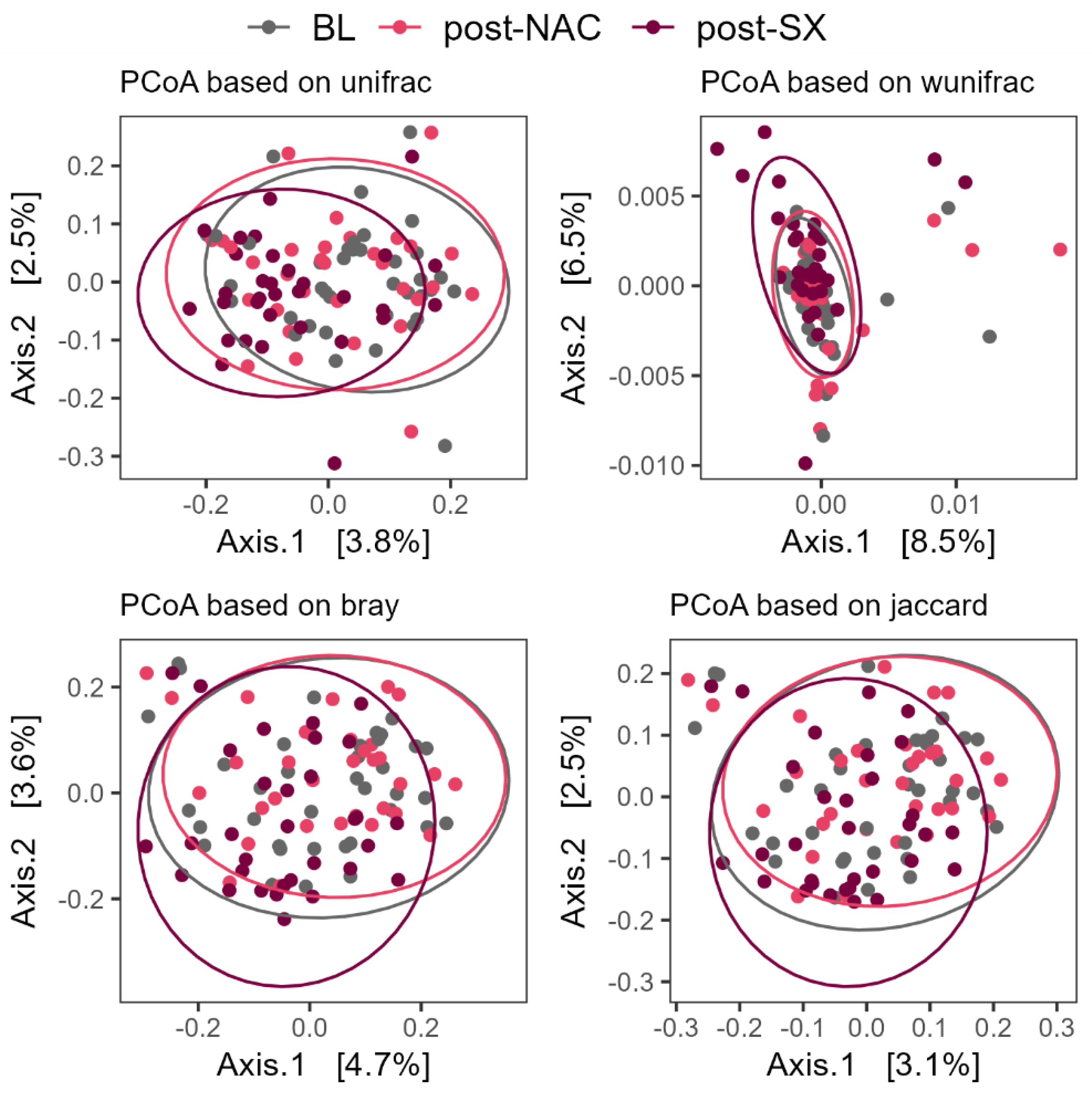
Beta diversity analysis, using principal coordinates analysis (PCoA) based on the unique fraction metric (unifrac), weighted unifrac (wunifrac), and the Bray–Curtis, and Jaccard dissimilarities, showed a significant difference between post-surgery and post-NAC timepoints (all p = 0.001) (Figure 2, Table S2). Redundancy analysis confirmed the profound impact of radical surgery on the gut microbiome changes (p = 0.014).

3.3. Microbiome Composition Changes Throughout the Treatment of GC

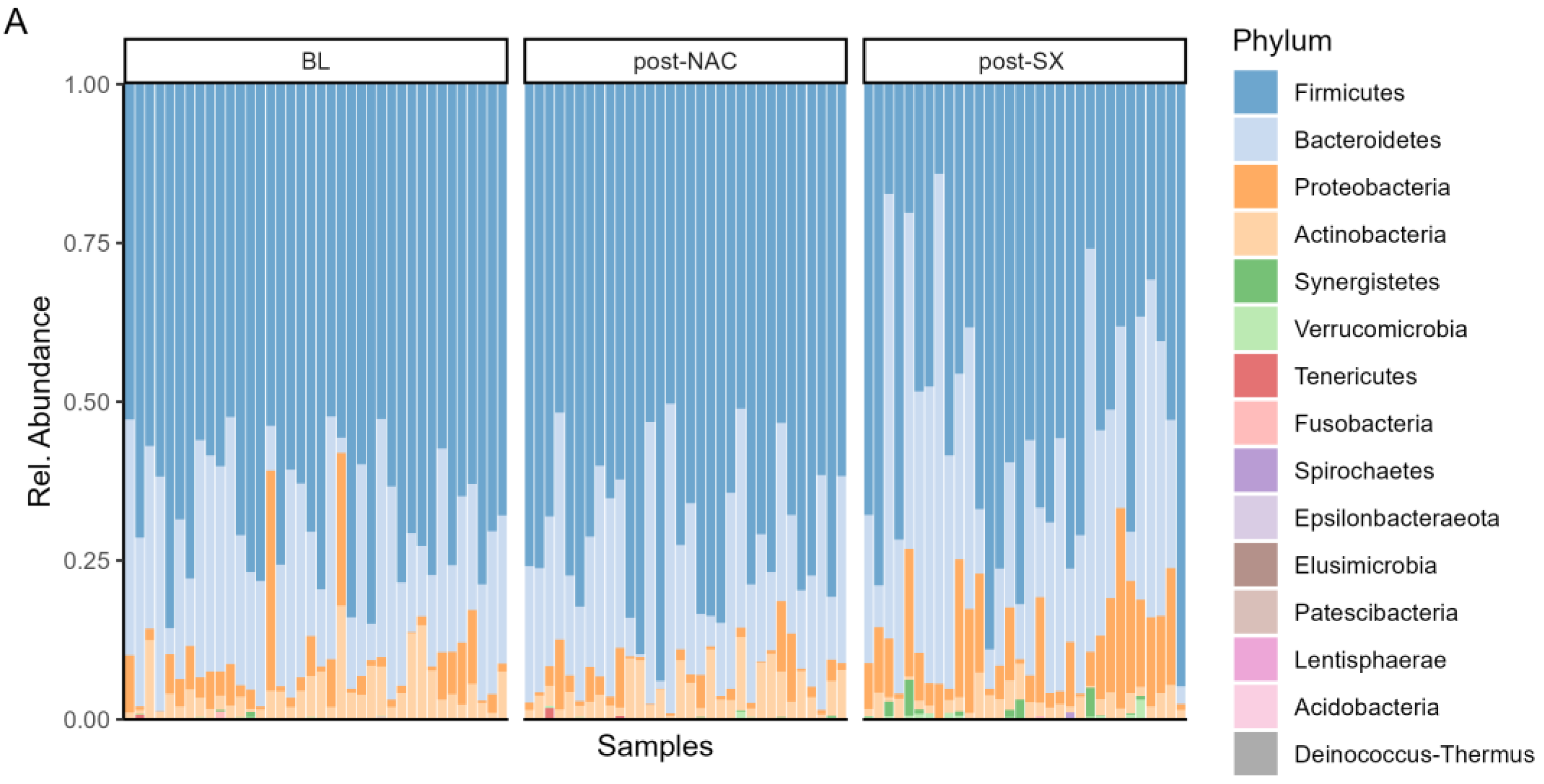
The study showed that the gut microbiome composition changed significantly throughout the treatment of GC. At the phylum level, NAC treatment did not alter the microbiome composition. In contrast, radical surgery led to an increased abundance of Bacteroidetes (p = 0.004) and Proteobacteria (p < 0.0001) and a decreased abundance of Firmicutes (p < 0.0001) and Actinobacteria (p = 0.001) compared to NAC (Figure 3A, Table S3).
At the genus level, NAC treatment resulted in a significant decrease in the Christensenellaceae R-7 group (p = 0.041) compared to BL samples. In contrast, radical surgery led to an increase in Prevotella 9 (p = 0.022), Streptococcus (p = 0.010), and Escherichia-Shigella (p = 0.001). This combination also caused a decrease in Lactobacillus (p = 0.008), Collinsella (p = 0.005), Faecalibacterium (p = 0.030), and the Ruminococcus torques group (p = 0.006) (Figure 3B, Table S4).

3.4. The Microbiome Composition After Surgery Is Enriched with Oralization-Associated Bacteria

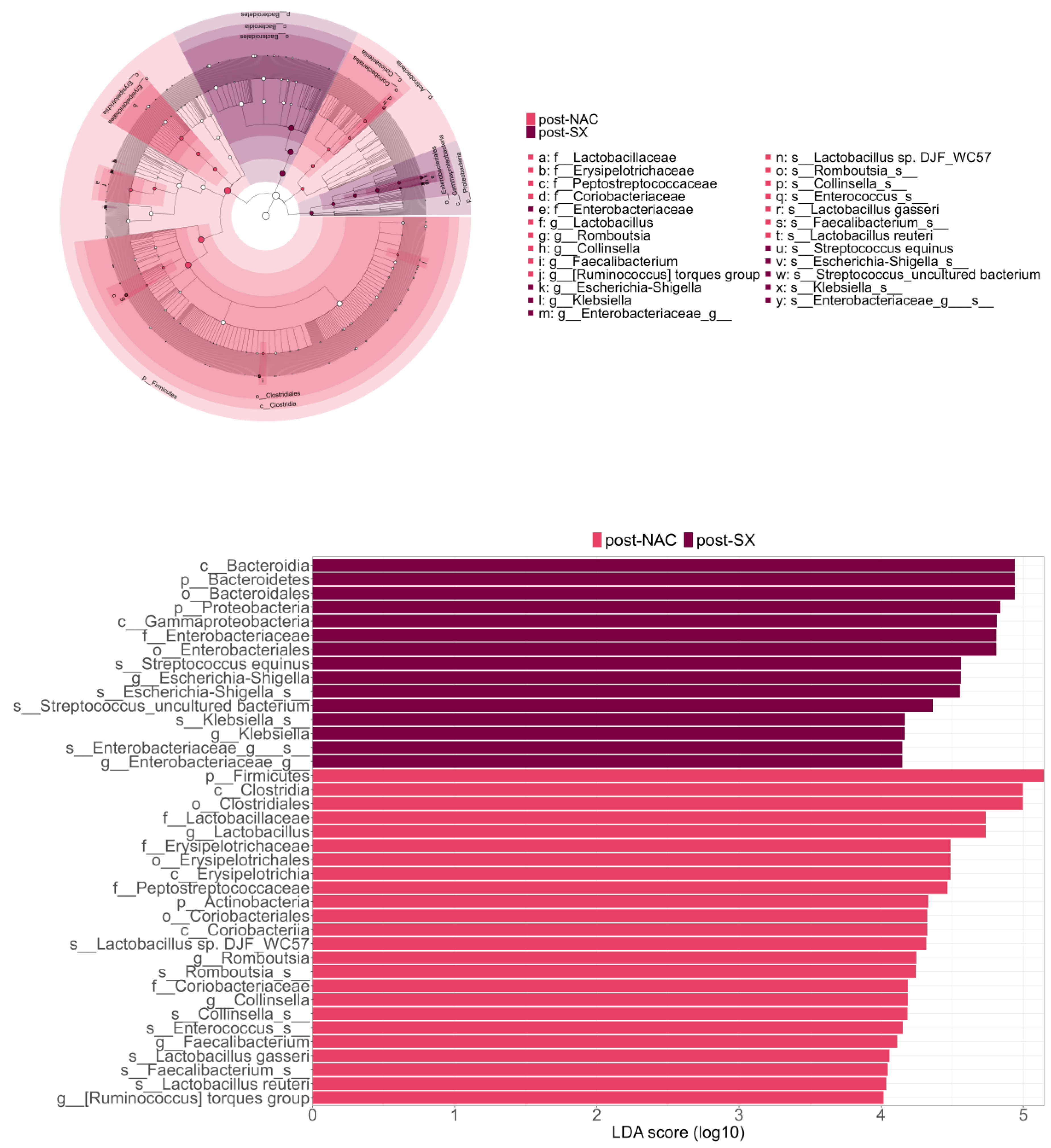
LEfSe analysis supported these findings, demonstrating that, post-surgery, patients exhibited a more enriched microbiome characterized by a higher abundance of Escherichia-Shigella species, Streptococcus equinus, uncultured Streptococcus species, and members of the Enterobacteriaceae family (Figure 4).
Additionally, ANCOM validated these results by demonstrating that post-SX patients had higher levels of Escherichia-Shigella and lower levels of Faecalibacterium and the Ruminococcus torques group. Linear models showed that the type of surgery, total or subtotal gastrectomy, had no impact on the gut microbiome composition except for the higher levels of Bacteroides in patients who received a total gastrectomy (Figure 5, Table S5).

3.5. Fecal and Blood Biomarkers Throughout the Treatment of GC

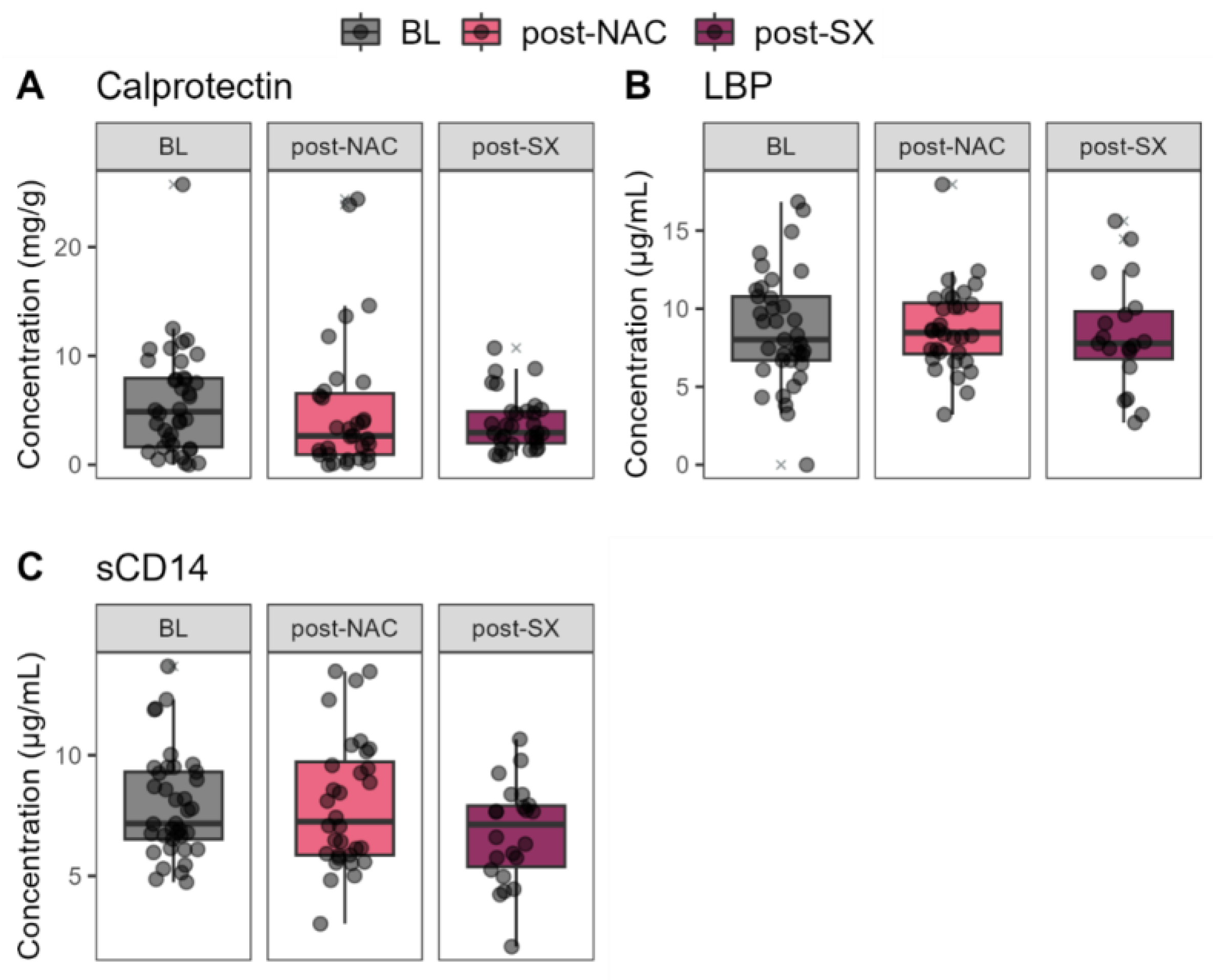
Fecal calprotectin, lipopolysaccharide-binding protein (LBP), and soluble Cluster Differentiation 14 (sCD14) biomarkers from the blood serum did not significantly change throughout the treatment (Figure 6).

4. Discussion

This study aimed to investigate gut microbiome changes throughout GC treatment with NAC and radical surgery and to explore the effect of treatment. The results indicate significant changes in the gut microbiome during treatment, driven primarily by radical surgery rather than NAC. NAC alone did not impact the gut microbiome composition at the phylum level. In contrast, radical surgery led to an increased abundance of Bacteroidetes and Proteobacteria phylum and a decreased abundance of Firmicutes and Actinobacteria phylum. Furthermore, NAC did not affect alpha or beta diversity, whereas radical surgery impacted both. The microbiome composition after radical surgery was enriched with oralization-associated bacteria, as LEfSe analysis showed an increased abundance of Escherichia-Shigella, Streptococcus equinus, and uncultured bacterium Streptococcus species, and species from Enterobacteriaceae family. ANCOM validated these results by demonstrating higher levels of Escherichia-Shigella and lower levels of Faecalibacterium genus and the Ruminococcus torques group after radical surgery.
Surprisingly, our study did not show a notable impact of NAC on gut microbiome composition. These findings contradict several previous studies of various malignancies [25,26,27,28], including gastrointestinal cancers [12,29]. For instance, a recent cross-sectional study by Li et al. reported that the gut microbiome of patients with metastatic or locally advanced esophagogastric or colorectal cancer undergoing chemotherapy differed from that of healthy controls, showing increased richness and compositional shifts in cancer patients [29]. However, this study was not longitudinal and did not include pre- and post-treatment comparisons, unlike our study. The longitudinal study by Kong et al. [12] investigated gut microbiome changes during colorectal cancer treatment with radical surgery and adjuvant capecitabine plus oxaliplatin (CapeOx) chemotherapy and showed that chemotherapy had an impact on the changes in the microbiome’s composition. Several methodological differences between Kong et al.’s [12] study and ours should be considered when interpreting these disparate findings. First, we assessed the effect of NAC on the microbiome before surgery, whereas Kong et al. [12] examined the impact of chemotherapy after colorectal resection. Second, we investigated the effects of FLOT chemotherapy in GC patients, while Kong et al. [12] focused on CapeOx in colorectal cancer patients. Most importantly, the timing of sample collection differed significantly. We collected samples approximately four weeks after NAC, just before surgery, whereas Kong et al. [12] collected samples after each cycle of chemotherapy. This suggests that chemotherapy-induced changes in the gut microbiome may be transient, with a four-week interval possibly allowing the microbiome to recover and reach a stable state. Another study by Chen et al. investigated the impact of surgery and chemotherapy on gut microbiome composition in GC patients [30]. Despite its retrospective cross-sectional design and lack of specificity regarding the type of chemotherapy and surgery, the study found no significant differences in gut microbiome composition between patients treated with and without chemotherapy [30], which aligns with our present findings. To sum up, this longitudinal study is the first to demonstrate that FLOT chemotherapy has no notable and lasting impact on gut microbiome composition in GC patients 4 weeks after the completion of neoadjuvant treatment.
The most important aspect of the present study is the demonstration that, contrary to NAC, radical surgery has a notable and relatively long-lasting impact on gut microbiome composition. Increased oralization-associated bacteria such as Streptococcus and Escherichia-Shigella genera can characterize radical surgery-induced dysbiosis. Our previous proof-of-concept study proposed that gastrectomy-associated gut microbiome oralization is a significant side effect of the treatment characterized by an increased abundance of bacteria typically found in the oral cavity, including Escherichia-Shigella, Enterococcus, Streptococcus, Veillonella, Oribacterium, and Mogibacterium [6]. The cross-sectional design of the previous study prevented us from drawing irrefutable conclusions. Therefore, this longitudinal study was necessary to confirm the hypothesis that the loss of the gastric barrier following gastrectomy creates conditions for oral cavity bacteria to colonize the distal gastrointestinal tract [6]. Moreover, our previous study demonstrated that gut microbiome oralization is linked to gastrointestinal symptoms, such as bloating, diarrhea, and abdominal discomfort, in GC survivors [6]. This oralization process is mainly associated with intestinal inflammation and the presence of Streptococcus in the stool [6]. Chronic intestinal inflammation is a known factor in the pathogenesis of chronic diarrhea [31], a common issue affecting about 40% of long-term survivors after gastrectomy [4,32,33,34]. The present study convincingly confirmed that gastrectomy increases the abundance of Streptococcus, a prevalent bacterial taxon in the oral cavity and commonly implicated in PPI-induced dysbiosis [8,9,35,36,37]. Additionally, the present study showed a radical surgery-induced increase in Escherichia-Shigella and lower levels of Faecalibacterium and the Ruminococcus genera. Escherichia is commonly implicated in small intestinal bacterial overgrowth [38], which frequently occurs in patients after gastrectomy and is associated with intestinal and postprandial symptoms [39]. Members of the genus Escherichia–Shigella are not sensitive to pH variations in their environment, and they appear to benefit from the altered milieu, as evidenced by their increased abundance observed in the general population after PPI intake [8,40]. In previous reports, Ruminococcus species have been associated with a stable human microbiome [41]. Decreased abundance of Ruminococcus has been linked to diarrhea in a porcine animal model [42], and specifically, Ruminococcus 1 was found to be depleted in GC survivors suffering from diarrhea [6].
It is not surprising that the type of surgery had no significant impact on dysbiosis, as shown in our results. Total gastrectomy leads to an instant loss of the gastric barrier, while subtotal gastrectomy results in a significant increase in gastric pH due to reduced gastric secretion, raising it above the threshold value of 4, which is considered necessary for a potent bactericidal effect [43,44].
To our surprise, the present study failed to show a surgery-induced increase in intestinal inflammation measured by fecal calprotectin. These findings contradict our previous study, which demonstrated elevated fecal calprotectin levels in gastrectomized patients compared to healthy controls [6]. However, the current longitudinal study compared post-surgery calprotectin levels with baseline levels. It is known that fecal calprotectin levels are elevated in patients with esophagogastric cancer [45], which was the case at our baseline timepoint. This pre-existing increase in calprotectin due to cancer may have masked any additional rise induced by the surgery, preventing us from detecting a surgery-induced increase in intestinal inflammation.
This study has some limitations that need to be considered. It is a side analysis of a previous randomized controlled trial [3], where the impact of treatment on gut microbiome composition was a secondary endpoint. Consequently, patients were enrolled without prior statistical power calculations to determine the optimal cohort size. As a result, the cohort size is relatively small, and the findings may require validation in a larger cohort. Also, expanding sample collection across multiple time points after NAC and radical surgery would be beneficial in improving the coherence and persuasiveness of the research findings. This approach would provide a more comprehensive depiction of the dynamic changes in the gut microbiome. However, this study also has some clear benefits. First, it employs a longitudinal design, allowing observation of changes in the microbiome throughout the complete treatment period. Additionally, the sequencing data used are of high quality, enabling precise data analysis that considers even the least common bacteria. Thus, the study provides clear evidence of how the gut microbiome changes throughout GC treatment with NAC and radical surgery.
The findings of the present study have significant clinical relevance. The results demonstrate that radical surgery, rather than NAC, exerts a long-term impact on the gut microbiome, resulting in oralization-defined dysbiosis. When combined with existing evidence associating this specific dysbiosis with gastrointestinal symptoms frequently reported by long-term gastric cancer (GC) survivors, these findings underscore the gut microbiome as a compelling therapeutic target for alleviating such symptoms. Future studies should explore gut microbiome-modulating interventions, such as probiotics, to alleviate gastrointestinal symptoms in long-term GC survivors.

5. Conclusions

Radical treatment of advanced GC with NAC and radical surgery has long-term effects on the gut microbiome, which is evident one year after treatment initiation. These sustained alterations primarily stem from radical gastrectomy rather than the NAC.

Supplementary Materials

The following supporting information can be downloaded at: https://www.mdpi.com/article/10.3390/cancers16234074/s1, Table S1: Linear mixed-effects model results for alpha-diversity parameters; Table S2: PERMANOVA analysis results of microbiome composition changes throughout the treatment of gastrointestinal cancer; Table S3: Linear mixed-effects model results for the relative abundance of the most common phylum, when the effect of NAC is compared to the baseline and radical surgery (SX). The table presents the estimates, standard errors (S.E.), t-values (t val.), degrees of freedom (d.f.), and p-values; Table S4: Linear mixed-effects model results for the relative abundance of the most common genera, when the effect of NAC is compared to the baseline and radical surgery (SX). The table presents the estimates, standard errors (S.E.), t-values (t val.), degrees of freedom (d.f.), and p-values; Table S5: Linear model results for the relative abundance of the most common genera when subtotal gastrectomy is compared to total gastrectomy.

Author Contributions

Conceptualization, R.B., R.S., A.H., V.S. and A.B.; methodology, K.Ž., A.H., A.M., S.J., V.S., R.B. and A.B.; software, K.Ž. and B.B.; validation, R.B., A.B. and A.H.; formal analysis, K.Ž. and B.B.; investigation, B.B, A.B., A.Š., A.M., R.S. and R.B.; resources, R.B.; data curation, K.Ž., A.Š., A.B., B.B. and R.B.; writing—original draft preparation, K.Ž. and A.B.; writing—review and editing, K.Ž., B.B., A.H., R.S., A.Š., A.M., S.J., V.S., R.B. and A.B.; visualization, K.Ž.; supervision, A.B. and R.B.; project administration, A.B.; funding acquisition, R.B. All authors have read and agreed to the published version of the manuscript.

Funding

This project has received funding from the Research Council of Lithuania (LMTLT), agreement No S-MIP-22-30.

Institutional Review Board Statement

The study was conducted according to the ethical standard of the Declaration of Helsinki of 2013, and approved by the Vilnius Regional Bioethics Committee (2020/1-1185-675).

Informed Consent Statement

All patients provided written informed consent before participation in the study.

Data Availability Statement

This side-study of a pilot two-arm randomized clinical trial is registered in the https://clinicaltrials.gov/ (NCT04223401) registry. Raw sequencing data are made publicly available in the National Centre for Biotechnology Information (NCBI) sequence read archive (SRA) at https://www.ncbi.nlm.nih.gov/sra (SRA data accession No. PRJNA1124058). Other data are available from the corresponding author upon reasonable request.

Conflicts of Interest

Authors Angela Horvath and Vanessa Stadlbauer were employed by the company CBmed GmbH. The remaining authors declare that the research was conducted in the absence of any commercial or financial relationships that could be construed as a potential conflict of interest.

References

  1. Sung, H.; Ferlay, J.; Siegel, R.L.; Laversanne, M.; Soerjomataram, I.; Jemal, A.; Bray, F. Global Cancer Statistics 2020: GLOBOCAN Estimates of Incidence and Mortality Worldwide for 36 Cancers in 185 Countries. CA Cancer J. Clin. 2021, 71, 209–249. [Google Scholar] [CrossRef] [PubMed]
  2. Li, Y.; Feng, A.; Zheng, S.; Chen, C.; Lyu, J. Recent Estimates and Predictions of 5-Year Survival in Patients with Gastric Cancer: A Model-Based Period Analysis. Cancer Control J. Moffitt Cancer Cent. 2022, 29, 10732748221099228. [Google Scholar] [CrossRef] [PubMed]
  3. Bausys, A.; Luksta, M.; Anglickiene, G.; Maneikiene, V.V.; Kryzauskas, M.; Rybakovas, A.; Dulskas, A.; Kuliavas, J.; Stratilatovas, E.; Macijauskiene, L.; et al. Effect of Home-Based Prehabilitation on Postoperative Complications after Surgery for Gastric Cancer: Randomized Clinical Trial. Br. J. Surg. 2023, 110, 1800–1807. [Google Scholar] [CrossRef] [PubMed]
  4. Karanicolas, P.J.; Graham, D.; Gönen, M.; Strong, V.E.; Brennan, M.F.; Coit, D.G. Quality of Life after Gastrectomy for Adenocarcinoma: A Prospective Cohort Study. Ann. Surg. 2013, 257, 1039–1046. [Google Scholar] [CrossRef] [PubMed]
  5. Lee, S.S.; Chung, H.Y.; Kwon, O.K.; Yu, W. Long-Term Quality of Life After Distal Subtotal and Total Gastrectomy: Symptom- and Behavior-Oriented Consequences. Ann. Surg. 2016, 263, 738–744. [Google Scholar] [CrossRef]
  6. Horvath, A.; Bausys, A.; Sabaliauskaite, R.; Stratilatovas, E.; Jarmalaite, S.; Schuetz, B.; Stiegler, P.; Bausys, R.; Stadlbauer, V.; Strupas, K. Distal Gastrectomy with Billroth II Reconstruction is Associated with Oralization of Gut Microbiome and Intestinal Inflammation: A Proof-of-Concept Study. Ann. Surg. Oncol. 2020, 28, 1198–1208. [Google Scholar] [CrossRef]
  7. Huang, L.; Xu, A.-M.; Li, T.-J.; Han, W.-X.; Xu, J. Should Peri-Gastrectomy Gastric Acidity Be Our Focus among Gastric Cancer Patients? World J. Gastroenterol. WJG 2014, 20, 6981. [Google Scholar] [CrossRef]
  8. Imhann, F.; Bonder, M.J.; Vich Vila, A.; Fu, J.; Mujagic, Z.; Vork, L.; Tigchelaar, E.F.; Jankipersadsing, S.A.; Cenit, M.C.; Harmsen, H.J.M.; et al. Proton Pump Inhibitors Affect the Gut Microbiome. Gut 2016, 65, 740–748. [Google Scholar] [CrossRef]
  9. Jackson, M.A.; Goodrich, J.K.; Maxan, M.-E.; Freedberg, D.E.; Abrams, J.A.; Poole, A.C.; Sutter, J.L.; Welter, D.; Ley, R.E.; Bell, J.T.; et al. Proton Pump Inhibitors Alter the Composition of the Gut Microbiota. Gut 2016, 65, 749–756. [Google Scholar] [CrossRef]
  10. Seto, C.T.; Jeraldo, P.; Orenstein, R.; Chia, N.; DiBaise, J.K. Prolonged Use of a Proton Pump Inhibitor Reduces Microbial Diversity: Implications for Clostridium Difficile Susceptibility. Microbiome 2014, 2, 42. [Google Scholar] [CrossRef]
  11. Shuwen, H.; Xi, Y.; Yuefen, P.; Jiamin, X.; Quan, Q.; Haihong, L.; Yizhen, J.; Wei, W. Effects of Postoperative Adjuvant Chemotherapy and Palliative Chemotherapy on the Gut Microbiome in Colorectal Cancer. Microb. Pathog. 2020, 149, 104343. [Google Scholar] [CrossRef] [PubMed]
  12. Kong, C.; Gao, R.; Yan, X.; Huang, L.; He, J.; Li, H.; You, J.; Qin, H. Alterations in Intestinal Microbiota of Colorectal Cancer Patients Receiving Radical Surgery Combined with Adjuvant CapeOx Therapy. Sci. China Life Sci. 2019, 62, 1178–1193. [Google Scholar] [CrossRef] [PubMed]
  13. Tong, J.; Zhang, X.; Fan, Y.; Chen, L.; Ma, X.; Yu, H.; Li, J.; Guan, X.; Zhao, P.; Yang, J. Changes of Intestinal Microbiota in Ovarian Cancer Patients Treated with Surgery and Chemotherapy. Cancer Manag. Res. 2020, 12, 8125–8135. [Google Scholar] [CrossRef] [PubMed]
  14. Stringer, A.M.; Al-Dasooqi, N.; Bowen, J.M.; Tan, T.H.; Radzuan, M.; Logan, R.M.; Mayo, B.; Keefe, D.M.K.; Gibson, R.J. Biomarkers of Chemotherapy-Induced Diarrhoea: A Clinical Study of Intestinal Microbiome Alterations, Inflammation and Circulating Matrix Metalloproteinases. Support. Care Cancer Off. J. Multinatl. Assoc. Support. Care Cancer 2013, 21, 1843–1852. [Google Scholar] [CrossRef]
  15. Fei, Z.; Lijuan, Y.; Xi, Y.; Wei, W.; Jing, Z.; Miao, D.; Shuwen, H. Gut Microbiome Associated with Chemotherapy-Induced Diarrhea from the CapeOX Regimen as Adjuvant Chemotherapy in Resected Stage III Colorectal Cancer. Gut Pathog. 2019, 11, 18. [Google Scholar] [CrossRef]
  16. Bausys, A.; Luksta, M.; Kuliavas, J.; Anglickiene, G.; Maneikiene, V.; Gedvilaite, L.; Celutkiene, J.; Jamontaite, I.; Cirtautas, A.; Lenickiene, S.; et al. Personalized Trimodal Prehabilitation for Gastrectomy. Medicine 2020, 99, e20687. [Google Scholar] [CrossRef]
  17. Caporaso, J.G.; Kuczynski, J.; Stombaugh, J.; Bittinger, K.; Bushman, F.D.; Costello, E.K.; Fierer, N.; Peña, A.G.; Goodrich, J.K.; Gordon, J.I.; et al. QIIME Allows Analysis of High-Throughput Community Sequencing Data. Nat. Methods 2010, 7, 335–336. [Google Scholar] [CrossRef]
  18. Callahan, B.J.; McMurdie, P.J.; Rosen, M.J.; Han, A.W.; Johnson, A.J.A.; Holmes, S.P. DADA2: High-Resolution Sample Inference from Illumina Amplicon Data. Nat. Methods 2016, 13, 581–583. [Google Scholar] [CrossRef]
  19. Bolyen, E.; Rideout, J.R.; Dillon, M.R.; Bokulich, N.A.; Abnet, C.C.; Al-Ghalith, G.A.; Alexander, H.; Alm, E.J.; Arumugam, M.; Asnicar, F.; et al. Reproducible, Interactive, Scalable and Extensible Microbiome Data Science Using QIIME 2. Nat. Biotechnol. 2019, 37, 852–857. [Google Scholar] [CrossRef]
  20. Bisanz, J.E. qiime2R. Importing QIIME2 Artifacts and Associated Data into R Sessions. Version 0.99. 2018, Volume 13. Available online: https://github.com/jbisanz/qiime2R (accessed on 14 June 2024).
  21. Oksanen, J.; Simpson, G.L.; Blanchet, F.G.; Kindt, R.; Legendre, P.; Minchin, P.R.; O’Hara, R.B.; Solymos, P.; Stevens, M.H.; Szoecs, E.; et al. Vegan: Community Ecology Package 2022. Available online: https://github.com/vegandevs/vegan (accessed on 14 June 2024).
  22. Cao, Y.; Dong, Q.; Wang, D.; Zhang, P.; Liu, Y.; Niu, C. MicrobiomeMarker: An R/Bioconductor Package for Microbiome Marker Identification and Visualization. Bioinforma. Oxf. Engl. 2022, 38, 4027–4029. [Google Scholar] [CrossRef]
  23. Bates, D.; Mächler, M.; Bolker, B.; Walker, S. Fitting Linear Mixed-Effects Models Using Lme4. J. Stat. Softw. 2015, 67, 1–48. [Google Scholar] [CrossRef]
  24. Yu, G. Convert Plot to “grob” or “Ggplot” Object. 2021. Available online: https://github.com/GuangchuangYu/ggplotify (accessed on 14 June 2024).
  25. Montassier, E.; Gastinne, T.; Vangay, P.; Al-Ghalith, G.A.; Bruley des Varannes, S.; Massart, S.; Moreau, P.; Potel, G.; de La Cochetière, M.F.; Batard, E.; et al. Chemotherapy-Driven Dysbiosis in the Intestinal Microbiome. Aliment. Pharmacol. Ther. 2015, 42, 515–528. [Google Scholar] [CrossRef] [PubMed]
  26. Zwielehner, J.; Lassl, C.; Hippe, B.; Pointner, A.; Switzeny, O.J.; Remely, M.; Kitzweger, E.; Ruckser, R.; Haslberger, A.G. Changes in Human Fecal Microbiota Due to Chemotherapy Analyzed by TaqMan-PCR, 454 Sequencing and PCR-DGGE Fingerprinting. PLoS ONE 2011, 6, e28654. [Google Scholar] [CrossRef] [PubMed]
  27. Dörffel, Y.; Swidsinski, A.; Loening-Baucke, V.; Wiedenmann, B.; Pavel, M. Common Biostructure of the Colonic Microbiota in Neuroendocrine Tumors and Crohn’s Disease and the Effect of Therapy. Inflamm. Bowel Dis. 2012, 18, 1663–1671. [Google Scholar] [CrossRef]
  28. Galloway-Peña, J.R.; Smith, D.P.; Sahasrabhojane, P.; Ajami, N.J.; Wadsworth, W.D.; Daver, N.G.; Chemaly, R.F.; Marsh, L.; Ghantoji, S.S.; Pemmaraju, N.; et al. The Role of the Gastrointestinal Microbiome in Infectious Complications during Induction Chemotherapy for Acute Myeloid Leukemia. Cancer 2016, 122, 2186–2196. [Google Scholar] [CrossRef]
  29. Li, N.; Bai, C.; Zhao, L.; Sun, Z.; Ge, Y.; Li, X. The Relationship Between Gut Microbiome Features and Chemotherapy Response in Gastrointestinal Cancer. Front. Oncol. 2021, 11, 781697. [Google Scholar] [CrossRef]
  30. Chen, C.; Shen, J.; Du, Y.; Shi, X.; Niu, Y.; Jin, G.; Liu, Y.; Shi, Y.; Lyu, J.; Lin, L. Characteristics of Gut Microbiota in Patients with Gastric Cancer by Surgery, Chemotherapy and Lymph Node Metastasis. Clin. Transl. Oncol. Off. Publ. Fed. Span. Oncol. Soc. Natl. Cancer Inst. Mex. 2022, 24, 2181–2190. [Google Scholar] [CrossRef]
  31. Anbazhagan, A.N.; Priyamvada, S.; Alrefai, W.A.; Dudeja, P.K. Pathophysiology of IBD associated diarrhea. Tissue Barriers 2018, 6, e1463897. [Google Scholar] [CrossRef]
  32. Yu, W.; Park, K.B.; Chung, H.Y.; Kwon, O.K.; Lee, S.S. Chronological Changes of Quality of Life in Long-Term Survivors after Gastrectomy for Gastric Cancer. Cancer Res. Treat. 2016, 48, 1030–1036. [Google Scholar] [CrossRef]
  33. Kim, A.R.; Cho, J.; Hsu, Y.-J.; Choi, M.G.; Noh, J.H.; Sohn, T.S.; Bae, J.M.; Yun, Y.H.; Kim, S. Changes of Quality of Life in Gastric Cancer Patients after Curative Resection: A Longitudinal Cohort Study in Korea. Ann. Surg. 2012, 256, 1008–1013. [Google Scholar] [CrossRef]
  34. Aoki, T.; Yamaji, I.; Hisamoto, T.; Sato, M.; Matsuda, T. Irregular Bowel Movement in Gastrectomized Subjects: Bowel Habits, Stool Characteristics, Fecal Flora, and Metabolites. Gastric Cancer 2012, 15, 396–404. [Google Scholar] [CrossRef]
  35. Tsuda, A.; Suda, W.; Morita, H.; Takanashi, K.; Takagi, A.; Koga, Y.; Hattori, M. Influence of Proton-Pump Inhibitors on the Luminal Microbiota in the Gastrointestinal Tract. Clin. Transl. Gastroenterol. 2015, 6, e89. [Google Scholar] [CrossRef] [PubMed]
  36. Bajaj, J.S.; Cox, I.J.; Betrapally, N.S.; Heuman, D.M.; Schubert, M.L.; Ratneswaran, M.; Hylemon, P.B.; White, M.B.; Daita, K.; Noble, N.A.; et al. Systems Biology Analysis of Omeprazole Therapy in Cirrhosis Demonstrates Significant Shifts in Gut Microbiota Composition and Function. Am. J. Physiol. Gastrointest. Liver Physiol. 2014, 307, G951–G957. [Google Scholar] [CrossRef] [PubMed]
  37. Clooney, A.G.; Bernstein, C.N.; Leslie, W.D.; Vagianos, K.; Sargent, M.; Laserna-Mendieta, E.J.; Claesson, M.J.; Targownik, L.E. A Comparison of the Gut Microbiome between Long-Term Users and Non-Users of Proton Pump Inhibitors. Aliment. Pharmacol. Ther. 2016, 43, 974–984. [Google Scholar] [CrossRef] [PubMed]
  38. Sachdev, A.H.; Pimentel, M. Gastrointestinal Bacterial Overgrowth: Pathogenesis and Clinical Significance. Ther. Adv. Chronic Dis. 2013, 4, 223–231. [Google Scholar] [CrossRef]
  39. Paik, C.N.; Choi, M.-G.; Lim, C.H.; Park, J.M.; Chung, W.C.; Lee, K.-M.; Jun, K.-H.; Song, K.Y.; Jeon, H.M.; Chin, H.-M.; et al. The Role of Small Intestinal Bacterial Overgrowth in Postgastrectomy Patients. Neurogastroenterol. Motil. Off. J. Eur. Gastrointest. Motil. Soc. 2011, 23, e191–e196. [Google Scholar] [CrossRef]
  40. Small, P.; Blankenhorn, D.; Welty, D.; Zinser, E.; Slonczewski, J.L. Acid and Base Resistance in Escherichia Coli and Shigella Flexneri: Role of RpoS and Growth PH. J. Bacteriol. 1994, 176, 1729–1737. [Google Scholar] [CrossRef]
  41. Engelbrektson, A.; Korzenik, J.R.; Pittler, A.; Sanders, M.E.; Klaenhammer, T.R.; Leyer, G.; Kitts, C.L. Probiotics to Minimize the Disruption of Faecal Microbiota in Healthy Subjects Undergoing Antibiotic Therapy. J. Med. Microbiol. 2009, 58, 663–670. [Google Scholar] [CrossRef]
  42. Yang, Q.; Huang, X.; Wang, P.; Yan, Z.; Sun, W.; Zhao, S.; Gun, S. Longitudinal development of the gut microbiota in healthy and diarrheic piglets induced by age-related dietary changes. Microbiologyopen 2019, 8, e923. [Google Scholar] [CrossRef]
  43. Martinsen, T.C.; Bergh, K.; Waldum, H.L. Gastric Juice: A Barrier against Infectious Diseases. Basic Clin. Pharmacol. Toxicol. 2005, 96, 94–102. [Google Scholar] [CrossRef]
  44. Carboni, M.; Guadagni, S.; Pistoia, M.A.; Amicucci, G.; Tuscano, D.; Negro, P.; Smith, P.L.; Walters, C.L. The Microflora of the Gastric Juice after Billroth I and Billroth II Partial Gastrectomy. Scand. J. Gastroenterol. 1986, 21, 461–470. [Google Scholar] [CrossRef]
  45. Vincent, Z.; Hornby, S.; Ball, S.; Sanders, G.; Ayling, R.M. Faecal Calprotectin as a Marker for Oesophago-Gastric Cancer. Ann. Clin. Biochem. 2015, 52, 660–664. [Google Scholar] [CrossRef]
Figure 1. Changes in alpha-diversity parameters in patients undergoing advanced gastric cancer treatment; outliers are marked in grey; Alpha-diversity parameters: (A) Richness, (B) Shannon index, (C) Inverse Simpson index, (D) Evenness, (E) Phylogenetic Diversity; BL–baseline, NAC–neoadjuvant chemotherapy, SX–surgery.
Cancers 16 04074 g001
Figure 2. Changes in beta-diversity parameters in patients undergoing chemotherapy and surgery combined with chemotherapy; BL–baseline, NAC–neoadjuvant chemotherapy, SX–surgery.
Cancers 16 04074 g002
Figure 3. Bacterial composition of the gut microbiome at the (A) phylum and (B) genus level at different treatment timepoints; BL–baseline, NAC–neoadjuvant chemotherapy, SX–surgery (radical gastrectomy).
Cancers 16 04074 g003aCancers 16 04074 g003b
Figure 4. The primary microbiome differences between post-surgery (post-SX) and post-neoadjuvant chemotherapy (post-NAC) treatment in patients with advanced gastric cancer.
Cancers 16 04074 g004
Figure 5. Comparative analysis of the relative abundance of various genera in patients undergoing total or subtotal gastrectomy.
Cancers 16 04074 g005
Figure 6. Blood parameters in gastric cancer patients post-surgery (post-SX) and post-neoadjuvant chemotherapy (post-NAC) treatment, compared to the baseline (BL).
Cancers 16 04074 g006
Table 1. Baseline and treatment characteristics of the study patients.
Characteristic x
Age (years), mean (SD)60 (11)
Male: Female24:14
Charlson comorbidity index score, mean (SD)4 (1.4)
BMI (kg/m2), mean (SD)25 (4)
cT, n (%)1–24 (10.5)
3–434 (89.5)
cN, n (%)010 (26.3)
1–328 (73.7)
cM, n (%)036 (94.7)
12 (5.3)
Tumor localization, n (%)Upper third6 (15.8)
Middle third14 (36.8)
Lower third16 (42.1)
Total2 (5.3)
Type of neoadjuvant chemotherapy, n (%)FLOT36 (94.7)
Cisplatin+5-FU2 (5.3)
Number of cycles, mean (SD)4 (1)
Type of surgery, n (%)Total gastrectomy11 (28.9)
Subtotal gastrectomy27 (71.1)
R0, n (%)37 (97.4)
D2 lymphadenectomy, n (%)38 (100)
Laparoscopic surgery, n (%)10 (26.3)
ypT, n (%)02 (5.3)
18 (21.0)
24 (10.5)
317 (44.7)
47 (18.5)
ypN, n (%)016 (42.1)
110 (26.3)
26 (15.8)
36 (15.8)
ypM, n (%)038 (100)
10 (0)
Disclaimer/Publisher’s Note: The statements, opinions and data contained in all publications are solely those of the individual author(s) and contributor(s) and not of MDPI and/or the editor(s). MDPI and/or the editor(s) disclaim responsibility for any injury to people or property resulting from any ideas, methods, instructions or products referred to in the content.