Endobronchial Ultrasound Using Guide Sheath-Guided Transbronchial Lung Biopsy in Ground-Glass Opacity Pulmonary Lesions without Fluoroscopic Guidance
Abstract
Simple Summary
Abstract
1. Introduction
2. Materials and Methods
2.1. Study Design and Subjects
2.2. CT and Bronchoscopy
2.3. Diagnostic Classification
2.4. Statistical Analyses
3. Results
3.1. Baseline Characteristics and Diagnostic Performance
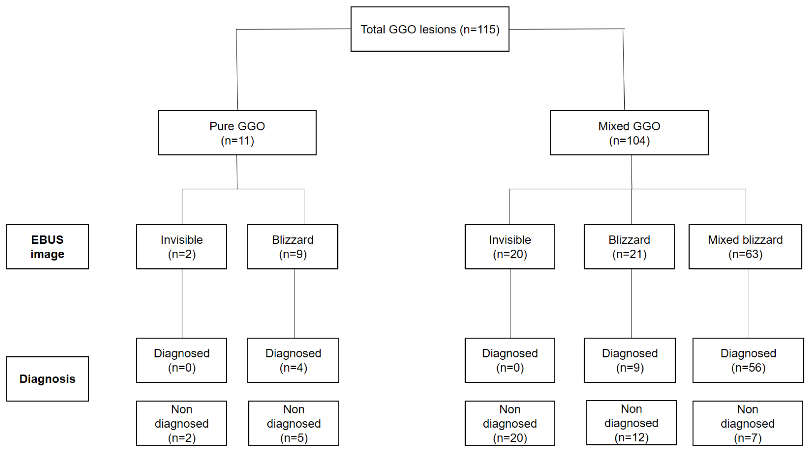
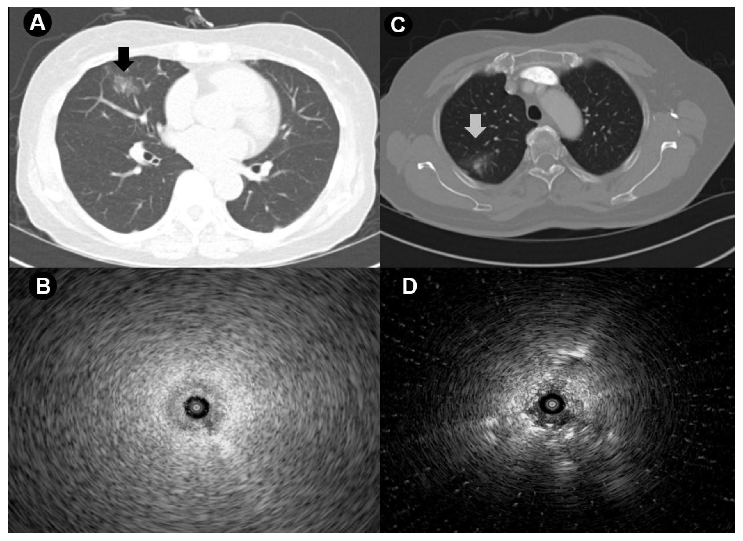
3.2. Correlation between Chest CT Findings and EBUS Image
3.3. Factors Affecting Diagnostic Success
3.4. Complications
4. Discussion
5. Conclusions
Author Contributions
Funding
Institutional Review Board Statement
Informed Consent Statement
Data Availability Statement
Conflicts of Interest
References
- National Lung Screening Trial Research Team; Aberle, D.R.; Adams, A.M.; Berg, C.D.; Black, W.C.; Clapp, J.D.; Fagerstrom, R.M.; Gareen, I.F.; Gatsonis, C.; Marcus, P.M.; et al. Reduced lung-cancer mortality with low-dose computed tomographic screening. N. Engl. J. Med. 2011, 365, 395–409. [Google Scholar] [CrossRef] [PubMed]
- National Lung Screening Trial Research Team; Church, T.R.; Black, W.C.; Aberle, D.R.; Berg, C.D.; Clingan, K.L.; Duan, F.; Fagerstrom, R.M.; Gareen, I.F.; Gierada, D.S.; et al. Results of initial low-dose computed tomographic screening for lung cancer. N. Engl. J. Med. 2013, 368, 1980–1991. [Google Scholar] [CrossRef] [PubMed]
- Lam, D.C.; Liam, C.K.; Andarini, S.; Park, S.; Tan, D.S.W.; Singh, N.; Jang, S.H.; Vardhanabhuti, V.; Ramos, A.B.; Nakayama, T.; et al. Lung Cancer Screening in Asia: An Expert Consensus Report. J. Thorac. Oncol. 2023, 18, 1303–1322. [Google Scholar] [CrossRef]
- Zhang, Y.; Jheon, S.; Li, H.M.; Zhang, H.B.; Xie, Y.Z.; Qian, B.; Lin, K.H.; Wang, S.P.; Fu, C.; Hu, H.; et al. Results of low-dose computed tomography as a regular health examination among Chinese hospital employees. J. Thorac. Cardiovasc. Surg. 2020, 160, 824–831.e4. [Google Scholar] [CrossRef]
- Lee, H.J.; Goo, J.M.; Lee, C.H.; Yoo, C.G.; Kim, Y.T.; Im, J.G. Nodular ground-glass opacities on thin-section CT: Size change during follow-up and pathological results. Korean J. Radiol. 2007, 8, 22–31. [Google Scholar] [CrossRef][Green Version]
- MacMahon, H.; Naidich, D.P.; Goo, J.M.; Lee, K.S.; Leung, A.N.C.; Mayo, J.R.; Mehta, A.C.; Ohno, Y.; Powell, C.A.; Prokop, M.; et al. Guidelines for Management of Incidental Pulmonary Nodules Detected on CT Images: From the Fleischner Society 2017. Radiology 2017, 284, 228–243. [Google Scholar] [CrossRef] [PubMed]
- American College of Radiology. Lung CT Screening Reporting and Data System (Lung-RADS); American College of Radiology: Reston, VA, USA, 2014. [Google Scholar]
- Lee, K.H.; Goo, J.M.; Park, S.J.; Wi, J.Y.; Chung, D.H.; Go, H.; Park, H.S.; Park, C.M.; Lee, S.M. Correlation between the Size of the Solid Component on Thin-Section CT and the Invasive Component on Pathology in Small Lung Adenocarcinomas Manifesting as Ground-Glass Nodules. J. Thorac. Oncol. 2014, 9, 74–82. [Google Scholar] [CrossRef]
- Lee, J.H.; Park, C.M.; Lee, S.M.; Kim, H.; McAdams, H.P.; Goo, J.M. Persistent pulmonary subsolid nodules with solid portions of 5 mm or smaller: Their natural course and predictors of interval growth. Eur. Radiol. 2016, 26, 1529–1537. [Google Scholar] [CrossRef]
- Lee, S.M.; Park, C.M.; Song, Y.S.; Kim, H.; Kim, Y.T.; Park, Y.S.; Goo, J.M. CT assessment-based direct surgical resection of part-solid nodules with solid component larger than 5 mm without preoperative biopsy: Experience at a single tertiary hospital. Eur. Radiol. 2017, 27, 5119–5126. [Google Scholar] [CrossRef] [PubMed]
- Yamauchi, Y.; Izumi, Y.; Nakatsuka, S.; Inoue, M.; Hayashi, Y.; Mukai, M.; Nomori, H. Diagnostic performance of percutaneous core needle lung biopsy under multi-CT fluoroscopic guidance for ground-glass opacity pulmonary lesions. Eur. J. Radiol. 2011, 79, e85–e89. [Google Scholar] [CrossRef]
- Kim, J.; Chee, C.G.; Cho, J.; Kim, Y.; Yoon, M.A. Diagnostic accuracy and complication rate of image-guided percutaneous transthoracic needle lung biopsy for subsolid pulmonary nodules: A systematic review and meta-analysis. Br. J. Radiol. 2021, 94, 20210065. [Google Scholar] [CrossRef]
- Kashiwabara, K.; Semba, H.; Fujii, S.; Tsumura, S. Preoperative Percutaneous Transthoracic Needle Biopsy Increased the Risk of Pleural Recurrence in Pathological Stage I Lung Cancer Patients With Sub-pleural Pure Solid Nodules. Cancer Investig. 2016, 34, 373–377. [Google Scholar] [CrossRef]
- Ikezawa, Y.; Sukoh, N.; Shinagawa, N.; Nakano, K.; Oizumi, S.; Nishimura, M. Endobronchial Ultrasonography with a Guide Sheath for Pure or Mixed Ground-Glass Opacity Lesions. Respiration 2014, 88, 137–143. [Google Scholar] [CrossRef] [PubMed]
- Nakai, T.; Matsumoto, Y.; Suzuk, F.; Tsuchida, T.; Izumo, T. Predictive factors for a successful diagnostic bronchoscopy of ground-glass nodules. Ann. Thorac. Med. 2017, 12, 171–176. [Google Scholar] [CrossRef]
- Hong, K.S.; Ahn, H.; Lee, K.H.; Chung, J.H.; Shin, K.C.; Jin, H.J.; Jang, J.G.; Lee, S.S.; Jang, M.H.; Ahn, J.H. Radial Probe Endobronchial Ultrasound Using Guide Sheath-Guided Transbronchial Lung Biopsy in Peripheral Pulmonary Lesions without Fluoroscopy. Tuberc. Respir. Dis. 2021, 84, 282–290. [Google Scholar] [CrossRef] [PubMed]
- Izumo, T.; Sasada, S.; Chavez, C.; Matsumoto, Y.; Tsuchida, T. Radial endobronchial ultrasound images for ground-glass opacity pulmonary lesions. Eur. Respir. J. 2015, 45, 1661–1668. [Google Scholar] [CrossRef]
- Yamagami, T.; Yoshimatsu, R.; Miura, H.; Yamada, K.; Takahata, A.; Matsumoto, T.; Hasebe, T. Diagnostic performance of percutaneous lung biopsy using automated biopsy needles under CT-fluoroscopic guidance for ground-glass opacity lesions. Br. J. Radiol. 2013, 86, 20120447. [Google Scholar] [CrossRef]
- Yang, J.S.; Liu, Y.M.; Mao, Y.M.; Yuan, J.H.; Yu, W.Q.; Cheng, R.D.; Hu, T.Y.; Cheng, J.M.; Wang, H.Y. Meta-analysis of CT-guided transthoracic needle biopsy for the evaluation of the ground-glass opacity pulmonary lesions. Br. J. Radiol. 2014, 87, 20140276. [Google Scholar] [CrossRef] [PubMed]
- Matsuguma, H.; Nakahara, R.; Kondo, T.; Kamiyama, Y.; Mori, K.; Yokoi, K. Risk of pleural recurrence after needle biopsy in patients with resected early stage lung cancer. Ann. Thorac. Surg. 2005, 80, 2026–2031. [Google Scholar] [CrossRef]
- Moon, S.M.; Lee, D.G.; Hwang, N.Y.; Ahn, S.; Lee, H.; Jeong, B.H.; Choi, Y.S.; Mog, Y.; Kim, T.J.; Lee, K.S.; et al. Ipsilateral pleural recurrence after diagnostic transthoracic needle biopsy in pathological stage I lung cancer patients who underwent curative resection. Lung Cancer 2017, 111, 69–74. [Google Scholar] [CrossRef] [PubMed]
- Inoue, M.; Honda, O.; Tomiyama, N.; Minami, M.; Sawabata, N.; Kadota, Y.; Shintani, Y.; Ohno, Y.; Okumura, M. Risk of pleural recurrence after computed tomographic-guided percutaneous needle biopsy in stage I lung cancer patients. Ann. Thorac. Surg. 2011, 91, 1066–1071. [Google Scholar] [CrossRef]
- Cho, J.; Ko, S.-J.; Kim, S.J.; Lee, Y.J.; Park, J.S.; Cho, Y.-J.; Yoon, H.I.; Cho, S.; Kim, K.; Jheon, S.; et al. Surgical resection of nodular ground-glass opacities without percutaneous needle aspiration or biopsy. BMC Cancer 2014, 14, 838. [Google Scholar] [CrossRef]
- Kim, Y.T. Management of ground-glass nodules: When and how to operate? Cancers 2022, 14, 715. [Google Scholar] [CrossRef]
- Ozeki, N.; Iwano, S.; Taniguchi, T.; Kawaguchi, K.; Fukui, T.; Ishiguro, F.; Fukumoto, K.; Nakamura, S.; Hirakawa, A.; Yokoi, K. Therapeutic surgery without a definitive diagnosis can be an option in selected patients with suspected lung cancer. Interact. Cardiovasc. Thorac. Surg. 2014, 19, 830–837. [Google Scholar] [CrossRef][Green Version]
- Mori, S.; Noda, Y.; Shibazaki, T.; Kato, D.; Matsudaira, H.; Hirano, J.; Ohtsuka, T. Definitive lobectomy without frozen section analysis is a treatment option for large or deep nodules selected carefully with clinical diagnosis of malignancy. Thorac. Cancer 2020, 11, 1996–2004. [Google Scholar] [CrossRef]
- Ettinger, D.S.; Wood, D.E.; Aisner, D.L.; Akerley, W.; Bauman, J.R.; Bharat, A.; Bruno, D.S.; Chang, J.Y.; Chirieac, L.R.; DeCamp, M.; et al. NCCN Guidelines® Insights: Non-Small Cell Lung Cancer, Version 2.2023. J. Natl. Compr. Cancer Netw. 2023, 21, 340–350. [Google Scholar] [CrossRef]
- Wood, D.E.; Kazerooni, E.A.; Aberle, D.; Berman, A.; Brown, L.M.; Eapen, G.A.; Ettinger, D.S.; Ferguson, J.S.; Hou, L.; Kadaria, D.; et al. NCCN Guidelines® Insights: Lung Cancer Screening, Version 1.2022. J. Natl. Compr. Cancer Netw. 2022, 20, 754–764. [Google Scholar] [CrossRef]
- Ikezawa, Y.; Shinagawa, N.; Sukoh, N.; Morimoto, M.; Kikuchi, H.; Watanabe, M.; Nakano, K.; Oizumi, S.; Nishimura, M. Usefulness of Endobronchial Ultrasonography With a Guide Sheath and Virtual Bronchoscopic Navigation for Ground-Glass Opacity Lesions. Ann. Thorac. Surg. 2017, 103, 470–475. [Google Scholar] [CrossRef][Green Version]


| Characteristic | Diagnosed (n = 69) | Undiagnosed (n = 46) | p-Value |
|---|---|---|---|
| Patients | |||
| Age, years | 65.7 ± 10.2 | 66.2 ± 10.7 | 0.830 |
| Male sex | 24 (34.8) | 16 (34.8) | 1.000 |
| Location of lesions | 0.126 | ||
| Right upper lobe | 21 (30.4) | 18 (39.1) | |
| Right middle lobe | 7 (10.1) | 1 (2.2) | |
| Right lower lobe | 12 (17.4) | 9 (19.6) | |
| Left upper lobe | 18 (26.1) | 16 (34.8) | |
| Left lower lobe | 11 (15.9) | 2 (4.3) | |
| Characteristics | 0.113 | ||
| Pure GGO | 4 (5.8) | 7 (15.2) | |
| Mixed GGO | 65 (94.2) | 39 (84.8) | |
| Size (mm), long axis | 21.9 ± 7.3 | 17.1 ± 6.6 | <0.001 |
| Distance from pleura (mm) | 13.9 ± 11.1 | 15.5 ± 11.8 | 0.479 |
| Bronchus sign in CT | <0.001 | ||
| Positive | 55 (79.7) | 18 (39.1) | |
| Negative | 14 (20.3) | 28 (60.9) | |
| Procedure | |||
| EBUS image | <0.001 | ||
| Blizzard | 13 (18.8) | 17 (37.0) | |
| Mixed blizzard | 56 (81.2) | 7 (15.2) | |
| Invisible | 0 (0.0) | 22 (47.8) | |
| Procedure time, min | 25.5 ± 11.1 | 21.2 ± 9.7 | 0.036 |
| Complications | 1.000 | ||
| Pneumothorax | 1 (1.4) | 1 (2.2) | |
| Hemoptysis | 2 (2.9) | 1 (2.2) | |
| Diagnosis | |||
| Adenocarcinoma | 61 (88.4) | 24 (52.2) | |
| Organizing pneumonia | 7 (10.1) | 1 (4.2) | |
| Sarcoidosis | 1 (1.4) | ||
| Stable disease | 13 (28.3) | ||
| Follow-up loss | 8 (17.4) |
| Lesion Size, mm | Total | Pure GGO | Mixed GGO |
|---|---|---|---|
| <20 | 29/58 (50.0) | 3/7 (42.9) | 26/51 (51.0) |
| 20–30 | 28/43 (65.1) | 0/3 (0.0) | 28/40 (70.0) |
| >30 | 12/14 (85.7) | 1/1 (80.0) | 11/13 (84.6) |
| Total | 69/115 (60.0) | 4/11 (36.3) | 65/104 (62.5) |
| Pure GGO | Mixed GGO | p-Value | |
|---|---|---|---|
| Subjects, n | 11 | 104 | |
| EBUS image | <0.001 | ||
| Blizzard | 9/11 (81.8) | 21/104 (20.2) | |
| Mixed blizzard | 0/11 (0) | 63/104 (60.6) | |
| Invisible | 2/11 (18.2) | 20/104 (19.2) |
| Diagnosed (n = 69) | Undiagnosed (n = 46) | Univariable Analyses | Multivariable Analyses | |||
|---|---|---|---|---|---|---|
| OR (95% CI) | p-Value | OR (95% CI) | p-Value | |||
| Age, years | 65.7 ± 10.2 | 66.2 ± 10.7 | 1.00 (0.97–1.04) | 0.828 | ||
| Male sex | 24 (34.8) | 16 (34.8) | 1.00 (0.46–2.19) | 1.000 | ||
| Characteristics | ||||||
| Mixed GGO | 65 (94.2) | 39 (84.8) | 2.92 (0.80–10.61) | 0.113 | ||
| Pure GGO | 4 (5.8) | 7 (15.2) | 1.00 | |||
| Size (mm), long axis | 21.9 ± 7.3 | 17.1 ± 6.6 | 1.11 (1.04–1.18) | 0.001 | 1.10 (1.00–1.16) | 0.042 |
| Bronchus sign in CT | ||||||
| Positive | 55 (79.7) | 18 (39.1) | 6.11 (2.66–14.07) | <0.001 | ||
| Negative | 14 (20.3) | 28 (60.9) | 1.00 | |||
| EBUS image | ||||||
| Mixed blizzard | 56 (81.2) | 7 (15.2) | 24.00 (8.78–65.61) | <0.001 | 20.92 (7.50–58.31) | <0.001 |
| Others | 13 (18.8) | 39 (84.8) | 1.00 | |||
Disclaimer/Publisher’s Note: The statements, opinions and data contained in all publications are solely those of the individual author(s) and contributor(s) and not of MDPI and/or the editor(s). MDPI and/or the editor(s) disclaim responsibility for any injury to people or property resulting from any ideas, methods, instructions or products referred to in the content. |
© 2024 by the authors. Licensee MDPI, Basel, Switzerland. This article is an open access article distributed under the terms and conditions of the Creative Commons Attribution (CC BY) license (https://creativecommons.org/licenses/by/4.0/).
Share and Cite
Park, J.; Kim, C.; Jang, J.G.; Lee, S.S.; Hong, K.S.; Ahn, J.H. Endobronchial Ultrasound Using Guide Sheath-Guided Transbronchial Lung Biopsy in Ground-Glass Opacity Pulmonary Lesions without Fluoroscopic Guidance. Cancers 2024, 16, 1203. https://doi.org/10.3390/cancers16061203
Park J, Kim C, Jang JG, Lee SS, Hong KS, Ahn JH. Endobronchial Ultrasound Using Guide Sheath-Guided Transbronchial Lung Biopsy in Ground-Glass Opacity Pulmonary Lesions without Fluoroscopic Guidance. Cancers. 2024; 16(6):1203. https://doi.org/10.3390/cancers16061203
Chicago/Turabian StylePark, Jongsoo, Changwoon Kim, Jong Geol Jang, Seok Soo Lee, Kyung Soo Hong, and June Hong Ahn. 2024. "Endobronchial Ultrasound Using Guide Sheath-Guided Transbronchial Lung Biopsy in Ground-Glass Opacity Pulmonary Lesions without Fluoroscopic Guidance" Cancers 16, no. 6: 1203. https://doi.org/10.3390/cancers16061203
APA StylePark, J., Kim, C., Jang, J. G., Lee, S. S., Hong, K. S., & Ahn, J. H. (2024). Endobronchial Ultrasound Using Guide Sheath-Guided Transbronchial Lung Biopsy in Ground-Glass Opacity Pulmonary Lesions without Fluoroscopic Guidance. Cancers, 16(6), 1203. https://doi.org/10.3390/cancers16061203


