


Ocular and Periocular Metastasis in Breast Cancer: Clinical Characteristics, Prognostic Factors and Treatment Outcome
 , , ,
, , ,  ,
, 
Abstract
Simple Summary
Abstract
1. Introduction
2. Materials and Methods
2.1. Inclusion and Exclusion Criteria
2.2. Ocular Metastasis: Characteristics, Definitions, and Treatment Approaches
2.3. Statistical Analysis
3. Results
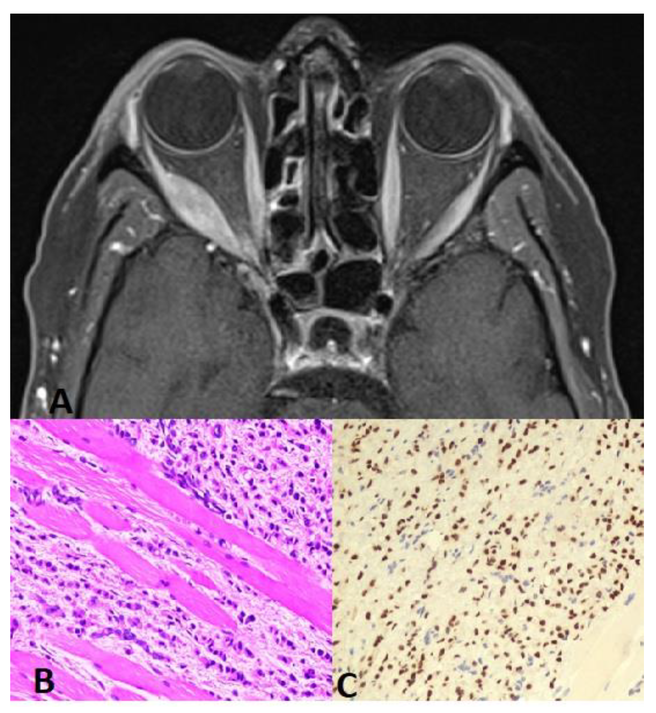
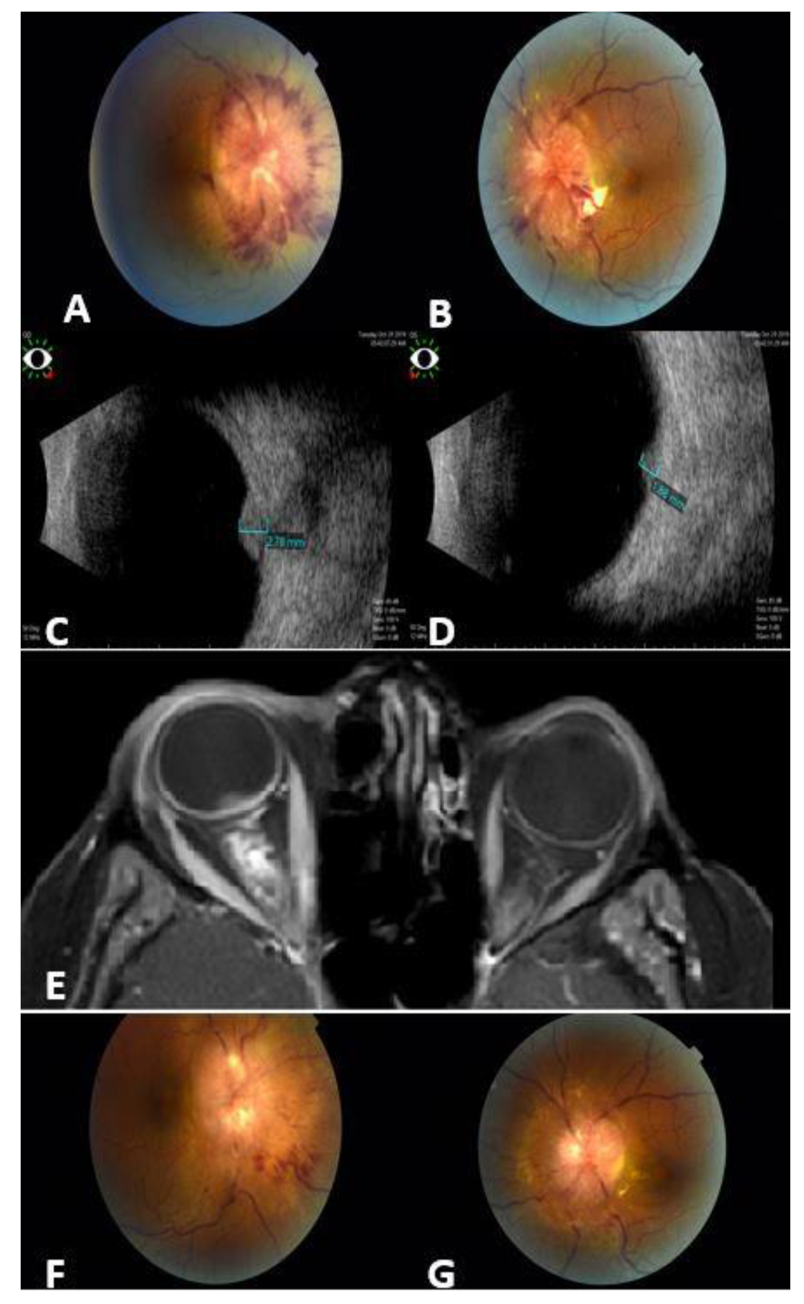
3.1. Presentation and Ocular Features
3.2. Prognostic Factors for Ocular Metastasis in Breast Cancer
3.3. Management and Outcome
4. Discussion
5. Conclusions
Author Contributions
Funding
Institutional Review Board Statement
Informed Consent Statement
Data Availability Statement
Acknowledgments
Conflicts of Interest
References
- Sung, H.; Ferlay, J.; Siegel, R.L.; Laversanne, M.; Soerjomataram, I.; Jemal, A.; Bray, F. Global Cancer Statistics 2020: GLOBOCAN Estimates of Incidence and Mortality Worldwide for 36 Cancers in 185 Countries. CA Cancer J. Clin. 2021, 71, 209–249. [Google Scholar] [CrossRef] [PubMed]
- GBD 2015 Eastern Mediterranean Region Cancer Collaborators. Burden of cancer in the Eastern Mediterranean Region, 2005–2015: Findings from the Global Burden of Disease 2015 Study. Int. J. Public Health 2018, 63, 151–164. [Google Scholar]
- World Health Organization, Breast Cancer Facts. Available online: https://www.who.int/news-room/fact-sheets/detail/breast-cancer (accessed on 16 February 2024).
- Abdel-Razeq, H.; Mansour, A.; Jaddan, D. Breast Cancer Care in Jordan. JCO Glob. Oncol. 2020, 6, 260–268. [Google Scholar] [CrossRef] [PubMed]
- Pestalozzi, B.C.; Zahrieh, D.; Mallon, E.; Gusterson, B.A.; Price, K.N.; Gelber, R.D.; Holmberg, S.B.; Lindtner, J.; Snyder, R.; Thürlimann, B.; et al. International Breast Cancer Study Group: Distinct clinical and prognostic features of infiltrating lobular carcinoma of the breast: Combined results of 15 International Breast Cancer Study Group clinical trials. J. Clin. Oncol. 2008, 26, 3006–3014. [Google Scholar] [CrossRef] [PubMed]
- Shields, J.A.; Shields, C.L.; Brotman, H.K. Carcinoma metastatic to the eye and orbit: A patient survival analysis. Am. J. Ophthalmol. 1997, 124, 276–283. [Google Scholar]
- Ahmad, S.M.; Esmaeli, B. Metastatic tumors of the orbit and ocular adnexa. Curr. Opin. Ophthalmol. 2007, 18, 405–413. [Google Scholar] [CrossRef] [PubMed]
- Mathis, T.; Jardel, P.; Loria, O.; Delaunay, B.; Nguyen, A.M.; Lanza, F.; Mosci, C.; Caujolle, J.P.; Kodjikian, L.; Thariat, J. New concepts in the diagnosis and management of choroidal metastases. Prog. Retin. Eye Res. 2019, 68, 144–176. [Google Scholar] [CrossRef] [PubMed]
- Eckardt, A.M.; Rana, M.; Essig, H.; Gellrich, N.C. Orbital metastases as first sign of metastatic spread in breast cancer: Case report and review of the literature. Head. Neck Oncol. 2011, 3, 37. [Google Scholar] [CrossRef] [PubMed]
- Vlachostergios, P.J.; Voutsadakis, I.A.; Papandreou, C.N. Orbital metastasis of breast carcinoma. Breast Cancer 2009, 3, 91–97. [Google Scholar] [CrossRef] [PubMed]
- Palmisciano, P.; Ferini, G.; Ogasawara, C.; Wahood, W.; Bin Alamer, O.; Gupta, A.D.; Scalia, G.; Larsen, A.M.G.; Yu, K.; Umana, G.E.; et al. Orbital Metastases: A Systematic Review of Clinical Characteristics, Management Strategies, and Treatment Outcomes. Cancers 2014, 14, 94. [Google Scholar] [CrossRef] [PubMed]
- Wickremasinghe, S.; Dansingani, K.K.; Tranos, P.; Liyanage, S.; Jones, A.; Davey, C. Ocular presentations of breast cancer. Acta Ophthalmol. Scand. 2007, 85, 133–142. [Google Scholar] [CrossRef] [PubMed]
- Shields, C.L.; Shields, J.A.; Peggs, M.; Singh, A.D. Metastatic carcinoma of the eye: The 2000 Sanford Gifford Memorial Lecture. Ophthalmic Plast. Reconstr. Surg. 2004, 20, 10–21. [Google Scholar] [CrossRef] [PubMed]
- Surace, D.; Piscioli, I.; Morelli, L.; Valduga, F.; Licci, S. Orbital metastasis as the first sign of “Dormant” breast cancer dissemination 25 years after mastectomy. Jpn. J. Ophthalmol. 2008, 52, 423–425. [Google Scholar] [CrossRef] [PubMed]
- Tinterri, C.; Sagona, A.; Barbieri, E.; Di Maria Grimaldi, S.; Jacobs, F.; Zambelli, A.; Trimboli, R.M.; Bernardi, D.; Vinci, V.; Gentile, D. Loco-Regional Treatment of the Primary Tumor in De Novo Metastatic Breast Cancer Patients Undergoing Front-Line Chemotherapy. Cancers 2022, 14, 6237. [Google Scholar] [CrossRef] [PubMed]
- Freedman, M.I.; Folk, J.C. Metastatic tumors to the eye and orbit: Patient survival and clinical characteristics. Arch. Ophthalmol. 1987, 105, 1215–1219. [Google Scholar] [CrossRef] [PubMed]
- American Cancer Society. Cancer Facts Figures 2021; American Cancer Society: Atlanta, GA, USA, 2021. [Google Scholar]
- Rakha, E.A.; Reis-Filho, J.S.; Baehner, F.; Dabbs, D.J.; Decker, T.; Eusebi, V.; Fox, S.B.; Ichihara, S.; Jacquemier, J.; Lakhani, S.R.; et al. Breast cancer prognostic classification in the molecular era: The role of histological grade. Breast Cancer Res. 2010, 12, 207. [Google Scholar] [CrossRef] [PubMed]
- Cristofanilli, M.; Budd, G.T.; Ellis, M.J.; Stopeck, A.; Matera, J.; Miller, M.C.; Reuben, J.M.; Doyle, G.V.; Allard, W.J.; Terstappen, L.W.; et al. Circulating tumor cells, disease progression, and survival in metastatic breast cancer. N. Engl. J. Med. 2004, 351, 781–791. [Google Scholar] [CrossRef] [PubMed]
- Barbera, G.; Todaro, M.; Saponaro, G.; Gasparini, G.; Moro, A. Orbital Exenteration in Recurrence Cancer: 5 Years Experience. J. Clin. Med. 2023, 12, 6180. [Google Scholar] [CrossRef] [PubMed]
- Nirmala, S.; Krishnaswamy, M.; Janaki, M.G.; Kaushik, K.S. Unilateral solitary choroid metastasis from breast cancer: Rewarding results of external radiotherapy. J. Cancer Res. Ther. 2008, 4, 206–208. [Google Scholar] [CrossRef]
- Shields, C.L.; Shields, J.A.; Baez, K.A.; Cater, J.R. Choroidal metastasis from carcinoma of the breast. Ophthalmology 1993, 100, 1645–1650. [Google Scholar]
- Arepalli, S.; Kaliki, S.; Shields, C.L.; Emrich, J. Ocular and orbital metastasis from breast cancer: A systematic review. Int. Ophthalmol. 2017, 37, 653–664. [Google Scholar]
- Janicijevic-Petrovic, M.; Sarenac, T.; Sreckovic, S.; Vulovic, D.; Janicijevic, K. Orbital metastases from breast cancer: A case report. Bosn. J. Basic. Med. Sci. 2011, 11, 253–255. [Google Scholar] [CrossRef] [PubMed][Green Version]
- Ashton, N.; Morgan, G. Discrete carcinomatous metastases in the extraocular muscles. Br. J. Ophthalmol. 1974, 58, 112–117. [Google Scholar] [CrossRef] [PubMed]
- Dossus, L.; Benusiglio, P.R. Lobular breast cancer: Incidence and genetic and non-genetic risk factors. Breast Cancer Res. 2015, 17, 37. [Google Scholar] [CrossRef] [PubMed]
- Yanagisawa, W.; Krishnan, S.; Fernandez, A. A rare case of lobular breast cancer metastasizing to large bowel. Clin. Case Rep. 2021, 9, e040812021. [Google Scholar] [CrossRef] [PubMed]
- Heitz, F.; Harter, P.; Lueck, H.J.; Fissler-Eckhoff, A.; Lorenz-Salehi, F.; Scheil-Bertram, S.; Traut, A.; du Bois, A. Triple-negative and HER2-overexpressing breast cancers exhibit an elevated risk and an earlier occurrence of cerebral metastases. Eur. J. Cancer. 2009, 45, 2792–2798. [Google Scholar] [CrossRef] [PubMed]
- Amer, M.H. Multiple brain metastases: Analysis of prognostic factors in 43 consecutive patients. J. Neurosurg. 1982, 56, 314–320. [Google Scholar]
- Cárdenas, J.R.V.; Penella, A.D.V.; Ibarra, E.C. Frameless radiosurgery for intraocular metastatic tumor: Case report. Rep. Pr. Oncol. Radiother. 2020, 25, 1–5. [Google Scholar] [CrossRef]
- Shields, J.A.; Shields, C.L.; Scartozzi, R. Survey of 1264 patients with orbital tumors and simulating lesions: The 2002 Montgomery Lecture, part. Ophthalmology 2004, 111, 997–1008. [Google Scholar] [CrossRef]
- Wiegel, T.; Bottke, D.; Kreusel, K.M.; Schmidt, S.; Bornfeld, N.; Foerster, M.H.; Hinkelbein, W.; German Cancer Society. External beam radiotherapy of choroidal metastases—Final results of a prospective study of the German Cancer Society (ARO 95-08). Radiother. Oncol. 2002, 64, 13–18. [Google Scholar] [PubMed]
- Kadivar, M.; Joulaee, A.; Kashkouli, M.B.; Kharazi, H.H.; Kalantari, M.; Kumar, P.V. Orbital metastasis as the first presentation of nonpalpable invasive lobular carcinoma of the breast. Breast J. 2006, 12, 75–76. [Google Scholar] [CrossRef] [PubMed]
- Gombos, D.S.; Mieler, W.F.; Reddy, R.K. Neoplastic disease of the eye. N. Engl. J. Med. 2002, 347, 317–326. [Google Scholar]


| Age at Diagnosis of Breast Cancer | Median 48, Mean 48.3, Range 31–71 Years | ||
|---|---|---|---|
| Age at Diagnosis of Ocular Metastasis | Median 50, Mean 50.6, Range 35–79 Years | ||
| Breast cancer laterality | Unilateral | 39 | 87% |
| Bilateral | 6 | 13% | |
| Pathology of breast cancer * | Invasive ductal | 28 | 62% |
| Invasive lobular | 17 | 38% | |
| Family history of breast cancer | Yes | 12 | 27% |
| No | 33 | 73% | |
| Site of ocular metastasis | Orbit # | 21 | 47% |
| Choroid | 18 | 40% | |
| Optic nerve | 5 | 11% | |
| Iris | 1 | 2% | |
| Presenting signs of ocular metastasis | Incidental by MRI (Asymptomatic) | 19 | 42% |
| Poor vision | 14 | 31% | |
| Orbital swelling/Ptosis | 5 | 11% | |
| Diplopia/Strabismus | 4 | 9% | |
| Proptosis | 3 | 7% | |
| Method of Diagnosis | Radiological | 21 | 47% |
| Clinical | 19 | 42% | |
| Pathological | 5 | 11% | |
| Laterality | Bilateral | 20 | 44% |
| Right or Left | 12 / 13 | 56% | |
| Vision at diagnosis (65 eyes) | less than 0.1 | 14 | 22% |
| 0.1–0.5 | 19 | 29% | |
| More than 0.5 | 32 | 49% | |
| Number Breast Cancer | Breast Cancer with Ocular Mets | p Value | |||
|---|---|---|---|---|---|
| 1 | Number of breast cancer patients | 9902 | 45 (0.45%) | ||
| 2 | Pathology | Invasive Ductal | 8028 | 28 (0.35%) | >0.0001 |
| Invasive Lobular | 797 | 17 (2.1%) | |||
| NA | 1077 | 0 | |||
| 3 | Age at diagnosis | >50 | 5148 | 25 (0.48%) | 0.656 |
| >50 | 4754 | 20 (0.4%) | |||
| 4 | Stage | Stage T1/2 | 4862 | 6 (0.12%) | 0.0019 |
| Stage T3/4 | 1687 | 9 (0.53%) | |||
| NA | 3351 | 30 (0.89%) | |||
| 5 | Distant metastasis | Metastatic | 1888 | 45 (2.4%) | >0.0001 |
| Nonmetastatic | 7644 | 0 (0%) | |||
| NA | 370 | 0 | |||
| 6 | Overall Survival | Alive | 7924 | 13 (0.16%) | >0.0001 |
| Dead | 1978 | 32 (1.6%) | |||
| 7 | Survival for metastatic breast cancer | Alive | 962 | 13 (1.4%) | 0.003 |
| Dead | 926 | 32 | |||
| Invasive Ductal | Invasive LOBULAR | p Value | ||
|---|---|---|---|---|
| Number | 28 (62%) | 17 (38%) | ||
| Laterality of ocular metastasis | Unilateral | 15 (54%) | 10 (59%) | 0.48 |
| Bilateral | 13 (46%) | 7 (41%) | ||
| Site of Ocular metastasis | Choroid | 11 (39%) | 7 (41%) | 0.62 |
| Orbit | 15 (53%) | 6 (35%) | 0.17 | |
| Optic nerve | 1 (4%) | 4 (24%) | 0.036 | |
| Iris | 1 (4%) | 0 (0%) | 1.0 | |
| Concomitant brain metastasis | Yes | 14 (50%) | 13 (76%) | 0.026 |
| No | 14 (50%) | 4 (24%) | ||
| Genetics of Breast Cancer * | Positive | 5 (36%) | 9 (64%) | 0.31 |
| Negative | 4 (57%) | 3 (43%) | ||
| Treatment of metastasis | Chemotherapy | 5 | 11% |
| Radiation | 40 | 89% | |
| Vision after treatment * | Less than 0.1 | 12 | 19% |
| 0.1–0.5 | 17 | 26% | |
| More than 0.5 | 36 | 55 | |
| Complications | Dry eye | 42 | 93% |
| Radiation keratopathy | 4 | 8% | |
| Radiation retinopathy | 3 | 7% | |
| Cataract | 2 | 4% | |
| Periocular redness | 6 | 13% | |
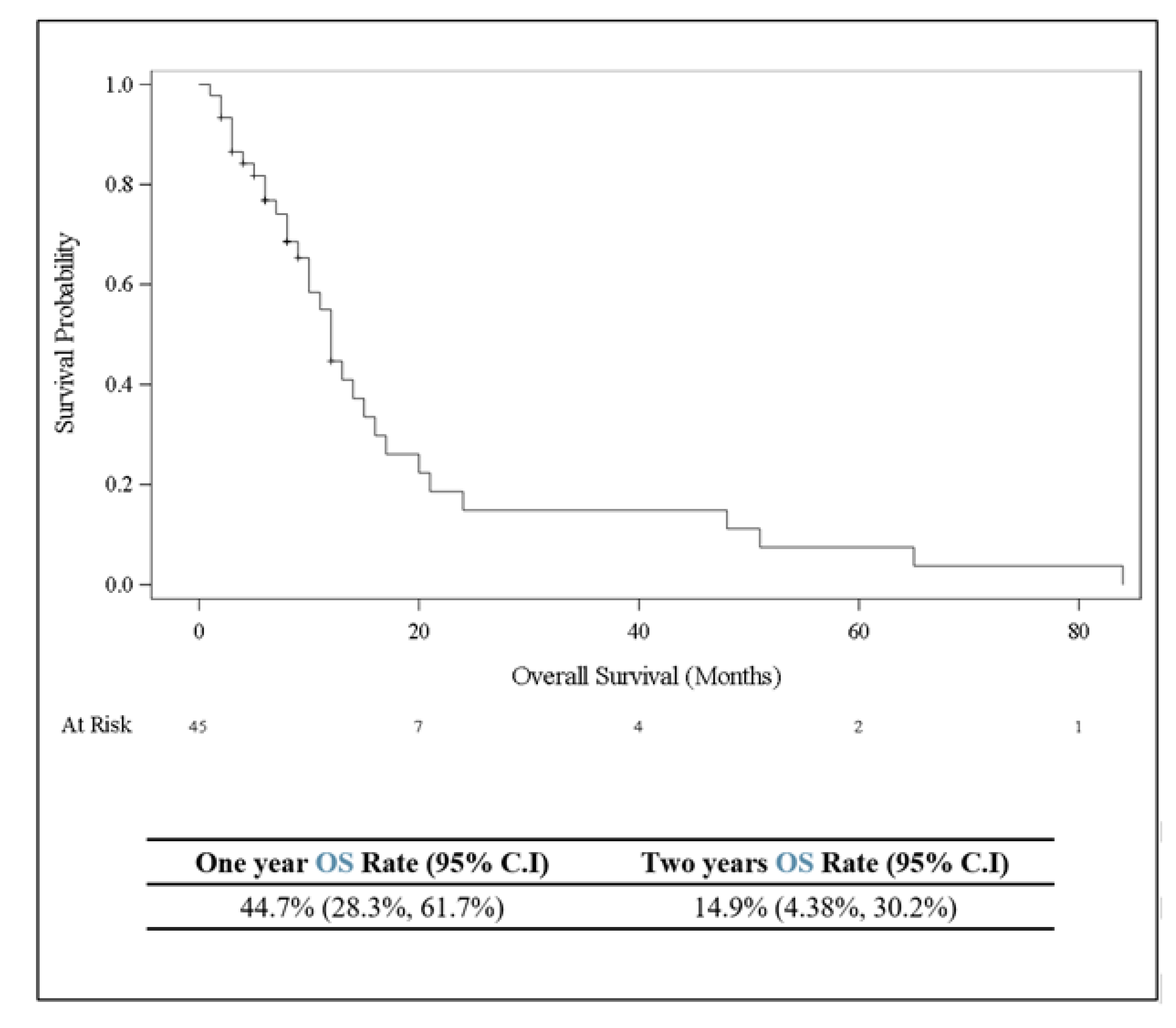
| Survival | Alive | 13 | 29% |
| Dead | 32 | 71% | |
| Duration between ocular metastasis to death | Median 10, Mean 16, Range 1–65 months | ||
Disclaimer/Publisher’s Note: The statements, opinions and data contained in all publications are solely those of the individual author(s) and contributor(s) and not of MDPI and/or the editor(s). MDPI and/or the editor(s) disclaim responsibility for any injury to people or property resulting from any ideas, methods, instructions or products referred to in the content. |
© 2024 by the authors. Licensee MDPI, Basel, Switzerland. This article is an open access article distributed under the terms and conditions of the Creative Commons Attribution (CC BY) license (https://creativecommons.org/licenses/by/4.0/).
Share and Cite
Yousef, Y.A.; Mohammad, M.; Khalil, H.; Khouri, T.; Alsweiti, R.; Khzouz, J.; Abu Laban, D.; Jaradat, I.; Ibrahimi, A.K.; Al-Ibraheem, A.; et al. Ocular and Periocular Metastasis in Breast Cancer: Clinical Characteristics, Prognostic Factors and Treatment Outcome. Cancers 2024, 16, 1518. https://doi.org/10.3390/cancers16081518
Yousef YA, Mohammad M, Khalil H, Khouri T, Alsweiti R, Khzouz J, Abu Laban D, Jaradat I, Ibrahimi AK, Al-Ibraheem A, et al. Ocular and Periocular Metastasis in Breast Cancer: Clinical Characteristics, Prognostic Factors and Treatment Outcome. Cancers. 2024; 16(8):1518. https://doi.org/10.3390/cancers16081518
Chicago/Turabian StyleYousef, Yacoub A., Mona Mohammad, Hanan Khalil, Tala Khouri, Rand Alsweiti, Jakub Khzouz, Dima Abu Laban, Imad Jaradat, Ahmad Kh. Ibrahimi, Akram Al-Ibraheem, and et al. 2024. "Ocular and Periocular Metastasis in Breast Cancer: Clinical Characteristics, Prognostic Factors and Treatment Outcome" Cancers 16, no. 8: 1518. https://doi.org/10.3390/cancers16081518
APA StyleYousef, Y. A., Mohammad, M., Khalil, H., Khouri, T., Alsweiti, R., Khzouz, J., Abu Laban, D., Jaradat, I., Ibrahimi, A. K., Al-Ibraheem, A., Masri, M. A., AlNawiaseh, I., & Abdel-Razeq, H. (2024). Ocular and Periocular Metastasis in Breast Cancer: Clinical Characteristics, Prognostic Factors and Treatment Outcome. Cancers, 16(8), 1518. https://doi.org/10.3390/cancers16081518